hira and daxx constitute two independent histone h3.3...
TRANSCRIPT
Histone octamers package the eukaryotic genome, notonly compacting the DNA, but also encoding and propa-gating a sophisticated additional layer of epigenetic infor-mation. Despite the inherent physical stability of thenucleosome particle, the resulting chromatin structuremust be highly dynamic to allow access to the DNA-en-coded information. H3/H4 and H2A/H2B units are de-posited and evicted from nucleosomes by different histonechaperones and ATP-dependent chromatin remodelers.Histone chaperones thereby serve two major functions:They (1) buffer the pool of nonchromatin-associated his-tones in predeposition complexes and (2) facilitate favor-able histone–DNA interactions during the stepwiseassembly of the histone octamer–DNA complex fromH3/H4 and H2A/H2B units. Consequently, mutants of his-tone chaperones in Saccharomyces cerevisiae cannot formproper chromatin structure (Sharp et al. 2002; Adkins andTyler 2004; Andrews et al. 2010). Metazoans have evolved histone variants to meet the di-
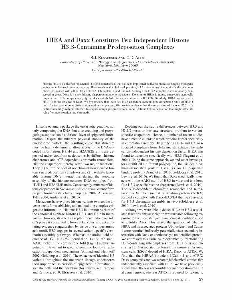
verse needs for establishing and maintaining complex epi-genetic information. Histone H3.3 is a minor variant ofthe canonical S-phase histones H3.1 and H3.2 in meta-zoans. However, its role as a replacement histone outsideof S phase is conserved to lower eukaryotes, and accumu-lating evidence suggests that, by virtue of a unique aminoacid motif, H3.3 engages in several variant-specific chro-matin assembly pathways. Whereas the amino acid se-quence of H3.3 is >95% identical to H3.1/2, the smallAAIG motif in the core histone fold (Fig. 1) allows tar-geting of the variant to specific genomic loci by a repli-cation-independent mechanism (Ahmad and Henikoff2002; Goldberg et al. 2010). The existence of identical H3variants throughout the metazoan lineage underscorestheir importance as carriers of epigenetic information insomatic cells and the germline (for review, see Camposand Reinberg 2010; Elsaesser et al. 2010).
Reading out the subtle differences between H3.3 andH3.1/2 poses an intricate structural problem to variant-specific chaperones. Hence, a number of recent studieshave aimed to elucidate which proteins confer specificityin chromatin assembly. By purifying H3.1- and H3.3-as-sociated complexes from HeLa nuclear extracts, the repli-cation-independent histone deposition factor HIRA wasfound to associate specifically with H3.3 (Tagami et al.2004). Using the same approach, we and other investiga-tors identified a different polypeptide, the Fas death-do-main–associated protein Daxx, as an H3.3-specificbinding protein (Drané et al. 2010; Goldberg et al. 2010;Lewis et al. 2010). We found that Daxx specifically inter-acts with the AAIG motif of H3.3 in vitro and is a bonafide H3.3-specific histone chaperone (Lewis et al. 2010).The ATP-dependent chromatin remodeler and α-tha-lassemia X-linked mental retardation protein (ATRX)formed a complex with Daxx-H3.3/H4 that was essentialfor H3.3 chromatin assembly in vivo (Goldberg et al.2010; Lewis et al. 2010). Although we were able to detect HIRA in H3.3-associ-
ated fractions, this association was unstable following ex-posure to the more stringent biochemical conditions usedto identify Daxx. This raised the question of whetherHIRA and its associated proteins Ubinuclein-1 and Cabin-1 were recruited indirectly, potentially via a secondary in-teraction with Daxx or another as yet unidentified protein.We addressed this issue by biochemically fractionatingH3.3-containing subcomplexes from HeLa cells and pu-rifying H3.3-associated proteins from mouse embryonicstem cells (ESCs) devoid of HIRA, Daxx, or ATRX. Wefind that the HIRA/Ubinuclein-1/Cabin-1 and ATRX/Daxx complexes are two separate biochemical entities thatindependently associate with H3.3. We have previouslyshown that HIRA is responsible for incorporation of H3.3at genic regions, whereas ATRX is required for telomeric
HIRA and Daxx Constitute Two Independent HistoneH3.3-Containing Predeposition Complexes
S.J. ELSAESSER AND C.D. ALLISLaboratory of Chromatin Biology and Epigenetics, The Rockefeller University,
New York, New York 10065Correspondence: [email protected]
Histone H3.3 is a universal replacement histone in metazoans that has been implicated in diverse processes ranging from geneactivation to heterochromatin silencing. Here, we show that, before deposition, H3.3 exists in two biochemically distinct com-plexes, associated with either Daxx or HIRA, Ubinuclein-1, and Cabin-1. Although the HIRA complex is evolutionarily con-served in yeast, Daxx is a novel histone chaperone unique to metazoans. Deletion of HIRA in mouse embryonic stem cellsimpairs the HIRA complex integrity but does not abolish Daxx association with H3.3/H4. Similarly, HIRA interacts withH3.3/H4 in the absence of Daxx. We hypothesize that these two H3.3 chaperone systems provide separate pools of H3/H4units for incorporation at distinct sites within the genome. We provide evidence that the association of histone H3.3 withdistinct assembly systems allows it to acquire unique posttranslational modifications before deposition that might affect itsrole after incorporation into chromatin.
Cold Spring Harbor Symposia on Quantitative Biology,Volume LXXV. ©2010 Cold Spring Harbor Laboratory Press 978-1-936113-07-1 27
deposition of H3.3 (Goldberg et al. 2010). Therefore,HIRA and Daxx/ATRX constitute two separate histoneH3.3-deposition machineries that act on distinct genomicloci.
MATERIALS AND METHODS
Nuclear extracts were prepared as described in Lewis etal. (2010) from HeLa cells stably expressing FLAG-HA–tagged H3.1 or H3 (Tagami et al. 2004) or mouse ESCs(Goldberg et al. 2010). Immunoprecipitations were per-formed in 20-mM HEPES•HCl (pH 7.5), 150 mM NaCl,10% glycerol, 0.2 mM EDTA, 2 mMMgCl
2in the presence
of DNase and RNase, washed five times with wash buffer(20 mM HEPES•HCl [pH 7.5], 250 mM NaCl, 10% glyc-erol, 0.2 mM EDTA, 0.01% NP-40 [Nonidet P-40]) andeluted with either FLAG peptide or 1% SDS. Eluted ma-terial was separated by SDS polyacrylamide gel elec-trophoresis (SDS PAGE) and analyzed by western blottingwith the following antibodies: α-FLAG-HRP (Sigma-Aldrich), α-HA-HRP A190-107P (Bethyl), α-HIRAWC119 (Peter Scambler), α-p150 sc-10772 (Santa Cruz),α-Daxx sc-7152 (Santa Cruz), α-p60 NB500-212 (NovusBiologicals), α-Cabin-1 ab3349 (Abcam), α-ATRX H-300(Santa Cruz), α-ASF1a/b 4A1/3 (Santa Cruz), α-H3K9Ac,No. 07-352 (Millipore), α-acetyl-H4, No. 06-598 (Milli-pore), and α-H4K5Ac, No. 06-759 (Millipore).
BIOCHEMICAL FRACTIONATIONOF H3.3-CONTAINING COMPLEXES
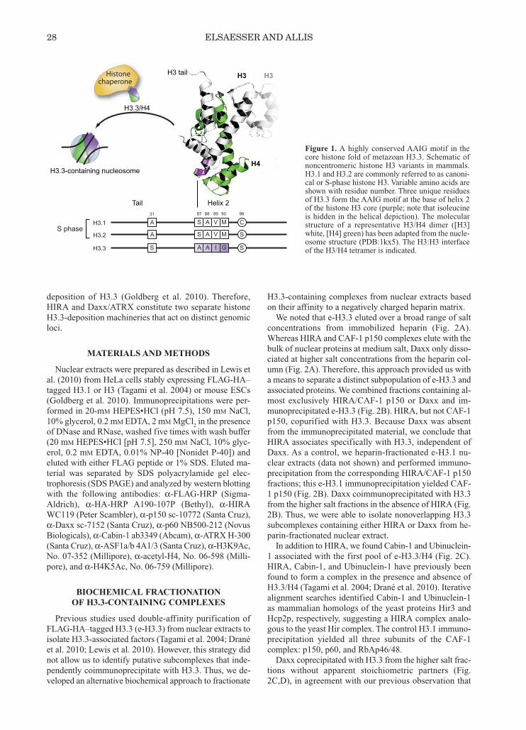
Previous studies used double-affinity purification ofFLAG-HA–tagged H3.3 (e-H3.3) from nuclear extracts toisolate H3.3-associated factors (Tagami et al. 2004; Dranéet al. 2010; Lewis et al. 2010). However, this strategy didnot allow us to identify putative subcomplexes that inde-pendently coimmunoprecipitate with H3.3. Thus, we de-veloped an alternative biochemical approach to fractionate
H3.3-containing complexes from nuclear extracts basedon their affinity to a negatively charged heparin matrix. We noted that e-H3.3 eluted over a broad range of salt
concentrations from immobilized heparin (Fig. 2A).Whereas HIRA and CAF-1 p150 complexes elute with thebulk of nuclear proteins at medium salt, Daxx only disso-ciated at higher salt concentrations from the heparin col-umn (Fig. 2A). Therefore, this approach provided us witha means to separate a distinct subpopulation of e-H3.3 andassociated proteins. We combined fractions containing al-most exclusively HIRA/CAF-1 p150 or Daxx and im-munoprecipitated e-H3.3 (Fig. 2B). HIRA, but not CAF-1p150, copurified with H3.3. Because Daxx was absentfrom the immunoprecipitated material, we conclude thatHIRA associates specifically with H3.3, independent ofDaxx. As a control, we heparin-fractionated e-H3.1 nu-clear extracts (data not shown) and performed immuno-precipitation from the corresponding HIRA/CAF-1 p150fractions; this e-H3.1 immunoprecipitation yielded CAF-1 p150 (Fig. 2B). Daxx coimmunoprecipitated with H3.3from the higher salt fractions in the absence of HIRA (Fig.2B). Thus, we were able to isolate nonoverlapping H3.3subcomplexes containing either HIRA or Daxx from he-parin-fractionated nuclear extract.In addition to HIRA, we found Cabin-1 and Ubinuclein-
1 associated with the first pool of e-H3.3/H4 (Fig. 2C).HIRA, Cabin-1, and Ubinuclein-1 have previously beenfound to form a complex in the presence and absence ofH3.3/H4 (Tagami et al. 2004; Drané et al. 2010). Iterativealignment searches identified Cabin-1 and Ubinuclein-1as mammalian homologs of the yeast proteins Hir3 andHcp2p, respectively, suggesting a HIRA complex analo-gous to the yeast Hir complex. The control H3.1 immuno-precipitation yielded all three subunits of the CAF-1complex: p150, p60, and RbAp46/48. Daxx coprecipitated with H3.3 from the higher salt frac-
tions without apparent stoichiometric partners (Fig.2C,D), in agreement with our previous observation that
28 ELSAESSER AND ALLIS
31 90 96898887
A MVASH3.1S phase
C
A MVASH3.2 S
SH3.3 GIAA S
H3
Helix 2Tail
H3 tail
H3.3/H4
H3.3-containing nucleosome
H3
H4A
A
G
Histone chaperone
Figure 1. A highly conserved AAIG motif in thecore histone fold of metazoan H3.3. Schematic ofnoncentromeric histone H3 variants in mammals.H3.1 and H3.2 are commonly referred to as canoni-cal or S-phase histone H3. Variable amino acids areshown with residue number. Three unique residuesof H3.3 form the AAIG motif at the base of helix 2of the histone H3 core (purple; note that isoleucineis hidden in the helical depiction). The molecularstructure of a representative H3/H4 dimer ([H3]white, [H4] green) has been adapted from the nucle-osome structure (PDB:1kx5). The H3:H3 interfaceof the H3/H4 tetramer is indicated.

the direct interaction of Daxx with H3.3/H4 is not depend-ent on other proteins such as ATRX (Lewis et al. 2010).In conclusion, we find that the known H3.3-associatedproteins fractionate into two biochemically distinct sub-complexes constituted by HIRA and Daxx.
GENETIC DISSECTIONOF H3.3-CONTAINING COMPLEXES
Next, we assessed how the deletion of either HIRA orDaxx histone chaperones affects H3.3 and its associatedcomplexes in respective knockout mouse ESC lines. Weused a zinc-finger–based genome editing strategy to specif-ically tag one allele of the endogenous H3.3B gene in wild-type W9.5, HIRA–/–, Daxx–/–, and ATRX–/– ESCs with acarboxy-terminal HA tag (H3.3-HA) (Goldberg et al.2010), assuring endogenous expression levels of the taggedhistone. We found elevated levels of H3.3-HA in nuclearextracts of HIRA–/– ESCs, as previously reported (Meshoreret al. 2006), whereas the soluble pool of H3.3-HA was re-duced in Daxx–/– ESCs (Fig. 3, left). We speculate that Daxxbuffers a considerable pool of H3.3 in the nucleus by form-ing a stable complex with H3/H4 units. Overall, H3 and H4levels (accounting for the untagged H3.3 as well as H3.1/2)in the nucleoplasm were unchanged, indicating that reducedlevels of H3.3 might be compensated by an increase in sol-uble H3.1/2. Levels of chromatin-bound H3.3-HA werecomparable in all cell lines (Fig. 3, bottom).As previously reported, ATRX levels were reduced in
Daxx–/– cells (Lewis et al. 2010). Unexpectedly, Cabin-1 wasdepleted in HIRA–/– nuclear extracts (Fig. 3, left panels) andwhole-cell lysates (data not shown). Cabin-1 mRNA levelsare not perturbed in HIRA–/– cells (Goldberg et al. 2010),suggesting that the Cabin-1 protein might be unstable anddegraded in the absence of HIRA. Therefore, Cabin-1 likelycontributes directly to the function of the HIRA complex, asalready shown for Ubinuclein-1 (Banumathy et al. 2009).
HIRA and Daxx ComplexesIndependentlyBind H3.3
Based on our biochemical fractionation of H3.3-contain-ing complexes, we hypothesized that association of HIRAand Daxx with H3.3 occurs independently. We tested thisby immunoprecipitating H3.3-HA from the above ESC nu-clear extract. Indeed, HIRA copurified with H3.3-HA inDaxx–/– cells and vice versa (Fig. 3, right panels). ATRXcoprecipitated with Daxx, but ATRX deletion did not in-fluence binding of either histone chaperone, as expectedfrom our earlier studies (Lewis et al. 2010). ASF1 was con-stitutively associated with H3.3-HA.We therefore conclude that the HIRA and Daxx com-
plexes independently interact with H3.3 and likely main-tain distinct pools of H3.3/H4 units in the nucleus. This isin line with our recent observation that HIRA andATRX/Daxx deposit histones at distinct genomic loca-tions: HIRA at genic regions (Goldberg et al. 2010) andATRX/Daxx at telomeres as well as pericentromeric het-erochromatin (Drané et al. 2010; Lewis et al. 2010). Although our in vitro studies confirmed that Daxx di-
rectly interacts with the H3.3 AAIG motif (Lewis et al.2010), similar direct evidence is thus far missing forHIRA. Together with previous data, our finding that HIRAconsistently copurifies with H3.3 in a complex withCabin-1 and Ubinuclein-1 (Tagami et al. 2004; Lewis etal. 2010) raises the possibility that these components con-tribute to binding and specificity as well.
HISTONE H3.3-CONTAINING COMPLEXES 29
A
B
C D
IN INe-H3.1
CAF-1 HIRA.com Daxx.com
CAF-1 HIRA.com Daxx.com Daxx.com
e-H3.3
e-H3.3
INα-CAF-1p150
α-HIRAα-Daxx
α-CAF-1p150
KCl800 mM
200 mM
α-HIRA
IN
α-Daxx
α-Flag
α-Flag
α-H4
α-CAF-1p150
α-HIRAα-Daxx
α-Flagα-H4
HIRACabin-1
HIRHCabinCabin
Ubn-
1 Daxx
p150HIRA
p60
p46/48
e-H3
H4
e-H3
H4
Cabin-1
Ubn-1Daxx
p48
p150ASF1p48
pp60
FLAG-IP FLAG-IP FLAG-IP
Heparin
Heparin
HeLa nuclear extract (e-H3.3)
NE (e-H3.1)
H3.1/H4 H3.3/H4
148
98
64
50
36
16
Figure 2. HIRA and Daxx constitute two biochemically distinctH3.3-containing subcomplexes. (A) Biochemical fractionationof nuclear H3.3 complexes: 200–800-mM KCl fractions from aheparin column were probed for e-H3.3, HIRA, CAF-1, andDaxx by western blotting. (B) Affinity purification of e-H3.3 ande-H3.1 complexes. e-H3.3/H3.1 complexes were immunoprecip-itated from indicated pools and analyzed by western blotting. Im-munoprecipitation from the medium-salt e-H3.3 and e-H3.1fractions yielded predominantly HIRA and CAF-1, respectively;Daxx coprecipitated with high-salt e-H3.3. (C) Silver staining ofeluted CAF-1, HIRA, and Daxx complexes from above. Indi-cated bands were all identified by mass spectrometry and/or ver-ified by western blotting. (D) Coomassie stain of the Daxxcomplex estimates protein abundance.

DO PREDEPOSITION COMPLEXESREPRESENT INDEPENDENT HISTONE POOLS?
Although H3.3 was originally characterized to be asso-ciated with actively transcribed genes (Ahmad andHenikoff 2002), the discovery of new H3.3-chaperonepathways and heterochromatic target regions (Hake et al.2005; van der Heijden et al. 2007; Drané et al. 2010;Lewis et al. 2010; Santenard et al. 2010) poses some in-triguing questions: Does H3.3 perform unique functionsat distinct genomic locations? How can a single replace-ment variant contribute to gene activation or repression,depending on the chromatin context? How do the special-ized deposition machineries contribute to the functionaloutcome of H3.3 incorporation? The functional diversity of H3.3 could be explained if the
distinct predeposition complexes would represent independ-ent pools of H3.3/H4 units. Assuming that the exchange be-tween those pools is slow due to the biochemical stabilityof the complexes or their distinct spatial or temporal distri-bution during the cell cycle, the associated histones couldacquire specific posttranslational modifications (PTMs)that might prime them for their respective functions at thegenomic target sites.Histone variants have been found enriched in PTMs that
correlate with the local chromatin environment at their targetsites (Hake et al. 2006). A set of specific modifications hasbeen identified for the soluble pool of histones composed ofnewly synthesized histones but possibly also evicted histones“recycled” from chromatin: ubiquitous H4K5/K12 diacety-lation (H4K5/K12diAc) (Sobel et al. 1995) and H3K9monomethylation (H3K9me1) on both H3.1/2 and H3.3, aswell as H3K9 dimethylation (H3K9me2) and H3K9 acety-
lation (H3K9Ac) enriched on H3.3 (Loyola et al. 2006).Given the overall heterogeneity of predeposition PTMs onthe single H3.3K9 residue, we wondered if any of thesemarks might be characteristic of a subset of predepositionH3.3 associated with a specific chaperone complex.
Distinct Histone Posttranslational ModificationsAre Present in Predeposition Complexes
Using heparin fractionation to separate e-H3.3-contain-ing complexes from HeLa cells, we identified a subpopu-lation of e-H3.3 that was enriched in H3K9Ac (Fig. 4A), amodification that is thought to be established on non-nu-cleosomal H3 by the histone acetyltransferase Gcn5 (Ad-kins et al. 2007). Because the enrichment of H3K9Actracked well with the HIRA complex, we further analyzedthe PTMs present on histones within the CAF-1, HIRA,and Daxx complexes (Fig. 4B). We found that H3K9Ac one-H3.3 associated with the HIRA complexes but was un-detectable in the Daxx complex. Both complexes containedlow levels of acetylated H4. e-H3.1 in the CAF-1 complexwas enriched in H4 acetylation, with H3K9Ac present at alower level than on e-H3.3. With the differential H3K9Acas a first indication, we conclude that unique H3.3/H4PTMs can be associated with predeposition complexes.
A MODEL FOR THE DIFFERENT PATHWAYSOF H3.3 CHROMATIN ASSEMBLY
Our biochemical characterization of the distinct H3.3deposition machineries allows us to propose a model forthe journey of histones from synthesis to incorporation atspecific genomic location. Newly synthesized histones in
30 ELSAESSER AND ALLIS
α-Daxx
α-ATRX
α-Cabin-1
α-HIRA
α-ASF1a/b
α-PCNA
α-HA
H3.3-HA
α-HA
α-H3
α-H4
- + + + +
- + + + +
wt ESC
HIRA-/-
Daxx-
/-
ATRX-/-
wt ESC
HIRA-/-
Daxx-
/-
ATRX-/-
HA-IP
Dax
x.co
mH
IRA
.com
HIRA
Cabin-1
Daxx
ATRX
Ub
n-1
Chromatin
Nuclear extract
Figure 3. HIRA and Daxx independently asso-ciate with nucleoplasmic H3.3. Nuclear extractswere prepared from wild-type, HIRA–/–, Daxx–/–,and ATRX–/– mouse ESC lines (Roberts et al.2002; Lewis et al. 2010). Where indicated, oneH3.3 allele has been endogenously tagged withan H3.3B-HA knockin. Protein levels in the nu-clear extracts and insoluble chromatin were as-sessed by immunoblotting (left panels). e-H3.3was immunoprecipitated with α-HA-agaroseand eluted with 1% SDS. Copurifying proteinswere analyzed by western blotting (right panels).

the cytosol are likely immediately chaperoned by abundantgeneral histone chaperones, such as ASF1 and/or NASP(Fig. 5, top). Furthermore, H4 is diacetylated on K5 andK12. ASF1-bound H3.3/H4 dimers then serve as substratesfor various assembly pathways. Although a Daxx-H3.3/H4complex has been found in the cytosol (Drané et al. 2010),HIRA seems to be exclusively localized to the nucleus andhas not been found in a cytosolic complex with H3.3/H4(Lorain et al. 1998; Tagami et al. 2004). ASF1 is ubiqui-tously detectable with H3.3/H4 (Tagami et al. 2004; Dranéet al. 2010), but our biochemical data suggest that it is nota required component of HIRA and Daxx predepositioncomplexes (Fig. 2C,D) (Lewis et al. 2010).
HIRA Complex and Histone Deposition atTranscribed Gene Bodies and Promoters
HIRA histone chaperone activity has been well studiedin Xenopus egg extracts, where it is necessary and suffi-cient for replication-independent chromatin assembly onexogenous DNA (Ray-Gallet et al. 2002, 2007; Tagami etal. 2004). However, in vivo, it accounts for only a subsetof replication-independent chromatin assembly (Fig. 5,pathway 1), namely, within transcribed regions, at promot-ers and some regulatory elements (REs) (Goldberg et al.2010). Evidence from yeast showing that the Hir complexand Asf1p genetically and physically interact with the Set2methyltransferase suggests that histone deposition mightbe functionally linked to H3K36 trimethylation. Subse-quently, histone deacetylases remove acetylation marksfrom nucleosomes after RNA Pol passage. H3.3 enrichment in gene bodies was consistently lost in
HIRA–/– ESCs, but gene-expression patterns and the tran-scription elongation mark H3K36me3 were only mildlyaffected (Goldberg et al. 2010). Several other histone
chaperones including Spt6 and FACT have also been im-plicated in histone exchange during transcription. Thus,the purpose of the transcription-associated H3.3 deposi-tion and possible compensatory mechanisms in the ab-sence of HIRA have yet to be defined. The homology of HIRA to the yeast Hir corepressor
complex subunits Hir1/2p suggests that its histone depo-sition activity might suppress basal transcription (Spectoret al. 1997; Anderson et al. 2009). Furthermore, it will benecessary to define the roles of the remaining complexmembers Cabin-1 and Ubinuclein-1. Cabin-1 has recentlybeen shown to act as a corepressor at p53 target genes, andit will be interesting to see if this activity requires HIRA(Jang et al. 2009). Ubinuclein-1 cooperates with HIRA ina chromatin assembly pathway specific to establishmentof facultative heterochromatin domains in senescent cells(Fig. 5, pathway 2) (Banumathy et al. 2009).
Daxx and ATRX Assemble H3.3 Chromatin atPericentric Heterochromatin and Telomeres
A considerable fraction of H3.3 is localized to telomeresin mouse ESCs, and we have recently shown that this en-richment is dependent on the Daxx/ATRX complex but notHIRA (Goldberg et al. 2010; Lewis et al. 2010). WhereasDaxx readily assembles H3.3/H4 chromatin in vitro, ATRXis required in vivo to target Daxx and H3.3/H4 to thetelomeres (Lewis et al. 2010) as well as to pericentric het-erochromatin (Drané et al. 2010). Therefore, a diffusibleDaxx-H3.3/H4 complex likely delivers histones to anATRX-containing complex associated with telomeric chro-matin (Fig. 5, pathway 3) (Wong et al. 2010). Furthermore,the chromatin-bound complex might contain the histonechaperone Dek and histone deacetylase HDACII, as sug-gested by biochemical studies (Hollenbach et al. 2002), to
HISTONE H3.3-CONTAINING COMPLEXES 31
A
B
KCl
CAF-1 HIRA.com Daxx.com
INHeparin
α-Flag
α-CAF-1p150α-HIRAα-Daxx
α-H3K9Ac
α-H4panAc
α-FLAG
α-H4K5Ac
α-H3K9Ac
FLAG-IPFLAG-IPFLAG-IPHeparinNE (e-H3.1)
NE (e-H3.3)
α-H4
Figure 4. H3.3 predeposition complexes are associatedwith distinct histone posttranslational modifications. (A)Western blot analysis of e-H3.3 lysine 9 acetylation(K9Ac) in the heparin salt gradient. K9Ac on e-H3.3 isenriched in fractions that correspond to the HIRA com-plex. (B) The CAF-1, HIRA, and Daxx complexes wereanalyzed for selected H3 and H4 posttranslational modi-fications. K9Ac is enriched on e-H3.3 within the HIRAcomplex but is undetectable in the Daxx complex.
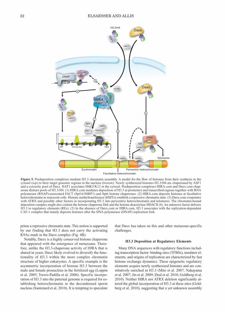
prime a repressive chromatin state. This notion is supportedby our finding that H3.3 does not carry the activatingK9Ac mark in the Daxx complex (Fig. 4B).Notably, Daxx is a highly conserved histone chaperone
that appeared with the emergence of metazoans. There-fore, unlike the H3.3-chaperone activity of HIRA that isshared in yeast, Daxx likely evolved to diversify the func-tionality of H3.3 within the more complex chromatinstructure of higher eukaryotes. A specific example is theasymmetric incorporation of histone H3.3 between themale and female pronucleus in the fertilized egg (Loppinet al. 2005; Torres-Padilla et al. 2006). Specific incorpo-ration of H3.3 into the paternal genome is required for es-tablishing heterochromatin in the decondensed spermnucleus (Santenard et al. 2010). It is tempting to speculate
that Daxx has taken on this and other metazoan-specificchallenges.
H3.3 Deposition at Regulatory Elements
Many DNA sequences with regulatory functions includ-ing transcription factor–binding sites (TFBSs), insulator el-ements, and origins of replication are characterized by fasthistone exchange dynamics. These epigenetic regulatoryelements acquire newly synthesized histones and are con-stitutively enriched in H3.3 (Mito et al. 2007; Nakayamaet al. 2007; Jin et al. 2009; Deal et al. 2010; Goldberg et al.2010). Neither HIRA nor ATRX deletion significantly al-tered the global incorporation of H3.3 at these sites (Gold-berg et al. 2010), suggesting that a yet unknown assembly
32 ELSAESSER AND ALLIS
DEKDEKHDACII
H3.3/H4
Daxx
Daxx
CIIKKKK Daxx
ATRX
DNAP p48p484pp4844
p60 p150
Euchromatin Pericentric heterochromatinFacultative heterochromatin
Telomeres
ASF1
ASF1
ASF1
GCN5
HAT1
HMTsHMTs
H3.3
enr
ichm
ent
RE Promoter TES
RNAP
P PP
HMTs
PP PP
HMTs
HDACs
Euchromatin
H3
RE Promoter STE
HIRACabin-111n-n-11
Ubn-1DEK?DEK?
Daxx?
1
2
3
4
5
1Spt6
PPP
FACT
Figure 5. Predeposition complexes mediate H3.3 chromatin assembly. A model for the flow of histones from their synthesis in thecytosol (top) to their target genomic regions in the nucleus (bottom). Newly synthesized histones H3.3/H4 are chaperoned by ASF1and a cytosolic pool of Daxx. HAT1 acetylates H4K5/K12 in the cytosol. Predeposition complexes HIRA.com and Daxx.com chap-erone distinct pools of H3.3/H4. (1) HIRA.com mediates deposition of H3.3 at promoters and transcribed regions together with RNApolymerase (RNAP)-associated FACT (Spt16/SSRP1) and Spt6 histone chaperones. (2) HIRA.com deposits histones at facultativeheterochromatin in senescent cells. Histone methyltransferases (HMTs) establish a repressive chromatin state. (3) Daxx.com cooperateswith ATRX and possibly other factors in incorporating H3.3 into pericentric heterochromatin and telomeres. The chromatin-bounddeposition complex might also contain the histone chaperone Dek and the histone deacetylase HDACII (4). An unknown factor deliversH3.3 to regulatory elements (REs). (5) In the absence of Daxx.com or HIRA.com, H3.3 associates with the replication-dependentCAF-1 complex that mainly deposits histones after the DNA polymerase (DNAP) replication fork.
pathway is used. Although general histone chaperones suchas ASF1 or NASP could provide the histones for ATP-de-pendent chromatin remodelers that occupy these locations,it is important to note that this pathway is also specific toH3.3 (Goldberg et al. 2010). We therefore speculate thatDaxx, Dek, or yet unknown factors mediate the depositionof H3.3 at regulatory elements (Fig. 5, pathway 4). Dek hasrecently been found to cooperate with ATP-dependent chro-matin remodelers and to act as a coactivator for nuclear re-ceptors (Sawatsubashi et al. 2010). Intriguingly, H3K4 monomethylation is another hall-
mark of regulatory elements; it tracks well with exchangehotspots and H3.3 enrichment in genome-wide analyses(Deal et al. 2010; Goldberg et al. 2010). Which histonemethyltransferase activity is responsible for establishingthe H3K4me1 mark remains elusive, but we speculate thatmethylation and histone exchange are functionally linkedat these genomic regions.
Alternate Pathways for H3.3 Deposition
As is apparent from the imperfect discrimination that canbe observed in pure in vitro assays with variant-specificchaperones (Drané et al. 2010; Lewis et al. 2010), only theinterplay and competition of variant-specific histone chap-erones can explain the exquisite specificity of H3.3 incor-poration observed in vivo. Whereas in unperturbed systems,H3.1 and H3.3 associate exclusively with their cognatechaperone systems (CAF-1, HIRA, and Daxx, respec-tively), we did observe CAF-1 copurifying with e-H3.3 inthe absence of Daxx and HIRA (Fig. 5, pathway 5) (Dranéet al. 2010; Lewis et al. 2010). Furthermore, there is evi-dence that low levels of H3.3 are broadly incorporated intochromosomes by the replication-dependent machinery inDrosophila cells (Ahmad and Henikoff 2002; Schwartz andAhmad 2005). Therefore, similar to the single yeast H3.3-like histone, H3.3 might represent a universal variant forreplication-dependent and -independent deposition path-ways, i.e., simply outnumbered by the large pool of H3.1/2present during S phase. Additionally, studies in flies suggestthat H3.1/2 can take over some but not all functions of H3.3(Hödl and Basler 2009; Sakai et al. 2009).
CONCLUSION
As illustrated by the replacement histone H3.3, rapidlyemerging literature points to remarkable complexity for theuse of relatively minor histone variants in punctuatingepigenomes in organisms ranging from yeast to man. Mul-tiple predeposition and assembly systems are called on todeposit histone variants in select genomic locations. Al-though these variants often differ in only a small number ofamino acids, mounting evidence suggests that, once assem-bled, they perform specialized functions that remain poorlydefined. It has been proposed that selective use of histonevariants may contribute to a “nucleosome code” (Bernsteinand Hake 2006), providing additional variation to that pro-vided by chromatin remodeling and posttranslational mod-ifications. We look forward to future experiments identifyingand characterizing the predeposition complexes that engage
these variants, giving the field more mechanistic insightsinto how these variants make their complicated journeysfrom synthesis to distinct chromatin functions.
ACKNOWLEDGMENTS
We thank Peter Scambler and David Pickett for celllines and Peter Adams for monoclonal α-HIRA antibodyWC119. We also thank Peter Lewis for experimental guid-ance and intellectual input and Laura Banaszynski, Chris-tian Zierhut, and Lindsey Baker for helpful discussionsand critical reading of the manuscript. S.J.E was supportedby a Boehringer Ingelheim Fonds fellowship; C.D.A ac-knowledges support from several National Institutes ofHealth grants and The Rockefeller University.
REFERENCES
Adkins MW, Tyler JK. 2004. The histone chaperone Asf1p medi-ates global chromatin disassembly in vivo. J Biol Chem 279:52069–52074.
Adkins MW, Carson JJ, English CM, Ramey CJ, Tyler JK. 2007.The histone chaperone anti-silencing function 1 stimulates theacetylation of newly synthesized histone H3 in S-phase. J BiolChem 282: 1334–1340.
Ahmad K, Henikoff S. 2002. The histone variant H3.3 marks activechromatin by replication-independent nucleosome assembly.Mol Cell 9: 1191–1200.
Anderson HE, Wardle J, Korkut SV, Murton HE, Lopez-Maury L,Bahler J, Whitehall SK. 2009. The fission yeast HIRA histonechaperone is required for promoter silencing and the suppres-sion of cryptic antisense transcripts. Mol Cell Biol 29: 5158–5167.
Andrews AJ, Chen X, Zevin A, Stargell LA, Luger K. 2010. Thehistone chaperone Nap1 promotes nucleosome assembly byeliminating nonnucleosomal histone DNA interactions. MolCell 37: 834–842.
Banumathy G, Somaiah N, Zhang R, Tang Y, Hoffmann J, AndrakeM, Ceulemans H, Schultz D, Marmorstein R, Adams PD. 2009.Human UBN1 is an ortholog of yeast Hpc2p and has an essen-tial role in the HIRA/ASF1a chromatin-remodeling pathway insenescent cells. Mol Cell Biol 29: 758–770.
Bernstein E, Hake SB. 2006. The nucleosome: A little variationgoes a long way. Biochem Cell Biol 84: 505–517.
Campos EI, Reinberg D. 2010. New chaps in the histone chaperonearena. Genes Dev 24: 1334–1338.
Deal RB, Henikoff JG, Henikoff S. 2010. Genome-wide kineticsof nucleosome turnover determined by metabolic labeling of his-tones. Science 328: 1161–1164.
Drané P, Ouararhni K, Depaux A, Shuaib M, Hamiche A. 2010.The death-associated protein DAXX is a novel histone chaper-one involved in the replication-independent deposition of H3.3.Genes Dev 24: 1253–1265.
Elsaesser SJ, Goldberg AD, Allis CD. 2010. New functions for anold variant: No substitute for histone H3.3. Curr Opin GenetDev 20: 110–117.
Goldberg AD, Banaszynski LA, Noh K-M, Lewis PW, ElsaesserSJ, Stadler S, Dewell S, Law M, Guo X, Li X, et al. 2010. Dis-tinct factors control histone variant H3.3 localization at specificgenomic regions. Cell 140: 678–691.
Hake SB, Garcia BA, Kauer M, Baker SP, Shabanowitz J, HuntDF, Allis CD. 2005. Serine 31 phosphorylation of histone vari-ant H3.3 is specific to regions bordering centromeres inmetaphase chromosomes. Proc Natl Acad Sci 102: 6344–6349.
Hake SB, Garcia BA, Duncan EM, Kauer M, Dellaire G, Sha-banowitz J, Bazett-Jones DP, Allis CD, Hunt DF. 2006. Expres-sion patterns and post-translational modifications associatedwith mammalian histone H3 variants. J Biol Chem 281: 559–568.
HISTONE H3.3-CONTAINING COMPLEXES 33
Hödl M, Basler K. 2009. Transcription in the absence of histoneH3.3. Curr Biol 19: 1221–1226.
Hollenbach AD, McPherson CJ, Mientjes EJ, Iyengar R, GrosveldG. 2002. Daxx and histone deacetylase II associate with chro-matin through an interaction with core histones and the chro-matin-associated protein Dek. J Cell Sci 115: 3319–3330.
Jang H, Choi SY, Cho EJ, Youn HD. 2009. Cabin1 restrains p53activity on chromatin. Nat Struct Mol Biol 16: 910–915.
Jin C, Zang C, Wei G, Cui K, Peng W, Zhao K, Felsenfeld G. 2009.H3.3/H2A.Z double variant-containing nucleosomes mark “nu-cleosome-free regions” of active promoters and other regulatoryregions. Nat Genet 41: 941–945.
Lewis PW, Elsaesser SJ, Noh K-M, Stadler SC, Allis CD. 2010.Daxx is an H3.3-specific histone chaperone and cooperates withATRX in replication-independent chromatin assembly at telo-meres. Proc Natl Acad Sci 107: 14075–14080.
Loppin B, Bonnefoy E, Anselme C, Laurençon A, Karr TL, CoubleP. 2005. The histone H3.3 chaperone HIRA is essential for chro-matin assembly in the male pronucleus. Nature 437: 1386–1390.
Lorain S, Quivy JP, Monier-Gavelle F, Scamps C, Lécluse Y, Al-mouzni G, Lipinski M. 1998. Core histones and HIRIP3, a novelhistone-binding protein, directly interact with WD repeat proteinHIRA. Mol Cell Biol 18: 5546–5556.
Loyola A, Bonaldi T, Roche D, Imhof A, Almouzni G. 2006. PTMson H3 variants before chromatin assembly potentiate their finalepigenetic state. Mol Cell 24: 309–316.
Meshorer E, Yellajoshula D, George E, Scambler PJ, Brown DT,Misteli T. 2006. Hyperdynamic plasticity of chromatin proteinsin pluripotent embryonic stem cells. Dev Cell 10: 105–116.
Mito Y, Henikoff JG, Henikoff S. 2007. Histone replacementmarks the boundaries of cis-regulatory domains. Science 315:1408–1411.
Nakayama T, Nishioka K, Dong YX, Shimojima T, Hirose S. 2007.Drosophila GAGA factor directs histone H3.3 replacement thatprevents the heterochromatin spreading. Genes Dev 21: 552–561.
Ray-Gallet D, Quivy JP, Scamps C, Martini EM, Lipinski M, Al-mouzni G. 2002. HIRA is critical for a nucleosome assemblypathway independent of DNA synthesis. Mol Cell 9: 1091–1100.
Ray-Gallet D, Quivy JP, Silljé HW, Nigg EA, Almouzni G. 2007.The histone chaperone Asf1 is dispensable for direct de novohistone deposition in Xenopus egg extracts. Chromosoma 116:487–496.
Roberts C, Sutherland HF, Farmer H, Kimber W, Halford S, CareyA, Brickman JM, Wynshaw-Boris A, Scambler PJ. 2002. Tar-geted mutagenesis of the Hira gene results in gastrulation de-
fects and patterning abnormalities of mesoendodermal deriva-tives prior to early embryonic lethality. Mol Cell Biol 22: 2318–2328.
Sakai A, Schwartz BE, Goldstein S, Ahmad K. 2009. Transcrip-tional and developmental functions of the H3.3 histone variantin Drosophila. Curr Biol 19: 1816–1820.
Santenard A, Ziegler-Birling C, Koch M, Tora L, Bannister AJ,Torres-Padilla M-E. 2010. Heterochromatin formation in themouse embryo requires critical residues of the histone variantH3.3. Nat Cell Biol 12: 853–862.
Sawatsubashi S, Murata T, Lim J, Fujiki R, Ito S, Suzuki E, TanabeM, Zhao Y, Kimura S, Fujiyama S, et al. 2010. A histone chap-erone, DEK, transcriptionally coactivates a nuclear receptor.Genes Dev 24: 159–170.
Schwartz BE, Ahmad K. 2005. Transcriptional activation triggersdeposition and removal of the histone variant H3.3. Genes Dev19: 804–814.
Sharp JA, Franco AA, Osley MA, Kaufman PD. 2002. Chromatinassembly factor I and Hir proteins contribute to building func-tional kinetochores in S. cerevisiae. Genes Dev 16: 85–100.
Sobel RE, Cook RG, Perry CA, Annunziato AT, Allis CD. 1995.Conservation of deposition-related acetylation sites in newlysynthesized histones H3 and H4. Proc Natl Acad Sci 92: 1237–1241.
Spector MS, Raff A, DeSilva H, Lee K, Osley MA. 1997. Hir1pand Hir2p function as transcriptional corepressors to regulatehistone gene transcription in the Saccharomyces cerevisiae cellcycle. Mol Cell Biol 17: 545–552.
Tagami H, Ray-Gallet D, Almouzni G, Nakatani Y. 2004. HistoneH3.1 and H3.3 complexes mediate nucleosome assembly path-ways dependent or independent of DNA synthesis. Cell 116:51–61.
Torres-Padilla M-E, Bannister AJ, Hurd PJ, Kouzarides T, Zer-nicka-Goetz M. 2006. Dynamic distribution of the replacementhistone variant H3.3 in the mouse oocyte and preimplantationembryos. Int J Dev Biol 50: 455–461.
van der Heijden GW, Derijck AAHA, Pósfai E, Giele M, PelczarP, Ramos L, Wansink DG, van der Vlag J, Peters AHFM, deBoer P. 2007. Chromosome-wide nucleosome replacement andH3.3 incorporation during mammalian meiotic sex chromo-some inactivation. Nat Genet 39: 251–258.
Wong LH, McGhie JD, Sim M, Anderson MA, Ahn S, HannanRD, George AJ, Morgan KA, Mann JR, Choo KHA. 2010.ATRX interacts with H3.3 in maintaining telomere structuralintegrity in pluripotent embryonic stem cells. Genome Res 20:351–360.
34 ELSAESSER AND ALLIS