hip results of birmingham hip resurfacing at 12 to 15 years · 1298 the bone & joint journal...
TRANSCRIPT

1298 THE BONE & JOINT JOURNAL
HIP
Results of Birmingham hip resurfacing at 12 to 15 yearsA SINGLE-SURGEON SERIES
J. Daniel,C. Pradhan,H. Ziaee,P. B. Pynsent,D. J. W. McMinn
From The McMinn Centre, Birmingham, United Kingdom
J. Daniel, FRCS, MS(Orth), DNBOrth, Director of Research, Staff Orthopaedic Surgeon H. Ziaee, BSc (Hons), Biomedical Scientist D. J. W. McMinn, MD FRCS, Consultant Orthopaedic SurgeonThe McMinn Centre, 25 Highfield Road, Birmingham, B15 3DP, UK.
C. Pradhan, FRCS, MCh(Orth), Staff Orthopaedic Surgeon P. B. Pynsent, PhD, Director, Research and Training CentreRoyal Orthopaedic Hospital, The Woodlands, Bristol Road South, Birmingham, B31 2AP, UK.
Correspondence should be sent to Mr J. Daniel; e-mail: [email protected]
©2014 The British Editorial Society of Bone & Joint Surgerydoi:10.1302/0301-620X.96B10. 33695 $2.00
Bone Joint J2014;96-B:1298–1306. Received 18 December 2013; Accepted after revision 26 June 2014
We report a 12- to 15-year implant survival assessment of a prospective single-surgeon series of Birmingham Hip Resurfacings (BHRs). The earliest 1000 consecutive BHRs including 288 women (335 hips) and 598 men (665 hips) of all ages and diagnoses with no exclusions were prospectively followed-up with postal questionnaires, of whom the first 402 BHRs (350 patients) also had clinical and radiological review.
Mean follow-up was 13.7 years (12.3 to 15.3). In total, 59 patients (68 hips) died 0.7 to 12.6 years following surgery from unrelated causes. There were 38 revisions, 0.1 to 13.9 years (median 8.7) following operation, including 17 femoral failures (1.7%) and seven each of infections, soft-tissue reactions and other causes. With revision for any reason as the end-point Kaplan–Meier survival analysis showed 97.4% (95% confidence interval (CI) 96.9 to 97.9) and 95.8% (95% CI 95.1 to 96.5) survival at ten and 15 years, respectively. Radiological assessment showed 11 (3.5%) femoral and 13 (4.1%) acetabular radiolucencies which were not deemed failures and one radiological femoral failure (0.3%).
Our study shows that the performance of the BHR continues to be good at 12- to 15-year follow-up. Men have better implant survival (98.0%; 95% CI 97.4 to 98.6) at 15 years than women (91.5%; 95% CI 89.8 to 93.2), and women < 60 years (90.5%; 95% CI 88.3 to 92.7) fare worse than others. Hip dysplasia and osteonecrosis are risk factors for failure. Patients under 50 years with osteoarthritis fare best (99.4%; 95% CI 98.8 to 100 survival at 15 years), with no failures in men in this group.
Cite this article: Bone Joint J 2014;96-B:1298–1306.
Modern metal-on-metal (MoM) hip resurfacing(HR) was developed as a conservative option foryoung patients with the objective of delaying theneed for a conventional total hip replacement(THR).1 National Registers demonstrate that theresults of HR are implant-specific, with the Bir-mingham Hip Resurfacing (BHR formerly Mid-land Medical Technologies, Birmingham, UnitedKingdom, now Smith & Nephew OrthopaedicsLtd, Warwick, United Kingdom) showingimplant survival of 93.7%2 and 92.9%3 at eightand ten years, respectively. The results from seriesof the BHRs undertaken by designer surgeons4,5
and from independent centres,6-8 show medium-term implant survival ranging from 92% to 97%at between ten and 14 years. The Oswestry Inter-national Register of 5000 BHRs, performed by141 surgeons worldwide, reports 95% implantsurvival at ten years.9
Advantages of HR over THR include areduced incidence of dislocation (as a result ofthe large diameter of the resurfaced femoralhead)4-6 and an uncomplicated femoral revisionwhich is attributable to the limited femoral boneresection needed for HR. There is some evidence
that HR provides better function of the hip10-12
and is associated with lower mortality13,14
compared with THR. While studies from somecentres have reported a low incidence ofadverse reaction to metal debris (ARMD)15 inthe ten years following BHR,4-8 others havepredicted high rates of failure by this mecha-nism at eight years.16
Of the 3500 BHRs performed by the designersurgeon (DJWM) in the last 15 years, the earliest1000 have now been followed up for between 12and 15 years. Of these, the first 402 hips (350patients) have undergone thorough clinical, radi-ological and functional assessment as requiredby the United States Food and Drug Administra-tion (FDA) premarket approval process.
The aim of the study is to report the compli-cations, failures and 12- to 15-year implantsurvivorship of the first 1000 BHRs, and theclinical, radiological and functional outcomesof the first 402 cases.
Patients and MethodsThis study represents a consecutive series ofthe first 1000 BHRs (886 patients) performed
RESULTS OF BIRMINGHAM HIP RESURFACING AT 12 TO 15 YEARS 1299
VOL. 96-B, No. 10, OCTOBER 2014
by a single surgeon (DJWM) between 1997 and 2000. Nopatient who underwent BHR surgery in the study periodhas been excluded from this analysis for any reason. Of the1000 BHRs (886 patients), 335 were implanted in women(288 patients) and 665 were in men (598 patients). Meanfollow-up was 13.7 years (12.3 to 15.3). The mean age ofthe patients at operation was 53 years (15 to 84 years) and817 hips (81.7%) were operated between the ages of
40 and 65 years. Primary osteoarthritis (OA) was the mostcommon diagnosis (763 hips (76%), 551 (83%) in men,212 (63%) in women), followed by dysplasia (103, (10%)of all hips). Diagnosis details are given in Table I (Fig. 1).
The BHR (Smith & Nephew) has a cemented femoralcomponent. The acetabular component has a porous sur-face coated with a calcium hydroxyapatite coating toallow cementless implantation. Both components are
Table I. Primary diagnoses of 1000 hips (886 patients (%)) treated with the Bimingham hip resurfacing implant
All ages, both genders (%)
Men under 60 years atoperation (%)
Men 60 years and above at operation (%)
Women under60 years at operation (%)
Women 60 years and above at operation (%)
n = 1000 n = 491 n = 174 n = 260 n = 75Osteoarthritis 763(76) 393 (80) 158 (91) 145 (56) 67 (89)Osteonecrosis 40 (4) 22 (4) 3 (2) 15 (6) 0 (0)Hip dysplasia 103(10) 20 (4) 7 (4) 68 (26) 8 (11)Destructive arthritis 37 (4) 13 (3) 5 (3) 19 (7) 0 (0)Inflammatory arthritis 18 (2) 10 (2) 0 (0) 8 (3) 0 (0)Slipped epiphysis and/or Perthes’ disease
25 (3) 23 (5) 0 (0) 2 (1) 0 (0)
Post-traumatic 14 (1) 10 (2) 1 (1) 3 (1) 0 (0)
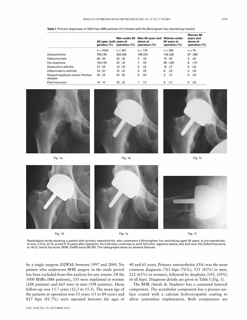
Fig. 1d
Radiological series showing a patient with primary osteoarthritis, who underwent a Birmingham hip resurfacing aged 38 years: a) pre-operatively,b) one, c) five, d) 10, e) and f) 15 years after operation. He is 54 now, continues to work full-time, regularly swims, skis and runs. His Oxford hip scoreis 14/12, Harris hip score 76/90. OsHIP score 95/100. The radiographs show no adverse features.
Fig. 1a Fig. 1b Fig. 1c
Fig. 1e Fig. 1f

1300 J. DANIEL, C. PRADHAN, H. ZIAEE, P. B. PYNSENT, D. J. W. MCMINN
THE BONE & JOINT JOURNAL
manufactured from as-cast high-carbon cobalt–chromealloy with 4 mm increments in the diameter of the femoralcomponent, and 2 mm increments for the acetabular com-ponent.
An extensile posterior approach was used1,17-19 to obtain a360° view of the acetabulum for optimal component posi-tioning. The intended acetabular component inclinationangle was 40°, with 20° anteversion, and the femoral com-ponent placed in neutral or mild valgus relative to the femo-ral neck. The acetabulum was under-reamed by 2 mmrelative to the component to allow primary press-fit fixation.The exceptions to this rule were patients, usually women,with very small sclerotic acetabulae who are under-reamedby 1 mm to avoid the risk of acetabular fracture, and in somelarge men with soft cancellous bone exposed in the reamedacetabulum, the socket is under-reamed by 3 mm.
Patients were reviewed at two months with clinical andradiological assessment. Between two months and tenyears, follow-up was conducted independently by theOswestry Outcomes Centre (OOC) by annual postal ques-tionnaires. From ten years onwards, postal follow-up hasbeen continued by the McMinn Centre.
We attempted to establish the reason for revision in everycase based on the history, clinico-radiological and labora-tory findings. Revision of either component for any reasonwas taken as the end-point for implant survival. All surviv-ing patients were contacted, by post, through their generalpractitioner or next of kin, or via the National StrategicTracing Service, and the status of their implant wasrecorded.
Clinical and radiological assessment was carried out onthe first 350 patients (402 hips). These results were auditedby the sponsor (Smith & Nephew) and by FDA-authorisedindependent auditors. Patients were sent a functional ques-tionnaire including Oxford Hip Score (OHS),20 Harris HipScore (HHS),21 Oswestry-modified Harris Hip Score(OsHIP, including a 5-point patient-satisfaction question),9
and modified University of California Los Angeles (UCLA)activity assessment.17 The OHS was used as described in itsoriginal form, with the score at 60 to 12, worst to best. Attheir appointment for clinical review, the completed ques-tionnaires were collected, and patients underwent conven-
tional radiographs (standing anteroposterior (AP) andhorizontal beam lateral views), metal ion measurement anda multi-slice metal artefact reduction sequence (MARS) CTscan. Activity level, in terms of steps taken per year, wasrecorded using the StepWatch 2 system (CymaCorp,Mountlake Terrace, Australia).22
Radiographs were assessed by a Consultant Musculo-skeletal Radiologist blinded to the clinical and functionaloutcomes. De Lee and Charnley acetabular zones23 andAmstutz femoral stem zones and scores24 were used. Grades7 to 9 (incomplete or complete radiolucencies ≥ 2 mm inthree zones with or without component migration of ≥ 3mm) were considered radiological failures. The surgeons(JD, CP) also assessed the radiographs during the clinicalreview and the two sets of independent observations (sur-geon and radiologist) were compared for inter-observervariability. Thinning of the femoral neck was assessed usingthe method described by Hing et al,25 with a 10% reductionin neck diameter considered significant. The acetabularinclination and stem–shaft angles were measured (HZ)using standard techniques.24,25 The interteardrop line wasused as the reference to measure the inclination angle. Theresults of the multi-slice CT assessment and metal ion levelswill be published separately.Statistical analysis. Statistical calculations were performedon MedCalc Version 12.2.1 (MedCalc Software, Ostend,Belgium) and the R statistical package (R Foundation forStatistical Computing, Vienna, Austria). The impact ofparameters (covariates) on survival were tested using theCox proportional hazard regression. The significance ofany model covariates was tested using the Wald statistic,which is actually the calculated covariate value divided byits standard error with the result squared. It has a chi-squared distribution. The 'quality' of a regression modelwas assessed using Akaike Information Criteria (AIC),where not only the goodness of the fit but also the numberof covariates is considered, thus providing a tool for regres-sion model selection.
Survival curves were produced based on the hazardratios (HR) calculated. Inter-observer agreement for theradiology was measured using weighted Kappa and thenomenclature of Landis and Koch.26 For all the statistics, a
Table II. Modes and times to failure in the 38 Birmingham Hip resurfacing revisions
Failure mode Number (%) Mean age at primary procedure in years (range)
Mean time to failure in years (range) Female Male
Femoral neck fracture 4 (0.4) 56.6 (34.7 to 75.1) 0.2 (0.1 to 0.4) 2 2Femoral head collapse 13 (1.3) 50.4 (34.5 to 58.8) 8.7 (0.7 to 14.0) 6 7Cup loosening 2 (0.2) 58.0 (50.3 to 65.7) 10.6 (9.7 to 11.6) 1 1Osteolysis 2 (0.2) 57.5 (50.2 to 64.8) 9.0 (7.0 to 10.9) 1 1Unexplained groin pain 3 (0.3) 44.8 (25.9 to 55.1) 8.5 (6.1 to 11.0) 2 1Debris Reaction ARMD* 7 (0.7) 53.7 (46.7 to 58.6) 10.6 (9.8 to 11.5) 7 0Infection 7 (0.7) 51.9 (39.6 to 61.8) 5.1 (2.5 to 11.6) 6 1
*ARMD, adverse reaction to metal debris

RESULTS OF BIRMINGHAM HIP RESURFACING AT 12 TO 15 YEARS 1301
VOL. 96-B, No. 10, OCTOBER 2014
p-value < 0.05 was considered statistically significant. 95%confidence intervals (CI) are shown where appropriate.
ResultsIn total, 59 patients (68 hips) died from unrelated causesbetween 0.7 and 12.6 years following surgery, of which twohad been revised prior to their death. Two patients hadintra-operative notching of the femoral neck, with no lateradverse effects. One patient sustained a posterior disloca-tion five years post-operatively, following a fall from a lad-der. Recurrent dislocation ensued which was successfullytreated with a double-breasted capsular repair.
There were 38 revisions (3.8%), in 36 patients whichwere performed at a median of 8.7 years (mean 7.6 years;
0.1 to 13.96) following implantation (Table II). In all, sixpatients with bilateral BHRs underwent revision; in two ofthese patients, both BHRs were revised. Of the 38 revisions,24 were performed in Birmingham and 14 were performedelsewhere.
Overall implant survival was 97.4% (95% confidenceinterval (CI) 96.9 to 97.9) at ten years and 95.8% (95%CI 95.1 to 96.5) survival at 15 years respectively (Fig. 2).Survival was worse in women compared with men at 15years (91.5%, 95% CI 89.8 to 93.2) in women versus(98.0%; 95% CI 97.4 to 98.6) in men (Fig. 3) and in youngwomen (< 60 years) compared with older women (≥ 60years) (90.5%; 95% CI 88.3 to 92.7 vs 95.9%; 95% CI94.6 to 97.2). Using Cox regression, the diagnosis of
Su
rviv
al p
rob
abili
ty (%
)
100
90
80
0 2 4 6 8 10 12 14 16
Number at risk
1000 991 980 964 942 925 902 292
99.0% 97.4% 95.8%
Follow-up (yrs)
Kaplan–Meier survival analysis with 95% confidence intervals shown,of the first 1000 consecutive BHRs, including all ages, all diagnoses andboth genders.
Fig. 2
100
90
80
0 2 4 6 8 10 12 14 16
Follow-up (years)
Survival differences byage and gender
95
85
Su
rviv
al p
rob
abili
ty (%
)
Sample size Implant survivorship
M ≥ 60
M < 60
F ≥ 60
F < 60
M ≥ 60M < 60F ≥ 60F < 60
M ≥ 60M < 60F ≥ 60F < 60
17449175260
99.4%98.3%95.9%94.9%
98.7%97.7%95.9%90.5%
100
90
80
0 2 4 6 8 10 12 14 16
Follow-up (years)
Survival differences byprimary diagnosis
95
85
Su
rviv
al p
rob
abili
ty (%
)
Sample size857
103
40
Others
Dysplasia
Osteonecrosis
Implant survivorship98.3%
93.2%
89.9%
97.1%
88.9%
87.4%
Others
Dysplasia
Osteonecrosis
Differences in Kaplan–Meier survivorship based on age, gender and primary diagnosis.
Fig. 3

1302 J. DANIEL, C. PRADHAN, H. ZIAEE, P. B. PYNSENT, D. J. W. MCMINN
THE BONE & JOINT JOURNAL
femoral head avascular necrosis (AVN) (p = 0.005) and hipdysplasia (p = 0.049) were found to have a significant influ-ence on implant survival, while age at operation (p =0.213), gender (p = 0.146), American Society of Anaesthe-siologists (ASA) grade27,28 (p = 0.674), head size (p =0.139), or whether unilateral or bilateral (p = 0.766) didnot. AVN alone was found to have a significant influence inmen (p < 0.001) and dysplasia (p = 0.049) alone in women(Table III). When reduced to just gender and diagnosis, themodel offers the best fit in terms of AIC.
For women with developmental hip dysplasia, implantsurvival was 96% (95% CI 93.8 to 98.2), 91% (95% CI87.7 to 94.3) and 85% (95% CI 80.2 to 89.8) at five, ten and15 years respectively. In patients of either gender < 50 yearsof age with osteoarthritis, survival was 100% at five yearsand 99.4% (95% CI 98.8 to 100) at ten and 15 years. Therewere no failures in men in this group, while in women inthis group, survival was 100%, 97.3% (95% CI 94.6 to100) and 97.3% (95% CI 94.6 to 100) at five, ten and 15years respectively.
Femoral failures (collapse of the femoral head or fractureof the femoral neck) occurred in 17 hips (1.7%) (Table II)(Fig. 4). All fractures of the femoral neck occurred in the firstfour months following surgery. In seven femoral failurestreatment was performed by conversion to a large diameterMoM THR, leaving the BHR acetabular component in situ.The remainder were revised to a non-MoM THR. Failuresrelated to wear, which were considered to include acetabularcomponent loosening, unexplained groin pain, osteolysisand ARMD, occurred in 14 hips (1.4%). All of these caseswere revised to a non-MoM THR. Of the six patients (sevenhips) that failed due to ARMD (0.7%), four patients (fivehips) showed strong reaction to nickel on lymphocyte trans-formation test, one did not undergo the test, and in the other,the cells were inadequate for it. No patient who underwentrevision for ARMD in our centre had major complications,muscle necrosis or re-revision. Of the patients who under-went revision elsewhere, none reported major complicationsor re-revision.
Of the 793 patients living with unrevised hip resurfacings(896 unrevised hips), 754 (95.1%) patients have at leastone completed current hip score, either OHS, OsHIP orHHS. Mean OsHIP scores have consistently exceeded 90%over the study period (Fig. 5). Of 754 cases, 18 (2.4%) cur-rently have a poor OsHIP score (< 70/100),29 and 10 of 603(1.7%) have a poor Oxford score (34 to 60 points).30 Themean UCLA activity Level Scale is 7.8, with 82% (467 of570 patients) remaining active (levels 7 to 10), 68% (387 of570 patients) regularly participating in very active events (8 to10), and 23% (133 of 570 patients) participating in impactsports.10,11
A subgroup of 350 patients, comprising the first 402 hipsin the series, underwent more detailed clinical and radio-logical follow-up. The mean age of these patients was53.2 years (23 to 84). At the latest follow-up, 23 patients(28 hips) had died, including one patient (one hip) who hadpreviously undergone revision, and 18 patients (20 hips)had undergone revision surgery. Of the remaining 355 hips,302 were reviewed in clinic and 14 sent radiographs andquestionnaire responses by post. As such, 316/355 surviv-ing hips (89%) had radiological follow-up and 325/355(91.5%) had clinical scores. Mean OSHIP score was 94%(standard deviation (SD) 9.1) and 93% (SD 9.9) at five andten years respectively, and 93% (SD 10.1) at the most recentfollow-up. Mean HHS was 85 (standard deviation (SD) 9.8)and mean OHS was 15 (SD 5.3) (Table IV). Mean activitylevel at the most recent follow-up was 1.8 million cycles peryear (0.5 to 4.1). The mean range of flexion of the operatedhip was 128° (85º to 140°). No patient had a fixed abduc-tion, adduction, or rotation deformity. Satisfaction wascategorised into two groups: ‘satisfied’ (comprising patientswho were ‘pleased’ or ‘extremely pleased’), and ‘dissatis-fied’ (comprising those reporting no improvement, andthose who report themselves worse or much worse thanpre-operatively). Fewer than 1% of patients were dissatis-fied at any interval in the study period.
The mean angle of inclination of the acetabular compo-nent was 43° (SD 5.1°), with three hips being outliers with
Table III. Cox Regression model in men and women. For categorical covariates (diagnosis and bilaterality), exp(b) offers theinstantaneous relative risk of an event, at any time, with the risk factor presentversus absent, given all other covariates areequal. For continuous covariates (age, American society of anaestheologists (ASA) grade and head size) it is the relative riskwith an increase of 1 in the value of the covariate. The diagnosis of dysplasia is the only covariate that has a significant influ-ence in women (p = 0.05), and the diagnosis of avascular necrosis (AVN) (femoral head osteonecrosis) has a significant influ-ence in men (p = 0.0001)
In women In men
Covariate HR (95% CI) Wald p-value HR (95% CI) Wald p-value
Age at operation 1.01 (0.97 to 1.05) 0.40 0.53 1.03 (0.97 to 1.09) 0.67 0.41ASA grade26 1.32 (0.55 to 3.17) 0.39 0.53 0.69 (0.18 to 2.66) 0.28 0.59Head size 1.03 (0.66 to 1.59) 0.01 0.90 0.96 (0.80 to 1.15) 0.19 0.66AVN 1.17 (0.15 to 9.27) 0.02 0.89 14.33 (3.69 to 55.70) 14.63 0.0001Dysplasia 2.46 (1.00 to 6.04) 3.82 0.049 0.00 (0.00 to 0.00) 0.00 0.97Bilateral (v unilateral) 0.86 (0.34 to 2.18) 0.10 0.75 0.68 (0.15 to 3.16) 0.23 0.63
HR, hazard ratio for revision; CI, confidence interval; Wald, Wald statistic

RESULTS OF BIRMINGHAM HIP RESURFACING AT 12 TO 15 YEARS 1303
VOL. 96-B, No. 10, OCTOBER 2014
inclination > 55°. The mean stem–shaft angle was 143°(SD 6.1°). Diffuse thinning of the femoral neck (comparedwith radiographs taken at two months) was present in16 hips (5.1%) in 15 patients, but none progressed after thefirst five years. Focal resorption of the inferomedial orsuperolateral neck (which may be the result of impinge-ment) was present in 14 hips (4.4%) in 14 patients.
Acetabular radiolucencies were present in a single zone(grades 1 to 3) in 9/316 hips (2.8%), and in two zones(grades 4 to 6) in 4/316 (1.3%). No hips had a radiolucencyin all three acetabular zones. Femoral radiolucencies werepresent in one zone in 11/316 (3.5%), in two zones in 0/316(0%) and in three zones in 1/316 (0.3%). Thus, there wereno radiological acetabular failures and one radiologicalfemoral failure. There was substantial agreement betweenthe degree of acetabular radiolucency as assessed by the
radiologist and the surgeons (weighted Kappa κ = 0.74,95% CI 0.62 to 0.86), but poor agreement in the assess-ment of femoral radiolucencies, with the surgeons diagnos-ing a greater number than the radiologist (κ = 0.29, 95% CI0.10 to 0.48). Inter-observer reliability for the summaryfindings was substantial (κ = 0.63, 95% CI 0.49 to 0.77).On CT, the acetabular radiolucencies (9/288) were a mean sizeof 1.6cm2 (1 to 3.4), and the femoral radiolucencies (4/288)were a mean size of 1.1cm2 (1 to 2.4). No radiolucency wasdeemed extensive enough to affect component stability orto justify a revision procedure. Aside from the seven hipsrevised for symptomatic effusions, no asymptomatic solidpseudotumours or large effusions were detected by CT.
Small effusions were seen in 16 of 288 hips (5.6%) of theBHRs and also in 2/79 (2.5%) of contralateral unoperatedhips where arthritic change was present at the time of
Radiological series of a 34-year-old female with developmental dysplasia treated with a Dysplasia Birmingham Hip resurfacing a) Initial radiographsshow good component position. Later, diagnosed with systemic lupus, she was administered steroids and immuno-suppressants. A year later herradiographs showed varus migration of femoral component, suggesting possible femoral loosening. The patient continued to mobilise withoutsymptoms. Eight years later she fell down, sustained a femoral head fracture and had to be converted to a total hip replacement. b) The retrievedfemoral head was sectioned and a radiograph was taken. The specimen was then stained with methylene blue/basic fuchsin c)12.5X and d) = 100Xmagnification) which confirm that the bone in the femoral head is vascular and viable; and that the femoral component was not loose as seen fromthe intact bone–cement and cement–implant interfaces. Histology also suggested a series of microfractures in the femoral head, which had sponta-neously healed, possibly resulting in the early migration. The migration, however, stabilised until the subsequent fall and basal fracture.
Fig. 4a
Fig. 4b Fig. 4c Fig. 4d

1304 J. DANIEL, C. PRADHAN, H. ZIAEE, P. B. PYNSENT, D. J. W. MCMINN
THE BONE & JOINT JOURNAL
review. There was no evidence of muscle necrosis or otherchanges in soft-tissue in either hip in any patient.
DiscussionThe introduction of the BHR followed 6.5 years of pre-clinical testing and clinical pilot studies.1 Despite the array ofpre-clinical and post-marketing strategies and trials, the cat-astrophic failure of other resurfacing devices demonstratethat these do not guarantee implant survivorship in reallife.31-36 Charnley first introduced the practice of followingup his first three years’ THR implantations through periodicreview or questionnaires over 15 years.37 We have followeda similar pattern of follow-up through prospective annualoutcomes scores by the OOC and periodic clinical and radi-ological assessments at our centre. This is the 15-year reportof the earliest three years and 1000 BHR implantations bythe inventor surgeon.
There are both strengths and limitations to this study. First,it is a prospective case series rather than a randomised con-trolled trial (RCT). However, unlike in pharmaceuticalresearch, RCTs are not the only study design suitable for the
introduction and monitoring of new surgical devices.38 Sec-ond, as this is a report from the designer centre, there is poten-tial for bias. In an attempt to minimise bias, prospectivequestionnaire evaluations were conducted by an independ-ent centre, and the radiological assessment included a con-tribution by a radiologist blinded to the clinical outcome.Third, this is a single surgeon series from a high volume hipsurgeon (DJWM) who had performed around 500 MoMresurfacings prior to the present study, which minimises thelearning curve effect and may limit the external validity ofthis study. However comparable medium-term results fromother centres4-8 and national registers2,3 suggest that,following adequate training, this technique is reproducible.
A strength of this study is the low rate of loss to follow-up.All patients had their survival and revision status confirmed;95.1% of surviving patients with unrevised hips had clinicaloutcome scores at latest follow-up and 89% of hips in theclinical and radiological review group (316/355) had a radi-ological assessment. Another strength is that the device usedin the present study and the implantation technique haveremained unchanged, (other than the subsequent addition of
100
90
80
70
60
50
96 97 96 96 94 93 94
EXCELLENT
GOOD
FAIR
POOR
Pre-opn = 723
Yr 1672
Yr 2695
Yr 3863
Yr 4851
Yr 7866
Yr 10846
Current842
58
Follow-up
Osw
estr
y H
ip S
core
mea
n a
nd
co
nfi
den
ce in
terv
als
Fig. 5
Oswestry modified Harris hip score (OsHIP) postal questionnaireresponses over the years following implantation. Marchetti’s gradeboundaries shown.
Table IV. Hip function scores in the clinico-radiological cohort.
OSHIP HHS OHS UCLA
Range (Best possible score to worst) (100 to 0) (90 to 0) (12 to 60) (10 to 1)Mean (expressed as % of best possible score) 93 (93) 85 (94) 15 (94) 7.8 Standard deviation 10.1 9.8 5.3 1.195% confidence interval 1.1 1.1 0.6 0.1Quartile 1 90 84 12 7.0Median (expressed as % of best possible score) 98 (98) 88 (98) 13 (98) 8 (80)Quartile 3 100 90 15 8Excellent + Good (%) 89 91 96 82 (level 7 to 10) Active
OSHIP, Oswestry Hip Score; OHS, Oxford Hip Score; HHS, Harris Hip Score; UCLA, University of California Los Angeles Activity scale

RESULTS OF BIRMINGHAM HIP RESURFACING AT 12 TO 15 YEARS 1305
VOL. 96-B, No. 10, OCTOBER 2014
2 mm head size increments), throughout the study and tothe present day. In a previous cohort, containing hipsbefore and after the addition of the additional head sizes, itwas found that the change had no effect on implant sur-vival,4 suggesting that the results in this report may betranslated to devices currently in use.
At 15 years, the results of this study in terms of all-causeimplant survival (98.0%; 95% CI 97.4 to 98.6 in men and91.5%; 95% CI 89.8 to 93.2 in women) compare well withresults from other centres4-8,12 and joint registers2,3 con-firming that the BHR is a reliable and effective procedure.A recent report has suggested that HR may only be suitablein men with bearing diameters > 54 mm.39 In contrast, theAustralian register demonstrates that in a national series,among men with osteoarthritis (OA) under 65 years (thelargest single group requiring a resurfacing), HR (of anydiameters) has a 1.3% lower cumulative implant revisionrate (6.5% to 6.7%) than THRs (7.8% to 8.7%) at ten-year follow-up.3
The risk of revision following conventional THR is threeto five times higher in young patients compared with olderpatients.40 In the Australian Register, the rate of revisionincreases from 7.3% (95% CI 6.0 to 8.9) in men over theage of 75, to 10.3% (95% CI 8.9 to 11.7) in men under 55,and this increase is more marked in women (from 5.0%;95% CI 4.6 to 5.5 to 12.7%; 95% CI 10.5 to 15.2).3 Thethree Nordic registers report a revision rate of 17% inyoung people (< 50 years) at ten years.41 A large series ofthe most widely-used cemented THR has reported a revi-sion rate of 8% at 12 years and 19% at 15.42,43 However, inthat series only 10% were under 50 years of age at the timeof surgery, 57.2% had died (at a mean of 8.3 years, 0 to 16years) by the time of analysis, and only 8% (26) of the orig-inal series (325) continued in low-stress sport or moderatemanual labour following surgery. In the present series, inwhich over 30% of hips (317/1000) were implanted inpatients below the age of 50 years, the overall revision rateof 4.2% compares well with previous series of THR. Thebest results in this series were seen in men and womenunder the age of 50 years with osteoarthritis. The BHR is aviable option in this relatively young active population whofare poorly with conventional THR.
Femoral failures (neck fractures and collapse) are unique toHR and represent a risk which has to be accepted in the inter-est of conserving bone. In our series, there were no instances offemoral loosening which is superior to a previous series of adifferent implant, with a different technique of cementing inwhich 8% of patients were revised for femoral loosening.44
Metal reactions with osseous and soft-tissue damage,45
with features representing hypersensitivity-induced necro-sis,46,47 cytotoxicity48 and osteolysis49 have been reportedfollowing HR. The incidence and severity of these compli-cations are implant-specific, with one centre predicting arate of failure by ARMD of 4% at eight years.16 Our seriesdemonstrates that with well-implanted BHRs the incidenceof ARMD is < 1% at 12 to 15 years. Of the seven revisions
for ARMD, three were revised at our centre. These patientshad effusions but had no muscle necrosis, and there wereno major complications or early re-revisions following revi-sion surgery contrary to the ARMD results from othercentres16 and with other devices.15 Our findings are sup-ported by results from other centres at nine to 11 years.5,6,12
Osteolysis, loosening, unexplained pain and ARMD mayall be a consequence of wear. At 15 years, the failure ratedue to all of these mechanisms combined is 1.4% overall,and only 0.5% in men. This low incidence strongly sup-ports the view that resurfacing is a suitable option for bothgenders, but is particularly suited to men.
Radiological assessment showed no impending acetabularfailures and only one radiological femoral failure, which sug-gests that there is unlikely to be a precipitous drop in survivalin the years following this report. In the present series, theprevalence of radiolucencies which were not deemed failures(those of grades 1 to 6), compare well with other seriesreporting such radiolucencies in 5% to 7% of femoral, and1% of acetabular component regions.7,44 Our results con-firm earlier reports that thinning of the neck does not pro-gress after five years.7 The number of cases with focalresorption representing impingement (4%) compares wellwith other reports of up to 20% impingement.44
This study reports a rate of implant survival of 95.8%(95% CI 95.1 to 96.5) in patients of all ages and diagnosesat 15 years, and of 98.0% (95% CI 97.4 to 98.6) in menalone, with a low incidence of radiological abnormalities.This suggests that the BHR is a viable conservative alterna-tive to THR. Patients (both men and women) under the ageof 50 years with osteoarthritis have the best results. Osteo-necrosis and dysplasia are risk factors for failure. The inci-dence of ARMD is less than 1%. The high hip function andpatient satisfaction scores suggest that the expectations ofthis relatively young cohort are adequately met.
Supplementary materialFour tables showing range of movement, longitudi-nal assessment, radiological findings and ten-year
results compared with the current study, as well as a figureshowing a radiological series of a patient with severe SUFEare available alongside the online version of this article atwww.bjj.boneandjoint.org.uk.
The authors gratefully acknowledge the expertise and support of Dr N. Evans(Consultant Radiologist, Royal Orthopaedic Hospital, Birmingham, United King-dom) for his independent expert radiographic assessment. We also thank Smith& Nephew Orthopaedics for project support for this investigation.
No benefits in any form have been received or will be received from a com-mercial party related directly or indirectly to the subject of this article.
This article was primary edited by A. Liddle and first proof edited by G. Scott.
References1. McMinn D, Treacy R, Lin K, Pynsent P. Metal on metal surface replacement of the
hip. Experience of the McMinn prothesis. Clin Orthop Relat Res 1996;(329Suppl):S89–S98.2. No authors listed. National Joint Registry: 10th Annual Report, National Joint Reg-
istry for England, Wales and Northern Ireland, 2013. http://www.njrcentre.org.uk/njr-centre/Portals/0/Documents/England/Reports/10th_annual_report/NJR%2010th%20Annual%20Report%202013%20B.pdf (date last accessed 26 June2014).

1306 J. DANIEL, C. PRADHAN, H. ZIAEE, P. B. PYNSENT, D. J. W. MCMINN
THE BONE & JOINT JOURNAL
3. No authors listed. Australian National Joint Registry Annual Report 2013 https://aoanjrr.dmac.adelaide.edu.au/annual-reports-2013 (date last accessed 14 July 2014).
4. McMinn DJ, Daniel J, Ziaee H, Pradhan C. Indications and results of hip resur-facing. Int Orthop 2011;35:231–237.
5. Treacy RB, McBryde CW, Shears E, Pynsent PB. Birmingham hip resurfacing: aminimum follow-up of ten years. J Bone Joint Surg [Br] 2011;93-B:27–33.
6. Holland JP, Langton DJ, Hashmi M. Ten-year clinical, radiological and metal ionanalysis of the Birmingham Hip Resurfacing: from a single, non-designer surgeon.J Bone Joint Surg [Br] 2012;94-B:471–476.
7. Coulter G, Young DA, Dalziel RE, Shimmin AJ. Birmingham hip resurfacing at amean of ten years: results from an independent centre. J Bone Joint Surg [Br]2012;94-B:315–321.
8. Van Der Straeten C, Van Quickenborne D, De Roest B, et al. Metal ion levelsfrom well-functioning Birmingham Hip Resurfacings decline significantly at ten years.Bone Joint J 2013;95-B:1332–1338.
9. Jaiswal A, Gilbert RE, Sunil Kumar KH, et al. Function and survival after revisionof hip resurfacing. Hip Int 2011;21:610–615.
10. Barrack RL, Ruh EL, Berend ME, et al. Do young, active patients perceive advan-tages after surface replacement compared to cementless total hip arthroplasty? ClinOrthop Relat Res 2013;471:3803–3813.
11. Aqil A, Drabu R, Bergmann JH, et al. The gait of patients with one resurfacing andone replacement hip: a single blinded controlled study. Int Orthop 2013;37:795–801.
12. Baker RP, Pollard TC, Eastaugh-Waring SJ, Bannister GC. A medium-termcomparison of hybrid hip replacement and Birmingham hip resurfacing in active youngpatients. J Bone Joint Surg [Br] 2011;93-B:158–163.
13. McMinn DJ, Snell KI, Daniel J, et al. Mortality and implant revision rates of hiparthroplasty in patients with osteoarthritis: registry based cohort study. BMJ2012;344:3319.
14. Kendal AR, Prieto-Alhambra D, Arden NK, Carr A, Judge A. Mortality rates at10 years after metal-on-metal hip resurfacing compared with total hip replacement inEngland: retrospective cohort analysis of hospital episode statistics. BMJ2013;347:6549.
15. Langton DJ, Joyce TJ, Jameson SS, et al. Adverse reaction to metal debris fol-lowing hip resurfacing: the influence of component type, orientation and volumetricwear. J Bone Joint Surg [Br] 2011;93-B:164–171.
16. Murray DW, Grammatopoulos G, Gundle R, et al. Hip resurfacing and pseudotu-mour. Hip Int 2011;21:279–283.
17. Daniel J, Pynsent PB, McMinn DJ. Metal-on-metal resurfacing of the hip inpatients under the age of 55 years with osteoarthritis. J Bone Joint Surg [Br] 2004;86-B:177–184.
18. McMinn DJW. Acetabular preparation and insertion of the standard BirminghamHip Resurfacing cup. In: McMinn D, ed. Modern Hip Resurfacing. London, England:Springer-Verlag; 2009:223–236.
19. McMinn DJW. Implantation of the Femoral Component of the Birmingham HipResurfacing. In: McMinn D, ed. Modern hip resurfacing. New York: Springer-Verlag,2009:265–299.
20. Dawson J, Fitzpatrick R, Carr A, Murray D. Questionnaire on the perceptions ofpatients about total hip replacement. J Bone Joint Surg [Br] 1996;78-B:185–190.
21. Mahomed NN, Arndt DC, McGrory BJ, Harris WH. The Harris hip score: compar-ison of patient self-report with surgeon assessment. J Arthroplasty 2001;16:575–580.
22. Daniel J, Ziaee H, Pradhan C, Pynsent PB, McMinn DJ. Blood and urine metalion levels in young and active patients after Birmingham hip resurfacing arthroplasty:four-year results of a prospective longitudinal study. J Bone Joint Surg [Br] 2007;89-B:169–173.
23. DeLee JG, Charnley J. Radiological demarcation of cemented sockets in total hipreplacement. Clin Orthop Relat Res 1976;121:20–32.
24. Beaulé PE, Dorey FJ, Le Duff MJ, Gruen T, Amstutz HC. Risk factors affectingoutcome of metal-on-metal surface arthroplasty of the hip. Clin Orthop Relat Res2004;41:87–93.
25. Hing CB, Back DL, Bailey M, et al. The results of primary Birmingham hip resur-facings at a mean of five years. An independent prospective review of the first 230hips. J Bone Joint Surg [Br] 2007;89-B:1431–1438.
26. Landis JR, Koch GG. The measurement of observer agreement for categorical data.Biometrics 1977;33:159–174.
27. No authors listed. American Society of Anesthesiologists: physical status classifi-cation system: new classification of physical status. Anesthesiology 1963;24:111.
28. Halaszynski TM, Juda R, Silverman DG. Optimizing postoperative outcomes withefficient preoperative assessment and management. Crit Care Med 2004;32(4Suppl):S76–S86.
29. Marchetti P, Binazzi R, Vaccari V, et al. Long-term results with cementless Fitek(or Fitmore) cups. J Arthroplasty 2005;20:730–737.
30. Kalairajah Y, Azurza K, Hulme C, Molloy S, Drabu KJ. Health outcome measuresin the evaluation of total hip arthroplasties--a comparison between the Harris hipscore and the Oxford hip score. J Arthroplasty 2005;20:1037–1041.
31. de Steiger RN, Hang JR, Miller LN, Graves SE, Davidson DC. Five-year resultsof the ASR XL Acetabular System and the ASR Hip Resurfacing System: an analysisfrom the Australian Orthopaedic Association National Joint Replacement Registry.J Bone Joint Surg [Am] 2011;93-A:2287–2293.
32. Long WT, Dastane M, Harris MJ, Wan Z, Dorr LD. Failure of the Durom Metasulacetabular component. Clin Orthop Relat Res 2010;468:400–405.
33. Malchau H, Bragdon CR, Muratoglu OK. The stepwise introduction of innovationinto orthopedic surgery: the next level of dilemmas. J Arthroplasty 2011;26:825–831.
34. Gross M. Innovations in surgery. A proposal for phased clinical trials. J Bone JointSurg [Br] 1993;75-B:351–354.
35. Vendittoli PA, Ganapathi M, Roy AG, Lusignan D, Lavigne M. A comparison ofclinical results of hip resurfacing arthroplasty and 28 mm metal on metal total hiparthroplasty: a randomised trial with 3-6 years follow-up. Hip Int 2010;20:1–13.
36. Isaac GH, Siebel T, Oakeshott RD, et al. Changes in whole blood metal ion levelsfollowing resurfacing: serial measurements in a multi-centre study. Hip Int2009;19:330–337.
37. Charnley J. Low friction arthroplasty of the hip: theory and practice. New York:Springer-Verlag; 1979:75.
38. Amstutz HC. Innovations in design and technology. The story of hip arthroplasty. ClinOrthop Relat Res 2000;378:23–30.
39. Smith AJ, Dieppe P, Howard PW, Blom AW; National Joint Registry for Eng-land and Wales. Failure rates of metal-on-metal hip resurfacings: analysis of datafrom the National Joint Registry for England and Wales. Lancet 2012;380:1759–1766.
40. Santaguida PL, Hawker GA, Hudak PL, et al. Patient characteristics affecting theprognosis of total hip and knee joint arthroplasty: a systematic review. Can J Surg2008;51:428–436.
41. Overgaard S, Petersen A, Havelln L, et al. The prognosis of total hip arthroplasty(THA) in patients younger than 50 years of age: results of 14,610 Primary THA. J BoneJoint Surg [Br] 2011;93-B(SuppII):87.
42. Carrington NC, Sierra RJ, Gie GA, et al. The Exeter Universal cemented femoralcomponent at 15 to 17 years: an update on the first 325 hips. J Bone Joint Surg [Br]2009;91-B:730–737.
43. Williams HD, Browne G, Gie GA, et al. The Exeter universal cemented femoralcomponent at 8 to 12 years. A study of the first 325 hips. J Bone Joint Surg [Br]2002;84-B:324–334.
44. Amstutz HC, Le Duff MJ, Campbell PA, Gruen TA, Wisk LE. Clinical and radio-graphic results of metal-on-metal hip resurfacing with a minimum ten-year follow-up.J Bone Joint Surg [Am] 2010;92-A:2663–2671.
45. Pandit H, Glyn-Jones S, McLardy-Smith P, et al. Pseudotumours associated withmetal-on-metal hip resurfacings. J Bone Joint Surg [Br] 2008;90-B:847–851.
46. Willert HG, Buchhorn GH, Fayyazi A, et al. Metal-on-metal bearings and hyper-sensitivity in patients with artificial hip joints. A clinical and histomorphologicalstudy. J Bone Joint Surg [Am] 2005;87-A:28–36.
47. Campbell P, Ebramzadeh E, Nelson S, et al. Histological features of pseudotu-mor-like tissues from metal-on-metal hips. Clin Orthop Relat Res 2010;468:2321–2327.
48. Mahendra G, Pandit H, Kliskey K, et al. Necrotic and inflammatory changes inmetal-on-metal resurfacing hip arthroplasties. Acta Orthop 2009;80:653–659.
49. Daniel J, Ziaee H, Kamali A, et al. Ten-year results of a double-heat-treated metal-on-metal hip resurfacing. J Bone Joint Surg [Br] 2010;92-B:20–27.