highly efficient and multidimensional extraction of …quanyuan.whu.edu.cn/pdf/2016 nano...
TRANSCRIPT

Highly efficient and multidimensional extraction of targets from complex matrices using aptamer-driven recognition
Jie Wang1, Haijing Shen1, Chi Huang1, Qinqin Ma1, Yaning Tan1, Fenglei Jiang1, Chao Ma2, and Quan Yuan1 ()
1 Key Laboratory of Analytical Chemistry for Biology and Medicine (Ministry of Education), College of Chemistry and Molecular Sciences,
Wuhan University, Wuhan 430072, China 2 Hefei National Laboratory for Physical Sciences at the Microscale, University of Science and Technology of China, Hefei 230026, China
Received: 26 June 2016
Revised: 5 August 2016
Accepted: 31 August 2016
© Tsinghua University Press
and Springer-Verlag Berlin
Heidelberg 2016
KEYWORDS
aptamer,
adsorbent,
selectivity,
separation,
extraction
ABSTRACT
Adsorbents are widely employed in both fundamental and applied research
areas such as separation technology, biotechnology, and environmental science.
Selectivity and reusability are two most important requirements for adsorbents.
Aptamers exhibit perfect selectivity and easy regeneration, which make them
uniquely effective adsorption materials. Herein, we have rationally designed
novel aptamer-based adsorbents and investigated their performance in extraction/
separation of targets from an aqueous solution. These adsorbents can selectively
extract targets from complicated sample matrices containing background com-
pounds. Moreover, they can also be easily recycled without a significant loss of
adsorption capacity. Notably, the adsorbents did not affect the activity of isolated
biological samples, revealing their potential for the purification/separation of
biomolecules. Composite adsorbents were constructed using aptamer-based
adsorbents and a porous polymer, displaying highly efficient target separation
from aqueous solution. Finally, separation columns were constructed, and targets
in the aqueous solution were efficiently separated by these columns. The aptamer-
based adsorbents described here exhibit great promise for potential applications
in separation technology, biotechnology, and environment-related areas.
1 Introduction
Adsorption is one of the most commonly encountered
phenomena in our daily life that has attracted broad
interest during the past decades [1–6]. Undoubtedly,
it is a very useful technique, with first practical
applications dated to ancient times. Nowadays, the
rapidly developing adsorption techniques are widely
employed in separation technology [7], biotechnology
[8], environmental science [9–16], and catalysis [17].
Separation technology, e.g., solid-phase extraction,
usually employs adsorbents to extract targets from
Nano Research
DOI 10.1007/s12274-016-1273-9
Address correspondence to [email protected]
| www.editorialmanager.com/nare/default.asp
2 Nano Res.
complicated sample matrices or pre-concentrate them
from dilute solutions to improve detection sensitivity
[18]. Biotechnology uses adsorbents to purify
biomolecules such as proteins, nucleic acids, and even
intact cells directly from crude samples [8]. Besides,
adsorption is one of the most convenient methods of
environmental science for removing pollutants from
wastewater [10]. Obviously, adsorption is of great
importance for both fundamental and applied research.
Although adsorbents are utilized in different application
areas, they share some key requirements, i.e., good
selectivity and reusability [2, 18].
Over the past several years, single-stranded DNA
and RNA (aptamers) have emerged as a novel class
of molecules that attract extensive attention, since they
are capable of recognizing a broad range of substances
with high affinity and specificity, ranging from metal
ions and small molecules to tissues [19–25]. Compared
to other functional biomolecules such as antibodies
and enzymes, aptamers offer the advantages of design
flexibility, synthetic accessibility, easy modification,
and chemical stability [26–30]. Several particularly
important features of aptamers make them uniquely
efficient components of adsorption materials, namely
perfect selectivity and easy regeneration. According
to previous studies, aptamers specifically bind their
targets with pico- to nanomolar dissociation constants
by folding into unique secondary or tertiary structures
that accommodate the molecular structure of the targets
[31–37], revealing their ability to extract even low
concentrations of targets in the presence of background
compounds from sample matrices. Furthermore, the
unique structures of aptamers can be efficiently
denaturized by changing pH or increasing the tem-
perature to release the captured targets into desired
media and the functional aptamers can be easily
regenerated within minutes [38, 39], indicating their
good reusability as adsorbents.
Herein, we have rationally designed a novel class
of adsorbents with good selectivity and reusability by
employing aptamers as binding groups. Specifically,
these adsorbents were constructed by immobilizing
aptamers on the surface of superparamagnetic magnetic
nanoparticles (MNPs). These adsorbents feature good
selectivity due to the special binding pockets of
aptamers and good reusability due to the rational
integration of their easy regenerability with magnetic
separation. It is worth noting that adsorbents for
inorganic metal ions, organic molecules, and even
intact living cells can be easily obtained by simply
changing the types of aptamers used. In addition,
unmodified MNPs can also be used as adsorbents to
separate phosphate from aqueous solution. Composite
adsorbents and separation columns were further
constructed to investigate the potential applications
(e.g., affinity chromatography) of these adsorbents
in device fabrication. The strategy described here
may therefore not only serve as a promising method
for the design of novel adsorbents, but also expand
the application of aptamers in separation technology,
biotechnology, and environmental science.
2 Experimental
2.1 Synthesis of mesoporous TiO2 (mTiO2)-coated
Fe3O4 nanoparticles (Fe3O4@mTiO2 MNPs)
To synthesis the nanoparticles, FeCl3·6H2O, trisodium
citrate, and sodium acetate were first dissolved in
ethylene glycol. The solution was then transferred
into a Teflon-lined stainless steel autoclave and heated
at 200 °C for 10 h. Subsequently, the as-prepared Fe3O4
nanoparticles, concentrated ammonia solution, and
tetrabutyl titanate were added to ethanol, and the
mixture was ultrasonicated for 15 min. The solution
was allowed to react at 45 °C for 24 h under continuous
mechanical stirring. The resulting products were dried
and calcined at 500 °C for 2 h to obtain Fe3O4@mTiO2
MNPs.
2.2 Fabrication of aptamer-based adsorbents
MNPs were immersed in an acetone solution of
3-aminopropyltriethoxysilane (APTES) (5%) for
10 h to introduce amino groups onto their surface.
Subsequently, a bifunctional cross-linker (Sulfo-SMCC)
was used to functionalize MNPs with aptamers.
MNPs (10 mg) and cross-linker (succinimidyl 4-(N-
maleimidomethyl)cyclohexane-1-carboxylate, Sulfo-
SMCC) (4 mg) were reacted in 2-(N-morpholino)-
ethansulfonic acid (MES) buffer (20 mM, pH 6.8) for
4 h. The resulting product was collected and washed.
The obtained MNPs and aptamer (20 nmol) were

www.theNanoResearch.com∣www.Springer.com/journal/12274 | Nano Research
3 Nano Res.
added to a phosphate buffer saline (20 mM, pH 7.2),
and the solution was shaken at room temperature for
24 h. The as-prepared aptamer-functionalized MNPs
were magnetically separated and washed with distilled
water for subsequent use.
2.3 Selectivity tests
Briefly, Apt-Hg-MNPs (1 mg) were dispersed in Tris-
nitrate buffer (1 mL, 10 mM, pH 8.0) containing Hg2+
(1 ppm), Na+ (10 ppm), Ca2+ (10 ppm), Cu2+ (10 ppm),
Cd2+ (10 ppm), Zn2+ (10 ppm), and Ag+ (10 ppm). The
above solution was allowed to react for 2 h at room
temperature. The adsorbents were isolated, and the
resulting solution was collected to determine the
residual concentrations of each ion. Additionally, Apt-
BPA-MNPs (1 mg) were suspended in Tris-nitrate
buffer containing bisphenol A (BPA, 60 ppm), bisphenol
F (BPF, 60 ppm), benzoic acid (60 ppm), phenol (60 ppm),
and hydroxybenzoic acid (60 ppm). The solution was
treated with Apt-BPA-MNPs obtained above. Further,
Apt-SA-MNPs (1 mg) were mixed with ~105 CFU/mL
Staphylococcus aureus (S. aureus) in 1 mL of stroke-
physiological saline solution and incubated for 2 h.
The supernatant was collected to determine the residual
number of S. aureus by plate count. An Escherichia coli
(E. coli) sample (~105 CFU/mL) was also treated with
Apt-SA-MNPs using the same procedure.
2.4 Cyclic separation of Hg2+, BPA, and S. aureus
Taking the cyclic separation of Hg2+ as an example,
Apt-Hg-MNPs (1 mg) were dispersed in a solution
of Hg2+ (1 mL, 1 ppm, pH 8.0, Tris-nitrate buffer), and
the mixture was allowed to react for 2 h at room tem-
perature. Subsequently, Apt-Hg-MNPs were separated,
and the amount of residual Hg2+ was determined.
The separated Apt-Hg-MNPs were washed with HCl
(pH 3) and deionized water for three times to remove
the adsorbed Hg2+ ions, and the regenerated Apt-Hg-
MNPs were used to separate Hg2+ ions from another
aqueous solution. Cyclic removal of BPA (60 ppm)
and S. aureus (~105 CFU/mL) was carried out using the
same procedure.
2.5 Cyclic separation of PO43–
Typically, MNPs (1 mg) were added to a solution of
sodium dihydrogen phosphate (1 mL, 1 ppm), and the
mixture was allowed to react at room temperature
for 2 h. The MNPs were separated and washed with
acetonitrile (50%) and trifluoroacetic acid (0.1%) before
being used in the next cycle. The concentration of PO43–
was measured using the ammonium molybdate spectro-
photometric method according to the National Standard
of the People’s Republic of China (GB 11893-89).
2.6 Preparation of composite adsorbents
Briefly, Apt-Hg-MNPs (1 mg) were dispersed in acetic
acid solution (5 mL, 0.1 M), followed by the addition of
chitosan powder (100 mg). Subsequently, a solution of
β-glycerol 2-phosphate disodium salt hydrate (β-GP)
(500 μL, 0.26 M) was added upon cooling in an ice-
water bath. Finally, the mixture was heated to 37 °C in
an oven to produce the composite adsorbents. The
related adsorbents for BPA and PO43– were fabricated
using the same protocol.
2.7 Separation of Hg2+, BPA, and PO43– using com-
posite adsorbents
In a typical experiment, the composite adsorbent for
mercury was added to a Tris-nitrate buffer containing
Hg2+ ions (1 ppm). The solution was incubated at room
temperature for 2 h under continuous mechanical
stirring. Subsequently, the composite adsorbent was
separated, and the solution was used for the following
color reactions. Separation of BPA and PO43– was
achieved using the same method. Dithizone, ferric
trichloride, and ammonium molybdate were utilized
to form colored complexes with Hg2+, BPA, and PO43–,
respectively.
2.8 Fabrication of separation columns
Separation columns were fabricated using plastic
medical syringes (10 mL) as containers for the composite
adsorbents. The acidic suspension was prepared
employing the abovementioned methods, followed by
addition of the β-GP solution. The composite hydrogel
was formed by transferring the above solution into
plastic syringes, and the resultant filters were heated
to 37 °C in an oven. Separation columns for Hg2+,
PO43–, BPA, and S. aureus were fabricated separately,
using the corresponding adsorbents.

| www.editorialmanager.com/nare/default.asp
4 Nano Res.
2.9 Removal of Hg2+, BPA, and PO43– by the separation
columns
Briefly, the aqueous solution containing Hg2+ (1 mL,
1 ppm) was allowed to pass through the syringe
filters under the action of gravity, and the filtrate was
collected to determine the residual Hg2+ concentration.
The separation of BPA (60 ppm), PO43– (1 ppm), and
S. aureus (~105 CFU/mL) was performed using the
same procedure.
3 Results and discussion
The approach used to construct aptamer-based
adsorbents is illustrated in Fig. 1(a). Core–shell MNPs
were prepared by coating Fe3O4 nanoparticles with
a layer of mesoporous titanium dioxide [40]. The
TiO2 shell can adsorb phosphate due to the classic
coordination reaction between titanium ions and
PO43–, with the amount of available active binding
sites greatly increased by the mesoporous structure of
TiO2. More importantly, the abundant hydroxy groups
on the surface of TiO2 can be used to introduce amino
groups via hydrolysis of 3-aminopropyltriethoxysilane
[41]. Thiol groups at the 3’-end of aptamers and the
amino groups on the MNP surface were further linked
by a bifunctional cross-linker (Sulfo-SMCC) to obtain
the final adsorbents [42]. In this study, three kinds
of aptamers binding Hg2+ (designated as Apt-Hg) [43],
bisphenol A (designated as Apt-BPA) [44], and
Figure 1 Characterization of adsorbents. (a) Schematic preparation of aptamer-based adsorbents. TEM (b), HAADF-STEM (c), and elemental mapping (d) images of adsorbents. (e) Aqueous dispersion of adsorbents and the separation test using a permanent magnet. (f) Magnetization field dependence plot of the adsorbents. (g) Zeta potentials of MNPs, amino-functionalized MNPs, Apt-Hg-MNPs, Apt-BPA-MNPs, and Apt-SA-MNPs.
www.theNanoResearch.com∣www.Springer.com/journal/12274 | Nano Research
5 Nano Res.
S. aureus (designated as Apt-SA) [45] were immobilized
on the surface of MNPs to construct the corresponding
adsorbents (designated as Apt-Hg-MNPs, Apt-BPA-
MNPs, and Apt-SA-MNPs). The sequences of these
aptamers were as follows:
Apt-Hg: 5’-CTT CTT TCT TCC CCT TGT TTG TTG
CCC CCC CCC-SH-3’
Apt-BPA: 5’-CCG GTG GGT GGT CAG GTG GGA
TAG CGT TCC GCG TAT GGC CCA GCG CAT CAC
GGG TTC GCA CCA TTT TTT TTT-SH-3’
Apt-SA: 5’-TCC CTA CGG CGC TAA CCC CCC CAG
TCC GTC CTC CCA GCC TCA CAC CGC CAC CGT
GCT ACA AC TTT TTT TTT-SH-3’
Transmission electron microscopy (TEM), high-angle
annular dark-field scanning transmission electron
microscopy (HAADF-STEM), and magnetization and
zeta potential measurements were performed to
characterize aptamer-based adsorbents. TEM imaging
showed that the adsorbents having an average particle
diameter of ca. 250 nm were well dispersed (Fig. 1(b)).
HAADF-STEM images (Fig. 1(c)), elemental mapping
(Fig. 1(d)), and line scan analysis (Fig. S6 in the
Electronic Supplementary Material (ESM)) show that
a thick layer of TiO2 was grown on the surface of the
Fe3O4 nanoparticles. The image in Fig. 1(e) suggests
that aptamer-based adsorbents can be well dispersed
in solution and be completely collected within minutes
(<2 min) using a permanent magnet, implying the
possibility of efficient extraction/separation. Magne-
tization measurements were performed to characterize
the magnetic properties of adsorbents and evaluate
their magnetic separation effectiveness. As shown
in Fig. 1(f), the magnetization saturation value was
determined to be 34.4 emu/g, and the magnetic
hysteresis loops clearly showed that MNPs exhibited
no remanence, revealing the superparamagnetic
character of these adsorbents. This superparamagnetism
can allow the combination of different adsorbents
with good dispersibility in aqueous solution due to
the absence of magnetic interactions between them.
Besides, these superparamagnetic adsorbents can also
rapidly respond to external magnetic fields, since they
can quickly generate a higher magnetic flux density
compared to ferromagnetic materials. Zeta potential
measurements were performed to verify the successful
immobilization of aptamers on the surface of MNPs.
As shown in Fig. 1(g), the MNPs were negatively
charged (15.9 mV) due to the presence of surface
hydroxyl groups, developing a high positive charge
(26.0 mV) after the introduction of amino groups.
Finally, the MNPs became negatively charged after
immobilization of aptamers, with the zeta potentials
determined to be 25.9 mV for Apt-Hg-MNPs, 21.9 mV
for Apt-BPA-MNPs, and 27.0 mV for Apt-SA-MNPs,
indicating that aptamers were successfully grafted
onto the surface of MNPs.
Unlike traditionally used organic functional groups
such as EDTA, aptamers showed little binding affinity
towards non-targets, which made them especially
appropriate for the extraction of targets from complex
matrices. As illustrated in Fig. 2(a), aptamer-based
adsorbents were dispersed in sample matrices con-
taining targets and other substances. Once exposed
to the targets, the aptamers on the surface of MNPs
instantly folded into unique structures to capture the
corresponding targets. In contrast, the background
compounds in solution were unaffected. The adsorbents
could subsequently be collected by an external magnetic
field, and the loaded targets could be eluted into the
desired medium for further usage. The selectivity of
aptamer-based adsorbents was investigated by Hg2+
extraction tests, using Apt-Hg-MNPs in the presence
of other metal ions. Similarly, Apt-BPA-MNPs were
used to extract BPA in the presence of other organic
molecules, and Apt-SA-MNPs were utilized to separate
S. aureus and E. coli. As shown in Fig. 2(b), more than
97.53% of Hg2+ ions were successfully extracted from
the solution, that is, the residual concentration of Hg2+
in the sample matrices was 0.026 ppm. In contrast,
the concentrations of Na+, Ca2+, Cu2+, Cd2+, Zn2+, and
Ag+ were virtually unchanged (Fig. S11 in the ESM),
suggesting that Apt-Hg-MNPs possessed little binding
affinity to non-targets in the sample matrices. As for
organic molecules (Fig. 2(c)), Apt-BPA-MNPs could
efficiently extract about 99.14% of BPA, exhibiting
negligible nonspecific adsorption towards BPF, benzoic
acid, phenol, and hydroxybenzoic acid (Fig. S14 in the
ESM). Similar results were obtained for large intact
cells. As shown in Fig. 2(d), above 99.80% of S. aureus
were removed from the original solution, while only
small amounts of E. coli were adsorbed. The above
results clearly demonstrate the good selectivity of

| www.editorialmanager.com/nare/default.asp
6 Nano Res.
aptamer-based adsorbents, attributed to the fact that
aptamers bind their targets by forming unique well-
accommodating structures. Such selectivity makes
these adsorbents especially suitable for the extraction/
separation of targets in separation technology and
the purification of biological macromolecules from
crude samples in biotechnology.
Apart from good selectivity, aptamer-based adsorbents
also possessed good reusability. The cyclic separation
of targets is illustrated in Scheme 1. The adsorbents
were dispersed in an aqueous solution containing
the targets of interest, and aptamers on the surface of
MNPs formed binding pocket structures once in contact
with their targets, leading to specific binding. After
capturing the targets, the adsorbents were efficiently
isolated using a portable permanent magnet. The
collected adsorbents were then regenerated by
denaturation of the aptamers, resulting in the release
of captured targets into the desired media at high
concentrations. The regenerated adsorbents could be
further used in the next separation cycle.
Cyclic tests were performed to separate Hg2+ and
BPA and test the feasibility of this design using the
corresponding adsorbents. As shown in Fig. 3(a), Hg2+
was efficiently separated in all of the ten cycles with
removal efficiencies above 90%, clearly suggesting
that Apt-Hg-MNPs can be easily recycled without a
significant loss of adsorption capacity. Such good
reusability was ascribed to the easy regeneration
of aptamers and the superior magnetic separation
effectiveness of MNPs. Similar results were also
obtained for BPA, which could be removed with
efficiencies higher than 98.7% (Fig. 3(b)) in all of the
tested cycles. The above results thus revealed that
aptamer-based adsorbents for metal ions and organic
molecules showed good reusability, being promising
for practical applications in separation technology or
environmental science.
Figure 2 Selectivity tests. (a) Schematic representation of selective target extraction. Extraction efficiencies of aptamer-based adsorbentstowards targets and background compounds in selectivity tests for Hg2+ (b), BPA (c), and S. aureus (d).
Scheme 1 Schematic representation of the cyclic separation of targets from aqueous solution.
www.theNanoResearch.com∣www.Springer.com/journal/12274 | Nano Research
7 Nano Res.
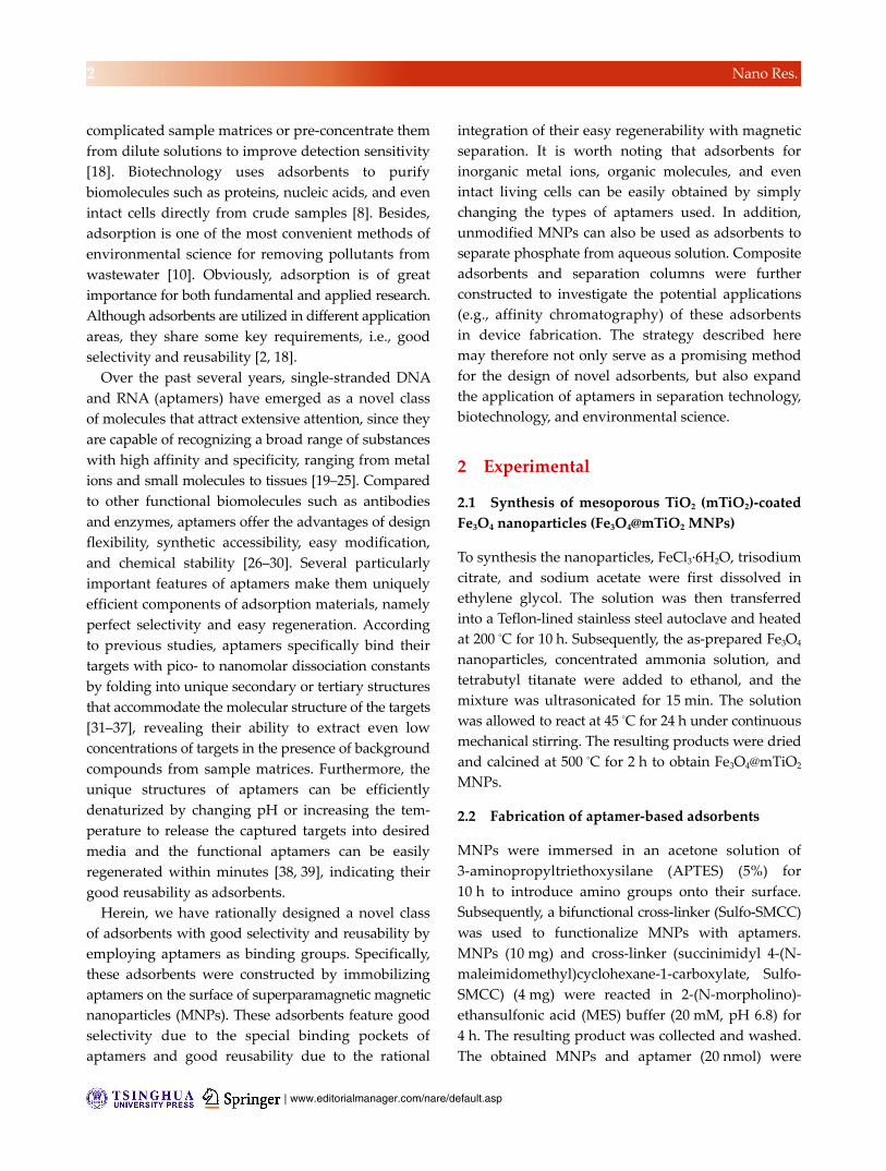
Figure 3 Cyclic extraction of Hg2+ and BPA. Cyclic separation efficiencies of aptamer-based adsorbents towards Hg2+ (a) and BPA (b) in ten cycles.
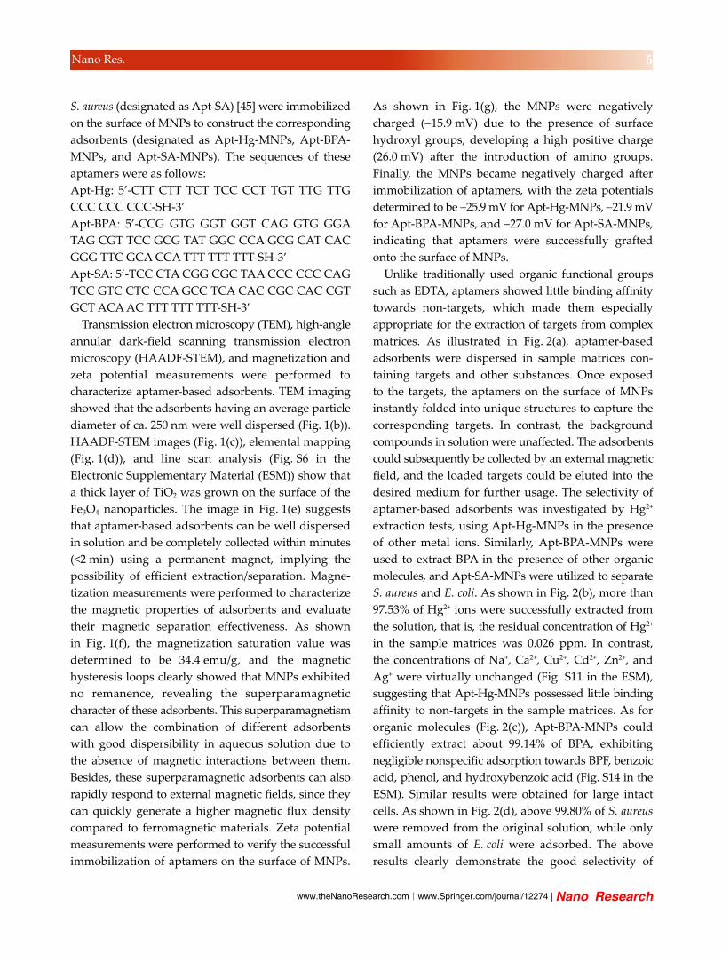
The results of reusability tests of aptamer-based
adsorbents for biological macromolecules are shown
in Fig. 4(a). Apt-SA-MNPs exhibited excellent perfor-
mance in extracting S. aureus from aqueous solutions,
with efficiencies above 96.8% in all cycles. More
importantly, the images of cultured agar plates (cycle
1, 3, 5, 7, 9, and 10) showed extensive growth of the
isolated S. aureus (Fig. 4(b)), clearly indicating that the
adsorbents do not significantly affect the activity of
target biological molecules. Such good reusability
and activity-friendly properties of these adsorbents
highlight their potential for the purification/separation
of biomolecules.
The above results clearly show that aptamer-based
adsorbents display good selectivity and reusability,
suggesting their great potential for practical applications
in separation technology, environmental science, and
biotechnology.
Figure 4 Cyclic separation of S. aureus. (a) Cyclic separation efficiencies of aptamer-based adsorbents towards S. aureus in ten cycles. (b) Culturing results of isolated S. aureus in cycle 1, 3, 5, 7, 9, and 10.
As mentioned above, the mesoporous TiO2 layer
can adsorb PO43– from aqueous solutions. Hence, the
cyclic phosphate separation performance of MNPs
was also tested. The mechanism for PO43– adsorption
by mesoporous MNPs is illustrated in Fig. 5(a). The
large surface area of the mesoporous structure provides
abundant active sites, allowing efficient phosphate
binding to MNPs via coordination. The concentration
of PO43– in aqueous solutions was determined by the
ammonium molybdate spectrophotometric method.
As shown in Fig. 5(b), the sample absorbance decreased
to nearly zero after separation, indicating complete
removal of PO43– by MNPs. This observation was
ascribed to the strong binding affinity of TiO2 towards
PO43– and the abundant surface active sites in the
mesoporous structures [46]. The results of the cyclic
removal of PO43– are presented in Fig. 5(c). Similar
to the results of the former cyclic tests, PO43– was
effectively separated from aqueous solutions, with

| www.editorialmanager.com/nare/default.asp
8 Nano Res.
separation efficiencies exceeding 92% even in the
tenth cycle, showing the great potential of the above
adsorbents for PO43– separation in environmental
applications.
Composite adsorbents, consisting of the above
aptamer-based adsorbents and three-dimensional (3D)
porous polymers, were further constructed. As shown
in Fig. 6(a), aptamer-based adsorbents were mixed
with a solution of chitosan to form homogeneous
dispersions, and this was followed by the addition
of β-GP to form porous composite adsorbents [47].
Scanning transmission electron microscopy (STEM) was
used to characterize the structure of these composite
adsorbents. As shown in Fig. 6(b), the latter exhibited
a 3D macroporous structure with interconnected open
pores after lyophilization, offering sufficient space
Figure 5 Cyclic separation of PO43–. (a) Schematic representation of the adsorption mechanism. (b) UV–Vis absorbance of water samples
before (black line) and after (red line) treatment. The absorbance is due to the colored complex formed by PO43– and ammonium
molybdate. (c) Removal efficiencies of MNPs towards PO43– in ten cycles.
Figure 6 Characterization of composite adsorbents. (a) Schematic fabrication of composite adsorbents. (b) and (c) STEM images of composite adsorbents at different magnifications.

www.theNanoResearch.com∣www.Springer.com/journal/12274 | Nano Research
9 Nano Res.
for the diffusion of targets to the binding sites. At
higher magnification (Fig. 6(c)), the aptamer-based
adsorbents were observed to be well dispersed on
the surface of the porous structure, thus, leading to
a rough surface morphology of the inner pores. In
addition, MNPs were also employed to prepare com-
posite adsorbents for the separation of PO43– using
the same strategy. Such 3D porous continuous bulk
structures were more suited for practical use, e.g., as
packing materials in affinity chromatography.
Composite adsorbents containing Apt-Hg-MNPs,
Apt-BPA-MNPs, and MNPs were employed to separate
Hg2+, BPA, and PO43–, respectively, from aqueous
solutions. Images of the original and treated solutions
are presented in Fig. 7. In order to make the separation
results more figurative, dithizone was used to form a
colored complex with Hg2+, ferric chloride was utilized
to form blue complexes with BPA, and molybdate
was utilized to form navy blue complexes with PO43–.
The organic phase (lower layer) of the original Hg2+
solution was dark brown, while the sample treated
with the composite adsorbent was light brown,
revealing that Hg2+ was efficiently separated from the
solution. As for BPA, the control solutions were deep
blue, while the sample treated with the composite
adsorbent was almost colorless, suggesting complete
separation. Similar results were also obtained for PO43–,
where the color of the original and treated solutions
showed a remarkable difference due to the high
removal efficiency of the composite adsorbent. These
results clearly indicated the robust adsorption abilities
of the porous composite adsorbents.
Figure 7 Removal of targets using composite adsorbents. Images of color reactions for Hg2+, BPA, and PO4
3– samples before and after separation. Dithizone, ferric chloride, and molybdate were used to form colored complexes with Hg2+, BPA, and PO4
3–, respectively.
After investigating the separation capabilities of the
above composite adsorbents, we constructed separation
columns using them as packing materials. The devices
were fabricated by transferring the chitosan solution
and aptamer-based adsorbents into plastic columns
to form composite adsorbents. In such columns, the
interconnected pores within these composites allowed
the penetration of aqueous solution, while the targets
were captured by the adsorbents (Fig. 8(a)). As shown
in Fig. 8(b), the residual concentration of Hg2+ in the
filtrate was 2.46 ppb, indicating that more than 99.75%
of Hg2+ was separated by the column. Similar results
were observed for the separation of BPA, PO43–, and S.
aureus. As shown in Figs. 8(c)–8(e), the residual con-
centrations of BPA, PO43–, and S. aureus in the filtrates
were ultra-low, and the separation efficiencies of these
three targets were above 99.39%, 99.92%, and 99.8%,
respectively. The above results clearly displayed the
excellent separation performance of the columns, and
we anticipated their use for adsorption of targets or
interferents in purification/pre-concentration stages
of separation technology and environmental science.
4 Conclusions
In summary, this work highlights the fabrication of
highly efficient adsorbents based on aptamers. These
adsorbents were capable of selectively extracting metal
ions, small organic molecules, and even intact cells
from complicated sample matrices. More importantly,
the adsorbents did not affect the activity of isolated
cells. These aptamer-based adsorbents could easily
be recycled several times without a significant loss of
adsorption capacity. In addition, the separation of
phosphate by MNPs was investigated. The MNPs
exhibited high separation efficiencies and excellent
reusability. Furthermore, aptamer-based adsorbents
and MNPs were incorporated into a 3D porous
polymer in order to investigate their potential practical
applications in device fabrication, and separation
columns were constructed. Both the composite hydrogel
and the columns displayed excellent separation of
targets from aqueous media, revealing the excellent
potential of the adsorbents for practical applications.
To conclude, the above strategy paves a way for the
development of multiple-use aptamer-based adsorbents,

| www.editorialmanager.com/nare/default.asp
10 Nano Res.
not only promoting the development of materials
science, but also providing efficient tools for separation
technology, biotechnology, and environmental fields.
Acknowledgements
This work was supported by the National Natural
Science Foundation of China (Nos. 51272186 and
21422105), “A Foundation for the Author of National
Excellent Doctoral Dissertation of P. R. China” (No.
201220), and Ten Thousand Talents Program for Young
Talents. Q. Y. thanks for large-scale instrument and
equipment sharing foundation of Wuhan University.
Electronic Supplementary Material: Supplementary
material (TEM characterization, line scan analysis,
XRD patterns, SEM images) is available in the online
version of this article at http://dx.doi.org/10.1007/
s12274-016-1273-9.
References
[1] Augusto, F.; Carasek, E.; Silva, R. G. C.; Rivellino, S. R.;
Batista, A. D.; Martendal, E. New sorbents for extraction
and microextraction techniques. J. Chromatogr. A 2010,
1217, 2533–2542.
[2] Dąbrowski, A. Adsorption—From theory to practice. Adv.
Colloid Interface Sci. 2001, 93, 135–224.
[3] Deng, Y. H.; Qi, D. W.; Deng, C. H.; Zhang, X. M.; Zhao, D.
Y. Superparamagnetic high-magnetization microspheres
with an Fe3O4@SiO2 core and perpendicularly aligned
mesoporous SiO2 shell for removal of microcystins. J. Am.
Chem. Soc. 2008, 130, 28–29.
[4] Crane, R. A.; Dickinson, M.; Popescu, I. C.; Scott, T. B.
Magnetite and zero-valent iron nanoparticles for the
remediation of uranium contaminated environmental water.
Water Res. 2011, 45, 2931–2942.
[5] Lukens, W. W., Jr.; Schmidt-Winkel, P.; Zhao, D. Y.; Feng,
J. L.; Stucky, G. D. Evaluating pore sizes in mesoporous
materials: A simplified standard adsorption method and a
simplified broekhoff-de Boer method. Langmuir 1999, 15,
5403–5409.
[6] Liu, B. W.; Liu, J. W. DNA adsorption by magnetic iron
oxide nanoparticles and its application for arsenate detection.
Chem. Commun. 2014, 50, 8568–8570.
[7] Huang, D. N.; Deng, C. H.; Zhang, X. M. Functionalized
magnetic nanomaterials as solid-phase extraction adsorbents
for organic pollutants in environmental analysis. Anal. Methods
2014, 6, 7130–7141.
[8] Borlido, L.; Azevedo, A. M.; Roque, A. C. A.; Aires-Barros,
M. R. Magnetic separations in biotechnology. Biotechnol.
Adv. 2013, 31, 1374–1385.
[9] Teng, W.; Wu, Z. X.; Fan, J. W.; Chen, H.; Feng, D.; Lv, Y. Y.;
Wang, J. X.; Asiri, A. M.; Zhao, D. Y. Ordered mesoporous
Figure 8 Removal of targets using separation columns. (a) Schematic illustration of the separation columns. Concentrations of Hg2+
(b), BPA (c), PO43– (d), and S. aureus (e) in the samples before and after separation using corresponding columns.
www.theNanoResearch.com∣www.Springer.com/journal/12274 | Nano Research
11 Nano Res.
carbons and their corresponding column for highly efficient
removal of microcystin-LR. Energy Environ. Sci. 2013, 6,
2765–2776.
[10] Kalia, S.; Kango, S.; Kumar, A.; Haldorai, Y.; Kumari, B.;
Kumar, R. Magnetic polymer nanocomposites for environ-
mental and biomedical applications. Colloid Polym. Sci.
2014, 292, 2025–2052.
[11] Qu, X. L.; Alvarez, P. J. J.; Li, Q. L. Applications of
nanotechnology in water and wastewater treatment. Water
Res. 2013, 47, 3931–3946.
[12] Soto, M. L.; Moure, A.; Domínguez, H.; Parajó, J. C. Recovery,
concentration and purification of phenolic compounds by
adsorption: A review. J. Food Eng. 2011, 105, 1–27.
[13] Wu, Z. X.; Li, W.; Webley, P. A.; Zhao, D. Y. General and
controllable synthesis of novel mesoporous magnetic iron
oxide@carbon encapsulates for efficient arsenic removal.
Adv. Mater. 2012, 24, 485–491.
[14] Wang, J.; Shen, H. J.; Hu, X. X.; Li, Y.; Li, Z. H.; Xu, J. F.;
Song, X. F.; Zeng, H. B.; Yuan, Q. A targeted “capture”
and “removal” scavenger toward multiple pollutants for
water remediation based on molecular recognition. Adv. Sci.
2016, 3, 1500289.
[15] Wang, P.; Shi, Q. H.; Liang, H. J.; Steuerman, D. W.;
Stucky, G. D.; Keller, A. A. Enhanced environmental mobility
of carbon nanotubes in the presence of humic acid and their
removal from aqueous solution. Small 2008, 4, 2166–2170.
[16] Huang, P. J. J.; Liu, J. W. Immobilization of DNA on
magnetic microparticles for mercury enrichment and detection
with flow cytometry. Chem.—Eur. J. 2011, 17, 5004–5010.
[17] Yokoi, T.; Kubota, Y.; Tatsumi, T. Amino-functionalized
mesoporous silica as base catalyst and adsorbent. Appl.
Catal. A: Gen. 2012, 421–422, 14–37.
[18] Xie, L. J.; Jiang, R. F.; Zhu, F.; Liu, H.; Ouyang, G. F.
Application of functionalized magnetic nanoparticles in
sample preparation. Anal. Bioanal. Chem. 2014, 406, 377–399.
[19] Bunka, D. H. J.; Stockley, P. G. Aptamers come of age—At
last. Nat. Rev. Microbiol. 2006, 4, 588–596.
[20] Liu, J. W.; Cao, Z. H.; Lu, Y. Functional nucleic acid sensors.
Chem. Rev. 2009, 109, 1948–1998.
[21] Tan, W. H.; Donovan, M. J.; Jiang, J. H. Aptamers from cell-
based selection for bioanalytical applications. Chem. Rev.
2013, 113, 2842–2862.
[22] Pei, H.; Zuo, X.; Zhu, D.; Huang, Q.; Fan, C. Functional
DNA nanostructures for theranostic applications. Acc. Chem.
Res. 2014, 47, 550–559.
[23] Fang, X. H.; Tan, W. H. Aptamers generated from Cell-
SELEX for molecular medicine: A chemical biology approach.
Acc. Chem. Res. 2010, 43, 48–57.
[24] Hamula, C. L. A.; Guthrie, J. W.; Zhang, H. Q.; Li, X. F.;
Le, X. C. Selection and analytical applications of aptamers.
TrAC Trend. Anal. Chem. 2006, 25, 681–691.
[25] Giljohann, D. A.; Mirkin, C. A. Drivers of biodiagnostic
development. Nature 2009, 462, 461–464.
[26] Hamula, C. L. A.; Zhang, H. Q.; Guan, L. L.; Li, X. F.; Le,
X. C. Selection of aptamers against live bacterial cells. Anal.
Chem. 2008, 80, 7812–7819.
[27] Liu, Q. L.; Jin, C.; Wang, Y. Y.; Fang, X. H.; Zhang, X. B.;
Chen, Z.; Tan, W. H. Aptamer-conjugated nanomaterials
for specific cancer cell recognition and targeted cancer
therapy. NPG Asia Mater. 2014, 6, e95.
[28] Song, S. P.; Wang, L. H.; Li, J.; Fan, C. H.; Zhao, J. L.
Aptamer-based biosensors. TrAC Trend. Anal. Chem. 2008,
27, 108–117.
[29] Jayasena, S. D. Aptamers: An emerging class of molecules
that rival antibodies in diagnostics. Clin. Chem. 1999, 45,
1628–1650.
[30] Guo, W.; Hong, F.; Liu, N. N.; Huang, J. Y.; Wang, B. Y.;
Duan, R. X.; Lou, X. D.; Xia, F. Target-specific 3D DNA
gatekeepers for biomimetic nanopores. Adv. Mater. 2015,
27, 2090–2095.
[31] Zheng, D.; Seferos, D. S.; Giljohann, D. A.; Patel, P. C.;
Mirkin, C. A. Aptamer nano-flares for molecular detection
in living cells. Nano Lett. 2009, 9, 3258–3261.
[32] Mairal, T.; Özalp, V. C.; Sánchez, P. L.; Mir, M.; Katakis, I.;
O’Sullivan, C. K. Aptamers: Molecular tools for analytical
applications. Anal. Bioanal. Chem. 2008, 390, 989–1007.
[33] Tombelli, S.; Minunni, M.; Mascini, M. Analytical applications
of aptamers. Biosens. Bioelectron. 2005, 20, 2424–2434.
[34] Liang, H.; Zhang, X. B.; Lv, Y. F.; Gong, L.; Wang, R. W.;
Zhu, X. Y.; Yang, R. H.; Tan, W. H. Functional DNA-
containing nanomaterials: Cellular applications in biosensing,
imaging, and targeted therapy. Acc. Chem. Res. 2014, 47,
1891–1901.
[35] Tombelli, S.; Minunni, M.; Mascini, M. Aptamers-based
assays for diagnostics, environmental and food analysis.
Biomol. Eng. 2007, 24, 191–200.
[36] Palchetti, I.; Mascini, M. Nucleic acid biosensors for
environmental pollution monitoring. Analyst 2008, 133,
846–854.
[37] Huang, P. J.; Liu, J. W. Flow cytometry-assisted detection
of adenosine in serum with an immobilized aptamer sensor.
Anal. Chem. 2010, 82, 4020–4026.
[38] Zhao, Q.; Li, X. F.; Le, X. C. Aptamer-modified monolithic
capillary chromatography for protein separation and detection.
Anal. Chem. 2008, 80, 3915–3920.
[39] Wang, J.; Wei, Y. R.; Hu, X. X.; Fang, Y. Y.; Li, X. Y.;
Liu, J.; Wang, S. F.; Yuan, Q. Protein activity regulation:
Inhibition by closed-loop aptamer-based structures and
restoration by near-IR stimulation. J. Am. Chem. Soc. 2015,
137, 10576–10584.

| www.editorialmanager.com/nare/default.asp
12 Nano Res.
[40] Li, W.; Yang, J. P.; Wu, Z. X.; Wang, J. X.; Li, B.; Feng, S.
S.; Deng, Y. H.; Zhang, F.; Zhao, D. Y. A versatile
kinetics-controlled coating method to construct uniform
porous TiO2 shells for multifunctional core–shell structures.
J. Am. Chem. Soc. 2012, 134, 11864–11867.
[41] Yuan, Y.; Chen, S.; Paunesku, T.; Gleber, S. C.; Liu, W. C.;
Doty, C. B.; Mak, R.; Deng, J. J.; Jin, Q. L.; Lai, B. et al.
Epidermal growth factor receptor targeted nuclear delivery
and high-resolution whole cell X-ray imaging of Fe3O4@TiO2
nanoparticles in cancer cells. ACS Nano 2013, 7, 10502–
10517.
[42] Yuan, Q.; Wu, Y.; Wang, J.; Lu, D. Q.; Zhao, Z. L.; Liu, T.;
Zhang, X. B.; Tan, W. H. Targeted bioimaging and
photodynamic therapy nanoplatform using an aptamer-
guided G-quadruplex DNA carrier and near-infrared light.
Angew. Chem., Int. Ed. 2013, 52, 13965 –13969.
[43] Dave, N.; Chan, M. Y.; Huang, P. J. J.; Smith, B. D.; Liu, J. W.
Regenerable DNA-functionalized hydrogels for ultrasensitive,
instrument-free mercury(II) detection and removal in water.
J. Am. Chem. Soc. 2010, 132, 12668–12673.
[44] Jo, M.; Ahn, J. Y.; Lee, J.; Lee, S.; Hong, S. W.; Yoo, J. W.;
Kang, J.; Dua, P. Lee, D. K.; Hong, S. et al. Development
of single-stranded DNA aptamers for specific bisphenol a
detection. Oligonucleotides 2011, 21, 85–91.
[45] Chang, Y. C.; Yang, C. Y.; Sun, R. L.; Cheng, Y. F.;
Kao, W. C.; Yang, P. C. Rapid single cell detection of
staphylococcus aureus by aptamer-conjugated gold nano-
particles. Sci. Rep. 2013, 3, 1863.
[46] Song, H.; Nor, Y. A.; Yu, M. H.; Yang, Y. N.; Zhang, J.;
Zhang, H. W.; Xu, C.; Mitter, N.; Yu, C. Z. Silica nanopollens
enhance adhesion for long-term bacterial inhibition. J. Am.
Chem. Soc. 2016, 138, 6455−6462. [47] Zhu, M.; Zhu, Y. F.; Zhang, L. X.; Shi, J. L. Preparation of
chitosan/mesoporous silica nanoparticle composite hydrogels
for sustained co-delivery of biomacromolecules and small
chemical drugs. Sci. Technol. Adv. Mat. 2013, 14, 045005.