highlights from the advanced radiation dosimetry european ... bioqart.pdf · phd with uni...
TRANSCRIPT

Highlights from the Advanced Radiation Dosimetry European Network Training initiative
(ARDENT)
Marco Silari (CERN) on behalf of the ARDENT consortium
1

M. Silari - Highlights from ARDENT BioQuaRT Satellite Workshop, Aix-en-Provence, June 2013
ARDENT February 2012 – January 2016
Advanced Radiation Dosimetry European Network Training initiative
Marie Curie Initial Training Network under EU FP7 – 4 M€ 8 Full Partners and 6 Associate Partners
Coordinator: CERN, Scientist-in-Charge: Dr. M. Silari
CERN (coordinator), Switzerland AIT Vienna, Austria CTU- IAEP Prague, Czech Republic IBA Dosimetry, Schwarzenbruck, Germany Jablotron, Prague, Czech Republic MI.AM, Milano, Italy Politecnico, of Milano, Italy Seibersdorf Laboratories, Austria
INFN Legnaro National Laboratories, Italy ST Microelectronics, Italy University of Erlangen, Germany University of Houston, USA University of Ontario, Canada University of Wollongong, Australia
2

M. Silari - Highlights from ARDENT BioQuaRT Satellite Workshop, Aix-en-Provence, June 2013
Three technologies • gas detectors [gas electron multipliers (GEM), tissue equivalent
proportional counters (TEPC), ionization chambers] • solid state detectors [Medipix, silicon micro-dosimeters] • track detector techniques [CR-39] Main objectives • Radiation dosimetry • Micro- (and maybe sub-micro-) dosimetry • Neutron spectrometry Applications • Characterization of radiation fields at particle accelerators • Characterization of radiation fields on-board aircrafts and in space • Assessment of secondary dose to RT patient • Measurement of properties of clinical hadron beams
Development of advanced instrumentation for radiation monitoring
3

M. Silari - Highlights from ARDENT BioQuaRT Satellite Workshop, Aix-en-Provence, June 2013
Supervisory Board (SB)
Technical Training Board (Leaders WP 1-4)
Administrative support unit (CERN)
Selection Committees
Dissemination and Outreach Board (WP 6)
Training Board (WP 5)
ITN Management Office Scientist in Charge
http://cern.ch/ardent
ARDENT management structure
o WP1: gas detectors o WP2: solid state detectors o WP3: track detectors o WP4: instrument inter-comparison
4

M. Silari - Highlights from ARDENT BioQuaRT Satellite Workshop, Aix-en-Provence, June 2013
Gas Electron Multiplier Silvia Puddu (CERN) – PhD with Uni Bern
Application of Gas Electron Multiplier (GEM) in dosimetry and microdosimetry around particle accelerators, space and environment, beam monitoring for experimental and medical particle beams
Beam profile measured at CERN n_TOF facility
GEM set-up in the ARDENT laboratory at CERN
5

M. Silari - Highlights from ARDENT BioQuaRT Satellite Workshop, Aix-en-Provence, June 2013
Test of GEM as a beam monitor at CERF (CERN)
With Boron converter, at n_TOF
Measurements with neutron detectors
• Intercomparison of neutron detectors in neutron pulsed fields in the HiRadMat facility and around the PS ring at CERN
• Measurements with BSS around the PS ring
FLUKA simulations For the study of the physical phenomena inside GEM and the design of the new neutron spectrometer
GEM-based Neutron Spectrometer Eleni Aza (CERN) – PhD with Uni Thessaloniki
6

M. Silari - Highlights from ARDENT BioQuaRT Satellite Workshop, Aix-en-Provence, June 2013
LUPIN Chris Cassell (POLIMI) – PhD with Uni Wollongong
• Characterisation of LUPIN (Long-interval, Ultra-wide dynamic, PIle-up free Neutron rem counter
• Instrument designed for use in Pulsed Neutron Fields (bursts of high-dose radiation over very short time periods) without saturation
• Experiments conducted at the Proton Synchrotron (PS) and Super Proton Synchrotron (SPS) at CERN, and at the SwissFEL (Swiss free-electon laser) at the Paul Scherrer Institute in Switzerland
Plot of normalised H*(10) from 6 detectors around the Proton Synchrotron at CERN. The intercomparison plot shows that the LUPIN presents almost no saturation effects, whereas most of the other detectors show a marked underestimation
7

M. Silari - Highlights from ARDENT BioQuaRT Satellite Workshop, Aix-en-Provence, June 2013
2D Ion Chamber Array for Clinical Applications Michele Togno (IBA Dosimetry) – PhD with TU Munich
Goal Benefit
Test new solutions for IC arrays (improve MatriXX detector)
Better future products
Reduce pitch and sensor size Improved spatial resolution
Reduce chamber sensitive volume Improved efficiency at high rate
Studies on materials and geometry Improved stability
Simple assembling Improved yeld, cost, service
Development and characterization of a new generation of ionization chambers arrays aimed at radiotherapy applications, particularly at the measurement of absorbed dose from photon, electron and proton radiation
Current activities:
▪ Iterative improvement of prototype design (first simple prototype is a small 1D array – picture beside shows an example sketch
▪ Measurements under gamma beam (60Co) and MV X-rays
▪ Studies on materials and geometry
▪ Monte Carlo simulations Sketch of a simple 1D array
8

M. Silari - Highlights from ARDENT BioQuaRT Satellite Workshop, Aix-en-Provence, June 2013
Characterization of Timepix and Medipix3RX Erik Fröjdh (CERN) – PhD with Mid-Sweden Uni
• Working together with the Medipix team • Characterization of Timepix and Medipix3RX • CdTe Sensor characterization • Measurements of scattered radiation around
clinical CT • Medipix support to other ARDENT ESRs
CdTe Sensor Defects flood exposure with X-rays
Cluster size as a function of beam position
9

M. Silari - Highlights from ARDENT BioQuaRT Satellite Workshop, Aix-en-Provence, June 2013
Evaluation of data in mixed radiation fields Benedikt Bergmann (CTU) – PhD with Uni Erlangen
Evaluation of data taken by the ATLAS-MPX detector network • Luminosity studies • Studies of activation and dose rates at
different sites in the ATLAS cavern • Example: measured count rate vs time for
detector MPX01
Calibration of MPX and TPX detector responses to different particle types • Measurements in well known radiation
fields (gamma, alpha, proton, neutron, …) • Example: detection efficiency vs energy
obtained in a pure neutron field with a TPX device and different converter foils
10

M. Silari - Highlights from ARDENT BioQuaRT Satellite Workshop, Aix-en-Provence, June 2013
Medipix-based radiation monitoring system Vijayaragavan Viswanathan (Jablotron)
(PhD from the Ecole Centrale de Lyon)
Overall aim of the project Market study Investigation of possible applications in radiation monitoring Design and development Testing and prototype verification Certification and production
Development of Medipix-based radiation monitoring system Design of Medipix/Timepix monitoring system
Interface development for hand held device Educational kit for high schools and universities
Progressed on PCB schematics and design Market study on application areas Medipix communication protocol Interface development (ongoing)
PCB design training – GM counter
Medipix – Voltage regulator – PCB layout -3D view
11

M. Silari - Highlights from ARDENT BioQuaRT Satellite Workshop, Aix-en-Provence, June 2013
Medipix in medical imaging Kevin Loo (CTU) – PhD with Uni Wollongong
1. Development of novel in-body imaging system: BrachyView Utilises Medipix technology in prostate cancer low dose rate brachytherapy procedures
Two main aspects: a) Intraoperative dose planning to avoid critical
structures and allow for implant adjustments as they occur. Dynamic quality assurance for patient dosimetry
b) Use of Medipix as imaging plane for post-implant CT dosimetry.
2. Further development of Medipix in use for mixed field radiation dosimetry, with possible applications in homeland security and radiation safety
In conjunction with Jablotron Alarm Systems (CZ)
12

M. Silari - Highlights from ARDENT BioQuaRT Satellite Workshop, Aix-en-Provence, June 2013
Dosepix Francesca Bisello (IBA Dosimetry) – PhD with Uni Erlangen
Hybrid Pixel Detector with read-out electronics 256 pixels of 220 µm pitch Photon radiation measurement using Si detector by Time over Threshold (ToT) calculation for each incoming photon
Project development:
- Chip Calibration Method - Chip Operation Mode (Dosi
mode, ToT mode) - Chip evaluation at different
radiation energy - Reconstruction and analysis of
radiation spectra - Monte Carlo simulations
0100200300400500600700800900
110
420
731
041
351
661
972
282
592
810
3111
3412
3713
4014
4315
4616
4917
5218
5519
58
Coun
ts
Analog Testpulse unit
Detector response after - Threshold equalisation - Analog Testpulse calibration
30keV X-rayspectrum
13

M. Silari - Highlights from ARDENT BioQuaRT Satellite Workshop, Aix-en-Provence, June 2013
GEMPIX at CERN F. Murtas and J. A. Alozy
A new detector concept where a GEM gas detector is coupled with a 260,000 Medipix “quad” readout of 55 µm x 55 µm area
Timepix readout card
14

M. Silari - Highlights from ARDENT BioQuaRT Satellite Workshop, Aix-en-Provence, June 2013
GEMPIX: two working modes
The readout is made with a Timepix chip The active area is 9 cm2
The particle track is analysed with 512 pixel in 3 cm length This is equivalent to 30 microns of tissue … with 17 samples/micron
Head-on
Side-on
Triple GEM
Mylar window
Particles to be analysed
Triple GEM Particles to be analysed
Length analysed Timepix
Timepix
15
M. Silari - Highlights from ARDENT BioQuaRT Satellite Workshop, Aix-en-Provence, June 2013
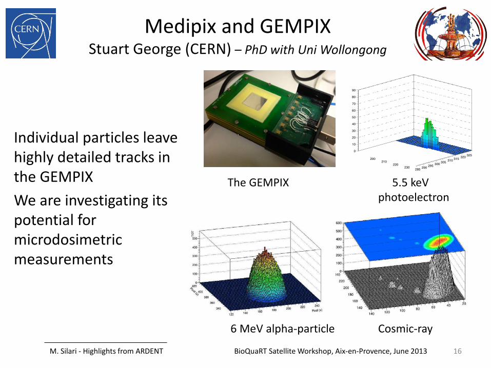
Medipix and GEMPIX Stuart George (CERN) – PhD with Uni Wollongong
Individual particles leave highly detailed tracks in the GEMPIX We are investigating its potential for microdosimetric measurements
Cosmic-ray 6 MeV alpha-particle
5.5 keV photoelectron
The GEMPIX
16

M. Silari - Highlights from ARDENT BioQuaRT Satellite Workshop, Aix-en-Provence, June 2013
Some events taken with GEMPIX
Cosmic (protons) Xray
Cesium Alphas Alphas
17

M. Silari - Highlights from ARDENT BioQuaRT Satellite Workshop, Aix-en-Provence, June 2013
Silicon microdosimeter at POLIMI (S. Agosteo and A. Pola)
SEGMENTED SILICON TELESCOPE
Constituted by a matrix of cylindrical ∆E elements (about 2 µm in thickness) and a single residual-energy E stage (500 µm in thickness)
Nominal diameter of the ∆E elements about 9 μm, the width of the pitch separating the elements about 41 µm
Minimum detectable energy limited to about 20 keV by the electronic noise
500 µm 14 µm
9 µm
E stage
Guard~2 μm
∆E element
500 µm 14 µm
9 µm
E stage
Guard~2 μm
∆E element
10 100 10000.0
0.1
0.2
0.3
0.4
0.5
0.6
0.7
0.8
y d(
y)
y (keV µm-1)
Pixelated silicon telescope Cylindrical TEPC (2.7 µm)
18

M. Silari - Highlights from ARDENT BioQuaRT Satellite Workshop, Aix-en-Provence, June 2013
Silicon microdosimeter Eleni Sagia (POLIMI) – PhD with POLIMI
500 µm 14 µm
9 µm
E stage
Guard~2 μm
∆E element
500 µm 14 µm
9 µm
E stage
Guard~2 μm
∆E element
n+
p+p+n+
E stage ( ≅ 500 μm)
ΔE stage ( ≅ 1.9 μm)
n+
p+p+n+
E stage ( ≅ 500 μm)
ΔE stage ( ≅ 1.9 μm)
Silicon telescope: a thin ∆E stage (1.9 μm thick) coupled to a residual energy E stage (500 μm thick) on the same silicon wafer.
More than 7000 pixels are connected in parallel to give an effective detection area of the ∆E stage of about 0.5 mm2 The ∆E stage acts as a microdosimeter while the E stage allows assessing the full energy of the recoil-protons, providing the LET-dependent correction for tissue-equivalency
19

M. Silari - Highlights from ARDENT BioQuaRT Satellite Workshop, Aix-en-Provence, June 2013
Silicon microdosimeter at POLIMI
Segmented silicon telescope irradiated inside a PMMA phantom exposed to the 62 MeV proton beam at the INFN-LNS CATANA facility.
Front-end electronics
Detector +
Mylar
PMMA phantom
PMMA foils
20

0 2 4 6 8 10 12 14 16 18 20 22 240.0
0.5
1.0
1.5
2.0
2.5
3.0
3.5
4.0
4.5
5.0
Dept
h do
se c
urve
(a.u
.)
depth in PMMA (mm)
1 10 100 10000.0
0.2
0.4
0.6
0.8
1.0 silicon telescope 21.8 mm cylindrical TEPC 22 mm
(threshold) cylindrical TEPC 22 mm
(no threshold)
y d(
y)
y (keV µm-1)
1 10 100 10000.0
0.2
0.4
0.6
0.8
1.0
silicon telescope 20.5 mm cylindrical TEPC 20.1 mm
(threshold) cylindrical TEPC 20.1 mm
(no threshold)
y d(
y)
y (keV µm-1)
1 10 100 10000.0
0.2
0.4
0.6
0.8
1.0 silicon telescope 21.2 mm cylindrical TEPC 21.4 mm
(threshold) cylindrical TEPC 21.4 mm
(no threshold)
y d(
y)y (keV µm-1)
1 10 100 10000.0
0.2
0.4
0.6
0.8
1.0 silicon telescope 21.4 mm cylindrical TEPC 21.6 mm
(threshold) cylindrical TEPC 21.6 mm
(no threshold)
y d(
y)
y (keV µm-1)
1 10 100 10000.0
0.2
0.4
0.6
0.8
1.0 silicon telescope 21.6 mm cylindrical TEPC 21.8 mm
(threshold) cylindrical TEPC 21.8 mm
(no threshold)
y d(
y)
y (keV µm-1)
Event-by-event TE correction!!!
DISTAL PART OF THE SOBP
BioQuaRT Satellite Workshop, Aix-en-Provence, June 2013 21
M. Silari - Highlights from ARDENT BioQuaRT Satellite Workshop, Aix-en-Provence, June 2013
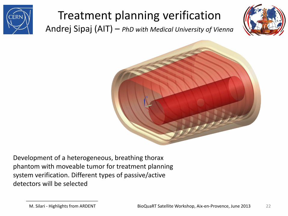
Treatment planning verification Andrej Sipaj (AIT) – PhD with Medical University of Vienna
Development of a heterogeneous, breathing thorax phantom with moveable tumor for treatment planning system verification. Different types of passive/active detectors will be selected
22

M. Silari - Highlights from ARDENT BioQuaRT Satellite Workshop, Aix-en-Provence, June 2013
LET reconstruction with CR39 at POLIMI (M. Caresana)
Longitudinal section and top view of etched tracks of 90 MeV 7-Li ions entering the detectors at 0° (left) and 40◦ (right) for diferent etching times, t (a) t = 3:5 h, (b) t = 4:33 h. Magnification: 950.
B. Dorschel et al. Radiation Measurements 37 (2003) 563 – 571
23

M. Silari - Highlights from ARDENT BioQuaRT Satellite Workshop, Aix-en-Provence, June 2013
LET reconstruction with CR39 at POLIMI
Ratio V of the track attack velocity to bulk attack velocity as a function of the etching time and the particle penetration in CR39
V = V(E,t) and V = V(E,x) functions calculated through the V = V(LET(E,x)) function for 1.2 MeV protons impinging perpendicularly on the detector surface. The simulated etching conditions are Vb=9.8 µm h-1 and 1.5 h of etching time.
24

M. Silari - Highlights from ARDENT BioQuaRT Satellite Workshop, Aix-en-Provence, June 2013
LET reconstruction with CR39 at POLIMI
LET calculation V = Vt/Vb = f[LETmean(E,x)]
V = (Dt,dt) where Dt and dt are the track major and minor axis
25

M. Silari - Highlights from ARDENT BioQuaRT Satellite Workshop, Aix-en-Provence, June 2013
LET reconstruction with CR39 at POLIMI
Measurement LET alfa Am-241 and Unat
Am-241 E=5.5 MeV Etching time 60’ Removed layer 10 µm Mean LET measured 130 keV/µm Mean LET calculated 140 keV/µm
Unat E1=4.2 MeV E2=4.77 MeV Etching time 40’ Removed layer 6.7 µm
Source Mean LET measured
Mean LET calculated
U-234 145 keV/µm 148 keV/µm U-338 170 keV/µm 187 keV/µm
26

M. Silari - Highlights from ARDENT BioQuaRT Satellite Workshop, Aix-en-Provence, June 2013
Dosimetry and spectrometry in complex neutron fields using CR-39 track detectors
Alvin Sashala Naik (MI.AM) – PhD with POLIMI
LET measurements in quasi-monoenergetic neutron beams at the PTB (Germany) and Ithemba labs (South Africa) calibration facilities
Development of personal and area neutron dosimeters with Monte Carlo simulations (MCNPX, FLUKA)
Development of the POLITRACKTM automatic detector reader for precision track image analysis in CR-39
Applications: • stray radiation measurement in Ion
beam therapy • complex neutron fields in high-altitude
flights and in space due to cosmic rays
Example of LET determination from track image parameters
(1) Raw image (2) Image analysis
(3) LET distribution
27
THANK YOU FOR YOUR ATTENTION
Highlights from the Advanced Radiation Dosimetry European Network Training Initiative (ARDENT)
Marco Silari (CERN)
on behalf of the ARDENT consortium
28