high-sensitivity troponin: a new tool for...
TRANSCRIPT

HIGH-SENSITIVITY TROPONIN: A NEW TOOLFOR PATHOPHYSIOLOGICAL INVESTIGATION
AND CLINICAL PRACTICE
Aldo Clerico,*,†,1 Alberto Giannoni,*,†
Concetta Prontera,* and Stefania Giovannini*
*Laboratory of Cardiovascular Endocrinology andCell Biology, G. Monasterio Foundation, CNR Regione
Toscana, 56126 Pisa, Italy†Scuola Superiore Sant’Anna, 56126 Pisa, Italy
1. Abstract . . . . . . . . . . . . . . . . . . . . . . . . . . . . . . . . . . . . . . . . . . . . . . . . . . . . . . . . . . . . . . . . . . . . . . . . . 2
2. Background and Aim. . . . . . . . . . . . . . . . . . . . . . . . . . . . . . . . . . . . . . . . . . . . . . . . . . . . . . . . . . . . 2
3. Introduction: Troponin Framework Within Myocardial Cells and Release
Kinetics After Myocardial Damage. . . . . . . . . . . . . . . . . . . . . . . . . . . . . . . . . . . . . . . . . . . . . . 4
4. Impact of the New Definition of Myocardial Infarction on Laboratory
Practice and Instrumentation: The Need for High-Sensitivity
cTnI and cTnT Methods . . . . . . . . . . . . . . . . . . . . . . . . . . . . . . . . . . . . . . . . . . . . . . . . . . . . . . . . 5
4.1. Quality Specifications for cTnI and cTnT Immunoassays. . . . . . . . . . . . . . . . . . . 5
4.2. The Development of High-Sensitivity Immunoassays
for cTnI and cTnT Measurement . . . . . . . . . . . . . . . . . . . . . . . . . . . . . . . . . . . . . . . . . . 7
4.3. Definition of Highly (Ultra) Sensitive Immunoassay for cTnI and cTnT . . . . 8
5. The Impact of High-Sensitivity cTnI and cTnT Methods on Clinical Practice . . . . 9
5.1. The Problem of Reliable Definition and Accurate Estimation of the 99th
Percentile Upper Reference Limit: Can Reference Values Be AVected by any
Characteristics of the Reference Population? . . . . . . . . . . . . . . . . . . . . . . . . . . . . . . . 10
5.2. More Myocardial Infarctions or More False Positive Results?. . . . . . . . . . . . . . 14
5.3. Clinical Relevance of Serially Measured Troponin Circulating Levels. . . . . . . 15
5.4. High-Sensitivity Troponin Methods in Patients with HF:
A Better Stratification of Cardiovascular Risk . . . . . . . . . . . . . . . . . . . . . . . . . . . . . 16
5.5. Early Detection of Myocardial Injury in Patients with Extracardiac Diseases
or Assuming Potentially Cardiotoxic Drugs . . . . . . . . . . . . . . . . . . . . . . . . . . . . . . . . 18
6. High-Sensitivity cTnI and cTnT Methods: A Powerful
Tool for Monitoring Physiological Renewal and Pathological Remodeling
of the Myocardial Tissue? . . . . . . . . . . . . . . . . . . . . . . . . . . . . . . . . . . . . . . . . . . . . . . . . . . . . . . . 19
1Corresponding author: Aldo Clerico, e-mail: [email protected]
1
0065-2423/09 $35.00 Copyright 2009, Elsevier Inc.DOI: 10.1016/S0065-2423(09)49001-2 All rights reserved.
ADVANCES IN CLINICAL CHEMISTRY, VOL. 49
7. Use of High-Sensitivity cTnI and cTnT Methods in a Multimarker Approach for
Early Screening: An Increase in Diagnostic and Prognostic EViciency?. . . . . . . . . . . 22
8. Conclusion . . . . . . . . . . . . . . . . . . . . . . . . . . . . . . . . . . . . . . . . . . . . . . . . . . . . . . . . . . . . . . . . . . . . . . 23
References. . . . . . . . . . . . . . . . . . . . . . . . . . . . . . . . . . . . . . . . . . . . . . . . . . . . . . . . . . . . . . . . . . . . . . . 24
1. Abstract
At the dawn of the new century, the advent of more specific myocardial
tissue markers, such as cardiac troponin I (cTnI) and T (cTnT), has led to a
new definition of acute myocardial infarction (AMI) by international guide-
lines. If we accept the concept that AMI is the portion of acutely necrotic
myocardial tissue (irrespective of size), some patients previously diagnosed
with severe angina may be currently considered to present minimal (even
microscopic) quantities of myocardial necrosis. Although increased cTnI or
cTnT values always indicate myocardial tissue damage, a positive test is not
able to identify the mechanism responsible for that cardiac damage (which
could be not due to ischemia). New cTnI and cTnT immunoassays with
increased analytical sensitivity may increase ‘‘false positive’’ results in
patients with cardiovascular disease, especially those with advanced age,
heart failure (HF), severe comorbidities (such as chronic renal insuViciency),or assuming potential cardiotoxic drugs. Hence, it may be not clear for most
patients and physicians whether high-sensitivity cTnI and cTnT methods will
lead to more clarity or confusion. The aim of this review is to update the
present knowledge in the field of cTnI and cTnT with particular attention on
the impact of immunoassays with increased analytical sensitivity on both
laboratory and clinical practice.
2. Background and Aim
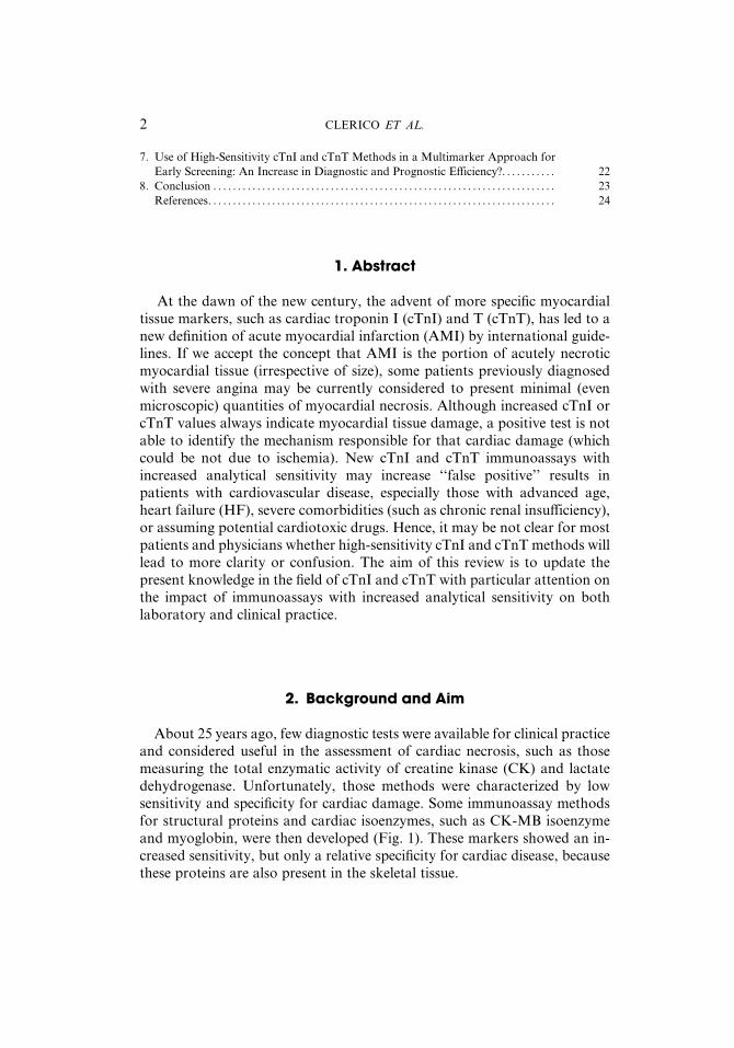
About 25 years ago, few diagnostic tests were available for clinical practice
and considered useful in the assessment of cardiac necrosis, such as those
measuring the total enzymatic activity of creatine kinase (CK) and lactate
dehydrogenase. Unfortunately, those methods were characterized by low
sensitivity and specificity for cardiac damage. Some immunoassay methods
for structural proteins and cardiac isoenzymes, such as CK-MB isoenzyme
and myoglobin, were then developed (Fig. 1). These markers showed an in-
creased sensitivity, but only a relative specificity for cardiac disease, because
these proteins are also present in the skeletal tissue.
2 CLERICO ET AL.

At the dawn of the new century, the advent of more specific myocardial
tissue markers, such as cardiac troponin I (cTnI) and T (cTnT), ledch to the
new definition of AMI by international guidelines [1, 2]. If we accept the
concept that AMI is the portion of the myocardial tissue (despite size) with
acute necrosis due to myocardial ischemia, several patients, previously diag-
nosed to have a severe angina, should be currently considered to present
minimal (even microscopic) quantities of myocardial necrosis [1, 2]. As a
result, the new definition of myocardial infarction has had a high impact on
both laboratory and clinical practice [3–8]. The clinical application of inter-
national guidelines [1] generated main social/economical eVects, leading to a
25–55% increment of diagnosed AMI [3–5].
Although increased cTnI or cTnT values always indicate myocardial tissue
damage, a positive test is unable to identify the mechanism responsible for
1950
AST in AMI
CK in AMI
Electrophoresys forCK and LD isoforms
RIA for myoglobinCK-MB activity
CK-MB immunoassay
cTnT in AMIcTnT in unstable angina
cTnT in cTnI in AMIfor risk stratification
High sensibility cTnT e cTnl
cTnI in AMI
Monoclonal antibody MB
1960
1970
1980
1990
2000
2010
1979 - WHO criterial for AMI
2000 - New criterial for AMI
2007 - Universal definition for AMI
FIG. 1. Brief history of cardiac marker for myocardial damage.
HIGH SENSITIVITY TROPONIN ASSAY 3

that cardiac damage (which could be not due to ischemia). The advent of the
new cTnI and cTnT immunoassays with increased analytical sensitivity may
increase ‘‘false positive’’ AMI results in patients with cardiovascular disease,
especially those with advanced age, HF, severe comorbidities (such as chron-
ic renal insuViciency) or being treated with potential cardiotoxic drugs [3–5].
Hence, it may be not clear for most patients and physicians whether the new
high-sensitivity cTnI and cTnT methods will lead to more clarity or
confusion.
To clarify these important clinical issues, a computerized literature search
on National Library of Medicine (i.e., PubMed access to MEDLINE cita-
tions, http://www3.ncbi.nlm.nih.gov/PubMed/) was performed in June 2009
using keywords such as ‘‘troponin assays’’ (>7000 articles) and
‘‘high-sensitive troponin assays’’ (�180 articles). The aim of this review is
to update the present knowledge of cTnI and cTnT with particular attention
to the impact of these new immunoassays with increased analytical sensitivity
(i.e., the so-called high-sensitivity cTnI and cTnT immunoassay methods) on
both laboratory and clinical practice.
3. Introduction: Troponin Framework WithinMyocardial Cells and Release Kinetics After
Myocardial Damage
Troponin is a complex of three integrated proteins essential for both
muscle contraction and relaxation, regulated by intracellular calcium con-
centration [9]. The troponin complex plays a fundamental role in the con-
traction of both cardiac and skeletal muscles, but not of smooth muscles.
This complex interacts with two key molecules of the contractile process, the
thin actin and the thick myosin filaments. Troponin is linked to the tropo-
myosin protein and is positioned among actin filaments within the muscle
tissue. The three complex subunits, troponin C (TnC), troponin I (TnI), and
troponin T (TnT), share diVerent physiologic properties. TnT binds the
troponin group to tropomyosin, forming a troponin–tropomyosin complex,
which is responsible for contraction. TnI binds to actin, secures the
troponin–tropomyosin complex, and leads to muscle relaxation by interrupt-
ing the actin–myosin linkage. TnC binds to calcium ions producing a struc-
tural change in TnI, in order to interrupt relaxation and to begin the
contraction cycle.
Skeletal isoforms of TnT and TnI are replaced by cardiac-specific isoforms
during fetal development of the human heart. At the end of the last century
(Fig. 1), specific immunoassays for identifying cardiac muscle damage were
4 CLERICO ET AL.

developed using antibodies to cTnI and cTnT. These assays were specific for
identifying cardiac muscle damage and were free from interferences due to
the presence of skeletal muscle isoforms [10]. First generation cTnT assays
were, however, susceptible to false positivity due to cross-reactivity with
skeletal TnT antibody [10]. Second generation immunoassay methods,
designed using more highly specific antibodies, solved the interference prob-
lem with skeletal muscle isoforms and showed comparable results with cTnI
assays [11–13]. Substantial data exist today that conclusively demonstrate
that methods that rely on cTnI and cTnT detection share absolute specificity
for myocardial damage.
Cardiac troponins appear in the serum relatively early following onset of
AMI (2–10 h), peak at 12–48 h, and remain abnormal for 4–14 days (cTnI
5–10 days and cTnT 5–14 days) [11–13]. These release kinetics can be
accounted for by examining the distribution of the proteins within the
myocardial cell. The great majority of both cTnI and cTnT is bound to
the myofibril (94–97%), and only a relatively small amount (�3% for cTnI
and 6% for cTnT) free in the cytoplasm [11, 14]. Following cardiac cell
injury and immediate release of the free cytoplasmic pool, there is a slow,
but continuous and prolonged release of troponins presumably from
myofibril-bound proteins [11, 14]. It is unclear, however, whether this
early releasable troponin pool is actually free in the cytoplasm or loosely
bound to myofilaments.
4. Impact of the New Definition of Myocardial Infarctionon Laboratory Practice and Instrumentation:
The Need for High-Sensitivity cTnI and cTnT Methods
4.1. QUALITY SPECIFICATIONS FOR CTNI AND CTNT IMMUNOASSAYS
According to the new definition of AMI [1, 2], cardiac-specific troponins
(cTnI and cTnT) are the preferred biomarkers, and if available, they
should be measured in all patients with typical chest pain. An increase
of cTnI or cTnT levels over the 99th percentile upper reference limit (99th
URL) (cut oV value) should be considered clinically relevant. Further-
more, it is recommended that cTnI and cTnT values corresponding to
the 99th URL should be measured with an imprecision, or coefficient of
variation (CV) �10% [1, 2]. Finally, it has been suggested by international
guidelines [1, 2] and quality specifications [6] that each laboratory inde-
pendently confirm reference intervals, although assay standardization is
preferable [4, 6, 7].
HIGH SENSITIVITY TROPONIN ASSAY 5

The first important analytical issue is epitope location on the troponin
molecule. It is important to note that the amino- and carboxy-terminal ends
are more susceptible to proteolysis and this degradation may be related to the
degree of tissue ischemia. Interestingly, these modified ‘‘partially degraded’’
products, not intact cTnI, were specifically detected in eVluents from severely
ischemic hearts [11]. International guidelines [6] for immunoassay develop-
ment have recommended that the epitope should be identified and located
within a stable region of the cTnI molecule. Furthermore, specific relative
responses are required for cTnI forms. These include free cTnI, the I–C
binary complex, the T–I–C ternary complex, and oxidized, reduced, and
phosphorylated isoforms of the three cTnI forms [6]. cTnI and cTnt can be
determined by a number of commercial immunoassays with diVerent epi-tope-specific antibodies. As such, it can be expected that diVerences in assay
response to the various troponin forms probably detect slightly diVerentpatient populations depending on the nature and timing of cardiac troponin
release [11, 13, 14]. These complications, in addition to diVerences in assay
generation, create a substantive problem for clinical and laboratory interpre-
tation of test results.
The second important analytical issue is specificity of troponin antibodies.
Apart from the cTnT method, which is oVered by one patent-protected
vendor, there are more than 20 cTnI immunoassays commercially available
[14, 15]. It can be safely assumed that antibodies in these diVerent assays donot bind all to the same epitope and therefore they measure diVerent cTnIforms. In addition, cTnI assays vary with respect to the antigen used for
calibration, antibody type itself, and indicator molecule. Detection of
antigen–antibody complexes also vary and may involve spectrophotometric,
fluorescent, chemiluminescent, or electrochemical methods. Consequently,
diVerent TnI assays do not produce equivalent concentration results [4–8]
and comparison of absolute troponin concentration should not be made [14].
Indeed, numerous manufacturers have developed their own cTnI assays,
leading to a situation in which cTnI measurements, using diVerent methods
on identical specimens, have been shown to diVer by more than 20-fold [4–8].
Unfortunately, standardization of cTnI methods, despite continuous solici-
tation and recommendations [4, 6–8], has been diVicult to achieve and
remains in progress [14, 15].
Many, or even most, commercially available cTnI and cTnT methods do
not actually report the 99th URL value, nor achieve the precision (10% CV)
required for assay reproducibility at the cutoV [4–8]. Increased assay
precision and improved standardization is mandatory in order to achieve
common reference and decision limits for troponin immunoassays in
accordance with international guidelines and quality specifications [1, 2, 6].
6 CLERICO ET AL.

4.2. THE DEVELOPMENT OF HIGH-SENSITIVITY IMMUNOASSAYS
FOR CTNI AND CTNT MEASUREMENT
The European Society of Cardiology (ESC), the American College of
Cardiology Foundation (ACCF), the American Heart Association (AHA),
and the World Heart Federation (WHF) now recommend a single cTnI and
cTnT decision cut-point for the diagnosis of myocardial infarction in patients
presentingwith suspectedmyocardial necrosis correspond to the 99thpercentile
upper reference limit (99th URL) [2]. This very low cut oV concentration,
however, creates a significant problem because most assays lack the analytical
sensitivity to consistently measure troponin in the blood of apparently healthy
individuals. This results in a high proportion of reference population values
below the limit of detection formostmethods.As such, the 99thURLcannot be
ascertained with any acceptable degree of analytic certainty or basis [15, 16].
Furthermore, the new definition of AMI [1, 2] that specifically requires assay
precision� 10%CV for the 99th percentile of the reference population, remains
a diVicult challenge for manufacturers of commercial cTnI and cTnT immu-
noassays. In fact, following establishment of the new AMI definition [1], no
commercial immunoassay was able to fulfill this recommendation [7, 8].
The development of more sensitive and better precision assays should
permit more reliable estimation of very low cTnI and cTnT concentration. It
is likely that significant improvement in troponin assay sensitivity is required to
reproducibly measure near or below the ng/L concentration where reference
values may be Gaussian-distributed [5, 14]. As a result of this challenge, next
generation of cTnI and cTnT assays have been recently developed to improve
the analytical performance and standardization [17–29]. It is noteworthy that
some of these new methods are characterized by improved low-end analytical
sensitivity and precision, which should increase precision at the cutoV (99th
percentile of the reference population) to about 10% or even better (Table 1).
TABLE 1
DETECTION LIMIT, ANALYTICAL SENSITIVITY, AND 99TH URL OF SOME HIGHLY SENSITIVE
IMMUNOASSAY METHODS FOR CTNI AND CTNT
Method DL (ng/L)
10% CV
(ng/L)
99th URL
(ng/L) Ratio References
cTnI Assay
Ultra ADVIA Centaur 6 57 72 0.8 21, 28, 29
Singulex Erenna 0.2 0.91 9 0.1 24
Ultra Accu TnI 6 14 40 0.35 18, 25, 45
cTnT Assay
Elecsys hs TnT 2 12 14 0.85 20, 22, 23
DL, Detection limit.
HIGH SENSITIVITY TROPONIN ASSAY 7
4.3. DEFINITION OF HIGHLY (ULTRA) SENSITIVE
IMMUNOASSAY FOR CTNI AND CTNT
An important issue in the development as well as in the practical use of
highly sensitive cTnI and cTnT immunoassays is the appropriate definition of
assay sensitivity. This definition directly impacts two aspects of assay perfor-
mance: limit of detection and assay precision [15]. Accurate discrimination of
‘‘minor’’ myocardial damage versus analytical noise requires assays with
excellent limit of detection and a high precision at low troponin concentration.
New generation cTnI and cTnT immunoassays have been characterized by a
limit of detection at the picogram or subpicogram level (Table 1). A simple
calculation may better explain the impact of increased analytical sensitivity in
clinical practice. For example, highly sensitive cTnI and cTnTmethods have a
limit of detection<10 pg/mL (Table 1). Cardiomyocytes contain�70mg cTnI
per grammyocardial tissue [30],�10 pg cTnI is contained in 1mgmyocardial
tissue [31]. As such, it is conceivable that necrotic damage to 10–50 mg of
myocardial tissue should be detectable by highly sensitive troponin methods.
There is a lackof consensus regarding thebestmethod toassess immunoassay
sensitivity at very lowanalyte concentration [15]. The limit of detectionhas been
defined by the Clinical and Laboratory Standards Institute as the lowest
amount of analyte in a biological sample that can be reliably detected by a
given analytical procedure [32]. The limit of detection for cTnI and cTnT is
usually estimatedas theprotein concentration that corresponds toa signal 2or 3
standard deviations (SD) above the mean of at least 20 replicates for a sample
absent in troponin (zero calibrator). This calculated value should be considered
as the limit of blank,which is defined as the highestmeasurement result likely to
be observed (with a stated probability) for a sample that contains no troponin,
rather that the true assay limit of detection [15, 32]. Thus, themajor diVerence inestimating assay limit of detection and limit of blank is the sample type used for
measurement. Zero calibrators, assay diluents, or serum troponin-free (recom-
mended) are only useful for determination of limit of blank, while in order to
haveanadequate estimationof the immunoassaydetection limit, serumsamples
containing cTnI or cTnI concentrations in the range from the blank value to
fourfold the blank value should be used [15, 32]. Therefore, limit of detection
values reported in the literature are generally lower than those obtained using
the recommended experimental procedure [32].
From a clinical point of view, the most important analytical characteristic
should be the limit of quantification [15, 32], also called functional sensitivity
[33]. This quantity is defined as the lowest amount of analyte (cTnI or cTnT)
that can be quantitatively measured with stated acceptable precision and bias
(i.e., measurement uncertainty) [34]. As previously mentioned, international
guidelines [1, 2] specifically require an assay precision �10% CV for the 99th
8 CLERICO ET AL.

percentile of the reference population. It is important to note that this degree
of precision (�10% CV) is slightly better than an optimal total error goal
(12% CV) as suggested by other authors considering the biologic variation of
cTnI [14, 35]. According to international guidelines and quality specifications
[1, 2, 8, 36], it is conceivable that an immunoassay for the measurement of cTnI
or cTnT should be defined to be highly (or ultra) sensitive if it is capable of
measuring the 99th percentile of the reference population with an total error
<10% CV (10% CV concentration to 99th percentile limit ratio<1). However,
there are some analytic and clinical problems concerning this definition.
First, there is lack of consensus in the literature about the best method to
assess and report precision data [14]. Manufacturer assay package inserts
often report only precision based on within-run or between-day evaluation of
samples with cTn concentrations much higher than the AMI cutoV. Further-more, these data do not usually include lot-to-lot and machine-to-machine
(interinstrument) variability [14]. As such, cTnI and cTnT assay variability
determined within the clinical laboratory is frequently higher than those
quoted by the manufacturer. For example, the ADVIA Siemens TnI-UltraTM
concentration at 10% CV determined across two hospital sites and five
analyzers using three reagent lots was 0.10 �g/L [26] compared with 0.03 �g/Lper package insert and 0.05–0.07 �g/L from literature studies [27–29, 37].
Second, some troponin values, measured in the reference population, may
be still below the analytical sensitivity of the new generation immunoassay
methods [28–39]. This suggests that the precision at low troponin values of
these methods should be further improved in order to measure the protein
concentration in each plasma sample of all healthy subjects. In consideration
of these findings, Apple [39] recently suggested to divide the new cTnI and
cTnT methods into four levels, according to the percentage of measurable
normal values below the 99th percentile: level 1 (contemporary) <50%; level
2 (first generation with high sensitivity) from 50% to <75%; level 3 (second
generation with high sensitivity) from 75% to<95%; level 4 (third generation
with high sensitivity) � 95%. However, the fraction of measurable normal
values may strongly depend on some demographic characteristics (i.e., gender,
age, and myocardial ventricular mass) of the reference population studied [16,
21, 28]. These issues will be discussed in the following sections.
5. The Impact of High-Sensitivity cTnI andcTnT Methods on Clinical Practice
International guidelines [1, 2] recommend the measurement of cardiac
damage markers (usually cTnI or cTnT) in each patient with suspected acute
coronary syndrome (ACS). From a clinical point of view, only cardiac
HIGH SENSITIVITY TROPONIN ASSAY 9

troponin measurement is able to distinguish patients with unstable angina
(UA) from those with non-ST-segment elevation myocardial infarction
(NSTEMI) (Fig. 2) [1, 2]. However, increased cTnI or cTnT values alone are
unable to indicate the pathophysiological mechanism underlying the detected
myocardial damage, which may be unrelated to ischemia. Therefore, an
increased marker value, without clinical indication of myocardial ischemia,
should prompt a search for other causes of cardiac damage (Table 2). On the
other hand, there is clear evidence that any amount of detectable cardiac
troponin is associated with an increased risk of new adverse cardiac events
in patients with ACS, with no record of a threshold below which troponin
elevation is harmless and meaningless for prognostic stratification [40–43].
These data suggest that there may be some unresolved problems in the defini-
tion of decisional (i.e., cut oV) values for diVerential diagnosis in ACS, as well
as, risk stratification of patients with cardiovascular diseases. These issues will
be discussed and clarified in the following sections.
5.1. THE PROBLEM OF RELIABLE DEFINITION AND ACCURATE ESTIMATION
OF THE 99TH PERCENTILE UPPER REFERENCE LIMIT: CAN REFERENCE VALUES
BE AFFECTED BY ANY CHARACTERISTICS OF THE REFERENCE POPULATION?
In their Universal Definition of AMI, the ESC/ACCF/AHA/WHF
Task Force for the Redefinition of AMI recommended, as criteria for
nonprocedure-related AMI, the evidence of increased or decreased cTnI or
1� step
Acute coronary syndrome
ECG
NSTEACS
Necrosis biomarker
Diagnostic Confirmatory
STEMINSTEMIUA
STEMI
2� step
@!
FIG. 2. Differential diagnosis of acute coronary syndromes according to consensus document
of the Joint European Society of Cardiology/American College of Cardiology Committee for the
redefinition of myocardial infarction [1].
10 CLERICO ET AL.
cTnT with one or more values above the 99th URL, found in a clinical setting
suggestive of myocardial ischemia, together with either clinical symptoms,
new ischemic ECG changes, or imaging findings of new loss of myocardium
[2]. According to this definition, a reliable estimation of the 99th URL
assumed a central role in the clinical diagnosis of AMI. In addition to
assay sensitivity, the main factor that may influence the 99th URL estimation
is the selection of the reference population, including number, type, age, and
gender of individuals enrolled in the study [17, 19, 21, 28, 38, 44–46]. Finally,
the matrix of the sample employed (serum or plasma) for this specific
evaluation may also aVect results [15].The sample size is an important factor to take into account for the 99th
URL estimation. International guidelines recommend a minimum of 120
reference individuals per group for appropriate statistical determination of
reference limits [47]. However, a sample size of at least 300 individuals is
required to reach the 95% probability, that at least 99% of the population will
fall below the highest observed analyte value [48] (Table 3). Under these
conditions, the uncertainty in defining true 99th URL is high because the cut
oV concentration is approximately equal to the individual having the third
highest cTnI or cTnT concentration. Thus, if three more apparently healthy
individuals with somewhat increased troponin concentrations were included
in the reference group, the calculated 99th URL would be substantially
changed [15].
Age- and gender-dependent diVerences may also have significant clinical
relevance on the 99th URL estimation. This has been demonstrated in recent
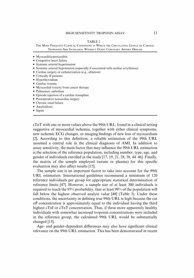
TABLE 2
THE MOST FREQUENT CLINICAL CONDITIONS IN WHICH THE CIRCULATING LEVELS OF CARDIAC
TROPONINS ARE INCREASED, WITHOUT OVERT CORONARY ARTERY DISEASE
� Myocarditis/pericarditis� Congestive heart failure� Systemic arterial hypertension� Systemic arterial hypotension (especially if associated with cardiac arrythmias)� Cardiac surgery or catheterization (e.g., ablation)� Critically ill patients� Hypothyroidism� Cardiac trauma� Myocardial toxicity from cancer therapy� Pulmonary embolism� Episode rejection of a cardiac transplant� Postoperative noncardiac surgery� Chronic renal failure� Amyloidosis� Sepsis
HIGH SENSITIVITY TROPONIN ASSAY 11

reference population studies that included at least 300 individuals and used
highly sensitive methods for cTnI or cTnT [28, 38, 45, 46]. A study from our
laboratory [28] included 692 apparently healthy subjects (311 males and 381
females) with a mean (SD) age of 45.3 (17.3) years [range 11–89 years;
females 46.5 (17.3) years, males 43.8 (17.1) years]. Our study found signifi-
cant gender-based diVerence in cTnI values (men: median 0.012 �g/L, rangefrom undetectable values to 0.196 �g/L; women: median 0.008 �g/L, rangefrom undetectable values to 0.130 �g/L; p < 0.0001) using the ADVIA TnI-
Ultra method (Siemens Medical Solutions Diagnostics SrL). Undetectable
cTnI concentrations were found in 168 individuals (24.3% of the samples
tested). All individuals used in this study were screened for preventive medi-
cine programs (laboratory staV, blood donors, or voluntary subjects), with
no acute or chronic diseases (excluded by history, accurate clinical examina-
tion, ECG, and laboratory tests), nor use of drugs for at least 2 weeks before
the sample collection. A gender-dependent cut oV value was also found in
another study [45] that used the recently refined Access AccuTnI assay
(Beckman-Coulter) to assess the distribution of cTnI in a community popu-
lation of elderly individuals [PIVUS (Prospective Study of the Vasculature in
Uppsala Seniors) study; n ¼ 1005]. Gender-dependent diVerences in 99th
URL for the highly sensitive cTnT assay by Roche Diagnostics was also
reported by Mingels et al. [46] in a reference population of 479 apparently
healthy individuals; the observed 99th percentile was 0.008 �g/L in 215
females and 0.018 �g/L in 264 males (p < 0.001).
In contrast, Collinson et al. [38] reported that the calculated 99th URL of
cTnI concentration, measured with the ADVIA TnI-Ultra method, was very
similar to that reported by the manufacturer (0.04 �g/L) and cTnI values
were not age- and gender-dependent [38]. Moreover, 165 (53.4%) of the 309
individuals (127 men, 182 women; median age 53 years, range 45–80 years)
enrolled were considered to have no measurable cTnI. The individuals who
participated in this study were randomly selected from a population of
TABLE 3
SAMPLE SIZES AND ASSOCIATED TOLERANCE LEVELS FOR
REPORTING 99TH PERCENTILE BASED ON THE LARGEST
OBSERVED VALUE (ACCORDING TO REFERENCE 48)
Sample size Tolerance level
100 0.63
200 0.87
300 0.95
400 0.98
500 0.99
12 CLERICO ET AL.

ostensibly healthy individuals, accurately screened to exclude any history of
vascular disease, diabetes mellitus, hypertension, heavy alcohol intake, use of
cardiac medication, or pathologic echocardiogram.
Some interesting observations may be derived from these studies [28, 38,
45, 46]. First, the selection of reference population may greatly influence the
calculation of 99th URL. In particular, the clinical protocol used to exclude
the presence of asymptomatic cardiac disease (especially in older subjects) is
likely to aVect the statistical analysis with respect to distribution of cTnI
measured by highly sensitive methods. Second, the presence of cTnI and
cTnT measurable values in apparently healthy subjects requires physiologic
explanation. Increased concentrations of cTnI and cTnT have been observed
in animal models of ischemia without histologic evidence of irreversible
injury [49]. Moreover, apoptotic cells have been described in normal adult
hearts, suggesting that myocyte replication is a significant component of
normal physiology and cellular processes, even in adults [50]. This finding
will be discussed in more detail in the following section. It can be suggested
that the release of cTnI from cardiomyocytes of healthy adult subjects may
result from a process related to the ‘‘physiological remodeling’’ of human
myocardium [28, 31].
Age-dependent increases in cTnI and cTnT in apparently healthy subjects
may suggest additional pathophysiological mechanisms. It is well known that
the incidence ofHFprogressively and steeply increases after the age of 55 years
(Table 4) and that this disease is the most common cause of death in elderly
people [51]. Several histological changes ofmyocardial tissue characterized by
loss of myocytes and subsequent hypertrophy of the remaining cells and
calcification of several cardiac structures can be found in most individuals
with aging [52, 53]. Moreover, the age-related loss of arterial compliance
contributes to isolated systolic hypertension and left ventricular hypertrophy
[52, 53]. Despite these changes, for the majority of apparently healthy older
TABLE 4
PREVALENCE OF SYSTOLIC AND DIASTOLIC DYSFUNCTION BY AGE (ACCORDING TO REFERENCE 51)
Dysfunction 45–54 years 55–64 years 65–74 years 75 and older Overall
Diastolic
Moderate 1.4% 6.0% 9.9% 14.6% 6.6%
Severe (restrictive) 0% 0.4% 0.7% 3.4% 0.7%
Systolic
LVEF � 50% 3.0% 4.8% 7.1% 12.9% 6.0%
LVEF � 40% 0.8% 1.3% 2.7% 4.4% 2.0%
LVEF, Left ventricular ejection fraction.
HIGH SENSITIVITY TROPONIN ASSAY 13

adults, cardiac output is well preserved by means of the Frank–Starling
principle, in the setting of reduced early diastolic filling [53]. In accordance
with these findings [52, 53], we suggest that increased levels of cTnI, measured
with high-sensitivity immunoassay methods in some apparently healthy older
adults [28, 45, 50], are likely to be due to increased remodeling of myocardial
tissue in this population. This hypothesis is well in agreement with the results
reported by Eggers et al. [54]. This study investigated the prevalence of cTnI
elevation in an elderly community population that included 1005 individuals
aged 70 years. Using a highly sensitive immunoassay, this study found that
increased cTnI was relatively common in elderly subjects and was associated
with cardiovascular high-risk features and/or impaired cardiac performance
[54].
Cumulatively, these data strongly indicate that calculation of 99th URL is
dependent on demographic and clinical characteristics of the reference pop-
ulation used in the study. Hence, clinical cutoVs using highly sensitive cTnI
and cTnT assays should be based on analytic definitions (i.e., CV) versus
distribution characteristics (i.e., percentiles) such that ‘‘true’’ troponin in-
crease may be identified [38]. Furthermore, the demographic and clinical
characteristics of the reference population enrolled for calculation of 99th
URL should be clearly delineated by the commercial manufacturers as well
as authors of published clinical studies.
5.2. MORE MYOCARDIAL INFARCTIONS OR MORE FALSE POSITIVE RESULTS?
It is reasonable that the new generation of high sensitivity cTnI and cTnT
methods can detect a greater number of patients with AMI than standard
methods, especially those individuals with very small infarct size [5, 55]. Data
reported by some recent studies appear to confirm this hypothesis [19, 55–59].
Unlike most commercial cTnI and cTnT methods, the use of highly sensitive
methods (i.e., with increased assay sensitivity at very low troponin concen-
tration) will allow earlier diagnosis of AMI (within 1–2 h after thoracic pain
onset), as well as recognition of very small (focal) areas of myocardial necrosis
as true AMI.
Although professional societies have recognized the importance of the
enhanced analytic performance of the newer and emerging cardiac troponin
assays, the clinical community has not uniformly embraced this trend [55].
Indeed, it is uncertain if the new highly sensitive troponin assays will lead to
increased clarity or more confusion for most physicians [59]. Predictably, the
application of assays with lower limits of detection has led to an increase in
patients evaluated in the emergency setting with detectable cardiac troponin
in a variety of acute and chronic medical conditions other than ACS [5, 16,
19, 31, 55, 56].
14 CLERICO ET AL.

It is also reasonable that increased cardiac troponin in apparently stable
populations, such as elderly subjects from the community and patients with
previous ACS, may primarily reflect left ventricular hypertrophy and/or
myocardial pump failure with a continuous loss of viable cardiac myocytes
caused by increased myocardial wall strain, chronic ischemia, or apoptosis.
It is well known that these conditions are often associated with ST-T segment
abnormalities that may mimic changes related to acute coronary ischemia.
Accordingly, several studies have already demonstrated that more sensitive
troponin assays increased the number and rapidity of AMI diagnosis, but
also increased the number of false positives, that is, non-ACS-related pathol-
ogy [16, 19, 25, 56, 57]. In fact, Eggers et al. [16] reported a�7%misdiagnosis
(i.e., false positivity rate) for AMI in troponin-positive patients with
preexisting ST-T segment abnormalities in patients admitted for nonischemic
chest pain or other symptoms indicative of myocardial ischemia.
In conclusion, these data confirm that it is very diVicult (or even impossible)
to reliably diagnose patients suspected with ACS using only one determination
of cTnI or cTnT due to the relatively low specificity of existing cardiac
troponin assays for ischemic myocardial injury. Indeed, international guide-
lines [1, 2] recommend at least two samples with a delay of time of 6–12 h for
measurement of cTnI and cTnT in these settings.
5.3. CLINICAL RELEVANCE OF SERIALLY MEASURED
TROPONIN CIRCULATING LEVELS
It is important to note that the detection of a true and significant increase/
decrease in serially measured troponin is of critical importance to correctly
establish the diagnosis of AMI in all patients without a diagnostically reliable
or recent electrocardiogram [1, 2] (Fig. 2) and to discriminate between
ischemic and other causes of troponin increase [1, 2, 16, 35, 60, 61].
Unfortunately, there is no consensus about the required degree of change
for serial measurement of cTnI and cTnT in AMI diagnosis in patients
with suspected ACS. The National Academy of Clinical Biochemistry has
recommended a 20% change as statistically significant [60]. However, these
recommendations assume that analytical assays have a precision of 5–7%
with three times the SD and produce 99% confidence at limit at the AMI
decision point [61].
A statistically more rigorous approach toward assessment of meaningful
serial markers in clinical laboratory tests would be to first ascertain biologic
variation [14, 35]. Unfortunately, troponin biologic variation cannot be
evaluated with certainty using the standard methods due to their inability
to detect the protein in the blood of healthy subjects with adequate precision.
Using new high-sensitivity assays, Wu et al. [35] were able to demonstrate
HIGH SENSITIVITY TROPONIN ASSAY 15

that cTnI biological variation was lower than other cardiovascular biomar-
kers, that is, cardiac natriuretic peptide hormone, creatine kinase-MB frac-
tion (CK-MB), myoglobin, C-reactive protein (CRP), myeloperoxidase, and
serum amyloid A [35]. Because this study was performed in healthy subjects,
there is concern regarding the applicability and reliability of these biologic
variation parameters in patients with ACS. One would expect that there are
diVerences in biologic variation parameters between healthy individuals and
patients with ACS. In comparison to cTnI, cTnT has diVerent release kineticsfrom myocytes and clearance in peripheral tissues and therefore its biologic
variation should be evaluated in specific studies [35]. Clearly, more compre-
hensive studies are required to confirm that measurement relative to biologic
variation is useful in evaluating the clinical significance of cardiac troponin in
patients with ACS.
5.4. HIGH-SENSITIVITY TROPONIN METHODS IN PATIENTS WITH HF:
A BETTER STRATIFICATION OF CARDIOVASCULAR RISK
HF is amajor public health problem in theNorthAmerica and Europe [62–
65]. The incidence and prevalence of HF increases significantly with aging in
these populations. After the age of 65, the incidence of HF approaches 10 per
1000 of population (�1:100) [51]. In the United States, HF is the most
common hospital discharge diagnosis, and more Medicare dollars are spent
for diagnosis and treatment of HF than for any other disease [62–64]. Similar
data have been reported from European countries [65].
HF may be considered as the fatal progression of all cardiovascular dis-
orders. For this reason, HF is considered a syndrome rather than a primary
diagnosis which results from any structural or functional cardiac disorder
that impairs the ability of the heart to function as a pump to supporting
physiologic circulation [5, 31]. We can assume that, if heart dysfunction is an
inevitable and ultimate fate, the measurement of some highly specific cardiac
biomarkers, such as cTnI, cTnT, B-type natriuretic peptide (BNP) and its
related forms, should be useful in detection of people at risk of a more
rapid progression toward symptomatic HF, thus in need of specific clinical
treatment [5, 31].
In 2001, the AHA/ACC task force for the diagnosis and management of
chronic HF introduced a new classification that focused on disease evolution
[62]. This classification, updated in 2005 [63] and 2009 [64], identified four
stages (A, B, C, and D) that account for symptoms, established risk factors
for HF development and structural myocardial abnormalities. Stage
A includes asymptomatic patients at risk for developing HF with no struc-
tural cardiac involvement. Stage B includes asymptomatic patients at risk for
developing HF with structural cardiac involvement. Stage C includes
16 CLERICO ET AL.

patients with past or current symptoms of HF associated with underlying
structural heart disease. Stage D includes symptomatic patients with end-
stage disease who require specialized treatment strategies, such as mechanical
circulatory support, continuous inotropic infusions, cardiac transplantation,
or hospice care. Unlike the NYHA classification which is based on clinical
severity of symptoms, this new classification emphasizes the progressive
nature of the pathophysiological processes responsible for development of
HF [64]. In fact, the first two stages (A and B) clearly do not include HF but
help in the early identification of patients at risk for developing HF [64].
Stages A and B patients are best defined as those individuals with risk factors
that clearly predispose to the development of HF.
Appropriate risk stratification depends on the availability of specific,
accurate, and eVective disease and risk markers [5, 31]. Highly sensitive
cTnI and cTnT immunoassay methods share the most important analytic
and clinical performance characteristics of an ideal cardiac biomarker
(Table 5) [5, 31].
It is well known that a relatively large proportion of HF patients (25–45%),
especially those with clinical history of coronary artery disease, has increased
cTnI and cTnT, even if measured by standard (i.e., not highly sensitive)
methods [64]. Recent studies [16, 54, 58, 59] have suggested that the fraction
of patients with HF and troponin values above the 99th URL may further
increase when highly sensitive cTnI and cTnT assays are used. In particular,
the ValHeFT study (including 4053 randomized patients with symptomatic
heart failure) demonstrated cTnT values above the cut-off level in 10.4% of
patients studied using a standard assay and this percentage increased to 92%
when a more sensitive method was used [66].
TABLE 5
DESIRABLE FEATURES OF AN IDEAL MOLECULAR CARDIAC BIOMARKER
� Absolute cardiospecificity� Acceptable to patient� Stability in vivo and in vitro
� Adequate analytical sensitivity (functional sensitivity) easy to perform� Good degree in reproducibility and accuracy� Complete automation of assay� Internationally standardized� Low cost� Low biological variation� Reference range and cut off values tested for gender, age, and ethnicity dependence� Good diagnostic and prognostic accuracy� Favorable cost-benefit ratio
HIGH SENSITIVITY TROPONIN ASSAY 17

It is well known that troponin and natriuretic peptide, when used as
biomarkers of cardiac disease, furnish complementary clinical information
[5, 31]. The progressive increase in both markers with aging [5, 20, 28, 31, 45]
suggests that there is progressive decline of cardiac function which can be
assessed and monitored. If heart dysfunction is an inevitable and ultimate
fate, the measurement of these analytes would be useful to initiate specific
clinical care for those individuals at risk of more rapid progression to
symptomatic cardiac failure.
5.5. EARLY DETECTION OF MYOCARDIAL INJURY IN PATIENTS WITH
EXTRACARDIAC DISEASES OR ASSUMING POTENTIALLY CARDIOTOXIC DRUGS
The Dallas Heart Study (3357 subjects) found that the prevalence of
increased cTnT in the general population was �1% using electrochemilumi-
nescence immunoassay (ECLIA) (Roche Diagnostics), a standard assay [67].
The prevalence of high-risk cardiovascular features was increased similarly in
subjects with cTnT levels in the minimally increased and increased range [67].
These data suggest that cTnT elevation is always indicative of cardiovascular
disease or at least a high-risk cardiovascular profile. As such, increased
cardiac troponin represents an important finding even in those patients
without coronary artery disease. This ‘‘index’’ of cardiac tissue damage
may suggest an appropriate diagnosis and, when necessary, a specific treat-
ment. Increased cTnI or cTnT in patients without evidence of ischemic
coronary artery disease represent an independent risk of future cardiac
events and poor prognosis [5, 31, 60]. It is important to point out that there
is no minimal threshold and that cardiovascular risk increases progressively
with increased troponin.
Increased troponin has been reported in patients treated with potentially
cardiotoxic drugs, such as high-dose chemotherapy [68, 69]. Indeed, a specific
pattern of troponin release following high-dose chemotherapy identified at-
risk patients for cardiac events in the subsequent 3 years [68]. Furthermore,
in patients treated with high-dose chemotherapy, increased cTnI suggested
early and appropriate treatment and was found to prevent the development
of late cardiotoxicity [69].
Another important application of highly sensitive troponin assay (along
with cardiac natriuretic peptides) may be the early detection of myocardial
damage in patients with systemic acute or chronic inflammatory and rheu-
matic diseases (i.e., systemic lupus erythematosus, systemic amyloidosis,
sarcoidosis, and rheumatoid arthritis) [70–73]. It is noteworthy that mortality
risk in these patients is strongly associated with heart complications versus
other organ involvement [72, 74–76]. As a result, the early detection of
cardiac involvement may have a tremendous clinical impact on the prognosis
18 CLERICO ET AL.

of patients with chronic inflammatory and rheumatic diseases, especially
those with systemic amyloidosis [72]. Early identification of these patients
is critical in the initiation of successful treatment strategies.
It is well known that cardiovascular events are also the major prognostic
determinants in patients with end-stage renal disease, with cardiovascular
deaths representing more than 50% of total mortality [77, 78]. The early
recognition of conditions such as left ventricular hypertrophy and coronary
artery disease may allow the identification of patients with chronic kidney
disease at higher risk of developing either HF or other major cardiovascular
events with consequent increased mortality [77, 78]. In a considerable num-
ber of chronic hemodialysis patients, increased troponin was found despite
absence of cardiac ischemia, even if older generation cTnI and cTnT immu-
noassay methods were used [77, 78]. The elevation of cardiac biomarkers in
patients with renal diseases showed a strong prognostic significance with
respect to cardiovascular morbidity and mortality [5, 31, 73, 77, 78]. Recent
studies have shown that highly sensitive cTnI and cTnT immunoassays were
able to detect a greater number of end-stage renal disease patients with
increased troponin [58, 78, 79]. As such, high-sensitivity cTnI and cTnT
immunoassays may provide a useful rationale for both occult cardiac disease
screening and better cardiovascular risk stratification in this unique group of
patients.
In conclusion, sensitive cTnI and cTnT immunoassays may provide a
useful tool for the early screening of occult cardiac disease either in patients
with extracardiac diseases (especially renal and chronic inflammatory dis-
eases) or in subjects undergoing treatment with potentially cardiotoxic drugs
(such as high-dose chemotherapy). However, further studies are necessary to
demonstrate the clinical impact and the cost-eVectiveness of this approachaccording to the evidence-based laboratory medicine principles (EBLM) [80].
6. High-Sensitivity cTnI and cTnT Methods: A PowerfulTool for Monitoring Physiological Renewal
and Pathological Remodeling of the Myocardial Tissue?
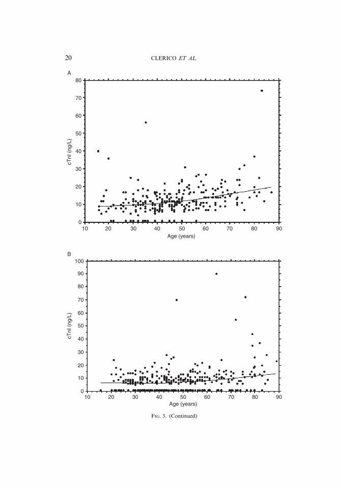
Increased analytical sensitivity of cTnI and cTnT methods has demon-
strated that measurable levels of these proteins are also present in apparently
healthy subjects [20, 28, 38] (Fig. 3). These findings suggest some interesting
pathophysiological considerations. At present time, the prevailing opinion,
based on the aggregate evidence to date, is that any reliably detected eleva-
tion of a cardiac troponin is abnormal and might represent cardiac necrosis
[81]. However, apoptotic cells have been described in normal adult hearts;
thus suggesting that myocyte replication is a significant component of the
HIGH SENSITIVITY TROPONIN ASSAY 19
80A
70
60
50
40
30
20
10
010 20 30 40 50
Age (years)
cTnI
(ng
/L)
60 70 80 90
100B
cTnI
(ng
/L)
90
80
70
60
50
40
30
20
10
010 20 30 40 50
Age (years)60 70 80 90
FIG. 3. (Continued)
20 CLERICO ET AL.

physiological cellular processes even in adults [50]. A very recent experimen-
tal study, based on DNA integration of the isotope 14C generated by nuclear
bomb tests during the Cold War, was able to establish cardiomyocyte age in
humans [82]. The results of this study suggested that cardiomyocytes renew
with a gradual decrease of annual turnover from 1% (age 25) to 0.45%
(age 75) with fewer than 50% of cardiomyocytes exchanged during a normal
life span [82].
At present time, there are no experimental data indicating that troponins
are degraded within the cardiomyocytes and released into the interstitial
space during apoptosis. There are two potential explanations for the tropo-
nin release in absence of lethal sarcolemmal disruption: (1) cellular release of
proteolytic troponin degradation products; (2) troponin leaks as an intact
nondegraded protein chain from reversibly damaged cardiomyocytes [83,
84]. Mechanical stretch of cardiomyocytes, for example, during pressure or
volume overload, may activate some intracellular proteases, such as metal-
loproteinase, that can degrade cardiac troponin intracellularly [85]. Over-
load-induced stretch at the cardiomyocyte level is sensed by integrins, which
are mechanotransducer molecules that link the extracellular matrix to the
intracellular cytoskeleton [86]. Hence, this mechanism may be involved in
stretch-induced release of troponin and its degradation products [83]. These
findings suggest that stretch stimulation of viable cardiomyocytes may lead
to intact cTnI release. Indeed, several studies have demonstrated that me-
chanically induced transient disruptions (wounding) of the sarcolemma are a
constitutive in vivo event [87–90]. This mechanism may account for the
release of proteins, like myocyte-derived growth factors that are released
despite lack of the classic signal peptide sequence that is normally associated
with secretion. These mechanically induced alterations in cardiomyocyte
sarcolemmal permeability may similarly be involved in the release of cTnI
from cytosolic pools in the absence of necrotic cell death. However, further
studies are necessary to accurately describe the cellular mechanisms respon-
sible for release of intact cTnI and cTnT in damaged cardiomyocytes.
FIG. 3. (A) Age-dependent distribution of cTnI values measured by the ADVIA method on
the Centaur Platform (Siemens Diagnostics) in 269 apparently healthy male subjects (age ranging
from 14 to 88 years). There is a very weak, although significant, correlation between age and cTnI
values (by Spearman Rank test, Rho ¼ 0.358, p < 0.0001). The trend, assessed by smooth spline
analysis, between age and cTnI values is indicated by a continuous line. (B) Age-dependent
distribution of cTnI values measured by the ADVIA method on the Centaur Platform (Siemens
Diagnostics) in 238 apparently healthy female subjects (age ranging from 14 to 88 years). There is
a very weak, although significant, correlation between age and cTnI values (by Spearman Rank
test, Rho ¼ 0.258, p < 0.0001). The trend, assessed by smooth spline analysis, between age
and cTnI values is indicated by a continuous line. Results obtained in the Authors’ laboratory
(see references 21, 28, 29).
HIGH SENSITIVITY TROPONIN ASSAY 21

The data, regarding gender- and age-related cTnI and cTnT levels in adult
healthy subjects [21, 23, 28, 45], support the hypothesis that small amounts of
cTnI can be released from cardiomyocytes even in apparently healthy sub-
jects due to a process related to the ‘‘physiological renewal or remodeling’’ of
human myocardium. Moreover, some findings obtained in healthy indivi-
duals after endurance exercise appear to confirm this hypothesis. Several
studies reported increased circulating cTnI or cTnT after strenuous exercise
(such as marathon runs or other endurance races), even in well-trained
athletes [91–98]. Middleton et al. [95] suggested that it is unlikely that
minor elevations in cTnI or cTnT subsequent to endurance exercise are due
to myocardial necrosis. These authors hypothesized that postexercise tropo-
nin release represents the reversible cardiomyocyte membrane damage dur-
ing remodeling processes [95]. According to this hypothesis, in a healthy
exercising population, cardiac troponins may be routinely released after
periods of increased myocardial demand, such as after endurance exercise.
As mentioned earlier, recently developed methods for cTnI and cTnT assays
may be able to detect a release of protein from a quantity of myocardial tissue
of a few milligrams in size [20–30] (Table 1).
Highly sensitive cTnI and cTnT immunoassays should be considered a useful
and potentially powerful tool to monitor the continuous and physiological
processes related to renewal and remodeling of the myocardial tissue.
7. Use of High-Sensitivity cTnI and cTnT Methods ina Multimarker Approach for Early Screening: An Increase
in Diagnostic and Prognostic Efficiency?
Many biomarkers exist for the diagnosis and prognosis of individuals with
cardiovascular diseases.Methods of assessment using a multimarker approach
have been considered the best model for risk prediction in individuals with
cardiovascular disease [5, 31, 99]. Despite the many suggested laboratory
biomarkers for diagnosis, risk assessment, and follow-up of patients with
cardiovascular disease, only cardiac troponins and natriuretic peptides have
been shown to have good diagnostic and prognostic accuracy, as well as
cost-eVectiveness, according to EBLM principles [5, 31, 99].
Indeed, these two biomarkers are characterized by high analytical and
clinical sensitivity as well as by absolute cardiospecificity. Cardiac troponins
and natriuretic peptides have diVerent physiological roles. Troponins are
structural proteins of actin–myosin complex involved in cardiac contraction
and relaxation, whereas the latter are peptide hormones with natriuretic and
vasodilative eVects produced by cardiomyocytes. Furthermore, diVerentpathophysiological mechanisms aVect the release of cardiac troponins and
22 CLERICO ET AL.

natriuretic peptides by cardiomyocytes [5, 31, 100–103]. Owing to their
diVerent pathophysiological roles, cardiac troponins and natriuretic peptides
may provide independent pathophysiological and clinical information. Due
to their unique roles, these markers may be combined in a multimarker
model for cardiovascular risk assessment and as predictive variables in
practically every acute and chronic cardiac disease, including those due to
ischemic, inflammatory, congenital, and traumatic conditions. A substantial
number of studies have confirmed this hypothesis [5, 13, 31, 60, 81, 104–109].
Furthermore, both biomarkers can be measured using highly sensitive immu-
noassays with detection limits below 10 pg/mL [5, 31, 108]. As such, these
biomarkers may be useful to assess disease progression and severity in
patients with stage B HF (ACC/AHA guidelines) [62–64] and patients with
end-stage renal disease [76]. Although some recent studies provide some
support [25, 54, 109–111], further and more comprehensive studies are
required to demonstrate the eVectiveness of highly sensitive cTnI and
cTnT, alone or together with BNP, in early screening of cardiac disease in
the general population or in high-risk individuals.
8. Conclusion
Improvement of analytical precision and clinical sensitivity of a laboratory
test is a goal for all laboratorians. This concept should be applied to cTnI and
cTnT immunoassays.
Careful evaluation with development of new diagnostic criteria (including
99th URL and decisional cut-oV values) will be necessary to improve patient
care. More accurate and earlier diagnosis should lead to more eVectivetherapies. In particular, highly sensitive cTnI and cTnT assays will better
define the true normal levels and the 99th URL. Moreover, the new genera-
tion of troponin assays will enable better risk stratification of patients who do
not have increased troponin by current assays and may allow risk stratifica-
tion of patients with chronic stable angina and patients with HF. It is really
important to stress that increased cTnI and cTnT represent an index of
cardiac tissue damage, even in the case of extracardiac diseases (including
chronic inflammatory disease, end-stage renal disease, or treatment with
powerful cardiotoxic drugs), suggesting an appropriate diagnosis and,
when necessary, a specific treatment. Despite these interesting and prelimi-
nary studies, further and more comprehensive studies with highly specific
assays are required to firmly establish the clinical usefulness of troponin in a
wide range of diseases.
HIGH SENSITIVITY TROPONIN ASSAY 23
REFERENCES
[1] J.S. Alpert, K. Thygesen, E. Antman, J.P. Bassand, Myocardial infarction redefined:
a consensus document of the Joint European Society of Cardiology/American College of
Cardiology Committee for the redefinition of myocardial infarction, J. Am. Coll. Cardiol.
36 (2000) 959–969.
[2] K. Thygesen, J.S. Alpert, H.D. White, Joint ESC/ACCF/AHA/WHF Task Force for the
Redefinition of Myocardial Infarction, Universal definition of myocardial infarction,
J. Am. Coll. Cardiol. 50 (2007) 2173–2195.
[3] J. Trevelyan, E.W.A. Needham, S.C.H. Smith, R.K. Mattu, Impact of the recommenda-
tions for the redefinition of miocardial infarction on diagnosis and prognosis in an
unselected United Kingdom cohort with suspected cardiac chest pain, Am. J. Cardiol.
93 (2004) 817–821.
[4] M. Panteghini, The new definition of myocardial infarction and the impact of troponin
determination on clinical practice, Int. J. Cardiol. 106 (2006) 298–306.
[5] S. Vittorini, A. Clerico, Cardiovascular biomarkers: increasing impact of laboratory
medicine in cardiology practice, Clin. Chem. Lab. Med. 46 (2008) 748–763.
[6] F.S. Apple, R.L. Jesse, L.K. Newby, et al., National Academy of Clinical Biochemistry
and IFCC Committee for Standardization of Markers of Cardiac Damage Laboratory
Medicine Practice Guidelines: analytical issues for biochemical markers of acute coronary
syndromes, Clin. Chem. 53 (2007) 547–551.
[7] R.H. Christenson, S.H. Duh, F.S. Apple, et al., Standardization of cardiac troponin
I assays: round robin of ten candidate reference materials, Clin. Chem. 47 (2001) 431–437.
[8] M. Panteghini, F. Pagani, K.T.J. Yeo, et al., Evaluation of imprecision for cardiac
troponin assays at low-range concentrations, Clin. Chem. 50 (2004) 327–332.
[9] S. Takeda, A. Yamashita, K. Maeda, Y. Maeda, Structure of the core domain of human
cardiac troponin in the Ca(2þ)-saturated form, Nature 424 (6944) (2003) 35–41.
[10] F.S. Apple, Cardiac troponin testing in renal failure and skeletal muscle disease patients,
in: A.H.B. Wu (Ed.), Cardiac Markers, second ed., Homan Press, Totowa, NJ, 2003,
pp. 139–147.
[11] M. Panteghini, Acute coronary syndrome. Biochemical strategies in the troponin era,
Chest 122 (2002) 1428–1435.
[12] F.S. Apple, M.M. Murakami, The diagnostic utility of cardiac biomarkers in detecting
myocardial infarction, Clin. Cornerstone 7 (Suppl. 1) (2005) S25–S30.
[13] A.S. JaVe, L. Babuin, F.S. Apple, Biomarkers in acute cardiac disease. The present and the
future, J. Am. Coll. Cardiol. 48 (2006) 1–11.
[14] M. Panteghini, Assay-related issues in the measurement of cardiac troponins, Clin. Chim.
Acta 402 (2009) 88–93.
[15] M. Panteghini, D.M. Bunk, R.H. Christenson, et al., Standardization of troponin I
measurements: an update, Clin. Chem. Lab. Med. 46 (2008) 1501–1506.
[16] K.M. Eggers, L. Lind, P. Venge, B. Lindahl, Will the universal definition of myocardial
infarction criteria result in an overdiagnosis of myocardial infarction? Am. J. Cardiol.
103 (2009) 588–591.
[17] F.S. Apple, M.A.M. Muratami, Serum and plasma cardiac troponin I 99th percentile
reference values for 3 2nd generation assays, Clin. Chem. 53 (2007) 1558–1560.
[18] K.M. Eggers, B. Lagerquist, P. Venge, L. Wallentin, B. Lindahl, Persistent troponin I
elevation in stabilized patients after an episode of acute coronary syndrome predicts long-
term mortality, Circulation 107 (2007) 1907–1914.
[19] S.E. Melanson, D.A. Morrow, P. Jarolim, Earlier detection of myocardial injury in a
preliminary evaluation using a new troponin I assay with improved sensitivity, Am. J. Clin.
Pathol. 128 (2007) 282–286.
24 CLERICO ET AL.

[20] D. Hermsen, F. Apple, L. Garcia-Beltran, et al., Results from a multicenter evaluation of
the 4th generation Elecsys Troponin T assay, Clin. Lab. 53 (2007) 1–9.
[21] C. Prontera, A. Fortunato, S. Storti, et al., Evaluation of analytical performance of the
siemens ADVIA TnI ultra immunoassay, Clin. Chem. 53 (2007) 1722–1723.
[22] J. Jarausch, S. Braun, A. Dolci, et al., Evaluation of a development version of the Elecsys
high sensitive troponin T assay, Clin. Chem. 54 (Suppl. 6) (2008) A91.
[23] R. Beyrau, S. Braun, R. Cooray, et al., Multicentre evaluation of a high sensitive Elecsys
troponin T assay, Clin. Chem. Lab. Med. 47 (2009) S128.
[24] M.S. Sabatine, D.A. Morrow, J.A. de Lemos, P. Jarolim, E. Braunwald, Detection of
acute changes in circulating troponin in the setting of transient stress test-induced
myocardial ischaemia using an ultrasensitive assay: results from TIMI 35, Eur. Heart J.
30 (2009) 162–169.
[25] P. Venge, S. James, L. Jansson, B. Lindahl, Clinical performance of two highly sensitive
cardiac troponin I assays, Clin. Chem. 55 (2009) 109–116.
[26] M. Redpath, G. Chalmers, C. Dibden, B. Morris, A. Blumsohn, Evaluation of imprecision
of ADVIA CentaurW TNI-UltraTM automated immunoassay, Ann. Clin. Biochem. 45
(Suppl. 1) (2008) 57.
[27] D. Van de Kerkhof, B. Peters, V. Scharnhorst, Performance of the Advia Centaur second-
generation troponin assay TnI-Ultra compared with the first-generation cTnI assay, Ann.
Clin. Biochem. 45 (2008) 316–317.
[28] A. Clerico, A. Fortunato, C. Prontera, A. Ripoli, G.C. Zucchelli, M. Emdin, Distribution
of plasma cardiac troponin-I values in healthy subjects: pathophysiological considerations,
Clin. Chem. Lab. Med. 46 (2008) 804–808.
[29] C. Prontera, A. Fortunato, S. Storti, et al., Evaluation of analytical performance of Advia
TnI ultra immunoassay and comparison with Access AccuTnI method, Immuno-Analyse
Biol. Spec. 23 (2008) 311–318.
[30] J.C.J.M. Swaanenburg, P.J. Visser-VanBrummen, M.J.L. DeJongste, Tiebosch ATHM.
The content and distribution of troponin I, troponin T, myoglobin, and alpha-hydrobytyric
acid dehydrogenase in the human heart, Am. J. Clin. Pathol. 115 (2001) 770–777.
[31] M. Emdin, S. Vittorini, C. Passino, A. Clerico, Old and new biomarkers of heart failure,
Eur. J. Heart Fail. 11 (2009) 331–335.
[32] Clinical and Laboratory Standards Institute, Approved guideline;Protocols for determi-
nation of limits of detection and limits of quantitation, (2004) CLSI document EP17-A.
[33] C.A. Spencer, M. Takeuchi, M. Kazarosyan, F. MacKenzie, G.J. Beckett, E. Wilkinson,
Interlaboratory/intermethod diVerences in functional sensitivity of immunometric assays
of thyrotropin (TSH) and impact on reliability of measurement of subnormal concentra-
tions of TSH, Clin. Chem. 41 (1995) 367–374.
[34] International vocabulary of metrology, Basic and general concepts and associated terms
(VIM). Document produced by the Working Group 2 of the Joint Committee for Guides
in Metrology (JCGM/WG 2), JCGM, (2008) 200.
[35] A.H.B. Wu, Q.A. Lu, J. Todd, J. Moecks, F. Wians, Short- and long-term biological
variation in cardiac troponin I measured with a high sensitivity assay: implications for
clinical practice, Clin. Chem. 55 (2009) 52–58.
[36] M. Zaninotto, M. Mion, S. Altinier, M. Forni, M. Plebani, Quality specifications for
biochemical markers of myocardial injury, Clin. Chim. Acta 346 (2004) 65–72.
[37] J.R. Tate, W. Ferguson, R. Bais, K. Kostner, T. Marwick, A. Carter, The determination of
the 99th centile level for troponin assays in an Australian reference population, Ann. Clin.
Biochem. 45 (2008) 275–288.
[38] P.O. Collinson, O. CliVord-Mobley, D. Gaze, F. Boa, R. Senior, Assay imprecision and
99th-percentile reference value of a high-sensitivity cardiac troponin I assay, Clin. Chem.
55 (2009) 1433–1434.
HIGH SENSITIVITY TROPONIN ASSAY 25

[39] F.S. Apple, A new season for cardiac troponin assays: it’s time to keep a scorecard, Clin.
Chem. 55 (2009) 1303–1306.
[40] S. James, P. Armstrong, R. CaliV, et al., Troponin T levels and risk of 30-day outcomes in
patients with the acute coronary syndrome: prospective verification in the GUSTO-IV
trial, Am. J. Med. 115 (2003) 178–184.
[41] B. Lindahl, E. Diderholm, B. Lagerqvist, P. Venge, L. Wallentin, Mechanisms behind the
prognostic value of troponin T in unstable coronary artery disease: a FRISC II substudy,
J. Am. Coll. Cardiol. 38 (2001) 979–986.
[42] D.A.Morrow, C.P. Cannon, N. Rifai, et al., Ability of minor elevations of troponins I and
T to predict benefit from an early invasive strategy in patients with unstable angina and
non-ST elevation myocardial infarction: results from a randomized trial, JAMA 286
(2001) 2405–2412.
[43] P. Venge, B. Lagerqvist, E. Diderholm, B. Lindahl, L. Wallentin, Clinical performance of
three cardiac troponin assays in patients with unstable coronary artery disease, Am.
J. Cardiol. 89 (2002) 1035–1041.
[44] J.R. Tale, Troponin revisited 2008: assay performance, Clin. Chem. Lab. Med. 46 (2008)
1489–1500.
[45] K.M. Eggers, A.S. JaVe, L. Lind, P. Venge, B. Lindahl, Value of cardiac troponin I
cutoV concentrations below the 99th percentile for clinical decision-making, Clin. Chem.
55 (2009) 85–92.
[46] A. Mingels, L. Jacobs, E. Michielsen, J. Swaanenburg, W. Wodzig, M. van Dieijen-Visser,
Reference population andmarathon runner sera assessed by highly sensitive cardiac tropo-
nin T and commercial cardiac troponin T and I assays, Clin. Chem. 55 (2009) 101–108.
[47] Clinical and Laboratory Standards Institute, Defining, establishing, and verifying
reference intervals in the clinical laboratory; Proposed guideline, third ed., Clinical and
Laboratory Standards Institute, Wayne, PA, 2008 CLSI document C28-P3.
[48] P.E. Hickman, T. Badrick, S.R. Wilson, D. McGill, Reporting of cardiac troponin—
problems with the 99th population centile, Clin. Chim. Acta 381 (2007) 182–183.
[49] Y. Chen, R.C. Serfass, S.M. Mackey-Bojack, K.L. Kelly, J.L. Titus, F.S. Apple, Cardiac
troponin T alterations in myocardium and serum of rats after stressful, prolonged intense
exercise, J. Appl. Physiol. 88 (2000) 1749–1755.
[50] P. Anversa, A. Leri, J. Kajstura, B. Nadal-Ginard, Myocyte growth and cardiac repair,
J. Mol. Cell. Cardiol. 34 (2002) 91–105.
[51] R.K. Illango, Utilizing NT-proBNP in the selection of risks for life insurance, J. Insur.
Med. 39 (2007) 182–191.
[52] J.M. Ribera-Casado, Ageing and the cardiovascular system, Z. Gerontol. Geriatr. 32 (1999)
412–419.
[53] K.G. Pugh, J.Y. Wei, Clinical implications of physiological changes in the aging heart,
Drugs Aging 18 (2001) 263–276.
[54] K.M. Eggers, L. Lind, H. Ahlstrom, et al., Prevalence and pathophysiological mechanisms
of elevated cardiac troponin I levels in a population-based sample of elderly subjects, Eur.
Heart J. 29 (2008) 2252–2258.
[55] D.A. Morrow, E.M. Antman, Evaluation of high-sensitivity assays for cardiac troponin,
Clin. Chem. 55 (2009) 5–9.
[56] S.E. Melanson, M.J. Conrad, N. Mosammaparast, P. Jarolim, Implementation of a highly
sensitive cardiac troponin I assay: test volumes, positivity rates and interpretation of
results, Clin. Chim. Acta 395 (2008) 57–61.
[57] P.A. Kavsak, A.R. MacRae, M.J. Yerna, A.S. JaVe, Analytical and clinical utility of a
next-generation, highly sensitive cardiac troponin I assay for early detection of myocardial
injury, Clin. Chem. 55 (2009) 573–577.
26 CLERICO ET AL.

[58] A.H. Wu, Interpretation of high sensitivity cardiac troponin I results: reference to
biological variability in patients who present to the emergency room with chest pain:
case report series, Clin. Chim. Acta 401 (2009) 170–174.
[59] H.D. White, Will new higher-precision troponins lead to clarify or confusion? Curr. Opin.
Cradiol. 23 (2008) 292–295.
[60] A.H. Wu, A.S. JaVe, F.S. Apple, et al., NACB Writing Group; Cannon CP, Storrow AB;
NACB Committee, National Academy of Clinical Biochemistry Laboratory Medicine
practice guidelines: use of cardiac troponin and B-type natriuretic peptide or N-terminal
proB-type natriuretic peptide for etiologies other than acute coronary syndromes and
heart failure, Clin. Chem. 53 (2007) 2086–2096.
[61] A.H.Wu, A.S. JaVe, The clinical need for high-sensitivity cardiac troponin assays for acute
coronary syndromes and the role for serial testing, Am. Heart J. 155 (2008) 208–214.
[62] S.A. Hunt, D.W. Baker, M.H. Chin, et al., ACC/AHA guidelines for the evaluation and
management of chronic heart failure in the adult: executive summary: a report of the
American College of Cardiology/American Heart Association Task Force on Practice
Guidelines (Committee to Revise the 1995 Guidelines for the Evaluation andManagement
of Heart Failure), J. Am. Coll. Cardiol. 38 (2001) 2101–2113.
[63] S.A. Hunt, American College of Cardiology; American Heart Association Task Force on
Practice Guidelines (Writing Committee to Update the 2001 Guidelines for the Evaluation
and Management of Heart Failure), ACC/AHA 2005 guideline update for the diagnosis
and management of chronic heart failure in the adult: a report of the American College of
Cardiology/American Heart Association Task Force on Practice Guidelines (Writing
Committee to Update the 2001 Guidelines for the Evaluation and Management of Heart
Failure), J. Am. Coll. Cardiol. 46 (2005) e1–e82.
[64] S.A. Hunt, W.T. Abraham, M.H. Chin, American College of Cardiology Foundation;
American Heart Association, 2009 Focused update incorporated into the ACC/AHA 2005
Guidelines for the Diagnosis and Management of Heart Failure in Adults A Report of the
American College of Cardiology Foundation/American Heart Association Task Force on
Practice Guidelines Developed in Collaboration With the International Society for Heart
and Lung Transplantation, J. Am. Coll. Cardiol. 53 (2009) e1–e90.
[65] K. Swedberg, J. Cleland, H. Dargie, et al., Task force for the diagnosis and treatment of
chronic heart failure of the European Society of Cardiology. Guidelines for the diagnosis
and treatment of chronic heart failure: executive summary (update 2005): the Task Force
for the Diagnosis and Treatment of Chronic Heart Failure of the European Society of
Cardiology, Eur. Heart J. 26 (2005) 1115–1140.
[66] R. Latini, S. Masson, I.S. Anand, et al., Prognostic value of very low plasma concentra-
tions of troponin T in patients with stable chronic heart failure, Circulation 116 (2007)
1242–1249.
[67] T.W. Wallace, S.M. Abdullah, M.H. Drazner, et al., Prevalence and determinants of
troponin T elevation in the general population, Circulation 113 (2006) 1958–1965.
[68] D. Cardinale, M.T. Sandri, A. Colombo, et al., Prognostic value of troponin I in cardiac
risk stratification of cancer patients undergoing high-dose chemotherapy, Circulation 109
(2004) 2749–2754.
[69] D. Cardinale, A. Colombo, M.T. Sandri, et al., Prevention of high-dose chemotherapy-
induced cardiotoxicity in high-risk patients by angiotensin-converting enzyme inhibition,
Circulation 114 (2006) 2474–2481.
[70] M. Maeder, T. Fehr, H. Rickli, P. Ammann, Sepsis-associated myocardial dysfunction:
diagnostic and prognostic impact of cardiac troponins and natriuretic peptides, Chest 129
(2006) 1349–1366.
HIGH SENSITIVITY TROPONIN ASSAY 27

[71] A. Dispenzieri, M.A. Gertz, R.A. Kyle, et al., Serum cardiac troponins and N-terminal
pro-brain natriuretic peptide: a staging system for primary systemic amyloidosis, J. Clin.
Oncol. 22 (2004) 3751–3757.
[72] G. Merlini, G. Palladini, Amyloidosis is a cure possible? Ann. Oncol. 19 (Suppl. 4) (2008)
iv63–iv66.
[73] C. Passino, R. Poletti, M. Fontana, et al., Clinical relevance of non-cardiac determinants
of natriuretic peptide levels, Clin. Chem. Lab. Med. 46 (2008) 1515–1523.
[74] T. Pham, L. Gossec, A. Constantin, et al., Cardiovascular risk and rheumatoid arthritis:
clinical practice guidelines based on published evidence and expert opinion, Joint Bone
Spine 73 (2006) 379–387.
[75] A. Kahan, Y. Allanore, Primary myocardial involvement in systemic sclerosis, Rheuma-
tology 45 (Suppl. 4) (2006) 14–17.
[76] N. Assous, E. Touze, C. Meune, A. Kahan, Y. Allanore, Cardiovascular disease in
rheumatoid arthritis: single-center hospital-based cohort study in France, Joint Bone
Spine 74 (2007) 66–72.
[77] A.Y. Wang, K.N. Lai, Use of cardiac biomarkers in end-stage renal disease, J. Am. Soc.
Nephrol. 19 (2008) 1643–1652.
[78] C. Sommerer, E. Giannitsis, V. Schwenger,M. Zeier, Cardiac biomarkers in haemodialysis
patients: the prognostic value of amino-terminal pro-B-type natriuretic peptide and cardi-
ac troponin T, Nephron Clin. Pract. 107 (2007) c77–c81.
[79] L.H. Jacobs, J. van deKerkhof, A.M.Mingels, et al., Haemodialysis patients longitudinally
assessed by highly sensitive cardiac troponin T and commercial cardiac troponin T and
cardiac troponin I assays, Ann. Clin. Biochem. 46 (2009) 283–290.
[80] D.A. Marshall, B.J. O’Brien, Economic evaluation of diagnostic tests, in: P.C. Price,
R.H. Christensen (Eds.), Evidence-based laboratory medicine—from principles to
outcomes, AACC Press, Washington, DC, 2003, pp. 159–186.
[81] D.A.Morrow, C.P. Cannon, R.L. Jesse, et al., National Academy of Clinical Biochemistry
Laboratory Medicine Practice Guidelines: clinical characteristics and utilization of
biochemical markers in acute coronary syndromes, Clin. Chem. 53 (2007) 552–574.
[82] O. Bergmann, R.D. Bhardwaj, S. Bernard, et al., Evidence for cardiomyocyte renewal in
humans, Science 324 (5923) (2009) 98–102.
[83] M.H. Hessel, D.E. Atsma, E.J. van der Valk, W.H. Bax, M.J. Schalij, A. van der Laarse,
Release of cardiac troponin I from viable cardiomyocytes is mediated by integrin stimula-
tion, Pflugers Arch. 455 (2008) 979–986.
[84] M.H. Hessel, E.C. Michielsen, D.E. Atsma, et al., Release kinetics of intact and degraded
troponin I and T after irreversible cell damage, Exp. Mol. Pathol. 85 (2008) 90–92.
[85] W. Wang, C.J. Schulze, W.L. Suarez-Pinzon, J.R. Dyck, G. Sawicki, R. Schulz, Intracel-
lular action of matrix metalloproteinase-2 accounts for acute myocardial ischemia and
reperfusion injury, Circulation 106 (2002) 1543–1549.
[86] J.Y. Shyy, S. Chien, Role of integrins in cellular responses to mechanical stress and
adhesion, Curr. Opin. Cell Biol. 9 (1997) 707–713.
[87] M.S. Clarke, R.W. Caldwell, H. Chiao, K.Miyake, P.L. McNeil, Contraction-induced cell
wounding and release of fibroblast growth factor in heart, Circ. Res. 76 (1995) 927–934.
[88] D. Kaye, D. Pimental, S. Prasad, et al., Role of transiently altered sarcolemmal membrane
permeability and basic fibroblast growth factor release in the hypertrophic response of
adult rat ventricular myocytes to increased mechanical activity in vitro, J. Clin. Invest. 97
(1996) 281–291.
[89] P.L. McNeil, R. Khakee, Disruptions of muscle fiber plasma membranes. Role in exercise-
induced damage, Am. J. Pathol. 140 (1992) 1097–1109.
28 CLERICO ET AL.

[90] E. Page, J. Upshaw-Earley, G. Goings, Permeability of rat atrial endocardium, epicardi-
um, and myocardium to large molecules. Stretch-dependent eVects, Circ. Res. 71 (1992)
159–173.
[91] R.E. Shave, E. Dawson, G.Whyte, et al., Evidence of exercise-induced cardiac dysfunction
and elevated cTnT in separate cohorts competing in an ultra-endurance mountain mara-
thon race, Int. J. Sports Med. 23 (2002) 489–494.
[92] M. Herrmann, J. Scharhag, M. Miclea, A. Urhausen, W. Herrmann, W. Kindermann,
Post-race kinetics of cardiac troponin T and I andN-terminal pro-brain natriuretic peptide
in marathon runners, Clin. Chem. 49 (2003) 831–834.
[93] H.J. Roth, R.M. Leithauser, H. Doppelmayr, et al., Cardiospecificity of the 3rd generation
cardiac troponin T assay during and after a 216 km ultra-endurance marathon run in
Death Valley, Clin. Res. Cardiol. 96 (2007) 359–364.
[94] E.B. Fortescue, A.Y. Shin, D.S. Greenes, et al., Cardiac troponin increases among runners
in the Boston Marathon, Ann. Emerg. Med. 49 (2007) 137–143.
[95] N. Middleton, K. George, G. Whyte, D. Gaze, P. Collinson, R. Shave, Cardiac troponin
release is stimulated by endurance exercise in healthy humans, J. Am. Coll. Cardiol. 52
(2008) 1813–1814.
[96] K.M. Hubble, D.M. Fatovich, J.M. Grasko, S.D. Vasikaran, Cardiac troponin increases
among marathon runners in the Perth Marathon: the Troponin in Marathons (TRIM)
study, Med. J. Aust. 190 (2009) 91–93.
[97] N. Mousavi, A. Czarnecki, K. Kumar, et al., Relation of biomarkers and cardiac magnetic
resonance imaging after marathon running, Am. J. Cardiol. 103 (2009) 1467–1472.
[98] D.S. Jassal, D. MoVat, J. Krahn, et al., Cardiac injury markers in non-elite marathon
runners, Int. J. Sports Med. 30 (2009) 75–79.
[99] S. Vasan, Biomarkers of cardiovascular disease: molecular basis and practical considera-
tions, Circulation 113 (2006) 2335–2362.
[100] A. Clerico, S. Vittorini, C. Passino, M. Emdin, New biomarkers of heart failure, Crit. Rev.
Clin. Lab. Sci. 46 (2009) 107–128.
[101] A. Clerico, F.A. Recchia, C. Passino,M. Emdin, Cardiac endocrine function is an essential
component of the homeostatic regulation network: physiological and clinical implications,
Am. J. Physiol. Heart Circ. Physiol. 290 (2006) H17–H29.
[102] A. Clerico, M. Fontana, S. Vittorini, M. Emdin, The search for a pathophysiological link
between gender, cardiac endocrine function, body mass regulation and cardiac mortality:
proposal for a working hypothesis, Clin. Chim. Acta 405 (2009) 1–7.
[103] A. De Bold, Determinants of brain natriuretic peptide gene expression and secretion in
acute cardiac allograft rejection, Curr. Opin. Cardiol. 22 (2007) 146–150.
[104] S. Potluri, H.O. Ventura, M. Mulumudi, M.R. Mehra, Cardiac troponin levels in heart
failure, Cardiol. Rev. 12 (2004) 21–25.
[105] S.D. Wiviott, J.A. de Lemos, D.A. Morrow, Pathophysiology, prognostic significance and
clinical utility of B-type natriuretic peptide in acute coronary syndromes, Clin. Chim. Acta
346 (2004) 119–128.
[106] P. Venge, J. Arnlov, B. Zethelius, Seemingly healthy 71-year-old men with minor eleva-
tions of cardiac troponin I and at risk of premature death in CVD have elevated levels of
NT-proBNP: report from the ULSAM study, Scand. Clin. Lab. Invest. 69 (2009)
418–424.
[107] J.C. Lega, Y. Lacasse, L. Lakhal, S. Provencher, Natriuretic peptides and troponins in
pulmonary embolism: a meta-analysis, Thorax 64 (2009) 868–875.
[108] A. Clerico, M. Emdin, Diagnostic accuracy and prognostic relevance of the measurement
of the cardiac natriuretic peptides: a review, Clin. Chem. 50 (2004) 33–50.
HIGH SENSITIVITY TROPONIN ASSAY 29

[109] M. Emdin, C. Passino, C. Prontera, et al., Comparison of brain natriuretic peptide (BNP)
and amino-terminal proBNP for early diagnosis of heart failure, Clin. Chem. 53 (2007)
1264–1272.
[110] J. Sundstrom, E. Ingelsson, L. Berglund, et al., Cardiac troponin-I and risk of heart failure:
a community-based cohort study, Eur. Heart J. 30 (2009) 773–781.
[111] G.C. Fonarow, T.B. Horwich, Combining natriuretic peptides and necrosis markers in
determining prognosis in heart failure, Rev. Cardiovasc. Med. 4 (Suppl. 4) (2003) S20–S28.
30 CLERICO ET AL.