hiatal hernia and short oesophagus in children · hiatal hernia and short oesophagus in children 59...
TRANSCRIPT

Thorax (1951), 6, 56.
HIATAL HERNIA AND SHORT OESOPHAGUS INCHILDREN
BY
ERIK HUSFELDT, GREGERS THOMSEN, AND ERIK WAMBERGFrom the Departments of Thoracic Surgery, Radiology, and Paediatrics, the University
Hospital, Copenhagen(RECEIVED FOR PUBLICATION SEPTEMBER 26, 1950)
Diseases and malformations of the oesophagus have hitherto been a neglectedfield in paediatrics. During recent years this attitude has been altered, becausethe progress of thoracic surgery has made operations on the oesophagus a muchsafer procedure and has thus increased the possibility of treating surgically oeso-phageal lesions in children. Among the diseases and malformations which havebecome the object of renewed interest is congenital hiatal hernia coexisting witha shortening of the oesophagus, which Scandinavian and French workers areaccustomed to call " brachyoesophagus."
PREVioUS REPORTSA review of about 70 cases reported up to 1947 can be found in a paper published
by one of us (Thomsen, 1949). In addition to several single cases, two large serieswere published in 1948, one from England and one from America, Allison reportingseven cases and'Olsen and Harrington eight among children. Considering thatthe condition has been known for almost 30 years, the number of published cases,about 100, is not impressive, and, judging by the literature, short oesophagus wouldappear to be rare among children. It would seem, however, that the literature doesnot convey the proper impression, since we have been able to collect 24 cases diag-nosed within the last decade, 14 of them since 1947.
AETIOLOGYAs stated in the previous paper (Wamberg, 1947) some disagreement has existed
as to the aetiology. According to Bund a persistence of the right pneumato-entericrecess produces a hernia in which the stomach forms the hernial contents. Tondorff,on the other hand, claims that the caudal shift of the diaphragm and abdominalcontents, which normally takes place in the fourth to fifth foetal week, is disturbedso that the longitudinal growth of the oesophagus ceases and a major or minorportion of the stomach remains in the thoracic cavity, whereas the diaphragm,completing its caudal movement, envelops the thoracic stomach in a thin layer ofconnective tissue. Recent papers, particularly from England (Allison, 1948), haveshown that none of these theories can be accepted. The fact that short oesophagusand peptic ulcer of the oesophagus are met with in a comparatively large numberof subjects over 50 years of age with no history of dyspeptic symptoms militatesagainst the idea of a congenital defect. In 1926 Akerlund described a form ofhiatal hernia, occurring particularly among elderly individuals, in which the hernial
on January 5, 2020 by guest. Protected by copyright.
http://thorax.bmj.com
/T
horax: first published as 10.1136/thx.6.1.56 on 1 March 1951. D
ownloaded from

HIATAL HERNJA AND SHORT OESOPHAGUS IN CHILDREN 57
.contents were the cardia and the adjacent part of the fundus of the stomach, theoesophagus being of normal length. This variety of hernia was usually visualizedonly in the horizontal position. It has now been shown by Allison that in thesepatients there is an incompetence of the closing mechanism of the cardial sphincter.In the horizontal position there will be a reflux of gastric contents into the hernia,whence they proceed into the oesophagus, since the resistance offered by the cardialsphincter is minimal. If the production of acid is preserved, the susceptible oeso-phageal mucosa will suffer and the result will be oesophagitis, possibly followedlater by peptic ulcer. After some time such oesophagitis, with or without ulceration,may lead to cicatricial changes with shortening and fixing of the oesophagus.
This explanation of the aetiology of short oesophagus in adults does not, however,apply directly to children. Although short oesophagus coexists with peptic ulcerin a rather large proportion of the children due to regurgitation of acid gastriccontents, the short oesophagus might easily be the primary feature. This is not,however, indicated by the operative results in our series, which seem to suggestthat in children as well as in adults hiatal hernia is primary. In children theoesophagus possesses considerable powers of spontaneous retraction, giving it theappearance of being too short; later this shortening may become fixed as a pepticulcer develops.
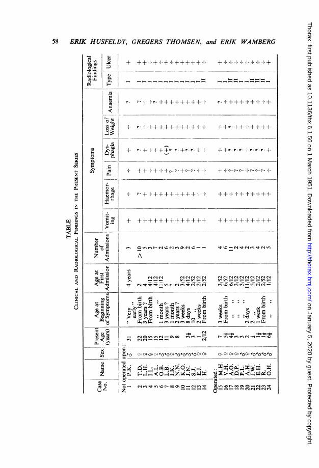
In addition to the four cases already published from the Queen Louise Hospitalfor Children in 1947 and one case from the Sundby Hospital in Copenhagen, wehave succeeded in collecting 19 cases of short oesophagus from the UniversityHospital in Copenhagen. The age and sex distribution will be seen in the table.As far as the older patients are concerned, the diagnoses were proved by perusalof the case reports and revision of the old radiographs. Of these 24 patients, lawere treated by surgical and 14 by conservative measures. The table sets out thepresent age of the patients, the time of onset of the disease, age upon first admission,number of admissions in the course of time, and, lastly, the most outstanding symp-toms. A perusal of the case histories leaves the following impression of the courseand symptoms of the disease.
SYMPTOMSAs a rule the symptoms manifest themselves immediately after birth or in the
course of the first month of life, rarely later and, if so, usually when the child beginsto take solid food. The most outstanding symptom is vomiting, which occurredin 100% of our series: it is nearly always periodical with intervening remissionsof a few weeks up to several months. During the attacks the child suffers fromdaily vomiting or spitting, usually during or shortly after meals. The vomitusis brought up without difficulty. (In one of the children it welled out of the mouthwhen he stood on his head.) The amount varies within wide limits and the vomitusis usually alimentary, slightly slimy, and not infrequently blood-tinged.
The symptom next in order of frequency is haemorrhage (in 70% of cases) inthe form of a slight bloody admixture in the vomitus or of regular haematemesisof extremely varying intensity. The latter is nearly always caused by the coexistentoesophagitis or a complicating peptic ulcer of the oesophagus. The haemorrhagesmay be alarming, and simple anaemia is no uncommon finding. Blood transfusionsmay be required.
on January 5, 2020 by guest. Protected by copyright.
http://thorax.bmj.com
/T
horax: first published as 10.1136/thx.6.1.56 on 1 March 1951. D
ownloaded from
58 ERIK HUSFELDT, GREGERS THOMSEN, and ERIK WAMBERG
.f_ J + +.1. .g + +1 + + + + + . ++1+11 1 1 1 1 1
. '.+ ++...I++ ++I ±.I .
E . .+.~.j + *++++++ + *1-++ -+ +
0.-
E~
I. ++'+++±±i++ + +++.I+.1.+++++
+ -++++±F-+++++ +++++.++++M !-
0
<CAC1~I~CIATh41
z E 1 > +- + + 1+
0 ~ ~ ~ ~ ~ ~ ~ ~ ~.0~~0C
___I. O\O_;i~~~~~~~~~~eeq7u en co elel
Z L Ei >, _ _ = -0 t tn s _en en m-==___=t
O~~~~~o-~j_noGL ; <t
C f- C0+oF C+Af ofo Ff0+fo "'-Of-o 0° ° 04- °4- C+°4- 0°+f f-0 fbI0b
z Z -2 iL°XO:0
-ma,0 Nm"t &n" -Ma D- e R
u1z .4 - _ o" - - ' NN"C0
on January 5, 2020 by guest. Protected by copyright.
http://thorax.bmj.com
/T
horax: first published as 10.1136/thx.6.1.56 on 1 March 1951. D
ownloaded from
HIATAL HERNIA AND SHORT OESOPHAGUS IN CHILDREN 59
In addition to these symptoms, older children may complain of dysphagia andpain occurring in varying intensity during meals and localized at the lower part ofthe sternum or high in the epigastrium. It is a characteristic feature in numerouscases that the pain is most marked in the horizontal position, subsiding or disappearingaltogether when the child is sitting. (One of our patients had formed the habit ofsleeping in the sitting position.) In addition to the pain, a few older children mayexhibit regular dysphagia, mostly for solids with marked periodicity, no doubtdue to exacerbation of the complicating oesophagitis and/or oesophageal ulcer.These patients, who have a feeling that the food is being held up in the oesophagus,are able to take solid food only in small portions which have to be thoroughlymasticated and swallowed with large amounts of fluid.
Such recurrent vomiting and pain, which do not infrequently make the childrefuse to eat, often result in considerable loss of weight, perhaps accompanied bya disturbance of the water balance and a poor general condition. The periodicityof the symptoms is an outstanding feature of the condition, and the patients haveusually been in hospital several times. The symptoms and the periodicity of thedisease are illustrated by the following typical case history.
CASE REPORT
M. H., a girl, was born on July 29, 1942. (University Hospital case record 890/47.)The vomiting set in at the age of 2 to 3 weeks. It was not profuse, but was at timesexplosive, and the vomitus was often blood-tinged. The condition remained unchangedduring the first year of life, after which it grew more periodical, as remissions of up to afortnight would intervene. At the age of 5 the condition grew much worse. The patientdeveloped daily vomiting, usually immediately after or exceptionally a few hours aftermeals. She failed to thrive, gradually lost weight, and suffered from dysphagia andsometimes from a feeling of being suffocated at meals. She was only able to take fluids.She had been in hospital four times, at 3 weeks, 8 months, 4 years, and 5 yearsof age. The first three times the treatment had been dietetic and symptomatic, but afterthe last admission she was transferred to the Ear, Nose, and Throat Department, whereshe obtained considerable relief from bouginage.
In April, 1949, the vomiting increased and the patient had a feeling that the food wasbeing held up behind the sternum. Bouginage was resumed. In June, 1949, transthoracicherniotomy was performed in the Department of Thoracic Surgery on the diagnosis of hiatalhernia of the oesophagus with oesophageal ulcer.
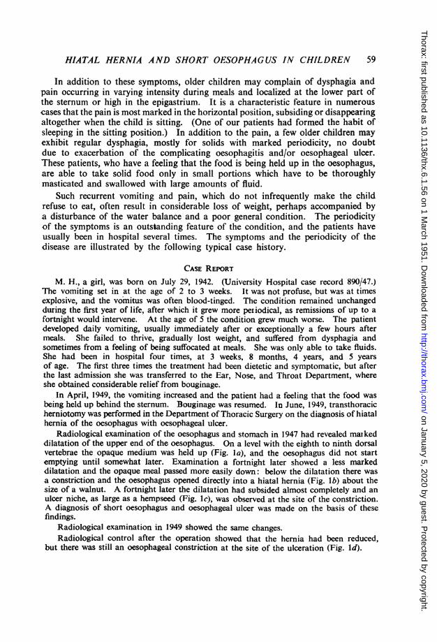
Radiological examination of the oesophagus and stomach in 1947 had revealed maikeddilatation of the upper end of the oesophagus. On a level with the eighth to ninth dorsalvertebrae the opaque medium was held up (Fig. Ia), and the oesophagus did not startemptying until somewhat later. Examination a fortnight later showed a less markeddilatation and the opaque meal passed more easily down: below the dilatation there wasa constriction and the oesophagus opened directly into a hiatal hernia (Fig. lb) about thesize of a walnut. A fortnight later the dilatation had subsided almost completely and anulcer niche, as large as a hempseed (Fig. lc), was observed at the site of the constriction.A diagnosis of short oesophagus and oesophageal ulcer was made on the basis of thesefindings.
Radiological examination in 1949 showed the same changes.Radiological control after the operation showed that the hernia had been reduced,
but there was still an oesophageal constriction at the site of the ulceration (Fig. Id).
on January 5, 2020 by guest. Protected by copyright.
http://thorax.bmj.com
/T
horax: first published as 10.1136/thx.6.1.56 on 1 March 1951. D
ownloaded from
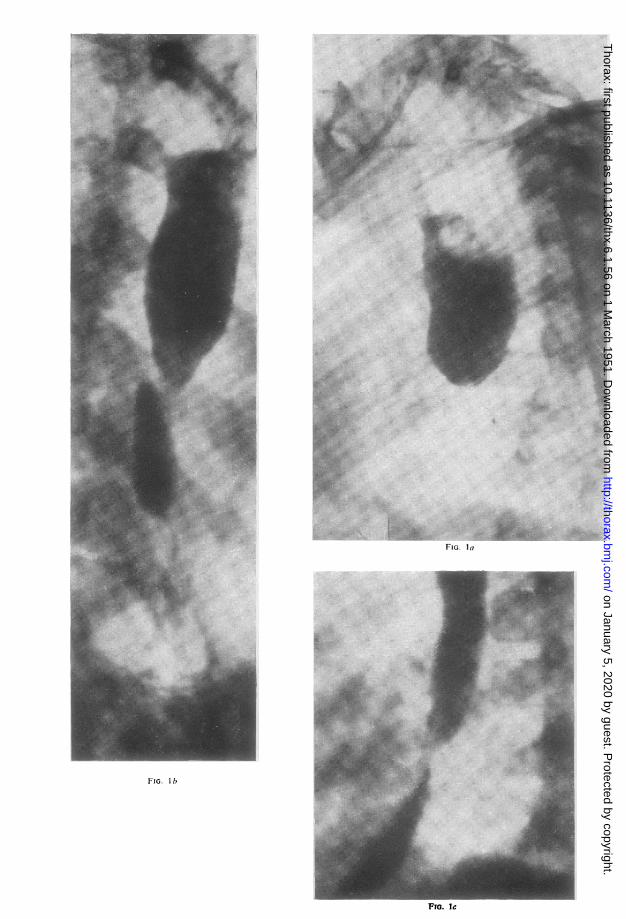
FIG. la
FIG. I b
Fra. Ic
Wt. .. .1. .... . W:
on January 5, 2020 by guest. Protected by copyright.
http://thorax.bmj.com
/T
horax: first published as 10.1136/thx.6.1.56 on 1 March 1951. D
ownloaded from
HIATAL HERNIA AND SHORT OESOPHAGUS IN CHILDREN 61
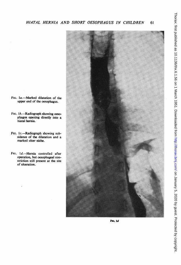
FIG. la.-Marked dilatation of theupper end of the oesophagus.
FIG. Ib.-Radiograph showing oeso-phagus opening directly into ahiatal hernia.
FIG. lc.-Radiograph showing sub-sidence of the dilatation and amarked ulcer niche.
FiG. Id.-Hernia controlled afteroperation, but oesophageal con-striction still present at the siteof ulceration.
FM. [d
on January 5, 2020 by guest. Protected by copyright.
http://thorax.bmj.com
/T
horax: first published as 10.1136/thx.6.1.56 on 1 March 1951. D
ownloaded from
62 ERIK HUSFELDT, GREGERS THOMSEN, and ERIK WAMBERG
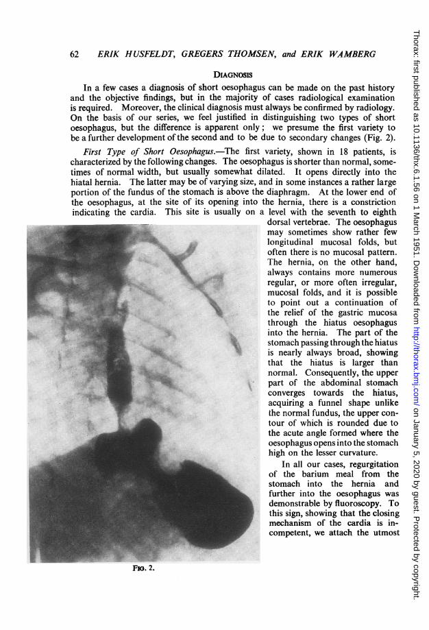
DiAGNOSISIn a few cases a diagnosis of short oesophagus can be made on the past history
and the objective findings, but in the majority of cases radiological examinationis required. Moreover, the clinical diagnosis must always be confirmed by radiology.On the basis of our series, we feel justified in distinguishing two types of shortoesophagus, but the difference is apparent only; we presume the first variety tobe a further development of the second and to be due to secondary changes (Fig. 2).
First Type of Short Oesophagus.-The first variety, shown in 18 patients, ischaracterized by the following changes. The oesophagus is shorter than normal, some-times of normal width, but usually somewhat dilated. It opens directly into thehiatal hernia. The latter may be of varying size, and in some instances a rather largeportion of the fundus of the stomach is above the diaphragm. At the lower end ofthe oesophagus, at the site of its opening into the hernia, there is a constrictionindicating the cardia. This site is usually on a level with the seventh to eighth
dorsal vertebrae. The oesophagusmay sometimes show rather fewlongitudinal mucosal folds, butoften there is no mucosal pattern.The hernia, on the other hand,always contains more numerousregular, or more often irregular,mucosal folds, and it is possibleto point out a continuation ofthe relief of the gastric mucosathrough the hiatus oesophagusinto the hernia. The part of thestomach passing through the hiatusis nearly always broad, showingthat the hiatus is larger thannormal. Consequently, the upperpart of the abdominal stomachconverges towards the hiatus,acquiring a funnel shape unlikethe normal fundus, the upper con-tour of which is rounded due tothe acute angle formed where theoesophagus opens into the stomachhigh on the lesser curvature.
In all our cases, regurgitationof the barium meal from thestomach into the hernia andfurther into the oesophagus wasdemonstrable by fluoroscopy. Tothis sign, showing that the closingmechanism of the cardia is in-competent, we attach the utmost
Fio. 2.
on January 5, 2020 by guest. Protected by copyright.
http://thorax.bmj.com
/T
horax: first published as 10.1136/thx.6.1.56 on 1 March 1951. D
ownloaded from
diagnostic significance. Massivereflux into the oesophagus ispresumably pathognomonic ofshort oesophagus. So far, it hasnot been possible to find thissign in normal children. A sharpdistinction must be made betweenregurgitation, which is a passivealteration of the distribution ofopaque medium, due partly tothe incompetent cardia, partly toexternal actions, and vomitingcaused by gastric contractions.In a few cases the reflux ofcontrast medium into the oeso-phagus may elicit vomiting. Insome children, regurgitation occursas soon as they lie down, but atother times not until they lie prone.Screening may show how the fill-ing of the oesophagus varies syn-chronously with the respirationdue to alterations in the intra-thoracic pressure. Similarly, anincrease in the intra-abdominalpressure caused by compressionof the abdomen may result in in-creased flow into the oesophagus.Other children have to be placedin Trendelenburg's position to fillthe hernia as completely as pos- FIG. 3sible. By tilting the table, theregurgitation is sometimes clearly illustrated, and screening shows how the opaquemedium in the oesophagus, hernia, and stomach follows the movements of the table.
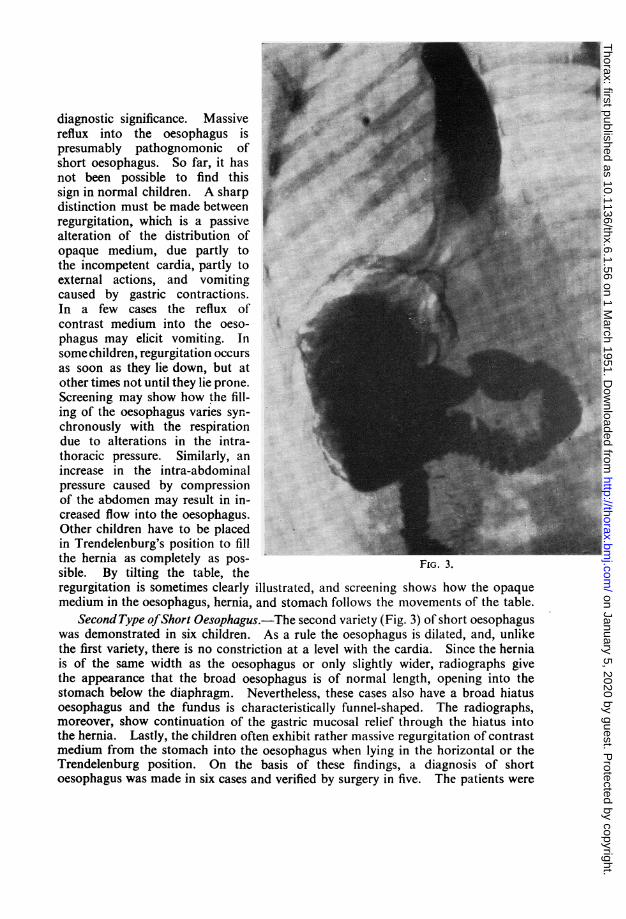
Second Type ofShort Oesophagus.-The second variety (Fig. 3) of short oesophaguswas demonstrated in six children. As a rule the oesophagus is dilated, and, unlikethe first variety, there is no constriction at a level with the cardia. Since the herniais of the same width as the oesophagus or only slightly wider, radiographs givethe appearance that the broad oesophagus is of normal length, opening into thestomach below the diaphragm. Nevertheless, these cases also have a broad hiatusoesophagus and the fundus is characteristically funnel-shaped. The radiographs,moreover, show continuation of the gastric mucosal relief through the hiatus intothe hernia. Lastly, the children often exhibit rather massive regurgitation of contrastmedium from the stomach into the oesophagus when lying in the horizontal or theTrendelenburg position. On the basis of these findings, a diagnosis of shortoesophagus was made in six cases and verified by surgery in five. The patients were
on January 5, 2020 by guest. Protected by copyright.
http://thorax.bmj.com
/T
horax: first published as 10.1136/thx.6.1.56 on 1 March 1951. D
ownloaded from
64 ERIK HUSFELDT, GREGERS THOMSEN, and ERIK WAMBERG
2 months, 18 months, 18 months, 21 months, 3 years, and 41 years of age respectively.The symptoms found in tliis group did not differ from those in the former group.It was not possible to demonstrate a peptic ulcer. This group may be only a precursorof the first, and as yet we have been unable to produce proof, since we have nothad the opportunity to follow the course of the disease in these children.
In 1947 Neuhauser and Berenberg described a syndrome in which the radiologicalchanges closely resembled this second variety of short oesophagus. In 12 infantswith persisting or recurrent vomiting they found a wide oesophagus, narrowing onlyslightly at the site of its passage through the hiatus. The cardial sphincter didnot function at all, and in the horizontal position regurgitation of opaque mediumwas seen from the stomach into the oesophagus. Neuhauser and Berenberg inter-preted the condition as a relaxation of the cardial sphincter due to a disturbance ofthe parasympathetic-sympathetic balance which regulates its function. Theystated that they had had no opportunity to investigate the correctness of this theoryat necropsy. They succeeded in controlling the symptoms by making the babiessit during and after meals and, in particularly pronounced cases, letting them assumea semi-sitting position day and night. At follow-up examination several monthslater even the radiological changes had subsided. If these cases are to be inter-preted as short oesophagus, as indicated by the films, the treatment may haveproduced a spontaneous reduction of the hernia.
COMPLICATIONSShort oesophagus is often complicated by oesophagitis or oesophagitis and
peptic ulcer of the oesophagus due to the action of gastric acid upon the oesophagealmucosa. In oesophagitis the mucosal relief is irregular or indistinct.
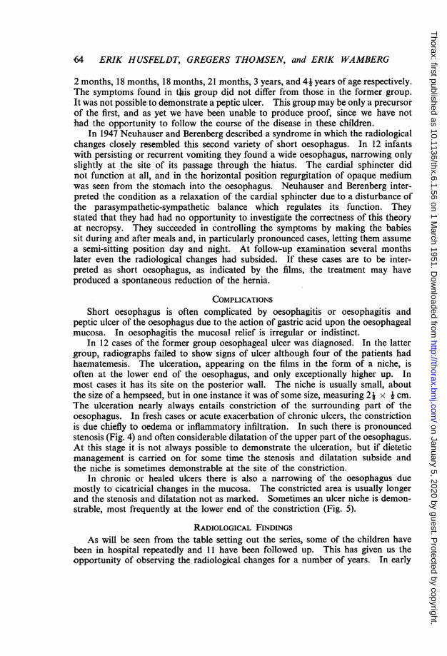
In 12 cases of the former group oesophageal ulcer was diagnosed. In the lattergroup, radiographs failed to show signs of ulcer although four of the patients hadhaematemesis. The ulceration, appearing on the films in the form of a niche, isoften at the lower end of the oesophagus, and only exceptionally higher up. Inmost cases it has its site on the posterior wall. The niche is usually small, aboutthe size of a hempseed, but in one instance it was of some size, measuring 2j x a cm.The ulceration nearly always entails constriction of the surrounding part of theoesophagus. In fresh cases or acute exacerbation of chronic ulcers, the constrictionis due chiefly to oedema or inflammatory infiltration. In such there is pronouncedstenosis (Fig. 4) and often considerable dilatation of the upper part of the oesophagus.At this stage it is not always possible to demonstrate the ulceration, but if dieteticmanagement is carried on for some time the stenosis and dilatation subside andthe niche is sometimes demonstrable at the site of the constriction.
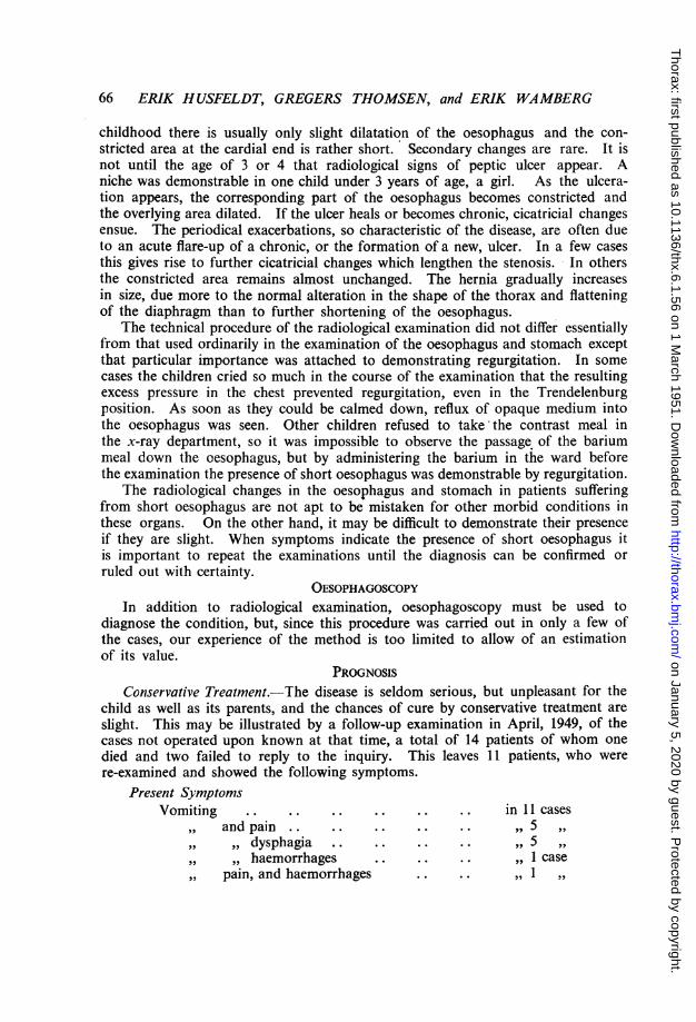
In chronic or healed ulcers there is also a narrowing of the oesophagus duemostly to cicatricial changes in the mucosa. The constricted area is usually longerand the stenosis and dilatation not as marked. Sometimes an ulcer niche is demon-strable, most frequently at the lower end of the constriction (Fig. 5).
RADIOLOGICAL FINDINGSAs will be seen from the table setting out the series, some of the children have
been in hospital repeatedly and 11 have been followed up. This has given us theopportunity of observing the radiological changes for a number of years. In early
on January 5, 2020 by guest. Protected by copyright.
http://thorax.bmj.com
/T
horax: first published as 10.1136/thx.6.1.56 on 1 March 1951. D
ownloaded from

FIG. 4.-Marked dilatation of theupper end of the oesophagus ina case of ulcer.
FiG. 5.-Stenosis ofthe oesophagus(arrow) in acase of chroniculcer.
on January 5, 2020 by guest. Protected by copyright.
http://thorax.bmj.com
/T
horax: first published as 10.1136/thx.6.1.56 on 1 March 1951. D
ownloaded from
66 ERIK HUSFELDT, GREGERS THOMSEN, and ERIK WAMBERG
childhood there is usually only slight dilatation of the oesophagus and the con-stricted area at the cardial end is rather short. Secondary changes are rare. It isnot until the age of 3 or 4 that radiological signs of peptic ulcer appear. Aniche was demonstrable in one child under 3 years of age, a girl. As the ulcera-tion appears, the corresponding part of the oesophagus becomes constricted andthe overlying area dilated. If the ulcer heals or becomes chronic, cicatricial changesensue. The periodical exacerbations, so characteristic of the disease, are often dueto an acute flare-up of a chronic, or the formation of a new, ulcer. In a few casesthis gives rise to further cicatricial changes which lengthen the stenosis. In othersthe constricted area remains almost unchanged. The hernia gradually increasesin size, due more to the normal alteration in the shape of the thorax and flatteningof the diaphragm than to further shortening of the oesophagus.
The technical procedure of the radiological examination did not differ essentiallyfrom that used ordinarily in the examination of the oesophagus and stomach exceptthat particular importance was attached to demonstrating regurgitation. In somecases the children cried so much in the course of the examination that the resultingexcess pressure in the chest prevented regurgitation, even in the Trendelenburgposition. As soon as they could be calmed down, reflux of opaque medium intothe oesophagus was seen. Other children refused to take the contrast meal inthe x-ray department, so it was impossible to observe the passage of the bariummeal down the oesophagus, but by administering the barium in the ward beforethe examination the presence of short oesophagus was demonstrable by regurgitation.
The radiological changes in the oesophagus and stomach in patients sufferingfrom short oesophagus are not apt to be mistaken for other morbid conditions inthese organs. On the other hand, it may be difficult to demonstrate their presenceif they are slight. When symptoms indicate the presence of short oesophagus itis important to repeat the examinations until the diagnosis can be confirmed orruled out with certainty.
OESOPHAGOSCOPYIn addition to radiological examination, oesophagoscopy must be used to
diagnose the condition, but, since this procedure was carried out in only a few ofthe cases, our experience of the method is too limited to allow of an estimationof its value.
PROGNOSISConservative Treatment.-The disease is seldom serious, but unpleasant for the
child as well as its parents, and the chances of cure by conservative treatment areslight. This may be illustrated by a follow-up examination in April, 1949, of thecases not operated upon known at that time, a total of 14 patients of whom onedied and two failed to reply to the inquiry. This leaves 11 patients, who werere-examined and showed the following symptoms.
Present SymptomsVomiting .. .. .. .. .. .. in 11 cases
and pain .. .. .. . . ,, 5,
dysphagia .. .. .. ,, 5,,, ,, haemorrhages .. .. .. ,, 1 case
pain, and haemorrhages .. .. ,, I
on January 5, 2020 by guest. Protected by copyright.
http://thorax.bmj.com
/T
horax: first published as 10.1136/thx.6.1.56 on 1 March 1951. D
ownloaded from
HIATAL HERNIA AND SHORT OESOPHAG US IN CHILDREN
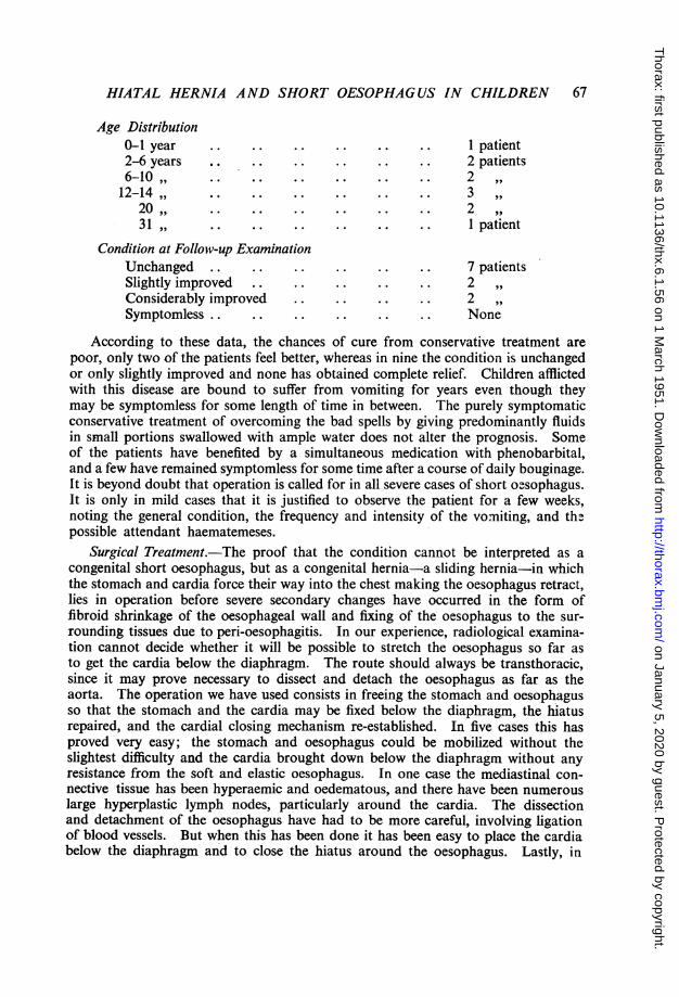
Age Distribution0-1 year .. .. .. 1 patient2-6 years .. .. 2 patients6-10, .. .. .. . . . 2 ,12-14, .. . .. . .. . 3 ,
20, .. . .. . .. . 2 ,331 ,, .I.. . . . patient
Condition at Follow-up ExaminationUnchanged .. .. .. .. .. .. 7 patientsSlightly improved .. .. .. .. 2Considerably improved .. .. .. 2Symptomless .. .. .. .. .. .. None
According to these data, the chances of cure from conservative treatment arepoor, only two of the patients feel better, whereas in nine the condition is unchangedor only slightly improved and none has obtained complete relief. Children afflictedwith this disease are bound to suffer from vomiting for years even though theymay be symptomless for some length of time in between. The purely symptomaticconservative treatment of overcoming the bad spells by giving predominantly fluidsin small portions swallowed with ample water does not alter the prognosis. Someof the patients have benefited by a simultaneous medication with phenobarbital,and a few have remained symptomless for some time after a course of daily bouginage.It is beyond doubt that operation is called for in all severe cases of short o3sophagus.It is only in mild cases that it is justified to observe the patient for a few weeks,noting the general condition, the frequency and intensity of the vo-miting, and thapossible attendant haematemeses.
Surgical Treatment.-The proof that the condition cannot be interpreted as acongenital short oesophagus, but as a congenital hernia-a sliding hernia-in whichthe stomach and cardia force their way into the chest making the oesophagus retract,lies in operation before severe secondary changes have occurred in the form offibroid shrinkage of the oesophageal wall and fixing of the oesophagus to the sur-rounding tissues due to peri-oesophagitis. In our experience, radiological examina-tion cannot decide whether it will be possible to stretch the oesophagus so far asto get the cardia below the diaphragm. The route should always be transthoracic,since it may prove necessary to dissect and detach the oesophagus as far as theaorta. The operation we have used consists in freeing the stomach and oesophagusso that the stomach and the cardia may be fixed below the diaphragm, the hiatusrepaired, and the cardial closing mechanism re-established. In five cases this hasproved very easy; the stomach and oesophagus could be mobilized without theslightest difficulty and the cardia brought down below the diaphragm without anyresistance from the soft and elastic oesophagus. In one case the mediastinal con-nective tissue has been hyperaemic and oedematous, and there have been numerouslarge hyperplastic lymph nodes, particularly around the cardia. The dissectionand detachment of the oesophagus have had to be more careful, involving ligationof blood vessels. But when this has been done it has been easy to place the cardiabelow the diaphragm and to close the hiatus around the oesophagus. Lastly, in
67
on January 5, 2020 by guest. Protected by copyright.
http://thorax.bmj.com
/T
horax: first published as 10.1136/thx.6.1.56 on 1 March 1951. D
ownloaded from

68 ERIK HUSFELDT, GREGERS THOMSEN, and ERIK WAMBERG
two cases the mediastinal connective tissue has been the seat of widespread fibroidchanges fixing the oesophagus to the surrounding tissues. The oesophageal wallproper has undergone fibroid changes and the lumen narrowed. Moreover, thereare cases of large peptic ulcers, and we have encountered two. In one the ulcer hadencroached upon the aortic wall and had to be removed by sharp dissection togetherwith a piece of the adventitia of the vessel.
It may be difficult to decide whether the oesophageal constriction is due to spasmor fibroid changes. In one instance it was due essentially to pressure from lymphnodes and peri-oesophagitic strands. In these cases it has been necessary to freethe stomach and the oesophagus from the diaphragm as far as the site where theoesophagus disappears behind the aorta. The oesophagus has had to be dissectedcarefully, from the inferior left pulmonary vein and the left main bronchus. It isimportant to detach the oesophagus as far as the aorta and pull the oesophagusfar forward in the hiatus, thus reaching the summit of the dome of the diaphragm.In that way 3 cm. can be gained.
After the detachment it has been possible to pull the cardia down in all casesbut two. In one of them it would no doubt have been possible with our presentexperience: in the other oesophago-gastrectomy was required because of a pene-trating peptic ulcer.
Operative Technique.-The anaesthetic was intratracheal nitrous oxide-oxygenether. In no case did tracheotomy become necessary because of glottic oedema.A postero-lateral incision was made on the left side, resecting the eighth rib.
After dividing the mediastinal pleura and detaching the stomach and oesophagus,the hernial sac was opened, the peritoneum attaching to the stomach being cutthrough. Thereupon the diaphragmatic crura are dissected, revealing the red bundlesof muscle, especially posteriorly, where they converge into a V-shaped figure. Asilk suture is passed through the crura anteriorly in the hiatus, including the oeso-phageal wall, after which the hiatus is closed from behind with three to four silksutures, the last of which includes the oesophageal wall. Moreover,- a few silksutures are passed through the crura and the oesophageal wall.
As a safety measure, a tube may be passed into the stomach and left in placeduring the process of suturing. The chest wall is closed with silk sutures and aPezzer catheter inserted into the chest and left there for 24 hours.
Results of Surgical Treatment.-One patient had a penetrating ulcer and requiredoesophago-gastrectomy. Now, he really has a too-short oesophagus affording apossibility of regurgitation, and the prognosis is doubtful, although vagotomy hasbeen performed.
One patient underwent operation, but at that time it was not considered possibleto place the cardia below the diaphragm. This patient is still suffering fromregurgitation and haematemeses and is to be admitted for another operation.
In eight cases herniotomy was performed on the hiatus and the cardia was fixedbelow the diaphragm.
Follow-up examinations from 15 to 2 months after the operation showed thatseven patients were in excellent health without symptoms, whereas one had arecurrence and probably will need another operation.
on January 5, 2020 by guest. Protected by copyright.
http://thorax.bmj.com
/T
horax: first published as 10.1136/thx.6.1.56 on 1 March 1951. D
ownloaded from
HIATAL HERNIA AND SHORT OESOPHAG US IN CHILDREN 69
CONCLUSIONThe numbers involved are small and the follow-up period short, but according
to our experience a congenital short oesophagus does not exist or is extremelyrare. These patients are suffering from a congenital hiatal hernia. Before operationit is impossible to know whether the cardia can be brought below the diaphragm.It is difficult to decide whether a narrowing of the oesophagus is due to spasm orstricture. The approach must be through the thorax.
In view of improved anaesthetic technique and thoracic operations in earlychildhood and the low mortality which they carry, all cases of so-called " congenitalshort oesophagus" should be submitted to operation, if possible before secondarychanges, such as shrinkage and fixation of the oesophagus, and peptic ulcer arecaused by the regurgitation. The operation is then a simple procedure which relievesthe patient of an extremely unpleasant and in some cases fatal disease.
SUMMARY"Brachyoesophagus," or congenital short oesophagus as it has been called, is
now interpreted as a congenital hernia through the hiatus oesophagus with simul-taneous shortening of the oesophagus. The anomaly was first described in 1918,and from 1930 it has been diagnosed in ever-increasing numbers, the number ofreported cases being about 100. We have diagnosed 24 cases, 14 of which wererecognized after 1947.
According to Allison (1948), the primary factor in adults is a hiatal hernia, andregurgitation of gastric contents produces oesophagitis and perhaps peptic ulcer of theoesophagus. Cicatricial changes contribute to shortening and fixing the oesophagus.
As a rule the symptoms set in immediately after birth, most commonly in theform of vomiting, nearly always periodical. Further symptoms are haemorrhage,either in the form of a slightly blood-tinged vomitus or of actual haematemesis,loss of weight, dysphagia, pain, anaemia, and in some cases dehydration.
The differential diagnostic possibilities are: hyper- or hypo-galactia, congenitalpyloric stenosis, atresia of the oesophagus and duodenal stenosis, and in olderchildren gastric lesions, especially gastric or duodenal ulcer.
The diagnosis is confirmed by radiological examination after administering anopaque medium. It is important to examine the patient in the Trendelenburgposition to demonstrate regurgitation. By this procedure it is possible to demon-strate two types of lesion. (1) A short and somewhat dilated oesophagus, openingdirectly into the hiatal hernia with a constriction at the site of transition. Thisis the most common variety and probably a further development of Type 2. (2) Adilated oesophagus without constrictions. The hernia is of the same width as theoesophagus, which appears to be of normal length and to open into the stomachbelow the diaphragm. The possibility of spontaneous reduction may be demonstrated.
The condition is often complicated by " oesophagitis " or by peptic ulcer ofthe oesophagus, more rarely by perforation. In most cases the disease runs aprolonged course, characterized by intermittent, ever-recurring vomiting.
In 11 patients who had been treated conservatively, the condition had remainedcompletely unchanged in seven. Two showed slight and two marked improvement,but none was symptomless, and all were still troubled by vomiting.
on January 5, 2020 by guest. Protected by copyright.
http://thorax.bmj.com
/T
horax: first published as 10.1136/thx.6.1.56 on 1 March 1951. D
ownloaded from

70 ERIK HUSFELDT, GREGERS THOMSEN, and ERIK WAMBERG
Conservative medical management must be abandoned, and early operationrecommended. At operation, the stomach and oesophagus are freed, so that thestomach and cardia may be fixed below the diaphragm. At the same time hernio-tomy is performed, and, after isolation of the diaphragmatic crura, sphincter functionis re-established by sutures through the crura and the oesophageal wall.
Follow-up examination of 10 operated cases from 15 to 2 months after theoperation revealed that seven patients were symptom-free and only one had recur-rence. In the remaining two cases the operation was technically impracticable.
REFERENCES
Akerlund, Ake (1926). Acta radiol., Stockh., 6, 3.Allison, P. R. (1948). Thorax, 3, 20.Neuhauser, E. B. D., and Berenberg, W. (1947). Radiology, 48, 480.Olsen, A. M., and Harrington, S. W. (1948). J. thorac. Surg., 17, 189.Thomsen, G. (1949). Acta radiol., Stockh., 32, 193.Wamberg, E. (1947). Acta paediatr., Stockh., 34, 293.
on January 5, 2020 by guest. Protected by copyright.
http://thorax.bmj.com
/T
horax: first published as 10.1136/thx.6.1.56 on 1 March 1951. D
ownloaded from