hhs public access derek c. medellina roaa m. kassim n3 and
TRANSCRIPT
Microtubule-Targeting 7-Deazahypoxanthines Derived from Marine Alkaloid Rigidins: Exploration of the N3 and N9 Positions and Interaction with Multi-Drug Resistance Proteins
Ramesh Dasaria,1, Andrzej Błaużb,1, Derek C. Medellina, Roaa M. Kassimc, Carlos Vierad, Maximo Santarosad, Danielle Turnerd, Alet E. van der Westhuyzene, Willem A. L. van Otterloe, Taryn Olivasa, Tugba Yildizf, Tania Betancourta,f, Charles B. Shusterc, Snezna Rogeljd, Błażej Rychlikb,*, Todd Hudnalla,*, Liliya V. Frolovad,*, and Alexander Kornienkoa,*
[a]Department of Chemistry and Biochemistry, Texas State University, San Marcos, Texas 78666, (USA) [b]Cytometry Lab, Department of Molecular Biophysics, Faculty of Biology and Environmental Protection, University of Łódź, ul. Pomorska 141/143, 90-236 Łódź, (Poland) [c]Department of Biology, New Mexico State University, Las Cruces, NM 88003, USA [d]Departments of Chemistry and Biology, New Mexico Tech, Socorro, NM 87801, USA [e]Department of Chemistry and Polymer Science, University of Stellenbosch, ZA-7602 Stellenbosch, South Africa [f]Materials Science and Engineering Program, Texas State University, San Marcos, Texas 78666
Abstract
Our laboratories have been investigating synthetic analogues of marine alkaloid rigidins
possessing promising anticancer activities. These analogues, based on the 7-deazahypoxanthine
skeleton are available in one- or two-step synthetic sequences and exert cytotoxicity by disrupting
microtubule dynamics in cancer cells. In the present work we extended the available structure-
activity relationship data to N3- and N9-substituted derivatives. Although the N3-substitution
results in a loss of activity, the N9-substituted compounds retain the nanomolar antiproliferative
activities and antitubulin mode of action of the original unsubstituted compounds. Furthermore,
work reported herein also demonstrated that MDR proteins do not confer resistance to both N9-
unsubstituted and substituted compounds. It was found that sublines overexpressing ABCG2,
ABCC1 and ABCB1 proteins were equally responsive to the rigidin analogues as their parental
cell lines. Thus, the study reported herein provides further impetus to investigate the rigidin-
inspired 7-deazahypoxanthines as promising anticancer agents.
Graphical Abstract
*Co-corresponding authors.1Co-first authors
HHS Public AccessAuthor manuscriptChemMedChem. Author manuscript; available in PMC 2020 February 05.
Published in final edited form as:ChemMedChem. 2019 February 05; 14(3): 322–333. doi:10.1002/cmdc.201800658.
Author M
anuscriptA
uthor Manuscript
Author M
anuscriptA
uthor Manuscript

Keywords
alkaloids; antitumor agents; 7-deazapurines; drug discovery; pyrrolo[2,3-d]pyrimidines
Introduction
Alkaloids isolated from various marine organisms have been found to possess various
biological activities and in many cases promising anticancer properties.[1–4] For example,
trabectedin (Yondelis®) isolated from the sea squirt Ecteinascidia turbinata is currently used
to treat patients with advanced soft tissue sarcoma and recurrent platinum-sensitive ovarian
cancer in combination therapy.[5–7] Recently, it was also approved by the FDA for the
treatment of liposarcoma and leiomyosarcoma as the second line of therapy.[8] The
development of Yondelis® had to overcome major hurdles associated with low natural
abundance and lengthy synthetic preparations.[9] One approach to solve the supply problem
is the discovery of analogues that can be accessed via short synthetic procedures.[10]
Using this strategy, our studies of synthetic analogues of marine alkaloid rigidins A, B, C, D
(Figure 1), isolated in minute amounts from the tunicate Eudistoma cf. rigida found near
Okinawa and New Guinea,[11–14] revealed compounds with potent antiproliferative activities
when the 7-deazaxanthine scaffold associated with the rigidins was replaced by the 7-
deazahypoxanthine variant through the removal of the carbonyl at C2, such as in 1 and 2 (Figure 1).[15–18] These compounds resulted from systematic investigation of the rigidin-
related scaffold A (top right corner in Figure 1) and they were found to target microtubule
network in cancer cells.[15–18] Compound 2 also exhibited promising in vivo efficacy in a
human colon cancer mouse model at doses as low as 2-3 mg/kg.[18] Compound 1 is
accessible via a one-step multicomponent reaction from commercially available starting
materials, whereas the synthesis of 2 was found to work best when it was split into two steps
(Figure 1).[16–18] As a logical progression of this work, we explore here the analogues
bearing substituents at positions N3 and N9 (Figure 1).
As part of preclinical evaluation of the rigidin-based anticancer agents, we also investigated
their interaction with multi-drug resistance proteins. Often, tumors initially respond to
chemotherapy but subsequently become refractory to the continuing treatment. Such
resistance is known as “acquired” and it is commonly based on the development of a multi-
drug resistant phenotype (MDR) impacting a wide range of structurally and mechanistically
distinct antitumor agents.[19,20] MDR has plagued conventional cancer drug therapies, for
example the widely used microtubule-disrupting agents such as the Vinca alkaloids[21] and
taxanes.[22] Thus, we thoroughly evaluated whether our rigidin-based anticancer agents,
Dasari et al. Page 2
ChemMedChem. Author manuscript; available in PMC 2020 February 05.
Author M
anuscriptA
uthor Manuscript
Author M
anuscriptA
uthor Manuscript

whose mode of action is also based on microtubule targeting, are capable of circumventing
MDR mechanisms and can combat drug-resistant cancers.
Results and Discussion
Synthesis of N3- and N9-substituted 7-deazahypoxanthines
Although there are four potential nucleophilic sites in the deprotonated forms of 1 and 2,
namely N1, N3, N9 and O6, we found that these molecules react exclusively at N9. This
finding was confirmed with an x-ray structure (Figure 2) of N9-t-butoxycarbonyl derivative
of 1 (compound 3 in Scheme 1A) obtained by the treatment of 1 with NaH and (Boc)2O.
Methylation of 3 at N3 and removal of the Boc group then furnished mono-methylated
compound 5. Alternatively, N9-methylated 6 was obtained by a direct reaction of 1 with
NaH and MeI (Scheme 1A). N9-sulfonylated compound 9 was obtained by a different route
involving a multicomponent condensation of benzaldehyde, malononitrile and
sulfonamidoacetophenone 7 (Scheme 1B). The obtained pyrrole 8 was then cyclized by
refluxing in formic acid, which was used as a solvent and reactant at the same time.
In our efforts to synthesize N-substituted analogues of 2, we were able to obtain sulfonylated
derivative 10 by a direct treatment of 2 with NaH and MsCl (Scheme 2). Compound 2 also
gave N9-Boc derivative 11 when reacted with NaH and (Boc)2O. Subsequent N3-
methylation and removal of Boc then gave mono-methylated 12. In contrast to direct
methylation of 1, similar conditions applied to 2 gave N3,N9-dimethyl derivative 14 along
with expected 13.
Structure-Activity Relationship studies
The synthesized analogues were evaluated for antiproliferative activities using the HeLa cell
line as a model for human cervical adenocarcinoma and MCF-7 cells as a model for breast
adenocarcinoma. The cells were treated with each compound for 48 h, and cell viability was
assessed using the MTT method[23] (Table 1). We found that analogues containing single
large substituents at N9 were equally or more potent than the original unsubstituted
compounds. These included N9-t-butoxycarbonyl compounds 3 and 11 as well as N9-
methylsulfonyl analogues 9 and 10. In contrast, compounds containing a smaller N9 group,
such as methyl in 6 and 13 were significantly less potent. Finally, the N3-substituted
analogues 5, 12 and 14 were totally inactive. In principle, these studies involving the
variations in the N-substituents are consistent with our earlier SAR work, which showed that
the modifications of the pyrimidine ring were not tolerated, whereas the pyrrole unit was a
useful site for optimizing potency in this series of compounds. The present SAR results are
similar between the 1- and 2-derived series of compounds indicating that the binding poses
must be similar for C2-H or C2-alkyl structures.[18]
Inhibition of tubulin assembly
To exclude the possibility that the mode action was not inadvertently changed when the
bulky substituents were added to the N9-position, the effects of these compounds on in vitro tubulin polymerization were evaluated.[24] In this assay, polymerization is monitored by the
Dasari et al. Page 3
ChemMedChem. Author manuscript; available in PMC 2020 February 05.
Author M
anuscriptA
uthor Manuscript
Author M
anuscriptA
uthor Manuscript

increase of fluorescence due to the incorporation of a fluorescent reporter, 4’,6-diamidino-2-
phenylindole (DAPI), into growing microtubules. As a control, a known microtubule
stabilizer paclitaxel induced potent enhancement of microtubule formation relative to the
effect of the DMSO control (Figure 3A and B). Consistent with our previous results
compounds 1 and 2 suppressed tubulin polymerization. In a similar manner, tubulin
polymerization was inhibited by N9-t-butoxycarbonyl compounds 3 and 11 (Figure 3A and
B), suggesting that the introduction of bulky groups at N9 of both 1 and 2 does not result in
a change in mechanism of action.
Effects on microtubule organization in cells
Additional confirmation of tubulin targeting as the mechanism responsible for anticancer
properties of the compounds in Table 1 came from studying their effects on the microtubule
cytoskeleton in intact cells. To this end, cultured HeLa cells were treated with the original
compounds 1 and 2 as well as their potent N9-substituted derivatives 3, 9 and 11 at 50 nM,
concentration relevant to their antiproliferative effects on HeLa cells (Table 1). Examination
of interphase (Figure 4, top row) and mitotic (Figure 4, bottom row) cells revealed that while
there were no pronounced effects on interphase microtubules, all compounds had marked
effects on spindle morphology and chromosome alignment. While most cells established
bipolar spindles, astral microtubules were frequently missing from one spindle pole,
possibly due to the inherent asymmetries between centrosomes.[25] Most notably, treated
cells invariably arrested in prometaphase with chromosomes clustered around the spindle
poles, suggesting an inability to congress to the metaphase plate.
Examination of cells treated with compounds 2 and 11 over a range of doses revealed that
effects on chromosome congression and spindle morphology were dose-dependent (Figure
5), with no noticeable effects on interphase microtubules, even at 100 nM (Figure 5B and C,
top rows). In cells treated at concentrations near the GI50 (25 nM), overall spindle
morphology was normal, but there were still chromosomes clustered at the spindle poles
with syntelic attachments (Figure 5B and C, bottom rows). Syntelic chromosome
attachments (where both sister chromatids are attached to the same spindle pole) activate the
spindle assembly checkpoint, resulting in mitotic arrest and eventual cell death.[26] In light
of the minimal effects on interphase microtubules (Figures 4 and 5) and the known
requirement of microtubule dynamics for proper spindle assembly, chromosome alignment
and segregation,[27] it is likely that the observed effects on growth inhibition are due to
inability of treated cells to establish proper bioriented chromosome attachments at the
metaphase plate rather than a gross disruption of the mitotic spindle.
Interaction with multi-drug resistance proteins
As mentioned in the introduction, “acquired” resistance is commonly based on the
development of an MDR phenotype impacting a wide range of structurally and
mechanistically distinct antitumor agents. This type of resistance is frequently mediated by
MDR proteins belonging to the vast superfamily of ABC transporters.[19,20] Due to their
ubiquitous tissue and organ distribution and extremely low specificity, they are able to
prevent many substances from entering the target cell. Moreover, they play an important role
in constituting the biochemical base of epithelial barriers, thus protecting vital organs
Dasari et al. Page 4
ChemMedChem. Author manuscript; available in PMC 2020 February 05.
Author M
anuscriptA
uthor Manuscript
Author M
anuscriptA
uthor Manuscript

located behind such barriers (e.g. brain, testes or fetal organs) against drugs or other
xenobiotics. Major drug regulatory agencies, e.g. the Food and Drug Administration, or
European Medicines Agency, require that the role of major MDR proteins be taken into
account. Thus, we assessed the potential role of ABCB1 (known also as MDR1 or Pgp),
ABCC1 (known also as MRP1) and ABCG2 (known also as BCRP) in conferring resistance
to rigidin analogues as well as potential modulatory effects of rigidin derivatives on selected
MDR proteins.
First, we studied whether the cytotoxic effects of the rigidin analogues depended on different
MDR protein profiles. We chose to work with colon cancer cells, as compound 2 showed
highly promising results in earlier in vivo experiments involving a colon cancer xenograft
model[18] and a panel of drug-resistant daughter lines of SW620 (human colorectal
adenocarcinoma) cells were obtained in our laboratories previously.[28] Specifically,
SW620C cells overexpress ABCG2; SW620E and SW620M cell lines are characterized by
overexpression of ABCC; SW620D, SW620E, and SW620V cells exhibit high expression
levels of ABCB1. We also included MDCKII (canine renal epithelium) cell line and
hABCG2- and hABCB1-transfected sublines (MDCKII-BCRP and MDCKII-MDR1,
respectively), to determine whether the two cell sets of different origin and basic metabolic
activity display different sensitivities to the rigidin analogues.
Analysis of results obtained with the neutral red accumulation assay[29] and presented in
Table 2 and Table 3 reveals that none of MDR proteins confers resistance to both the parent
compounds 1 and 2 as well as their N9-t-butoxycarbonyl derivatives 3 and 11. Toxicity of
these substances is generally lower for SW620 cells (in ten to hundred nanomolar range,
Table 2) than for MDCKII cells (between 14 and 24 nM, Table 3). The SW620 and ABCC1-
overexpressing SW620M cell lines appear to be more sensitive to compound 11 than other
lines, which could suggest some involvement of ABCB1 in its detoxication as virtually no
ABCB1 is expressed in this cell line. However, no such tendency is seen in the MDCKII
panel.
Despite negative results of cytotoxicity assay, we proceeded to determine if the rigidin
analogues added at a low and virtually non-toxic concentration (10 nM) were able to
modulate a well-defined resistance to some model cytotoxic drugs. We chose mitoxantrone
as a model substrate for human ABCG2[30] and vincristine as a model substrate for ABCB1.[31] The resistance factors (IC50 ratio for resistant and sensitive cells for a given drug)
observed in our system for these drugs were 8 for mitoxantrone and ABCG2 and 38 for
vincristine and ABCB1 (see Table 4). If sensitizing effects (i.e., significant reduction of
resistance factor) were observed, it would suggest direct interactions of rigidin derivatives
with MDR proteins. However, as seen in Table 4, no such results were detected. 10 µM
verapamil (Ver) and 10 µM Ko143 were used as positive controls (both compounds are
specific inhibitors of ABCB1 and ABCG2, respectively). Values observed for investigated
rigidin analogues did not differ significantly from controls (the confidence intervals fairly
overlapped). Therefore, it can be induced that rigidins do not exert any long-term effects on
MDR protein expression or function.
Dasari et al. Page 5
ChemMedChem. Author manuscript; available in PMC 2020 February 05.
Author M
anuscriptA
uthor Manuscript
Author M
anuscriptA
uthor Manuscript

As it is possible that rigidin analogues undergo conversion to metabolites that do not interact
with MDR proteins, we finally determined whether there is any direct interaction between
investigated compounds and transporters in a real-time transport assay.[32] We employed
three different assays specific for a given protein of interest – calcein accumulation assay for
determination of ABCB1 activity,[33] modified BCECF extrusion assay[34] for ABCC1
assessment and pheophorbide a accumulation to measure ABCG2 function[35] (Table 5). In
this case, despite Ver and Ko143 as inhibitors of ABCB1 and ABCG2, respectively, MK571
was used as a specific inhibitor of ABCC1. As evident from Table 5, no direct interaction
between investigated rigidin analogues and MDR proteins exists, as none of the compounds
is able to inhibit transporter activity even partially. Summing up all results presented in
Tables 2 – 5, rigidins exhibit no modulatory activity on MDR protein function. Taking into
account also their antiproliferative properties, they can be considered agents of possibly
great therapeutic potential as they pose no risk of development of classical multidrug
resistance.
Conclusion
Previous work with the synthetic analogues of marine alkaloid rigidins, involving promising
in vitro and in vivo data, revealed the potential of these compounds as anticancer agents.
While various parts of this 7-deazahypoxanthine skeleton had been investigated to derive
SAR data, the N3- and N9-positions remained unexplored. As reported herein, we developed
synthetic chemistry to derivatize both nitrogens separately. The derivatization at N9 led to
compounds with retained activities and the antitubulin mode of action of the original N3-
and N9-unsubstituted compounds. Position N9 is thus identified as a site that can be utilized
to optimize the properties of these compounds in further preclinical work.
Work reported herein also demonstrated that MDR proteins do not confer resistance to the
previously investigated N3- and N9-unsubstituted compounds as well as their new N9-t-butoxycarbonyl derivatives. Moreover, no direct interaction of these compounds with any of
the transporters tested was demonstrated, either in short-term or long-term assays. It can thus
be postulated that the investigated rigidin analogues are promising cytotoxic agents that are
able to cross epithelial barriers guarded by multidrug resistance transporters.
Experimental Section
Chemistry
General: All reagents, solvents and catalysts were purchased from commercial sources
(Acros Organics and Sigma-Aldrich) and used without purification. All reactions were
performed in oven-dried flasks open to the atmosphere or under nitrogen and monitored by
thin layer chromatography (TLC) on TLC precoated (250 μm) silica gel 60 F254 glass-
backed plates (EMD Chemicals Inc.). Visualization was accomplished with UV light. Flash
column chromatography was performed on silica gel (32-63 μm, 60 Å pore size). 1H and 13C NMR spectra were recorded on a Bruker 400 spectrometer. Chemical shifts (δ) are
reported in ppm relative to the TMS internal standard. Abbreviations are as follows: s
(singlet), d (doublet), t (triplet), q (quartet), m (multiplet). HRMS analyses were performed
using Waters Synapt G2 LCMS.
Dasari et al. Page 6
ChemMedChem. Author manuscript; available in PMC 2020 February 05.
Author M
anuscriptA
uthor Manuscript
Author M
anuscriptA
uthor Manuscript
tert-Butyl 6-benzoyl-4-oxo-5-phenyl-1,4-dihydro-7H-pyrrolo[2,3-d]pyrimidine-7-carboxylate (3): To a solution of 115 (35.0 mg, 0.111 mmol) in DMF (1 mL) was added
NaH (60%) (6.6 mg, 0.17 mmol) at 0 °C and stirred for 15 minutes. To that (Boc)2O (35.7
μL, 0.155 mmol) and DMAP (2.7 mg, 0.022 mmol) were added at 0 °C. The resultant
mixture was allowed to warm up to room temperature and stirred for 4 h. After completion,
the reaction was quenched with saturated NH4Cl and then extracted with ethyl acetate. The
organic layer was washed with water and dried over anhydrous Na2SO4 and then
concentrated to give crude residue. The crude product was purified by column
chromatography using 2.5% MeOH/DCM solvent system and the product was obtained in
73% yield (33.7 mg). 1H NMR (500 MHz, DMSO-d6) δ 12.49 (s, 1H), 8.18 (d, J = 2.8 Hz,
1H), 7.64 (dd, J = 8.3, 1.2 Hz, 2H), 7.56 – 7.50 (m, 1H), 7.40 – 7.35 (m, 2H), 7.30 – 7.25
(m, 2H), 7.20 – 7.15 (m, 3H), 1.26 (s, 9H). 13C NMR (126 MHz, DMSO) δ 187.4, 157.8,
149.3, 147.8, 146.2, 136.8, 133.5, 130.4 (2C), 128.9 (2C), 128.6 (2C), 128.0, 127.5, 127.2
(2C), 125.1, 107.2, 85.9, 79.1, 26.7 (3C). HRMS (ESI) calcd for C24H22N3O4 (M+H)
416.1610, found 416.1611.
tert-Butyl 6-benzoyl-1-methyl-4-oxo-5-phenyl-1,4-dihydro-7H-pyrrolo[2,3-d]pyrimidine-7-carboxylate (4): To a solution of 3 (15.0 mg, 0.036 mmol) in DMF (1.5
mL) was added NaH (60%) (1.9 mg, 0.047 mmol) at 0 °C and stirred for 10 minutes. To that
MeI (2.6 μL, 0.043 mmol) was added at 0 °C. The resultant mixture was allowed to warm up
to rt and stirred for 2 h. After completion, the reaction was quenched with ice and then
extracted with ethyl acetate. The organic layer was washed with water and dried over
anhydrous Na2SO4 and then concentrated to give crude residue. The crude product was
purified by preparative TLC using 2.0% MeOH/DCM solvent system and the product was
obtained in 65% yield (10.0 mg). 1H NMR (500 MHz, CD3OD) δ 8.36 (s, 1H), 7.73 – 7.69
(m, 2H), 7.50 – 7.46 (m, 1H), 7.35 – 7.29 (m, 4H), 7.17 – 7.13 (m, 3H), 3.58 (s, 3H), 1.33 (s,
9H). 13C NMR (125 MHz, DMSO-d6) δ 187.4, 157.4, 150.5, 148.7, 146.1, 136.8, 133.6,
130.3 (2C), 128.9 (2C), 128.6 (2C), 128.3, 127.5, 127.2 (2C), 124.8, 106.4, 85.9, 79.2, 33.5,
26.7 (3C). HRMS (ESI) calcd for C25H23N3NaO4 (M+Na) 452.1586, found 452.1588.
6-Benzoyl-1-methyl-5-phenyl-1,7-dihydro-4H-pyrrolo[2,3-d]pyrimidin-4-one (5): To a solution of 4 (8.0 mg, 0.018 mmol) in DCM (1.5 mL) was added 100 μL of TFA at
0 °C and stirred for 10 min. The resultant mixture was allowed to warm up rt and stirred for
2 h. After completion, the reaction was diluted with hexanes, co-distilled with toluene to
obtain the product in 97% yield (5.9 mg). 1H NMR (500 MHz, DMSO-d6) δ 12.76 (s, 1H),
8.35 (s, 1H), 7.45 – 7.41 (m, 2H), 7.35 – 7.30 (m, 1H), 7.19 – 7.11 (m, 4H), 7.06 – 7.00 (m,
3H), 3.43 (s, 3H). 13C NMR (125 MHz, DMSO-d6) δ 187.8, 158.2, 149.7, 148.9, 137.4,
131.9, 131.1 (2C), 129.1 (2C), 127.9, 127.7 (2C), 126.8 (2C), 126.7 (2C), 126.5, 105.5, 99.5,
33.2. HRMS (ESI) calcd for C20H16N3O2 (M+H) 330.1242, found 330.1242.
6-Benzoyl-7-methyl-5-phenyl-1,7-dihydro-4H-pyrrolo[2,3-d]pyrimidin-4-one (6): To a solution of 115 (6.0 mg, 0.019 mmol) in DMF (1.5 mL) was added NaH (60%) (1.0
mg, 0.024 mmol) at 0 °C and stirred for 10 minutes. To that MeI (1.4 μL, 0.022 mmol) was
added at 0 °C. The resultant mixture was allowed to warm up to rt and stirred for 2 hours.
After completion, the reaction was quenched with ice and then extracted with ethyl acetate.
Dasari et al. Page 7
ChemMedChem. Author manuscript; available in PMC 2020 February 05.
Author M
anuscriptA
uthor Manuscript
Author M
anuscriptA
uthor Manuscript

The organic layer was washed with water and dried over anhydrous Na2SO4 and then
concentrated to give crude residue. The crude product was purified by preparative TLC
using 3.0% MeOH/DCM solvent system and the product was obtained in 57% yield (3.6
mg). 1H NMR (500 MHz, CDCl3) δ 11.40 (s, 1H), 7.86 (s, 1H), 7.62 – 7.56 (m, 2H), 7.31 –
7.27 (m, 3H), 7.14 – 7.04 (m, 5H), 3.97 (s, 3H). 13C NMR (125 MHz, CDCl3) δ 189.5,
160.2, 149.8, 144.6, 137.6, 132.7, 132.0, 131.1 (2C), 129.9 (2C), 129.8, 127.9 (2C), 127.4
(2C), 127.3, 126.8, 105.5, 31.0. HRMS (ESI) calcd for C20H16N3O2 (M+H) 330.1242,
found 330.1238.
2-Amino-5-benzoyl-1-(methylsulfonyl)-4-phenyl-1H-pyrrole-3-carbonitrile (8): To a stirred solution of N-methyl sulfonamidoacetophenone (0.2 mmol), benzaldehyde
(0.26 mmol) and malononitrile (0.26 mmol) in acetonitrile (3 mL) was added Et3N (0.073
mmol). The mixture was refluxed until the starting sulfonamide disappeared (TLC). After
this time the reaction mixture was cooled to rt and DDQ (0.6 mmol) was added under the
nitrogen atmosphere. The reaction mixture was stirred at rt for 6 h. The crude product was
concentrated and purified by column chromatography using 1:40 EtOAc/DCM solvent
system and the product was obtained in 52% yield (38 mg). 1H NMR (400 MHz, acetone-
d6) δ 7.68 (d, J = 8.4 Hz, 2H), 7.38 – 7.34 (m, 1H), 7.23 – 7.14 (m, 7H), 6.90 (s, 2H), 3.95
(s, 3H); 13C NMR (100 MHz, aceton-d6) δ 186.2, 137.8, 133.5, 132.7, 131.0, 129.7 (2C),
129.3, 129.1 (2C), 128.3, 128.2 (2C), 128.0, 127.9 (2C), 122.2, 114.1, 43.1. HRMS (ESI)
calcd for C19H16N3O3S (M+H) 366.0912, found 366.0917.
6-Benzoyl-7-(methylsulfonyl)-5-phenyl-1,7-dihydro-4H-pyrrolo[2,3-d]pyrimidin-4-one (9): To compound 8 (0.1 mmol), was added formic acid (4 mL) and
the mixture was refluxed under nitrogen atmosphere for 6 h. The formation of the compound
9 was monitored by TLC. After compound 8 disappeared, the mixture was cooled to rt and
the crude product was purified by preparative TLC using 1:2 EtOAc/hexanes solvent system
and the product was obtained in 21% yield (8.0 mg). 1H NMR (400 MHz, acetone-d6) δ 11.45 (s, 1H), 8.29 (s, 1H), 7.79 (d, J = 7.2Hz, 2H), 7.56 – 7.52 (m, 1H), 7.41 – 7.40 (m,
4H), 7.21–7.20 (m, 3H), 3.75 (s, 3H); 13C NMR (100 MHz, aceton-d6) δ 187.9, 157.2,
149.8, 147.0, 137.8, 133.4, 130.6, 130.4 (2C), 129.2 (2C), 128.5 (2C), 127.6 (2C), 127.3
(2C), 124.1, 108.7, 43.0. HRMS (ESI) calcd for C20H15N3O4SNa (M+Na) 416.0681, found
416.0680.
6-Benzoyl-7-(methylsulfonyl)-2-(pent-4-yn-1-yl)-5-phenyl-1,7-dihydro-4H-pyrrolo[2,3-d]pyrimidin-4-one (10): To a solution of 218 (20.0 mg, 0.052 mmol) in
DMF (1.0 mL) was added NaH (60%) (3.1 mg, 0.078 mmol) at 0 °C and stirred for 10
minutes. To that MsCl (5.6 μL, 0.073 mmol) and DMAP (1.2 mg, 0.010 mmol) were added
at 0 °C. The resultant mixture was heated at 70 oC and stirred for 24 h. The reaction was
quenched with ice and then extracted with ethyl acetate, organic layer was washed with
water, dried over anhydrous Na2SO4 and then concentrated to give crude residue. The crude
product was purified by preparative TLC using 2.5% MeOH/DCM solvent system, resulting
in recovered 2 (14 mg) and obtained 10 in 46% yield (3.3 mg of product from 6 mg of 2). 1H
NMR (500 MHz, DMSO) δ 12.55 (s, 1H), 7.72 – 7.65 (m, 2H), 7.59 – 7.52 (m, 1H), 7.46 –
7.39 (m, 2H), 7.32 – 7.27 (m, 2H), 7.24 – 7.18 (m, 3H), 3.73 (s, 3H), 2.89 – 2.76 (m, 3H),
Dasari et al. Page 8
ChemMedChem. Author manuscript; available in PMC 2020 February 05.
Author M
anuscriptA
uthor Manuscript
Author M
anuscriptA
uthor Manuscript

2.38 – 2.29 (m, 2H), 2.02 – 1.89 (m, 2H). 13C NMR (125 MHz, DMSO) δ 187.7, 159.9,
158.2, 149.7, 137.4, 133.6, 130.3, 130.0 (2C), 128.9 (2C), 128.7 (2C), 127.6, 127.4 (2C),
127.2, 123.6, 105.9, 83.8, 71.8, 43.2, 32.6, 25.2, 17.0. HRMS (ESI) calcd for C25H22N3O4S
(M+H) 460.1331, found 460.1334.
tert-Butyl 6-benzoyl-4-oxo-2-(pent-4-yn-1-yl)-5-phenyl-1,4-dihydro-7H-pyrrolo[2,3-d]pyrimidine-7-carboxylate (11): To a solution of 218 (50.0 mg, 0.131
mmol) in DMF (2 mL) was added NaH (60%) (7.8 mg, 0.20 mmol) at 0 °C and stirred for
15 minutes. To that (Boc)2O (45.1 μL, 0.196 mmol) and DMAP (3.2 mg, 0.026 mmol) were
added at 0 °C. The resultant mixture was allowed to warm up to rt and stirred for 4 h. After
completion, the reaction was quenched with saturated NH4Cl and then extracted with ethyl
acetate. The organic layer was washed with water and dried over anhydrous Na2SO4 and
then concentrated to give crude residue. The crude product was purified by column
chromatography using 2.0% MeOH/DCM solvent system and the product was obtained in
78% yield (4.9 mg). 1H NMR (500 MHz, CDCl3) δ 12.31 (s, 1H), 7.74 – 7.70 (m, 2H), 7.45
– 7.38 (m, 3H), 7.32 – 7.25 (m, 2H), 7.23 – 7.17 (m, 3H), 2.85 – 2.81 (m, 2H), 2.24 (td, J =
7.0, 2.6 Hz, 2H), 2.07 – 2.00 (m, 2H), 1.96 (t, J = 2.6 Hz, 1H), 1.38 (s, 9H). 13C NMR (125
MHz, DMSO-d6) δ 187.3, 159.9, 158.6, 149.6, 146.7, 137.0, 133.3, 130.4 (2C), 128.8 (2C),
128.5 (2C), 127.6, 127.4, 127.2 (2C), 125.4, 105.2, 85.6, 83.9, 71.7, 54.9, 32.7, 26.9 (3C),
25.1, 17.2. HRMS (ESI) calcd for C29H28N3O4 (M+H) 482.2080, found 482.2077.
6-Benzoyl-1-methyl-2-(pent-4-yn-1-yl)-5-phenyl-1,7-dihydro-4H-pyrrolo[2,3-d]pyrimidin-4-one (12): To a solution of 11 (10.0 mg, 0.021 mmol) in DMF (1.5 mL)
was added NaH (60%) (1.1 mg, 0.027 mmol) at 0 °C and the mixture was stirred for 10 min.
To the mixture was added MeI (1.6 μL, 0.025 mmol) at 0 °C. The resultant mixture was
allowed to warm up to rt and stirred for 3 h. After completion, the reaction was quenched
with ice and then extracted with ethyl acetate. The organic layer was washed with water,
dried over anhydrous Na2SO4 and then concentrated to give N9-Boc-N3-methylated
compound in 69% yield (7.1 mg). It was used for the boc-deprotection reaction without
further purification. To a solution of N9-boc-N3-methylated compound (5.0 mg, 0.010
mmol) in DCM (1.5 mL) was added 100 μL of TFA at 0 °C and stirred for 10 min. The
resultant mixture was allowed to warm up to rt and stirred for 2 h. After completion, the
reaction was diluted with DCM, washed with saturated NaHCO3 solution and H2O. The
organic layer was dried over anhydrous Na2SO4 and then concentrated to give crude residue.
The crude product was purified by preparative TLC using 2.0% MeOH/CHCl3 solvent
system and to obtain 12 in 55% yield (2.1 mg). 1H NMR (500 MHz, DMSO-d6) δ 12.57 (s,
1H), 7.44 – 7.40 (m, 2H), 7.34 – 7.30 (m, 1H), 7.19 – 7.11 (m, 4H), 7.07 – 7.00 (m, 3H),
3.00 – 2.92 (m, 2H), 2.85 (t, J = 2.6 Hz, 1H), 2.37 (td, J = 7.1, 2.6 Hz, 2H), 2.02 – 1.93 (m,
2H); 13C NMR (125 MHz, DMSO-d6) δ 187.6, 159.1, 158.7, 147.8, 137.5, 132.5, 131.8,
131.1 (2C), 129.0 (2C), 127.7 (2C), 127.5, 126.7 (2C), 126.63, 126.61, 103.6, 84.1, 71.8,
33.5, 29.3, 24.9, 17.3. HRMS (ESI) calcd for C25H21N3NaO2 (M+Na) 418.1531, found
418.1536.
6-Benzoyl-7-methyl-2-(pent-4-yn-1-yl)-5-phenyl-1,7-dihydro-4H-pyrrolo[2,3-d]pyrimidin-4-one (13) and 6-benzoyl-1,7-dimethyl-2-(pent-4-yn-1-yl)-5-
Dasari et al. Page 9
ChemMedChem. Author manuscript; available in PMC 2020 February 05.
Author M
anuscriptA
uthor Manuscript
Author M
anuscriptA
uthor Manuscript

phenyl-1,7-dihydro-4H-pyrrolo[2,3-d]pyrimidin-4-one (14): To a solution of 218
(10.0 mg, 0.026 mmol) in DMF (1.0 mL) was added NaH (60%) (1.3 mg, 0.034 mmol) at
0 °C and stirred for 10 min. To that MeI (1.9 μL, 0.031 mmol) was added at 0 °C. The
resultant mixture was allowed to warm up to rt and stirred for 3 h. After completion, the
reaction was quenched with ice and then extracted with ethyl acetate. The organic layer was
washed with water and dried over anhydrous Na2SO4 and then concentrated to give crude
residue. The crude product was purified by preparative TLC using 3.0% MeOH/CHCl3
solvent system resulting in two compounds 13 in 56% (5.8 mg) and 14 in 23% (2.5 mg).
Compound 13: 1H NMR (500 MHz, DMSO-d6) δ 12.03 (s, 1H), 7.52 – 7.49 (m, 2H), 7.35 –
7.31 (m, 1H), 7.18 – 7.13 (m, 4H), 7.03 – 6.99 (m, 3H), 3.81 (s, 3H), 2.85 (t, J = 2.6 Hz,
1H), 2.78 – 2.73 (m, 2H), 2.30 (td, J = 7.1, 2.6 Hz, 2H), 1.98 – 1.91 (m, 2H); 13C NMR (125
MHz, DMSO-d6) δ 188.7, 159.0, 158.5, 149.7, 137.7, 132.5, 132.3, 130.9 (2C), 129.5 (2C),
128.4, 127.8 (2C), 126.9 (2C), 126.7, 125.9, 103.0, 83.8, 71.8, 32.9, 30.5, 25.6, 17.3. HRMS
(ESI) calcd for C25H21N3NaO2 (M+Na) 418.1531, found 418.1530. Compound 14: 1H
NMR (500 MHz, DMSO-d6) δ 7.53 – 7.49 (m, 2H), 7.36 – 7.31 (m, 1H), 7.18 – 7.13 (m,
4H), 7.04 – 6.99 (m, 3H), 3.82 (s, 3H), 3.48 (s, 3H), 3.01 – 2.96 (m, 2H), 2.85 (t, J = 2.6 Hz,
1H), 2.40 (td, J = 7.1, 2.6 Hz, 2H), 2.06 – 1.98 (m, 2H); 13C NMR (125 MHz, DMSO) δ 188.7, 158.8, 158.4, 147.7, 137.6, 132.5, 132.2, 130.9 (2C), 129.5 (2C), 128.7, 127.8 (2C),
126.8 (2C), 126.7, 125.8, 102.1, 84.1, 71.8, 33.4, 30.2, 29.4, 24.7, 17.2. HRMS (ESI) calcd
for C26H24N3O2 (M+H) 410.1869, found 410.1871.
Biology
Cell culture: Human cancer cell lines were obtained from the American Type Culture
Collection (ATCC, Manassas, VA, USA). The HeLa (ATCC CCL-2) and MCF-7 (ATCC
HTB-22) cells were cultured in RPMI media supplemented with 10% heat-inactivated fetal
calf serum (FCS), 4 mM L-glutamine, 100 mg/mL gentamicin, 200 U/mL penicillin, and
200 mg/mL streptomycin. These cell lines were maintained and grown at 37 ºC, 95%
humidity, 5% CO2. SW620 (human colorectal adenocarcinoma) and a panel of drug-resistant
daughter lines were obtained as described elsewhere,[28] while MDCKII (canine renal
epithelium) and hABCG2- and hABCB1-transfected sublines (MDCKII-BCRP and
MDCKII-MDR1, respectively) were purchased from Solvo (Solvo Biotechnology, Szeged,
Hungary). These cell lines were cultured in standard cell culture conditions (37 °C, 5% CO2,
95% relative humidity) in high-glucose Dulbecco’s Modified Eagle Medium buffered with
HEPES, supplemented with Glutamax-I and 10% v/v fetal bovine serum (Thermo Fisher
Scientific Inc., Waltham, MA USA). Care was taken to avoid cross-contamination between
the cell lines. The cells were tested every 3 months for Mycoplasma contamination with a
MycoProbe® Mycoplasma Detection Kit by R&D (Minneapolis, Minnesota, USA).
Antiproliferative Properties:
MTT assay (Table 1): The cells were prepared by trypsinizing each cell line and seeding 4
× 103 cells per well into microtiter plates. All compounds were dissolved in DMSO at a
concentration of either 100 mM or 25 mM prior to cell treatment. The cells were grown for
24 h before treatment at concentrations ranging from 0.004 to 100 μM and incubated for 48
h in 200 µL media. 20 µL of MTT reagent in serum free medium (5 mg/mL) was added to
each well and incubated further for 2 h. Media was removed, and the resulting formazan
Dasari et al. Page 10
ChemMedChem. Author manuscript; available in PMC 2020 February 05.
Author M
anuscriptA
uthor Manuscript
Author M
anuscriptA
uthor Manuscript

crystals were re-solubilized in 100 µL of DMSO. A490 was measured using a Thermomax
Molecular Device plate reader. Cells treated with 0.1% DMSO were used as a control.
Neutral red accumulation assay (Tables 2, 3, 4): Cells suspended in 100 µL of a complete
medium were seeded on 96-well plates at a density of 104/well. The cells were allowed to
attach for 24 h and then the investigated substance was added at the desired concentration.
Stock solutions were prepared in DMSO and the solvent concentration was maintained
constant in all wells, including the controls. The final DMSO concentration did not exceed
0.1% v/v and was determined to be non-toxic to the cells. After 70 h of incubation, neutral
red was added to the medium to a final concentration of 1.15 mM. After further 2 h, the
medium was removed, the plate was rinsed twice with phosphate-buffered saline and 200
mm3 of 16.65 mM acetic acid in 50% v/v ethanol were added to solubilize the cells. The
absorbance was measured at 540 nm analytic wavelength and 690 nm reference wavelength.
The results were turned into percentage of controls and the GI50 values for each cell line and
substance were calculated with the GraphPad Prism 5.02 software (GraphPad Inc.) using a
four-parameter nonlinear logistic regression.
Transport assays: Drug-resistant variants of SW620 cells expressing elevated number of
ABCB1 (SW620V), ABCC1 (SW620M) or ABCG2 (SW620C) protein copies were
trypsinized and suspended in a complete cell culture medium pre-warmed to 37 °C at a final
density of approx. 1×10⁶/mL. Tested compounds were added from DMSO stock solutions to
a final concentration of 1 µM. Respective samples were supplemented with a specific
fluorescent marker (calcein-AM for ABCB1, BCECF-AM for ABCC1 and pheophorbide a for ABCG2) at 100 nM final concentration. For control, given protein-specific inhibitors (10
µM verapamil for ABCB1, 25 µM MK571 for ABCC1 and 10 µM Ko143 for ABCG2) were
added to parallel, tested compound-free, samples. For ABCB1 and ABCG2 assays (dye
accumulation assays), intracellular fluorescence was measured immediately after dye
addition (time 0) and every 3–5 minutes up to approx. 30 minutes using a flow cytometer
(LSRII, Becton Dickinson, East Rutherford, NJ, USA) set at 488 nm excitation and 530/30
emission for calcein/ABCB1 assay and 405 nm excitation and 655/8 nm emission for
pheophorbide a/ABCG2 assay. For BCECF/ABCC1 assay (dye exclusion assay), the sample
was preincubated for 10 minutes at 37°C, then centrifuged (100×g, 10’, 4°C) and re-
suspended in a fresh medium. Intracellular BCECF concentration was then immediately
measured at 488 nm excitation and 530/30 emission (time 0). Further measurements were
performed in 15-minute intervals. All samples were incubated at 37°C between
measurements. Accumulation/exclusion curves were plotted and accumulation rate or dye
half-life as a measure of its cytoplasmic retention were assessed with the GraphPad Prism
5.02 software (GraphPad Inc.).
In Vitro Tubulin Polymerization Assay: To investigate whether the test compound
bound and inhibited polymerization of tubulin, experiments were performed with the tubulin
polymerization assay obtained from Cytoskeleton, Inc. A 10x stock solution of a test
compound was prepared using ultrapure water. The tubulin reaction mix was prepared by
mixing 243 µL of buffer 1 [80 mM PIPES sequisodium salt; 2.0 mM MgCl2; 0.5 mM
ethylene glycol-bis(2-aminoethyl ether)-N,N,N’,N’-tetraacetic acid, pH 6.9, 10 µM DAPI],
Dasari et al. Page 11
ChemMedChem. Author manuscript; available in PMC 2020 February 05.
Author M
anuscriptA
uthor Manuscript
Author M
anuscriptA
uthor Manuscript

112 µL tubulin glycerol buffer [80 mM PIPES sequisodium salt; 2.0 mM MgCl2; 0.5 mM
ethylene glycol-bis(2-aminoethyl ether)-N,N,N’,N’-tetraacetic acid, 60% v/v glycerol, pH
6.9], 1 mM GTP (final concentration), and 2 mg/mL tubulin protein (final concentration).
The reaction mixture was kept on ice and used within an hour of preparation. The 10x test
compounds were pipetted into the corresponding wells and warmed in the plate reader for 1
min, after which time they were diluted with the reaction mixture to their final 1x
concentrations and placed in the plate reader. The test compounds were incubated with the
tubulin reaction mixture at 37 °C. The effect of each agent on tubulin polymerization was
monitored in a temperature-controlled BioTek Synergy H4 Hybrid Multi-Mode
Fluorescence, Absorbance and Luminescence Microplate Reader for one hour, with readings
acquired every 60 s.
Morphological Analysis of Microtubule Organization in HeLa Cells: HeLa cells
were incubated in the absence or presence of a test compound for 4 h prior to fixation with
3.7% formaldehyde in phosphate-buffered saline (PBS) and permeabilization in 0.1% Triton
X-100 in PBS. Cells were then briefly blocked with 3% Bovine Serum Albumin in PBS, and
then probed with antibodies specific for tubulin (Sigma, St. Louis, MO) and the centromere
marker CENP-B (Abcam, Cambridge, MA). Hoescht 33342 (Life Technologies) was
included to highlight DNA. Samples were imaged using a Leica TCS-SP5 II confocal
microscope at the Core University Research Resources Laboratory at New Mexico State
University, and figures were prepared using Adobe Photoshop CS5.
Supplementary Material
Refer to Web version on PubMed Central for supplementary material.
Acknowledgments
This project was supported by the grant from the National Cancer Institute (CA186046-01A1). SR and LF acknowledge their NMT Presidential Research Support. RMK and CBS acknowledge support from the Cowboys for Cancer Research. WvO and AvdW thank the National Research Foundation (NRF)-South Africa for support in terms of CPRR (113322) and postgraduate research funding respectively.
References
[1]. Urban S, Hickford SJH, Blunt JW, Munro MHG, Curr. Org. Chem 2000, 4, 765–807.
[2]. Hill RA, Annu. Rep. Prog. Chem., Sect. B, 2005, 101, 124–136.
[3]. Simmons TL, Andrianasolo E, McPhail K, Flatt P, Gerwick WH, Mol. Cancer Ther 2005, 4, 333–342. [PubMed: 15713904]
[4]. Dembitsky VM, Gloriozova TA, Poroikov VV, Mini-Rev. Med. Chem 2005, 5, 319–336. [PubMed: 15777266]
[5]. Petek BJ, Loggers ET, Pollack SM, Jones RL, Mar. Drugs 2015, 13, 974–983. [PubMed: 25686274]
[6]. Mascilini F, Amadio G, Di Stefano MG, Ludovici M, Di Legge A, Conte C, De Vincenzo R, Ricci C, Mascuillo V, Salutari V, Scambia G, Ferrandina G, Onco Targ. Ther 2014, 7, 1273–1284.
[7]. Mabuchi S, Hisamatsu T, Kawase C, Hayashi M, Sawada K, Mimura K, Takahashi K, Takahashi T, Kurachi H, Kimura T, Clin. Cancer Res 2011, 17, 4462–4473. [PubMed: 21622721]
[8]. Barone A, Chi DC, Theoret MR, Chen H, He K, Kufrin D, Helms WS, Subramaniam S, Zhao H, Patel A, Goldberg KB, Keegan P, Pazdur R, Clin. Cancer Res 2017, 23, 7448–7453. [PubMed: 28774898]
Dasari et al. Page 12
ChemMedChem. Author manuscript; available in PMC 2020 February 05.
Author M
anuscriptA
uthor Manuscript
Author M
anuscriptA
uthor Manuscript

[9]. Cuevas C, Francesch A, Nat. Prod. Rep 2009, 26, 322–337. [PubMed: 19240944]
[10]. Gomes NGM, Dasari R, Chandra S, Kiss R, Kornienko A, Mar. Drugs 2016, 14, 98.
[11]. Kobayashi J, Cheng J, Kikuchi Y, Ishibashi M, Yamamura S, Ohizumi Y, Ohta T, Nozoec S, Tetrahedron Lett. 1990, 31, 4617–4620.
[12]. Tsuda M, Nozawa K, Shimbo K, Kobayashi J, J. Nat. Prod 2003, 66, 292–294. [PubMed: 12608870]
[13]. Davis RA, Christensen LV, Richardson AD, Moreira da Rocha R, Ireland CM, Mar. Drugs 2003, 1, 27–33.
[14]. Frolova LV, Evdokimov NM, Hayden K, Malik I, Rogelj S, Kornienko A, Magedov IV, Org. Lett 2011, 13, 1118–1121. [PubMed: 21268660]
[15]. Frolova LV, Magedov IV, Romero A, Karki M, Otero I, Hayden K, Evdokimov NM, Banuls LMY, Rastogi SK, Smith WR, Lu SL, Kiss R, Shuster CB, Hamel E, Betancourt T, Rogelj S, Kornienko A, J. Med. Chem 2013, 56, 6886–6900. [PubMed: 23927793]
[16]. Scott R, Karki M, Reisenauer MR, Rodrigues R, Dasari R, Smith WR, Pelly SC, van Otterlo WAL, Shuster CB, Rogelj S, Magedov IV, Frolova LV, Kornienko A, ChemMedChem. 2014, 9, 1428–1435. [PubMed: 24644272]
[17]. Dasari R, Kornienko A, Chem. Heterocycl. Compd 2014, 50, 139–144.
[18]. Medellin DC, Zhou Q, Scott R, Hill RM, Frail SK, Dasari R, Ontiveros SJ, Pelly SC, van Otterlo WAL, Betancourt TB, Shuster CB, Hamel E, Bai R, LaBarbera DV, Rogelj S, Frolova LV, Kornienko A, J. Med. Chem 2016, 59, 480–485. [PubMed: 26641132]
[19]. Gottesman MM, Fojo T, Bates SE, Nat. Rev. Cancer 2002, 2, 48–58. [PubMed: 11902585]
[20]. Saraswathy M, Gong SQ, Biotechnol. Adv 2013, 31, 1397–1407. [PubMed: 23800690]
[21]. Chen GK, Duran GE, Mangili A, Beketic-Oreskovic L, Sikic BI, Br. J. Cancer 2000, 83, 892–898. [PubMed: 10970691]
[22]. Geney R, Ungureanu M, Li D, Ojima I, Clin. Chem. Lab. Med 2002, 40, 918–925. [PubMed: 12435109]
[23]. Mosmann T, Immunol J. Methods 1983, 65, 55–63.
[24]. Jordan MA, Wilson L, Nature Rev. Cancer 2004, 4, 253–265. [PubMed: 15057285]
[25]. Pelletier L, Yamashita YM, Curr. Opin. Cell Biol 2012, 24, 541–546. [PubMed: 22683192]
[26]. Lischetti T, Nilsson J, Mol. Cell Oncol 2015, 2, e970484. [PubMed: 27308407]
[27]. Wittmann T, Hyman A, Desai A, Nat. Cell Biol 2001, 3, E28–34. [PubMed: 11146647]
[28]. Błauż A, Rychlik B, J. Pharmacol. Toxicol. Methods 2017, 84, 57–65. [PubMed: 27838457]
[29]. Borenfreund E, Puerner JA, Toxicol. Lett 1985, 24, 119–124. [PubMed: 3983963]
[30]. Doyle LA, Yang W, Abruzzo LV, Krogmann T, Gao Y, Rishi AK, Ross DD, Proc. Natl. Acad. Sci. U S A 1998, 95, 15665–15670. [PubMed: 9861027] Erratum in: Proc. Natl. Acad. Sci. U S A 1999, 96, 2569.
[31]. Mechetner EB, Roninson IB, Proc. Natl. Acad. Sci. U S A 1992, 89, 5824–5828. [PubMed: 1352877]
[32]. Wieczorek A, Błauż A, Zakrzewski J, Rychlik B, Plażuk D, ACS Med. Chem. Lett 2016, 7, 612–617. [PubMed: 27326336]
[33]. Hollo Z, Homolya L, Davis CW, Sarkadi B, Biochim. Biophys. Acta 1994, 1191, 384–388. [PubMed: 7909692]
[34]. Draper MP, Martell RL, Levy SB, Eur. J. Biochem 1997, 243, 219–224. [PubMed: 9030742]
[35]. Jonker JW, Buitelaar M, Wagenaar E, Van Der Valk MA, Scheffer GL, Scheper RJ, Plosch T, Kuipers F, Elferink RP, Rosing H, Beijnen JH, Schinkel AH, Proc. Natl. Acad. Sci. U S A 2002, 99, 15649–15654. [PubMed: 12429862]
Dasari et al. Page 13
ChemMedChem. Author manuscript; available in PMC 2020 February 05.
Author M
anuscriptA
uthor Manuscript
Author M
anuscriptA
uthor Manuscript

Figure 1. Systematic SAR investigation of rigidin analogues.
Dasari et al. Page 14
ChemMedChem. Author manuscript; available in PMC 2020 February 05.
Author M
anuscriptA
uthor Manuscript
Author M
anuscriptA
uthor Manuscript
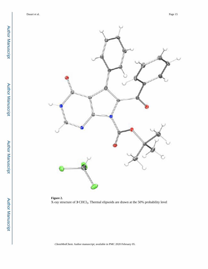
Figure 2. X-ray structure of 3·CHCl3. Thermal elipsoids are drawn at the 50% probability level
Dasari et al. Page 15
ChemMedChem. Author manuscript; available in PMC 2020 February 05.
Author M
anuscriptA
uthor Manuscript
Author M
anuscriptA
uthor Manuscript

Figure 3. Effects of compounds 1 and 3 (A), and 2 and 11 (B) on in vitro tubulin polymerization.
Taxol (3 µM) promotes (A, B), while colchicine (25 µM) suppresses (A) microtubule
formation relative to 0.1% DMSO control. 1, 3, 2 and 11 (all at 25 µM) suppress tubulin
polymerization.
Dasari et al. Page 16
ChemMedChem. Author manuscript; available in PMC 2020 February 05.
Author M
anuscriptA
uthor Manuscript
Author M
anuscriptA
uthor Manuscript

Figure 4. Microtubule organization in HeLa cells during interphase (top row) and mitosis (bottom
row). HeLa cells were treated for 3 h with compounds 1, 2, 3, 9, 11 at a concentration of 50
nM. Following drug treatment, cells were probed for microtubules (red), centromeres
(green) and DNA (blue). Bar, 10 μm.
Dasari et al. Page 17
ChemMedChem. Author manuscript; available in PMC 2020 February 05.
Author M
anuscriptA
uthor Manuscript
Author M
anuscriptA
uthor Manuscript

Figure 5. Microtubule organization in HeLa cells during interphase and mitosis when HeLa cells were
treated for 3 h with compounds 2 (B) and its N9-t-butoxycarbonyl derivative 11 (C) at a
range of concentrations. Following drug treatment, cells were probed for microtubules (red),
the centromeres (green) and DNA (blue). Bar, 10 μm.
Dasari et al. Page 18
ChemMedChem. Author manuscript; available in PMC 2020 February 05.
Author M
anuscriptA
uthor Manuscript
Author M
anuscriptA
uthor Manuscript
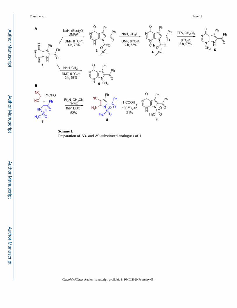
Scheme 1. Preparation of N3- and N9-substituted analogues of 1
Dasari et al. Page 19
ChemMedChem. Author manuscript; available in PMC 2020 February 05.
Author M
anuscriptA
uthor Manuscript
Author M
anuscriptA
uthor Manuscript

Scheme 2. Preparation of N3- and N9-substituted analogues of 2
Dasari et al. Page 20
ChemMedChem. Author manuscript; available in PMC 2020 February 05.
Author M
anuscriptA
uthor Manuscript
Author M
anuscriptA
uthor Manuscript

Author M
anuscriptA
uthor Manuscript
Author M
anuscriptA
uthor Manuscript
Dasari et al. Page 21
Table 1.
Antiproliferative activities of N3- and N9-substituted analogues of 1 and 2 synthesized in this work
# structure cell viabilitya
GI50, μM, ± SD
# structure cell viabilityGI50, μM, ± SD
HeLa MCF-7 HeLa MCF-7
1 0.035 ± 0.007 0.040 ± 0.024 2 0.022 ± 0.002 0.038 ± 0.018
3 0.029 ± 0.002 0.029 ± 0.001 10 0.11 ± 0.01 0.10 ± 0.01
4 81 ± 3 81 ± 4 11 0.021 ± 0.003 0.074 ± 0.005
5 44 ± 2 47 ± 0 12 >100 >100
6 2.8 ± 0.6 5.8 ± 0.1 13 10.3 ± 0.5 20.6 ± 5.5
9 0.028 ± 0.001 0.029 ± 0.001 14 29.2 ± 3.5 45.4 ± 3.4
aConcentration required to reduce the viability of cells by 50% after a 48 h treatment with the indicated compounds relative to a DMSO control ±
SD from two independent experiments, each performed in 4 replicates, as determined by the MTT assay.
ChemMedChem. Author manuscript; available in PMC 2020 February 05.

Author M
anuscriptA
uthor Manuscript
Author M
anuscriptA
uthor Manuscript
Dasari et al. Page 22
Table 2.
Cytotoxic effects of rigidin analogues in the SW620 cell line panel.
SW620 SW620C SW620D SW620E SW620M SW620V
2
159.9a 178.4 207.8 192.7 137.7 186.0
133.6 – 191.4 140.8 – 226.1 176.6 – 244.4 154.6 – 240.2 117.6 – 161.3 157.6 – 219.5
3
116.9 160.0 97.4 76.5 92.8 74.5
76.5 – 178.7 97.3 – 263.2 56.8 – 167.1 46.3 – 126.6 53.8 – 159.9 47.1 – 117.6
11
30.5 84.8 68.0 118.7 20.3 81.5
17.2 – 54.1 45.8 – 157.1 40.2 – 115.0 65.8 – 214.1 11.7 – 35.4 55.4 – 119.9
1
90.5 101.8 128.1 103.4 123.5 134.3
51.5 – 159.0 69.2 – 149.8 87.2 – 188.3 72.2 – 148.1 87.8 – 173.8 98.0 – 184.1
aThe results (GI50 in nM) are presented along with their respective 95% confidence intervals (lower line). Calculations were performed using data
obtained in three independent experiments.
ChemMedChem. Author manuscript; available in PMC 2020 February 05.

Author M
anuscriptA
uthor Manuscript
Author M
anuscriptA
uthor Manuscript
Dasari et al. Page 23
Table 3.
Cytotoxic effects of rigidin analogues in MDCKII cell line panel.
MDCKII MDCKII-BCRP MDCKII-MDR
2
~ 24.1a
16.8 21.1
(Very wide) 14.5 – 19.4 19.3 – 23.0
3
15.2 14.2 15.0
11.7 – 19.8 13.5 – 15.0 14.1 – 15.9
11
18.3 14.2 18.9
14.4 – 23.3 12.8 – 15.8 17.3 – 20.7
1
15.8 15.0 17.3
13.5 – 18.5 14.2 – 15.8 15.0 – 20.1
aThe results (GI50 in nM) are presented along with their respective 95% confidence intervals (lower line). Calculations were performed using data
obtained in three independent experiments.
ChemMedChem. Author manuscript; available in PMC 2020 February 05.

Author M
anuscriptA
uthor Manuscript
Author M
anuscriptA
uthor Manuscript
Dasari et al. Page 24
Tab
le 4
.
Poss
ible
mod
ulat
ory
effe
cts
of r
igid
in a
nalo
gues
on
mito
xant
rone
and
vin
cris
tine
resi
stan
ce in
, res
pect
ivel
y, M
DC
KII
-BC
RP
and
MD
CK
II-M
DR
1 ce
lls.
Mit
oxan
tron
e to
xici
ty
MD
CK
IIM
DC
KII
+Ko1
43M
DC
KII
+2M
DC
KII
+3M
DC
KII
+11
MD
CK
II+1
MD
CK
II-B
CR
PM
DC
KII
-BC
RP
+Ko1
43M
DC
KII
-BC
RP
+2M
DC
KII
-BC
RP
+3M
DC
KII
-BC
RP
+11
MD
CK
II-B
CR
P+1
18.7
a21
.314
.517
.415
.016
.914
9.3
44.6
145.
716
5.0
131.
612
6.5
14.7
– 2
3.8
12.2
– 3
7.3
11.9
– 1
7.6
13.6
– 2
2.2
11.5
– 1
9.5
13.4
– 2
1.4
117.
4 –
189.
835
.6 –
56.
012
0.8
– 17
5.9
133.
3 –
204.
211
3.8
– 15
2.1
109.
2 –
146.
6
Vin
cris
tine
tox
icit
y
MD
CK
IIM
DC
KII
+Ver
MD
CK
II+2
MD
CK
II+3
MD
CK
II+1
1M
DC
KII
+1M
DC
KII
-MD
RM
DC
KII
-MD
R+V
erM
DC
KII
-MD
R+2
MD
CK
II-M
DR
+3M
DC
KII
-MD
R+1
1M
DC
KII
-MD
R+1
11.5
11.1
10.5
11.8
11.8
11.8
434.
015
.540
9.3
442.
632
1.6
397.
5
10.9
– 1
2.1
10.1
– 1
2.2
9.9
– 11
.210
.0 –
13.
811
.0 –
12.
710
.7 –
13.
036
6.2
– 51
4.4
14.2
– 1
6.9
334.
1 –
501.
437
6.7
– 52
0.1
259.
3 –
398.
832
7.7
– 48
2.1
a The
res
ults
(G
I 50
in n
M)
are
pres
ente
d al
ong
with
thei
r re
spec
tive
95%
con
fide
nce
inte
rval
s (l
ower
line
). C
alcu
latio
ns w
ere
perf
orm
ed u
sing
dat
a ob
tain
ed in
thre
e in
depe
nden
t exp
erim
ents
.
ChemMedChem. Author manuscript; available in PMC 2020 February 05.

Author M
anuscriptA
uthor Manuscript
Author M
anuscriptA
uthor Manuscript
Dasari et al. Page 25
Tab
le 5
.
Dir
ect e
ffec
ts o
f ri
gidi
n an
alog
ues
on m
ultid
rug
resi
stan
ce p
rote
in a
ctiv
ity.
Cal
cein
acc
umul
atio
n ra
te (
AB
CB
1 ac
tivi
ty)
[AU
/min
]
SW62
0SW
620
+Ver
SW62
0+2
SW62
0+3
SW62
0+1
1SW
620
+1SW
620V
SW62
0V+V
erSW
620V
+2SW
620V
+3SW
620V
+11
SW62
0V+1
4265
9 ±
146
4a43
073
± 8
8036
190
± 4
0841
254
± 4
0723
220
± 2
593
2639
3 ±
265
629
7 ±
31
2541
6 ±
451
319
± 3
338
5 ±
47
299
± 3
231
4 ±
50
Phe
opho
rbid
e a
accu
mul
atio
n ra
te (
AB
CG
2 ac
tivi
ty)
[AU
/min
]
SW62
0SW
620
+Ko1
43SW
620
+2SW
620
+3SW
620
+11
SW62
0+1
SW62
0CSW
620C
+Ko1
43SW
620C
+2SW
620C
+3SW
620C
+11
SW62
0C+1
3422
4 ±
336
130
113
± 3
023
3092
2 ±
299
431
938
± 2
971
3238
0 ±
276
930
778
± 2
946
1749
3 ±
162
136
522
± 3
314
1527
7 ±
140
916
572
± 1
769
1787
1 ±
130
914
248
± 1
292
BC
EC
F h
alf-
life
in t
he c
ell (
AB
CC
1 ac
tivi
ty)
[min
]
SW62
0SW
620
+MK
571
SW62
0+2
SW62
0+3
SW62
0+1
1SW
620
+1SW
620M
SW62
0M+M
K57
1SW
620M
+2SW
620M
+3SW
620M
+11
SW62
0M+1
33.3
649
.55
38.8
839
.69
47.5
138
.15
16.0
968
.39
20.4
617
.21
17.1
217
.41
8.89
- ∞
10.7
6 - ∞
9.72
- ∞
9.43
- ∞
10.3
1 - ∞
9.82
- ∞
5.48
- ∞
21.0
6 - ∞
6.64
- ∞
5.75
- ∞
5.93
- ∞
5.75
- ∞
a Mea
n va
lues
± s
tand
ard
devi
atio
ns a
re p
rese
nted
exc
ept f
or B
CE
CF
assa
y w
here
95%
con
fide
nce
inte
rval
is p
rese
nted
(lo
wer
line
). A
vera
ged
data
for
thre
e in
depe
nden
t exp
erim
ents
.
ChemMedChem. Author manuscript; available in PMC 2020 February 05.