herpes simplex virus 1 infection: misleading findings in ... filebrospinal fluid and pleural liquid...
TRANSCRIPT

NEW MICROBIOLOGICA, 36, 307-313, 2013
Herpes Simplex Virus 1 infection: misleadingfindings in an infant with disseminated disease
Maria Grazia Capretti1, Concetta Marsico1, Tiziana Lazzarotto2, Liliana Gabrielli2, Alberto Bagni3, Morena De Angelis1, Roberto Rossini1, Giacomo Faldella1
1Department of Obstetrical, Gynaecological and Paediatric Sciences, Operative Unit of Neonatology,St. Orsola-Malpighi General Hospital, University of Bologna, Italy;
2Operative Unit of Clinical Microbiology, St. Orsola-Malpighi General Hospital, University of Bologna, Italy;3Department of Haematology, Oncology and Laboratory Medicine, “F. D’Addari” Pathology Institute,
St. Orsola-Malpighi General Hospital, University of Bologna, Italy
INTRODUCTION
Herpes Simplex Virus (HSV) infection is a majorcause of morbidity and mortality in infants. Theincidence of neonatal HSV infection is 31.2 per100,000 live births (Brown et al., 2003), with awidely variable range (8 per 100,000-60 per100,000 live births) (Corey et al., 2009). The clin-ical presentation of HSV infection in the neona-tal period has been divided into three categories
Corresponding authorMaria Grazia CaprettiOperative Unit of NeonatologySt. Orsola-Malpighi General HospitalUniversity of BolognaVia Massarenti, 11 - 40138 Bologna, ItalyE-mail: [email protected]
according to the extent of disease: skin, eyes andmouth disease (45% of cases of neonatal HSV),the central nervous system (CNS)-associated dis-ease (30%), and disseminated disease (25%). HSVdisseminated disease involves multiple organs;approximately 30% of affected infants die andsurviving infants present neurological sequelaein 20% of cases (Kimberlin et al., 2001b). Early high-dose intravenous Acyclovir at 60mg/kg for 21 days has been found to reduce themortality rate of neonatal HSV infection, and toimprove the rate of infected infants without neu-rological impairment for the disseminated dis-ease (Kimberlin et al., 2001a). We describe a disseminated HSV-1 infection ina newborn who succumbed to severe pulmonarycomplications, liver dysfunction and coagulopa-thy.
Neonatal Herpes Simplex Virus (HSV) infection is a serious illness with significant mortality and morbidity for dis-seminated disease. Clinical diagnosis of neonatal HSV infection is often difficult without evidence of HSV exposure,for example, absence of a rash or the presence of non-specified manifestations in an infant. Early recognition and treat-ment with high-dose Acyclovir may dramatically improve the short and long-term outcomes. We describe an infantwith disseminated disease due to HSV-1 infection, who first presented clinical and radiologic features of pneumonia.The diagnosis was performed post-mortem by Real-Time Polymerase Chain Reaction (PCR) analysis of blood, cere-brospinal fluid and pleural liquid of the infant. Tissue PCR revealed a disseminated HSV-1 infection, with a high vi-ral load detected in liver, lungs, brain, heart, striated muscle, kidneys, and thymus tissues. This case report highlightsthe need for neonatologists to raise awareness about the different clinical manifestations of disseminated neonatal HSVinfection. HSV infections should be prominent in the differential diagnosis of an infant under four weeks of age withfever, pneumonia, unexplained seizures or sepsis-like disease, particularly if unresponsive to antibiotics. Early initia-tion of appropriate antiviral therapy for high-risk infants undergoing testing for HSV infection can be essential toprevent significant morbidity and mortality.
KEY WORDS: HSV-1, Disseminated disease, Neonatal infection.
SUMMARY
Received January 21, 2013 Accepted April 2, 2013
CASE REPORT
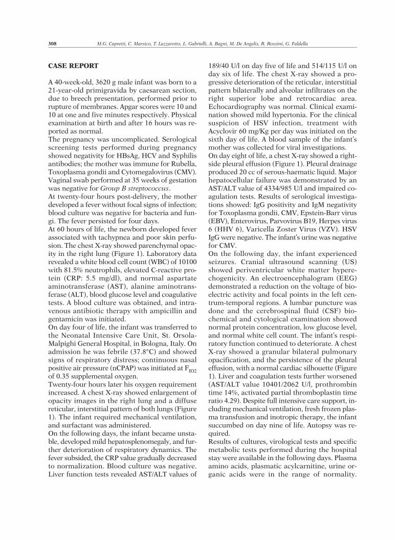
A 40-week-old, 3620 g male infant was born to a21-year-old primigravida by caesarean section,due to breech presentation, performed prior torupture of membranes. Apgar scores were 10 and10 at one and five minutes respectively. Physicalexamination at birth and after 16 hours was re-ported as normal.The pregnancy was uncomplicated. Serologicalscreening tests performed during pregnancyshowed negativity for HBsAg, HCV and Syphilisantibodies; the mother was immune for Rubella,Toxoplasma gondii and Cytomegalovirus (CMV).Vaginal swab performed at 35 weeks of gestationwas negative for Group B streptococcus.At twenty-four hours post-delivery, the motherdeveloped a fever without focal signs of infection;blood culture was negative for bacteria and fun-gi. The fever persisted for four days. At 60 hours of life, the newborn developed feverassociated with tachypnea and poor skin perfu-sion. The chest X-ray showed parenchymal opac-ity in the right lung (Figure 1). Laboratory datarevealed a white blood cell count (WBC) of 10100with 81.5% neutrophils, elevated C-reactive pro-tein (CRP: 5.5 mg/dl), and normal aspartateaminotransferase (AST), alanine aminotrans-ferase (ALT), blood glucose level and coagulativetests. A blood culture was obtained, and intra-venous antibiotic therapy with ampicillin andgentamicin was initiated. On day four of life, the infant was transferred tothe Neonatal Intensive Care Unit, St. Orsola-Malpighi General Hospital, in Bologna, Italy. Onadmission he was febrile (37.8°C) and showedsigns of respiratory distress; continuous nasalpositive air pressure (nCPAP) was initiated at FIO2of 0.35 supplemental oxygen.Twenty-four hours later his oxygen requirementincreased. A chest X-ray showed enlargement ofopacity images in the right lung and a diffusereticular, interstitial pattern of both lungs (Figure1). The infant required mechanical ventilation,and surfactant was administered.On the following days, the infant became unsta-ble, developed mild hepatosplenomegaly, and fur-ther deterioration of respiratory dynamics. Thefever subsided, the CRP value gradually decreasedto normalization. Blood culture was negative.Liver function tests revealed AST/ALT values of
189/40 U/l on day five of life and 514/115 U/l onday six of life. The chest X-ray showed a pro-gressive deterioration of the reticular, interstitialpattern bilaterally and alveolar infiltrates on theright superior lobe and retrocardiac area.Echocardiography was normal. Clinical exami-nation showed mild hypertonia. For the clinicalsuspicion of HSV infection, treatment withAcyclovir 60 mg/Kg per day was initiated on thesixth day of life. A blood sample of the infant’smother was collected for viral investigations. On day eight of life, a chest X-ray showed a right-side pleural effusion (Figure 1). Pleural drainageproduced 20 cc of serous-haematic liquid. Majorhepatocellular failure was demonstrated by anAST/ALT value of 4334/985 U/l and impaired co-agulation tests. Results of serological investiga-tions showed: IgG positivity and IgM negativityfor Toxoplasma gondii, CMV, Epstein-Barr virus(EBV), Enterovirus, Parvovirus B19, Herpes virus6 (HHV 6), Varicella Zoster Virus (VZV). HSVIgG were negative. The infant’s urine was negativefor CMV.On the following day, the infant experiencedseizures. Cranial ultrasound scanning (US)showed periventricular white matter hypere-chogenicity. An electroencephalogram (EEG)demonstrated a reduction on the voltage of bio-electric activity and focal points in the left cen-trum-temporal regions. A lumbar puncture wasdone and the cerebrospinal fluid (CSF) bio-chemical and cytological examination showednormal protein concentration, low glucose level,and normal white cell count. The infant’s respi-ratory function continued to deteriorate. A chestX-ray showed a granular bilateral pulmonaryopacification, and the persistence of the pleuraleffusion, with a normal cardiac silhouette (Figure1). Liver and coagulation tests further worsened(AST/ALT value 10401/2062 U/l, prothrombintime 14%, activated partial thromboplastin timeratio 4.29). Despite full intensive care support, in-cluding mechanical ventilation, fresh frozen plas-ma transfusion and inotropic therapy, the infantsuccumbed on day nine of life. Autopsy was re-quired. Results of cultures, virological tests and specificmetabolic tests performed during the hospitalstay were available in the following days. Plasmaamino acids, plasmatic acylcarnitine, urine or-ganic acids were in the range of normality.
308 M.G. Capretti, C. Marsico, T. Lazzarotto, L. Gabrielli, A. Bagni, M. De Angelis, R. Rossini, G. Faldella

Cultures of pleural liquid, devices and CSF werenegative for bacteria, fungi and Mycobacteria.Real-Time PCR in infant’s CSF and blood werenegative for: HHV6, EBV, VZV, CMV, HSV2, andEnterovirus.
Laboratory methodsEnzyme immunoassay (EIA - Enzygnost,Siemens Healthcare Diagnostics, Marburg,Germany) was used for the quantitative determi-nation of specific IgG and IgM antibodies in hu-man serum. IgG or IgM index >1.3 was consid-ered positive. DNA was extracted from maternal
and neonatal samples using the NucliSenseasyMAG System (bioMerieux, Marcy l’Etoile,France) according to the manufacturer’s recom-mendations. DNA extraction from paraffin-em-bedded tissue was performed on five micron tis-sue slices using a BioSprint 15 DNA Blood Kit(Qiagen GmbH, Hilden, Germany) according tothe manufacturer’s package insert and one mi-crogram DNA was used for PCR determinations.HSV-1, HSV-2, HHV6, EBV, VZV, CMV andEnterovirus were quantified using real-Time PCRassays (Nanogen Advanced Diagnostics SRL,Turin, Italy). Amplification, detection and analy-
Neonatal HSV-1 infection 309
FIGURE 1 - Radiographic studies. Chest X-ray obtained within 24 h after the infant’s admission to our hospital (a),on day 5 of life (b), on day 8 of life (c), and during the final period of clinical deterioration on day 9 of life (d).

sis were performed using the ABI PRISM 7300platform (Applied Biosystems, Foster City, CA,USA). Viral load was reported as number ofcopies/ml or number of copies/microgram DNA.Immunohistochemical staining for HSV glyco-protein B (Advanced Biotechnologies Incor -
porated, Columbia, MD, USA) was performed toidentify HSV-1 positive cells.
Virological and autopsy diagnosisHSV1 DNA was found in the infant’s blood byReal-Time PCR (viral load>10.000.000 copies/ml)
310 M.G. Capretti, C. Marsico, T. Lazzarotto, L. Gabrielli, A. Bagni, M. De Angelis, R. Rossini, G. Faldella
FIGURE 2 - a) Adrenal histology, H&E, x250: adrenal cortex with multiple foci of necrosis of various size (arrows).b) HSV immunistochemistry, x100: brown staining depicts necrotic spots and scattered adrenal cells. c) Lung his-tology, H&E, x60: diffuse interstitial pneumonitis and blood vascular congestion. d) Liver histology, H&E, x60: mul-tiple foci of coagulative and haemorrhagic necrosis, mainly around central veins.

collected at day eight of life, drained pleural liq-uid (>10.000.000 copies/ml) collected at day eightof life and CSF (210.000 copies/ml) collected atday nine of life.Viral investigations were performed on the ma-ternal blood sample stored on day seven post-de-livery, and showed slightly positive HSV IgG (EIAIgG index: 1.5) and positive HSV-1 DNA PCR (vi-ral load 200.000 copies/ml). After one month, themother was tested again for HSV-1, and viral in-vestigations demonstrated an increased HSV IgGindex (EIA IgG index: 3.2), and a HSV-1 DNAPCR of 800 copies/ml. Cervical and vaginal swabsobtained at this time were positive for HSV-1DNA. At post-mortem examination lung histologyshowed interstitial pneumonitis, hyperaemia andfocal intralveolar haemorrhages. The livershowed a diffuse necrotizing hepatitis with con-fluent coagulation necrosis. Multiple foci ofnecrosis were present in the adrenal glands. Thespleen revealed hyperaemia of the red pulp.Immunohistochemistry staining of the lungs, liv-er and adrenal glands were positive for HSV(Figure 2). HSV-1 DNA was extracted from allpost-mortem specimens. Viral loads in each sam-ple are reported in Table 1. The autopsy and vi-rological findings were consistent with a severedisseminated systemic HSV-1 infection withnecrotizing hepatitis and diffuse interstitial pneu-monitis. The maternal antibody profile and the
evidence of HSV-1 DNA in maternal blood andthe cervical swab provided a diagnosis of primaryHSV-1 infection which had occurred near term.
DISCUSSION
Neonatal HSV infection is a rare but severe ill-ness. We described an uncommon onset of HSV-1 infection of an infant born by caesarean sec-tion presenting symptoms of pneumonia and on-ly days later developed the typical features of dis-seminated disease. This case report highlights thedifficulty of making an early diagnosis of HSVneonatal infection when the mother’s infection isnot apparent and mucocutaneous lesions are ab-sent in the infant. The majority of reported neonatal HSV infectionsare caused by HSV-2 (Corey et al., 2009; Rudnicket al., 2002), since the passage through the in-fected birth canal is the principal route of trans-mission. However, in recent years, it has beenshown that an increasing proportion of genitalherpes is caused by HSV-1 (Lafferty et al., 2000;Westhoff et al., 2011). During pregnancy, the riskof transmission is significantly higher in moth-ers acquiring a primary HSV infection than moth-ers experiencing a reactivation of the virus(Brown et al., 1991)The important aspect of this case report is thatthe mother acquired HSV primary infection nearterm. HSV has been transmitted to the infant inutero with intact fetal membranes by maternalviremia when protective IgG antibodies have notyet been produced by the mother. Diagnosis ofHSV infection was performed post-mortem, bydetection of HSV-1 DNA in the infant’s CSF, pleu-ral liquid and blood. At autopsy, an extensive vi-ral dissemination was demonstrated, with a mas-sive involvement of lungs, liver, spleen, brain andadrenal glands. HSV-1 DNA was detected in alltested organs, even in striated muscle, kidneys,heart and thymus where histology did not showabnormalities.The onset of the HSV disseminated disease wason day 3 of life, that is clearly earlier than usual.Disseminated HSV disease presentation is usual-ly around days 10 to 12 of life (Kimberlin, 2007),but based on antiviral treatment trials and caseseries, there is a widely variable range from birthto the fourth week of life (Campbell et al., 1983;
Neonatal HSV-1 infection 311
TABLE 1 - HSV-1 load detected by Real-Time PCRin post-mortem specimens.
Tested organ HSV-1 load (copies/microgram DNA)
Lung 68.000.000
Spleen 7.000.000
Kidney 1.800.000
Brain 1.000.000
Thymus 220.000
Heart 214.000
Striated muscle 130.000
Liver 8.000

Withley et al., 1991; Kimberlin et al., 2001b;Kropp et al., 2006; Long et al., 2011; Catlin et al.,2012). No single association of presenting symptomsand signs identifies all infants with HSV dissem-inated disease: the most common features areskin vesicles, fever, lethargy, respiratory distress,disseminated intravascular coagulopathy,seizures, and poor feeding. Most cases of HSV in-fection in infants under 21 days of age have anonspecific presentation (Long et al., 2011)This case report shows an early uncommon man-ifestation of the HSV disseminated disease.Although the presence of pulmonary infiltratesat the onset of infection is rare, it has been pre-viously reported (Campbell et al., 1983; Lissaueret al., 1984; Langlet et al., 2003; Knezevic et al.,2007). This means that early and rapidly pro-gressive pneumonia might be considered one ofthe different initial manifestations of dissemi-nated neonatal HSV. The early appearance of therespiratory distress, the consolidation on thechest radiography and the elevated CRP valuewhich decreased after antibiotic therapy, lead tothe clinical suspicion of bacterial sepsis. However,the progressive radiological findings were sug-gestive of HSV-pneumonia: normal lung in thefirst stage, reticular and interstitial pattern in thesecond, alveolar infiltrates without hyperinflationin the third and total pulmonary opacificationwith pleural effusion in the last stage (Dominguezet al., 1984). On day nine of life the infant devel-oped seizures as a manifestation of CNS diseasewithout relevant cranial US abnormalities, butwith abnormal bioelectric activity of the tempo-ral lobe at EEG examination. However, the in-volvement of the temporal lobe is considered tobe typical of HSV encephalitis after the neonatalperiod (Kimberlin, 2007).Clinical deterioration in spite of antibiotic treat-ment, low CRP and increasing transaminase lev-els detected from day 5 of life might have helpedin making a differential diagnosis. The clinical course of the infant described in thiscase report emphasizes the need for neonatolo-gists to raise awareness about the different waysthat disseminated HSV infection may present inthe neonatal period. Acyclovir treatment was de-layed due to a misinterpretation of the presentingsymptoms as bacterial pneumonia. The findingthat early manifestations of HSV infection are of-
ten nonspecific should be considered whenweighing management strategies of an acutely illinfant under 21 days of age: empiric acyclovirstrategy narrowly restricted to infants with onsetof illness under 21 days of age has been reportedto capture 90% of HSV cases and to anticipatethe rate of CNS involvement similar to that ofbacterial meningitis (Long et al., 2011).In our opinion, infants under four weeks of agewith fever, pneumonia, unexplained seizures orsepsis-like disease, particularly if unresponsiveto antibiotics, should be promptly evaluated forHSV infection. Early initiation of appropriateantiviral therapy for high-risk infants can be es-sential to prevent significant morbidity and mor-tality.
No external funding was secured for this brief re-port.Ther authors have no financial relationships rel-evant to this manuscript to disclose.The authors have no conflict of interest to dis-close.
REFERENCES
BROWN Z.A., BENEDETTI J., ASHLEY R., BURCHETT S.,SELKE S., BERRY S., VONTVER L.A., COREY L. (1991).Neonatal herpes simplex virus infection in relationto asymptomatic maternal infection at the time oflabor. N. Engl. J. Med. 324, 1247-1252.
BROWN Z.A., WALD A., MORROW R.A., SELKE S., ZEH J.,COREY L. (2003). Effect of serologic status and ce-sarean delivery on transmission rates of herpes sim-plex virus from mother to infant. JAMA. 289, 203-209.
CAMPBELL A.N., O’DRISCOLL M.C., ROBINSON D.L., READ
S.E. (1983). A case of neonatal herpes simplex withpneumonia. Can. Med. Assoc. J. 129, 725-726.
CATLIN E.A., WARREN S., SHAILAM R., LAHOUD-RAHME
M., LEW M. (2012). Case records of the massachu-setts general hospital. N. Engl. J. Med. 366, 2409-2419.
COREY L., WALD A. (2009). Maternal and neonatal her-pes simplex virus infections. N. Engl. J. Med. 361,1376-1385.
DOMINGUEZ R., RIVERO H., GAISIE G., TALMACHOFF P.,AMORTEGUI A., YOUNG L.W. (1984). Neonatal herpessimplex pneumonia: radiographic findings.Radiology. 153, 395-399.
KIMBERLIN D.W., LIN C.Y., JACOBS R., POWELL D.A.,COREY L., GRUBER W.C., RATHORE M., BRADLEY J.S.,DIAZ P.S., KUMAR M., ARVIN A., GUTIERREZ K.,
312 M.G. Capretti, C. Marsico, T. Lazzarotto, L. Gabrielli, A. Bagni, M. De Angelis, R. Rossini, G. Faldella

SHELTON M., WEINER L.B., SLEASMAN J.W., DE
SIERRA T.M., WELLER S., SOONG S.J., KIELL J.,LAKEMAN F.D., WHITLEY R.J. AND THE NATIONAL
INSTITUTE OF ALLERGY AND INFECTIOUS DISEASES
COLLABORATIVE ANTIVIRAL STUDY GROUP (2001a).Safety and efficacy of high-dose intravenous acy-clovir in the management of neonatal herpes sim-plex virus infections. Pediatrics. 108, 230-238.
KIMBERLIN, D.W., LIN C.Y., JACOBS R., POWELL D.A.,FRENKEL L.M., GRUBER W.C., RATHORE M., BRADLEY
J.S., DIAZ P.S., KUMAR M., ARVIN A.M., GUTIERREZ
K., SHELTON M., WEINER L.B., SLEASMAN J.W.,MURGUIA DE SIERRA T., SOONG S.J., KIELL J., LAKEMAN
F.D., WHITLEY R.J.; AND THE NATIONAL INSTITUTE OF
ALLERGY AND INFECTIOUS DISEASES COLLABORATIVE
ANTIVIRAL STUDY GROUP. (2001b). Natural history ofneonatal herpes simplex virus infections in the acy-clovir era. Pediatrics. 108, 223-229.
KIMBERLIN D.W. (2007). Herpes Simplex VirusInfections of the newborn. Semin. Perinatol. 31,19-25.
KNEZEVIC A., MARTIC J., STANOJEVIC M., JANKOVIC S.,NEDELJKOVIC J., NIKOLIC L., PASIC S., JANKOVIC B.,JOVANOVIC T. (2007). Disseminated neonatal herpescaused by herpes simplex virus types 1 and 2.Emerg. Infect. Dis. 13, 302-304.
KROPP R.Y., WONG T., CORMIER L., RINGROSE A., BURTON
S., EMBREE J.E., STEBEN M. (2006). Neonatal herpessimplex virus infections in Canada: results of a 3-year national prospective study. Pediatrics. 117,1955-1962.
LAFFERTY W.E., DOWNEY L., CELUM C., WALD A. (2000).Herpes simplex virus type 1 as a cause of genitalherpes: impact on surveillance and prevention. J.Infect. Dis. 181, 1454-1457.
LANGLET C., GAUGLER C., CASTAING M., ASTRUC D.,FALKENRODT A., NEUVILLE A., MESSER J. (2003). Anuncommon case of disseminated neonatal herpessimplex infection presenting with pneumonia andpleural effusions. Eur. J. Pediatr. 162, 532-533.
LISSAUER T.J., SHAW P.J., UNDERHILL G. (1984). Neonatalherpes simplex pneumonia. Arch. Dis. Child. 59,668-670.
LONG S.S., POOL T.E., VODZAK J., DASKALAKI I., GOULD
J.M. (2011). Herpes simplex virus infection inyoung infants during 2 decades of empiric acyclo-vir therapy. Pediatr. Infect. Dis J. 30, 556-561.
RUDNICK C.M., HOEKZEMA G.S. (2002). Neonatal her-pes simplex virus infections. Am. Fam. Physician.65, 1138-1143.
WESTHOFF G.L., LITTLE S.E., CAUGHEY A.B. (2011).Herpes simplex virus and pregnancy: a review ofthe management of antenatal and peripartum her-pes infections. Obstet. Gynecol. Surv. 66, 629-638.
WITHLEY R., ARVIN A., PROBER C., BURCHETT S., COREY
L., POWELL D., PLOTKIN S., STARR S., ALFORD C.,CONNOR J., JACOBS R., NAHMIAS A., SOONG SJ., AND
THE NATIONAL INSTITUTE OF ALLERGY AND INFECTIOUS
DISEASES COLLABORATIVE ANTIVIRAL STUDY GROUP.(1991). A controlled trial comparing vidarabinewith acyclovir in neonatal herpes simplex virus in-fection. N. Engl. J. Med. 324, 444-449.
Neonatal HSV-1 infection 313
