hepatocyte growth factor in renal failure: promise and reality
TRANSCRIPT

Kidney International, Vol. 57 (2000), pp. 1426–1436
PERSPECTIVES IN BASIC SCIENCE
Hepatocyte growth factor in renal failure: Promise and reality
GUSTAVO A. VARGAS, ANDREAS HOEFLICH, and PETER M. JEHLE
Department of Internal Medicine II, Division of Nephrology, University of Ulm, Ulm, and Institute of Molecular AnimalBreeding, Gene Center, Munich, Germany
Hepatocyte growth factor in renal failure: Promise and reality. possible therapeutic applications of hepatocyte growthCan science discover some secrets of Greek mythology? In the factor (HGF) and its receptor, c-met [3–5].case of Prometheus, we can now suppose that his amazinghepatic regeneration was caused by a peptide growth factorcalled hepatocyte growth factor (HGF). Increasing evidence HEPATOCYTE GROWTH FACTOR/SCATTERindicates that HGF acts as a multifunctional cytokine on differ- FACTOR STRUCTURE AND SYNTHESISent cell types. This review addresses the molecular mechanisms
The biological features of HGF are summarized inthat are responsible for the pleiotropic effects of HGF. HGFTable 1. Previous studies described several mitogenic andbinds with high affinity to its specific tyrosine kinase receptormotogenic polypeptides that were identified later as HGFc-met, thereby stimulating not only cell proliferation and differ-
entiation, but also cell migration and tumorigenesis. The three [6–9]. Independently, the existence of a polypeptide se-fundamental principles of medicine—prevention, diagnosis, creted by embryonic fibroblasts was reported that promotesand therapy—may be benefited by the rational use of HGF. In the scattering of Madin-Darby canine kidney (MDCK)renal tubular cells, HGF induces mitogenic and morphogenetic and other epithelial cells by paracrine mechanismsresponses. In animal models of toxic or ischemic acute renal
[10–12]. This polypeptide was named scatter factor (SF)failure, HGF acts in a renotropic and nephroprotective manner.[13] and was later on identified as HGF [14–18]. Simulta-HGF expression is rapidly up-regulated in the remnant kidneyneously, fibroblast tumor cytotoxic factor (F-TCF) [19]of nephrectomized rats, inducing compensatory growth. In aand the broad spectrum mitogen produced by lung fi-mouse model of chronic renal disease, HGF inhibits the pro-
gression of tubulointerstitial fibrosis and kidney dysfunction. broblasts [20] were also found to be identical to HGF.Increased HGF mRNA transcripts were detected in mesenchy- The human HGF gene is localized on chromosome bandsmal and tubular epithelial cells of rejecting kidney. In trans- 7q11.2-21 [14]. The HGF gene promoter contains posi-planted patients, elevated HGF levels may indicate renal rejec- tive and negative regulatory elements and is highly con-tion. When HGF is considered as a therapeutic agent in human
served among human and rodent species [21]. Nativemedicine, for example, to stimulate kidney regeneration afterHGF is secreted as an inactive precursor (pro-HGF),acute injury, strategies need to be developed to stimulate cellwhich is converted to the active form after extracellularregeneration and differentiation without an induction of tumor-
igenesis. proteolysis by several activators [22–25] and in responseto tissue injury [26]. HGF activation could be inhibitedby a specific serine protease inhibitor [27].
It has been suggested that HGF, macrophage stimulat-Growth factors are involved in multiple responses such ing protein (MSP; a growth factor with biological effects
as cell proliferation, differentiation, and motility mediat- similar to HGF), apolipoprotein-a, and plasminogen haveing their effects via receptor tyrosine kinases [1, 2]. The evolved from a common ancestral gene [28, 29]. Twotype of response triggered depends on receptors and truncated variants of HGF containing the N-terminalligands that are differentially expressed by various cell region and the first (HGF/NK1) or the first and thetypes. This review focuses on the structure, function, and second (HGF/NK2) kringle domains are generated by
alternative splicing. NK2 behaves as an HGF antagonist[30]. NK1 is derived from a 2.2 kb transcript [4] and isable to exhibit agonistic and antagonistic actions [30–33].In hepatocytes, NK1 acts primarily as an HGF antagonistKey words: growth factors, HGF, cell proliferation, tumorigenesis,
nephroprotection, renotropic growth factor. [34] but elicits an agonistic response in the presenceof heparin [35]. NK1 transgenic mice reveal the typicalReceived for publication June 29, 1999phenotype of HGF transgenic mice, underscoring its ago-and in revised form October 18, 1999
Accepted for publication November 2, 1999 nistic action in vivo [36]. A shorter synthetic HGF vari-ant, HGF/NK4, can act as an antagonist [37]. 2000 by the International Society of Nephrology
1426

Vargas et al: HGF in renal failure 1427
Table 1. Synopsis on biologic features of hepatocyte Table 2. Synopsis on biologic features of c-metgrowth factor (HGF)
Polypeptide features of c-met• product of the c-met proto-oncogeneSynonymous polypeptides
• fibroblast tumor cytotoxic factor (F-TCF) • synonymous polypeptides not known• 190 kD precursor• hepatocyte growth factor (HGF)
• hepatopoietin A • N-glycosylated mature protein• two disulfide-linked subunits• hepatotropin
• scatter factor (SF) a-chain, 50 kD, extracellular locationb-chain, 145 kD, extracellular, transmembrane and intracellularRelated polypeptides
• apolipoprotein (a) two major isoforms differing by a 47-aa segment in the juxta-membrane domain• blood coagulation factor XIIa
• macrophage-stimulating protein (MSP) single noninterrupted tyrosine kinase domain in the b-chainRelated polypeptides• plasminogen
Biologic effects • Ron tyrosine kinase receptor (ligand MSP)• Sea tyrosine kinase receptor• mitogen
• morphogen c-met gene localization• human chromosome 7, bands 7q21-q23• motogen
• inductor of tubulogenesis Biologic function• receptor unique for HGF• promotor of invasiveness
HGF gene localization c-met expression• almost all epithelia (e.g., kidney, liver, stomach, small intestine,• human chromosome 7, bands 7q11.2-q21
Polypeptide characteristics brain, breast)• endothelial cells• monomeric inactive precursor (pro-HGF)
• heterodimeric form (a/b-chain) after proteolytic cleavage (728 aa) • increased expression in several carcinoma• various isoforms (truncation, alternative splicing)• four kringle domains• one inactive serine protease domain• four potential glycosylation sites• heparin and glycosaminoglycans binding sites A specific Tyr at position 1356 in the C-terminal tail is
HGF expressionneeded for cell motility and morphogenesis [57]. Heparin• during organ development (e.g., kidney, liver, trophoblast)
• in adult life in tissue of mesodermal origin (e.g., fibroblasts, blood or similar extracellular matrix compounds can modulatemononuclear cells, platelets) HGF binding and action [58, 59]. Heparin-like molecules
can stabilize HGF oligomers and enhance the mitogenicpotency, probably by facilitating c-met receptor dimer-ization and activation [60], whereas heparan sulfate pro-
HEPATOCYTE GROWTH FACTOR RECEPTOR teoglycans act as negative modulators [61].BINDING AND SIGNALING
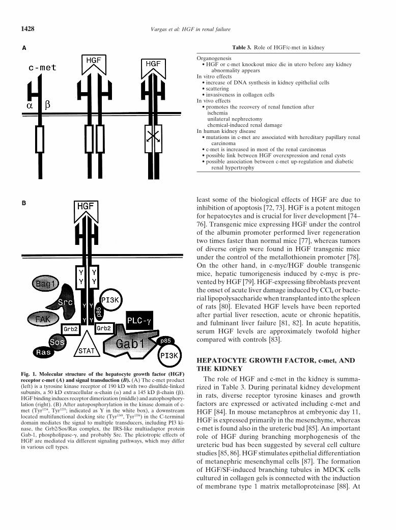
The HGF receptor is the product of the c-met proto-EXPRESSION OF HEPATOCYTE GROWTHoncogene [38]. A synopsis on the biological features ofFACTOR AND c-met AND BIOLOGICAL EFFECTSc-met is given in Table 2. The c-met product is a tyrosine
Hepatocyte growth factor is expressed in tissues ofkinase receptor [39–43] with two disulfide-linked sub-mesodermal origin [62], whereas c-met is expressed inunits and a single noninterrupted kinase domain sharingepithelia, including liver and kidney [63], and in severalhomology with the src family of tyrosine kinases (Fig. 1).human carcinomas [64]. Steroid hormones, cytokines [tu-The c-met tyrosine protein kinase oncogene family ismor necrosis factor-a (TNF-a), interleukin (IL)-1, IL-6],related to cell invasiveness and comprises v-ros, the insu-and basic fibroblast growth factor (bFGF) regulate HGFlin-like growth factor (IGF)-I receptor, the 220 kD SEXand c-met expression [5, 65, 66]. According to the find-protein, Ron (MSP receptor), and Sea (sarcoma, erythro-ings in double transgenic HGF/transforming growth fac-blastosis, and anemia; unknown ligand) [44–49]. Ron andtor (TGF)-a mice, HGF produces a partially inhibitorySea can induce the same biological effects as c-met [47].effect on hepatocellular carcinogenesis induced by TGF-aAfter HGF binding and subsequent ligand-induced re-[67]. Furthermore, HGF can reverse the arrest on cellceptor dimerization, c-met is activated by autophosphory-growth induced by TGF-b [68], whereas TGF-b waslation (Fig. 1A) [16, 39, 41, 43, 50–52]. The principal siteshown to enhance the motogenic effects of epidermalfor autophosphorylation is located in the kinase domaingrowth factor (EGF) but not HGF [69]. EGF can nega-at Tyr 1235, which together with Tyr 1230 and Tyr 1234,tively modulate HGF-induced cell migration [70]. In con-comprises a common “three tyrosine” motif that is con-trast, the HGF-induced increase in DNA synthesis inserved in the insulin and platelet-derived growth factorhepatocytes of HGF-transgenic mice is intensified by(PDGF) receptor tyrosine kinases [53]. The phosphory-EGF or insulin, and the growth inhibitory effects oflation of Ser 985 [54] and a protein phosphatase [55] canTGF-b are depressed [71].negatively modulate c-met kinase activity. Experiments
Besides its renotropic action (vide infra), HGF exertswith receptor chimeras showed that c-met is the firstdownstream effector in the HGF signaling cascade [56]. pleiotropic biological effects on different cell types. At
Vargas et al: HGF in renal failure1428
Table 3. Role of HGF/c-met in kidney
Organogenesis• HGF or c-met knockout mice die in utero before any kidney
abnormality appearsIn vitro effects
• increase of DNA synthesis in kidney epithelial cells• scattering• invasiveness in collagen cells
In vivo effects• promotes the recovery of renal function after
ischemiaunilateral nephrectomychemical-induced renal damage
In human kidney disease• mutations in c-met are associated with hereditary papillary renal
carcinoma• c-met is increased in most of the renal carcinomas• possible link between HGF overexpression and renal cysts• possible association between c-met up-regulation and diabetic
renal hypertrophy
least some of the biological effects of HGF are due toinhibition of apoptosis [72, 73]. HGF is a potent mitogenfor hepatocytes and is crucial for liver development [74–76]. Transgenic mice expressing HGF under the controlof the albumin promoter performed liver regenerationtwo times faster than normal mice [77], whereas tumorsof diverse origin were found in HGF transgenic miceunder the control of the metallothionein promoter [78].On the other hand, in c-myc/HGF double transgenicmice, hepatic tumorigenesis induced by c-myc is pre-vented by HGF [79]. HGF-expressing fibroblasts preventthe onset of acute liver damage induced by CCl4 or bacte-rial lipopolysaccharide when transplanted into the spleenof rats [80]. Elevated HGF levels have been reportedafter partial liver resection, acute or chronic hepatitis,and fulminant liver failure [81, 82]. In acute hepatitis,serum HGF levels are approximately twofold highercompared with controls [83].
HEPATOCYTE GROWTH FACTOR, c-met, ANDTHE KIDNEY
Fig. 1. Molecular structure of the hepatocyte growth factor (HGF)The role of HGF and c-met in the kidney is summa-receptor c-met (A) and signal transduction (B). (A) The c-met product
(left) is a tyrosine kinase receptor of 190 kD with two disulfide-linked rized in Table 3. During perinatal kidney developmentsubunits, a 50 kD extracellular a-chain (a) and a 145 kD b-chain (b). in rats, diverse receptor tyrosine kinases and growthHGF binding induces receptor dimerization (middle) and autophosphory-
factors are expressed or activated including c-met andlation (right). (B) After autoposphorylation in the kinase domain of c-met (Tyr1234, Tyr1235; indicated as Y in the white box), a downstream HGF [84]. In mouse metanephros at embryonic day 11,located multifunctional docking site (Tyr1349, Tyr1356) in the C-terminal
HGF is expressed primarily in the mesenchyme, whereasdomain mediates the signal to multiple transducers, including PI3 ki-nase, the Grb2/Sos/Ras complex, the IRS-like multiadaptor protein c-met is found also in the ureteric bud [85]. An importantGab-1, phospholipase-g, and probably Src. The pleiotropic effects of role of HGF during branching morphogenesis of theHGF are mediated via different signaling pathways, which may differ
ureteric bud has been suggested by several cell culturein various cell types.studies [85, 86]. HGF stimulates epithelial differentiationof metanephric mesenchymal cells [87]. The formationof HGF/SF-induced branching tubules in MDCK cellscultured in collagen gels is connected with the inductionof membrane type 1 matrix metalloproteinase [88]. At

Vargas et al: HGF in renal failure 1429
ing is based on the ability of HGF to inhibit junctionalcommunications, to enhance proteolysis of connexin 43[96], and to change the phosphorylation patterns of junc-tional molecules, thereby increasing their stability [97].In MDCK cells transformed with SV40 large T antigen,scattering is specifically triggered via inactivation of reti-noblastoma protein inducing an HGF autocrine loop[98]. In renal epithelial cells, the ectopic overexpressionof HGF is able to up-regulate the level of c-met and tostimulate cell scattering [99].
The pleiotropic effects of HGF are mediated via differ-ent signaling pathways, which may show differences invarious cell types. In all cases, a multifunctional dockingsite (Tyr at position 1349 and 1356 in the C-terminaldomain) mediates the signal of activated c-met receptorsto multiple transducers (Fig. 1B), including PI3 kinase,the Grb2/Sos/Ras complex, the insulin receptor substrate(IRS)-like multiadaptor protein Gab-1, and probably Src[100–102]. It is important to note that c-met–mediatedbiological effects can be dissected on the basis of differ-ent signaling requirements [100]. In MDCK cells, HGF-induced cell scattering is dependent on the Ras–mitogen-activated protein kinase (MAPK) signaling pathway[103]. In rabbit proximal renal tubular cells, we demon-strated that the growth-promoting effects of HGF in-volve the activation of Src, whereas the HGF-inducedtubulogenic differentiation (for example, formation oflong microvilli at the apical cell membrane) was notFig. 2. Morphogenic effects of HGF on cultured proximal tubular cells.
The differentiated rabbit proximal tubular cell line PT-1 was cultured influenced by src-interfering tyrosine kinase inhibitorsin Dulbecco’s modified Eagle’s medium (DMEM) as described [94, 95]. [95]. Invasive cell growth upon stimulation of c-met re-Cells were incubated for 48 hours in serum-free medium alone (A; bar 5
quires the concomitant activation of Ras and PI-3 kinase10 mm) or in the presence of HGF (1029 mol/L; B). HGF induced cellscattering (thin arrows) and tubulogenesis (thick arrows). Reprinted [49] or Src and PI3 kinase [104]. Scattering needs PI3-with permission from Jehle et al [94]. kinase activation [105–107] and in some cell types also
requires an additional effector [108]. Morphologicalchanges induced by HGF in MDCK cells are related tomodifications in cell polarity with alteration of synthesisthe first stages of kidney tubulogenesis, other growth fac-rate and interaction between b-catenin and E-cadherintors may replace the morphogenetic role of HGF. c-met[109]. The cell spreading response in this model is depen-knockout kidney cells form tubules in vitro upon stimula-dent on the activation of the small guanine 59-triphos-tion with TGF-a [89]. Another study showed that TGF-aphate (GTP)-binding proteins Ras and Rac. PI3 kinaseand EGF induce tubulogenesis of murine inner medul-is also involved in mediating the effects of HGF on tubu-lary collecting duct cells [90]. Coculture experiments re-logenesis [110]. In MDCK cells, HGF-induced tubulo-vealed that HGF produced by mesangial cells stimulatesgenesis and branching are enhanced by several compo-endothelial cell growth and that this interaction is nega-nents of the cell matrix, such as laminin, fibronectin, andtively modulated by TGF-b and angiotensin II [73].entactin, but are inhibited by type IV collagen, heparanThese authors suggested that negative regulation of localsulfate proteoglycan, vitronectin, and TGF-b [111]. InHGF production by both factors may play an importantthese cells, the branching development is regulated byrole in the pathogenesis of renal disease.phosphorylation processes mediated by protein kinase AThe morphogenetic and motogenic effects of HGF(PKA), protein kinase C (PKC), and Ca21/calmodulin-were first described in the MDCK cell line [91] and weredependent kinases [111]. Another protein involved inconfirmed in a variety of other epithelial cells [92–94].the scattering and tubulogenesis mediated by HGF inIn differentiated rabbit proximal tubular cells, HGF ex-kidney cells is the membrane-cytoskeleton linker ezrin,hibits potent mitogenic, morphogenetic, motogenic, andwhich is phosphorylated by c-met [112]. Downstream ofepithelial cell-differentiating effects under serum-freeGrb2 [106], the tubulogenic HGF response is dependentconditions [95]. Among its pleiotropic effects, the induc-
tion of cell scattering plays a pivotal role (Fig. 2). Scatter- on the STAT pathway [113].

Vargas et al: HGF in renal failure1430
nephrectomy or partial hepatectomy [118]. An increaseof HGF and its mRNA was also detected in rats afterunilateral nephrectomy and CCl4 treatment, with con-comitant internalization of c-met [119], and a transientincrease of HGF precedes the regenerating events in ratkidney after vitamin E depletion combined with glutathi-one depletion [120]. HGF and HGF mRNA increase inrat kidney after ischemia or HgCl2-induced nephrotoxic-ity and levels remain augmented during the phase oftubular regeneration [121]. On the other hand, the renalexpression of c-met increases in rats after ischemia, uni-lateral nephrectomy, or folic acid treatment [122]. A linkbetween renal hypertrophy observed in diabetic rats was
Fig. 3. Stimulation of c-met gene expression by cytokines and growth also proposed in relationship with the elevation of c-metfactors in cultured renal epithelial cells. Mouse inner medullary collect-in medullar and cortical tubular epithelium [123]. Aing duct epithelial cells (mIMCD-3) were incubated for 24 hours in
serum-free medium without (control) or with specific cytokines or spontaneous mouse model of chronic renal disease [im-growth factors at a concentration of 10 ng/mL. The total cellular RNA mune complex glomerulonephritis (ICGN) strain devel-was isolated and hybridized using rat c-met cDNA as a probe. The
oping glomerular sclerotic injury, tubular atrophy, andsame blot was stripped and reprobed with GAPDH to confirm equalloading of the RNA. Similar results were obtained using proximal renal dysfunction until 17 weeks of age] was used totubule-derived opossum kidney epithelial cells. Reprinted with permis- investigate whether HGF may inhibit the progression ofsion from Liu et al [116].
tubulointerstitial fibrosis [124]. When recombinant HGFwas injected into these mice during a four-week period(from weeks 14 through 17 after birth), DNA synthesisof tubular epithelial cells was found to be 4.4-fold higher
Evidence that HGF may represent an important reno- than in mice without HGF injection, thereby suggestingtropic growth factor came from cell culture studies, ani- tubular parenchymal expansion promoted by HGF. No-mal experiments, and findings in human kidney disease. tably, HGF suppressed the expression of TGF-b and ofHGF promotes DNA synthesis of several renal cell types, PDGF as well as myofibroblast formation in the affectedincluding tubular and glomerular epithelial cells [94, 114, kidney. Consequently, HGF completely inhibited the on-115]. The mitogenic effects of HGF on renal tubular set of tubulointerstitial fibrosis and attenuated the pro-epithelial cells are regulated by cell–cell interactions [114]. gression of glomerulosclerosis, both preventing manifesta-DNA synthesis stimulated by HGF was high at a low cell tion of renal dysfunction. From these results, supplementdensity and was strongly suppressed at a higher one. These therapy with HGF may be taken into consideration as aresults suggest that HGF may act not only as an inductor, novel option for prevention and treatment of chronicbut also as a modulator in renal regeneration and com- renal disease.pensatory renal growth. In conditions of renal injury medi- Increased HGF mRNA transcripts were detected inated by folic acid treatment, HGF up-regulates c-met in mesenchymal and tubular epithelial cells of rejecting kid-a tissue-specific manner, acting primarily at the transcrip- ney [125], and elevated serum HGF levels were sug-tional level by stimulating the c-met promoter activity [116]. gested to indicate renal rejection [126]. Serum HGF lev-This study also demonstrated up-regulation of c-met ex- els correlate with serum creatinine in chronic renalpression in renal epithelial cells in vitro by interleukins, failure patients, indicating a response to the organ dam-TGF-b, EGF, insulin-like growth factors (IGFs), and age or decreased clearance by the insufficient kidney.platelet-derived growth factor (PDGF) (Fig. 3). Because In acute renal failure, elevated urine HGF levels aretissue regeneration results from the integration of the described as consistent with a role for HGF in promotingcomplex interplay among many different growth factors, tubule cell proliferation [127], whereas patients withc-met seems to be at a convergence point and may play chronic glomerular or polycystic disease, as well as pa-a key role in the processes leading to the completion of tients with advanced chronic renal insufficiency andthe entire regenerative course. In the adult mouse kid- healthy controls, show detectable but low urine HGFney, in situ hybridization experiments localized c-met in levels [127, 128]. Hemodialysis with cellulosic or biocom-proximal and distal tubules [63]. During experimental patible membranes and with or without heparin stimu-acute renal failure, elevated HGF mRNA levels were lates the increase of HGF levels in circulation, and serumfound in kidney and liver, but c-met mRNA was up- of dialysis patients induces the HGF production in mono-regulated selectively at the site of greatest tubular injury nuclear cells and fibroblasts [129]. HGF was also detectedor hypertrophy [117]. HGF mRNA and peptide levels in cyst fluids in cases of renal cystic disease, suggesting
a possible causal coherence with this pathology [130]. Fur-increases in kidney and spleen in response to unilateral

Vargas et al: HGF in renal failure 1431
sion. In hepatic nonparenchymal cell lines, HGF trans-fection produces a loss of cell contact inhibition and thegeneration of large, invasive tumors in nude mice [134].Hepatocyte cell lines expressing HGF display the samecharacteristics [133]. In fibroblasts, simultaneous expres-sion of HGF and c-met is required for tumorigenesis[135]. Bladder carcinoma cells exhibit a scattered pheno-type when transfected with HGF and are more invasivethan untransfected cells [136]. In renal carcinogenesis,c-met plays a role as an inductor of invasiveness. In 87%of renal carcinomas and in one renal carcinoma cell line,c-met was detected [137]. In SMTK-R3, another renalcarcinoma cell line, HGF stimulates not only cell motilityand proliferation, but also the glycolipid sulfotransfer-ases activity, indicating multiple biological effects in re-nal cell carcinomas [138]. In this context, several reportssuggested a relationship between the human hereditarypapillary renal carcinoma and mutations in the tyrosinekinase domain of c-met [100]. In contrast, both HGFand c-met mRNA levels are normal in nephroblastomas[139]. Abnormal overexpression of HGF produces mes-enchymal and epithelial tumors in mice with c-met acti-vation by an autocrine loop [140].
The role of HGF/c-met in tumor progression may beexplained in part by an association of the multifunctionaldocking site of c-met (Fig. 1B) with the anti-apoptoticprotein BAG-1 and other substrates [141, 142]. In oralsquamous carcinoma cell lines, HGF acts as a promoterof cell migration and invasion with tyrosine phosphoryla-Fig. 4. Endothelin-1 release by cultured proximal tubular (PT-1) cells,
coincubated with cyclosporine A and hepatocyte growth factor (HGF, tion of p125FAK kinase [143]. In contrast to these data,1028 mol/L) or epidermal growth factor (EGF, 1028 mol/L). Data are the antiproliferative effects of HGF were described inmeans 6 SEM, N 5 6. ***P , 0.001 vs. cyclosporine 50 mg/L; 111P ,
melanoma, squamous carcinoma, and hepatocellular car-0.001 vs. cyclosporine 500 mg/L. Reprinted with permission from Hauget al [132]. cinoma cell (HCC) lines [144]. The introduction of an
HGF vector in HCCs inhibits cell growth in vitro anddiminishes tumor growth in vivo, but stimulates growthof normal hepatocytes [145]. Growth inhibition and in-duction of apoptosis by HGF have been shown also inthermore, HGF may be involved in the pathogenesis oftransformed rat liver epithelial cells [146].inflammatory renal diseases. Rat mesangial cells coexpress
HGF and c-met in response to IL-6 stimulation in vitro[131]. In cultured renal proximal tubular cells, we showed DIAGNOSTIC AND THERAPEUTICthat HGF potently inhibits the cyclosporine A- or tacroli- IMPLICATIONS OF HGF/c-met INmus-induced endothelin-1 release (Fig. 4) [132]. Taken KIDNEY DISEASEStogether, the above-mentioned studies indicate that HGF,
A plethora of clinical applications for treatment withdespite its activity on promoting renal regeneration inHGF or HGF analogue peptides might be raised in theacute processes, may also be involved in the developmentnear future [147]. Clinical indications for HGF treatmentof histologic changes associated with the loss of kidneywere suggested in kidney, liver, and lung regeneration,functionality in chronic diseases.as well as in the treatment of diabetes mellitus, gastriculcers, and vascular or neuroneal diseases [147]. A recent
HEPATOCYTE GROWTH FACTOR, c-met, AND report describes the reversion of liver cirrhosis in ratsTUMOR CELL GROWTH injected with an HGF expression vector [148]. Reports
connect the severity of arterial hypertension with signifi-Hepatocyte growth factor and c-met are involved incantly increased circulating HGF levels, probably as amalignant cell growth. HGF transforms immortalizedconsequence of endothelial cell damage [149]. A markedmouse epithelial cells [133], and several lines of evidence
suggest that its scatter ability is critical in tumor progres- elevation of HGF serum levels could also be found in

Vargas et al: HGF in renal failure1432
the first stages of myocardial infarction but not in other thesis in tubular epithelial cells, attenuates glomerulo-sclerosis, and inhibits almost totally the development ofheart diseases [150]. Thus, HGF may be used as a serum
marker to monitor the course of myocardium infarction tubulointerstitial fibrosis [124]. In contrast, transgenicmice expressing HGF under control of the metallothio-and hypertension-induced endothelial dysfunction. In di-
verse neoplastic diseases, HGF and/or c-met were also nein promoter develop glomerulosclerosis, tubular hy-perplasia, and polycystic kidney disease [154]. The sameproposed as novel clinical markers of diagnostic or prog-
nostic value. Probably the most exciting possibilities of animals show also a marked incidence of tumors of di-verse origin [140], probably because of the systemic over-the clinical utilization of HGF and/or c-met are related
to therapy with two basic variants: HGF or c-met block- expression of HGF. In fact, in the less affected metallo-thionein-HGF mice line, the severity of kidney diseaseade in proliferative diseases or HGF-induced tissue re-
generation [147]. Protocols including the preadministra- is not correlated to the HGF level in serum, but to theliver weight. These findings were interpreted as a contri-tion of HGF to prevent nephrotoxic side effects of drugs
or to induce restoration of functional integrity in diverse bution of the liver hypertrophy and the systemic HGFtissues after surgical manipulation may be of clinical levels to the onset of kidney disease in these animals.relevance. Finally, our comprehension of the factors involved in
Several experimental models indicate a possible thera- the subtle equilibrium between HGF-induced cell andpeutic application of HGF in kidney diseases. In animal organ regeneration and tumorigenesis is still incomplete,models, HGF is at least as potent as EGF or IGF-1 to and several problems have to be solved before consider-recover the renal function after acute injury [121, 151]. ing HGF as a therapeutical agent in human kidney dis-The administration of HGF accelerates recovery from ease. The short half-life and fast clearance of HGF fromacute ischemic renal injury in rats by enhancing regener- circulation may be overcome by HGF variants with aation of proximal tubular epithelium [152]. In this study, longer life in circulation [61]. However, the use of suchthe beneficial effects of HGF on the clinical outcome agents may be limited by enhanced systemic side-effects.are indicated by significantly lower creatinine and blood A therapeutical challenge for the future could be theurea nitrogen levels, enhanced insulin clearances, re- design of tissue-specific vectors to express HGF in dam-duced mortality, and much less injury in kidney histolog- aged organs, most importantly in kidney during acuteies. Furthermore, the administration of HGF may even renal failure. This could be an approach to avoid theprevent acute renal failure when given in a prophylactic deleterious consequences of systemic HGF expressionmanner [121]. HGF not only activates tubular repair [140] and to use the amazing biological potency of thisprocesses but also ameliorates the initial injury (for ex- renotropic growth factor.ample, cisplatin toxicity) by protecting renal epithelialcells from undergoing apoptosis [72]. It is important, ACKNOWLEDGMENTShowever, to discuss the renoprotective action of adminis-
This work was supported by the Landesforschungsschwerpunkt Ba-tered HGF on the basis of endogenous HGF expression, den-Wurttemberg: Modulation von Wachstumsfaktoren als Therapie-
prinzip (P.M.J.). We thank PD Dr. Cornelia Haug for providing Figure 4.which changes in acute renal failure. When this conditionwas induced in rats by ischemia or by HgCl2 administra-
Reprint requests to PD Dr. Peter M. Jehle, University of Ulm, Internaltion, DNA synthesis occurred predominantly in the renal Medicine II, Division of Nephrology, Robert-Koch-Straße 8, 89081 Ulm,tubular cells located in the outer medulla, with a peak Germany.
E-mail: [email protected] 48 hours after the treatments [153]. In both renalinjuries, mRNA levels and activity of HGF in the kidney
REFERENCESincreased markedly, reaching a maximum 6 to 12 hoursafter the treatments. HGF was expressed in renal inter- 1. Ullrich A, Schlesinger J: Signal transduction by receptors with
tyrosine kinase activity. Cell 61:203–212, 1990stitial cells, presumably endothelial cells and macro-2. Heldin CH: Dimerization of cell surface receptors in signal trans-phages, but not in tubular epithelial cells. In the folic duction. Cell 80:213–223, 1995
acid rat model of acute renal failure, an extremely rapid 3. Furlong RA: The biology of hepatocyte growth factor/scatterfactor. Bioessays 14:613–617, 1992induction of renal HGF was observed beginning one
4. Rubin JS, Bottaro DP, Aaronson SA: Hepatocyte growth factor/hour after the injection of folic acid, with an approxi-scatter factor and its receptor, the c-met proto-oncogene product.
mately 16-fold rise in circulating plasma HGF levels [116]. Biochem Biophys Acta 1155:357–371, 19935. Zarnegar R, Michalopoulos GK: The many faces of hepatocyteHepatocyte growth factor may also be of interest in
growth factor: From hepatopoiesis to hematopoiesis. J Cell Bioltreatment of chronic renal failure. In cultured proximal129:1177–1180, 1995
tubular cells, we recently showed that HGF is more po- 6. Michalopoulos G, Houck KA, Dolan ML, Luetteke NC: Con-trol of hepatocytes replication by two serum factors. Cancer Restent than EGF to counteract the cyclosporine- or tacroli-44:4414–4419, 1984mus-induced stimulation of endothelin-1 synthesis and
7. Thaler J, Michalopoulos G: Hepatopoietin A: Partial character-release (Fig. 4) [132]. In the ICGN mouse strain, a model ization and trypsin activation of a hepatocyte growth factor. Can-
cer Res 45:2545–2549, 1985of chronic renal disease, HGF promotes the DNA syn-
Vargas et al: HGF in renal failure 1433
8. Nakamura T, Nawa K, Ichihara A, Kaise N, Nishino T: Purifi- 26. Miyazawa K, Shimomura T, Naka D, Kitamura N: Proteolyticactivation of hepatocyte growth factor in response to tissue injury.cation and subunit structure of hepatocyte growth factor from
rat platelets. FEBS Lett 224:311–316, 1987 J Biol Chem 269:8966–8970, 199427. Shimomura T, Denda K, Kitamura A, Kawaguchi T, Kito M,9. Zarnegar R, Michalopoulos G: Purification and biological char-
acterization of human hepatopoietin A, a polypeptide growth Kondo J, Kagaya S, Qin L, Takata H, Miyazawa K, KitamuraN: Hepatocyte growth factor activator inhibitor, a novel Kunitz-factor for hepatocytes. Cancer Res 49:3314–3320, 1989
10. Stoker M, Perryman M: An epithelial scatter factor released by type serine protease inhibitor. J Biol Chem 272:6370–6376, 199728. Donate LE, Gheradi E, Srinivasan N, Sowdhamini R, Aparicioembryo fibroblasts. J Cell Sci 77:209–223, 1985
11. Stoker M, Gherardi E, Perryman M, Gray J: Scatter factor is S, Blundell TL: Molecular evolution and domain structure ofplasminogen-related growth factors (HGF/SF and HGF1/MSP).a fibroblast-derived modulator of epithelial cell mobility. Nature
327:239–242, 1987 Protein Sci 3:2378–2394, 199429. Yoshimura T, Yuhki N, Wang MH, Skeel A, Leonardt EJ:12. Stoker M: Effect of scatter factor on motility of epithelial cells
and fibroblasts. J Cell Physiol 139:565–569, 1989 Cloning, sequencing and expression of human macrophage stimu-lating protein (MSP, MST1) confirms MSP as a member of the13. Gherardi E, Gray J, Stoker M, Perryman M, Furlong R: Puri-
fication of scatter factor, a fibroblast-derived basic protein that family of kringle proteins and locates the MSP gene on chromo-some 3. J Biol Chem 268:15461–15468, 1993modulates epithelial interactions and movement. Proc Natl Acad
Sci USA 86:5844–5848, 1989 30. Chan AM, Rubin JS, Bottaro DP, Hirschfield DW, ChedidM, Aaronson SA: Identification of a competitive HGF antagonist14. Weidner KM, Arakaki N, Hartmann G, Vandekerchove J,
Weingardt S, Rieder H, Fonatsch C, Tsubiuchi H, Hishida T, encoded by an alternative transcript. Science 254:1382–1385, 199131. Hartmann G, Naldini L, Weidner KM, Sachs M, Vigna E,Daikuhara Y, Birchmeier W: Evidence for the identity of human
scatter factor and human hepatocyte growth factor. Proc Natl Comoglio P, Birchmeier W: A functional domain in the heavychain of scatter factor/hepatocyte growth factor binds the c-metAcad Sci USA 88:7001–7005, 1991
15. Furlong RA, Takehara T, Taylor WG, Nakamura T, Rubin receptor and induces cell dissociation but not mitogenesis. ProcNatl Acad Sci USA 89:11574–11578, 1992JS: Comparison of biological and immunochemical properties
indicates that scatter factor and hepatocyte growth factor are 32. Miyazawa K, Kitamura A, Naka D, Kitamura N: An alterna-tively processed mRNA generated from human hepatocyteindistinguishable. J Cell Sci 100:173–177, 1991
16. Naldini L, Weidner KM, Vigna E, Gaudino G, Bardelli A, growth factor gene. Eur J Biochem 197:15–22, 199133. Lokker NA, Presta LG, Godowski PJ: Mutational analysis andPonzetto C, Narshiman RP, Hartmann G, Zamegar R, Micha-
lopoulos GK: Scatter factor and hepatocyte growth factor are molecular modeling of the N-terminal kringle-containing domainof hepatocyte growth factor identifies amino acid side chainsindistinguishable ligands for the MET receptor. EMBO J 10:2867–
2878, 1991 important for interaction with the c-met receptor. Protein Eng7:895–903, 199417. Miyazawa K, Tsubouchi H, Naka D, Takahashi K, Okigaki M,
Arakaki N, Nakayama H, Hirono S, Sakiyama O, Takahashi 34. Lokker NA, Godowski PJ: Generation and characterization ofa competitive antagonist of human hepatocyte growth factor,K, Gohda E, Daikuhara I, Kitamura N: Molecular cloning and
sequence analysis of cDNA for human hepatocyte growth factor. HGF/NK1. J Biol Chem 268:17145–17150, 199335. Schwall RH, Chang LY, Godowski PJ, Kahn DW, Hillan KJ,Biochem Biophys Res Commun 163:967–973, 1989
18. Nakamura T, Nishizawa T, Hagiya M, Nakamura T, Nishizawa Bauer KD, Zioncheck TF: Heparin induces dimerization andconfers proliferative activity onto the hepatocyte growth factorT, Hagiya M, Seki T, Shimonishi M, Sugimura A, Tashiro K,
Shimisu S: Molecular cloning and expression of human hepatocyte antagonists NK1 and NK2. J Cell Biol 133:709–718, 199636. Jakubczak JL, Larochelle WJ, Merlino G: NK1, a naturalgrowth factor. Nature 342:440–443, 1989
19. Higashio K, Shima N, Goto M, Itagaki Y, Nagao M, Yasudo splice variant of hepatocyte growth factor/scatter factor, is a par-tial agonist in vivo. Mol Cell Biol 18:1275–1283, 1998H, Morinaga T: Identity of a tumour cytotoxic factor from human
fibroblasts and hepatocyte growth factor. Biochem Biophys Res 37. Date K, Matsumoto K, Shimura H, Tanaka M, Nakamura T:HGF/NK4 is a specific antagonist for pleotropic actions of hepato-Commun 170:397–404, 1990
20. Rubin JS, Chan AM-L, Bottaro DP, Burgess WH, Taylor cyte growth factor. FEBS Lett 420:1–6, 199738. Bottaro DP, Rubin S, Faletto DL, Chan A-L, Kmiecik TE,WG, Cech AC, Hirschfield DW, Wong J, Miki T, Finch PW,
Aaronson SA: A broad spectrum human lung fibroblast-derived van de Woude GF, Aronson SA: Identification of the hepatocytegrowth factor receptor as the c-met proto-oncogene product. Sci-mitogen is a variant of hepatocyte growth factor. Proc Natl Acad
Sci USA 88:415–419, 1991 ence 251:802–804, 199139. Dean M, Park M, Le-Beau MM, Robins TS, Diaz MO, Rowley21. Plaschke-Schlutter A, Behrens J, Gherardi E, Birchmeier W:
Characterization of the scatter factor/hepatocyte growth factor JD, Blair DG, van de Woude GF: The human met oncogeneis related to the tyrosine kinase oncogenes. Nature 318:385–388,gene promoter: Positive and negative regulatory elements direct
gene expression to mesenchymal cells. J Biol Chem 270:830–836, 198540. Giordano S, Ponzetto C, Di Renzo RF, Cooper CS, Comoglio1995
22. Naldini L, Tamagnone L, Vigna E, Sachs M, Hartmann G, PM: Tyrosine kinase receptor indistinguishable from the c-metprotein. Nature 339:155–156, 1989Birchmeier W, Daikuhara Y, Tsubouchi H, Blasi F, Comoglio
PM: Extracellular proteolytic cleavage by urokinase is required 41. Giordano S, Di Renzo MF, Narsimhan R, Cooper CS, Rosa C,Comoglio PM: Biosynthesis of the protein encoded by the c-metfor activation of hepatocyte growth factor/scatter factor. EMBO
J 11:4825–4833, 1992 proto-oncogene. Oncogene 4:1383–1388, 198942. Lee CC, Yamada KM: Alternatively spliced juxtamembrane do-23. Mars WM, Zarnegar R, Michalopoulos GK: Activation of he-
patocyte growth factor by the plasminogen activators uPA and main of a tyrosine kinase receptor is a multifunctional regulatorysite: Deletion alters cellular tyrosine phosphorylation pattern andtPA. Am J Pathol 143:949–958, 1993
24. Miyazawa K, Shimomura T, Kitamura A, Kondo J, Morimoto facilitates binding of phosphatidylinositol-3-OH kinase to the he-patocyte growth factor receptor. J Biol Chem 270:507–510, 1995Y, Kitamura N: Molecular cloning and sequence analysis of the
cDNA for a human serine protease responsible for activation of 43. Park M, Dean M, Kaul K, Braun MJ, Gonda MA, van deWoude G: Sequence of MET protooncogene cDNA has featureshepatocyte growth factor: Structural similarity of the protease
precursor to blood coagulation factor XII. J Biol Chem 268:10024– characteristic of the tyrosine kinase family of growth-factor recep-tors. Proc Natl Acad Sci USA 84:6379–6383, 198710028, 1993
25. Shimomura T, Miyazawa K, Komiyama Y, Hiraoka H, Naka D, 44. Neckameyer WS, Wang LH: Nucleotide sequence of avian sar-coma virus UR2 and comparison of its transforming gene withMorimoto Y, Kitamura N: Activation of hepatocyte growth fac-
tor by two homologous proteases, blood-coagulation factor XIIa other members of the tyrosine protein kinase oncogene family.J Virol 53:879–884, 1985and hepatocyte growth factor activator. Eur J Biochem 229:257–
261, 1995 45. Ullrich A, Bell JR, Chen EY, Herrera R, Petruzzelli LM,

Vargas et al: HGF in renal failure1434
Dull TJ, Gray A, Coussens L, Liao YC, Tsubokawa M, Mason 63. Yang XM, Park M: Expression of the hepatocyte growth factor/scatter factor receptor tyrosine kinase is localized to epithelia inA, Seeburg PH, Grunfeld C, Rosen OM, Ramachandran J:
Human insulin receptor and its relationship to the tyrosine kinase the adult mouse. Lab Invest 73:483–491, 199564. Di Renzo MF, Narsimhan RP, Olivero M, Bretti S, Giordanofamily of oncogenes. Nature 313:756–761, 1985
46. Gaudino G, Follenzi A, Naldini L, Collesi C, Santoro M, S, Medico E, Gaglia P, Zara P, Comoglio PM: Expression ofthe Met/HGF receptor in normal and neoplastic human tissues.Gallo KA, Godowski PJ, Comoglio PM: RON is a heterodimeric
tyrosine kinase receptor activated by the HGF homologue MSP. Oncogene 6:1997–2003, 199165. Roletto F, Galvani AP, Cristiani C, Valsasina B, Landonio A,EMBO J 13:3524–3532, 1994
47. Medico E, Mongiovi AM, Huff J, Jelinek M-A, Follenzi A, Bertolero F: Basic fibroblast growth factor stimulates hepatocytegrowth factor/scatter factor secretion by human mesenchymalGaudino G, Parsons JT, Comoglio PM: The tyrosine kinase
receptors Ron and Sea control “scattering” and morphogenesis cells. J Cell Physiol 166:105–111, 199666. Shimizu M, Takakuwa Y, Nitta S: Study of stimulation-secretionof liver progenitor cells in vivo. Mol Biol Cell 7:495–504, 1996
48. Maestrini E, Tamagnone L, Longati P, Cremona O, Gulisano coupling in a flow culture system: Periodic secretion of hepatocytegrowth factor by interleukin-1 alpha-stimulated human embry-M, Bione S, Tamanini F, Neel BG, Toniolo D, Comoglio PM:
A family of transmembrane proteins with homology to the MET- onic lung fibroblasts. Biochim Biophys Acta 1244:357–362, 199567. Shiota G, Kawasaki H, Nakamura T, Schmidt EV: Characteriza-hepatocyte growth factor receptor. Proc Natl Acad Sci USA
93:674–678, 1996 tion of double transgenic mice expressing hepatocye growth factorand transforming growth factor alpha. Res Commun Mol Pathol49. Maggiora P, Gambarotta G, Olivero M, Giordano S, Di Renzo
MF, Comoglio PM: Control of invasive growth by the HGF recep- Pharmacol 90:17–24, 199568. Borset M, Waage A, Sundan A: Hepatocyte growth factor re-tor family. J Cell Physiol 173:183–186, 1997
50. Gonzatti-Haces M, Seth A, Park M, Copeland T, Oroszlan verses the TGF-beta-induced growth inhibition of CCL-64 cells:A novel bioassay for HGF and implications for the TGF-betaS, van de Woude GF: Characterization of the TPR-MET onco-
gene p65 and the MET protooncogene p140 protein-tyrosine ki- bioassay. J Immunol Methods 189:59–64, 199669. Stolz DB, Michalopoulos GK: Synergistic enhancement ofnases. Proc Natl Acad Sci USA 85:21–25, 1988
51. Naldini L, Vigna E, Narsimhan RP, Gaudino G, Zarnegar R, EGF, but not HGF, stimulated hepatocyte motility by TGF-beta1 in vitro. J Cell Physiol 170:57–68, 1997Michalopoulos GK, Comoglio PM: Hepatocyte growth factor
(HGF) stimulates the tyrosine kinase activity of the receptor 70. Takeuchi K, Shibamoto S, Hayakawa M, Hori T, Miyazawa K,Kitamura N, Iyo F: Hepatocyte growth factor (HGF)-inducedencoded by the proto-oncogene c-MET. Oncogene 6:501–504,
1991 cell migration is negatively modulated by epidermal growth factorthrough tyrosine phosphorylation of the HGF receptor. Exp Cell52. Zhen Z, Giordano S, Longati P, Medico E, Campiglio M, Com-
oglio PM: Structural and functional domains critical for constitu- Res 223:420–425, 199671. Shiota G, Kawasaki H, Nakamura T, Schmidt EV: Assessmenttive activation of the HGF-receptor (Met). Oncogene 9:1691–
1697, 1994 of in vitro growth potential of hepatocytes expressing hepatocytegrowth factor in an autocrine fashion. Res Commun Mol Pathol53. Ferracini R, Longati P, Naldini L, Vigna E, Comoglio PM:
Identification of the major autophosphorylation site of the Met/ Pharmacol 85:151–156, 199472. Liu Y, Sun AM, Dworkin LD: Hepatocyte growth factor protectshepatocyte growth factor receptor tyrosine kinase. J Biol Chem
266:19558–19564, 1991 renal epithelial cells from apoptotic cell death. Biochem BiophysRes Commun 246:821–826, 199854. Gandino L, Longati P, Medico E, Prat M, Comoglio PM: Phos-
phorylation of serine 985 negatively regulates the hepatocyte 73. Yo Y, Morishita R, Yamamoto K, Tomita N, Kida I, HayashiS, Moriguchi A, Kato S, Matsumoto K, Nakamura T, Higakigrowth factor receptor kinase. J Biol Chem 269:1815–1820, 1994
55. Villa Moruzzi E, Lapi S, Prat M, Gaudino G, Comoglio PM: J, Ogihara T: Actions of hepatocyte growth factor as a localmodulator in the kidney: Potential role in pathogenesis of renalA protein tyrosine phosphatase activity associated with the hepa-
tocyte growth factor/scatter factor receptor. J Biol Chem 268: disease. Kidney Int 53:50–58, 199874. Gohda E, Yamasaki T, Tsubouchi H, Kurobe M, Sakiyama O,18176–18180, 1993
56. Zhu H, Naujokas MA, Park M: Receptor chimeras indicate that Aoki H, Hiidani N, Shin S, Hayashi K, Hashimoto S, DaikuharaY, Yamamoto I: Biological and immunological properties of hu-the met tyrosine kinase mediates the motility and morphogenic
responses of hepatocyte growth/scatter factor. Cell Growth Differ man hepatocyte growth factor from human plasma of patientswith fulminant hepatic failure. Biochim Biophys Acta 1053:21–26,5:359–366, 1994
57. Zhu H, Naujokas MA, Fixman ED, Torossian K, Park M: Tyro- 199075. Schmidt C, Bladt F, Goedecke S, Brinkmann V, Zschiesche W,sine 1356 in the carboxyl-terminal tail of the HGF/SF receptor
is essential for the transduction of signals for cell motility and Sharpe M, Gherardi E, Birchmeier W: Scatter factor/hepatocytegrowth factor is essential for liver development. Nature 373:699–morphogenesis. J Biol Chem 269:29943–29948, 1994
58. Ashikari S, Habuchi H, Kimata K: Characterization of heparan 702, 199576. Roos F, Ryan AM, Chamow SM, Bennett GL, Schwall RH:sulfate oligosaccharides that bind to hepatocyte growth factor.
J Biol Chem 270:29586–29593, 1995 Induction of liver growth in normal mice by infusion of hepatocytegrowth factor/scatter factor. Am J Physiol 268:G380–G386, 199559. Sakata H, Stahl SJ, Taylor WG, Rosenberg JM, Sakaguchi
K, Wingfield PT, Rubin JS: Heparin binding and oligomerization 77. Shiota G, Wang TC, Nakamura T, Schmidt EV: Hepatocytegrowth factor in transgenic mice: Effects on hepatocyte growth,of hepatocyte growth factor/scatter factor isoforms: Heparan sul-
fate glycosaminoglycan requirement for Met binding and signal- liver regeneration and gene expression. Hepatology 19:962–972,1994ing. J Biol Chem 272:9457–9463, 1997
60. Zioncheck TF, Richardson L, Liu J, Chang L, King KL, Ben- 78. Sakata H, Takayama H, Sharp R, Rubin JS, Merlino G, Laro-chelle WJ: Hepatocyte growth factor/scatter factor overexpres-nett GL, Fugedi P, Chamow SM, Schwall RH, Stack RJ: Sul-
fated oligosaccharides promote hepatocyte growth factor associa- sion induces growth, abnormal development, and tumor forma-tion in transgenic mouse livers. Cell Growth Differ 7:1513–1523,tion and govern its mitogenic activity. J Biol Chem 270:16871–16878,
1995 199679. Santoni-Rugiu E, Preisegger KH, Kiss A, Audolfsson T, Shi-61. Hartmann G, Prospero T, Brinkmann V, Ozcelik O, Winter
G, Hepple J, Batley S, Bladt F, Sachs M, Birchmeier C, Birch- ota G, Schmidt EV, Thorgeirsson SS: Inhibition of neoplasticdevelopment in the liver by hepatocyte growth factor in a trans-meier W, Gherardi E: Engineered mutants of HGF/SF with re-
duced binding to heparan sulphate proteoglycans, decreased genic mouse model. Proc Natl Acad Sci USA 93:9577–9582, 199680. Kaido T, Yamaoka S, Seto S, Funaki N, Kasamatsu T, Tanaka J,clearance and enhanced activity in vivo. Curr Biol 8:125–134, 1997
62. Nakamura H, Tashiro K, Nakamura T, Shiokawa K: Molecular Nakamura T, Imanura M: Continuous hepatocyte growth factorsupply prevents lipopolysaccharide-induced liver injury in rats.cloning of Xenopus HGF cDNA and its expression studies in
Xenopus early embryogenesis. Mech Dev 49:123–131, 1995 FEBS Lett 411:378–382, 1997

Vargas et al: HGF in renal failure 1435
81. Tsubouchi H, Hirono S, Gohda E, Nakayama H, Takahashi Inactivation of retinoblastoma family proteins by SV40 T antigenresults in creation of a hepatocyte growth factor/scatter factorK, Sakiyama O, Miyazaki H, Sugihara J, Tomita E, Muto Y,
Daikuhara Y, Hashimoto S: Clinical significance of human hepa- autocrine loop associated with an epithelial-fibroblastoid conver-sion and invasiveness. Cell Growth Differ 8:165–178, 1997tocyte growth factor in blood from patients with fulminant hepatic
failure. Hepatology 96:875–881, 1989 99. Liu Y, Centracchio JN, Lin L, Sun AM, Dworkin LD: Constitu-tive expression of HGF modulates renal epithelial cell phenotype82. Tsubouchi H, Hirono S, Gohda E, Nakayama H, Takahashi K,
Sakiyama O, Kimoto M, Kawakami S, Miyoshi H, Kubozono O, and induces c-met and fibronectin expression. Exp Cell Res242:174–185, 1998Kawarada Y, Mizumoto R, Arakaki N, Daikuhara Y, Hashi-
moto S: Human hepatocyte growth factor in blood of patients 100. Bardelli A, Pugliese L, Comoglio PM: “Invasive growth” sig-naling by the Met/HGF receptor: The hereditary renal carcinomawith fulminant hepatic failure. I. Clinical aspects. Dig Dis Sci
36:780–784, 1991 connection. Biochim Biophys Acta 1333:M43–M51, 1997101. Chen HC, Chan PC, Tang MJ, Cheng CH, Chang TJ: Tyrosine83. Tsubouchi H, Niitani Y, Hirono S, Nakayama H, Gohda E,
Arakaki N, Sakiyama S, Takahashi K, Kimoto M, Kawakami phosphorylation of focal adhesion kinase stimulated by hepato-cyte growth factor leads to mitogen-activated protein kinase acti-S, Setoguchi M, Tachikawa T, Shin S, Arima T, Dasikuhara
Y: Levels of the human hepatocyte growth factor in serum of vation. J Biol Chem 273:25777–25782, 1998102. Cantley LG, Cantley LC: Signal transduction by the hepatocytepatients with various liver diseases determined by an enzyme-
linked immunosorbent assay. Hepatology 13:1–5, 1991 growth factor/scatter factor receptor, c-met: Activation of thephosphatidylinositol 3-kinase. J Am Soc Nephrol 5:1872–1881,84. Kee N, McTavish AJ, Papillon J, Cybulsky AV: Receptor pro-
tein tyrosine kinases in perinatal developing rat kidney. Kidney 1995103. Tamimura S, Chatani Y, Hoshino R, Sato M, Watanabe S,Int 52:309–317, 1997
85. Woolf AS, Kolatsi-Joannou M, Hardman P, Andermacher E, Kataoka T, Nakamura T, Kohno M: Activation of the 41/43 kDamitogen-activated protein kinase signaling pathway is requiredMoorby C, Fine LG, Jat PS, Noble MD, Gherardi E: Roles of
hepatocyte growth factor/scatter factor and the met receptor in for hepatocyte growth factor-induced cell scattering. Oncogene17:57–65, 1998the early development of the metanephros. J Cell Biol 128:171–
184, 1995 104. Kotelevets L, Noe V, Bruynee E, Myssiakine E, Chastre E,Marcel M, Gespach C: Inhibition by a platelet-activating factor86. Santos OF, Nigam SK: HGF-induced tubulogenesis and branching
of epithelial cells is modulated by extracellular matrix and TGF- of src- and hepatocyte growth factor-dependent invasiveness ofintestinal and kidney epithelial cells. J Biol Chem 273:14138–beta. Dev Biol 160:293–302, 1993
87. Karp SL, Ortiz-Arduan A, Li S, Neilson EG: Epithelial differ- 14145, 1998105. Royal I, Park M: Hepatocyte growth factor-induced scatter ofentiation of metanephric mesenchymal cells after stimulation with
hepatocyte growth factor or embryonic spinal cord. Proc Natl Madin-Darby canine kidney cells requires phosphatidylinositol3-kinase. J Biol Chem 270:27780–27787, 1995Acad Sci USA 91:5286–5290, 1994
88. Kadono Y, Shibahara K, Namiki M, Watanabe Y, Seiki M, 106. Royal I, Fournier TM, Park M: Differential requirement ofGrb2 and PI3-kinase in HGF/SF induced cell motility and tubulo-Satoh H: Membrane type 1-matrix metalloproteinase is involved
on the formation of hepatocyte growth factor/scatter factor-induced genesis. J Cell Physiol 173:196–201, 1997107. Khwaja A, Lehmann K, Marte BM, Downward J: Phosphoino-branching tubules in madin-darby canine kidney epithelial cells.
Biochem Biophys Res Commun 251:681–687, 1998 sitide 3-kinase induces scattering and tubulogenesis in epithelialcells through a novel pathway. J Biol Chem 273:18793–18801,89. Kjelsberg C, Sakurai H, Spokes K, Birchmeier C, Drummond
I, Nigam S, Cantley LG: Met–/– kidneys express epithelial cells 1998108. Ridley AJ, Comoglio PM, Hall A: Regulation of scatter factor/that chemotax and form tubules in response to EGF receptor
ligands. Am J Physiol 272:F222–F228, 1997 hepatocyte growth factor responses by Ras, Rac, and Rho inMDCK cells. Mol Cell Biol 15:1110–1122, 199590. Barros EJ, Santos OF, Matsumoto K, Nakamura T, Nigam SK:
Differential tubulogenic and branching morphogenetic activities 109. Balkovetz DF, Pollack L, Mostov KE: Hepatocyte growth fac-tor alters the polarity of Madin-Darby canine kidney cell mono-of growth factors: Implications for epithelial tissue development.
Proc Natl Acad Sci USA 92:4412–4416, 1995 layers. J Biol Chem 272:3471–3477, 1997110. Derman MP, Cunha MJ, Barros EJ, Nigam SK, Cantley LG:91. Li Y, Joseph A, Bhargava MM, Rosen EM, Nakamura T, Gold-
berg I: Effect of scatter factor and hepatocyte growth factor on HGF-mediated chemotaxis and tubulogenesis require activationof the phosphatidylinositol 3-kinase. Am J Physiol 268:F1211–motility and morphology of MDCK cells. In Vitro Cell Dev Biol
28A:364–368, 1992 F1217, 1995111. Santos OF, Moura LA, Rosen EM, Nigam SK: Modulation of92. Brinkmann V, Foroutan H, Sachs M, Weidner KM, Birchmeier
W: Hepatocyte growth factor/scatter factor induces a variety of HGF-induced tubulogenesis and branching by multiple phosphor-ylation mechanisms. Dev Biol 159:535–548, 1993tissue-specific morphogenic programs in epithelial cells. J Cell
Biol 131:1573–1586, 1995 112. Crepaldi T, Gautreau A, Comoglio PM, Louvard D, Arpin M:Ezrin is an effector of hepatocyte growth factor-mediate migration93. Soriano JV, Pepprer MS, Nakamura T, Orci L, Montesano R:
Hepatocyte growth factor stimulates extensive development of and morphogenesis in epithelial cells. J Cell Biol 138:423–434,1997branching duct-like structures by cloned mammary gland epithe-
lial cells. J Cell Sci 108:413–430, 1995 113. Bocaccio C, Ando M, Tamagnone L, Bardelli A, Michieli P,Battistini C, Comoglio PM: Induction of epithelial tubules by94. Jehle PM, Stracke S, Ernst F, Jehle DR, Grunewald RW,
Haller H, Keller F: Pleiotropic effects of hepatocyte growth growth factor HGF depends on the STAT pathway. Nature391:285–288, 1998factor in proximal tubule involve different signaling pathways.
Kidney Int 54(Suppl 67):S152–S154, 1998 114. Igawa T, Kanda S, Kanetake H, Saitoh Y, Ichihara A, TomitaY, Nakamura T: Hepatocyte growth factor is a potent mitogen95. Stracke S, Ernst F, Jehle DR, Grunewald RW, Haller H,
Keller F, Jehle PM: Differentiating and proliferative effects of for cultured rabbit renal tubular epithelial cells. Biochem BiophysRes Commun 174:831–838, 1991HGF in renal proximal tubular cells are mediated via different
signaling pathways. Nephrol Dial Transplant 13:1398–1405, 1998 115. Kawaguchi M, Kawashima F, Ohshima K, Kawaguchi S, WadaH: Hepatocyte growth factor is a potent promoter of mitogenesis96. Moorby CD, Stoker M, Gherardi E: HGF/SF inhibits junctional
communication. Exp Cell Res 219:657–663, 1995 in cultured rat visceral glomerular epithelial cells. Cell Mol Biol(Noisy-le-grand) 40:1103–1111, 199497. Pasdar M, Li Z, Marreli M, Nguyen BT, Park M, Wong K:
Inhibition of junction assembly in cultured epithelial cells by 116. Liu Y, Tolbert EM, Lin L, Thursby MA, Sun AM, NakamuraT, Dworkin LD: Up-regulation of hepatocyte growth factor re-hepatocyte growth factor/scatter factor is concomitant with in-
creased stability and altered phosphorylation of the soluble junc- ceptor: An amplification and targeting mechanism for hepatocytegrowth factor action in acute renal failure. Kidney Int 55:442–453,tional molecules. Cell Growth Differ 8:451–462, 1997
98. Martel C, Harper F, Cereghini S, Noe V, Mareel M, Cremisi C: 1999
Vargas et al: HGF in renal failure1436
117. Joannidis M, Spokes K, Nakamura T, Faletto D, Cantley LG: scatter factor autocrine loop in carcinoma cells induces invasiveRegional expression of hepatocyte growth factor/c-met in experi- properties associated with increased tumorigenicity. Oncogenemental renal hypertrophy and hyperplasia. Am J Physiol 267: 9:1091–1099, 1994F231–F236, 1994 137. Natali PG, Prat M, Nicotra MR, Bigotti A, Olivero M, Comog-
118. Kono S, Nagaike M, Matsumoto K, Nakamura T: Marked induc- lio PM, Di Renzo MF: Overexpression of the met/HGF receptortion of hepatocyte growth factor mRNA in intact kidney and in renal cell carcinomas. Int J Cancer 69:212–217, 1996spleen in response to injury of distant organs. Biochem Biophys 138. Kobayashi T, Honke K, Gasa S, Miyazaki T, Tajima H, Matsu-Res Commun 186:991–998, 1992 moto K, Nakamura T, Makita A: Hepatocyte growth factor ele-
119. Nagaike M, Hirao S, Tajima H, Noji S, Taniguchi S, Mastsu- vates the activity levels of glycolipid sulfotransferases in renalmoto K, Nakamura S: Renotropic functions of hepatocyte growth cell carcinoma cells. Eur J Biochem 219:407–413, 1994factor in renal regeneration after unilateral nephrectomy. J Biol 139. Higinbotham KG, Karavanova ID, Diwan BA, Perantoni AO:Chem 266:22781–22784, 1991 Deficient expression of mRNA for the putative inductive factor
120. Yano T, Yano Y, Horikawa S, Osaza H, Okada S, Otani S, bone morphogenetic protein-7 in chemically initiated rat nephro-Hagiwara K: Regenerative response in acute renal failure due blastomas. Mol Carcinog 23:53–61, 1998to vitamin E deficiency and glutathione depletion in rats. Biochem 140. Takayama H, Larochelle WJ, Sharp R, Otsuka T, Kriebel P,Pharmacol 56:543–546, 1998 Anver M, Aaronson SA, Merlino G: Diverse tumorigenesis
121. Kawaida K, Matsumoto K, Shimazu H, Nakamura T: Hepato- associated with aberrant development in mice overexpressing he-cyte growth factor prevents acute renal failure and accelerates patocyte growth factor/scatter factor. Proc Natl Acad Sci USArenal regeneration in mice. Proc Natl Acad Sci USA 91:4357–4361, 94:701–706, 19971994 141. Bardelli A, Longati P, Albero D, Goruppi S, Schneider C,
122. Ishibashi K, Sasaki S, Sakamoto H, Hoshino Y, Nakamura T, Ponzetto C, Comoglio P: HGF receptor associates with the anti-Marumo F: Expressions of receptor gene for hepatocyte growth apoptotic protein BAG-1 and prevents cell death. EMBO Jfactor in kidney after unilateral nephrectomy and renal injury. 15:6205–6212, 1996Biochem Biophys Res Commun 187:1454–1459, 1992 142. Bardelli A, Longati P, Gramaglia D, Stella MC, Comoglio123. Liu Y, Tolbert EM, Sun AM, Dworkin LD: In vivo and in vitro PM: Gab1 coupling to the HGF/Met receptor multifunctionalevidence for increased expression of HGF receptor in kidney of docking site requires binding of Grb2 and correlates with thediabetic rat. Am J Physiol 271:F1202–F1210, 1996
transforming potential. Oncogene 15:3103–3111, 1997124. Mizuno S, Kurosawa T, Matsumoto K, Mizuno-Horikawa Y,143. Matsumoto K, Nakamura T, Kramer RH: Hepatocyte growthOkamoto M, Nakamura T: Hepatocyte growth factor prevents
factor/scatter factor induces tyrosine phosphorylation of focalrenal fibrosis and dysfunction in a mouse model of a chronic renaladhesion kinase (p125FAK) and promotes migration and invasiondisease. J Clin Invest 101:1827–1834, 1998by oral squamous cell carcinoma cells. J Biol Chem 269:31807–125. Yamaguchi K, Nalesnik MA, Michalopoulos GK: Expression31813, 1994of HGF mRNA in human rejecting kidney as evidenced by in
144. Tajima H, Matsumoto K, Nakamura T: Hepatocyte growth factorsitu hybridization. Urol Res 24:349–354, 1996has potent anti-proliferative activity in various tumor cell lines.126. Takada S, Namiki M, Takahara S, Matsumiya K, Kondoh N,FEBS Lett 291:229–232, 1991Kokado Y, Matsumoto K, Nakamura T, Okuyama A: Serum
145. Shiota G, Rhoads DB, Wang TC, Nakamura T, Schmidt EV:HGF levels in acute renal rejection after living related renalHepatocyte growth factor inhibits growth of hepatocellular carci-transplantation. Transplant Int 9:151–154, 1996noma cells. Proc Natl Acad Sci USA 89:373–377, 1992127. Libetta C, Rampino T, Esposito C, Fornoni A, Semeraro L,
146. Conner EA, Wirth PJ, Kiss A, Santoni-Rugiu E, ThorgeirssonDal Canton A: Stimulation of hepatocyte growth factor in humanSS: Growth inhibition and induction of apoptosis by HGF inacute renal failure. Nephron 80:1–41, 1998transformed rat liver epithelial cells. Biochem Biophys Res Com-128. Taman M, Liu Y, Tolbert E, Dworkin LD: Increase urinarymun 236:396–401, 1997hepatocyte growth factor excretion in human acute renal failure.
147. Bradbury J: A two-pronged approach to the clinical use of HGF.Clin Nephrol 48:241–245, 1997Lancet 351:272, 1998129. Rampino T, Libetta C, De Simone W, Ranghino A, Soccio C,
148. Ueki T, Kaneda Y, Tsutsui H, Nakanishi K, Sawa Y, MorishitaGregorini M, Guallini P, Tamagnone L, Dal Canton A: Hemo-R, Matsumoto K, Nakamura T, Takahashi H, Okamoto E, Fuji-dialysis stimulates hepatocyte growth factor release. Kidney Intmoto J: Hepatocyte growth factor gene therapy of liver cirrhosis53:1382–1388, 1998in rats. Nat Med 5:226–230, 1999130. Horie S, Higashihara E, Nutahara K, Mikami Y, Okubo A,
149. Nakamura S, Moriguchi A, Morishita R, Aoki M, Yo Y, Hay-Kano M, Kawabe K: Mediation of renal cyst formation by hepato-ashi S, Nakano N, Katsuya T, Nakata S, Takami S, Matsumotocyte growth factor. Lancet 344:789–791, 1994K, Nakamura T, Higaki J, Ogihara T: A novel vascular modula-131. Liu Y, Tolbert EM, Sun AM, Dworkin LD: Primary structure oftor, hepatocyte growth factor (HGF), as a potential index of therat HGF receptor and induced expression in glomerular mesangialseverity of hypertension. Biochem Biophys Res Commun 241:238–cells. Am J Physiol 271:F679–F688, 1996243, 1998132. Haug C, Grill C, Schmid-Kotsas A, Gruenert A, Jehle PM:
150. Matsumori A, Furukawa Y, Hashimoto T, Ono K, Shioi T,Endothelin release by rabbit proximal tubule cells: ModulatoryOkada M, Iwasaki A, Nishio R, Sasayama S: Increased circulat-effects of cyclosporine A, tacrolimus, HGF and EGF. Kidney Inting hepatocyte growth factor in the early stage of acute myocardial54:1626–1636, 1998infarction. Biochem Biophys Res Commun 221:391–395, 1996133. Kanda H, Tajima H, Lee G-H, Nomura K, Ohtake K, Matsu-
151. Hammerman MR, Miller SB: Therapeutic use of growth factorsmoto K, Nakamura T, Kitagawa T: Hepatocyte growth factorin renal failure. J Am Soc Nephrol 5:1–11, 1994transforms immortalized mouse liver epithelial cells. Oncogene
152. Miller SB, Martin DR, Kissane J, Hammerman MR: Hepato-8:3047–3053, 1993cyte growth factor accelerates recovery from acute ischemic renal134. Johnson M, Koukoulis G, Kochhar K, Kubo C, Nakamura T,injury in rats. Am J Physiol 266:F129–F134, 1994Iyer A: Selective tumorigenesis in non-parenchymal liver epithe-
153. Igawa T, Matsumoto K, Kanda S, Saito Y, Nakamura T: Hepa-lial cell lines by hepatocyte growth factor transfection. Cancertocyte growth factor may function as a renotropic factor for regen-Lett 96:37–48, 1995eration in rats with acute renal injury. Am J Physiol 265:F61–F69,135. Rong S, Bodescot M, Blair D, Dunn J, Nakamura T, Mizuno1993K, Park M, Chan A, Aaronson S, van de Woude GF: Tumorige-
154. Takayama H, Larochelle WJ, Sabnis SG, Otsuka T, Merlinonicity of the met proto-oncogene and the gene for hepatocyteG: Renal tubular hyperplasia, polycystic disease, and glomerulo-growth factor. Mol Cell Biol 12:5152–5158, 1992sclerosis in transgenic mice overexpressing hepatocyte growth136. Bellusci S, Moens G, Gaudino G, Comoglio P, Nakamura T,
Thiery J-P, Journeau J: Creation of an hepatocyte growth factor/ factor/scatter factor. Lab Invest 77:131–138, 1997