hepatitis b virus-associated vasculitis: a case report of
TRANSCRIPT

1
doi: 10.2169/internalmedicine.3012-19
Intern Med Advance Publication
http://internmed.jp
【 CASE REPORT 】
Hepatitis B Virus-associated Vasculitis: A Case Report ofMultiple Cavitary Masses in the Lung Mimicking
Granulomatous Polyangiitis
Masahiro Nemoto 1, Kenjin Nishioka 1, Jun Fukuoka 2,3 and Masahiro Aoshima 1
Abstract:Hepatitis B virus (HBV) is one of the main causes of polyarteritis nodosa (PAN). We herein report a rare
case of HBV-associated vasculitis presenting with multiple pulmonary nodules, mimicking granulomatous
polyangiitis (GPA), with no abnormalities of the ear, nose, or kidney. A surgical lung biopsy revealed geo-
graphic necrosis surrounded by palisading granuloma and capillaritis. Because the HBV surface antigen was
positive with a serum HBV-DNA level of 2.9 log10 copies/ml, we first treated the patient with entecavir and
2 weeks of prednisone 50 mg/day. The pulmonary nodules resolved, and seroconversion was observed after
one month.
Key words: hepatitis B virus-associated vasculitis, granulomatous polyangiitis, seroconversion, entecavir,
short-term corticosteroid
(Intern Med Advance Publication)(DOI: 10.2169/internalmedicine.3012-19)
Introduction
Hepatitis B virus (HBV)-associated vasculitis is a vasculi-
tis associated with probable etiology, and most cases de-
velop HBV-polyarteritis nodosa (PAN), according to the
2012 revised international Chapel Hill consensus conference
nomenclature of vasculitis (1). In general, HBV-PAN shows
very similar symptoms to those of non-HBV-PAN. PAN is a
necrotizing arteritis of the medium and small arteries, typi-
cally accompanied by a fever, malaise, weight loss, arthral-
gia, peripheral neuropathy, palpable purpura, and gastroin-
testinal bleeding. However, PAN is not associated with anti-
neutrophil cytoplasmic antibodies (ANCA), and the lungs
are usually spared (2). While a few reports of various clini-
cal presentations of HBV-PAN have been published (3-6),
there are no reports of HBV-associated vasculitis presenting
with granulomatous polyangiitis (GPA)-like granulomatous
lung nodules.
With respect to the treatment of HBV-PAN, plasma ex-
change (PE), antiviral drugs, and short-term corticosteroids
were proposed in a French study, with the aim of clearing
immune complexes, suppressing HBV replication, and con-
trolling the ongoing inflammatory process (2). In one large
prospective observational study conducted by the French
Vasculitis Study Group, seroconversion was a promising fac-
tor for no future relapse (7). However, precisely how to in-
duce seroconversion remains unclear, as no randomized con-
trolled studies have been conducted, due to the condition’s
rarity.
We herein report a rare case of HBV-associated vasculitis
presenting with multiple cavitary nodules of necrosis, pali-
sading granuloma, and capillaritis mimicking GPA in the
lung. Seroconversion was successfully induced, and the lung
nodules all resolved with entecavir and two weeks of short-
term prednisone.
Case Report
A 44-year-old woman presented to our clinic with a chief
complaint of a few months’ history of hemoptysis, cough,
general fatigue, and weight loss. Her family doctor diag-
1Department of Pulmonary Medicine, Kameda Medical Center, Japan, 2Department of Pathology, Kameda Medical Center, Japan and 3Depart-
ment of Pathology, Nagasaki University Graduate School of Biomedical Sciences, Japan
Received: March 12, 2019; Accepted: April 17, 2019; Advance Publication by J-STAGE: June 27, 2019
Correspondence to Dr. Masahiro Aoshima, [email protected]

Intern Med Advance Publication DOI: 10.2169/internalmedicine.3012-19
2
Figure 1. Imaging findings at the initial examination. A: Chest radiograph showing bilateral con-solidation of the mediastinal side of the lower lung fields. B, C, D: High-resolution CT revealing mul-tiple cavitary nodules in both lower lobes.
nosed her with pneumonia and prescribed clarithromycin
and sultamicillin tosylate sequentially. She took these medi-
cations for about a month; however, her symptoms did not
resolve. The doctor then referred her to our clinic with a
suspicion of pulmonary tuberculosis.
Her medical history was notable for subarachnoid hemor-
rhaging of unknown etiology nine years earlier. She was a
non-smoker who worked in a variety retail store, and her
dust and bird exposure were unremarkable. According to her
family history, her sister was an HBV carrier from mother-
to-child transmission. The patient showed no abnormal vital
signs or findings on a physical examination, including lung
auscultation, skin, and musculoskeletal assessments. The
modified Medical Research Council scale was 0. Laboratory
examinations showed leukocytosis (white blood cells 18,400/
μL with 90% neutrophils), thrombocythemia (570,000/μL),
normal liver and renal functions, and high C-reactive prote-
inemia (6.19 mg/dL). Autoimmune antibody tests were
negative for the following: anti-nuclear antibody, rheumatoid
factor (8.0 IU/ml), anti-CCP antibody (3.4 U/ml), PR3-
ANCA (1.0> U/ml), and MPO-ANCA (13.1 U/ml). C4, C1
q, and CH50 were within normal limits. HBV surface anti-
gen was positive (5,268 IU/ml); however, HB surface anti-
body and envelope antigen/antibody were negative. The HB-
DNA level was 2.9 log10 copies/ml, and the HBV was type
C. Urine was negative for blood, casts, and protein.
Chest X-ray revealed bilateral increased density in the
lower lung fields (Fig. 1A). Chest computed tomography
(CT) revealed bilateral multiple cavitary nodule-like consoli-
dations, predominantly in peripheral sites in the lower lobes
(Fig. 1B). Acid-fast staining of her sputum was tested for
three consecutive days, with negative findings shown.
We therefore started amoxicillin and clavulanate potas-
sium for a week; however, the imaging studies did not
change at all, and the sputum culture grew normal flora.
Given the history of non-resolving pneumonia with antibiot-
ics, we hospitalized her for further testing. On admission
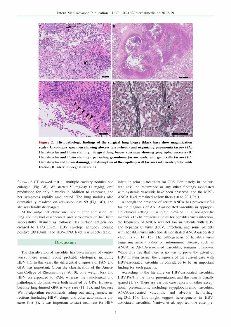
day 3, a transbronchial cryobiopsy of one of the nodules in
the right lower lobe revealed an abscess, organizing pneu-
monia, and marked hemosiderin deposition, suggesting in-
fection or vasculitis associated with hemorrhaging; neverthe-
less, the biopsy was not diagnostic (Fig. 2A). No microor-
ganisms were detected on lung tissue culture. On admission
day 12, a surgical lung biopsy was performed
(Fig. 2B, C and D), and the specimen revealed geographic
necrosis surrounded by palisading granuloma and multinu-
cleated giant cells. Surrounding lung parenchyma showed
capillaritis infiltrated with neutrophils, and no microorgan-
isms were identified with Ziehl-Neelsen and Grocott stains.
Therefore, histological GPA was suspected. However, there
were no abnormal findings in her ears or nose as observed
by an otolaryngologist, nor were any kidney abnormalities
noted.
In a multidisciplinary discussion, we decided on a diag-
nostic/therapeutic plan according to the classification algo-
rithm of vasculitis proposed by Watts et al. (8) for lung-
limited granulomatous polyangiitis after treating HBV-
associated vasculitis. Entecavir treatment was started from
admission day 34 (Fig. 3A). After a week of treatment with
entecavir, her symptoms of hemoptysis worsened, and
Intern Med Advance Publication DOI: 10.2169/internalmedicine.3012-19
3
Figure 2. Histopathologic findings of the surgical lung biopsy (black bars show magnification scale). Cryobiopsy specimen showing abscess (arrowhead) and organizing pneumonia (arrow) (A: Hematoxylin and Eosin staining). Surgical lung biopsy specimen showing geographic necrosis (B: Hematoxylin and Eosin staining), palisading granuloma (arrowheads) and giant cells (arrow) (C: Hematoxylin and Eosin staining), and disruption of the capillary wall (arrow) with neutrophilic infil-tration (D: silver impregnation stain).
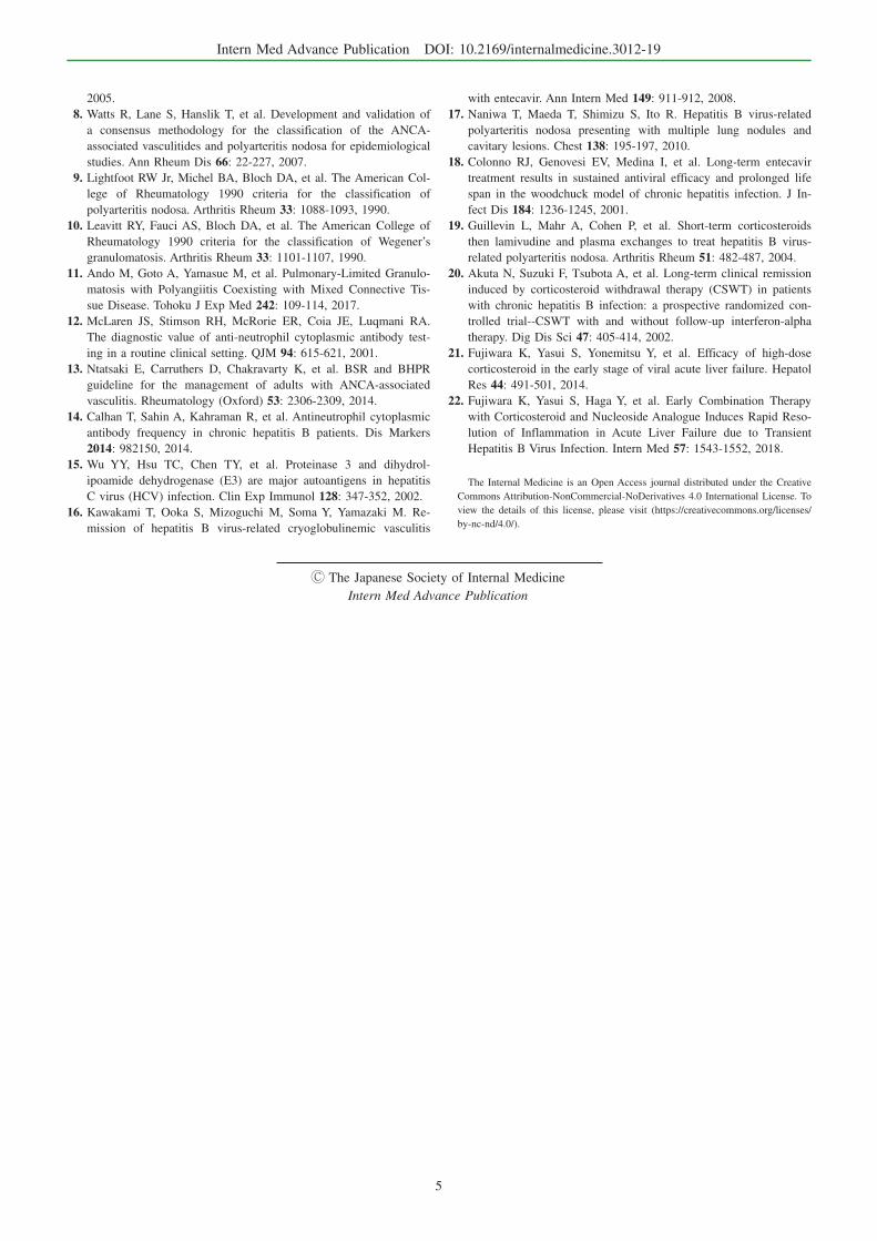
follow-up CT showed that all multiple cavitary nodules had
enlarged (Fig. 3B). We started 50 mg/day (1 mg/kg) oral
prednisone for only 2 weeks in addition to entecavir, and
her symptoms rapidly ameliorated. The lung nodules also
dramatically resolved on admission day 59 (Fig. 3C), and
she was finally discharged.
At the outpatient clinic one month after admission, all
lung nodules had disappeared, and seroconversion had been
successfully attained as follows: HB surface antigen de-
creased to 1,173 IU/ml, HBV envelope antibody became
positive (99 IU/ml), and HBV-DNA level was undetectable.
Discussion
The classification of vasculitis has been an area of contro-
versy; there remain some probable etiologies, including
HBV (1). In this case, the differential diagnosis of PAN and
GPA was important. Given the classification of the Ameri-
can College of Rheumatology (9, 10), only weight loss and
HBV corresponded to PAN, whereas the radiological and
pathological domains were both satisfied by GPA. However,
because lung-limited GPA is very rare (11, 12), and because
Watt’s algorithm recommends ruling out malignancies, in-
fections (including HBV), drugs, and other autoimmune dis-
eases first (8), it was important to start treatment for HBV
infection prior to treatment for GPA. Fortunately, in the cur-
rent case, no recurrence or any other findings associated
with systemic vasculitis have been observed, and the MPO-
ANCA level remained at low titers (10 to 20 U/ml).
Although the presence of serum ANCA has proven useful
for the diagnosis of ANCA-associated vasculitis in appropri-
ate clinical setting, it is often elevated in a non-specific
manner. (13) In previous studies for hepatitis virus infection,
the frequency of ANCA was not low in patients with HBV
and hepatitis C virus (HCV) infection, and some patients
with hepatitis virus infection demonstrated ANCA-associated
vasculitis (3, 14, 15). The pathogenesis of hepatitis virus
triggering autoantibodies or autoimmune disease, such as
ANCA or ANCA-associated vasculitis, remains unknown.
While it is true that there is no way to prove the extent of
HBV in lung tissue, the diagnosis of the current case with
HBV-associated vasculitis is considered to be an important
finding for such patients.
According to the literature on HBV-associated vasculitis,
HBV-PAN is the major presentation, and the lung is usually
spared (1, 7). There are various case reports of other excep-
tional presentations, including cryoglobulinemic vasculitis,
ANCA-associated vasculitis, and alveolar hemorrhag-
ing (3-5, 16). This might suggest heterogeneity in HBV-
associated vasculitis. Naniwa et al. reported one case pre-

Intern Med Advance Publication DOI: 10.2169/internalmedicine.3012-19
4
Figure 3. High-resolution CT findings at day 34 (A), 40 (B), and 59 (C). A: The bilateral nodules in the lower lobes were stable compared with the initial evaluation. B: The nodules had all grown, and there were increased numbers of cavitary le-sions. C: The nodules were dramatically resolved.
senting with multiple lung nodules, similar to the current
case, although without granulomatous changes. That case
turned out to be HBV-PAN (17). The current case demon-
strated pathological rarity in light of its lung-limited GPA-
like inflammation due to HBV infection, contributing to the
diversity among reports of HBV-associated vasculitis.
Considering the risk of HBV reactivation with corticoster-
oid treatment, we decided to treat her with entecavir first.
However, both her hemoptysis and lung nodules worsened
dramatically in the week following entecavir administration.
In terms of the pathogenesis of HBV-associated vasculitis,
this was assumed to be a type III or immune complex reac-
tion with HB surface antibody affecting the vascular
wall (2). Colonno et al. reported transient increases in HB
surface antigen levels within the initial eight weeks from en-
tecavir treatment in a woodchuck model (18); therefore, the
initial exacerbation in the current case might have been due
to immune complex reaction, although this cannot be
proven.
The efficacy of oral prednisone in the initial two weeks to
control ongoing organ or life-threatening inflammatory proc-
esses was reported in several French studies (7, 19). Indeed,
the lung nodules in the current case immediately resolved
after two weeks of oral prednisone. According to these
French studies, seroconversion of HBV was a key prognos-
tic factor for deterring the relapse of HBV-associated vascu-
litis. Short-term oral prednisone with abrupt stoppage might
enhance the immunological clearance of HBV-infected hepa-
tocytes and induce seroconversion from HB surface antigen
to HB envelope antibody (7, 19). In a Japanese randomized
controlled trial of 42 patients, short-term corticosteroid and
abrupt discontinuation were evident for seroconversion in
general HBV inflammation (20-22). In the current case,
combination therapy with entecavir and short-term predni-
sone and abrupt discontinuation was appropriate for sup-
pressing HBV replication to control the ongoing inflamma-
tion and induce seroconversion.
In conclusion, the current case report describes a rare
form of HBV-associated vasculitis presenting with multiple
cavitary nodules of necrosis, granuloma, and capillaritis,
mimicking GPA in the lung. Corticosteroid treatment led to
the resolution of signs and symptoms as well as successful
seroconversion.
The authors state that they have no Conflict of Interest (COI).
This research did not receive any specific grant from funding
agencies in the public, commercial, or not-for-profit sectors.
We would like to thank Editage (www.editage.jp) for English
language editing.
References
1. Jennette JC, Falk RJ, Bacon PA, et al. 2012 revised International
Chapel Hill Consensus Conference Nomenclature of Vasculitides.
Arthritis Rheum 65: 1-11, 2013.
2. Sharma A, Sharma K. Hepatotropic viral infection associated sys-
temic vasculitides-hepatitis B virus associated polyarteritis nodosa
and hepatitis C virus associated cryoglobulinemic vasculitis. J Clin
Exp Hepatol 3: 204-212, 2013.
3. Joshi U, Subedi R, Gajurel BP. Hepatitis B virus induced cytoplas-
mic antineutrophil cytoplasmic antibody-mediated vasculitis caus-
ing subarachnoid hemorrhage, acute transverse myelitis, and neph-
ropathy: a case report. J Med Case Rep 11: 91, 2017.
4. Mazzaro C, Dal Maso L, Urraro T, et al. Hepatitis B virus related
cryoglobulinemic vasculitis: A multicentre open label study from
the Gruppo Italiano di Studio delle Crioglobulinemie - GISC. Dig
Liver Dis 48: 780-784, 2016.
5. Chen B, Yang X, Sun S, et al. Propylthiouracil-Induced Vasculitis
With Alveolar Hemorrhage Confirmed by Clinical, Laboratory,
Computed Tomography, and Bronchoscopy Findings: A Case Re-
port and Literature Review. Iran Red Crescent Med J 18: e23320,
2016.
6. Ferreira C, Costa T, Marques AV. Diffuse alveolar haemorrhage
secondary to propylthiouracil-induced vasculitis. BMJ Case Rep
2015.
7. Guillevin L, Mahr A, Callard P, et al. Hepatitis B virus-associated
polyarteritis nodosa: clinical characteristics, outcome, and impact
of treatment in 115 patients. Medicine (Baltimore) 84: 313-322,
Intern Med Advance Publication DOI: 10.2169/internalmedicine.3012-19
5
2005.
8. Watts R, Lane S, Hanslik T, et al. Development and validation of
a consensus methodology for the classification of the ANCA-
associated vasculitides and polyarteritis nodosa for epidemiological
studies. Ann Rheum Dis 66: 22-227, 2007.
9. Lightfoot RW Jr, Michel BA, Bloch DA, et al. The American Col-
lege of Rheumatology 1990 criteria for the classification of
polyarteritis nodosa. Arthritis Rheum 33: 1088-1093, 1990.
10. Leavitt RY, Fauci AS, Bloch DA, et al. The American College of
Rheumatology 1990 criteria for the classification of Wegener’s
granulomatosis. Arthritis Rheum 33: 1101-1107, 1990.
11. Ando M, Goto A, Yamasue M, et al. Pulmonary-Limited Granulo-
matosis with Polyangiitis Coexisting with Mixed Connective Tis-
sue Disease. Tohoku J Exp Med 242: 109-114, 2017.
12. McLaren JS, Stimson RH, McRorie ER, Coia JE, Luqmani RA.
The diagnostic value of anti-neutrophil cytoplasmic antibody test-
ing in a routine clinical setting. QJM 94: 615-621, 2001.
13. Ntatsaki E, Carruthers D, Chakravarty K, et al. BSR and BHPR
guideline for the management of adults with ANCA-associated
vasculitis. Rheumatology (Oxford) 53: 2306-2309, 2014.
14. Calhan T, Sahin A, Kahraman R, et al. Antineutrophil cytoplasmic
antibody frequency in chronic hepatitis B patients. Dis Markers
2014: 982150, 2014.
15. Wu YY, Hsu TC, Chen TY, et al. Proteinase 3 and dihydrol-
ipoamide dehydrogenase (E3) are major autoantigens in hepatitis
C virus (HCV) infection. Clin Exp Immunol 128: 347-352, 2002.
16. Kawakami T, Ooka S, Mizoguchi M, Soma Y, Yamazaki M. Re-
mission of hepatitis B virus-related cryoglobulinemic vasculitis
with entecavir. Ann Intern Med 149: 911-912, 2008.
17. Naniwa T, Maeda T, Shimizu S, Ito R. Hepatitis B virus-related
polyarteritis nodosa presenting with multiple lung nodules and
cavitary lesions. Chest 138: 195-197, 2010.
18. Colonno RJ, Genovesi EV, Medina I, et al. Long-term entecavir
treatment results in sustained antiviral efficacy and prolonged life
span in the woodchuck model of chronic hepatitis infection. J In-
fect Dis 184: 1236-1245, 2001.
19. Guillevin L, Mahr A, Cohen P, et al. Short-term corticosteroids
then lamivudine and plasma exchanges to treat hepatitis B virus-
related polyarteritis nodosa. Arthritis Rheum 51: 482-487, 2004.
20. Akuta N, Suzuki F, Tsubota A, et al. Long-term clinical remission
induced by corticosteroid withdrawal therapy (CSWT) in patients
with chronic hepatitis B infection: a prospective randomized con-
trolled trial--CSWT with and without follow-up interferon-alpha
therapy. Dig Dis Sci 47: 405-414, 2002.
21. Fujiwara K, Yasui S, Yonemitsu Y, et al. Efficacy of high-dose
corticosteroid in the early stage of viral acute liver failure. Hepatol
Res 44: 491-501, 2014.
22. Fujiwara K, Yasui S, Haga Y, et al. Early Combination Therapy
with Corticosteroid and Nucleoside Analogue Induces Rapid Reso-
lution of Inflammation in Acute Liver Failure due to Transient
Hepatitis B Virus Infection. Intern Med 57: 1543-1552, 2018.
The Internal Medicine is an Open Access journal distributed under the Creative
Commons Attribution-NonCommercial-NoDerivatives 4.0 International License. To
view the details of this license, please visit (https://creativecommons.org/licenses/
by-nc-nd/4.0/).
Ⓒ The Japanese Society of Internal Medicine
Intern Med Advance Publication