hepatic metastasis from choriocarcinoma: angiographic ... · pdf file260 korean j radiol 3(4),...
TRANSCRIPT

260 Korean J Radiol 3(4), December 2002
Hepatic Metastasis fromChoriocarcinoma: Angiographic Findingsin Two Cases
We report two cases of hepatic metastases from choriocarcinoma in women ofchildbearing age in whom imaging studies performed at presentation revealedthe presence of liver masses, and who had clinically progressive anemia or intra-abdominal hemorrhage. CT demonstrated heterogeneously enhanced livermasses. Characteristic angiographic findings included hypervascular hepaticmasses with aneurysmal dilatations of the peripheral hepatic arteries at the arteri-al phase and persistent vascular lakes at the venous phase.
horiocarcinoma is a neoplasm of syncytiotrophoblastic origin, of whichapproximately 50% occur after a molar pregnancy, 25% after a normalpregnancy, and 24% after spontaneous abortion (1, 2). It is a highly vas-
cularized tumor and because of the affinity of trophoblasts for blood vessels, showsrapid hematogeneous metastasis. This is commonly to the lung and vagina, althoughother sites including the brain, genitourinary and gastrointestinal system have alsobeen reported. When manifestation of metastasis precedes that of the primary lesion,the findings of imaging studies present a diagnostic challenge.
We report two cases of choriocarcinoma which metastasized to the liver in patientswith acute and massive abdominal bleeding and characteristic angiographic findings.
CASE REPORTS
Case 1A 33-year-old Asian woman (gravida 3, para 2) presented with upper abdominal
pain and distension, and weight loss of 4kg in one month. Her medical history was un-remarkable, though an intrauterine device had been inserted after her last pregnancy,four years earlier.
Computed tomography (CT) revealed multiple, irregularly shaped, heterogeneouslyenhanced mass lesions of variable size in the liver and spleen, suggesting hepaticmetastasis from splenic angiosarcoma or multifocal hepatocellular carcinoma. The con-tour of the uterus appeared to be normal. Diagnostic celiac angiography demonstratedmultiple hypervascular masses with abnormal tumor vessels and saccular aneurysmaldilatation of the peripheral end of hepatic arteries at the arterial phase, and persistentvisualization of hepatic and splenic vascular lakes at the venous phase, but activebleeding was not apparent (Figs. 1A, B). The size and distribution of the masses weremore extensive than CT initially suggested. SMA angiography demonstrated normalportal flow.
Diagnostic laparoscopy was performed, and the findings included multiple hepaticmasses, splenomegaly with multiple nodules, and a large (7 8-cm) cystic and solid
Yun Jung Kang, MD1
Joo Hyeong Oh, MD1
Yup Yoon, MD1
Eui Jong Kim, MD1
Deog Yoon Kim, MD2
Heung Sun Kang, MD2
Index terms:ChoriocarcinomaLiver neoplasm, metastasesLiver neoplasm, angiography
Korean J Radiol 2002;3:260-263Received March 2, 2002; accepted after revision July 29, 2002.
Departments of 1Diagnostic Radiologyand 2Internal Medicine, Kyung HeeUniversity College of Medicine
Address reprint requests to:Joo Hyeong Oh, MD, Department ofDiagnostic Radiology, Kyung HeeUniversity Hospital, 1 Hoeki-dong,Dongdaemun-gu, Seoul 130-702, Korea.Telephone: (822) 958-8622Fax: (822) 968-0787e-mail: [email protected]
C

mass in the left ovary. Laparoscopic biopsy of the hepaticand ovarian masses followed by histolopathologic exami-nation revealed metastatic choriocarcinoma of the liverand a hemorrhagic lutein cyst in the left ovary. The serumbeta hCG concentration was very high, at 2,000,000mIU/mL, but chest radiography revealed nothing abnor-mal.
Five days after admission, sudden changes in the labora-tory data (hemoglobin, 8.7 mg/dl; hematocrit, 26.3%;platelet count, 80,000/mm3) suggested active bleeding, andexploratory laparotomy revealed 4000 cc of blood in theperitoneal cavity and active bleeding from the liver, spleenand both ovaries. Despite bilateral oophorectomy andsurgery known as the Pringle method to control bleedingfrom the liver, the flow could not be staunched and emer-gency angiography was again performed. Celiac angiogra-phy demonstrated massive extravasation of contrast mediafrom the posterior superior and inferior segmental branch-es of the right hepatic artery and lateral segmental branchof the left hepatic artery, as well as from the branch in theupper pole of the spleen, and immediate embolization wassuccessfully performed using 16 Tornado microcoils (two,4 mm 2 cm in size; four, 3 mm 2 cm; five, 5 mm 2 cm;five, 6 mm 2 cm) and gelfoam particles after superselec-tion of the segmental arteries of both hepatic lobes and thespleen. Post-embolization angiography demonstrated nofurther extravasation of contrast media and intact portalflow. The patient was then observed for evidence of fur-ther bleeding.
AST and ALT levels continuously and rapidly increased,however, suggesting progression to hepatic failure and aci-
dosis, and follow-up CT performed 13 days after emboliza-tion showed an extensive area of low attenuation with aminimally enhanced region in the enlarged liver andspleen, representing massive hepatic infarction and necro-sis. Although the exact cause of this infarction was un-known, it was assumed that embolization was the probablecause of hepatic failure, though some normal parenchymalportions and intact portal flow remained. However, the pa-tient’s condition continued to deteriorate, and she died 22days after embolization.
Case 2A 33-year-old Asian woman (gravida 4, para 0) was hos-
pitalized via the emergency department for severe abdomi-nal pain without vaginal bleeding. A hydatidiform molewith pulmonary metastasis had been diagnosed about fiveyears earlier and the patient had subsequently undergoneeleven cycles of chemotherapy. One year before hospital-ization, however, she was lost to follow-up.
A urinary pregnancy test was positive, showing a serumbeta hCG level of over 2,800,000 mIU/mL. Chest CTdemonstrated that multiple hematogenous metastatic nod-ules extended throughout both lungs, while abdominal/pelvic CT revealed huge, ill-defined, heterogeneously en-hanced masses in both lobes of the liver and thrombosis inthe left portal vein (Fig. 2A). At precontrast CT, somewhathighly attenuated ascites, suggestive of hemoperitoneum,was apparent. The contour of the uterus was normal. Thedifferential diagnoses included other metastatic diseases,primary hepatocellular carcinoma, and other hypervascu-lar hepatic tumors. After the confirmation of laboratory
Angiographic Findings of Hepatic Metastasis from Choriocarcinoma
Korean J Radiol 3(4), December 2002 261
Fig. 1. Case 1. Hepatic metastasis from choriocarcinoma in a 33-year-old woman.A. Arterial-phase celiac angiogram depicts multiple hypervascular masses with abnormal tumor vessels and saccular aneurysmal dilata-tion of the peripheral end of hepatic arteries in the liver and spleen.B. Venous phase celiac angiogram demonstrates aneurysmal hepatic and splenic vascular lakes, but active bleeding is not apparent.
A B

data indicating massive intraperitoneal hemorrhage, ex-ploratory laparotomy was performed.
During surgery, 1500 cc of blood was evacuated fromthe peritoneum, and the liver was found to have beenlargely replaced by numerous friable tumor deposits.Several of these in the left lobe were, moreover, ruptured,and were bleeding profusely and continuously. The hepaticmasses were ligated, but hemostasis was not achieved.
Two days after surgery a sudden overnight drop in thehemoglobin level, from 9.5 g to 5.5 g, occurred, and thepatient’s condition continued to deteriorate. A second la-parotomy was undertaken to investigate the reason for thisdrop, and during surgery severe hemorrhage from the fri-able liver tumors was observed. Hemostasis could not,however, be attained.
On the same day, emergency angiography was per-formed. Hepatic arterial-phase arteriography demonstratedthe presence of huge, multiple, exophytic hypervascularmasses with abnormal tumor vessels, saccular aneurysmaldilatation of the hepatic arteries, and arteriovenous shunts,
and at the venous phase, persistent bilobar vascular lakeswere observed. Active bleeding, was not apparent (Figs.2B, C). SMA portography revealed thrombosis of the leftportal vein, and using gelfoam particles, superselective em-bolization of the right and left hepatic arteries was success-fully performed. Post-embolization angiography demon-strated residual tumor staining supplied by the cystic arteryand medial segmental branch of the left hepatic artery, butthe previously noted staining of other masses was not ob-served. The laboratory data also indicated that bleedinghas ceased.
Despite the treatment described, the patient’s conditioncontinued to deteriorate rapidly. Respiratory failure en-sued, and she died 17 days after admission to hospital.
DISCUSSION
Choriocarcinoma is a rare malignant trophoblastic tumorcomplicating between 1:13,000 and 1:50,000 pregnanciesin Caucasian populations and a higher proportion among
Kang et al.
262 Korean J Radiol 3(4), December 2002
A B
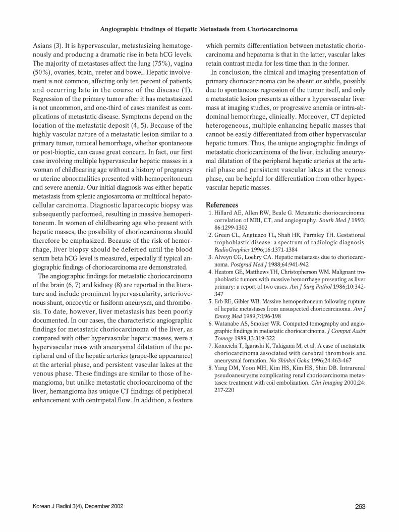
Fig. 2. Case 2. Hepatic metastasis from choriocarcinoma in a33-year-old woman.A. Contrast-enhanced CT image depicts huge, ill-defined, het-erogeneously enhanced masses in both lobes of the liver, andthrombosis in the left portal vein.B. Arterial-phase hepatic arteriogram reveals huge, multiple,exophytic hypervascular masses with saccular aneurysmal di-latation of the peripheral hepatic arteries and arteriovenousshunting in both lobes of the liver.C. Venous-phase hepatic arteriogram demonstrates persistenthepatic vascular lakes.
C
Angiographic Findings of Hepatic Metastasis from Choriocarcinoma
Korean J Radiol 3(4), December 2002 263
Asians (3). It is hypervascular, metastasizing hematoge-nously and producing a dramatic rise in beta hCG levels.The majority of metastases affect the lung (75%), vagina(50%), ovaries, brain, ureter and bowel. Hepatic involve-ment is not common, affecting only ten percent of patients,and occurring late in the course of the disease (1).Regression of the primary tumor after it has metastasizedis not uncommon, and one-third of cases manifest as com-plications of metastatic disease. Symptoms depend on thelocation of the metastatic deposit (4, 5). Because of thehighly vascular nature of a metastatic lesion similar to aprimary tumor, tumoral hemorrhage, whether spontaneousor post-bioptic, can cause great concern. In fact, our firstcase involving multiple hypervascular hepatic masses in awoman of childbearing age without a history of pregnancyor uterine abnormalities presented with hemoperitoneumand severe anemia. Our initial diagnosis was either hepaticmetastasis from splenic angiosarcoma or multifocal hepato-cellular carcinoma. Diagnostic laparoscopic biopsy wassubsequently performed, resulting in massive hemoperi-toneum. In women of childbearing age who present withhepatic masses, the possibility of choriocarcinoma shouldtherefore be emphasized. Because of the risk of hemor-rhage, liver biopsy should be deferred until the bloodserum beta hCG level is measured, especially if typical an-giographic findings of choriocarcinoma are demonstrated.
The angiographic findings for metastatic choriocarcinomaof the brain (6, 7) and kidney (8) are reported in the litera-ture and include prominent hypervascularity, arteriove-nous shunt, oncocytic or fusiform aneurysm, and thrombo-sis. To date, however, liver metastasis has been poorlydocumented. In our cases, the characteristic angiographicfindings for metastatic choriocarcinoma of the liver, ascompared with other hypervascular hepatic masses, were ahypervascular mass with aneurysmal dilatation of the pe-ripheral end of the hepatic arteries (grape-lke appearance)at the arterial phase, and persistent vascular lakes at thevenous phase. These findings are similar to those of he-mangioma, but unlike metastatic choriocarcinoma of theliver, hemangioma has unique CT findings of peripheralenhancement with centripetal flow. In addition, a feature
which permits differentiation between metastatic chorio-carcinoma and hepatoma is that in the latter, vascular lakesretain contrast media for less time than in the former.
In conclusion, the clinical and imaging presentation ofprimary choriocarcinoma can be absent or subtle, possiblydue to spontaneous regression of the tumor itself, and onlya metastatic lesion presents as either a hypervascular livermass at imaging studies, or progressive anemia or intra-ab-dominal hemorrhage, clinically. Moreover, CT depictedheterogeneous, multiple enhancing hepatic masses thatcannot be easily differentiated from other hypervascularhepatic tumors. Thus, the unique angiographic findings ofmetastatic choriocarcinoma of the liver, including aneurys-mal dilatation of the peripheral hepatic arteries at the arte-rial phase and persistent vascular lakes at the venousphase, can be helpful for differentiation from other hyper-vascular hepatic masses.
References1. Hillard AE, Allen RW, Beale G. Metastatic choriocarcinoma:
correlation of MRI, CT, and angiography. South Med J 1993;86:1299-1302
2. Green CL, Angtuaco TL, Shah HR, Parmley TH. Gestationaltrophoblastic disease: a spectrum of radiologic diagnosis.RadioGraphics 1996;16:1371-1384
3. Alveyn CG, Loehry CA. Hepatic metastases due to choriocarci-noma. Postgrad Med J 1988;64:941-942
4. Heatom GE, Matthews TH, Christopherson WM. Malignant tro-phoblastic tumors with massive hemorrhage presenting as liverprimary: a report of two cases. Am J Surg Pathol 1986;10:342-347
5. Erb RE, Gibler WB. Massive hemoperitoneum following ruptureof hepatic metastases from unsuspected choriocarcinoma. Am JEmerg Med 1989;7:196-198
6. Watanabe AS, Smoker WR. Computed tomography and angio-graphic findings in metastatic choriocarcinoma. J Comput AssistTomogr 1989;13:319-322
7. Komeichi T, Igarashi K, Takigami M, et al. A case of metastaticchoriocarcinoma associated with cerebral thrombosis andaneurysmal formation. No Shinkei Geka 1996;24:463-467
8. Yang DM, Yoon MH, Kim HS, Kim HS, Shin DB. Intrarenalpseudoaneurysms complicating renal choriocarcinoma metas-tases: treatment with coil embolization. Clin Imaging 2000;24:217-220