hepatic glucagon receptor signaling enhances insulin ... · ser473; this effect was reproduced in...
TRANSCRIPT

Hepatic Glucagon Receptor Signaling EnhancesInsulin-Stimulated Glucose Disposal in RodentsTeayoun Kim,1 Cassie L. Holleman,1 Shelly Nason,1 Deanna M. Arble,2 Nickki Ottaway,3 Joseph Chabenne,4
Christine Loyd,1 Jeong-a Kim,1 Darleen Sandoval,5 Daniel J. Drucker,6 Richard DiMarchi,4,7
Diego Perez-Tilve,3 and Kirk M. Habegger1
Diabetes 2018;67:2157–2166 | https://doi.org/10.2337/db18-0068
Glucagon receptor (GCGR) agonists cause hyperglyce-mia but also weight loss. However, GCG-like peptide1 receptor (GLP1R)/GCGR mixed agonists do not exhibitthe diabetogenic effects often attributed to GCGR activ-ity. Thus, we sought to investigate the effect of glucagonagonism on insulin action and glucose homeostasis.Acute GCGR agonism induced immediate hyperglyce-mia, followed by improved glucose tolerance and en-hanced glucose-stimulated insulin secretion. Moreover,acute GCGR agonism improved insulin tolerance in adose-dependent manner in both lean and obese mice.Improved insulin tolerance was independent of GLP1R,FGF21, and hepatic glycogenolysis. Moreover, we ob-served increased glucose infusion rate, disposal, uptake,and suppressed endogenous glucose production duringeuglycemic clamps. Mice treated with insulin and GCGRagonist had enhanced phosphorylation of hepatic AKT atSer473; this effect was reproduced in isolated mouse pri-mary hepatocytes and resulted in increased AKT kinaseactivity. These data reveal that GCGR agonism enhancesglucose tolerance, in part, by augmenting insulin action,with implications for the use of GCGR agonism in thera-peutic strategies for diabetes.
Glucagon (GCG) is a 29–amino acid peptide released froma-cells of the pancreatic islet. Its role as a primary counter-regulatory hormone to insulin action has long receivedscientific attention, yet its broader therapeutic potential
is underappreciated (1,2). Intriguingly, the antidiabeticactions of GCG-like peptide 1 receptor (GLP1R) agonismare enhanced by the catabolic and hypolipidemic proper-ties of GCG receptor (GCGR) agonism (3–5). Importantly,GLP1R/GCGR mixed agonists do not exhibit the hyper-glycemia and glucose intolerance often attributed to GCGRactivity, findings that were confirmed in human subjects(6). Moreover, postprandial elevations of GCG and GLP-1may contribute to improved postprandial glucose homeo-stasis in Roux-en-Y gastric bypass patients (7,8). HowGCGR monoagonism promotes transient glucose intoler-ance, yet simultaneously contributes to the antidiabeticactions of incretin-based therapies is still unknown. Asa counterregulatory hormone with a role in maintainingfasting blood glucose, it is tempting to assume that GCGopposes all actions of insulin. However, the increasedconcentrations and action of GCG in the fasting stateare well suited to potentiate subsequent, insulin-mediatedglucose control. In the context of normal physiology, ex-ercise induces GCG secretion (9) and modulates the ce-phalic response of meal assimilation (10). Altogether thesedata suggest that GCG may in fact contribute to thesestates of heightened insulin sensitivity.
Chronic GCGR agonism in diet-induced obese (DIO)mice stimulates weight loss, hyperglycemia, and glucoseintolerance (11). Unexpectedly, GCGR signaling in db/dbmice improved the rate of insulin-stimulated glucosedisappearance (kg) (11). Here we demonstrate that, in
1Comprehensive Diabetes Center and Division of Endocrinology, Diabetes, andMetabolism, Department of Medicine, University of Alabama at Birmingham,Birmingham, AL2Department of Biological Sciences, Marquette University, Milwaukee, WI3Metabolic Diseases Institute and Division of Endocrinology, Diabetes, andMetabolism, Department of Medicine, University of Cincinnati, Cincinnati, OH4Novo Nordisk Research Center, Indianapolis, IN5Department of Surgery, University of Michigan, Ann Arbor, MI6Lunenfeld-Tanenbaum Research Institute, Mt. Sinai Hospital, Department ofMedicine, University of Toronto, Toronto, Ontario, Canada7Department of Chemistry, Indiana University, Bloomington, IN
Corresponding author: Kirk M. Habegger, [email protected], or DiegoPerez-Tilve, [email protected].
Received 16 January 2018 and accepted 10 August 2018.
This article contains Supplementary Data online at http://diabetes.diabetesjournals.org/lookup/suppl/doi:10.2337/db18-0068/-/DC1.
© 2018 by the American Diabetes Association. Readers may use this article aslong as the work is properly cited, the use is educational and not for profit, and thework is not altered. More information is available at http://www.diabetesjournals.org/content/license.
Diabetes Volume 67, November 2018 2157
METABOLISM
addition to its counterregulatory role, hepatic GCGR ago-nism enhances systemic insulin-stimulated glucose dis-posal. Together, these data mechanistically elucidate howglucose control may be improved by therapies characterizedby GCG agonism.
RESEARCH DESIGN AND METHODS
Animal ModelsAll studies were approved by and performed according tothe guidelines of the Institutional Animal Care and UseCommittee of the University of Alabama at Birmingham orthe University of Cincinnati. Mice were single or grouphoused on a 12:12-h light-dark cycle (light on from 0600 hto 1800 h) at 22°C and constant humidity with free accessto food and water, except as noted. Fgf21-deficient, Gcgr-floxed, and Glp1r-deficient mice were generated as pre-viously described (12–15), and Alb-Cre mice were obtainedfrom The Jackson Laboratory (Bar Harbor, ME). All micewere maintained in our facilities on a C57Bl/6J backgroundand fed a standard chow (5.6% fat, Teklad LM-485) or high-fat diet (58.0 kcal% fat, D12331; Research Diets, NewBrunswick, NJ).
Peptides and InhibitorsNovel GCGR agonists (IUB288 and ASP28, GLU29-GCG)were synthesized as previously described (11,16). NativeGCG and insulin (Humulin R) were obtained from Amer-ican Peptide Co. and Eli Lilly and Co., respectively. Gly-cogen phosphorylase a/b inhibitor (BAY R3401) wasobtained from Sigma-Aldrich and diluted in 0.5% methylcellulose.
Glucose and Insulin Tolerance TestsGlucose and insulin tolerance tests (GTTs and ITTs) wereperformed in 6 h–fasted 8- to 10-week-old chow-fed or24-week-old DIO male C57Bl/6J mice by i.p. injection ofIUB288 (10 nmol/kg) or GCG (10 nmol/kg) 60 min prior toi.p. injection of glucose (2 g/kg, 20% weight for volume[w/v] D-glucose [Sigma-Aldrich] in 0.9% w/v saline) or in-sulin (0.25–1.0 units/kg in 0.9% w/v saline). ITTs wereconducted in lean chow-fed male Glp1r2/2 and Fgf21Δliver
mice at 10–14 weeks old. GTTs and ITTs were conductedin 8- to 12- and 10- to 14-week-old GcgrΔliver mice, respec-tively. Blood glucose was determined by the TheraSenseFreestyle Glucometer. Glucose disappearance rate (kg) wasdefined as (D blood glucose/min). Insulin and C-peptidewere measured from blood collected 60 min after IUB288challenge.
Intravenous Glucose-Stimulated Insulin Secretion andGCGR Agonist Infusion TestsCatheters were surgically implanted as previously de-scribed (17). Four days after surgery, lean chow-fed14-week-old male C57Bl/6J mice were fasted for 5 h.IUB288 injection (10 nmol/kg i.p.) was administered60 min prior to glucose bolus (1 g/kg) delivered via venouscatheter. Plasma samples were collected immediately be-fore and 2, 5, 10, and 15 min after infusion. For GCGR
agonist infusion studies, chow-fed 14-week-old maleC57Bl/6J mice were fasted for 4 h before 120 min ASP28,GLU29-GCG infusion (0.00064 and 0.0064 nmol/min). Micewere administered 0.25 units/kg insulin i.p. and euthanized20 min later.
Euglycemic ClampsClamps were conducted as previously described (17). Inbrief, catheters were implanted in male 8-week-old chow-fed C57Bl/6J mice. Four to six days postoperative, micewere fasted for 5 h with saline or IUB288 (10 nmol/kg) in-jected (s.c.) during the final 60 min. Insulin (4 mU/kg/min,diluted in saline) was infused through the venous cathe-ter, and euglycemia (140 mg/dL) was maintained by ad-justing the infusion rate of a 20% glucose solution. A tracerequilibration period (t = 2120 to 0 min) was used asfollows: a 5 mCi bolus of [3-3H]-glucose (PerkinElmer,Boston, MA) was given at t = 2120 min followed bya 0.05 mCi/min infusion for 2 h. Somatostatin (SST;EMD Millipore, Temecula, CA), mixed with the [3-3H]-glucose, was infused at 1.5 mg/kg/min from t = 2120 to0 min and at 3mg/kg/min from t = 0min to 120 min. At t =0, [3-3H]-glucose infusion was increased to 0.1 mCi/min tominimize changes in specific activity (SA). The variationof SA was ,10% from mean during the last 40 min ofclamp, and the slope of SA over time was not significantlydifferent from t = 0. Blood samples (100 mL) were takenat2120,260,230,25, 90, 100, 110, and 120min for theassessment of glucose, insulin, and glucose SA in plasma.Red blood cells from these samples were recovered bycentrifugation and injected via arterial catheter to preventa hematocrit deficit. A 10 mCi [1-14C]-2 deoxy-D-glucose([1-14C]-2DG) (MP Biomedicals, Santa Ana, CA) bolus wasinjected at t = 75 min via carotid arterial catheter to assessglucose uptake. After the clamp, mice were euthanized andtissues (gastrocnemius, soleus, extensor digitorum longus[EDL], quadriceps, liver, gonadal white adipose tissue[WAT], and interscapular brown adipose tissue [BAT])were snap frozen in liquid nitrogen.
Biochemical AssaysPlasma [3-3H]-glucose, [1-14C]-2DG, and 3H2O were mea-sured to determine rate of endogenous glucose production(Ra), rate of glucose disposal (Rd), and uptake as previouslydescribed (17). Clamp plasma glucose was measured from20 mL of deproteinized samples (Glucose Assay Kit; CellBiolabs, San Diego, CA). Tissue-specific [1-14C]-2DG-6-phosphate ([1-14C]-2DGp) content was determined viaperchloric acid–extracted supernatant with Somogyi pro-cedure (18). Glycogen was assessed from [3-3H]-glucoseincorporated into ethanol-precipitated glycogen in KOH-digested tissues (19). Plasma glucose SA was calculatedfrom the ratio of plasma glucose radioactivity (dpm) overplasma glucose, multiplied by the ratio of chemical standardevaporated (CSE) to chemical recovery standard (CRS). The[3-3H]-glucose infusion rate (dpm/kg/min) was then calcu-lated fromCSE. Rd (mg/kg/min) was determined as the ratio
2158 Glucagon Receptor and Glucose Disposal Diabetes Volume 67, November 2018

of the [3-3H]-glucose infusion rate and the plasma glucoseSA of (dpm/mg) at the end of the basal period and duringthe final 30 min of the clamps. Hepatic glucose productionrates (Endo Ra; mg/kg/min) were determined by subtractingthe steady-state glucose infusion rate (GIR) from Rd. Plasma[14C]-2DG SA (dpm/mg) was obtained by multiplying theradioactivity disappearance area under the curve (AUC) of[14C] in plasma samples by CSE/CRS and then dividing bythe average blood glucose during the clamped time course.Finally, tissue-specific 2DG uptake (Rg; mg glucose/mgtissue/min) was determined as the ratio of the [14C]-2DGpin tissue per tissue weight to plasma [14C]-2DG SA. Plasmainsulin and C-peptide were measured using mouse ELISAkits (Crystal Chem, Downers Grove, IL).
Primary Hepatocyte IsolationPrimary hepatocytes were prepared from anesthetized leanchow-fed male C57Bl/6J mice as previously described (20).In brief, perfusion buffer (Krebs Ringer with glucose and0.1 mmol/L EGTA), followed by digestion buffer (KrebsRinger with glucose, 1.4 mmol/L CaCl2, 50 mg/mL liberase[05401119001; Roche]), was infused into the vena cava viaperistaltic pump. Hepatocytes were recovered by centrifu-gation (50g 3 min, three times) and seeded on rat tail type 1collagen–coated plates in DMEM (10% FBS, 1% penicillin/streptomycin) with all experiments conducted ,24 h post-isolation. Hepatocytes were serum starved 60 min prior to2.5 min insulin or GCG treatment for signaling studies. Forglycogen assay, hepatocytes were fasted overnight in serum-free minimum essential medium alpha (1 g/L glucose) prior to2-h insulin or IUB288 treatment with [3-3H]-glucose.
Immunoblot AnalysesCell extracts were prepared in lysis buffer (20 mm Tris [pH6.8], 3.8 mm dithiothreitol, 10% glycerol, 1% SDS, 0.3mol/L phenylmethylsulfonyl fluoride, and HALT proteaseinhibitor cocktail [Thermo Fisher Scientific]), rotated for15 min at 4°C, and centrifuged for 10 min at 4°C. Equiv-alent protein amounts were separated by 7.5% SDS-PAGE.Resolved fractions were transferred to polyvinylidenefluoride (Bio-Rad Laboratories, Hercules, CA), and phos-phorylation was detected using phosphospecific antibod-ies to AKT473, AKT308, p44/42 MAPK202/204, forkhead boxprotein O124 (FOXO124), glycogen synthase kinase (GSK)3a/b21/9 (Cell Signaling Technology, Danvers, MA), andIRS1612 (LifeTechnologies, Frederick, MD). Phosphoryla-tion was normalized by TGX stain-free technology and byimmunoblot analysis with anti-AKT, anti-IRS1, FOXO1,and anti-GSK3 (Cell Signaling Technology). Immunoblotswere labeled with goat anti-rabbit horseradish peroxidase–conjugated secondary antibodies, and protein bands weredetected and quantified using Clarity ECL, ChemDoc im-aging system, and Image Lab 5.0 software (Bio-Rad Lab-oratories).
AKT Kinase Activity AnalysisKinase analysis was performed in freshly isolated primaryhepatocytes after 2.5 min of insulin/GCG treatments using
AKT Kinase Assay Kit (9840; Cell Signaling Technology).Blots from subsequent immunoblot analysis were imagedusing LumoGLO substrate provided in kit and normalizedto total protein by TGX stain-free technology.
StatisticsAll data are represented as mean and SEM. Statisticalsignificance was determined using unpaired Student t testsor, where appropriate, one- and two-way ANOVA withmultiple comparisons Tukey and Sidak posttest, respec-tively. Statistics were completed using GraphPad Prismversion 6.0 for Macintosh (GraphPad Software, San Diego,CA) and significance assigned when P , 0.05.
RESULTS
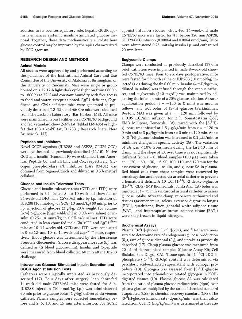
Acute GCGR Agonism Enhances Insulin Action andSecretion in MiceTo interrogate GCGR agonism in glucose metabolism, weadministered the GCGR agonist IUB288 (11,15) to leanchow-fed C57Bl/6J mice via i.p. injection (10 nmol/kg).IUB288 administration resulted in a rapid rise in glycemiaand a subtle increase in plasma insulin (Fig. 1A and B), bothreturning to baseline levels within 60 min. Surprisingly, ani.p. GTT performed 60 min after single injection of IUB288in lean mice revealed a significant enhancement of glucosetolerance compared with vehicle (Fig. 1C). Importantly,this effect was recapitulated by native GCG (Supplemen-tary Fig. 1A and inset). Consistent with prior observationsof GCGR-enhanced glucose-stimulated insulin secretion(GSIS) (21), IUB288 pretreatment (260 min) increasedglucose-stimulated insulin levels in response to an i.v. glu-cose challenge (1 g/kg) (Fig. 1D and inset). These data andprevious findings (21) suggest that GCG-stimulated en-hancements in GSIS likely contribute to the improvedglucose tolerance after GCGR agonism.
Acute GCGR Agonism Enhances Insulin Sensitivityin MiceAlthough GCGR-dependent potentiation of GSIS may con-tribute to enhanced glucose tolerance, this mechanism isunlikely to account for any enhancement of insulin action.To assess the effects of GCGR agonism on insulin-dependentglucose disposal, we treated lean mice with IUB288 60 minprior to an ITT (0.5 units/kg). IUB288 significantly enhancedthe response to insulin, as determined by both the nadirglucose and the calculated kg over the initial 30-min period(Fig. 1E). To assay endogenous insulin during this test, wemeasured circulating C-peptide (Fig. 1F) and observed thatthe enhanced insulin action was independent of increasedinsulin secretion. Importantly, this sensitizing effect wasrecapitulated by native GCG during both 0.25 units/kgand 0.5 units/kg ITTs without an increase in circulatingC-peptide (Supplementary Fig. 1B–D) and was similarlyobserved during coadministration of IUB288 and insulin(Supplementary Fig. 1E).
To assess how long improved insulin action remainedevident after GCGR agonism, we increased the pretreatment
diabetes.diabetesjournals.org Kim and Associates 2159
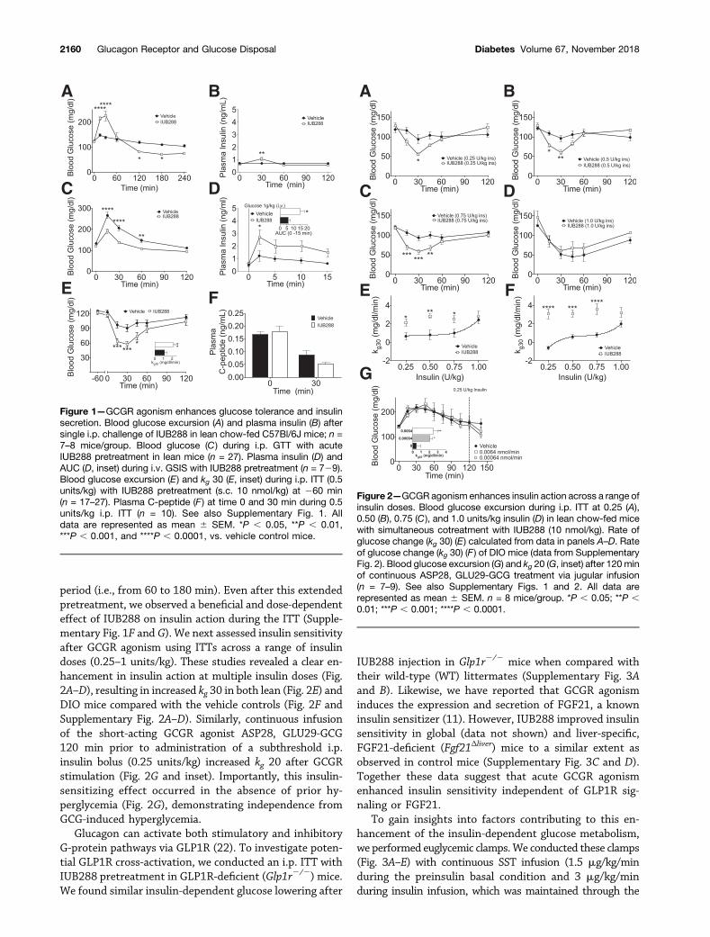
period (i.e., from 60 to 180 min). Even after this extendedpretreatment, we observed a beneficial and dose-dependenteffect of IUB288 on insulin action during the ITT (Supple-mentary Fig. 1F and G). We next assessed insulin sensitivityafter GCGR agonism using ITTs across a range of insulindoses (0.25–1 units/kg). These studies revealed a clear en-hancement in insulin action at multiple insulin doses (Fig.2A–D), resulting in increased kg 30 in both lean (Fig. 2E) andDIO mice compared with the vehicle controls (Fig. 2F andSupplementary Fig. 2A–D). Similarly, continuous infusionof the short-acting GCGR agonist ASP28, GLU29-GCG120 min prior to administration of a subthreshold i.p.insulin bolus (0.25 units/kg) increased kg 20 after GCGRstimulation (Fig. 2G and inset). Importantly, this insulin-sensitizing effect occurred in the absence of prior hy-perglycemia (Fig. 2G), demonstrating independence fromGCG-induced hyperglycemia.
Glucagon can activate both stimulatory and inhibitoryG-protein pathways via GLP1R (22). To investigate poten-tial GLP1R cross-activation, we conducted an i.p. ITT withIUB288 pretreatment in GLP1R-deficient (Glp1r2/2) mice.We found similar insulin-dependent glucose lowering after
IUB288 injection in Glp1r2/2 mice when compared withtheir wild-type (WT) littermates (Supplementary Fig. 3Aand B). Likewise, we have reported that GCGR agonisminduces the expression and secretion of FGF21, a knowninsulin sensitizer (11). However, IUB288 improved insulinsensitivity in global (data not shown) and liver-specific,FGF21-deficient (Fgf21Δliver) mice to a similar extent asobserved in control mice (Supplementary Fig. 3C and D).Together these data suggest that acute GCGR agonismenhanced insulin sensitivity independent of GLP1R sig-naling or FGF21.
To gain insights into factors contributing to this en-hancement of the insulin-dependent glucose metabolism,we performed euglycemic clamps.We conducted these clamps(Fig. 3A–E) with continuous SST infusion (1.5 mg/kg/minduring the preinsulin basal condition and 3 mg/kg/minduring insulin infusion, which was maintained through the
Figure 1—GCGR agonism enhances glucose tolerance and insulinsecretion. Blood glucose excursion (A) and plasma insulin (B) aftersingle i.p. challenge of IUB288 in lean chow-fed C57Bl/6J mice; n =7–8 mice/group. Blood glucose (C ) during i.p. GTT with acuteIUB288 pretreatment in lean mice (n = 27). Plasma insulin (D) andAUC (D, inset) during i.v. GSIS with IUB288 pretreatment (n = 729).Blood glucose excursion (E) and kg 30 (E, inset) during i.p. ITT (0.5units/kg) with IUB288 pretreatment (s.c. 10 nmol/kg) at 260 min(n = 17–27). Plasma C-peptide (F ) at time 0 and 30 min during 0.5units/kg i.p. ITT (n = 10). See also Supplementary Fig. 1. Alldata are represented as mean 6 SEM. *P , 0.05, **P , 0.01,***P , 0.001, and ****P , 0.0001, vs. vehicle control mice.
Figure 2—GCGR agonism enhances insulin action across a range ofinsulin doses. Blood glucose excursion during i.p. ITT at 0.25 (A),0.50 (B), 0.75 (C ), and 1.0 units/kg insulin (D) in lean chow-fed micewith simultaneous cotreatment with IUB288 (10 nmol/kg). Rate ofglucose change (kg 30) (E) calculated from data in panels A–D. Rateof glucose change (kg 30) (F ) of DIO mice (data from SupplementaryFig. 2). Blood glucose excursion (G) and kg 20 (G, inset) after 120 minof continuous ASP28, GLU29-GCG treatment via jugular infusion(n = 7–9). See also Supplementary Figs. 1 and 2. All data arerepresented as mean 6 SEM. n = 8 mice/group. *P , 0.05; **P ,0.01; ***P , 0.001; ****P , 0.0001.
2160 Glucagon Receptor and Glucose Disposal Diabetes Volume 67, November 2018

termination of the clamp) to control for increased insulinlevels observed in IUB288-treated mice (Fig. 1). We alsoincluded D-[3-3H]-glucose to interrogate hepatic glucose me-tabolism and [14C]-2DG to assess tissue-specific glucose up-take. Mice receiving IUB288 initially displayed hyperglycemia(Fig. 3A) that was resolved by the second hour of insulininfusion. However, under clamp conditions (t = 90–120), weobserved a striking increase in GIR in IUB288-treated mice(Fig. 3B). Importantly, we observed similar GIR potentiationin euglycemic clamp studies conducted without SST (datanot shown). Plasma insulin measured at baseline (t = 0) and120 min in our SST-based euglycemic clamp revealed a slightelevation (P = 0.0025, two-way ANOVA) (Fig. 3C). Impor-tantly, the enhanced GIR observed in IUB288-treated micewas not attributable to differential levels of circulating insulin(Fig. 3C). Rd was likewise increased over vehicle treatment(Fig. 3D). Altogether, these data demonstrate that GCGRagonism enhances whole-body insulin sensitivity.
Ra in IUB288-treated mice was reduced to a greaterextent as compared with vehicle controls (P , 0.0001 insteady state) (Fig. 3E and inset), suggesting hepatic insulinsensitivity was enhanced despite the fact that baselineRa was predictably elevated by IUB288 (P , 0.01). GCGRagonism significantly reduced liver glycogen content dur-ing euglycemic clamp (P, 0.01) (Fig. 4A). Likewise, insulinfailed to revert the reduction in glycogen promoted byIUB288 in isolated primary hepatocytes (Fig. 4B). Hepa-tocyte glycogen depletion enhances glycogen synthesis and
glucose uptake (23) and also stimulates adipocyte lipolysisvia a hepatic–central nervous system–adipose signaling axis(24). To test if depletion of hepatic glycogen stores alsoacts as a precipitating signal to enhance whole-body insu-lin sensitivity, we blocked glycogenolysis via the glycogenphosphorylase a/b inhibitor BAY R3401 (25). BAY R3401was administered 60 min prior to IUB288 treatment (i.e.,t = 2120) and was sufficient to block 70% of the acuteGCGR agonist–stimulated hyperglycemia and to reduce gly-cemia in lean chow-fed mice (Supplementary Fig. 4A and B).BAYR3401 pretreatment likewise improved glucose tolerance(Supplementary Fig. 4C), yet did not reduce the incrementalAUC, in these mice (Supplementary Fig. 4D, bars 1 and 3). Asin our prior studies, IUB288 pretreatment induced transienthyperglycemia, yet improved glucose tolerance (excursion andincremental AUC), in vehicle-pretreatedmice (SupplementaryFig. 4C and D). However, inhibition of glycogenolysis failedto reduce GCG-stimulated enhancement of glucose tolerance(Supplementary Fig. 4D, bars 2 and 4). Altogether, thesedata suggest that GCGR signaling cooperates with insu-lin to reduce glucose output independent of its effectson glycogen metabolism.
Unlike in liver, skeletal muscle glycogen was unchangedby IUB288 (Fig. 4A). However, we observed elevated [14C]-2DG uptake into EDL, quadriceps, gastrocnemius, andsoleus (Fig. 4C). In contrast to skeletal muscle, glucoseuptake was unchanged in WAT (Fig. 4C), yet BAT appearsto be the primary site of IUB288-stimulated glucose dis-posal, as [14C]-2DG accumulation was increased by fivefoldcompared with saline (P , 0.005) (Fig. 4D).
Interaction of Hepatic Glucagon and Insulin ReceptorSignalingConsidering high-level expression of both GCGR and in-sulin receptor (INSR) in liver, we reasoned that hepatic
Figure 3—GCGR agonism enhances hepatic insulin sensitivity dur-ing euglycemic clamp. Blood glucose (A) and GIR (B) during labeledeuglycemic clamp. Plasma insulin (C),Rd (D),Ra (E ), and suppressionof Ra (E, inset) during labeled euglycemic clamp. All data arerepresented asmean6SEM (n = 6–7mice). See also SupplementaryFig. 5. *P , 0.05, **P , 0.01, ***P , 0.001, and ****P , 0.0001, vs.baseline time point; #P , 0.05, ##P , 0.01, and ###P , 0.001, vs.vehicle within time points.
Figure 4—GCGR agonism enhances insulin-stimulated nonhepaticglucose uptake during euglycemic clamp. Liver and quadricepsglycogen content (A) after labeled euglycemic clamp. Cellular gly-cogen levels (B) after 2-h IUB288 or IUB288 and insulin cotreatmentin primary hepatocytes (n = 3–4 observations). [14C]-2DG uptake intoEDL, quadriceps, gastrocnemius, soleus, and epididymal WAT (C )and BAT (D). All data are represented as mean 6 SEM (n = 6–7 mice).See also Supplementary Fig. 5. *P, 0.05; ***P, 0.001; ****P, 0.0001.
diabetes.diabetesjournals.org Kim and Associates 2161
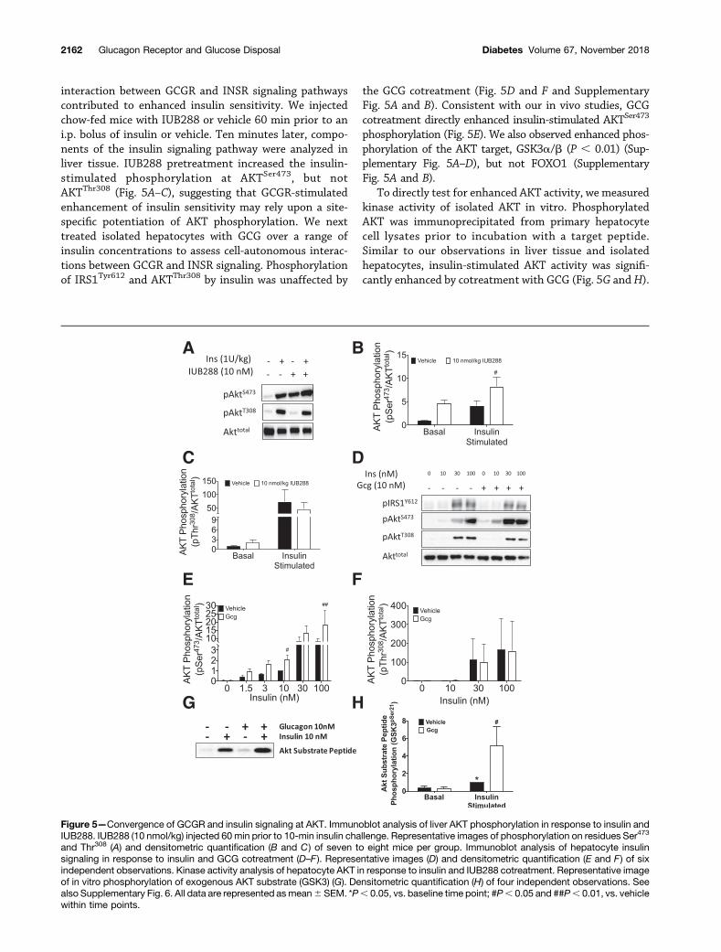
interaction between GCGR and INSR signaling pathwayscontributed to enhanced insulin sensitivity. We injectedchow-fed mice with IUB288 or vehicle 60 min prior to ani.p. bolus of insulin or vehicle. Ten minutes later, compo-nents of the insulin signaling pathway were analyzed inliver tissue. IUB288 pretreatment increased the insulin-stimulated phosphorylation at AKTSer473, but notAKTThr308 (Fig. 5A–C), suggesting that GCGR-stimulatedenhancement of insulin sensitivity may rely upon a site-specific potentiation of AKT phosphorylation. We nexttreated isolated hepatocytes with GCG over a range ofinsulin concentrations to assess cell-autonomous interac-tions between GCGR and INSR signaling. Phosphorylationof IRS1Tyr612 and AKTThr308 by insulin was unaffected by
the GCG cotreatment (Fig. 5D and F and SupplementaryFig. 5A and B). Consistent with our in vivo studies, GCGcotreatment directly enhanced insulin-stimulated AKTSer473
phosphorylation (Fig. 5E). We also observed enhanced phos-phorylation of the AKT target, GSK3a/b (P , 0.01) (Sup-plementary Fig. 5A–D), but not FOXO1 (SupplementaryFig. 5A and B).
To directly test for enhanced AKT activity, we measuredkinase activity of isolated AKT in vitro. PhosphorylatedAKT was immunoprecipitated from primary hepatocytecell lysates prior to incubation with a target peptide.Similar to our observations in liver tissue and isolatedhepatocytes, insulin-stimulated AKT activity was signifi-cantly enhanced by cotreatment with GCG (Fig. 5G andH).
Figure 5—Convergence of GCGR and insulin signaling at AKT. Immunoblot analysis of liver AKT phosphorylation in response to insulin andIUB288. IUB288 (10 nmol/kg) injected 60 min prior to 10-min insulin challenge. Representative images of phosphorylation on residues Ser473
and Thr308 (A) and densitometric quantification (B and C) of seven to eight mice per group. Immunoblot analysis of hepatocyte insulinsignaling in response to insulin and GCG cotreatment (D–F ). Representative images (D) and densitometric quantification (E and F) of sixindependent observations. Kinase activity analysis of hepatocyte AKT in response to insulin and IUB288 cotreatment. Representative imageof in vitro phosphorylation of exogenous AKT substrate (GSK3) (G). Densitometric quantification (H) of four independent observations. Seealso Supplementary Fig. 6. All data are represented asmean6 SEM. *P, 0.05, vs. baseline time point; #P, 0.05 and ##P, 0.01, vs. vehiclewithin time points.
2162 Glucagon Receptor and Glucose Disposal Diabetes Volume 67, November 2018

Together these data suggest that GCGR-dependent en-hancement of insulin sensitivity is mediated via increasedAKT activity.
Analysis of liver samples from clamped mice identifieda similar increase in liver AKTSer473 phosphorylation (Sup-plementary Fig. 6A and E) with reduced phosphorylationat IRS1Tyr612, AKTThr308, and p44/42 MAPKThr202/Tyr204
(Supplementary Fig. 6B–E). Although glucose uptake wasenhanced in EDL, AKTSer473 phosphorylation was reducedin this tissue, whereas AKTThr308 phosphorylation waselevated and phosphorylation at IRS1Tyr612 and p44/42MAPKThr202/Tyr204 was unchanged (Supplementary Fig.6A–D and F). Finally, in BAT, where glucose uptake wasmost positively regulated, phosphorylation at AKTSer473,AKTThr308, and p44/42 MAPKThr202/Tyr204 were all en-hanced, with a similar trend at IRS1Tyr612 (SupplementaryFig. 6A–D and G). Together these data further supporta direct and indirect contribution of multiple tissues to theGCGR-dependent enhancement of glucose clearance.
Hepatic Glucagon Receptors Contribute to GCGR-Mediated Improvements in Insulin ActionTo investigate the physiological contribution of hepaticGCGRs, we used mice deficient for hepatic Gcgr (GcgrΔliver).As previously described (14,15), these mice exhibit reducedfasting blood glucose (Fig. 6A), are refractory to a singleprovocative IUB288 challenge (Fig. 6B), and display dra-matically enhanced glucose tolerance (Fig. 6C). Unlike inWT mice, IUB288 had little effect on glucose excursionin GcgrΔliver mice (Fig. 6C and D). To further dissect thecontribution of the hepatic GCGR, we challenged micewith an ITT (0.25 units/kg) and observed reduced bloodglucose (Fig. 6E) and enhanced kg (Fig. 6F) in WT, but notGcgrΔliver, mice. Moreover, GCG cotreatment enhancedinsulin-stimulated AKTSer473 phosphorylation in isolatedprimary hepatocytes fromWT, but not GcgrΔliver, mice (Fig.6G and H), suggesting that hepatic GCGR signaling con-tributes to insulin-dependent improvement in glycemiccontrol.
DISCUSSION
Data presented here elucidate a novel role for GCGR sig-naling in insulin sensitivity. As a counterregulatory hor-mone, it is tempting to assume that GCG opposes all ofinsulin’s actions. However, GCG secretion and action in thefasting state make it well-suited to potentiate subsequentinsulin action. In normal physiology, these data and thepreviously described prandial GCG spike (26) suggest thatGCG may act as a preparatory component for the post-prandial state. Thus, whereas chronic GCGR activationimpairs glucose tolerance, acute agonism synergisticallyenhances insulin action and may also potentiate glucose-dependent insulin secretion.
Mechanistically, we observed that hepatic GCGR-deficient mice were resistant to GCG-mediated potentia-tion of insulin action, suggesting that the liver is likely theprimary site of action. Although these findings highlight
the liver (and presumably hepatocytes) as the tissue re-sponsible for this novel GCGR action, a caveat must be ac-knowledged in this interpretation. Specifically, the GcgrΔliver
mouse is characterized by supraphysiological levels of GLP-1and FGF21 (27), both potent sensitizers of insulin action,which may contribute to the reported improvement inglucose tolerance (27). In the context of this study, thesefactors may act to mask any subtle differences between theIUB288- and vehicle-pretreated GcgrΔliver mice. Consistentwith this observation, we observed a small, but statisti-cally significant, difference in glucose excursion betweenthe IUB288- and vehicle-pretreated GcgrΔliver mice 30 minafter glucose challenge. Thus, although the liver is likelythe primary site of action, it is possible that the directpotentiation of AKTSer473 phosphorylation and subse-quent activity observed in liver may also occur in othertissues.
The acute action of GCGR agonsim to elevate glycemiainvokes the possibility that this transient hyperglycemiamay trigger insulin secretion and thus improve glucosehomeostasis. However, infusion of a short-acting GCGRagonist at doses insufficient to induce hyperglycemia stillresulted in a clear and significant increase in insulin action.Likewise, blockade of glycogenolysis (via BAY R3401) (Sup-plementary Fig. 4) ablated GCGR agonist–induced hypergly-cemia, but not IUB288 enhancement of glucose tolerance.Moreover, our euglycemic clamp protocol included contin-uous SST infusion to eliminate potential endogenous insulinsecretion. We observed GCG-enhanced insulin actions, in-cluding improved glucose tolerance, suppressed hepatic glu-cose output, and enhanced glucose uptake in the presence ofSST. Together we interpret these data to suggest that insulinsecretion, downstream of GCGR agonist–induced hypergly-cemia, is not the mechanism underlying this enhancedglucose homeostasis.
Although these data cannot exclude a possible cross-activation of other receptors (i.e., GIPR), these insulin-sensitizing effects are clearly GCGR dependent. Alongthese lines, hepatic glycogen is known to regulate adipo-cyte lipolysis via a hepatic–central nervous system–adiposesignaling axis (24). This regulatory pathway is of interest inthat it is stimulated by depleted hepatic glycogen levels,similar to what we observe after GCGR agonsim. However,pharmacological blockade of glycogenolysis had no effecton GCG-stimulated enhancement of glucose tolerance.We interpret these results to conclude that the effectsof GCGR agonsim on glucose tolerance are independent ofits effects on glycogen metabolism. Although we hypoth-esize that GCGR and INSR signaling are interacting ina cell-autonomous manner at the hepatocyte, insulin alsosuppresses hepatic glucose production via reduction ofcirculating free fatty acids (28) (i.e., independent of liverINSR or GCGR signaling). Thus, although the liver is likelythe primary site of action, it is possible that the directpotentiation of AKTSer473 phosphorylation and subsequentactivity observed in liver may also occur in other tissues.Of particular note was the enhanced uptake observed in
diabetes.diabetesjournals.org Kim and Associates 2163

skeletal muscle. Given that GCGR is poorly expressed inskeletal muscle (if at all) (29), this effect is likely mediatedindirectly via an alternative endocrine signal and not thedirect interactions of GCGR and INSR signaling in these cells.
Importantly, during the preparation of this article,Alonge et al. (30) reported the cooperative intersectionof GCG and insulin signaling in the transcriptional regula-tion of Fgf21 expression. This report, along with our currentstudies, provides strong evidence for intracellular andcooperative overlap between these two counterregulating
hormones. The physiological relevance of Ser473 phosphor-ylation is controversial. However, an emerging view suggeststhat it precedes Thr308 phosphorylation, facilitating activa-tion by PDK1 (31). The mechanistic target of rapamycincomplex 2 (mTORC2) is responsible for insulin-stimulatedAKTSer473 phosphorylation (32). Thus, potentiation of Ser473
phosphorylation suggests that GCGR agonism may aug-ment this pathway. However, other known mTORC2 tar-gets, paxillin, protein kinase Ca (33), and the serum- andglucocorticoid-induced protein kinase (34), remained unaffected
Figure 6—Hepatic receptors contribute to GCGR-mediated improvements in insulin action. Fasting blood glucose (A) and i.p. IUB288-stimulated blood glucose excursion (B) in WT and GcgrΔliver mice. GTT (C) and AUC analysis (D) in WT and GcgrΔliver mice with IUB288pretreatment. ITT (E ) and kg 15 analysis (F ) in WT and GcgrΔliver mice with 60-min IUB288 pretreatment administered s.c. at 10 nmol/kgthroughout. Immunoblot analysis of AKT phosphorylation in response to insulin and GCG cotreatment in hepatocytes isolated from WT orGcgrΔliver mice (G andH). All data are represented asmean6SEM. *P, 0.05, **P, 0.01, ***P, 0.001, and ****P, 0.0001,WT vehicle vs. WTIUB288; #P , 0.05, GcgrΔliver vehicle vs. GcgrΔliver IUB288; &&P , 0.01, WT insulin vs. WT insulin + IUB288; $$$$P , 0.0001, WT insulin +IUB288 vs. GcgrΔliver insulin + IUB288.
2164 Glucagon Receptor and Glucose Disposal Diabetes Volume 67, November 2018

by GCG (data not shown). These data suggest target-specific activation of the complex after GCGR agonism, orinhibition of a phosphatase specifically targeting AKT-Ser473, such as PH domain leucine-rich repeat proteinphosphatase (35). Testing these hypotheses will requirefurther experimentation.
The findings described here add to the growing ther-apeutic attributes of GCGR activation. Specifically, recentreports suggest that coagonists containing GCGR activityproduce superior glucose control as compared with GLP-1,GIP, or thyroid hormones alone (3–5,36). Likewise, ourdata may provide mechanistic insight into the paradoxicalimprovements in the average level of glycemia and a re-duction in hyperglycemic events observed in patients usinga wearable, bihormonal (GCG and insulin) bionic pancreas(37). Thus, these data suggest that GCGR agonism actsboth at the level of the liver and pancreas to improvepostprandial glycemia. Further, they provide mechanisticinsight into the “paradoxical” improvement in glucosehomeostasis seen in GCGR-targeted therapeutics.
Acknowledgments. The authors acknowledge David D’Alessio (DukeUniversity), Stuart Frank and Anath Shalev (University of Alabama at Birmingham)for helpful discussion, and Jessica Antipenko (University of Alabama at Birming-ham), Joyce Sorrel, Sarah Amburgy, Jenna Holland (University of Cincinnati), andChelsea Penny and Leslie Wilkinson (University of Alabama at Birmingham) fortheir technical assistance.Funding. This project was supported by the National Heart, Lung, and BloodInstitute (R01-HL-128695 [J.K.]) and the National Institute of Diabetes andDigestive and Kidney Diseases (R01-DK-082480 [D.S.], R01-DK-077975 [D.P.-T.],5K01-DK-098319 and 1R01-DK-112934 [K.M.H.], and P30-DK-079626). Thiswork was also supported by the Canada Research Chairs Program (D.J.D.), theCanadian Institutes of Health Research (136942 and 154321), and the AmericanDiabetes Association (ADA) (1-13-JF-21 [K.M.H.]).Duality of Interest. No potential conflicts of interest relevant to this articlewere reported.Author Contributions. T.K. conceived and designed the study, generatedexperimental data, analyzed and interpreted data, and drafted the manuscript. C.L.H.,S.N., D.M.A., N.O., J.C., and C.L. generated experimental data. J.K., D.S.,D.J.D., R.D., and D.P.-T. advised on the study concept and critically revised themanuscript. K.M.H. conceived and designed the study, analyzed and interpreteddata, and drafted the manuscript. K.M.H. is the guarantor of this work and, assuch, had full access to all the data in the study and takes responsibility for theintegrity of the data and the accuracy of the data analysis.Prior Presentation. Portions of this work were presented at the 74thScientific Sessions of the American Diabetes Association, San Francisco, CA, 13–17 June 2014; the 75th Scientific Sessions of the American Diabetes Association,Boston, MA, 5–9 June 2015; and the 76th Scientific Sessions of the AmericanDiabetes Association, New Orleans, LA, 10–14 June 2016.
References1. Habegger KM, Heppner KM, Geary N, Bartness TJ, DiMarchi R, Tschöp MH.The metabolic actions of glucagon revisited. Nat Rev Endocrinol 2010;6:689–6972. Müller TD, Finan B, Clemmensen C, DiMarchi RD, Tschöp MH. The newbiology and pharmacology of glucagon. Physiol Rev 2017;97:721–7663. Day JW, Ottaway N, Patterson JT, et al. A new glucagon and GLP-1co-agonist eliminates obesity in rodents. Nat Chem Biol 2009;5:749–7574. Clemmensen C, Chabenne J, Finan B, et al. GLP-1/glucagon coagonism re-stores leptin responsiveness in obese mice chronically maintained on an obesogenicdiet. Diabetes 2014;63:1422–1427
5. Finan B, Yang B, Ottaway N, et al. A rationally designed monomeric peptidetriagonist corrects obesity and diabetes in rodents. Nat Med 2015;21:27–366. Tan TM, Field BC, McCullough KA, et al. Coadministration of glucagon-likepeptide-1 during glucagon infusion in humans results in increased energy ex-penditure and amelioration of hyperglycemia. Diabetes 2013;62:1131–11387. Habegger KM, Heppner KM, Amburgy SE, et al. GLP-1R responsivenesspredicts individual gastric bypass efficacy on glucose tolerance in rats. Diabetes2014;63:505–5138. Campos GM, Rabl C, Havel PJ, et al. Changes in post-prandial glucose andpancreatic hormones, and steady-state insulin and free fatty acids after gastricbypass surgery. Surg Obes Relat Dis 2014;10:1–89. Krishna MG, Coker RH, Lacy DB, Zinker BA, Halseth AE, Wasserman DH.Glucagon response to exercise is critical for accelerated hepatic glutaminemetabolism and nitrogen disposal. Am J Physiol Endocrinol Metab 2000;279:E638–E64510. Powley TL. The ventromedial hypothalamic syndrome, satiety, and a cephalicphase hypothesis. Psychol Rev 1977;84:89–12611. Habegger KM, Stemmer K, Cheng C, et al. Fibroblast growth factor 21 mediatesspecific glucagon actions. Diabetes 2013;62:1453–146312. Hotta Y, Nakamura H, Konishi M, et al. Fibroblast growth factor 21 regulateslipolysis in white adipose tissue but is not required for ketogenesis and triglycerideclearance in liver. Endocrinology 2009;150:4625–463313. Scrocchi LA, Brown TJ, MaClusky N, et al. Glucose intolerance but normalsatiety in mice with a null mutation in the glucagon-like peptide 1 receptor gene.Nat Med 1996;2:1254–125814. Longuet C, Robledo AM, Dean ED, et al. Liver-specific disruption of themurine glucagon receptor produces a-cell hyperplasia: evidence for a circulatinga-cell growth factor. Diabetes 2013;62:1196–120515. Kim T, Nason S, Holleman C, et al. Glucagon-receptor signaling regulatesenergy metabolism via hepatic farnesoid X receptor and fibroblast growth factor21. Diabetes 201816. Lockie SH, Heppner KM, Chaudhary N, et al. Direct control of brown adiposetissue thermogenesis by central nervous system glucagon-like peptide-1 receptorsignaling. Diabetes 2012;61:2753–276217. Kim T, He L, Johnson MS, et al. Carnitine palmitoyltransferase 1b deficiencyprotects mice from diet-induced insulin resistance. J Diabetes Metab 2014;5:36118. Mészáros K, Lang CH, Bagby GJ, Spitzer JJ. Contribution of different organsto increased glucose consumption after endotoxin administration. J Biol Chem1987;262:10965–1097019. Kim HJ, Higashimori T, Park SY, et al. Differential effects of interleukin-6 and-10 on skeletal muscle and liver insulin action in vivo. Diabetes 2004;53:1060–106720. Li WC, Ralphs KL, Tosh D. Isolation and culture of adult mouse hepatocytes.Methods Mol Biol 2010;633:185–19621. Huypens P, Ling Z, Pipeleers D, Schuit F. Glucagon receptors on human isletcells contribute to glucose competence of insulin release. Diabetologia 2000;43:1012–101922. Weston C, Poyner D, Patel V, Dowell S, Ladds G. Investigating G proteinsignalling bias at the glucagon-like peptide-1 receptor in yeast. Br J Pharmacol2014;171:3651–366523. Winnick JJ, An Z, Kraft G, et al. Liver glycogen loading dampens glycogensynthesis seen in response to either hyperinsulinemia or intraportal glucose in-fusion. Diabetes 2013;62:96–10124. Izumida Y, Yahagi N, Takeuchi Y, et al. Glycogen shortage during fastingtriggers liver-brain-adipose neurocircuitry to facilitate fat utilization [publishedcorrection appears in Nat Commun 2013;4:2930]. Nat Commun 2013;4:231625. Bergans N, Stalmans W, Goldmann S, Vanstapel F. Molecular mode ofinhibition of glycogenolysis in rat liver by the dihydropyridine derivative, BAYR3401: inhibition and inactivation of glycogen phosphorylase by an activatedmetabolite. Diabetes 2000;49:1419–142626. Geary N. Pancreatic glucagon signals postprandial satiety. Neurosci BiobehavRev 1990;14:323–338
diabetes.diabetesjournals.org Kim and Associates 2165

27. Omar BA, Andersen B, Hald J, Raun K, Nishimura E, Ahrén B. Fibroblastgrowth factor 21 (FGF21) and glucagon-like peptide 1 contribute to diabetesresistance in glucagon receptor-deficient mice. Diabetes 2014;63:101–11028. Titchenell PM, Chu Q, Monks BR, Birnbaum MJ. Hepatic insulin signalling isdispensable for suppression of glucose output by insulin in vivo. Nat Commun2015;6:707829. Hansen LH, Abrahamsen N, Nishimura E. Glucagon receptor mRNA distri-bution in rat tissues. Peptides 1995;16:1163–116630. Alonge KM, Meares GP, Hillgartner FB. Glucagon and insulin cooperativelystimulate fibroblast growth factor 21 gene transcription by increasing theexpression of activating transcription factor 4. J Biol Chem 2017;292:5239–525231. Scheid MP, Marignani PA, Woodgett JR. Multiple phosphoinositide 3-kinase-dependent steps in activation of protein kinase B. Mol Cell Biol 2002;22:6247–6260
32. Sarbassov DD, Guertin DA, Ali SM, Sabatini DM. Phosphorylation andregulation of Akt/PKB by the rictor-mTOR complex. Science 2005;307:1098–110133. Sarbassov DD, Ali SM, Kim DH, et al. Rictor, a novel binding partner of mTOR,defines a rapamycin-insensitive and raptor-independent pathway that regulatesthe cytoskeleton. Curr Biol 2004;14:1296–130234. Laplante M, Sabatini DM. mTOR signaling in growth control and disease. Cell2012;149:274–29335. Gao T, Furnari F, Newton AC. PHLPP: a phosphatase that directly de-phosphorylates Akt, promotes apoptosis, and suppresses tumor growth. Mol Cell2005;18:13–2436. Finan B, Clemmensen C, Zhu Z, et al. Chemical hybridization of glucagon andthyroid hormone optimizes therapeutic impact for metabolic disease. Cell 2016;167:843–857.e81437. Russell SJ, El-Khatib FH, Sinha M, et al. Outpatient glycemic control witha bionic pancreas in type 1 diabetes. N Engl J Med 2014;371:313–325
2166 Glucagon Receptor and Glucose Disposal Diabetes Volume 67, November 2018