hemostasis and thrombosis in obstetrics & gynecology · obstetrics & gynecology michael j....
TRANSCRIPT

P1: SFK/UKS P2: SFK Color: 1C
fm BLBK321-Paidas September 10, 2010 16:41 Trim: 244mm X 172mm
Hemostasis andThrombosis inObstetrics &GynecologyMichael J. Paidas, MDYale University School of Medicine, New Haven, CT, USA
Nazli Hossain, MBBS, FCPSDow University of Health Sciences, Karachi, Pakistan
Tahir S. Shamsi, MBBS, FRCPath, FCPPNational Institute of Blood Disease and Bone Marrow Transplantation
Karachi, Pakistan and University of Health Sciences, Lahore, Pakistan
Marc A. Rodger, MD, MScOttowa Hospital General, ON, Canada
Jens Langhoff-Roos, MD, DMScRoskilde Hospital, Roskilde and Rigshopitalet, Copenhagen, Denmark
Charles J. Lockwood, MD, MHCMYale University School of Medicine, New Haven, CT, USA
A John Wiley & Sons, Ltd., Publication
iii

P1: SFK/UKS P2: SFK Color: 1C
fm BLBK321-Paidas September 10, 2010 16:41 Trim: 244mm X 172mm
ii

P1: SFK/UKS P2: SFK Color: 1C
fm BLBK321-Paidas September 10, 2010 16:41 Trim: 244mm X 172mm
Hemostasis and Thrombosis inObstetrics & Gynecology
i

P1: SFK/UKS P2: SFK Color: 1C
fm BLBK321-Paidas September 10, 2010 16:41 Trim: 244mm X 172mm
ii

P1: SFK/UKS P2: SFK Color: 1C
fm BLBK321-Paidas September 10, 2010 16:41 Trim: 244mm X 172mm
Hemostasis andThrombosis inObstetrics &GynecologyMichael J. Paidas, MDYale University School of Medicine, New Haven, CT, USA
Nazli Hossain, MBBS, FCPSDow University of Health Sciences, Karachi, Pakistan
Tahir S. Shamsi, MBBS, FRCPath, FCPPNational Institute of Blood Disease and Bone Marrow Transplantation
Karachi, Pakistan and University of Health Sciences, Lahore, Pakistan
Marc A. Rodger, MD, MScOttowa Hospital General, ON, Canada
Jens Langhoff-Roos, MD, DMScRoskilde Hospital, Roskilde and Rigshopitalet, Copenhagen, Denmark
Charles J. Lockwood, MD, MHCMYale University School of Medicine, New Haven, CT, USA
A John Wiley & Sons, Ltd., Publication
iii
P1: SFK/UKS P2: SFK Color: 1C
fm BLBK321-Paidas September 10, 2010 16:41 Trim: 244mm X 172mm
This edition first published 2011, C© 2011 by Michael J. Paidas, Nazli Hossain, Tahir S. Shamsi, MarcA. Rodger, Jens Langhoff-Roos and Charles J. Lockwood.
Blackwell Publishing was acquired by John Wiley & Sons in February 2007. Blackwell’s publishingprogram has been merged with Wiley’s global Scientific, Technical and Medical business to formWiley-Blackwell.
Registered office: John Wiley & Sons Ltd, The Atrium, Southern Gate, Chichester, West Sussex, PO198SQ, UK
Editorial offices: 9600 Garsington Road, Oxford, OX4 2DQ, UKThe Atrium, Southern Gate, Chichester, West Sussex, PO19 8SQ, UK111 River Street, Hoboken, NJ 07030-5774, USA
For details of our global editorial offices, for customer services and for information about how toapply for permission to reuse the copyright material in this book please see our website atwww.wiley.com/wiley-blackwell
The right of the author to be identified as the author of this work has been asserted in accordancewith the UK Copyright, Designs and Patents Act 1988.
All rights reserved. No part of this publication may be reproduced, stored in a retrieval system, ortransmitted, in any form or by any means, electronic, mechanical, photocopying, recording orotherwise, except as permitted by the UK Copyright, Designs and Patents Act 1988, without theprior permission of the publisher.
Designations used by companies to distinguish their products are often claimed as trademarks. Allbrand names and product names used in this book are trade names, service marks, trademarks orregistered trademarks of their respective owners. The publisher is not associated with any product orvendor mentioned in this book. This publication is designed to provide accurate and authoritativeinformation in regard to the subject matter covered. It is sold on the understanding that thepublisher is not engaged in rendering professional services. If professional advice or other expertassistance is required, the services of a competent professional should be sought.
The contents of this work are intended to further general scientific research, understanding, anddiscussion only and are not intended and should not be relied upon as recommending or promotinga specific method, diagnosis, or treatment by physicians for any particular patient. The publisher andthe author make no representations or warranties with respect to the accuracy or completeness ofthe contents of this work and specifically disclaim all warranties, including without limitation anyimplied warranties of fitness for a particular purpose. In view of ongoing research, equipmentmodifications, changes in governmental regulations, and the constant flow of information relatingto the use of medicines, equipment, and devices, the reader is urged to review and evaluate theinformation provided in the package insert or instructions for each medicine, equipment, or devicefor, among other things, any changes in the instructions or indication of usage and for addedwarnings and precautions. Readers should consult with a specialist where appropriate. The fact thatan organization or Website is referred to in this work as a citation and/or a potential source offurther information does not mean that the author or the publisher endorses the information theorganization or Website may provide or recommendations it may make. Further, readers should beaware that Internet Websites listed in this work may have changed or disappeared between whenthis work was written and when it is read. No warranty may be created or extended by anypromotional statements for this work. Neither the publisher nor the author shall be liable for anydamages arising herefrom.
Library of Congress Cataloging-in-Publication Data
Hemostasis and thrombosis in obstetrics & gynecology / Michael J. Paidas . . . [et al.].p. ; cm.
Other title: Hemostasis and thrombosis in obstetrics and gynecologyIncludes bibliographical references.ISBN 978-1-4051-8399-41. Blood coagulation disorders in pregnancy. 2. Women–Diseases. 3. Hemostasis.
4. Thrombosis. 5. Pregnancy–Complications. I. Paidas, Michael J. II. Title: Hemostasis andthrombosis in obstetrics and gynecology.
[DNLM: 1. Hemostasis–physiology–Handbooks. 2. Blood Coagulation Disorders–Handbooks.3. Pregnancy Complications, Hematologic–Handbooks. 4. Thrombosis–physiopathology–Handbooks. WH 310 H4893 2010]
RG580.B56H33 2010618.3–dc22
2010023429
A catalogue record for this book is available from the British Library.
This book is published in the following electronic formats: ePDF 9781444328349; Wiley OnlineLibrary 9781444328332.
Set in 9.5/13 pt Meridien by Aptara R© Inc., New Delhi, India
1 2011
iv
P1: SFK/UKS P2: SFK Color: 1C
fm BLBK321-Paidas September 10, 2010 16:41 Trim: 244mm X 172mm
Contents
List of Contributors, vi
Preface, vii
Acknowledgments and Dedication, vii
1 Hematologic Changes in Pregnancy, 1
Michael J. Paidas & Nazli Hossain
2 Red Cell Disorders, 12
Tahir S. Shamsi
3 Hemolytic Disease of the Newborn, 28
Tahir S. Shamsi, Nazli Hossain & Michael J. Paidas
4 Maternal and Fetal Thrombocytopenia, 41
Michael J. Paidas & Nazli Hossain
5 Inherited and Acquired Thrombophilia in Obstetrics, 67
Michael J. Paidas, Christina S. Han, Nazli Hossain, and Charles
J. Lockwood
6 Anticoagulant Therapy During Pregnancy and Gynecology, 111
Marc A. Rodger & Genevieve Le Templier
7 Inherited Bleeding Disorders in Obstetrics, 153
Nazli Hossain & Michael J. Paidas
8 Inherited Bleeding Disorders in Gynecology, 161
Nazli Hossain & Michael J. Paidas
9 Postpartum Hemorrhage, 167
Nazli Hossain, Jens Langhoff-Roos & Michael J. Paidas
10 Disseminated Intravascular Coagulation, 182
Nazli Hossain & Michael J. Paidas
11 Transfusion of Blood and Blood Products in Obstetrics, 195
Tahir S. Shamsi & Nazli Hossain
Index, 216
v
P1: SFK/UKS P2: SFK Color: 1C
fm BLBK321-Paidas September 10, 2010 16:41 Trim: 244mm X 172mm
List of Contributors
Christina S. Han, MDClinical instructor, Division of Maternal Fetal Medicine, Department of Obstetrics, Gynecology &
Reproductive Sciences, Yale University School of Medicine, New Haven, CT, USA
Nazli Hossain, MBBS, FCPSAssociate Professor, Department of Obstetrics and Gynecology, Dow University of Health Sciences,
Karachi, Pakistan
Jens Langhoff-Roos, MD, DMScDepartment of Obstetrics and Gynaecology, Roskilde Hospital, Roskilde, and Department of
Obstetrics, Rigshospitalet, Copenhagen, Denmark
Charles J. Lockwood, MD, MHCMAnita O’Keeffe Young Professor and Chair, Department of Obstetrics, Gynecology and Reproductive
Sciences, Yale University School of Medicine, New Haven, CT, USA
Michael J. Paidas, MDAssociate Professor
Co-Director, Yale Women and Children’s Center for Blood Disorders
Co- Director, National Hemophilia Foundation- Baxter Clinical Fellowship Program at Yale
Division of Maternal Fetal Medicine
Department of Obstetrics, Gynecology and Reproductive Sciences
Yale University School of Medicine, New Haven, CT, USA
Marc A. Rodger, MD, MSc (Epidemiology)Professor of Medicine
University of Ottawa Research Chair in Thrombosis and Thrombophilia Ottowa Hospital General,
ON, Canada
Tahir S. Shamsi, MBBS, FRCPath, FCPPProfessor of Haematology, Consultant Haematologist and Transplant Physician, National Institute of
Blood Disease and Bone Marrow Transplantation, Karachi, Pakistan and University of Health
Sciences, Lahore, Pakistan
Genevieve Le Templier, MD, FRCPCOttawa Hospital, Ottawa Health Research Institute, University of Ottawa, Ottawa, ON, Canada
vi
P1: SFK/UKS P2: SFK Color: 1C
fm BLBK321-Paidas September 10, 2010 16:41 Trim: 244mm X 172mm
Preface
Interest in reproductive hemostasis continues to develop among care
providers, basic science and translational researchers across a variety of
disciplines. This phenomenon, it seems, is not a regional, country or
even a continent-specific evolution, but rather a global response. Several
factors have probably contributed to this niche area which is garnering
widespread appeal. International meetings of our respective primary spe-
cialties encourage dialogue with members of other disciplines to tackle
common scientific and clinical dilemmas in women’s health. At many
higher academic institutions, investments in infrastructure to foster a more
‘thematic’ and integrated approach are being promoted. It is obvious to ev-
eryone that communication among colleagues separated by great physical
distances is not as difficult as in the past. Several programs are in place that
support interaction between the developing and the developed countries.
Finally, drugs or biologics, which represent present and future interven-
tions, are definitely global endeavors.
Our goal for this textbook is to provide a contemporary, international,
multidisciplinary approach to common obstetric, gynecologic and hema-
tologic issues, and present the data in a comprehensive, but simplified
manner. This textbook is written for trainees including residents and fel-
lows, seasoned care providers, obstetrician/gynecologists, hematologists,
primary care providers, consultants, and students. We have made delib-
erate attempts to address concerns in the developing and the developed
countries. The chapters are divided according to relevant clinical condi-
tions. We have intended that this textbook be a useful ‘pocket’ reference
for residents, fellows and care providers who might not have immediate
internet access to search a particular question, but need an answer quickly.
This book will provide firm guidelines for obstetricians in the management
of pregnant women in labor and delivery suites.
We have enjoyed writing this book and hope that it stimulates further
collaborative research and discourse directed toward a better understand-
ing of hemostasis and thrombosis issues in the reproduction sciences.
Michael J. Paidas MD
Nazli Hossain MBBS FCPS
vii

P1: SFK/UKS P2: SFK Color: 1C
fm BLBK321-Paidas September 10, 2010 16:41 Trim: 244mm X 172mm
Acknowledgments andDedication
There are a few individuals who deserve special mention because they
have influenced my career path that intersects obstetrics, gynecology, the
reproductive sciences, and hematology. I credit Michael J. Haut, MD, who
cultivated my interest in maternal and fetal hematologic disorders dur-
ing residency at Pennsylvania Hospital. Together, we studied aspects of
both maternal and fetal platelet disorders, and he encouraged me to pur-
sue evaluation of in utero fetal platelet function. I have always admired
Fernand Daffos, MD, as a pioneer in many aspects of fetal medicine and
appreciated his encouragement early in my career. Richard L. Berkowitz,
MD, taught me many things, but two lessons in particular have resonated
with me throughout my entire career. Dr. Berkowitz stressed that we, as
physicians, must dedicate ourselves to providing our patients, who entrust
their lives in our hands, with the best care possible. They deserve noth-
ing less than 100% of our best efforts. Secondly, he provided me with
much insight and guidance with his contributions to defining manage-
ment of antenatal alloimmune thrombocytopenia. Perhaps more than any
other individual, I learned from Mary D’Alton, MD, to insist upon car-
rying out good clinical trials in obstetrics to drive practice management
guidelines. For several years, Yale S. Arkel, MD, and Wayne Ku, PhD, gal-
vanized our clinical research efforts focused on the protein C system and
hemostatic derangements associated with pregnancy complications. Both
of them taught me so much about hemostasis and laboratory medicine.
Finally, I owe much to my mentor, collaborator, and colleague, Charles J.
Lockwood, MD, MHCM, who I first met over 25 years ago, for nurturing
my career over such a long period.
Over the years, my patients have served as a daily source of inspiration.
Their courage and determination have been infectious and I wish to thank
them collectively for allowing me to share some aspect of their lives. I wish
to thank my parents, Angela and Nicholas Paidas, who have sacrificed so
much of their life for their children. Finally, I wish to thank my family,
Anne Marie, Nicholas, and Lauren because without their patience, love,
and support, I would not have been able to devote time and energy to this
endeavor and my profession in general.
viii

P1: SFK/UKS P2: SFK Color: 1C
fm BLBK321-Paidas September 10, 2010 16:41 Trim: 244mm X 172mm
Acknowledgments and Dedication ix
This book is dedicated to Diana S. Beardsley, MD, PhD, Director of the
Yale Hemophilia Treatment Center. For 24 years, from 1986–2010, Dr.
Beardsley was the face of Pediatric Hemostasis at Yale. She was a superb
physician, brilliant scientist, and tireless advocate and mentor for her fel-
lows, who she mentored during the past three decades. Diana was beloved
by her patients, colleagues, and students. This textbook is a tribute to her
legacy.
Diana S. Beardsley, MD, PhD, December 8, 1947 to March 30, 2010
Michael J. Paidas, MD.
We would like to acknowledge our parents and our families who have
helped us in taking this path; we dedicate this book to mothers who suf-
fered because of ignorance of their disease and taught us how to manage
blood disorders in women.
Nazli Hossain
Tahir S. Shamsi
Marc A. Rodger
Jens Langhoff-Roos
For Nancy, Sarah, John and Billy
Charles J. Lockwood
P1: SFK/UKS P2: SFK Color: 1C
c01 BLBK321-Paidas August 17, 2010 19:1 Trim: 244mm X 172mm
C H A P T E R 1
Hematologic Changes inPregnancyMichael J. Paidas & Nazli Hossain
Introduction
Normal pregnancy is characterized by profound changes in nearly every
organ system to accommodate the demands of the fetoplacental unit. Ma-
ternal hematological adaptations to the pregnant state are reviewed in this
chapter. The most significant hematological changes are physiologic ane-
mia, neutrophilia, mild thrombocytopenia, increased procoagulant factors,
and diminished fibrinolysis.
This chapter will review the pregnancy-associated changes in plasma
volume, red blood cells, white blood cells, platelets, and coagulation
factors.
Plasma Volume
Plasma volume increases by 10–15% at 6–12 weeks of gestation [1–3],
expands rapidly until 30–34 weeks, after which there is only a modest
rise. The total gain of plasma volume at term averages 1100–1600 mL
and results in a plasma volume of 4700–5200 mL, 30–50% above that
found in nonpregnant women [1, 4]. Plasma volume decreases immedi-
ately postpartum, then increases again 2–5 days after delivery, possibly
because of a simultaneous rise in aldosterone secretion. Plasma volume
then decreases; it is still elevated by 10–15% above nonpregnant levels at
3 weeks postpartum, but is usually at normal nonpregnant levels at
6 weeks postpartum.
During pregnancy, plasma renin activity is typically increased and atrial
natriuretic peptide levels are slightly reduced, suggesting that the increase
in plasma volume represents underfilling due to systemic vasodilatation
and the ensuing rise in vascular capacitance, rather than true blood
Hemostasis and Thrombosis in Obstetrics & Gynecology, 1st edition. By Michael J. Paidas,
Nazli Hossain, Tahir S. Shamsi, Marc A. Rodger, Jens Langhoff-Roos, and
Charles J. Lockwood. Published 2011 by Blackwell Publishing Ltd.
1

P1: SFK/UKS P2: SFK Color: 1C
c01 BLBK321-Paidas August 17, 2010 19:1 Trim: 244mm X 172mm
2 Hemostasis and Thrombosis in Obstetrics & Gynecology
volume expansion, which would produce the opposite hormonal pro-
file (low plasma renin activity, elevated atrial natriuretic peptide) [5, 6].
Furthermore, the degree of sodium retention is physiologically regulated,
as increasing sodium intake does not produce further volume expansion
[7].
Red Blood Cells
Red blood cell mass begins to increase at 8–10 weeks of gestation and
steadily rises by 20–30% (250–450 mL) above nonpregnant levels by the
end of pregnancy in women receiving iron supplementation [4, 8–11].
Among women not on iron supplements, the red cell mass may only in-
crease by 15–20% [12]. Erythrocyte life span is slightly decreased during
normal pregnancy [13].
Erythropoietin levels increase by 50% in normal pregnancies and vary
according to the presence of pregnancy complications [14]. The increased
plasma erythropoietin induces the rise in red cell mass, which partially
supports the higher metabolic requirement for oxygen during pregnancy
[15]. Mean corpuscular volume decreases during pregnancy and averages
80–84 fL in the third trimester [16].
AnemiaA greater expansion of plasma volume relative to the increase in
hemoglobin mass and erythrocyte volume is responsible for the mod-
est fall in hemoglobin levels (i.e., physiological or dilutional anemia of
pregnancy) observed in healthy pregnant women. The greatest dispro-
portion between the rates at which plasma and erythrocytes are added
to the maternal circulation occurs during the late second to early third
trimester. (Lowest hematocrit is typically measured at 28–36 weeks [16].)
Nearer to term, hemoglobin concentration increases due to cessation of
plasma expansion and continuing increase in hemoglobin mass. Con-
versely, the absence of physiologic anemia appears to be a risk factor for
stillbirth [17].
Determining a good definition of anemia in pregnant women is not
straightforward, given the pregnancy-associated changes in plasma vol-
ume and red cell mass, normal differences in hemoglobin concentrations
between women and men, ethnic variation between white and black
women, and the frequent use of iron supplementation in pregnancy.
The Centers for Disease Control and Prevention has defined anemia as
hemoglobin levels of less than 11 g/dL (hematocrit less than 33%) in the
first and third trimesters and less than 10.5 g/dL (hematocrit less than
32%) in the second trimester [18]. Since hemoglobin and hematocrit
P1: SFK/UKS P2: SFK Color: 1C
c01 BLBK321-Paidas August 17, 2010 19:1 Trim: 244mm X 172mm
Chapter 1 Hematologic Changes in Pregnancy 3
levels are lower in African-American adults, the Institute of Medicine rec-
ommends lowering of the hemoglobin cutoff level by 0.8 g/dL in this pop-
ulation [19].
Women with hemoglobin values below these levels can be considered
anemic and should undergo a standard evaluation [20]. Sixteen to twenty-
nine percent of pregnant women become anemic in the third trimester
[21].
Severe anemia with maternal hemoglobin below 6 g/dL has been asso-
ciated with reduced amniotic fluid volume, fetal cerebral vasodilation, and
nonreassuring fetal heart rate patterns [22]. Increased risks of prematu-
rity, spontaneous abortion, low birth weight, growth restriction, and fetal
death have also been reported [23]. The administration of lactoferrin to
treat iron deficiency anemia in pregnancy requires further investigation.
Lactoferrin chelates two ferric ions, decreases interleukin-6, thereby de-
creasing hepcidin and increasing ferroportin expression.
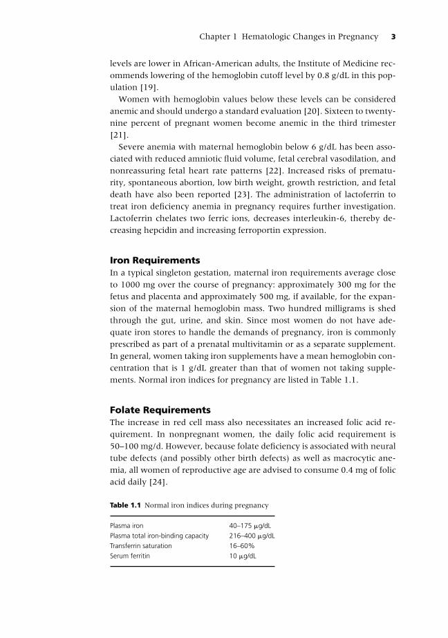
Iron RequirementsIn a typical singleton gestation, maternal iron requirements average close
to 1000 mg over the course of pregnancy: approximately 300 mg for the
fetus and placenta and approximately 500 mg, if available, for the expan-
sion of the maternal hemoglobin mass. Two hundred milligrams is shed
through the gut, urine, and skin. Since most women do not have ade-
quate iron stores to handle the demands of pregnancy, iron is commonly
prescribed as part of a prenatal multivitamin or as a separate supplement.
In general, women taking iron supplements have a mean hemoglobin con-
centration that is 1 g/dL greater than that of women not taking supple-
ments. Normal iron indices for pregnancy are listed in Table 1.1.
Folate RequirementsThe increase in red cell mass also necessitates an increased folic acid re-
quirement. In nonpregnant women, the daily folic acid requirement is
50–100 mg/d. However, because folate deficiency is associated with neural
tube defects (and possibly other birth defects) as well as macrocytic ane-
mia, all women of reproductive age are advised to consume 0.4 mg of folic
acid daily [24].
Table 1.1 Normal iron indices during pregnancy
Plasma iron 40–175 �g/dL
Plasma total iron-binding capacity 216–400 �g/dL
Transferrin saturation 16–60%
Serum ferritin 10 �g/dL

P1: SFK/UKS P2: SFK Color: 1C
c01 BLBK321-Paidas August 17, 2010 19:1 Trim: 244mm X 172mm
4 Hemostasis and Thrombosis in Obstetrics & Gynecology
Platelet Count
Although platelet counts remain in the normal nonpregnant range in most
women during uncomplicated pregnancies [25], mean platelet counts of
pregnant women may be slightly lower than in healthy nonpregnant
women [26]. Serial platelet counts during uncomplicated pregnancies may
[27] or may not [28] decrease, but the mean values in these groups do not
necessarily reflect both increases and decreases in individual women [29].
The lower limit of normal platelet counts in pregnancy has been reported
to be 106,000–120,000 platelets/�L.
ThrombocytopeniaThe most significant obstetrical consideration concerning platelet physiol-
ogy in pregnancy is thrombocytopenia, which may be related to complica-
tions of pregnancy (e.g., severe preeclampsia, HELLP syndrome), medical
disorders (e.g., idiopathic thrombocytopenic purpura, thrombotic throm-
bocytopenic purpura-hemolytic uremic syndrome), or gestational. Ges-
tational or incidental thrombocytopenia is characterized by mild asymp-
tomatic thrombocytopenia occurring in the third trimester in a patient
without any history of thrombocytopenia (other than in a prior preg-
nancy). It is not associated with maternal, fetal, or neonatal sequelae and
spontaneously resolves postpartum [30–32]. Platelet counts are typically
greater than 70,000/�L.
White Blood Cells
Pregnancy is associated with leukocytosis, primarily related to increased
circulation of neutrophils. The neutrophil count begins to increase in the
second month of pregnancy and plateaus in the second or third trimester,
at which time the total white blood cell counts ranges from 9000 to
15,000 cells/�L [33]. Data from two series reported mean white blood
cell counts of 10,000–16,000 cells/�L in laboring patients, with an upper
level as high as 29,000 cells/�L [34,35]; the mean count increased linearly
with the duration of elapsed labor [35]. The white blood cell count falls to
the normal nonpregnant range by the sixth day postpartum. Dohle bodies
(blue staining cytoplasmic inclusions in granulocytes) are a normal finding
in pregnant women.
In healthy women with normal pregnancies, there is no change in the
absolute lymphocyte count and no significant changes in the relative num-
bers of T and B lymphocytes [36]. The monocyte count is generally stable;
the basophil count may slightly decrease and the eosinophil count may
slightly increase. Normal pregnant women can have a small number of
myelocytes or metamyelocytes in the peripheral circulation.

P1: SFK/UKS P2: SFK Color: 1C
c01 BLBK321-Paidas August 17, 2010 19:1 Trim: 244mm X 172mm
Chapter 1 Hematologic Changes in Pregnancy 5
Coagulation
Normal pregnancy is a prothrombotic state [37–46]. The circulating levels
of several coagulation factors change during pregnancy (Table 1.2):� Protein S activity and free protein S antigen decrease due to estrogen-
induced increases in the complement 4b binding protein and possibly
due to other mechanisms related to the hormonal changes of pregnancy.� Resistance to activated protein C increases in the second and third
trimesters.� Fibrinogen, factors II, VII, VIII, and X increase by 20–200% [47]; von
Willebrand factor also increases.� Activity of the fibrinolytic inhibitors, thrombin activatable fibrinolytic
inhibitor (TAFI), PAI-1, and PAI-2 increases [48].� Factors V and IX remain unchanged and factor XI levels decrease by 30%
[47].
The net effect of these changes is to increase the tendency toward throm-
bus formation, extension, and stability. Normalization of coagulation pa-
rameters varies depending on the factor, but all should return to baseline
by 8 weeks postpartum.
Table 1.2 Hemostatic changes in pregnancy.
Variables (mean ± SD) First tri∗ Second tri∗ Third tri∗ Normal range
Platelet (× 109 1−1) 275 ± 64 256 ± 49 244 ± 52 150–400
Fibrinogen (g/L) 3.7 ± 0.6 4.4 ± 1.2 5.4 ± 0.8 2.1–4.2
Prothrombin complex (%) 120 ± 27 140 ± 27 130 ± 27 70–30
Antithrombin (U/mL) 1.02 ± 0.10 1.07 ± 0.14 1.07 ± 0.11 0.85–1.25
Protein C (U/mL) 0.92 ± 0.13 1.06 ± 0.17 .94 ± 0.2 0.68–1.25
Protein S, total (U/mL) 0.83 ± 0.11 0.73 ± 0.11 0.77 ± 0.10 0.70–1.70
Protein S, free (U/mL) 0.26 ± 0.07 0.17 ± 0.04 0.14 ± 0.04 0.20–0.50
Soluble fibrin (nmol/L) 9.2 ± 8.6 11.8 ± 7.7 13.4 ± 5.2 <15
Thrombin–antithrombin (�g/L) 3.1 ± 1.4 5.9 ± 2.6 7.1 ± 2.4 <2.7
D-dimers (�g/L) 91 ± 24 128 ± 49 198 ± 59 <80
Plasminogen activator
inhibitor-1 (AU/mL)
7.4 ± 4.9 14.9 ± 5.2 37.8 ± 19.4 <15
Plasminogen activator
inhibitor-2 (�g/L)
31 ± 14 84 ± 16 160 ± 31 <5
Cardiolipin antibodies positive 2/25 2/25 3/23 0
Protein Z (�g mL−1)† 2.01 ± 0.76 1.47 ± 0.45 1.55 ± 0.48
Protein S (%)† 34.4 ± 11.8 27.5 ± 8.4
∗First tri, 12–15 weeks; second tri, week 24; third tri, week 35.†First tri, 0–14 weeks; second tri, 14–27 weeks; third tri, 27 weeks or more.
tri, trimester.
Adapted from Bremme [46], table 3, p. 157 and Paidas et al. [51], with permission.

P1: SFK/UKS P2: SFK Color: 1C
c01 BLBK321-Paidas August 17, 2010 19:1 Trim: 244mm X 172mm
6 Hemostasis and Thrombosis in Obstetrics & Gynecology
Protein SProtein S (PS) is a vitamin K-dependent glycoprotein with several antico-
agulant functions [49]. In the presence of PS, activated protein C inacti-
vates factor Va and factor VIIIa, resulting in reduced thrombin generation.
PS also serves as a cofactor for protein C enhancement of fibrinolysis. PS
has a direct anticoagulant effect independent of its co-factor function with
activated protein C. It prevents the binding of surface phospholipids with
factors such as Va, Xa, and VIIIa, thereby decreasing the activation of the
factors.
Pregnancy is associated with decreased levels of PS activity and free
PS antigen [44, 50]. The significance and degree of decrease in PS levels
commonly seen in pregnancy has not been vigorously evaluated. To ad-
dress this question, we compared second and third trimester PS levels in
51 healthy women with a normal pregnancy outcome with 51 healthy
women with a poor pregnancy outcome [51]. Protein S levels were sig-
nificantly lower in the second and third trimesters among patients with
adverse pregnancy outcome compared to patients with normal pregnancy
outcome (second trimester 34.4 ± 11.8% versus 38.9 ± 10.3%, respec-
tively; and third trimester 27.5 ± 8.4 versus 31.2 ± 7.4%, respectively).
Resistance to Activated Protein CDuring pregnancy, normal women acquire some degree of resistance to
activated protein C (APC), when measured by the first generation global
assays and tests that measure endogenous thrombin potential [45,52,53].
Factor XFactor X, its activation to FXa and participation in the activation of
prothrombin, is a central element in the generation of thrombin [54]. It
is possible that derangements in the control of factor Xa contributes to
adverse prothrombotic sequelae in pregnancy.
Protein ZProtein Z (PZ) is a 62 kDa vitamin K-dependent plasma protein that serves
as a co-factor for a PZ-dependent protease inhibitor (ZPI) of Factor Xa
[55, 56]. It is a component in the regulation of factor Xa activity in ad-
dition to tissue factor pathway inhibitor [57–59]. PZ deficiency increases
the prothrombotic phenotype in factor V Leiden patients and has been
associated with various adverse clinical sequelae [60–63].
There is a reported increased prevalence of PZ deficiency in patients with
unexplained early fetal loss (10–19 weeks of gestation) and other adverse
pregnancy outcomes [51,64–67]. As an example:� One study reported the odds ratio for fetal loss associated with PZ defi-
ciency was 6.7 (95% CI 3.1–14.8) and noted that the patients with late
fetal loss and recurrent miscarriages had lower PZ levels [65].
P1: SFK/UKS P2: SFK Color: 1C
c01 BLBK321-Paidas August 17, 2010 19:1 Trim: 244mm X 172mm
Chapter 1 Hematologic Changes in Pregnancy 7
� Another study found that women with a variety of adverse pregnancy
outcomes (e.g., intrauterine growth restriction, preeclampsia, preterm
delivery, and antepartum bleeding) had significantly lower PZ levels in
each trimester than women with normal pregnancy outcomes [51]. Pro-
tein Z levels at the twentieth percentile (1.30 mcg/mL) were associated
with an increased risk of adverse pregnancy outcome (OR 4.25, 95% CI
1.5–11.8, sensitivity 93%, specificity 32%).� An inverse correlation was found between anti-protein Z IgM antibody
levels and protein Z concentrations (p = −0.43) in patients with re-
current embryonic loss and PZ deficiency [66]. However, the relation-
ship between PZ antibodies and PZ levels is not straightforward. Anti-
protein Z IgG antibody and anti-protein Z IgM antibody levels were not
correlated with protein Z levels in the entire cohort of patients with
normal and abnormal outcomes. The immunological response to coag-
ulation factors in pregnancy requires further inquiry. A recent meta-
analysis of 28 case-control studies (33 patient cohorts), including 4,218
patients with thrombotic diseases and 4,778 controls, were analyzed
[68]. Low protein Z levels were associated with an increased risk of
thrombosis (odds ratio [OR] 2.90, 95% confidence interval [CI] 2.05–
4.12; p �0.00001). A significant association was found between low
protein Z levels and arterial vascular diseases (OR 2.67, 95% CI 1.60–
4.48; p = 0.0002), pregnancy complications (OR 4.17, 95% CI 2.31–
7.52; p �0.00001), and venous thromboembolic diseases (OR 2.18, 95%
CI 1.19–4.00; p = 0.01). Thus, protein Z deficiency appears to play a role
in thrombotic diseases, including arterial thrombosis, pregnancy compli-
cations and venous thromboembolism.
Activation MarkersActivation markers are often increased in pregnancy. Normal pregnancy is
associated with both increased thrombin activity, increased soluble fibrin
levels (9.2–13.4 nmol/L) and increased thrombin–antithrombin complexes
(3.1–7.1 mcg/L), and fibrinolysis, as evidenced by increased levels of fibrin
D-dimer (91–198 mcg/L) [69].
Summary and Key Points
The major hematological changes during pregnancy are physiologic ane-
mia, neutrophilia, mild thrombocytopenia, increased procoagulant factors,
and diminished fibrinolysis.� Plasma volume increases by 10–15% at 6–12 weeks of gestation, and
then expands rapidly until 30–34 weeks, after which there is only a
modest rise.

P1: SFK/UKS P2: SFK Color: 1C
c01 BLBK321-Paidas August 17, 2010 19:1 Trim: 244mm X 172mm
8 Hemostasis and Thrombosis in Obstetrics & Gynecology
� Red blood cell mass begins to increase at 8–10 weeks of gestation and
steadily rises by 20–30% (250–450 mL) above nonpregnant levels by
the end of pregnancy.� A greater expansion of plasma volume relative to the increase in
hemoglobin mass and erythrocyte volume is responsible for the mod-
est fall in hemoglobin levels (i.e., physiological or dilutional anemia of
pregnancy) observed in healthy pregnant women.� The Centers for Disease Control in the United States and Prevention has
defined anemia as hemoglobin levels of less than 11 g/dL in the first and
third trimesters and less than 10.5 g/dL in the second trimester.� Mean platelet counts of pregnant women may be slightly lower than in
healthy nonpregnant women.� The neutrophil count begins to increase in the second month of preg-
nancy and plateaus in the second or third trimester, at which time the
total white blood cell counts ranges from 9000 to 15,000 cells/�L.� There is no change in the absolute lymphocyte count.� The circulating levels of several coagulation factors change during preg-
nancy and contribute to the prothrombotic and antifibrinolytic changes
associated with pregnancy.
References
1. Lund CJ, Donovan JC. Blood volume during pregnancy. Significance of plasma and
red cell volumes. Am J Obstet Gynecol 1967; 98:394–403.
2. Bernstein IM, Ziegler W, Badger GJ. Plasma volume expansion in early pregnancy.
Obstet Gynecol 2001; 97:669–72.
3. Whittaker PG, Lind T. The intravascular mass of albumin during human pregnancy:
a serial study in normal and diabetic women. Br J Obstet Gynaecol 1993; 100:587–
92.
4. Pritchard JA. Changes in the blood volume during pregnancy and delivery. Anesthe-
siology 1965; 26:393–9.
5. Schrier RW. Pathogenesis of sodium and water retention in high-output and low-
output cardiac failure, nephrotic syndrome, cirrhosis, and pregnancy (2) [published
erratum appears in. N Engl J Med 1988 Oct 27;319(17):112734. Review. Erratum in:
N Engl J Med 1989 Mar 9;320(10):676.
6. Nadel AS, Ballermann BJ, Anderson S, Brenner BM. Interrelationships among atrial
peptides, renin, and blood volume in pregnant rats. Am J Physiol 1988; 254:R793–
800.
7. Lindheimer MD, Katz AI. Sodium and diuretics in pregnancy. N Engl J Med 1973;
288:891–4.
8. Metcalfe J, Stock MK, Barron DH. Maternal physiology during gestation. In: K Kno-
bil and L Ewing (eds), The Physiology of Reproduction, 1988. New York, Raven Press.
p. 2145.
9. McLennan CE. Plasma volume late in pregnancy. Am J Obstet Gynecol 1950; 59:
662–6.

P1: SFK/UKS P2: SFK Color: 1C
c01 BLBK321-Paidas August 17, 2010 19:1 Trim: 244mm X 172mm
Chapter 1 Hematologic Changes in Pregnancy 9
10. Campbell DM, MacGillivray I. Comparison of maternal response in first and second
pregnancies in relation to baby weight. J Obstet Gynaecol Br Commonw 1972; 79:684–
93.
11. Ueland K. Maternal cardiovascular dynamics. VII. Intrapartum blood volume
changes. Am J Obstet Gynecol 1976; 126:671–7.
12. Hytten FE, Lind T. (1973) Indices of cardiovascular function. In: FE Hytten, T Lind
(eds), Diagnostic Indices in Pregnancy. Documenta Geigy, Basel.
13. Lurie S, Mamet Y. Red blood cell survival and kinetics during pregnancy. Eur J Obstet
Gynecol Reprod Biol 2000; 93:185–92.
14. Harstad TW, Mason RA, Cox SM. Serum erythropoietin quantitation in pregnancy
using an enzyme-linked immunoassay. Am J Perinatol 1992; 9:233–5.
15. Milman N, Graudal N, Nielsen OJ, Agger AO. Serum erythropoietin during normal
pregnancy: relationship to hemoglobin and iron status markers and impact of iron
supplementation in a longitudinal, placebo-controlled study on 118 women. Int J
Hematol 1997; 66:159–68.
16. Whittaker PG, Macphail S, Lind T. Serial hematologic changes and pregnancy out-
come. Obstet Gynecol 1996; 88:33–9.
17. Stephansson O, Dickman PW, Johansson A, Cnattingius S. Maternal hemoglobin
concentration during pregnancy and risk of stillbirth. JAMA 2000; 284:2611–7.
18. CDC criteria for anemia in children and childbearing-aged women. MMWR Morb
Mortal Wkly Rep 1989; 38:400–4.
19. Institute of Medicine. (1993) Iron deficiency anemia: recommended guidelines for
the prevention, detection, and management among US children and women of
childbearing age. Washington, DC.
20. ACOG Practice Bulletin No. 95: anemia in pregnancy. Obstet Gynecol 2008; 112:201–
7.
21. Bailit JL, Doty E, Todia W. Repeated hematocrit measurements in low-risk pregnant
women. J Reprod Med 2007; 52:619–22.
22. Carles G, Tobal N, Raynal P, et al. Doppler assessment of the fetal cerebral hemody-
namic response to moderate or severe maternal anemia. Am J Obstet Gynecol 2003;
188:794–9.
23. Sifakis S, Pharmakides G. Anemia in pregnancy. Ann N Y Acad Sci 2000; 900:125–36.
24. ACOG practice bulletin. Clinical management guidelines for obstetrician-
gynecologists. Number 44, July 2003. (Replaces Committee Opinion Number 252,
March 2001). Obstet Gynecol 2003; 102:203–13.
25. Giles C, Inglis TCM. Thrombocytopenia and macrothrombocytosis in gestational hy-
pertension. Br J Obstet Gynaecol 1981; 88:1115–9.
26. Matthews JH, Benjamin S, Gill DS, et al. Pregnancy-associated thrombocytopenia:
definition, incidence and natural history. Acta Haematol 1990; 84:24–9.
27. Verdy E, Bessous V, Dreyfus M, et al. Longitudinal analysis of platelet count and
volume in normal pregnancy. Thromb Haemost 1997; 77:806–7.
28. Ahmed Y, Van Iddekinge B, Paul C, et al. Retrospective analysis of platelet numbers
and volumes in normal pregnancy and in pre-eclampsia. Br J Obstet Gynaecol 1993;
100:216–20.
29. Minakami H, Kuwata T, Sato I. Gestational thrombocytopenia: is it new? [letter].
Am J Obstet Gynecol 1996; 175:1676–7.
30. Burrows RF, Kelton JG. Fetal thrombocytopenia and its relation to maternal throm-
bocytopenia. N Engl J Med 1993; 329:1463–6.
31. Rouse DJ, Owen J, Goldenberg RL. Routine maternal platelet count: an assess-
ment of a technologically driven screening practice. Am J Obstet Gynecol 1998; 179:
573–6.
P1: SFK/UKS P2: SFK Color: 1C
c01 BLBK321-Paidas August 17, 2010 19:1 Trim: 244mm X 172mm
10 Hemostasis and Thrombosis in Obstetrics & Gynecology
32. George JN, Woolf SH, Raskob GE, et al. Idiopathic thrombocytopenic purpura: a
practice guideline developed by explicit methods for the American Society of Hema-
tology. Blood 1996; 88:3–40.
33. Kuvin SF, Brecher G. Differential neutrophil counts in pregnancy. N Engl J Med
1962; 266:877–8.
34. Molberg P, Johnson C, Brown TS. Leukocytosis in labor: what are its implications?
Fam Pract Res J 1994; 14:229–36.
35. Acker D, Johnson MP, Sachs BP, Friedman EA. The leukocyte count in labor. Am J
Obstet Gynecol 1985; 153:737–9.
36. Kuhnert M, Strohmeier R, Stegmuller M, Halberstadt E. Changes in lymphocyte
subsets during normal pregnancy. Eur J Obstet Gynecol Reprod Biol 1998; 76:147–51.
37. Paidas MJ, Ku DH, Arkel YS. Screening and management of inherited thrombophil-
ias in the setting of adverse pregnancy outcome. Clin Perinatol 2004; 31:783–805.
38. Greer IA. Epidemiology, risk factors and prophylaxis of venous thrombo-embolism
in obstetrics and gynaecology. Baillieres Clin Obstet Gynaecol 1997; 11:403–30.
39. Greer IA. Thrombosis in pregnancy:maternal and fetal issues. Lancet 1999;
353:1258–65.
40. Lindqvist P, Dahlback B, Marsal K. Thrombotic risk during pregnancy: a population
study. Obstet Gynecol 1999; 94:595–9.
41. Andersen BS, Steffensen FH, Sorensen HT, et al. The cumulative incidence of
venous thromboembolism during pregnancy and puerperium–an 11 year Danish
population-based study of 63,300 pregnancies. Acta Obstet Gynecol Scand 1998; 77:
170–3.
42. Hellgren M, Blomback M. Studies on blood coagulation and fibrinolysis in preg-
nancy, during delivery and in the puerperium. I. Normal condition Gynecol Obstet Invest
1981; 12:141–54.
43. Stirling Y, Woolf L, North WR, et al. Haemostasis in normal pregnancy. Thromb
Haemost 1984; 52:176–82.
44. Comp PC, Thurnau GR, Welsh J, Esmon CT. Functional and immunologic protein S
levels are decreased during pregnancy. Blood 1986; 68:881–5.
45. Cumming AM, Tait RC, Fildes S, et al. Development of resistance to activated protein
C during pregnancy. Br J Haematol 1995; 90:725–7.
46. Bremme KA. Haemostatic changes in pregnancy. Best Pract Res Clin Haematol 2003;
16:153–68.
47. Esmon CT. Molecular events that control the protein C anticoagulant pathway.
Thromb Haemost 1993; 70:29–35.
48. Ku DH, Arkel YS, Paidas MP, Lockwood CJ. Circulating levels of inflammatory cy-
tokines (IL-1 beta and TNF-alpha), resistance to activated protein C, thrombin and
fibrin generation in uncomplicated pregnancies. Thromb Haemost 2003; 90:1074–9.
49. Dahlback B. Protein S and C4b-binding protein: components involved in the regu-
lation of the protein C anticoagulant system. Thromb Haemost 1991; 66:49–61.
50. Paidas M, Ku DW, Arkel Y, et al. Normal pregnancy is associated with the develop-
ment of Protein S and Protein Z antibodies, independent of PS and PZ level. Am J
Obstet Gynecol 2004; 191:S491.
51. Paidas MJ, Ku DH, Lee MJ, et al. Protein Z, protein S levels are lower in patients with
thrombophilia and subsequent pregnancy complications. J Thromb Haemost 2005;
3:497–501.
52. Brenner B. Haemostatic changes in pregnancy. Thromb Res 2004; 114:409–14.
53. Sugimura M, Kobayashi T, Kanayama N, Terao T. Detection of decreased response to
activated protein C during pregnancy by an endogenous thrombin potential-based
assay. Semin Thromb Hemost 1999; 25:497–502.

P1: SFK/UKS P2: SFK Color: 1C
c01 BLBK321-Paidas August 17, 2010 19:1 Trim: 244mm X 172mm
Chapter 1 Hematologic Changes in Pregnancy 11
54. Prager NA, Abendschein DR, McKenzie CR, Eisenberg PR. Role of thrombin com-
pared with factor Xa in the procoagulant activity of whole blood clots. Circulation
1995; 92:962–7.
55. Han X, Fiehler R, Broze GJ Jr. Characterization of the protein Z-dependent protease
inhibitor. Blood 2000; 96:3049–55.
56. Kemkes-Matthes B, Matthes KJ. Protein Z. Semin Thromb Hemost 2001; 5:551–6.
57. Broze GJ Jr. Protein Z-dependent regulation of coagulation. Thromb Haemost 2001;
86:8–13.
58. Vasse M, Guegan-Massardier E, Borg JY, et al. Frequency of protein Z deficiency in
patients with ischaemic stroke. Lancet 2001; 357:933–4.
59. Han X, Huang ZF, Fiehler R, Broze GJ Jr. The protein Z-dependent protease inhibitor
is a serpin. Biochemistry 1999; 38:11073–8.
60. Kemkes-Matthes B, Nees M, Kuhnel G, Matzdorff A, Matthes KJ. Protein Z influ-
ences the prothrombotic phenotype in factor V Leiden patients. Thromb Res 2002;
106:183–5.
61. McColl MD, Deans A, Maclean P, Tait RC, Greer IA, Walker ID. Plasma protein Z
deficiency is common in women with antiphospholipid antibodies. Br J Haematol
2003; 120:913–4.
62. Steffano B, Forastiero R, Martinuzzo M, Kordich L. Low plasma protein Z levels in
patients with antiphospholipid antibnodies. Blood Coagul Fibrinolysis 2001; 12:411–2.
63. Gamba G, Bertolino G, Montani N, et al. Bleeding tendency of unknown origin and
protein Z levels. Thromb Res 1998; 90:291–5.
64. Gris JC, Quere I, Dechaud H, Mercier E, Pincon C, Hoffet M, Vasse M, Mares P. High
frequency of protein Z deficiency in patients with unexplained early fetal loss. Blood
2002; 99:2606–8.
65. Gris JC, Mercier E, Quere I I, Lavigne-Lissalde G, Cochery-Nouvellon E, Hoffet M,
Ripart-Neveu S, Tailland ML, Dauzat M, Mares P. Low-molecular-weight heparin
versus low-dose aspirin in women with one fetal loss and a constitutional throm-
bophilic disorder. Blood 2004; 103:3695–9.
66. Gris JC, Amadio C, Mercier E, Lavigne-Lissalde G, Dechaud H, Hoffet M, Quere I,
Amiral J, Dauzat M, Mares P. Anti- protein Z antibodies in women with pathologic
pregnancies. Blood 2003; 101:4850–2.
67. Bretelle F, Arnoux D, Shojai R, et al. Protein Z in patients with pregnancy complica-
tions. Am J Obstet Gynecol 2005; 193:1698–702.
68. Sofi F, Cesari F, Abbate R, Gensini GF, Broze G Jr, Fedi S. A meta-analysis of poten-
tial risks of low levels of protein Z for diseases related to vascular thrombosis. Thromb
Haemost 2010; 103(4):749–56.
69. Bremme K, Ostlund E, Almqvist I, et al. Enhanced thrombin generation and fib-
rinolytic activity in normal pregnancy and the puerperium. Obstet Gynecol 1992;
80:132–7.

P1: SFK/UKS P2: SFK Color: 1C
c02 BLBK321-Paidas August 17, 2010 19:3 Trim: 244mm X 172mm
C H A P T E R 2
Red Cell DisordersTahir S. Shamsi
Introduction
Anemia during pregnancy is a well-known risk factor for mother and
fetus [1]. Fetal consequences include risk of intrauterine growth restric-
tion, prematurity, intrauterine death, preterm rupture of membranes and
infection [2]. Maternal consequences of anemia include cardiovascular
symptoms, reduced physical and mental performance, reduced immune
function, tiredness, reduced peripartum blood reserves and finally in-
creased risk of postpartum hemorrhage and need for blood transfusion in
the postpartum period [2–4]. Severe anemia is associated with increased
low birth weight babies, induction rates, operative deliveries, preterm de-
liveries and prolonged labor [5–8].
Anemia
Sixty million pregnant women worldwide are anemic; four million live in
industrialized countries [1]. Worldwide, the prevalence lies between 25
and 50%, reflecting race, socio-economic factors, nutritional habits, med-
ical care, and the frequency of malaria and other parasitic illnesses. In de-
veloping countries, it ranges between 35 and 75%. It is lower in developed
countries, with estimates of 18–20%. The prevalence of iron deficiency
without anemia (which is termed latent iron deficiency) is much higher.
It is most prevalent in pregnant women, infants, and children and is more
common in lower socio-economic groups and in uneducated [3, 9]. Ane-
mia is caused by inadequate diet (mostly insufficient iron but also dietary
deficiencies of folate and vitamin B12); impaired absorption; or blood loss
resulting from hemorrhage or helminths or, in women, from menstru-
ation, childbirth, or repeated pregnancies. Non-nutritional anemia may
be caused by thalassemia and other disorders such as malaria and sickle
Hemostasis and Thrombosis in Obstetrics & Gynecology, 1st edition. By Michael J. Paidas,
Nazli Hossain, Tahir S. Shamsi, Marc A. Rodger, Jens Langhoff-Roos, and
Charles J. Lockwood. Published 2011 by Blackwell Publishing Ltd.
12

P1: SFK/UKS P2: SFK Color: 1C
c02 BLBK321-Paidas August 17, 2010 19:3 Trim: 244mm X 172mm
Chapter 2 Red Cell Disorders 13
cell disease (SCD). Rarely, hematological malignancies and aplastic ane-
mia present during pregnancy.
Pathophysiology During Pregnancy
Fifty percent of iron deficiency anemia (IDA) occurs after the twenty-fifth
gestational week; it is low during the first trimester and increases during
the second trimester. Pregnancy and lactation stress iron balance. Begin-
ning in the sixth week of pregnancy, maternal plasma volume expands
by approximately 50% during the first and second trimester; whereas the
corresponding increases in red cell mass are only 20–30%. A dilutional
anemia results, so that the lower limit of normal hemoglobin (Hb) concen-
tration is approximately 10.5 g/dL between 16 and 40 weeks of pregnancy.
The increase in red cell mass is due to transfer of iron to the fetus, which
takes place largely in the third trimester, and blood loss during labor, to-
gether they impose a requirement of about 800–1000 mg of iron, so that
iron deficiency frequently arises in mothers with normal or reduced iron
stores if not treated with supplemental iron. It is typically present before
or aggravated by pregnancy. Under normal conditions, up to 10–15% of
nutritional iron is absorbed in the intestine. The main factors influencing
intestinal absorption during pregnancy are the iron demands of the ma-
ternal red cell pool, the fetus and the placenta. Up to 30% of transferrin-
bound iron is released to placental transferrin receptors. There is a positive
feedback mechanism between the placental iron needs and intestinal iron
absorption reaching a maximum rate of 5 mg iron per day. In contrast, iron
need during pregnancy is estimated to be 6–7 mg per day (median need
4.6 mg/d) or 800–1200 mg for the entire pregnancy. As a result, up to
10 years of normal dietary intake is required to replace the loss of iron in-
curred with each pregnancy. A store of more than 500 mg, present in only
20% of menstruating women, is required to avoid iron deficiency during
pregnancy. Thus, the iron deficit grows despite increasing iron absorption.
If iron stores are low before pregnancy, negative iron balance will surely
end in iron deficiency and ultimately IDA.
Anemia is an end result of iron deficiency. The latent and pre-latent
stages of iron deficiency, which produces ineffective erythropoiesis and
defective heme synthesis, are often not detected. Iron absorption is re-
lated to the maternal ferritin concentration, but the influence of maternal
iron stores is limited. Decrease in ferritin concentration of 10 mg/L in-
creases iron absorption by 1.5%. Women with iron deficiency during the
third trimester are able to increase the percentage of iron absorbed above
20–25%. Red blood cell incorporation of iron varies between 76 and 92%,
depending on whether or not iron supplements are given. Recent studies