hemodynamic monitoring for the critically ill patient - · pdf filehemodynamic monitoring for...
TRANSCRIPT

Hemodynamic Monitoring for the Critically Ill Patient
Michael Gough, MD, FCCP
Pulmonary and Critical Care Medicine
Kenmore Mercy Hospital

Disclosure Statement
• I have no conflicts of interest relevant to topics covered in this presentation.

Learning Objectives
• Review the rationale for hemodynamic monitoring critically ill patients.
• Compare different modalities for hemodynamic monitoring in ICU patients and the evidence for their use.
• Consider which modalities for hemodynamic monitoring we may use most in the future and how they will be used.

Shock Pathophysiology
• Impaired oxygen delivery and resultant end organ dysfunction.
• Organ failure progresses and death ensues.
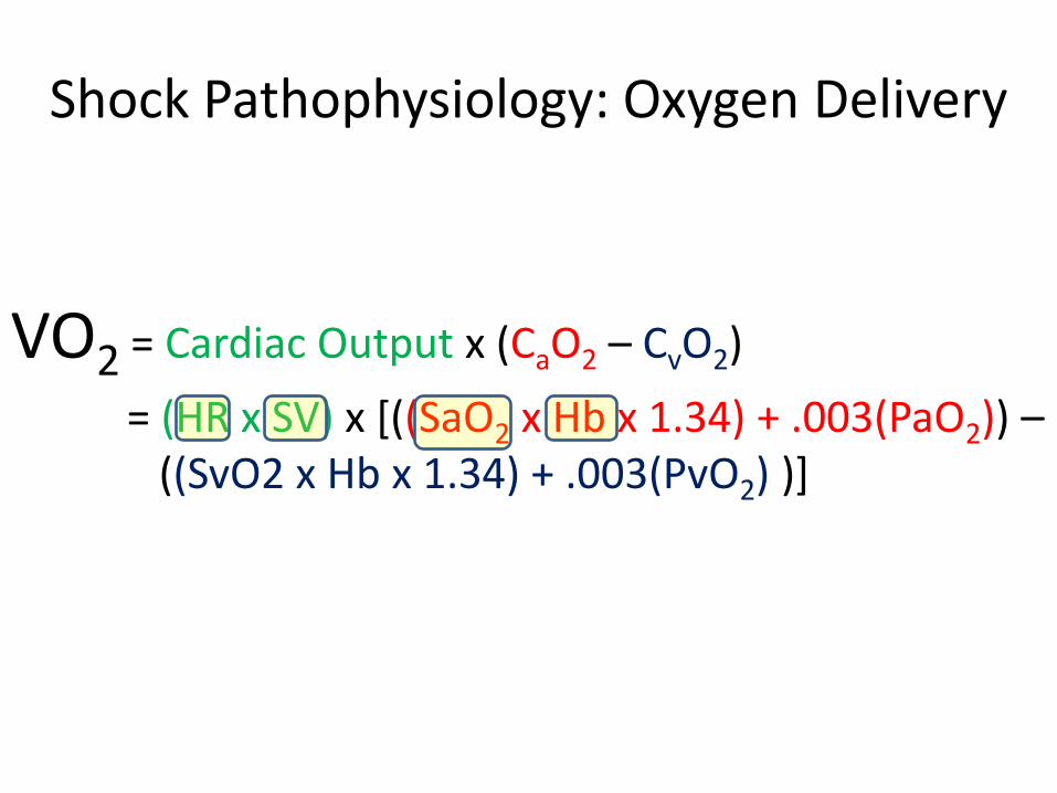
Shock Pathophysiology: Oxygen Delivery
VO2 = Cardiac Output x (CaO2 – CvO2)
= (HR x SV) x [((SaO2 x Hb x 1.34) + .003(PaO2)) – ((SvO2 x Hb x 1.34) + .003(PvO2) )]

Shock Pathophysiology: Mean Arterial Pressure
MAP = (CO x SVR)
= ((HR x SV) x SVR)

Therapeutic Interventions We Can Make
• Improve Cardiac Filling Pressures (Preload)
»Fluid Boluses
• Increase SVR and therefore MAP

Therapeutic Interventions We Can Make
• Improve Cardiac Filling Pressures (Preload) » Fluid Boluses
• Increase SVR and therefore MAP » α receptor agonism » vasopressin » dopamine
• Increase Cardiac Output » Dobutamine β1 and β2 agonism » Milrinone » Intra-aortic balloon pump
• Improve the oxygen content of the blood » Improve SaO2 via mechanical ventilation » Increase O2 carrying capacity of blood (transfusions)

• MAP ≥ 60mmHg is necessary to maintain end organ perfusion in most cases.
• Higher MAP may be necessary in select cases • Intracranial Hypertension – Hemorrage, Cerebral
Edema, Closed Head trauma, Large Strokes
• Chronic HTN
Shock Pathophysiology: Mean Arterial Pressure

What Do We Monitor, and Why?
• Variables we can directly influence to reverse abberant physiology
• MAP
• HR
• CVP
• Variables that we monitor to tell if our interventions are reversing aberrant physioglogy
• Lactate
• Urine output
• Mixed venous oxygen saturation

What Do We Monitor, and Why?
• Cuff Blood Pressure
• Intra-arterial blood pressure
• Central Venous Pressure
• Pulmonary Artery Catheter • RV Pressure
• PA Pressure
• Pulmonary Capillary Wedge Pressure
• CO/CI
• Cardiac Output
• Echocardiography / IVC Diameter

Monitoring: Cuff Blood Pressure
• Oscillatory Automated Blood Pressure Monitors

Monitoring: Cuff Blood Pressure
Situations where potentially unreliable:
• Caridiac Arrythmias (especially rapid Afib)
• Increased SVR / Circulatory Shock
• Calcified Arteries

Monitoring: Cuff Blood Pressure
• Manual Cuff Pressures / Ausculatory Method
– Inflate Cuff and listen for Korotkoff Sounds to determine systolic and diastolic pressures.
• Calculate MAP = (SPB + 2(DBP)) / 3

Monitoring: Intra Arterial Blood Pressure
• The gold standard for accurate assessment of blood pressure
• Provides continuous data
• Not essential, but probably should be considered any time vasopressor agents are used

Monitoring: Intra Arterial Blood Pressure

Monitoring: Central Venous Pressure

Monitoring: Central Venous Pressure
a wave = atrial contraction c wave = ventricular contraction against closed tricuspid x descent = closed tricuspid valve being pulled toward ventricle by ventricular contraction v wave = atrial filling y descent = ventricular filling, tricuspid valve open

Monitoring: Central Venous Pressure
• Estimates cardiac filling pressures and helps us estimate whether hypotension might be fluid responsive or not.

Central Venous Pressure: Severe Sepsis and Septic Shock
Rivers, E. et al. NEJM 345(19): 1368. 2001

NHLBI ARDS Clinical Trials Network. NEJM 2006. 354:2564
Central Venous Pressure: Acute Respiratory Distress Syndrome (ARDS)

Monitoring: The Pulmonary Artery Catheter
• Cardiac Output • Cardiac Index • Systemic Vascular Resistance • Pulmonary Vascular Resistance
Redrawn from Marino Pl. The ICU Book, Philadelphia, Lea and Feliger, 1991, p. 103.Up To Date Online. Graphic 75601 Version 2.0
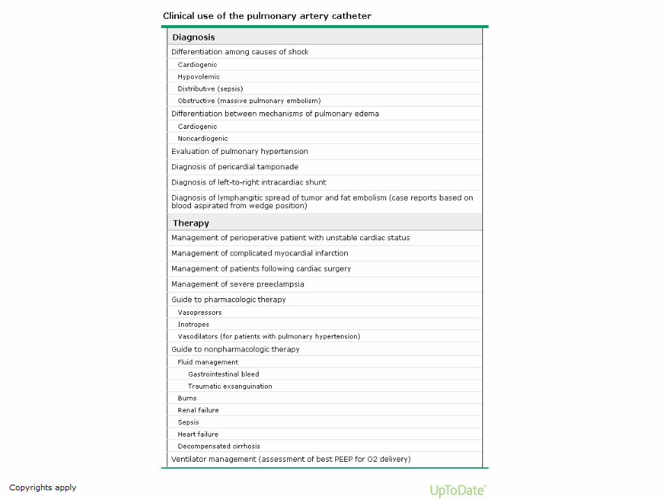

• Studies have shown no benefit with use of pulmonary artery catheters
• Different experienced provdiders come to different conclusions based on the same data from PAC’s
• Use is now very limited. Usually used to answer a very specific question.
Monitoring: The Pulmonary Artery Catheter

Its All About Fluid Responsiveness!
• Trial and Error • Fluid Challenge
• Passive Leg Raising Maneuvre
• New High Tech Evaluations • PiCCO
• Pulse Pressure Variation
• Bedside Ultrasound Evaluation of the IVC

Its All About Fluid Responsiveness!

Hemodynamic Monitoring: Pulse Pressure Variation
• Relies on the principal that stroke volume and therefore blood pressure will vary with alterations in preload induced by respiratory cycles on positive pressure ventilation.

• PiCCO and LiDCO Both use arterial waveform tracings to derive cardiac output by using proprietary algorithms to convert pressure based signals into flow measurements.
• They also use thermodilution to estimate intrathoracic blood volume, extravascular lung volume, and cardiac output.
• They require and CVP and an arterial line.
Hemodynamic Monitoring: Pulse Pressure Variation
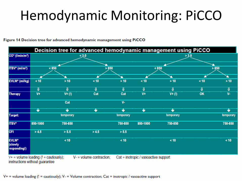
Hemodynamic Monitoring: PiCCO

Hemodynamic Monitoring: Bedside Ultrasonography and Focused
Echocardiography
• IVC diameter and variation with respiration has been used validated as predictive for fluid responsiveness patients breathing spontaneously and on mechanical ventilation.
• Hyperdynamic ventricles or severe ventricular dysfunction

Hemodynamic Monitoring: Bedside Ultrasonography and Focused
Echocardiography
From Muller, L. et al. Critical Care. 2012. 16:R188

How Do We Use These New Technologies?
• Extrapolation of what the data obtained means to our patient’s current pathophysiology
• We need algorithmic data that show how these data should be interpreted and intervened upon, and that those interventions improve outcomes.

Axioms of Effective Critical Care Delivery
1. Make your patients get evidence based care and therapies that we KNOW will improve outcome FIRST!
2. Don’t allow other data to push you to interventions that contradict evidence based therapies.
3. Use hemodynamic data to try to maintain normal physiologic parameters and reverse deteriorating organ function.
References
• NHLBI ARDS Clincial Trials Network. Comparison of two fluid management strategies in Acute Lung Injury. NEJM 2006. 354:2564
• Vincent, JL et al. Clinical Review: Update on Hemodynamic Monitoring – A Consensus of 16. Critical Care 2011. 15:229
• Antonelli, M. et al. Hemodynamic Monitoring in Shock and Implications for Management. Intensive Care Medicine 2007. 33:575.
• Pinksy, M. Hemodynamic Evaluation and Monitoring in the ICU. Chest 2007. 132:2020.
• Muller, L. et al. Respiratory Variation s in Inferior Vena Cava to Predict Fluid Responsiveness in Spontaneously Breathing Patients with Acute Circulatory Failure: Need for a Cautious Use. Critical Care. 2012. 16:R188
• Up to Date Online. Pulmonary artery catheterization.