helminth parasites of the japanese monkey, … · title helminth parasites of the japanese monkey,...
TRANSCRIPT

Instructions for use
Title HELMINTH PARASITES OF THE JAPANESE MONKEY, MACACA FUSCATA FUSCATA IN EHIMEPREFECTURE, JAPAN
Author(s) ITOH, Kazuhiro; OKU, Yuzaburo; OKAMOTO, Munehiro; OHBAYASHI, Masashi; KITAMURA, Yukitoshi;SHIBAHARA, Toshiyuki
Citation Japanese Journal of Veterinary Research, 36(3-4): 235-247
Issue Date 1988-12-05
DOI 10.14943/jjvr.36.3-4.235
Doc URL http://hdl.handle.net/2115/3133
Type bulletin
File Information KJ00002377125.pdf
Hokkaido University Collection of Scholarly and Academic Papers : HUSCAP

jpn. ]. Vet. Res., 36, 235-247 (1988)
HELMINTH PARASITES OF THE JAPANESE MONKEY, MACACA FUSCATA FUSCATA
IN EHIME PREFECTURE, JAPAN
Kazuhiro ITOH, Yuzaburo OKU, Munehiro OKAMOTO, Masashi OHBAYASHI, Yukitoshi KITAMURA1 and Toshiyuki SHIBAHARA2
(Accepted for publication October 28, 1988)
Thirty-six Japanese monkeys, Macaca fuscata fuscata captured in Ehime Prefecture, Japan, during 1986-1987 were subjected to postmortem examination.
The survey revealed 4 species of helminths: Streptopharagus pigmentatus (69.9%
of monkeys), Strongyloides fuelleborni (52.9%), Trichuris sp. (52.9%) and
Oesophagostomum aculeatum (5.6%). No cestodes or trematodes were found.
The intensity of infection in the monkeys was low except for 3 cases of heavy infection.
Key words: Macaca fuscata fuscata, parasitological survey, Streptopharagus, Trichuris, Oesophagostomum, Strongyloides, Japan
INTRODUCTION
Japanese monkeys, Macaca fuscata fuscata, are widely distributed in Japan. A few helminthological surveys on the monkeys have been performed, in which five
species have been reported; Streptopharagus pigmentatus, Strongyloides fuelleborni, Trichuris trichiura, OesoPhagostomum aculeatum [NEMATODA] and Bertiella sp. [CESTODA] (HAYAMA & NIGI, 1963; YAMASHITA, 1963; TANAKA & NIGI, 1967; NIGI,
1983). Most of the surveys, however, have been carried out using the fecal examination. In the present survey, the prevalence and intensity of the parasites were
determined by postmortem examination.
MATERIALS AND METHODS
Thirty-six wild Japanese monkeys were captured in May 1985, Feb. and Nov. 1986, and Jan. 1987, from two areas in the southern part of Ehime Prefecture: 15 from Misho-choh; 21 from Nishiumi-choh. Viscera were removed from carcasses and
Department of Parasitology, Faculty of Veterinary Medicine, Hokkaido University, Sapporo 060, Japan
1 Division of Animal Experimentation, Sapporo Medical College, Sapporo 060, Japan 2 Laboratory Animal Research Center, School of Medicine, Tottori University, Yonago 683,
Japan

236 ITOH, K. et al.
preserved in 10% formalin solution. The heart, liver, lungs, kidneys and spleen were
examined macroscopically. Tissues with gross lesions were subjected to histological
examination. The gastrointestinal tract was divided into 4 parts; esophagus-stomach, upper
part of small intestine, lower' part of small intestine and large intestine. Each part was opened longitudinally. From the solution containing intestinal contents and mucosa, parasites were collected and counted under the dissecting microscope.
The collected worms were preserved in 10% formalin solution, and prior ~? the morphological observations the parasites were immersed in lacto-phenol solution.
Use of prevalence, intensity, mean intensity, and range of intensity followed the definition of MARGOLIS et al. (1982).
T. trichiura from Japanese monkey in Maruyama Zoo, Sapporo, was observed. Trichuris sp. and T. trichiura were prepared for electron microscopy as done by Y ABU
& TAKAYANAGI (1988). Representative specimens have been deposited in the Parasite Collection in the
Department of Parasitology, Faculty of Veterinary Medicine, Hokkaido University.
RESULTS
Four speCIes of nematodes were found in the alimentary tract of 32 out of 36 monkeys examined, and species detected are as follows; S. pigmentatus, S. fuelleborni, Oesophagostomum aculeatum and Trichuris sp. No trematodes or cestodes were detected. In the infected monkeys, the number of cases parasitized by I, 2 and 3 species of helminths was 15, 13 and 4, respectively. The species detected, preva
lence and intensity are shown in Table 1.
No helminth was detected in the heart, liver, lungs, kidneys or spleen. Streptopharagus pigmentatus (LINSTOW, 1897) (SPIRUROIDAE)
S. pigmentatus was the most prevalent parasite at 69.9%. This parasite was
found in the stomach and small intestine, especially in the upper part of the small
TABLE 1 Prevalence and intensi~y of helminth parasites in Japanese monkeys
in Ehime Prefecture
Helminth Prevalence (%)
S tr ep topha rag us p igmen ta tus 25/36 (69.9)
Strongyloides (uelleborni 13/36 (52.9)
Trichuris sp. 13/36 (52.9)
Oe s ophago s tomum a culea tum 2/36 ( 5.6)
Range of intensity
3~52
many
2~125
1~2
Sites
Stomach & small intestine
Small intestine
Caecum & colon
Colon

TABLE 2 Measurements of Streptopharagus pigmentatus (in mm, egg in ,urn)
Male Female
Present authors YAMAGUTI (1941) Present authors YAMAGUTI (1935*,1941) n=45 n= 5 n=40 n=5*,n=8
Body length 26.7 45.6 30 40 44.4 72.6 47 50 width 0.78 1. 56 0.9 1.1 1.11 2.22 1.0 1.5 ::x::
('l)
(mid boby) §" Head diameter 0.17 0.19 0.15 0.19 0.18 0.20 0.15 0.24
S· ..... ::r'
Nerve ring 0.48 0.74 0.52 0.63 0.67 0.81 0.53 0.80 ~
from head end ~ "1 ~
Excretory pore 0.58 0.60 0.68 0.79 0.62 0.85 0.60 0.87 g:. ...... from head end
('l) rJl
Pharynx length 0.32 0.53 0.28 0.37 0.23 0.37 0.32 0.40 0 ......
width 0.063- 0.083 0.06 0.09 0.0'/8- 0.086 0.065- 0.115 ..... ::r
Esophagus ('l)
...... muscular part ~
~ ~
length 0.51 0.86 0.30 0.55 0.47 0.51 0.37 0.60 ::s ('l)
width 0.11 0.13 0.12 0.15 0.11 0.13 0.12 0.19 rJl ('l)
glandular part a length 4.08 6.52 5.5 7.9 7.1 7.3 6.7 9.1
0 ::s :;<;"
width 0.31 0.35 0.25 0.33 0.27 0.32 0.24 0.36 ('l)
'< Tail length 0.45 0.58 0.47 0.55 0.56 - 0.66 0.45 0.65
Spicule length right 0.66 0.79 0.62 0.66 left 3.75 4.80 4.8 5.4
Gubernaculum 0.07 0.10 0.06 0.09 Egg 33-35 X 16-20 36-45 X 18-22 Vulva 6.08 9.80 9.7*
from head end N v.., '-J

238 IToH, K. et al.
intestine. The mean intensity was 14.8. The stomach of one male monkey was parasitized by more than 50 worms with mucosal congestion.
The measurement (Table 2) and morphology (Figures 1-5) of the worms were similar to those of YAMAGUTI (1935, 1941) except for the spicule length and egg size.
Spicules were unequal: left spicule was more elongated and slender, and distal end
sharp; right spicule was massive, and distal end plump and shorter than that of YAMAGUTI (1941). The gubernaculum was broad, rectangular in form. In addition, the complex verrucosity at distal end of the tail was found. Egg size was smaller than that of YAMAGUTI (1941).
Trichuris sp. (TRICHURIDAE) Trichuris sp. was the second most prevalent parasite in this study. The mean
intensity was 26.1. Two cases showed heavy infection (more than 100 worms) with marked mucosal congestion in their caecum, and they were relatively emaciated. Young « 1 year old) monkeys had a significantly higher prevalence value of 71 % than either juveniles (1- 5 years old) or adults (> 5 years old) « 15%). -
The measurements of Trichuris which were obtained in this survey are shown in
Table 3, with the measurements of specimens from Ehime Pref. and from Maruyama Zoo, and descriptions by KONNO (1958) and SKRJABIN et a1. (1957, the specimens
parasitizing humans). The body length of the parasites from Ehime Pref. tended to be smaller than that of others. A significant difference in spicule length between
those from Ehime Pref. and other specimens was noted. The worms from Ehime Pref. were not thought to be immature worms, because mature eggs were present in the uterus. SpiCUlar sheath of the parasites from Ehime Pref. was cylindrical as T. suis (Figures 6-9). Moreover, in the scanning electron microscopic observation, the shape of spines on the spiCUlar sheath was seen to be similar to that of T. suis rather than T. trichiura· (specimens from monkey in Maruyama Zoo, and those of KONNO
(1958».
In other species Oesophagostomum aculeatum (LINSTOW, 1879) (TRICHONEMATOIDAE) was found
in the caecum and colon of monkeys obtained from Nishiumi-choh at a prevalence of 9.5% (2 cases). Some nodules were found on the mucosal surface of the large intestine of the infected monkeys.
Strongyloides juelleborni, LINSTOW, 1905 (STRONGYLOIDAE) was identified according to PREMVATI (1958). It was the second most prevalent parasite recovered
as Trichuris sp, and many in the small intestine were detected.
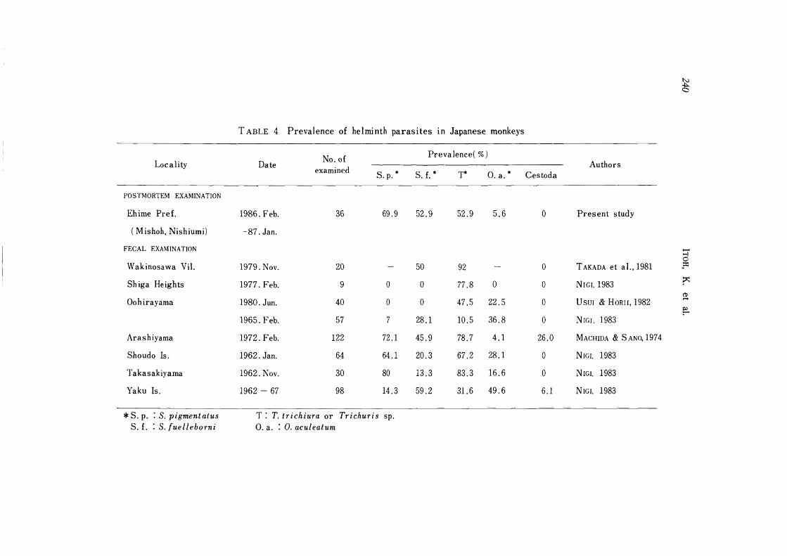
DISCUSSION
Present data by postmortem examination were compared with data reported elsewhere in other areas of Japan (Table 4, Figure 10). In Arashiyama, five species including Bertiella sp. are found, and positive rate of fecal examination was more than

TABLE 3 Comparison of measurements of Trichuris (in mm, egg in j.Lm)
Species Trichuris sp. T. ir ichiura
Ehime Pref. Maruyama Zoo KONNO (1958) S KR.JAB[:-.J et a I. ( 1957) Host Jpn. monkey Jpn. monkey Jpn. monkey Human
Male n=35 n= 3 n= 5
Body length 28.8 33.3 34.0 38.6 28.7 41.3 38 42
Esophagus length 15.4 19.1 22.1 25.1 18.1 26.0* 25 29
Spicule length 1. 56 1. 65 2.1 2.6 2.21 2.26 2.7 --3.9
Female n=28 n= :1 n= 4
Body length 25.8 35.8 40.7 46.3 30.2 45.0 48 50
Esophagus length 15.4 19.1 29.3 30.1 19.6 29.3* 25 29
46.8 58.6 52.3 58.6 51 57 50 65 Egg X X X X
19.5 22.1 21.5 25.4 21 27 20 30
* : Conversion by present authors
T. SUIS
Y AMAGUTI (1942) Pig
n= 7
36 - 50
1.8 2.3
n=12
35 - 50
54 60 X
26 29
:::t:: ('T)
a Er ,..,. ::r '0 I:>l '"1 I:>l C/)
::;: ('T) C/)
0 -,..,. ::r ('T)
'-I:>l '0 I:>l ::l ('T) C/) ('T)
a 0 ::l ~ ('T)
'<
N W \0
~ c
TABLE 4 Prevalence of helminth parasites in Japanese monkeys
No. of Prevalence( %) Locality Date Authors
examined S. p. * S. f. * T* O. a. * Cestoda
POSTMORTEM EXAMINATION
Ehime Pre£. 1986. Feb. 36 69.9 52.9 52.9 5.6 0 Present study
(Mishoh, Nishiumi) -87. Jan.
FECAL EXAM INA TION ...... -l 0
Wakinosawa Vil. 1979. Nov. 20 50 92 0 T AKADA et a I., 1981 ~::r:
Shiga Heights 1977. Feb. 9 0 0 77.8 0 0 NIGI,1983 r (1)
Oohirayama 1980. Jun. 40 0 0 47.5 22.5 0 USUI & HORII, 1982 ~
~ 1965. Feb. 57 7 28.1 10.5 36.8 0 NrGI, 1983
Arashiyama 1972. Feb. 122 72.1 45.9 78.7 4.1 26.0 MACHIDA & S ANO, 1974
Shoudo Is. 1962. Jan. 64 64.1 20.3 67.2 28.1 0 NIGr. 1983
Takasakiyama 1962. Nov. 30 80 13.3 83.3 16.6 0 NrGI, 1983
Yaku Is. 1962 - 67 98 14.3 59.2 31.6 49.6 6.1 NIGI. 1983
* S. p. : S. pigmentatus T: T. trichiura or Trichuris sp. S. f. : S. {uelleborni O. a. : O. aculeatum

Helminth parasites of the Japanese monkey 241
Fig. 10 Localities collected hosts and fecal samples (in Table 4)
I
95%. In Wakinosawa ViI. two species (5. fuelleborni, T. trichiura) are reported. On the Shiga Heights, only T. trichiura is found.
In Ehime Pref. in this study, 4 species except for Bertiella sp. were detected,
and the prevalence of parasites was similar to that of Shoudo Is. and Takasakiyama (NIGI, 1983), and these three areas are located in southern part of Japan (Figure. 10).
5. pigmentatus requires the intermediate host (beetles) in their life cycle. ARAKI et al. (1977) succeeded the experimental infection of Japanese monkey with larvae
obtained from the body cavity of Geotrupes laevistriatus, Onthophagus ater and
Onthophagus atripennis. In the present investigation, beetles were found in the stomach content (5 cases). The difference of the prevalence of S. pigmentatus among
cold, snow and warm region might be related to the distribution of these beetles and their active season.
O. aculeatum was detected only in 2 cases in this survey. This parasite is one of the noticeable parasites which cause heavy diarrhea among monkeys (HONJO et al.,
1963). Trichuris obtained in this survey were not indentified. According to CHANDLER
(1925) the specific features of whipworms are the size of the spicule and the structure of cloaca in male. SCHWARTZ (1926) attached much importance to the size of spicule,
structure of spicular sheath and size of embryonated egg in classification of Trichur
idae. The validity of T. suis and T. trichiura is a subject of controversy, and some

242 ITOH, K et al.
workers do not believe the two parasites to be identical. Experimental and accidental
infection of humans with T. suis have been reported (BEER, 1976). Therefore, there
is a great possibility for T. suis to infect the Japanese monkey.
Recently, many chances for humans to come into contact with Japanese monkeys
are given, as more wild monkey parks have built in every part of Japan. And much
attention has been paid to problems of "zoonoses". KOYAMA et al. (1978) showed that
Japanese monkey as an experimental animal was infected with entamebiasis. And the
experimental infection to men by eggs of Trichuris sp. from Japanese monkeys was
successful (lMADA et al., 1980). Furthermore, PAMPIGLIONE & RICCIARDI (1972) re
ported that humans are susceptible to infection with S. fuelleborni. For public health
importances more attention should be paid to parasites of monkeys.
ACKNOWLEDGEMENTS
We thank Mr. Y. NISHINE, Maruyama Zoo, Sapporo, for offering specimens (T.
trichiura) from Japanese monkeys. And we wish to express our thanks to Mr. S.
CHISEMBE, Faculty of Veterinary Medicine, University of Zambia, for reading the
manuscript.
REFERENCES
1) ARAKI,}., MACH IDA, M., KOYAMA, T., KUMADA, M., KAWABATA, M., HORII, Y., IMADA, I., HONJO, S., TAKAsAKA, M., TIBA, T. & MATSUBAYASHI, K (1977): The
life-cycle of Streptopharagus sp. : I. jpn.]' Parasitol., 26 (Suppl.), 80 (in Japanese) (Abstr.)
2) BEER, R. J. S. (1976): The relationship between Trichuris trichiura (LINNAEUS 1758)
of man and Trichuris suis (SCHRANK 1788) of the pig. Res. Vet. Sci., 20, 47-54
3) CHANDLER, A. C. (1925): Characters for use in identification of species of the genus
Trichuris. l. P arasitol. , 16, 97. (Abstr. ) 4) HAYAMA, S. & NIGI, H. (1963): Investigation on the helminth parasites in the Japan
Monkey Centre during 1959-1961. Primates, 4, 97-112
5) HONJO, S., MUTO, K, FUJIWARA, T., SUZUKI, Y. & IMAIZUMI, K (1963): Significance
of the natural infection of OesoPhagostomum sp. in cynomolgus monkeys (M acaca irus) used as experimental animals. lpn. l. Med. Sci. Bioi., 16, 225-227
6) IMADA, I., HORII, Y. & USUI, M. (1980): The experiment of artificial infection to
men and monkeys by eggs of Trichuris trichiura from Japanese monkeys. jpn.]' Parasitol., 29 (1, Suppl.), 30 (in Japanese) (Abstr.)
7) KONNO, S. (1958): On Trichuris trichiura in Macaca fusucata fusucata and Papio papio. Med. and Bioi., 48, 21-24 (in Japanese)
8) KOYAMA, T., KUMADA, M., KODAMA, K, SAITO, R., SHIGA, M., MIYAO, Y. &
IKEHARA, T. (1978): Amoeba infection in rhesus monkeys, Macaca mulatta and in
Japanese monkey, Macaca fuscata fuscata. lPn. l. Parasitol., 27 (Suppl.), 60 (in Japanese) (Abstr.)
9) MACHIDA, M. & SANO, N. (1974): Fecal examination of Japanese monkey

Helminth parasites of the japanese monkey
(Arashiyama A-group). lpn. l. Parasitol., 23 (Suppl.) , 7 (in japanese) (Abstr.)
10) MARGOLIS, L., ESCH, G. W., HOLMES,]' C., KURIS, A. M. & SCHAD, G. A. (1982):
The use of ecological terms in parasitology (report of an ad hoc committee of the
American Society of Parasitologists). l. Parasitol., 68, 131-133
11) NIGI, H. (1983): (translated title) Survey on helminth parasites of the japanese
monkey. Reichohrui-to-Shippei, 38-40 (in Japanese)
12) PAMPIGLIONE, S. & RICCIARDI, M. L. (1972): Experimental infection with human
strain Strongyloides fuelleborni in man. Lancet, 7752, 663-665
13) PREMVATI. (1958): Studies on Strongyloides of primates. I. Morphology and life
history of Strongyloides fuelleborni von LINSTOW, 1905. Can. I Zool., 36, 57-77
14) SCHWARTZ, B. (1926): The specific identity of whipworms from swine. ]. Agric.
Res., 33, 311-316
15) SKRJABIN, K. 1., SHIKHOBALOVA, N. P. & ORLOV, 1. V. (1957): Osnovy Nematodologii.
VI, Moskva Izd. AN SSSR. (in Russian)
16) TAKADA, N., HUANG, W. & FUJITA, H. (1981): (translated title) Fecal examination of
Japanese monkey inhabitant Aomori prefecture. Hirosaki Igaku, 33, 67-76 (in
Japanese)
17) TANAKA, T. & NIGI, H. (1967): Clinical examinations of the Japanese monkey
(Macaca fusucata). Primates, 8, 91-106
18) USUI, M. & HORII, Y. (1982): A survey on helminth parasites of the Japanese
monkey (Macaca fusucata). Bull. Fac. Agric., Miyazaki Univ., 29, 269-274 (in
Japanese with English summary)
19) YABU, Y. & TAKAYANAGI, T. (1988): Trypsin stimulated transformation of Trypanoso
ma brucei gambiense bloodstream forms to procyc1ic forms in vitro. Parasitol. Res., 74, 501-506
20) YAMAGUTI. S. (1935): Studies on the helminth fauna of Japan. Part 13. Mammalian
nematodes. lPn. ]. Zool., 6, 453-454
21) Y AMAGUTI, S. (1941): Studies on the helminth fauna of Japan. Part 35. Mammalian
nematodes. lPn. I Zool., 9, 453-454
22) YAMAGUTI, S. (1942): Studies on the helminth fauna of Japan. Part 41. Mammalian
nematodes III. jpn. ]. Zool., 1O, 2-3
23) YAMASHITA, J. (1963): Ecological relationships between parasites and primates. I.
Helminth parasites and primates. Primates, 4, 1-96
243

244 ITOH, K. et al.
EXPLANATION OF PLATES
PLATE I
FIGS. 1-5 Streptopharagus pigmentatus
Fig. 1 Anterior end, lateral view
Fig. 2 Cephalic end, apical view
Fig. 3 Egg
Fig. 4 Posterior end of male, lateral view
Fig. 5 Posterior end of female, lateral view

ITOH, K. et al.
3 3
4
0.2 mm
o e".)
3 3
PLATE I
2
5

246 ITOH, K. et al.
PLATE IT
FIGS. 6-7 Trichuris sp.
Fig. 6 Vulvar region, lateral view
Fig. 7 Posterior end of male, lateral view
Fig. 8 Egg
Fig. 9 Posterior end of female, lateral VIew

ITOH, K. et al.
3 3
1
3 3
PLATE IT
7
8 9
il