helena biosciences europeperamed.com/peramed/docs/201300_5060169851955_en.pdfthe reagent vials...
TRANSCRIPT

SAS-1 LD Vis-12 Isoenzyme
Instructions For UseREF 201300REF 201301 (gel kit only), REF 201302 (reagent kit only)
SAS-1 Isoenzyme LDH VIS-12Fiche techniqueREF 201300REF 201301 (gels uniquement) REF 201302 (réactifs uniquement)
SAS-1 LDH Vis-12 Isoenzym-KitAnleitungREF 201300REF 201301 (Nur Gel-Kit), REF 201302 (Nur Reagenz-Kit)
Kit per Isoenzimi LDH VIS-12 SAS-1Istruzioni per l'usoREF 201300REF 201301 (solo kit gel) REF 201302 (solo kit reagente)
Kit de isoenzimas LDH SAS-1 Vis-12Instrucciones de usoREF 201300REF 201301 (sólo el kit de gel)REF 201302 (sólo el kit de reactivos)
helenawww.helena-biosciences.com
BioSciencesEurope
Contents
English..................................................1Français................................................8Deutsch ...............................................15Italiano .................................................22Español ................................................29


IINNTTEENNDDEEDD PPUURRPPOOSSEEThe SAS-1 LD Vis-12 Isoenzyme Kit is intended for the separation and quantitation of LactateDehydrogenase Isoenzymes in serum or plasma by agarose gel electrophoresis.
Lactate dehydrogenase (LD) (EC 1.1.1.27) is an enzyme found in virtually all human tissues, with theliver, skeletal muscle, heart and kidney having the greatest concentrations. The wide distribution ofLD in body tissues limits the usefulness of total LD determinations in diagnosis. Testing for the sourceof elevated LD activity may be indicated with isoenzyme assessment
1.
Five isoenzymes of LD can be demonstrated in human serum. Each isoenzyme is designated by anumber which is related to its electrophoretic mobility. The fastest moving fraction (most anodic) isdesignated LD1 and is found primarily in heart muscle. The slowest moving (most cathodic) is LD5found primarily in liver and skeletal muscle. The others - LD2, LD3, and LD4 are found in varyingdegrees along with LD1 and LD5 in all tissues
1,2.
The most important use of LD isoenzymes is in the diagnosis of myocardial damage. LD2 is found inhighest concentration in normal human serum. The ratio LDI/LD2 is therefore less than one.Following myocardial infarction (MI), there is substantial elevation in LD1 so that the LD1/LD2 ratiofollowing Ml will approach or even exceed 1, a phenomenon referred to as ‘flipped LD’. The LD levelbegins to rise approximately 12-24 hours following myocardial infarction, frequently reaching levelstwo to three times (or greater) the upper limit of normal. Peak activity is usually reached on day 3-4and activity may remain elevated for as long as two weeks after infarction
2.
The most definitive testing in the diagnosis of Ml is accomplished by performing creatine kinase (CK)isoenzyme studies in conjunction with LD isoenzyme studies
1-5. The specificity and sensitivity achieved
with these two tests has eliminated the necessity for additional enzyme studies in accurately diagnosingMl. Studies have shown that CK and LD isoenzyme analyses, in conjunction with the proper clinicalsetting and electrocardiogram results, are virtually 100% accurate in properly diagnosing myocardialinfarction
2,6.
The isoenzymes of LD have been determined by various methods7-11
. Electrophoresis provides farmore information than the other methods because it allows complete separation of all five isoenzymeswith no risk of carryover. The support media used in electrophoresis includes cellulose acetate, agar,agarose and acrylamide gels
1.
The SAS-1 LD Vis-12 Isoenzyme Kit utilises a modification of the method of Preston8and separates the
lactate dehydrogenase isoenzymes according to mobility in an agarose gel. The isoenzyme bands arethen visualised with a colorimetric reagent which quantitatively visualises the isoenzymes according tothe following reaction sequence:
The patterns may be quantitated by densitometry at 595nm
WWAARRNNIINNGGSS AANNDD PPRREECCAAUUTTIIOONNSSAll reagents are for in-vitro diagnostic use only. Do not ingest or pipette by mouth any kit component.Wear gloves when handling all kit components. Refer to the product safety data sheet for risk andsafety phrases and disposal information.
1
SAS-1 LD VIS-12 ISOENZYME
English
L-lactate + NAD
NADH + Tetrazolium salts
LDH
PMSPyruvate + NADH
NADH + Formazan Dye (insoluble)

CCOOMMPPOOSSIITTIIOONN11.. SSAASS--11 LLDD IIssooeennzzyymmee GGeell ((xx1100))
Contains agarose in a barbital buffer with sodium azide as preservative. The gel is ready for use aspackaged.
22.. LLDD IIssooeennzzyymmee RReeaaggeenntt ((1100xx 11mmll))Contains those components required for the identification of lactate dehydrogenase isoenzymesaccording to the reaction sequence shown above. See STEP-BY-STEP PROCEDURE forreconstitution instructions.
33.. LLDD IIssooeennzzyymmee DDiilluueenntt ((11xx 1155mmll))Contains AMP, bicine, barbital and aspartate buffer and sodium azide as preservative. The diluentis ready for use as packaged.
44.. OOtthheerr KKiitt CCoommppoonneennttssEach kit contains Instructions For Use, and sufficient Blotters B, C and Reagent Spreading Films tocomplete 10 gels.
SSTTOORRAAGGEE AANNDD SSHHEELLFF--LLIIFFEE11.. SSAASS--11 LLDD IIssooeennzzyymmee GGeell
Gels should be stored at 15...30°C and are stable until the expiry date indicated on the package.DO NOT REFRIGERATE OR FREEZE. Deterioration of the gel may be indicated by 1) crystallineappearance indicating the gel has been frozen, 2) cracking and peeling indicating drying of the gelor 3) visible contamination of the agarose from bacterial or fungal sources.
22.. LLDD IIssooeennzzyymmee RReeaaggeenntt The reagent vials should be stored at 2...6°C and are stable until the expiry date indicated on thelabel. Reconstituted reagent is stable for 4 hours at 15...30°C or 48 hours at 2...6°C.
33.. LLDD IIssooeennzzyymmee DDiilluueenntt The diluent should be stored at 2...6°C and is stable until the expiry date indicated on the label.Avoid contamination.
IITTEEMMSS RREEQQUUIIRREEDD BBUUTT NNOOTT PPRROOVVIIDDEEDDCat. No. 210200 Applicator Blades (1 x 10)Cat. No. 210300 Applicator Blades (5 x 10)Cat. No. 210100 Disposable Sample Cups (100)Cat. No. 5014 Development weightCat. No. 4062 Incubation ChamberCat. No. 3100 REP PrepDrying Oven with forced air capable of 60...70°CIncubator capable of 45°CDestain solution: Mix 100ml of glacial acetic acid and 900ml of purified water. Store in a tightlystoppered bottle.Purified water
2

SSAAMMPPLLEE CCOOLLLLEECCTTIIOONN AANNDD PPRREEPPAARRAATTIIOONNFresh serum is the specimen of choice. Heparin or EDTA anticoagulated plasma can also be used,Oxalate inhibits LD activity and should not be used
12. Plasma should be platelet-free as platelets contain
LD13
. Samples should be tested as soon as possible after collection as no one storage condition isoptimal for all isoenzymes
12,14,15,16. If required, samples can be stored refrigerated at 15...30°C or 2...6°C
for up to 48 hours12
. DO NOT FREEZE - freezing destroys LD5 activity12
.SSaammppllee CCoolllleeccttiioonn:: Correct timing of sample collection is critical for the accurate interpretation of LDIsoenzyme patterns in the assessment of MI. A minimum of 3 samples should be collected - one onadmission, one 6-13 hours later and the third 24-37 hours following admission
2-4.
IInntteerrffeerriinngg ffaaccttoorrss::1) Haemolysis may affect LD1 and LD2 quantitations as erythrocytes contain high levels of LD
activity1-2,12
.2) LD activity is reduced in uraemic sera due to the presence of inhibitors such as urea and oxalate.
LD5 is particularly affected17
.3) Acetone and chloroform inhibit all isoenzymes except LD1
14.
4) Certain drugs can affect LD isoenzyme activity18
.
SSTTEEPP--BBYY--SSTTEEPP PPRROOCCEEDDUURREE1. Pipette 35µl of the sample into the appropriate well of the sample tray or disposable sample cups.ii)) SSAASS--11 && SSAASS--11 PPlluuss uusseerrss:: Use SAS-1 sample tray. Carefully place the sample tray onto the
applicator drawer. Ensure that the tray is pushed firmly down into position.iiii)) SSAASS--33 uusseerrss:: Use SPIFE / SAS-3 sample tray. Carefully locate the sample tray using the sample base
locating pins. Ensure that the tray is positioned securely.2. Remove the gel from the packaging and:ii)) SSAASS--11 uusseerrss:: place the gel in the SAS-1, agarose side up, aligning the positive and negative sides
with the corresponding electrode posts.iiii)) SSAASS--11 PPlluuss uusseerrss:: dispense 400µL of REP Prep onto the heat sink. Place the gel onto the heat sink,
agarose side up, aligning the positive and negative sides with the corresponding electrode posts,taking care to avoid air bubbles under the gel.
iiiiii)) SSAASS--33 uusseerrss:: place the alignment guide onto the pins and dispense 400µL of REP Prep onto thecentre of the chamber. Place the gel into the chamber agarose side up, using the guide, align thepositive and negative sides with the corresponding electrode posts, taking care to avoid air bubblesunder the gel.
3. Blot the surface of the gel with a blotter C, discard the blotter.4. ii)) SSAASS--11 uusseerrss:: attach the electrodes onto the top side of the electrode posts so that they are in
contact with the gel blocks.iiii)) SSAASS--11 PPlluuss uusseerrss:: (as above). Place the cover over the gel and electrodes and press firmly for 5seconds to ensure contact.iiiiii)) SSAASS--33 uusseerrss:: attach the electrodes onto the the electrode posts so that they are in contact withthe gel blocks.
5. Place 1 applicator blade assembly into the top position on the instrument, ((SSAASS--33 uusseerrss:: slot 8).6. Perform the LD electrophoresis:ii)) SSAASS--11 uusseerrss:: 100 volts, 13 mins, 5 applicationsiiii)) SSAASS--11 PPlluuss uusseerrss:: Electrophoresis: 80 volts, 20 mins, 15°C, 5 applications
Incubation Step 1: 25 mins, 45°C
3
SAS-1 LD VIS-12 ISOENZYME
English

iiiiii)) SSAASS--33 uusseerrss::
SStteepp TTiimmee ((mmmm::ssss)) TTeemmppeerraattuurree ((°°CC)) VVoollttaaggee OOtthheerrElectrophoresisLoad Sample 00:10 21 Speed 1Apply Sample 00:10 21 Speed 1*RReeppeeaatt sstteeppss ‘‘ llooaadd ssaammppllee,, aappppllyy ssaammppllee’’ ffoorr aa ttoottaall ooff 66 aapppplliiccaattiioonnss..Electrophoresis 15:00 20 100
* Use Location 2
7. SSAASS--11 aanndd SSAASS--33 uusseerrss:: Whilst the samples are electrophoresing, place a tissue moistened withwater into the incubation chamber and place in the incubator set at 45°C to equilibrate.
8. Approximately 3-4 minutes before the end of electrophoresis, reconstitute 1 vial of LD IsoenzymeReagent by adding 1ml of LD Isoenzyme Diluent and mix well.
9. Following electrophoresis, SSAASS--11 aanndd SSAASS--33 uusseerrss:: place the gel agarose side up into the incubationchamber.
10. Pour the contents of the LD Isoenzyme Reagent vial along the middle of the gel. Carefully applyone piece of reagent spreading film to spread the reagent, avoiding air bubbles.
11. SSAASS--11 aanndd SSAASS--33 uusseerrss:: Incubate the gel: 25 minutes, 45°C.SSAASS--11 PPlluuss uusseerrss:: Incubate the gel.
12. Remove the reagent spreading film and remove both gel blocks using the Gel Block Remover.13. Wash the gel in destain solution for 2 minutes.14. Place the gel on a flat surface and place a blotter B (wetted in destain solution), onto the surface
of the gel, followed by 3 folded paper towels. Press the gel using the development weight for 5minutes.
15. Wash the gel in destain solution for 1 minute.16. Wash the gel in purified water and dry at 60...70°C.
IINNTTEERRPPRREETTAATTIIOONN OOFF RREESSUULLTTSSIt is recommended that any evaluation of the gels is performed against normal values which have beenproduced for this test in each individual laboratory. Completed gels should be stored in the dark at alltimes.
QQuuaalliittaattiivvee EEvvaalluuaattiioonn:: The SAS-1 LD Vis-12 Isoenzyme gel can be visually inspected for the presenceor absence of bands of interest.QQuuaannttiittaattiivvee EEvvaalluuaattiioonn:: Scan the gels at 595nm, within 2 hours of completion. Completed gels shouldbe stored in the dark at all times.
Following electophoresis, 5 zones of LD activity are normally detected. The fastest zone (LD1) moveswith a migration similar to the alpha1 globulins and the slowest zone (LD5) moves with the gammaglobulins. The other 3 zones have intermediate mobilities. The LD activity in serum reflects thebreakdown of numerous cell types and all 5 zones can be seen. LD2 predominates, followed by LD1and LD3. LD4 and LD5 occur in minor amounts.
4

1. LD2 is the LD Isoenzyme present in the largest amounts in normal serum1-4,12
.2. LD1 is elevated and may be greater than LD2 in:a) Myocardial infarction
1-4,12.
b) Duchenne’s muscular dystrophy presents a pattern like MI but clinical symptoms help in easilydifferentiating the two diseases
19,20.
c) Haemolysis (including haemolytic anaemias) should be strongly considered whenever total serumLD reaches levels greater than 5x normal and the isoenzymes show a greater LD1 and LD2. TotalLD is much higher in haemolytic anaemia than in MI unless the MI is accompanied by severe shock.Pernicious anaemia in relapse gives an LD pattern like haemolysis and some of the highest totalserum LD levels are found with this condition
2,14.
d) Renal infarct2,12
.3. LD3 is elevated in pulmonary infarctions
7,12,21.
4. LD4 elevation has not been associated with any particular pathology.5. LD5 is elevated in hepatic and muscular damage and skin diseases
1.
6. When total LD is markedly elevated but all of the isoenzymes have normal percentages, this iscalled an isomorphic pattern. Widely divergent groups of clinical diagnoses have shown this typeof pattern and include cardiorespiratory diseases, malignancy, fracture, diseases of the centralnervous system, infection and inflammation, hepatic cirrhosis, alcoholism, trauma without fracture,infectious mononucleosis, hypothyroidism, uraemia, necrosis, pseudomononucleosis, viraemia andintestinal obstruction
1,2,22.
7. LD and CK isoenzymes are less specific following open heart surgery than they are in mostdiagnostic situations. The CK-MB will be elevated due to myocardial damage from the operativeprocedure and trauma caused by manipulation and cannulation of the heart. The LD2/LD1 may beelevated secondary to haemolysis from extracorporeal circulation
22-24.
QQUUAALLIITTYY CCOONNTTRROOLLThe CK / Control (Cat. No. 5134) can be used to verify all phases of the procedure and should be usedon each plate run. Refer to the package insert provided for acceptable assay values.
LLIIMMIITTAATTIIOONNSSThe SAS-1 LD Vis-12 Isoenzyme method is not designed to identify tumour markers.
Refer to SAMPLE COLLECTION AND HANDLING for other interfering factors.Further testing required:
1. Total LD activity may be determined. Conflicting reports exist about the true value of total serumenzyme levels and the severity of a disease
1,4,25.
2. In diagnosing MI, CK Isoenzyme studies should also be performed1,4
.3. Haptoglobin studies may be performed to rule out haemolysis as a cause of elevated LD1 and LD2.
5
SAS-1 LD VIS-12 ISOENZYME
English

RREEFFEERREENNCCEE VVAALLUUEESSThe following normal values were obtained in studies at Helena BioSciences. These values should onlyserve as a guideline. Each laboratory should establish its own expected normal values for this method.
LD1 = 20.6 - 32.0%LD2 = 31.1 - 35.9%LD3 = 19.8 - 25.7%LD4 = 7.0 - 10.4%LD5 = 6.5 - 14.0%
PPEERRFFOORRMMAANNCCEE CCHHAARRAACCTTEERRIISSTTIICCSSLLiinneeaarriittyyThis system is linear to 650 IU/L per band or a total LD activity of 1300 IU/L.
BBIIBBLLIIOOGGRRAAPPHHYY1. Brish, L.K. ‘CK & LD Isoenzymes A Self-Instructional Text’ , Am. Soc. of Clin. Path. Press,
Chicago, 1984: 85-120.2. Hadden, D.M. and Prentiss, T., Cardiac Profiling by Electrophoresis’, Lab. Mgt., 1977; May: 19-24.3. Henry, J.D. ‘Clinical Diagnosis and Management by Laboratory Methods’, 16th Edition, 1979; 1:
366-369, W.B. Saunders Co., Philadelphia.4. Galen, R.S., Reiffel, J.A. and Gambino, S.R., Diagnosis of Acute Myocardial Infarction’, J. Am. Med.
Assoc., 1975; 232(2): 145-147.5. Frolich, J., Brosseuk, A., Grant, A. and McLennan, M., ‘Study of the Value of CPK and LDH
Isoenzyme Determinations in the Differential Diagnosis of Ischemic Chest Pain’, Clin. Biochem.,1978; 11(6): 232-234.
6. Wagner, G.S., Roe, C.R., Limbird, L.E., Rosati, R.A. and Wallace, A.G., ‘The Importance ofIdentification of the Myocardial-Specific Isoenzyme of Creatine Phosphokinase (MB Form) in theDiagnosis of Acute Myocardial Infarction’, Circulation, 1973; 47: 263-269.
7. Nerenberg, S.T., ‘Electrophoretic Screening Procedures’, 1973: 96-125, Lea & Febiger,Philadelphia.
8. Preston, J.A., Briere, R.O. and Batsakis, J.G., ‘Rapid Electrophoretic Separation of LactateDehydrogenase Isoenzymes on Cellulose Acetate’, Am. J. Clin. Pathol., 1965; 43(3): 256-260.
9. Hsu, M-Y., Kohler, M.M., Barolia, L. and Bondar, R.J.L., ‘Separation of Five Isoenzymes of SerumLactate Dehydrogenase by Discontinuous Gradient Elution from a Miniature Ion-ExchangeColumn’, Clin. Chem., 1979; 25(8): 1453-1458.
10. Usategui-Gomez, M., Wicks, R.W. and Warshaw, M., ‘Immunochemical Determination of theHeart Isoenzyme of Lactate Dehydrogenase(LDH1) in Human Serum’, Clin. Chem., 1979; 25(5):729-734.
11. Rotenberg, Z., Weinberger, I., Sagie, A., Fuchs, J., Sperling, O. and Agmon, J., ‘LactateDehydrogenase Isoenzymes in Serum During Recent Acute Myocardial Infarction’, Clin. Chem.,1987; 33(8): 1419-1420.
12. Tietz, N.W. et al., ‘Textbook of Clinical Chemistry’, 1986: 691-700, W.B. Saunders Co.,Philadelphia.
13. Rothwell, D.J., Jendrzejczak, B., Becker, M. and Doumas, B.T., ‘Lactate Dehydrogenase Activitiesin Serum and Plasma’, Clin. Chem., 1976; 22(7): 1024-1026.
14. Latner, A.L. and Skillen, A.W., ‘Isoenzymes in Biology and Medicine’, 1968: 146-157, AcademicPress, London.
6

15. Kreutzer, H.H. and Fennis, W.H.S., ‘Lactic Dehydrogenase Isoenzymes in Blood Serum afterStorage at Different Temperatures’, Clin. Chim. Acta, 1964; 9: 64-68.
16. Galen, R.S., Personal Communication, Dec. 1981.17. Clark, P.I., Kostuk, W.J. and Henderson, A.R., ‘Time-Dependency of Human Lactate
Dehydrogenase Isoenzyme 5 Inhibition by Urea’, Clin. Chem., 1976; 22(12): 2059.18. Young, D.S., ‘Effects of Drugs on Clinical Laboratory Tests’, 3rd Ed., 1990, AACC Press,
Washington D.C.19. Roses, A.D., Roses, M.J., Nicholson, G.A. and Roe, C.R., ‘Lactate Dehydrogenase Isoenzyme 5 in
Detecting carriers of Duchenne Muscular Dystrophy’, Neurology, 1977; 27(May): 414-421.20. Yasmineh, W.G., Ibrahim, G.A., Abbasnezhad, M. and Awad, E.A., ‘Isoenzyme Distribution of
Creatine Kinase and lactate Dehydrogenase in Serum and Skeletal Muscle in Duchenne MuscularDystrophy, Collagen Disease, and Other Muscular Disorders’, Clin. Chem., 1978; 24(11): 1985-1989.
21. Papadopoulos, N.M., ‘Clinical Applications of Lactate Dehydrogenase Isoenzymes’, Annals of Clin.Lab. Sci., 1977; 7(6): 506-510.
22. Jacobs, D.S., Robinson, R.A., Clark, G.M. and Tucker, J.M., ‘Clinical Significance of the IsomorphicPattern of the Isoenzymes of Serum Lactate Dehydrogenase’, Annals of Clin. and Lab. Sci., 1977;7(5): 411-421.
23. Galen, R.S., ‘Diagnostic Tests in Cardiac and Non-Cardiac Disorders’ Diag. Med., 1978 Feb., 74-87.
24. Mohiuddin, S.M., Raffetto, J., Sketch, M.H., Lynch, J.D., Schultz, R.D. and Runco, V., ‘LDHIsoenzymes and Myocardial Infarction in patients Undergoing Coronary Bypass Surgery: AnExcellent Correlation’, Am. Heart J., 1976; 92(5): 584-588.
25. Chapelle, J-P., Albert, A., Smeets, J-P., Marechal, J-P.,Heusghem, C. and Kulbertus, H.E., ‘DoesLactate Dehydrogenase Isoenzyme-5 Contribute to the Predictive Power of Total LactateDehydrogenase in Myocardial Infarction?’ Clin. Chem., 1983; 29(5): 774-777.
7
SAS-1 LD VIS-12 ISOENZYME
English

UUTTIILLIISSAATTIIOONNLe kit SAS-1 Isoenzyme LDH Vis-12 est destiné à la séparation et la quantification des isoenzymes dela lactate déshydrogénase du sérum ou du plasma par électrophorèse en gel d’agarose.
La lactate déshydrogénase (LDH) (EC 1.1.1.27) est une enzyme se trouvant dans pratiquement tousles tissus humains, avec une concentration plus importante dans le foie, dans les muscles squelettiques,dans le cœur et dans les reins. Étant donné que la LDH est largement présente dans les tissuscorporels, l’utilité du dosage de la LDH totale dans les diagnostics est limitée. Une analyse del’isoenzyme peut indiquer la source d’une activité LDH élevée
1.
Il est possible de trouver dans le sérum humain cinq isoenzymes de la LDH. Chaque isoenzyme estdésignée par un chiffre en rapport avec sa mobilité électrophorétique. La fraction se déplaçant le plusrapidement (principalement anodique) est appelée LDH-1 et se trouve principalement dans le musclecardiaque. Celle se déplaçant le plus lentement (principalement cathodique) est la LDH-5 et se trouveprincipalement dans le foie et les muscles squelettiques. Les autres (LDH-2, LDH-3 ET LDH-3), ainsique la LDH-1 et la LDH-5, sont présentes à divers degrés dans tous les tissus
1,2.
Les isoenzymes LDH sont principalement utilisées pour le diagnostic des lésions myocardiques. La LDH-2 est la plus présente dans le sérum humain normal. Le rapport LDH-1/LDH-2 est doncinférieur à un. Suite à un infarctus du myocarde (IDM), il y a une élévation substantielle du taux deLDH-1 si bien que le rapport LDH-1/LDH-2 post-IDM se rapproche ou même dépasse 1, phénomèneappelé « rapport LDH inversé ». Le taux de LDH commence à augmenter environ 12-24 heuresaprès un infarctus du myocarde et atteint souvent des valeurs deux à trois fois (ou plus) supérieures àla limite supérieure normale. L’activité maximale est en général atteinte 3-4 jours après et elle resteélevée deux semaines après l’infarctus
2.
L’étude conjointe des isoenzymes de la créatine kinase (CK) et de celles de la LDH constitue l’analysede référence pour le diagnostic d’IDM
1-5. La spécificité et la sensibilité offertes par ces deux dosages
ont rendu inutile toute analyse d’autres enzymes pour obtenir un diagnostic d’IDM exact. Des étudesont montré que l’analyse des isoenzymes de la CK et de la LDH, unie aux résultats del’électrocardiogramme et à un tableau clinique approprié, permet de réaliser un diagnostic correct d’uninfarctus du myocarde avec une fiabilité de presque 100%
2,6.
Plusieurs méthodes permettent de doser les isoenzymes de la LDH7-11
. L’électrophorèse fournit plusd’informations que les autres méthodes car elle permet d’obtenir une séparation complète des cinqisoenzymes sans aucun risque de contamination. Les gels d’acétate de cellulose, de gélose, d’agaroseet d’acrylamide sont les supports utilisés pour l’électrophorèse
1.
Le kit SAS-1 Isoenzyme LDH Vis-12 utilise une variation de la méthode de Preston8
et sépare lesisoenzymes de la lactate déshydrogénase suivant leur mobilité sur un gel d’agarose. Les bandesd’isoenzymes sont ensuite mises en évidence de façon quantitative par un réactif de colorationconformément à la série de réactions suivante:
Un densitomètre à 595 nm permet de quantifier les bandes.
8
L-Lactate + NAD
NADH + Sels de tétrazolium
LDH
PMSPyruvate + NADH
NADH + Formazan coloré (insoluble)

9
SAS-1 ISOENZYME LDH VIS-12
Français
PPRRÉÉCCAAUUTTIIOONNSSTous les réactifs sont à usage diagnostic in-vitro uniquement. Ne pas ingérer ou pipeter à la boucheaucun composant. Porter des gants pour la manipulation de tous les composants. Se reporter auxfiches de sécurité des composants du kit pour la manipulation et l’élimination.
CCOOMMPPOOSSIITTIIOONN11.. PPllaaqquuee SSAASS--11 IIssooeennzzyymmee LLDDHH ((xx1100))
Contient de l’agarose dans un tampon barbital additionné d’azide de sodium comme conservateur.Le gel est prêt à l’emploi.
22.. RRééaaccttiiff IIssooeennzzyymmee LLDDHH ((1100xx 11mmll))Contient les composants nécessaires pour identifier les isoenzymes de la lactate déshydrogénaseconformément à la série de réactions indiquée auparavant. La section MÉTHODOLOGIE fournitles instructions nécessaires à la reconstitution.
33.. DDiilluuaanntt IIssooeennzzyymmee LLDDHH ((11xx 1155mmll))Contient un tampon AMP, bicine, barbital et aspartate additionné d’azide de sodium commeconservateur. Le diluant est prêt à l’emploi.
44.. AAuuttrreess ccoommppoossaannttss dduu kkiittChaque kit contient également une fiche technique et des buvards B, C et des films étaleurs deréactif pour 10 gels.
SSTTOOCCKKAAGGEE EETT CCOONNSSEERRVVAATTIIOONN11.. PPllaaqquuee SSAASS--11 IIssooeennzzyymmee LLDDHH
Les gels doivent être conservés entre 15...30°C; ils sont stables jusqu’à la date de péremptionindiquée sur l’emballage. NE PAS RÉFRIGÉRER OU CONGELER. Les conditions suivantesindiquent une détérioration du gel: 1) des cristaux visibles indiquant que le gel a été congelé, 2) descraquelures indiquant une déshydratation du gel, 3) une contamination visible, bactérienne oufongique.
22.. RRééaaccttiiff IIssooeennzzyymmee LLDDHHLes flacons de réactif doivent être conservés entre 2...6°C; ils sont stables jusqu’à la date depéremption indiquée sur l’étiquette. Après reconstitution, le réactif est stable 4 heures entre15...30°C ou 48 heures entre 2...6°C.
33.. DDiilluuaanntt IIssooeennzzyymmee LLDDHHLe diluant doit être conservé entre 2...6°C; il est stable jusqu’à la date de péremption indiquée surl’étiquette. Éviter toute contamination.
MMAATTÉÉRRIIEELLSS NNÉÉCCEESSSSAAIIRREESS NNOONN FFOOUURRNNIISSRéf. 210200 Applicateurs échantillons (1 x 10)Réf. 210300 Applicateurs échantillons (5 x 10)Réf. 210100 Cupules échantillons jetables (100)Réf. 5014 Poids à développementRéf. 4062 Chambre d’incubationRéf. 3100 Solution de REP-prepÉtuve de séchage à convection forcée offrant une température entre 60...70°CIncubateur offrant une température de 45°CSolution décolorante: Mélanger 100ml d’acide acétique glacial avec 900ml d’eau distillée. Conserveren bouteille hermétiquement fermée.Eau distillée
PPRRÉÉLLÈÈVVEEMMEENNTTSS DDEESS ÉÉCCHHAANNTTIILLLLOONNSSL’utilisation de sérums fraîchement prélevés est fortement recommandée. Il est aussi possible d’utiliserdu plasma prélevé sur héparine ou EDTA. L’oxalate ne doit pas être utilisé car il inhibe l’activité de laLDH
12. Le plasma doit être sans plaquettes car elles contiennent de la LDH
13. Les échantillons doivent
être analysés le plus rapidement possible après le prélèvement puisque aucune méthode deconservation n’est optimale pour toutes les isoenzymes
12, 14, 15, 16. En cas de besoin, il est possible de
conserver les échantillons entre 15...30°C ou entre 2...6°C 48 heures maximum12
. NE PASCONGELER: l’activité de la LDH-5 en serait détruite
12.
PPrrééllèèvveemmeenntt ddee ll’’éécchhaannttiilllloonn:: Il est essentiel que le prélèvement des échantillons soit correctementplanifié pour bien interpréter les types d’isoenzymes de la LDH lors du diagnostic d’IDM. Troiséchantillons au moins sont nécessaires: le premier à l’admission du patient, le deuxième 6-13 heuresaprès et le troisième 24-37 heures après
2-4.
FFaacctteeuurrss iinntteerrfféérreennttss::1) En cas d’hémolyse, le dosage de la LDH-1 et de la LDH-2 risque d’être altéré puisque les
érythrocytes contiennent un taux élevé d’activité LDH1-2, 12
.2) L’activité LDH est réduite dans les sérums urémiques en raison de la présence d’inhibiteurs
comme l’urée et l’oxalate. La LDH-5 est tout particulièrement affectée17
.3) L’acétone et le chloroforme inhibent toutes les isoenzymes sauf la LDH-1
14.
4) Certains médicaments peuvent altérer l’activité des isoenzymes de la LDH18
.
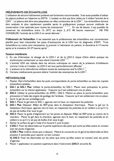
MMÉÉTTHHOODDOOLLOOGGIIEE1. Pipeter 35µl d’échantillon dans les puits correspondants du porte-échantillon ou dans les cupules
échantillons jetables.ii)) SSAASS--11 eett SSAASS--11 PPlluuss:: Utiliser le porte-échantillon du SAS-1. Placer avec précaution le porte-
échantillon sur le chariot applicateur. S’assurer qu’il est solidement mis en place.iiii)) SSAASS--33:: Utiliser le porte-échantillon du SPIFE / SAS-3. Mettre en place le porte-échantillon avec
précaution à l’aide des ergots de guidage de l’embase. S’assurer qu’il est solidement mis en place.2. Sortir le gel de son emballage puis:ii)) SSAASS--11:: Placer le gel dans le SAS-1, agarose vers le haut, en respectant les polarités.iiii)) SSAASS--11 PPlluuss:: Déposer 400µl de REP-prep dans le dissipateur thermique. Placer le gel sur le
dissipateur thermique, agarose vers le haut, en respectant les polarités et en veillant à ce qu’il n’yait pas de bulles d’air sous le gel.
iiiiii)) SSAASS--33:: Placer le guide d’alignement sur les picots et déposer 400µl de REP-prep au centre de lachambre. Placer le gel dans la chambre, agarose vers le haut, en respectant les polarités et enveillant à ce qu’il n’y ait pas de bulles d’air sous le gel.
3. Sécher la surface du gel à l’aide d’un buvard C puis le jeter.4. ii)) SSAASS--11:: Fixer les électrodes sur la partie supérieure des plots que sorte qu’elles soient en contact
avec les ponts d’agarose.iiii)) SSAASS--11 PPlluuss:: (même chose que ci-dessus). Mettre le couvercle sur le gel et les électrodes et fairepression 5 secondes pour assurer un bon contact.iiiiii)) SSAASS--33:: Fixer les électrodes sur les plots que sorte qu’elles soient en contact avec les pontsd’agarose.
5. Placer 1 applicateur en position supérieure dans l’instrument ((SSAASS--33:: encoche 8).
10

6. Réaliser l’électrophorèse LDH:ii)) SSAASS--11:: 100 volts, 13 min., 5 dépôts.iiii)) SSAASS--11 PPlluuss:: Électrophorèse : 80 volts, 20 min., 15°C, 5 dépôts.
Incubation étape 1 : 25 min., 45°Ciiiiii)) SSAASS--33::
ÉÉttaappee DDuurrééee ((mmmm::ssss)) TTeemmppéérraattuurree ((°°CC)) TTeennssiioonn AAuuttrreeÉÉlleeccttrroopphhoorrèèsseeCharger échantillon 00:10 21 Vitesse 1Déposer échantillon 00:10 21 Vitesse 1*RRééppéétteezz lleess ééttaappeess CChhaarrggeerr éécchhaannttiilllloonn,, DDééppoosseerr éécchhaannttiilllloonn ppoouurr uunn ttoottaall ddee 66 ddééppôôttss..Électrophorèse 15:00 20 100
* Utiliser Emplacement 2 (Loc 2)
7. SSAASS--11 eett SSAASS--33:: Pendant que la migration a lieu, placer un papier absorbant humidifié avec de l’eaudans la chambre d’incubation et la régler à 45°C pour qu’elle s’équilibre.
8. Environ 3-4 minutes avant la fin de l’électrophorèse, reconstituer 1 flacon de réactif IsoenzymeLDH en ajoutant 1ml de diluant Isoenzyme LDH et bien mélanger.
9. Une fois l’électrophorèse terminée, SSAASS--11 eett SSAASS--33:: Placer le gel, agarose vers le haut, dans lachambre d’incubation.
10. Verser le contenu du flacon de réactif Isoenzyme LDH au milieu du gel. Déposer avec précautionun film étaleur pour étaler le réactif, en évitant la formation de bulles d’air.
11. SSAASS--11 eett SSAASS--33:: Incuber le gel pendant 25 minutes à 45°C.SSAASS--11 PPlluuss:: Incuber le gel.
12. Enlever le film étaleur et les deux ponts d’agarose à l’aide de la raclette.13. Laver le gel dans la solution décolorante pendant 2 minutes.14. Placer le gel sur une surface plane et déposer dessus un buvard B (imbibé de solution décolorante)
puis 3 papiers absorbants pliés. Presser le gel à l’aide du poids à développement pendant 5minutes.
15. Laver le gel dans la solution décolorante pendant 1 minute.16. Rincer le gel avec de l’eau distillée et sécher entre 60...70°C.
IINNTTEERRPPRRÉÉTTAATTIIOONN DDEESS RRÉÉSSUULLTTAATTSSIl est recommandé de réaliser chaque évaluation en comparant les gels à un modèle normal obtenudans les mêmes conditions pour chaque laboratoire. Les gels terminés doivent toujours être conservésà l’abri de la lumière.
ÉÉvvaalluuaattiioonn qquuaalliittaattiivvee:: Une inspection visuelle du gel SAS-1 Isoenzyme LDH Vis-12 permet dedéterminer si les bandes des isoenzymes sont présentes ou non.ÉÉvvaalluuaattiioonn qquuaannttiittaattiivvee:: Lire le gel à 595nm dans les 2 heures suivant la fin du processus. Les gelsterminés doivent toujours être conservés à l’abri de la lumière.
11
SAS-1 ISOENZYME LDH VIS-12
Français

La migration permet normalement de détecter 5 zones d’activité LDH. La zone la plus rapide (LDH-1) a une migration similaire à celle des alpha-1-globulines et la plus lente (LDH-5) migre avec lesgammaglobulines. Les 3 autres zones ont des mobilités intermédiaires. L’activité LDH dans le sérumreflète la répartition de différents types de cellules et il est possible de voir les 5 zones. La LDH-2 estla plus présente, suivie de la LDH-1 et de la LDH-3. La LDH-4 et la LDH-5 sont présentes en moindrequantité.
1. La LDH-2 est l’isoenzyme de la LDH la plus présente dans la sérum normal1-4,12
.2. Le taux de LDH-1 est élevé et même supérieur à celui de la LDH-2 dans les cas suivants:a) Infarctus du myocarde
1-4,12.
b) Dystrophie musculaire de Duchenne: le résultat de l’électrophorèse est similaire à celui obtenuchez un patient présentant un IDM, mais les symptômes cliniques permettent de différencierfacilement les deux maladies
19,20.
c) Hémolyse (y compris les anémies hémolytiques): doit être envisagée si le taux de LDH totalsérique est 5 fois supérieur à la normale et que la LDH-1 et la LDH-2 sont plus élevées. Le tauxde LDH total est plus élevé en cas d’anémie hémolytique qu’en cas d’IDM à moins qu l’IDM ne soitaccompagné d’un état de choc grave. Une rechute d’anémie pernicieuse donne des résultatssimilaires à ceux obtenus en cas d’hémolyse et c’est dans ce cas que l’on trouve les taux de LDHtotale les plus élevés
2,14.
d) Infarctus rénal2,12
.3. La LDH-3 est élevée en cas d’infarctus pulmonaire
7,12,21.
4. L’augmentation du taux de la LDH-4 n’a été associée avec aucune pathologie spécifique.5. La LDH-5 est élevée en cas de lésions hépatiques ou musculaires et de maladies de la peau
1.
6. Si le taux de LDH total est nettement élevé mais que le pourcentage relatif des isoenzymes estnormal, on parle de résultats isomorphes. Ce genre de résultat correspond à des pathologiesdiverses: maladies cardiorespiratoires, malignité, fracture, maladies du système nerveux central,infection et inflammation, cirrhose hépatique, alcoolisme, trauma sans fracture, mononucléoseinfectieuse, hypothyroïdie, urémie, nécrose, pseudo-mononucléose, virémie, occlusion intestinale,entre autres
1,2,22.
7. Les isoenzymes de la LDH et de la CK sont moins spécifiques suite à une opération à cœur ouvertque dans la plupart des situations diagnostiques. La CK-MB est élevée en raison des lésions dumyocarde lors de l’opération et du trauma lié à la manipulation et à la canulation du cœur. Le rapport LDH-2/LDH-1 risque d’être élevé suite à l’hémolyse due à la circulationextracorporelle
22-24.
CCOONNTTRRÔÔLLEE QQUUAALLIITTÉÉLe contrôle CK/LDH (Réf. 5134) peut être utilisé afin de vérifier toutes les phases de la méthode etdoit être déposé sur chaque plaque. La notice jointe indique les valeurs acceptables du dosage.
LLIIMMIITTEESSLa méthode SAS-1 Isoenzyme LDH Vis-12 n’est pas conçue pour identifier des marqueurs tumoraux.
La section PRÉLÈVEMENT DES ÉCHANTILLONS fournit des renseignements à propos des facteursinterférents.
12

Analyses supplémentaires nécessaires:1. Il est possible de déterminer l’activité totale de la LDH. Il existe des rapports non concordants sur
la valeur normale du taux d’enzymes totales dans le sérum et son rapport avec la gravité desmaladies
1,4,25.
2. Pour le diagnostic d’un IDM, il est aussi nécessaire d’analyser les isoenzymes de la CK1,4
.3. Il est possible d’analyser l’haptoglobine pour écarter la possibilité d’une hémolyse causant des taux
de LDH-1 et de LDH-2 élevés.
VVAALLEEUURRSS DDEE RRÉÉFFÉÉRREENNCCEELes valeurs normales suivantes ont été obtenues grâce à des études réalisées par Helena BioSciences.Elles ne sont données qu’à titre indicatif. Chaque laboratoire doit déterminer ses propres valeursnormales prévues pour cette méthode.
LDH-1 = 20,6 - 32,0%LDH-2 = 31,1 - 35,9%LDH-3 = 19,8 - 25,7%LDH-4 = 7,0 - 10,4%LDH-5 = 6,5 - 14,0%
PPEERRFFOORRMMAANNCCEESSLLiinnééaarriittééCe système est linéaire jusqu’à 650 UI/L par bande ou une activité LDH totale de 1300 UI/L.
BBIIBBLLIIOOGGRRAAPPHHIIEE1. Brish, L. K., ‘CK & LD Isoenzymes A Self-Instructional Text’, Am. Soc. of Clin. Path. Press,
Chicago, 1984 : 85-120.2. Hadden, D. M. et Prentiss, T., ‘Cardiac Profiling by Electrophoresis’, Lab. Mgt., 1977 ; mai : 19-
24.3. Henry, J. D., ‘Clinical Diagnosis and Management by Laboratory Methods’, 16e édition, 1979 ; 1:
366-369, W. B. Saunders Co., Philadelphie.4. Galen, R. S., Reiffel, J. A. et Gambino, S. R., ‘Diagnosis of Acute Myocardial Infarction’, J. Am.
Med. Assoc., 1975 ; 232(2) : 145-147.5. Frolich, J., Brosseuk, A., Grant, A. et McLennan, M., ‘Study of the Value of CPK and LDH
Isoenzyme Determinations in the Differential Diagnosis of Ischemic Chest Pain’, Clin. Biochem.,1978 ; 11(6) : 232-234.
6. Wagner, G. S., Roe, C. R., Limbird, L. E., Rosati, R. A. et Wallace, A. G., ‘The Importance ofIdentification of the Myocardial-Specific Isoenzyme of Creatine Phosphokinase (MB Form) in theDiagnosis of Acute Myocardial Infarction’, Circulation, 1973 ; 47 : 263-269.
7. Nerenberg, S. T., ‘Electrophoretic Screening Procedures’, 1973 : 96-125, Lea & Febiger,Philadelphie.
8. Preston, J. A., Briere, R. O. et Batsakis, J. G., ‘Rapid Electrophoretic Separation of LactateDehydrogenase Isoenzymes on Cellulose Acetate’, Am. J. Clin. Pathol., 1965 ; 43(3) : 256-260.
9. Hsu, M-Y., Kohler, M. M., Barolia, L. et Bondar, R. J. L., ‘Separation of Five Isoenzymes of SerumLactate Dehydrogenase by Discontinuous Gradient Elution from a Miniature Ion-ExchangeColumn’, Clin. Chem., 1979 ; 25(8) : 1453-1458.
13
SAS-1 ISOENZYME LDH VIS-12
Français

10. Usategui-Gomez, M., Wicks, R. W. et Warshaw, M., ‘Immunochemical Determination of theHeart Isoenzyme of Lactate Dehydrogenase (LDH1) in Human Serum’, Clin. Chem., 1979 ; 25(5): 729-734.
11. Rotenberg, Z., Weinberger, I., Sagie, A., Fuchs, J., Sperling, O. et Agmon, J., ‘LactateDehydrogenase Isoenzymes in Serum During Recent Acute Myocardial Infarction’, Clin. Chem.,1987 ; 33(8) : 1419-1420.
12. Tietz, N. W. et al., ‘Textbook of Clinical Chemistry’, 1986 : 691-700, W. B. Saunders Co.,Philadelphie.
13. Rothwell, D. J., Jendrzejczak, B., Becker, M. et Doumas, B. T., ‘Lactate Dehydrogenase Activitiesin Serum and Plasma’, Clin. Chem., 1976 ; 22(7) : 1024-1026.
14. Latner, A. L. et Skillen, A. W., ‘Isoenzymes in Biology and Medicine’, 1968 : 146-157, AcademicPress, Londres.
15. Kreutzer, H. H. et Fennis, W. H. S., ‘Lactic Dehydrogenase Isoenzymes in Blood Serum afterStorage at Different Temperatures’, Clin. Chim. Acta, 1964 ; 9 : 64-68.
16. Galen, R. S., Personal Communication, déc. 1981.17. Clark, P. I., Kostuk, W. J. et Henderson, A. R., ‘Time-Dependency of Human Lactate
Dehydrogenase Isoenzyme 5 Inhibition by Urea’, Clin. Chem., 1976 ; 22(12) : 2059.18. Young, D. S., ‘Effects of Drugs on Clinical Laboratory Tests’, 3e éd., 1990, AACC Press,
Washington D.C.19. Roses, A. D., Roses, M. J., Nicholson, G. A. et Roe, C. R., ‘Lactate Dehydrogenase Isoenzyme 5
in Detecting carriers of Duchenne Muscular Dystrophy’, Neurology, 1977 ; 27 (mai) : 414-421.20. Yasmineh, W. G., Ibrahim, G. A., Abbasnezhad, M. et Awad, E. A., ‘Isoenzyme Distribution of
Creatine Kinase and lactate Dehydrogenase in Serum and Skeletal Muscle in Duchenne MuscularDystrophy, Collagen Disease, and Other Muscular Disorders’, Clin. Chem., 1978 ; 24(11) : 1985-1989.
21. Papadopoulos, N. M., ‘Clinical Applications of Lactate Dehydrogenase Isoenzymes’, Annals ofClin. Lab. Sci., 1977 ; 7(6) : 506-510.
22. Jacobs, D. S., Robinson, R. A., Clark, G. M. et Tucker, J. M., ‘Clinical Significance of theIsomorphic Pattern of the Isoenzymes of Serum Lactate Dehydrogenase’, Annals of Clin. and Lab.Sci., 1977 ; 7(5) : 411-421.
23. Galen, R. S., ‘Diagnostic Tests in Cardiac and Non-Cardiac Disorders’, Diag. Med., fév. 1978, 74-87.
24. Mohiuddin, S. M., Raffetto, J., Sketch, M. H., Lynch, J. D., Schultz, R. D. et Runco, V., ‘LDHIsoenzymes and Myocardial Infarction in patients Undergoing Coronary Bypass Surgery: AnExcellent Correlation’, Am. Heart J., 1976 ; 92(5) : 584-588.
25. Chapelle, J-P., Albert, A., Smeets, J-P., Marechal, J-P.,Heusghem, C. et Kulbertus, H. E., ‘DoesLactate Dehydrogenase Isoenzyme-5 Contribute to the Predictive Power of Total LactateDehydrogenase in Myocardial Infarction?’ Clin. Chem., 1983 ; 29(5) : 774-777.
14

AANNWWEENNDDUUNNGGSSBBEERREEIICCHHDas SAS-1 LDH Vis-12 Isoenzym-Kit dient der Auftrennung und Quantifizierung von Isoenzymen derLactat-Dehydrogenase im Serum oder Plasma durch Elektrophorese im Agarose-Gel.
Lactat-Dehydrogenase (LDH) (EC 1.1.1.27) ist ein Enzym, das praktisch in allen menschlichenGeweben nachzuweisen ist, wobei Leber, Skelettmuskulatur, Herz und Nieren die höchstenKonzentrationen enthalten. Diese weite Verbreitung des LDH im menschlichen Gewebe beschränktfür Diagnosezwecke den Nutzen einer Bestimmung der Gesamt-LDH. Eine Befundung der Isoenzymekann angezeigt sein, um die Ursache für eine erhöhte LDH-Aktivität festzustellen
1.
Fünf LDH-Isoenzyme können im Humanserum nachgewiesen werden. Jedes Isoenzym wird mit einerNummer bezeichnet, die sich auf ihre elektrophoretische Mobilität bezieht. Die Fraktion, die sich amschnellsten bewegt (d. h. nahe der Anode) wird LDH-1 genannt und findet sich in erster Linie imHerzmuskel. LDH-5 ist die langsamste Fraktion (d. h. nahe der Kathode) und findet sich in erster Liniein Leber und Skelettmuskulatur. Die anderen – LDH-2, LDH-3 und LDH-4 sind in unterschiedlichenMengen neben LDH-1 und LDH-5 in allen Geweben zu finden
1,2.
Die Bestimmung der LDH-Isoenzyme spielt in der Diagnose einer Myokardverletzung eine wichtigeRolle. LDH-2 findet man in hoher Konzentration im normalen Humanserum. Das Verhältnis LDH-1/LDH-2 ist deswegen unter Eins. Nach einem Myokardinfarkt (MI) kommt es zu einerdeutlichen Erhöhung der LDH-1, so dass nach einem MI das Verhältnis LDH-1/LDH-2 gegen 1 odersogar darüber sein kann. Dieses Phänomen wird als „umgekehrten LDH-Quotienten“ („flipped LD“)bezeichnet. Die LDH-Werte beginnen ca. 12-24 Stunden nach dem Myokardinfarkt zu steigen underreichen dabei häufig Werte, die zwei- bis dreimal höher sind als die Grenze des oberenNormalbereichs oder noch darüber. Die Spitze der Aktivität wird in der Regel am 3-4 Tag erreichtund kann noch bis zu zwei Wochen nach dem Infarkt erhöht sein
2.
Die definitiv beste Diagnose des Ml wird durch die Bestimmung der Kreatinkinase-(CK)-Isoenzyme inVerbindung mit den LDH-Isoenzymen erzielt
1-5. Die Spezifität und Sensitivität, die mit diesen zwei
Tests erreicht wird, hat die Notwendigkeit zusätzlicher Enzymtests für eine genaue MI-Diagnoseüberflüssig gemacht. Studien habe gezeigt, dass CK- und LDH-Isoenzymbestimmungen in Verbindungmit dem entsprechenden klinischen Bild und den Ergebnissen des Elektrokardiogramms eine praktisch100 % richtige Diagnose des Myokardinfarkts gewährleisten
2,6.
Die LDH-Isoenzyme sind mit den verschiedensten Methoden bestimmt worden7-11
. Die Elektrophorese bietet eine bei weitem größere Informationsfülle als andere Methoden, weil sieeine vollständige Trennung aller 5 Isoenzyme ohne Verschleppungsgefahr gestattet. Trägermedien zurElektrophorese sind unter anderem Zelluloseacetat-, Agar-, Agarose- und Acrylamid-Gele
1.
Das SAS-1 LDH Vis-12 Isoenzym Kit benutzt eine nach Preston modifizierte Methode8
und trennt dieIsoenzyme der Lactat-Dehydrogenase entsprechend ihrer Mobilität im Agarose-Gel auf. Die Isoenzym-Banden werden dann mit einem kolorimetrischen Reagenz sichtbar gemacht, dass dieIsoenzyme quantitativ gemäß dem folgenden Reaktionsschema darstellen:
Die Bandenmuster können quantitativ mittels Densitometrie bei 595nm gemessen werden.
15
SAS-1 LDH VIS-12 ISOENZYM-KIT
Deutsch
L-Lactat + NAD
NADH + Tetrazolsalze
LDH
PMSPyruvat + NADH
NADH + Formazan-Farbstoff (unlöslich)

WWAARRNNHHIINNWWEEIISSEE UUNNDD VVOORRSSIICCHHTTSSMMAASSSSNNAAHHMMEENNAlle Reagenzien sind nur zur in-vitro Diagnostik bestimmt. Nicht einnehmen oder mit dem Mundpipettieren. Beim Umgang mit den Kit-Komponenten ist das Tragen von Handschuhen erforderlich.Siehe Sicherheitsdatenblatt mit den Gefahrenhinweisen und Sicherheitsvorschlägen sowieInformationen zur Entsorgung.
IINNHHAALLTT11.. SSAASS--11 LLDD IIssooeennzzyymm--GGeell ((xx1100))
Enthält Agarose in einem Barbital-Puffer mit Natriumazid als Konservierungsmittel. Das Gel istgebrauchsfertig verpackt.
22.. LLDD--IIssooeennzzyymm--RReeaaggeennzz ((1100xx 11mmll))Enthält die wie im obigen Reaktionsablauf gezeigten und zur Identifikation von Lactat-Dehydrogenase-Isoenzymen benötigten Bestandteile. Siehe SCHRITT-FÜR-SCHRITT Methodefür die Rekonstitutionsanleitung.
33.. LLDD--IIssooeennzzyymm--VVeerrddüünnnnuunnggssllöössuunngg ((11xx 1155mmll))Enthält AMPD-, Bicine-, Barbital- und Asparat-Puffer mit Natriumazid als Konservierungsmittel.Der Verdünnungspuffer ist gebrauchsfertig verpackt.
44.. WWeeiitteerree KKiitt--KKoommppoonneenntteennJedes Kit enthält eine Methodenbeschreibung sowie ausreichend Blotter B, C undReagenzverteilerfolien für 10 Gele.
LLAAGGEERRUUNNGG UUNNDD SSTTAABBIILLIITTÄÄTT11.. SSAASS--11 LLDD IIssooeennzzyymm--GGeell
Gele sollten bei 15...30°C gelagert werden und sind bis zum aufgedruckten Verfallsdatum stabil.NICHT IM KÜHLSCHRANK ODER TIEFKÜHLSCHRANK AUFBEWAHREN! Der Zustand desGels kann sich verschlechtern. Dafür gibt es folgende Merkmale: 1) Kristallisation weist aufvorangegangenes Einfrieren hin, 2) Risse und Ablösen weisen auf ein Austrocknen des Gels hin,und 3) sichtbare Kontamination der Agarose durch Bakterien oder Pilze.
22.. LLDD--IIssooeennzzyymm--RReeaaggeennzzDie Reagenzfläschchen sollten bei 2...6°C gelagert werden und sind bis zum aufgedrucktenVerfallsdatum stabil. Rekonstituiertes Reagenz ist bei 15...30°C 4 Stunden oder 48 Stunden bei2...6°C stabil.
33.. LLDD--IIssooeennzzyymm--VVeerrddüünnnnuunnggssllöössuunnggDer Verdünnungspuffer sollte bei 2...6°C gelagert werden und ist bis zum aufgedrucktenVerfallsdatum stabil. Kontamination vermeiden.
16

NNIICCHHTT MMIITTGGEELLIIEEFFEERRTTEESS,, AABBEERR BBEENNÖÖTTIIGGTTEESS MMAATTEERRIIAALLKat. Nr. 210200 Applikatorkämme (1 x 10)Kat. Nr. 210300 Applikatorkämme (5 x 10)Kat. Nr. 210100 Einweg-Probengefäße (100)Kat. Nr. 5014 EntwicklungsgewichtKat. Nr. 4062 InkubationskammerKat. Nr. 3100 REP-Prep-Lösung Trockenschrank mit Umluft und einer Temperaturleistung von 60...70°C.Inkubator mit einer Temperaturleistung von 45°C.Entfärbelösung: 100ml Eisessig mit 900ml dest. Wasser mischen. In einer fest verschlossenen Flascheaufbewahren.Destilliertes Wasser
PPRROOBBEENNEENNTTNNAAHHMMEE UUNNDD VVOORRBBEERREEIITTUUNNGGFrisches Serum ist das Untersuchungsmaterial der Wahl. Es kann auch Heparin- oder EDTA-Plasmaverwendet werden. Oxalat hemmt die LDH-Aktivität und sollte daher nicht verwendet werden
12.
Plasma sollte frei von Thrombozyten sein, da sie LDH enthalten13
. Proben sollten nach der Entnahmeso schnell wie möglich untersucht werden, da es für Isoenzyme keine optimale Lagerbedingungengibt
12,14,15,16. Falls erforderlich können die Proben gekühlt bei 15...30°C oder 2...6°C bis zu 48 Stunden
aufbewahrt werden12
. NICHT EINFRIEREN – Einfrieren zerstört die Aktivität der LDH-512
.
PPrroobbeenneennttnnaahhmmee:: Bei der Befundung eines MI ist der richtige Zeitpunkt zur Probenentnahme für einegenaue Interpretation der LDH-Isoenzym-Muster entscheidend. Es sollten mindestens 3 Probenentnommen werden – eine bei der Einweisung, eine 6-13 Stunden danach und eine dritte 24-37Stunden nach der Einweisung
2-4.
SSttöörrffaakkttoorreenn::1) Hämolyse kann die LDH-1 und LDH-2 Quantifizierung beeinträchtigen, da Erythrozyten einen
hohen Anteil an LDH-Aktivität enthalten1-2,12
.2) LDH-Aktivität ist in urämischen Seren aufgrund von Hemmfaktoren wie Harnstoff und Oxalat
reduziert. LDH-5 ist besonders beeinträchtigt17
.3) Aceton und Chloroform hemmen alle Isoenzyme bis auf LDH-1
14.
4) Bestimmte Medikamente können die Aktivität der LDH-Isoenzyme beeinflussen18
.
SSCCHHRRIITTTT--FFÜÜRR--SSCCHHRRIITTTT MMEETTHHOODDEE1. 35µl Probe in die entsprechende Vertiefung der Probenplatte oder Einweg- Probengefäße
pipettieren.ii)) SSAASS--11 && SSAASS--11 PPlluuss BBeennuuttzzeerr:: SAS-1 Probenplatte verwenden. Die Probenplatte vorsichtig auf
den Applikatoreinschub stellen. Darauf achten, dass die Platte in Position fest eingerastet ist.iiii)) SSAASS--33 BBeennuuttzzeerr:: SPIFE / SAS-3 Probenplatte verwenden. Mit den Haltestiften der Probenbasis die
Probenplatte vorsichtig einrichten. Sicherstellen, dass die Platte richtig eingesetzt ist.2. Das Gel aus der Verpackung nehmen und:ii)) SSAASS--11 BBeennuuttzzeerr:: Gel in das SAS-1 (Agarose nach oben) legen. Die positiven und negativen Seiten
auf die passenden Elektrodenhalter ausrichten.iiii)) SSAASS--11 PPlluuss BBeennuuttzzeerr:: 400µL REP-Prep-Lösung auf die Kühlplatte verteilen. Das Gel mit der
Agaroseseite nach oben auf die Kühlplatte legen, positive und negative Seite an den passendenElektrodenhaltern ausrichten. Darauf achten, dass unter dem Gel keine Luftblasen sind.
17
SAS-1 LDH VIS-12 ISOENZYM-KIT
Deutsch
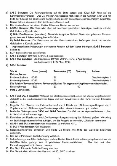
iiiiii)) SSAASS--33 BBeennuuttzzeerr:: Die Führungsschiene auf die Stifte setzen und 400µl REP Prep auf dieKammermitte verteilen. Das Gel mit der Agaroseseite nach oben in die Kammer legen und mitHilfe der Schiene die positive und negative Seite an den passenden Elektrodenhaltern ausrichten.Darauf achten, dass unter dem Gel keine Luftblasen sind.
3. Die Geloberfläche mit einem Blotter C blotten, Blotter verwerfen.4. ii)) SSAASS--11 BBeennuuttzzeerr:: Die Elektroden oben auf den Elektrodenhaltern befestigen, damit sie mit den
Gelblöcken in Kontakt sind.iiii)) SSAASS--11 PPlluuss BBeennuuttzzeerr:: (wie oben). Die Abdeckung über Gel und Elektroden geben und für einenguten Kontakt 5 Sekunden lang fest aufdrücken.iiiiii)) SSAASS--33 BBeennuuttzzeerr:: Die Elektroden auf den Elektrodenhaltern befestigen, damit sie mit denGelblöcken in Kontakt stehen.
5. 1 Applikatorkamm-Halterung in der oberen Position auf dem Gerät anbringen, ((SSAASS--33 BBeennuuttzzeerr::Schlitz 8).
6. LDH-Elektrophorese durchführen:ii)) SSAASS--11 BBeennuuttzzeerr:: 100 Volt, 13 Min., 5 Applikationeniiii)) SSAASS--11 PPlluuss BBeennuuttzzeerr:: Elektrophorese: 80 Volt, 20 Min., 15°C, 5 Applikationen
Inkubationsschritt 1: 25 Min., 45°C.iiiiii)) SSAASS--33 BBeennuuttzzeerr::
SScchhrriitttt DDaauueerr ((mmmm::ssss)) TTeemmppeerraattuurr ((°°CC)) SSppaannnnuunngg AAnnddeerreeEElleekkttrroopphhoorreesseeProbenaufnahme 00:10 21 Geschwindigkeit 1Probenapplikation 00:10 21 Geschwindigkeit 1DDiiee SScchhrriittttee ‘‘PPrroobbeennaauuffnnaahhmmee,, PPrroobbeennaapppplliikkaattiioonn’’ ffüürr iinnssggeessaammtt 66 AApppplliikkaattiioonneenn wwiieeddeerrhhoolleenn..Elektrophorese 15:00 20 100
* Platz 2 verwenden
7. SSAASS--11 uunndd SSAASS--33 BBeennuuttzzeerr:: Während die Elektrophorese läuft, einen mit Wasser angefeuchtetenZellstoff in die Inkubationskammer legen und zum Anwärmen in den 45°C warmen Inkubatorstellen.
8. Ungefähr 3-4 Minuten vor Elektrophorese-Ende 1 Fläschchen LDH-Isoenzym-Reagenz durchZugabe von 1ml LDH-Isoenzym-Verdünnungspuffer rekonstituieren und gut mischen.
9. Nach der Elektrophorese, SSAASS--11 uunndd SSAASS--33 BBeennuuttzzeerr:: Das Gel mit der Agarose-Seite nach obenin die Inkubationskammer legen.
10. Den Inhalt des Fläschchens mit LDH-Isoenzym-Reagenz entlang der Gelmitte gießen. Vorsichtigein Stück Reagenzverteilerfolie auflegen, um das Reagenz zu verteilen, Luftblasen vermeiden.
11. ii)) SSAASS--11 uunndd SSAASS--33 BBeennuuttzzeerr:: Gel inkubieren: 25 Minuten, 45°C.iiii)) SSAASS--11 PPlluuss BBeennuuttzzeerr:: Gel inkubieren.
12. Reagenzverteilerfolie entfernen und beide Gel-Blöcke mit Hilfe des Gel-Block-Entfernersentfernen.
13. Das Gel 2 Minuten in Entfärberlösung waschen.14. Gel auf eine gerade Oberfläche legen und ein Blotter B (mit Entfärbelösung angefeuchtet) auf die
Gel-Oberfläche gefolgt von 3 gefalteten Papierhandtüchern. Das Gel mit demEntwicklungsgewicht 5 Minuten pressen.
15. Das Gel 1 Minute in Entfärberlösung waschen.16. Das Gel mit dest. Wasser abspülen und bei 60...70°C trocknen.
18

AAUUSSWWEERRTTUUNNGG DDEERR EERRGGEEBBNNIISSSSEEEs wird empfohlen, die Auswertung der Gele gegen Normalwerte vorzunehmen, die für diesen Testin jedem einzelnen Labor ermittelt worden sind. Fertige Gels sollten immer dunkel aufbewahrtwerden.
QQuuaalliittaattiivvee AAuusswweerrttuunngg:: Das SAS-1 LDH Vis-12 Isoenzym-Gel kann optisch auf das Vorhandenseinoder Fehlen von Banden untersucht werden.QQuuaannttiittaattiivvee AAuusswweerrttuunngg:: Gele bei 595nm innerhalb von 2 Stunden nach Fertigstellung scannen.Fertige Gels sollten immer dunkel aufbewahrt werden.
Nach der Elektrophorese werden in der Regel 5 Zonen von LDH-Aktivität nachgewiesen. Die sich amschnellsten bewegende Zone (LDH-1) entspricht in der Wanderung den Alpha-1-Globulinen, und diesich am langsamsten bewegende Zone (LDH-5) den Gammaglobulinen. Die anderen 3 Zonen habeneine mittlere Mobilität. Die LDH-Aktivität im Serum spiegelt den Abbau zahlreicher Zelltypen widerund können als 5 Zonen beobachtet werden. LDH-2 ist vorherrschend, gefolgt von LDH-1 und LDH-3. LDH-4 und LDH-5 kommen nur in geringen Mengen vor.
1. LDH-2 ist das LDH-Isoenzym, dass in einem normalen Serum in größeren Mengen vorkommt1-4,12
.2. LDH-1 ist erhöht und kann höher als LDH-2 sein bei:a) Myokardinfarkt
1-4,12.
b) Duchenne’sche Muskelatrophie zeigt ein Bandenmuster wie ein MI; man kann aber anhand derklinischen Symptome leicht differenziert werden
19,20.
c) Hämolyse (einschließlich hämolytische Anämien) sollten immer in Erwägung gezogen werden,wenn das Gesamt-LDH im Serum Werte über das 5-fache des Normalwerts erreichen und dieIsoenzyme ein höheres LDH-1 und LDH-2 zeigen. Das Gesamt-LDH ist bei einer hämolytischenAnämie viel höher als bei einem MI, es sei denn, der MI wird von einem schweren Schockbegleitet. Rezidive perniziöse Anämie ergibt eine LDH-Muster ähnlich einer Hämolyse, und inderen Zusammenhang sind einige der höchsten Gesamt-LDH-Werte nachgewiesen worden
2,14.
d) Niereninfarkt2,12
.3. LDH-3 ist erhöht bei Lungeninfarzierung
7,12,21.
4. Erhöhung des LDH-4 ist nicht mit speziellen, pathologischen Krankheitsbildern in Verbindunggebracht worden.
5. LDH-5 ist bei Leber- und Muskelverletzungen und Hauterkrankungen erhöht1.
6. Man spricht von einem isomorphen Muster, wenn die Gesamt-LDH deutlich erhöht ist, aber alleIsoenzyme normale Prozentwerte haben. Weitgehend divergierende Gruppen von klinischenDiagnosen haben diesen Bandenmuster-Typ gezeigt wie kardiorespiratorische Erkrankungen,Malignität, Frakturen, Erkrankungen des zentralen Nervensystems, Infektion und Entzündung,Leberzirrhose, Alkoholismus, Trauma ohne Fraktur, infektiöse Mononukleose,Hypothyreoidismus, Urämie, Nekrose, Pseudomononukleose, Virämie und Ileus
1,2,22.
7. LDH- und CK-Isoenzyme sind nach kardiochirurgischen Eingriffen weniger spezifisch als in denmeisten diagnostischen Situationen. Das CK-MB ist wegen der Verletzung des Myokards durch dieOperation und dem Trauma der Manipulation und Punktion des Herzens erhöht. LDH-2/LDH-1kann durch Hämolyse aufgrund des extrakorporalen Kreislaufs erhöht sein
22-24.
19
SAS-1 LDH VIS-12 ISOENZYM-KIT
Deutsch

QQUUAALLIITTÄÄTTSSKKOONNTTRROOLLLLEEDie CK / LDH-Kontrolle (Kat. Nr. 5134) überprüft alle Phasen der Methode und sollte bei jedem Laufmitgeführt werden. Zulässige Test-Werte sind der Packungsbeilage zu entnehmen.
EEIINNSSCCHHRRÄÄNNKKUUNNGGEENNDie SAS-1 LDH Vis-12 Isoenzym-Methode ist nicht zur Identifikation von Tumor-Markern geeignet.
Siehe PROBENENTNAHME UND VORBEREITUNG für weitere Störfaktoren.
Weitere notwendige Untersuchungen:1. Bestimmung der Gesamt-LDH-Aktivität. Es gibt widersprüchliche Berichte über den wahren Wert
einer Gesamtenzymbestimmung und die Schwere einer Krankheit1,4,25
.2. Bei einer MI-Diagnose sollten auch die CK-Isoenzyme bestimmt werden
1,4.
3. Zum Ausschluss einer Hämolyse kann als mögliche Ursache eines erhöhten LDH-1 und LDH-2 dasHaptoglobin bestimmt werden.
RREEFFEERREENNZZWWEERRTTEEDie folgenden Normalwerte wurden bei Studien von Helena BioSciences ermittelt. Diese Wertedienen lediglich als Richtlinie. Jedes Labor sollte seine eigenen Normalwerte für diese Methodeerstellen.
LDH-1 = 20,6 - 32,0%LDH-2 = 31,1 - 35,9%LDH-3 = 19,8 - 25,7%LDH-4 = 7,0 - 10,4%LDH-5 = 6,5 - 14,0%
LLEEIISSTTUUNNGGSSEEIIGGEENNSSCCHHAAFFTTEENNLLiinneeaarriittäättDas System zeigt bis zu 650 IU/l pro Bande oder einer Gesamt-LDH-Aktivität von 1300 IU/l einenlinearen Verlauf.
LLIITTEERRAATTUURR1. Brish, L.K. ‘CK & LD Isoenzymes A Self-Instructional Text’ , Am. Soc. of Clin. Path. Press, Chicago,
1984: 85-120.2. Hadden, D.M. and Prentiss, T., Cardiac Profiling by Electrophoresis’, Lab. Mgt., 1977; May: 19-
24.3. Henry, J.D. ‘Clinical Diagnosis and Management by Laboratory Methods’, 16th Edition, 1979; 1:
366-369, W.B. Saunders Co., Philadelphia.4. Galen, R.S., Reiffel, J.A. and Gambino, S.R., Diagnosis of Acute Myocardial Infarction’, J. Am. Med.
Assoc., 1975; 232(2): 145-147.5. Frolich, J., Brosseuk, A., Grant, A. and McLennan, M., ‘Study of the Value of CPK and LDH
Isoenzyme Determinations in the Differential Diagnosis of Ischemic Chest Pain’, Clin. Biochem.,1978; 11(6): 232-234.
6. Wagner, G.S., Roe, C.R., Limbird, L.E., Rosati, R.A. and Wallace, A.G., ‘The Importance ofIdentification of the Myocardial-Specific Isoenzyme of Creatine Phosphokinase (MB Form) in theDiagnosis of Acute Myocardial Infarction’, Circulation, 1973; 47: 263-269.
20

7. Nerenberg, S.T., ‘Electrophoretic Screening Procedures’, 1973: 96-125, Lea & Febiger,Philadelphia.
8. Preston, J.A., Briere, R.O. and Batsakis, J.G., ‘Rapid Electrophoretic Separation of LactateDehydrogenase Isoenzymes on Cellulose Acetate’, Am. J. Clin. Pathol., 1965; 43(3): 256-260.
9. Hsu, M-Y., Kohler, M.M., Barolia, L. and Bondar, R.J.L., ‘Separation of Five Isoenzymes of SerumLactate Dehydrogenase by Discontinuous Gradient Elution from a Miniature Ion-ExchangeColumn’, Clin. Chem., 1979; 25(8) : 1453-1458.
10. Usategui-Gomez, M., Wicks, R.W. and Warshaw, M., ‘Immunochemical Determination of theHeart Isoenzyme of Lactate Dehydrogenase(LDH1) in Human Serum’, Clin. Chem., 1979; 25(5) :729-734.
11. Rotenberg, Z., Weinberger, I., Sagie, A., Fuchs, J., Sperling, O. and Agmon, J., ‘LactateDehydrogenase Isoenzymes in Serum During Recent Acute Myocardial Infarction’, Clin. Chem.,1987; 33(8) : 1419-1420.
12. Tietz, N.W. et al., ‘Textbook of Clinical Chemistry’, 1986: 691-700, W.B. Saunders Co.,Philadelphia.
13. Rothwell, D.J., Jendrzejczak, B., Becker, M. and Doumas, B.T., ‘Lactate Dehydrogenase Activitiesin Serum and Plasma’, Clin. Chem., 1976; 22(7) : 1024-1026.
14. Latner, A.L. and Skillen, A.W., ‘Isoenzymes in Biology and Medicine’, 1968: 146-157, AcademicPress, London.
15. Kreutzer, H.H. and Fennis, W.H.S., ‘Lactic Dehydrogenase Isoenzymes in Blood Serum afterStorage at Different Temperatures’, Clin. Chim. Acta, 1964; 9: 64-68.
16. Galen, R.S., Personal Communication, Dec. 1981.17. Clark, P.I., Kostuk, W.J. and Henderson, A.R., ‘Time-Dependency of Human Lactate
Dehydrogenase Isoenzyme 5 Inhibition by Urea’, Clin. Chem., 1976; 22(12) : 2059.18. Young, D.S., ‘Effects of Drugs on Clinical Laboratory Tests’, 3rd Ed., 1990, AACC Press,
Washington D.C.19. Roses, A.D., Roses, M.J., Nicholson, G.A. and Roe, C.R., ‘Lactate Dehydrogenase Isoenzyme 5 in
Detecting carriers of Duchenne Muscular Dystrophy’, Neurology, 1977; 27(May): 414-421.20. Yasmineh, W.G., Ibrahim, G.A., Abbasnezhad, M. and Awad, E.A., ‘Isoenzyme Distribution of
Creatine Kinase and lactate Dehydrogenase in Serum and Skeletal Muscle in Duchenne MuscularDystrophy, Collagen Disease, and Other Muscular Disorders’, Clin. Chem., 1978; 24(11) : 1985-1989.
21. Papadopoulos, N.M., ‘Clinical Applications of Lactate Dehydrogenase Isoenzymes’, Annals of Clin.Lab. Sci., 1977; 7(6): 506-510.
22. Jacobs, D.S., Robinson, R.A., Clark, G.M. and Tucker, J.M., ‘Clinical Significance of the IsomorphicPattern of the Isoenzymes of Serum Lactate Dehydrogenase’, Annals of Clin. and Lab. Sci., 1977;7(5): 411-421.
23. Galen, R.S., ‘Diagnostic Tests in Cardiac and Non-Cardiac Disorders’ Diag. Med., 1978 Feb., 74-87.
24. Mohiuddin, S.M., Raffetto, J., Sketch, M.H., Lynch, J.D., Schultz, R.D. and Runco, V., ‘LDHIsoenzymes and Myocardial Infarction in patients Undergoing Coronary Bypass Surgery: AnExcellent Correlation’, Am. Heart J., 1976; 92(5): 584-588.
25. Chapelle, J-P., Albert, A., Smeets, J-P., Marechal, J-P.,Heusghem, C. and Kulbertus, H.E., ‘DoesLactate Dehydrogenase Isoenzyme-5 Contribute to the Predictive Power of Total LactateDehydrogenase in Myocardial Infarction?’ Clin. Chem., 1983; 29(5) : 774-777.
21
SAS-1 LDH VIS-12 ISOENZYM-KIT
Deutsch

PPRRIINNCCIIPPIIOOIl kit per isoenzimi LDH Vis-12 SAS-1 è stato formulato per separare e quantificare gli isoenzimi dellalattato deidrogenasi nel siero o nel plasma mediante elettroforesi in gel di agarosio.
La lattato deidrogenasi (LDH) (EC 1.1.1.27) è un enzima riscontrato in pressoché tutti i tessuti umani,con la massima concentrazione a livello del fegato, del muscolo scheletrico, del cuore e dei reni. La vasta distribuzione della LDH nei tessuti corporei limita l’utilità delle determinazioni della LDHtotale in fase diagnostica. I test per la determinazione dell’origine di un’elevata attività LDH possonoaccompagnati dalla valutazione degli isoenzimi
1.
Nel siero umano possono è possibile osservare la presenza di 5 isoenzimi della LDH. Ogni isoenzimaè indicato da un numero, correlato alla sua mobilità elettroforetica. La frazione in più rapidomovimento (più anodica) è denominata LDH1 ed è riscontrabile principalmente nel muscolo cardiaco.La frazione con movimento più lento (più catodica) è invece denominata LDH5, riscontrabileprincipalmente nel fegato e nel muscolo scheletrico. Le altre frazioni, denominate LDH2, LDH3 eLDH4, sono presenti in concentrazioni variabili unitamente alla LDH1 e alla LDH5 in tutti i tessuti
1,2.
L’impiego più importante degli isoenzimi della LDH interessa la diagnosi dei danni miocardici. La LDH2presenta la massima concentrazione nel siero umano normale. Il rapporto LDH1/LDH2 è pertantoinferiore a 1. In seguito ad infarto del miocardio (IM), vi è un notevole aumento della LDH1 cosicchéil rapporto LDH1/LDH2 successivo a tale evento si avvicinerà o addirittura supererà l’unità; talefenomeno è noto con il nome di “flipped LD”. Il livello di LDH inizia ad aumentare a circa 12-24 oredi distanza dall’infarto miocardico, raggiungendo spesso livelli doppi o tripli (o ancora maggiori) dellimite superiore del range normale. L’attività di picco viene solitamente raggiunta il terzo o quartogiorno e può rimanere elevata anche per 2 settimane dopo l’infarto
2.
I test maggiormente precisi nella diagnosi dell’infarto miocardico vengono compiuti studiandol’isoenzima della creatinchinasi (CK) unitamente agli isoenzimi della lattato deidrogenasi
1-5.
La specificità e la sensibilità raggiunte con questi due test hanno eliminato la necessità di effettuareulteriori studi sugli enzimi per giungere ad una diagnosi accurata dell’infarto miocardico. Dagli studi èemerso che le analisi degli isoenzimi CK e LDH, unitamente ad un corretto quadro clinico e ai risultatidell’elettrocardiogramma, offrono una precisione pressoché totale nel diagnosticare adeguatamentel’infarto miocardico
2,6.
Gli isoenzimi della LDH sono stati determinati con vari metodi7-11
. L’elettroforesi fornisce tuttaviamolte più informazioni rispetto agli altri metodi, in quanto consente una separazione completa di tuttii 5 isoenzimi senza alcun rischio di carryover. Il mezzo di supporto utilizzato nell’elettroforesicomprende cellulosa acetato, agar, agarosio e gel di acrilamide
1.
Il kit per isoenzimi LDH Vis-12 SAS-1 utilizza una variante del metodo di Preston8e separa gli isoenzimi
della lattato deidrogenasi in base alla mobilità in un gel di agarosio. Le bande di isoenzimi vengonoquindi visualizzate con un reagente colorimetrico, che visualizza quantitativamente gli isoenzimi in basealla sequenza di reazioni sotto riportata:
22
L-lattato + NAD
NADH + Sali di tetrazolio
LDH
PMSPiruvato + NADH
NADH + Colorante Formazan (insolubile)

AAVVVVEERRTTEENNZZEE EE PPRREECCAAUUZZIIOONNIITutti i reagenti devono essere utilizzati esclusivamente per diagnosi in vitro. Non ingerire né pipettarecon la bocca i componenti del kit. Indossare guanti protettivi durante l’uso dei componenti del kit.Riferirsi alle schede tecniche e dati di sicurezza per le avvertenze sui componenti dei Kit.
CCOOMMPPOOSSIIZZIIOONNEE11.. GGeell SSAASS--11 ppeerr iissooeennzziimmii LLDDHH ((xx1100))
Contiene agarosio in un tampone barbiturico con sodio azide come conservante. Il gel è prontoall’uso.
22.. RReeaaggeennttee ppeerr iissooeennzziimmii LLDDHH ((1100xx 11mmll))Contiene i componenti necessari all’identificazione degli isoenzimi della lattato deidrogenasisecondo la sequenza di reazioni sopra illustrata. Ved. il paragrafo PROCEDURA per le istruzionidi ricostituzione.
33.. DDiilluueennttee ppeerr iissooeennzziimmii LLDDHH ((11xx 1155mmll))Contiene tampone AMP, bicina, barbital e aspartato con sodio azide come conservante. Il gel èpronto all’uso così come viene fornito.2.
44.. AAllttrrii ccoommppoonneennttii ddeell kkiittCiascun kit contiene un foglio procedurale, blotter B e C e pellicole di distribuzione del reagentein quantità sufficiente per completare 10 gel.
CCOONNSSEERRVVAAZZIIOONNEE EESSTTAABBIILLIITTAA’’11.. GGeell SSAASS--11ppeerr iissooeennzziimmaa LLDD
I gel devono essere conservati a 15...30°C e sono stabili fino alla data di scadenza riportata sullaconfezione. NON REFRIGERARE NÉ CONGELARE. Il deterioramento del gel può essereindicato da 1) formazioni cristalline per effetto di congelamento, 2) screpolature e fessurazione pereffetto di essiccamento oppure, 3) contaminazione visibile dell’agarosio causata da batteri o funghi.
22.. RReeaaggeennttee ppeerr iissooeennzziimmii LLDDHH Conservare i flaconi di reagente ad una temperatura compresa tra 2...6°C: sono stabili fino alla datadi scadenza indicata sull’etichetta. Il reagente ricostituito è stabile per 4 ore a 15...30°C o per 48ore a 2...6°C.
33.. DDiilluueennttee ppeerr iissooeennzziimmii LLDDHH Il colorante concentrato deve essere conservato a 2...6°C ed è stabile fino alla data di scadenzaindicata sull’etichetta della bottiglia. Evitare la contaminazione.
MMAATTEERRIIAALLII NNEECCEESSSSAARRII,, MMAA NNOONN IINN DDOOTTAAZZIIOONNEECod. N. 210200 Lamelle di applicazione (1 x 10)Cod. N. 210300 Lamelle di applicazione (5 x 10)Cod. N. 210100 Coppette per campioni monouso (100)Cod. N. 5014 Peso di sviluppo Cod. N. 4062 Camera d’incubazione Cod. N. 3100 Preparazione REP Forno di essiccazione ad aria forzata con temperature di 60...70°CIncubatrice prevista per 45°CSoluzione decolorante: Miscelare 100ml di acido acetico glaciale con 900ml di acqua distillata.Conservare in una bottiglia tappata ermeticamente.Acqua distillata
23
KIT PER ISOENZIMI LDH VIS-12 SAS-1
Italiano

RRAACCCCOOLLTTAA DDEEII CCAAMMPPIIOONNII EE PPRREEPPAARRAAZZIIOONNEEIl siero fresco è il campione da preferire. Può anche essere utilizzato plasma anticoagulato con eparinao EDTA. L’ossalato inibisce l’attività della LDH e pertanto non deve essere utilizzato
12. Il plasma deve
essere privo di piastrine, in quanto queste ultime contengono LDH13. I campioni devono essere testatiappena possibile dopo la raccolta, in quanto non esistono condizioni di conservazione ottimali per tuttigli isoenzimi
12, 14, 15, 16. Se necessario, i campioni possono essere refrigerati a 15...30°C oppure a 2...6°C
per un massimo di 48 ore12. NON CONGELARE: il congelamento distrugge infatti l’attività LDH512
.
RRaaccccoollttaa ddeell ccaammppiioonnee:: Un’adeguata tempestività nella raccolta dei campioni è di cruciale importanzaper l’esatta interpretazione dei pattern degli isoenzimi della LDH nella valutazione dell’infarto delmiocardio. Devono essere raccolti almeno 3 campioni, di cui uno al momento del ricovero, uno dopo6-13 ore e il terzo a 24-37 ore di distanza dal ricovero
2-4.
FFaattttoorrii dd’’iinntteerrffeerreennzzaa::1) L’emolisi può influire sulle quantificazioni di LDH1 e LDH2, in quanto gli eritrociti contengono
livelli elevati di attività LDH1-2,12
.2) L’attività LDH appare ridotta nei sieri uremici a causa della presenza di inibitori, quali l’urea e
l’ossalato. La LDH5 risulta particolarmente interessata da tali inibitori17
.3) L’acetone e il cloroformio inibiscono tutti gli isoenzimi tranne la LDH1
14.
4) Alcuni farmaci possono influenzare l’attività degli isoenzimi della LDH18
.
PPRROOCCEEDDUURRAA1. Pipettare 35µl di campione nel pozzetto appropriato del vassoio per campioni o nelle coppette per
campioni monouso.ii)) PPeerr ggllii uuttiilliizzzzaattoorrii ddii SSAASS--11 ee SSAASS--11 PPlluuss:: Utilizzare il vassoio per campioni SAS-1. Collocare con
cautela il vassoio per campioni sul cassetto di applicazione. Assicurarsi che il vassoio sia saldamenteinserito in posizione.
iiii)) PPeerr ggllii uuttiilliizzzzaattoorrii ddii SSAASS--33:: Utilizzare il vassoio per campioni SPIFE / SAS-3. Collocare con cautelail vassoio per campioni utilizzando i fermi di posizionamento della base di campioni. Assicurarsi cheil vassoio sia posizionato saldamente.
2. Rimuovere il gel dalla confezione e:ii)) PPeerr ggllii uuttiilliizzzzaattoorrii ddii SSAASS--11:: Collocare la piastra all’interno della camera con il gel verso l’alto e
allineando il lato positivo + e iiii)) PPeerr ggllii uuttiilliizzzzaattoorrii ddii SSAASS--11 PPlluuss:: Distribuire 400µL di preparazione REP sul dissipatore di calore.
Collocare il gel sul dissipatore di calore con l’agarosio rivolto verso l’alto, allineando i lati positivoe negativo rispetto ai corrispondenti puntali degli elettrodi, prestando attenzione ad evitare bolled’aria sotto il gel.
iiiiii)) PPeerr ggllii uuttiilliizzzzaattoorrii ddii SSAASS--33:: Collocare la guida di allineamento sui fermi e distribuire 400µL dipreparazione REP sul centro della camera. Collocare il gel nella camera con l’agarosio rivolto versol’alto; utilizzando la guida, allineare i lati positivo e negativo rispetto ai corrispondenti puntali deglielettrodi, prestando attenzione ad evitare bolle d’aria sotto il gel.
3. Asciugare la superficie del gel con un blotter C, quindi gettarlo.4. ii)) PPeerr ggllii uuttiilliizzzzaattoorrii ddii SSAASS--11:: fissare gli elettrodi sul lato superiore dei puntali, in modo tale che
entrino a contatto con i blocchi di gel.iiii)) PPeerr ggllii uuttiilliizzzzaattoorrii ddii SSAASS--11 PPlluuss:: (come sopra). Sistemare il coperchio sul gel e sugli elettrodi epremere con decisione per 5 secondi per consentire il contatto.
24
iiiiii)) PPeerr ggllii uuttiilliizzzzaattoorrii ddii SSAASS--33:: fissare gli elettrodi sul lato superiore dei puntali, in modo tale cheentrino a contatto con i blocchi di gel .
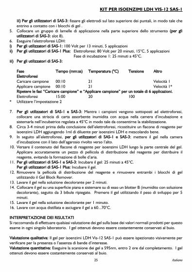
5. Collocare un gruppo di lamelle di applicazione nella parte superiore dello strumento ((ppeerr gglliiuuttiilliizzzzaattoorrii ddii SSAASS--33:: slot 8).
6. Eseguire l’elettroforesi LDH:ii)) PPeerr ggllii uuttiilliizzzzaattoorrii ddii SSAASS--11:: 100 Volt per 13 minuti, 5 applicazioniiiii)) PPeerr ggllii uuttiilliizzzzaattoorrii ddii SSAASS--11 PPlluuss:: Elettroforesi: 80 Volt per 20 minuti, 15°C, 5 applicazioni
Fase di incubazione 1: 25 minuti a 45°C.iiiiii)) PPeerr ggllii uuttiilliizzzzaattoorrii ddii SSAASS--33::
FFaassee TTeemmppoo ((mmmm::ssss)) TTeemmppeerraattuurraa ((°°CC)) TTeennssiioonnee AAllttrrooEElleettttrrooffoorreessiiCaricare campione 00:10 21 Velocità 1Applicare campione 00:10 21 Velocità 1*RRiippeetteerree llee ffaassii ““CCaarriiccaarree ccaammppiioonnee”” ee ““AApppplliiccaarree ccaammppiioonnee”” ppeerr uunn ttoottaallee ddii 66 aapppplliiccaazziioonnii..Elettroforesi 15:00 20 100
* Utilizzare l’impostazione 2
7. PPeerr ggllii uuttiilliizzzzaattoorrii ddii SSAASS--11 ee SSAASS--33:: Mentre i campioni vengono sottoposti ad elettroforesi,collocare una striscia di carta assorbente inumidita con acqua nella camera d’incubazione esistemarla nell’incubatrice regolata a 45°C in modo tale da consentirne la stabilizzazione.
8. Circa 3-4 minuti prima della conclusione dell’elettroforesi, ricostituire un flacone di reagente perisoenzimi LDH aggiungendo 1ml di diluente per isoenzimi LDH e mescolando bene.
9. In seguito all’elettroforesi, ppeerr ggllii uuttiilliizzzzaattoorrii ddii SSAASS--11 ee SSAASS--33:: mettere il gel nella camerad’incubazione con il lato dell’agarosio rivolto verso l’alto.
10. Versare il contenuto del flacone di reagente per isoenzimi LDH lungo la parte centrale del gel.Applicare accuratamente un pezzo di pellicola di distribuzione del reagente per distribuire ilreagente, evitando la formazione di bolle d’aria.
11. PPeerr ggllii uuttiilliizzzzaattoorrii ddii SSAASS--11 ee SSAASS--33:: Incubare il gel: 25 minuti a 45°C.PPeerr ggllii uuttiilliizzzzaattoorrii ddii SSAASS--11 PPlluuss:: Incubare il gel.
12. Rimuovere la pellicola di distribuzione del reagente e rimuovere entrambi i blocchi di gelutilizzando il Gel Block Remover.
13. Lavare il gel nella soluzione decolorante per 2 minuti.14. Collocare il gel su una superficie piana e sistemare su di esso un blotter B (inumidito con soluzione
decolorante), seguito da 3 bibule ripiegate. Premere il gel utilizzando il peso di sviluppo per 5minuti.
15. Lavare il gel nella soluzione decolorante per 1 minuto.16. Lavare con acqua distillata e asciugare il gel a 60...70°C.
IINNTTEERRPPRREETTAAZZIIOONNEE DDEEII RRIISSUULLTTAATTIISi raccomanda di effettuare qualsiasi valutazione dei gel sulla base dei valori normali prodotti per questoesame in ogni singolo laboratorio. I gel ottenuti devono essere costantemente conservati al buio.
VVaalluuttaazziioonnee qquuaalliittaattiivvaa:: Il gel per isoenzimi LDH Vis-12 SAS-1 può essere ispezionato visivamente perverificare per la presenza o l’assenza di bande d’interesse.VVaalluuttaazziioonnee qquuaannttiittaattiivvaa:: Eseguire la scansione dei gel a 595nm, entro 2 ore dal completamento. I gelottenuti devono essere costantemente conservati al buio.
25
KIT PER ISOENZIMI LDH VIS-12 SAS-1
Italiano

In seguito all’elettroforesi, vengono solitamente individuate 5 zone di attività LDH. La zona più rapida(LDH1) si muove con una migrazione simile alle alfa1-globuline, mentre la zona più lenta (LDH5) simuove con le gammaglobuline. Le altre 3 zone presentano mobilità intermedie. L’attività LDH nelsiero rispecchia l’analisi di molti tipi di cellule ed è possibile osservare tutte le 5 zone. La LDH2 èpredominante, seguita dalla LDH1 e dalla LDH3. La LDH4 e la LDH5 compaiono in quantità minori.
1. La LDH2 è l’isoenzima della LDH presente in quantità massime nel siero normale1-4,12
.2. La LDH1 è elevata e può essere maggiore della LDH2 nelle seguenti condizioni:a) Infarto del miocardio
1-4,12.
b) La distrofia muscolare di Duchenne presenta un pattern analogo all’IM, ma i sintomi clinicicontribuiscono a distinguere facilmente le due patologie
19,20.
c) L’emolisi (comprese le anemie emolitiche) devono essere prese in grande considerazioneogniqualvolta la LDH totale nel siero raggiunge livelli superiori a 5 volte i valori normali e gliisoenzimi mostrano maggiori livelli di LDH1 e LDH2. La LDH totale è molto più elevatanell’anemia emolitica di quanto lo sia nell’IM, salvo qualora l’IM sia accompagnato da grave shock.L’anemia perniciosa recidivante fornisce un pattern della LDH simile a quello dell’emolisi; in questacondizione si riscontrano alcuni dei massimi livelli sierici di LDH totale
2,14.
d) Infarto renale2,12
.3. La LDH3 è elevata negli infarti polmonari
7,12,21.
4. L’aumento della LDH4 non è stato associato a patologie particolari.5. La LDH5 è elevata nei danni epatici e muscolari e nelle malattie cutanee
1.
6. Quando la LDH totale risulta notevolmente elevata mentre tutti gli isoenzimi presentanopercentuali nella norma, si parla di pattern isomorfico. Gruppi ampiamente divergenti di diagnosicliniche hanno mostrato questo tipo di pattern; tra di essi rientrano malattie cardiorespiratorie,tumori maligni, fratture, patologie del sistema nervoso centrale, infezioni e infiammazioni, cirrosiepatica, alcolismo, traumi senza fratture, mononucleosi infettiva, ipotiroidismo, uremia, necrosi,pseudo-mononucleosi, viremia ed occlusione intestinale
1,2,22.
7. Gli isoenzimi della LDH e della CK sono meno specifici in seguito ad un intervento a cuore apertorispetto a quanto lo siano nella maggior parte delle situazioni diagnostiche. Il CK-MB aumenterà acausa del danno miocardico dovuto alla procedura di intervento e del trauma causato dallamanipolazione e dall’incannulamento del cuore. Gli isoenzimi LDH2/LDH1 possono essere elevatisecondariamente all’emolisi dovuta alla circolazione extracorporea
22-24.
CCOONNTTRROOLLLLOO QQUUAALLIITTÀÀIl controllo CK (Cod. N. 5134) può essere utilizzato per verificare tutte le fasi della procedura e deveessere impiegato su ogni piastra. Fare riferimento alle schede all’interno della confezione per i valoriaccettabili del campione.
LLIIMMIITTAAZZIIOONNIIIl metodo degli isoenzimi LDH Vis-12 SAS-1 non è destinato ad identificare i marker tumorali.
Fare riferimento al paragrafo RACCOLTA DEI CAMPIONI E PREPARAZIONE per altri fattorid’interferenza.
26

Ulteriori esami necessari:1. Può essere determinata l’attività LDH totale. Esistono rapporti contrastanti in merito al valore
reale dei livelli enzimatici totali nel siero e alla gravità di una malattia1,4,25
.2. Nella diagnosi dell’IM devono essere eseguiti anche gli studi sugli isoenzimi della CK
1,4.
3. Possono essere svolti studi sull’aptoglobina, per escludere l’emolisi come causa di elevati livelli diLDH1 e LDH2.
VVAALLOORRII DDIIRRIIFFEERRIIMMEENNTTOOI valori normali riportati di seguito sono stati ottenuti nel corso di studi eseguiti presso HelenaBioSciences. Tali valori devono fungere esclusivamente da valori di riferimento. Ciascun laboratoriodovrà determinare i propri valori normali previsti per il metodo in questione.
LDH1 = 20.6 - 32.0%LD2 = 31.1 - 35.9%LD3 = 19.8 - 25.7%LD4 = 7.0 - 10.4%LD5 = 6.5 - 14.0%
CCAARRAATTTTEERRIISSTTIICCHHEE PPRREESSTTAAZZIIOONNAALLII LLiinneeaarriittààQuesto sistema è lineare a 650 IU/L per banda o ad un’attività LDH totale di 1300 IU/L.
BBIIBBLLIIOOGGRRAAFFIIAA1. Brish, L.K. ‘CK & LD Isoenzymes A Self-Instructional Text’ , Am. Soc. of Clin. Path. Press, Chicago,
1984 : 85-120.2. Hadden, D.M. and Prentiss, T., Cardiac Profiling by Electrophoresis’, Lab. Mgt., 1977; May: 19-24.3. Henry, J.D. ‘Clinical Diagnosis and Management by Laboratory Methods’, 16th Edition, 1979; 1:
366-369, W.B. Saunders Co., Philadelphia.4. Galen, R.S. Reiffel, J.A. and Gambino, S.R., Diagnosis of Acute Myocardial Infarction’, J. Am. Med.
Assoc., 1975; 232(2): 145-147.5. Frolich, J., Brosseuk, A., Grant, A. and McLennan, M., ‘Study of the Value of CPK and LDH
Isoenzyme Determinations in the Differential Diagnosis of Ischemic Chest Pain’, Clin. Biochem.,1978; 11(6): 232-234.
6. Wagner, G.S., Roe, C.R., Limbird, L.E., Rosati, R.A. and Wallace, A.G., ‘The Importance ofIdentification of the Myocardial-Specific Isoenzyme of Creatine Phosphokinase (MB Form) in theDiagnosis of Acute Myocardial Infarction’, Circulation, 1973; 47: 263-269.
7. Nerenberg, S.T., ‘Electrophoretic Screening Procedures’, 1973: 96-125, Lea & Febiger,Philadelphia.
8. Preston, J.A., Briere, R.O. and Batsakis, J.G., ‘Rapid Electrophoretic Separation of LactateDehydrogenase Isoenzymes on Cellulose Acetate’, Am. J. Clin. Pathol., 1965; 43(3): 256-260.
9. Hsu, M-Y., Kohler, M.M., Barolia, L. and Bondar, R.J.L., ‘Separation of Five Isoenzymes of SerumLactate Dehydrogenase by Discontinuous Gradient Elution from a Miniature Ion-ExchangeColumn’, Clin. Chem., 1979; 25(8): 1453-1458.
10. Usategui-Gomez, M., Wicks, R.W. and Warshaw, M., ‘Immunochemical Determination of theHeart Isoenzyme of Lactate Dehydrogenase(LDH1) in Human Serum’, Clin. Chem., 1979; 25(5):729-734.
27
KIT PER ISOENZIMI LDH VIS-12 SAS-1
Italiano

11. Rotenberg, Z., Weinberger, I., Sagie, A., Fuchs, J., Sperling, O. and Agmon, J., ‘LactateDehydrogenase Isoenzymes in Serum During Recent Acute Myocardial Infarction’, Clin. Chem.,1987; 33(8): 1419-1420.
12. Tietz, N.W. et al., ‘Textbook of Clinical Chemistry’, 1986: 691-700, W.B. Saunders Co.,Philadelphia.
13. Rothwell, D.J., Jendrzejczak, B., Becker, M. and Doumas, B.T., ‘Lactate Dehydrogenase Activitiesin Serum and Plasma’, Clin. Chem., 1976; 22(7): 1024-1026.
14. Latner, A.L. and Skillen, A.W., ‘Isoenzymes in Biology and Medicine’, 1968: 146-157, AcademicPress, London.
15. Kreutzer, H.H. and Fennis, W.H.S., ‘Lactic Dehydrogenase Isoenzymes in Blood Serum afterStorage at Different Temperatures’, Clin. Chim. Acta, 1964; 9: 64-68.
16. Galen, R.S. Personal Communication, Dec. 1981.17. Clark, P.I., Kostuk, W.J. and Henderson, A.R., ‘Time-Dependency of Human Lactate
Dehydrogenase Isoenzyme 5 Inhibition by Urea’, Clin. Chem., 1976; 22(12): 2059.18. Young, D.S., ‘Effects of Drugs on Clinical Laboratory Tests’, 3rd Ed., 1990, AACC Press,
Washington D.C.19. Roses, A.D., Roses, M.J., Nicholson, G.A. and Roe, C.R., ‘Lactate Dehydrogenase Isoenzyme 5 in
Detecting carriers of Duchenne Muscular Dystrophy’, Neurology, 1977; 27(May): 414-421.20. Yasmineh, W.G., Ibrahim, G.A., Abbasnezhad, M. and Awad, E.A., ‘Isoenzyme Distribution of
Creatine Kinase and lactate Dehydrogenase in Serum and Skeletal Muscle in Duchenne MuscularDystrophy, Collagen Disease, and Other Muscular Disorders’, Clin. Chem., 1978; 24(11): 1985-1989.
21. Papadopoulos, N.M., ‘Clinical Applications of Lactate Dehydrogenase Isoenzymes’, Annals of Clin.Lab. Sci., 1977; 7(6): 506-510.
22. Jacobs, D.S., Robinson, R.A., Clark, G.M. and Tucker, J.M., ‘Clinical Significance of the IsomorphicPattern of the Isoenzymes of Serum Lactate Dehydrogenase’, Annals of Clin. and Lab. Sci., 1977;7(5): 411-421.
23. Galen, R.S. ‘Diagnostic Tests in Cardiac and Non-Cardiac Disorders’ Diag. Med., 1978 Feb., 74-87.
24. Mohiuddin, S.M., Raffetto, J., Sketch, M.H., Lynch, J.D., Schultz, R.D. and Runco, V., ‘LDHIsoenzymes and Myocardial Infarction in patients Undergoing Coronary Bypass Surgery: AnExcellent Correlation’, Am. Heart J., 1976; 92(5): 584-588.
25. Chapelle, J-P., Albert, A., Smeets, J-P., Marechal, J-P.,Heusghem, C. and Kulbertus, H.E., ‘DoesLactate Dehydrogenase Isoenzyme-5 Contribute to the Predictive Power of Total LactateDehydrogenase in Myocardial Infarction?’ Clin. Chem., 1983; 29(5): 774-777.
28

UUSSOO PPRREEVVIISSTTOOEl kit de isoenzimas SAS-1 LDH Vis-12 está diseñado para la separación y cuantificación de lasisoenzimas de la lactato deshidrogenasa en suero o plasma mediante electroforesis en gel de agarosa.
La lactato deshidrogenasa (LDH) (EC 1.1.1.27) es una enzima que se encuentra prácticamente entodos los tejidos humanos, siendo el hígado, el músculo esquelético, el corazón y el riñón los que tienenmayores concentraciones . La amplia distribución de la LDH en los tejidos corporales limita la utilidadde las determinaciones de la LDH total en diagnóstico. Pueden estar indicados los estudios sobre elorigen de la actividad elevada de LDH mediante valoración de isoenzimas
1.
Pueden demostrarse cinco isoenzimas de LDH en el suero humano. Cada isoenzima se conoce conun número que está relacionado con su movilidad electroforética. La fracción de movimiento másrápido (la más anódica) se denomina LDH1 y se encuentra fundamentalmente en el músculo cardíaco.La de movimiento más lento (la más catódica) es la LDH5, que se encuentra fundamentalmente en elhígado y el músculo esquelético. Las otras - LDH2, LDH3 y LDH4 se encuentran en diversascantidades junto con la LDH1 y la LDH5 en todos los tejidos
1,2.
El uso más importante de las isoenzimas de la LDH es en el diagnóstico de las lesiones miocárdicas. En el suero humano normal, la LDH2 se encuentra en las mayores concentraciones. Por tanto, elcociente LDH1/LDH2 es menor que uno. Después de un infarto de miocardio (IM), hay una elevaciónsustancial de la LDH1, de manera que el cociente LDH1/LDH2 después del IM se acercará o inclusosuperará el 1, un fenómeno conocido como ‘vuelco de la LDH’. El nivel de LDH comienza a elevarseaproximadamente 12-24 horas después del infarto de miocardio, alcanzando con frecuencia niveles dosa tres veces (o más) el límite superior de la normalidad. La actividad máxima suele alcanzarse el día 3-4 y la actividad puede seguir elevada durante hasta dos semanas después del infarto
2.
La prueba más definitiva en el diagnóstico de la IM se consigue realizando estudios de isoenzimas de lacreatina cinasa (CK) junto con estudios de las isoenzimas de la LDH de la 1 a la 5. La especificidad ysensibilidad conseguidas con estas dos pruebas ha eliminado la necesidad de realizar más estudios deenzimas para diagnosticar con exactitud un IM. Hay estudios que han demostrado que los análisis deisoenzimas de CK y LDH, junto con un contexto clínico y resultados del electrocardiogramaadecuados, son exactos prácticamente en un 100% para diagnosticar adecuadamente un infarto demiocardio
2,6.
Se han determinado las isoenzimas de LDH mediante diversos métodos7-11
. La electroforesisproporciona mucha más información que los otros métodos, porque permite la separación completade las cinco isoenzimas sin riesgo de contaminación entre ellas. El medio de soporte usado en laelectroforesis incluye geles de acetato de celulosa, agar, agarosa y acrilamida
1.
El kit de isoenzimas SAS-1 LDH Vis-2 utiliza una modificación del método de Preston8
y separa lasisoenzimas de lactato deshidrogenasa de acuerdo con su movilidad en un gel de agarosa. Luego sevisualizan las bandas con un reactivo colorimétrico que visualiza cuantitativamente las isoenzimas deacuerdo con la siguiente secuencia de reacción:
29
KIT DE ISOENZIMAS LDH SAS-1 VIS-12
Español
L-lactato + NAD
NADH + Sales de tetrazolio
LDH
PMSPiruvato + NADH
NADH + colorante de formazán (insoluble)

AADDVVEERRTTEENNCCIIAASS YY PPRREECCAAUUCCIIOONNEESSTodos los reactivos son exclusivamente para uso diagnóstico in-vitro. No ingerir ni chupar con la bocaningún componente del kit. Usar guantes para manejar todos los componentes del kit. Consultar lahoja con los datos de seguridad del producto acerca de los riesgos de los componentes, avisos deseguridad y consejos para su eliminación.
CCOOMMPPOOSSIICCIIÓÓNN11.. GGeell ddee iissooeennzziimmaass SSAASS--11 LLDDHH ((xx1100))
Contiene agarosa en un tampón de barbital con azida de sodio como conservante. El gel vieneenvasado listo para usar.
22.. RReeaaccttiivvooss ddee iissooeennzziimmaass ddee LLDDHH ((1100xx 11mmll))Contiene los compuestos necesarios para identificar de las isoenzimas de la lactato deshidrogenasade acuerdo con la secuencia de reacción que ya hemos mostrado anteriormente. Véase elprocedimiento PASO A PASO para las instrucciones de reconstitución.
33.. DDiilluuyyeennttee ddee iissooeennzziimmaass ddee LLDDHH ((11xx 1155mmll))Contiene AMPD, bicina, barbital, y tampón aspartato con azida de sodio como conservante. El diluyente viene envasado listo para usar.
44.. OOttrrooss ccoommppoonneenntteess ddeell kkiittCada kit contiene una hoja de instrucciones y suficientes secantes B, C y extendedores de reactivospara completar 10 geles.
AALLMMAACCEENNAAMMIIEENNTTOO YY PPEERRÍÍOODDOO DDEE VVAALLIIDDEEZZ11.. GGeell ddee iissooeennzziimmaass SSAASS--11 LLDDHH
Los geles han de almacenarse a 15...30°C y permanecen estables hasta la fecha de caducidadindicada en el envase. NO REFRIGERAR NI CONGELAR. El deterioro del gel puede estarindicado por: 1) apariencia cristalina, indicativo de que el gel ha sido congelado, 2) agrietamiento ydescamación, indicativo del resecamiento del gel, o 3) contaminación visible de la agarosa porfuentes bacterianas o micóticas.
22.. RReeaaccttiivvooss ddee iissooeennzziimmaass ddee LLDDHHLos viales de reactivos deben almacenarse a 2...6°C y permanecen estables hasta la fecha decaducidad indicada en la etiqueta. El reactivo reconstituido permanece estable durante 4 horas a15...30°C o durante 48 horas a 2...6°C.
33.. DDiilluuyyeennttee ddee iissooeennzziimmaass ddee LLDDHHEl diluyente debe guardarse a 2...6°C y permanece estable hasta la fecha de caducidad indicada enla etiqueta. Evite la contaminación.
30

AARRTTÍÍCCUULLOOSS NNEECCEESSAARRIIOOSS NNOO SSUUMMIINNIISSTTRRAADDOOSSNº de catálogo 210200 Aplicadores 1 x 10Nº de catálogo 210300 Aplicadores 5 x 10Nº de catálogo 210100 Vasos de recogida de muestras desechables (100)Nº de catálogo 5014 Peso de desarrolloNº de catálogo 4062 Cámara de IncubaciónNº de catálogo 3100 REP PrepHorno de secado con aire a presión con capacidad para alcanzar 60...70°CIncubadora capaz de alcanzar 45°CSolución decolorante: Mezclar 100ml de ácido acético cristalizado con 900ml de agua destilada.Guardar en un frasco herméticamente cerrado.Agua destilada
RREECCOOGGIIDDAA YY PPRREEPPAARRAACCIIÓÓNN DDEE MMUUEESSTTRRAASSLa muestra elegida ha de ser suero recién recogido. El plasma anticoagulado con heparina o EDTApuede usarse también, el oxalato inhibe la actividad de LDH y no debe usarse
12. El plasma no debe
contener plaquetas, puesto que las plaquetas contienen LDH13
. Las muestras deben estudiarse cuantoantes después de su recogida, puesto que ningunas condiciones de conservación son óptimas paratodas las isoenzimas
12,14,15,16. Si es necesario, las muestras pueden conservarse refrigeradas a 15...30°C
o 2...6°C durante hasta 48 horas12
. NO CONGELAR – la congelación destruye la actividad de laLDH5
12.
RReeccooggiiddaa ddee mmuueessttrraass:: La selección del momento correcto de recogida de muestras es fundamentalpara la interpretación exacta de los patrones de isoenzimas de LDH en la valoración del IM. Deberecogerse un mínimo de 3 muestras – una al ingreso, una 6-13 horas más tarde y la tercera 24-37 horasdespués del ingreso
2-4.
FFaaccttoorreess qquuee iinntteerrffiieerreenn::1) La hemólisis puede afectar a las cuantificaciones de LDH1 y LDH2, porque los eritrocitos
contienen niveles elevados de actividad de LDH1-2,12
.2) La actividad de la LDH está reducida en sueros urémicos, debido a la presencia de inhibidores
como la urea y el oxalato. La LDH5 se ve especialmente afectada17
.3) La acetona y el cloroformo inhiben todas las isoenzimas excepto la LDH1
14.
4) Determinados fármacos pueden afectar a la actividad de isoenzimas de LDH18
.
PPRROOCCEEDDIIMMIIEENNTTOO PPAASSOO AA PPAASSOO1. Con una pipeta, introducir 35µl de la muestra en la cavidad correspondiente de la bandeja de
muestras o en vasos de recogida de muestras desechables.ii)) UUssuuaarriiooss ddee SSAASS--11 yy SSAASS--11 PPlluuss:: Utilizar la bandeja de muestras de SAS-1. Colocar
cuidadosamente la bandeja con la muestra en la gaveta del aplicador. Asegurarse de que la bandejaestá firmemente introducida en su posición.
iiii)) UUssuuaarriiooss ddee SSAASS--33:: Utilizar la bandeja de muestras de SPIFE / SAS-3. Colocar cuidadosamente labandeja con la muestra utilizando los pasadores de localización de la base de la muestra.Asegurarse de que la bandeja está en una posición segura.
2. Sacar el gel del envase y:ii)) UUssuuaarriiooss ddee SSAASS--11:: colocar el gel en el SAS-1, con la agarosa hacia arriba, alineando los lados
positivo y negativo con los bordes de los electrodos correspondientes.
31
KIT DE ISOENZIMAS LDH SAS-1 VIS-12
Español

iiii)) UUssuuaarriiooss ddee SSAASS--11 PPlluuss:: pipetear 400µL de REP Prep en el disipador térmico. Colocar el gel en eldisipador térmico, con el lado de agarosa hacia arriba, alineando los lados positivo y negativo conlos bordes, teniendo cuidado de evitar las burbujas aéreas debajo del gel.
iiiiii)) UUssuuaarriiooss ddee SSAASS--33:: colocar la guía de alineación en los pasadores y pipetear 400µL de REP Prepen el centro de la cámara. Colocar el gel en la cámara con la agarosa hacia arriba, utilizar la guíapara alinear los lados positivo y negativo con los bordes, teniendo cuidado de evitar las burbujasaéreas debajo del gel
3. Secar la superficie del gel con un secante C y luego desechar el secante.4. ii)) UUssuuaarriiooss ddee SSAASS--11:: conectar los electrodos a la parte superior de los bordes, de forma que estén
en contacto con los bloques de gel.iiii)) UUssuuaarriiooss ddee SSAASS--11 PPlluuss:: (igual que el anterior). Colocar la cubierta del gel sobre el gel y loselectrodos; presionar con firmeza durante 5 segundos para asegurar un buen contacto.iiiiii)) UUssuuaarriiooss ddee SSAASS--33:: conectar los electrodos a los bordes, de forma que estén en contacto conlos bloques de gel.
5. Colocar 1 unidad del aplicador en la posición del instrumento ((uussuuaarriiooss ddee SSAASS--33:: ranura 8).6. Realizar la electroforesis de LDH:ii)) UUssuuaarriiooss ddee SSAASS--11:: 100 voltios, 13 min., 5 aplicacionesiiii)) UUssuuaarriiooss ddee SSAASS--11 PPlluuss:: Electroforesis: 80 voltios, 20 min., 15°C, 5 aplicaciones
Incubation step 1 (Paso de incubación 1) 25 min., 45°Ciiiiii)) UUssuuaarriiooss ddee SSAASS--33::
PPaassoo HHoorraa ((mmmm::ssss)) TTeemmppeerraattuurraa ((ººCC)) TTeennssiióónn OOttrroossEElleeccttrrooffoorreessiissCargar muestra 00:10 21 Velocidad 1Aplicar muestra 00:10 21 Velocidad 1*RReeppeettiirr ppaassooss ‘‘ccaarrggaarr mmuueessttrraa,, aapplliiccaarr mmuueessttrraa’’ ppaarraa uunn ttoottaall ddee 66 aapplliiccaacciioonneess..Electroforesis 15:00 20 100
* Utilizar posición 2
7. UUssuuaarriiooss ddee SSAASS--11 yy SSAASS--33:: Mientras se está realizando la electroforesis, colocar un papelhumedecido con agua en la cámara de incubación y colocarlo en el incubador a 45°C para que seequilibre.
8. Aproximadamente 3-4 minutos antes del final de la electroforesis, reconstituir un vial de reactivode isoenzimas de LDH añadiendo 1 ml de diluyente de isoenzimas de LDH y mezclar bien.
9. Finalizada la eletroforesis, los uussuuaarriiooss ddee SSAASS--11 yy SSAASS--33:: colocar el gel con la agarosa hacia arribasobre la cámara de incubación.
10. Verter el contenido del vial de Reactivos de isoenzimas de LDH en el centro del gel. Aplicar concuidado un trozo de película extendedora de reactivos para extender el reactivo evitando lasburbujas de aire.
11. UUssuuaarriiooss ddee SSAASS--11 yy SSAASS--33:: Incubar el gel: 25 minutos, 45°C.UUssuuaarriiooss ddee SSAASS--11 PPlluuss:: Incubar el gel.
12. Retirar la película extendedora de reactivos y retirar ambos bloques de gel utilizando el extractorde bloques de gel.
13. Lavar el gel en la solución decolorante durante 2 minutos.14. Colocar el gel sobre una superficie plana y colocar un secante B (humedecido en solución
decolorante) sobre la superficie del gel, seguido de 3 toallitas de papel dobladas. Presionar el gelusando una potencia de desarrollo durante 5 minutos.
32
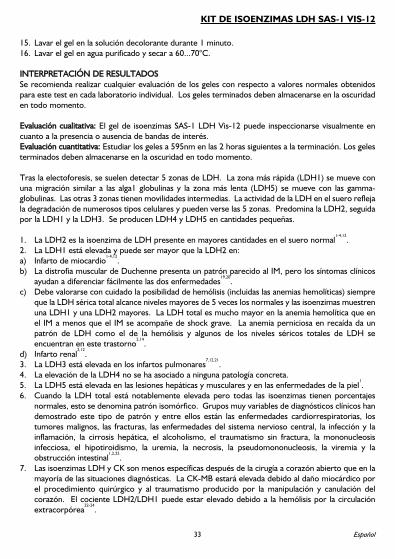
15. Lavar el gel en la solución decolorante durante 1 minuto.16. Lavar el gel en agua purificado y secar a 60...70°C.
IINNTTEERRPPRREETTAACCIIÓÓNN DDEE RREESSUULLTTAADDOOSSSe recomienda realizar cualquier evaluación de los geles con respecto a valores normales obtenidospara este test en cada laboratorio individual. Los geles terminados deben almacenarse en la oscuridaden todo momento.
EEvvaalluuaacciióónn ccuuaalliittaattiivvaa:: El gel de isoenzimas SAS-1 LDH Vis-12 puede inspeccionarse visualmente encuanto a la presencia o ausencia de bandas de interés.EEvvaalluuaacciióónn ccuuaannttiittaattiivvaa:: Estudiar los geles a 595nm en las 2 horas siguientes a la terminación. Los gelesterminados deben almacenarse en la oscuridad en todo momento.
Tras la electoforesis, se suelen detectar 5 zonas de LDH. La zona más rápida (LDH1) se mueve conuna migración similar a las alga1 globulinas y la zona más lenta (LDH5) se mueve con las gamma-globulinas. Las otras 3 zonas tienen movilidades intermedias. La actividad de la LDH en el suero reflejala degradación de numerosos tipos celulares y pueden verse las 5 zonas. Predomina la LDH2, seguidapor la LDH1 y la LDH3. Se producen LDH4 y LDH5 en cantidades pequeñas.
1. La LDH2 es la isoenzima de LDH presente en mayores cantidades en el suero normal1-4,12
.2. La LDH1 está elevada y puede ser mayor que la LDH2 en:a) Infarto de miocardio
1-4,12.
b) La distrofia muscular de Duchenne presenta un patrón parecido al IM, pero los síntomas clínicosayudan a diferenciar fácilmente las dos enfermedades
19,20.
c) Debe valorarse con cuidado la posibilidad de hemólisis (incluidas las anemias hemolíticas) siempreque la LDH sérica total alcance niveles mayores de 5 veces los normales y las isoenzimas muestrenuna LDH1 y una LDH2 mayores. La LDH total es mucho mayor en la anemia hemolítica que enel IM a menos que el IM se acompañe de shock grave. La anemia perniciosa en recaída da unpatrón de LDH como el de la hemólisis y algunos de los niveles séricos totales de LDH seencuentran en este trastorno
2,14.
d) Infarto renal2,12
.3. La LDH3 está elevada en los infartos pulmonares
7,12,21.
4. La elevación de la LDH4 no se ha asociado a ninguna patología concreta.5. La LDH5 está elevada en las lesiones hepáticas y musculares y en las enfermedades de la piel
1.
6. Cuando la LDH total está notablemente elevada pero todas las isoenzimas tienen porcentajesnormales, esto se denomina patrón isomórfico. Grupos muy variables de diagnósticos clínicos handemostrado este tipo de patrón y entre ellos están las enfermedades cardiorrespiratorias, lostumores malignos, las fracturas, las enfermedades del sistema nervioso central, la infección y lainflamación, la cirrosis hepática, el alcoholismo, el traumatismo sin fractura, la mononucleosisinfecciosa, el hipotiroidismo, la uremia, la necrosis, la pseudomononucleosis, la viremia y laobstrucción intestinal
1,2,22.
7. Las isoenzimas LDH y CK son menos específicas después de la cirugía a corazón abierto que en lamayoría de las situaciones diagnósticas. La CK-MB estará elevada debido al daño miocárdico porel procedimiento quirúrgico y al traumatismo producido por la manipulación y canulación delcorazón. El cociente LDH2/LDH1 puede estar elevado debido a la hemólisis por la circulaciónextracorpórea
22-24.
33
KIT DE ISOENZIMAS LDH SAS-1 VIS-12
Español

CCOONNTTRROOLL DDEE CCAALLIIDDAADDEl Control CK/LDH (Nº de catálogo 5134) puede usarse para verificar todas las fases delprocedimiento y debe usarse en cada prueba. Para obtener los valores apropiados de los ensayos,consultar el folleto adjunto en el envase.
LLIIMMIITTAACCIIOONNEESSEl método de isoenzimas SAS-1 LDH Vis-12 no está diseñado para identificar marcadores tumorales.
Consúltese RECOGIDA Y MANIPULACIÓN DE MUESTRAS para conocer otros factores deinterferencia.
Otras pruebas adicionales necesarias son:1. Puede determinarse la actividad de LDH total. Existen informes contradictorios sobre el valor
verdadero de los niveles séricos enzimáticos totales y la gravedad de una enfermedad1,4,25
.2. Al diagnosticar un IM, deben realizarse también estudios de las isoenzimas de la CK
1,4.
3. Pueden realizarse estudios de la haptoglobina para descartar una hemólisis como causa deelevación de LDH1 y LDH2.
VVAALLOORREESS DDEE RREEFFEERREENNCCIIAALos siguientes valores normales se obtuvieron en estudios de Helena BioSciences. Estos valores seutilizarán únicamente como guía. Cada laboratorio deberá establecer sus propios valores normalesesperados para este método.
LDH1 = 20,6 - 32,0%LDH2 = 31,1 - 35,9%LDH3 = 19,8 - 25,7%LDH4 = 7,0 - 10,4%LDH5 = 6,5 - 14,0%
CCAARRAACCTTEERRÍÍSSTTIICCAASS FFUUNNCCIIOONNAALLEESSLLiinneeaalliiddaaddSe trata de un sistema lineal hasta 650 IU/l por banda o una actividad de LDH total de 1300 IU/l.
BBIIBBLLIIOOGGRRAAFFÍÍAA1. Brish, L.K. ‘CK & LD Isoenzymes A Self-Instructional Text’ , Am. Soc. of Clin. Path. Press, Chicago,
1984: 85-120.2. Hadden, D.M. and Prentiss, T., Cardiac Profiling by Electrophoresis’, Lab. Mgt., 1977; May: 19-24.3. Henry, J.D. ‘Clinical Diagnosis and Management by Laboratory Methods’, 16th Edition, 1979; 1:
366-369, W.B. Saunders Co., Philadelphia.4. Galen, R.S., Reiffel, J.A. and Gambino, S.R., Diagnosis of Acute Myocardial Infarction’, J. Am. Med.
Assoc., 1975; 232(2): 145-147.5. Frolich, J., Brosseuk, A., Grant, A. and McLennan, M., ‘Study of the Value of CPK and LDH
Isoenzyme Determinations in the Differential Diagnosis of Ischemic Chest Pain’, Clin. Biochem.,1978; 11(6): 232-234.
6. Wagner, G.S., Roe, C.R., Limbird, L.E., Rosati, R.A. and Wallace, A.G., ‘The Importance ofIdentification of the Myocardial-Specific Isoenzyme of Creatine Phosphokinase (MB Form) in theDiagnosis of Acute Myocardial Infarction’, Circulation, 1973; 47: 263-269.
34

7. Nerenberg, S.T., ‘Electrophoretic Screening Procedures’, 1973: 96-125, Lea & Febiger,Philadelphia.
8. Preston, J.A., Briere, R.O. and Batsakis, J.G., ‘Rapid Electrophoretic Separation of LactateDehydrogenase Isoenzymes on Cellulose Acetate’, Am. J. Clin. Pathol., 1965; 43(3): 256-260.
9. Hsu, M-Y., Kohler, M.M., Barolia, L. and Bondar, R.J.L., ‘Separation of Five Isoenzymes of SerumLactate Dehydrogenase by Discontinuous Gradient Elution from a Miniature Ion-ExchangeColumn’, Clin. Chem., 1979; 25(8): 1453-1458.
10. Usategui-Gomez, M., Wicks, R.W. and Warshaw, M., ‘Immunochemical Determination of theHeart Isoenzyme of Lactate Dehydrogenase(LDH1) in Human Serum’, Clin. Chem., 1979; 25(5):729-734.
11. Rotenberg, Z., Weinberger, I., Sagie, A., Fuchs, J., Sperling, O. and Agmon, J., ‘LactateDehydrogenase Isoenzymes in Serum During Recent Acute Myocardial Infarction’, Clin. Chem.,1987; 33(8): 1419-1420.
12. Tietz, N.W. et al., ‘Textbook of Clinical Chemistry’, 1986: 691-700, W.B. Saunders Co.,Philadelphia.
13. Rothwell, D.J., Jendrzejczak, B., Becker, M. and Doumas, B.T., ‘Lactate Dehydrogenase Activitiesin Serum and Plasma’, Clin. Chem., 1976; 22(7): 1024-1026.
14. Latner, A.L. and Skillen, A.W., ‘Isoenzymes in Biology and Medicine’, 1968: 146-157, AcademicPress, London.
15. Kreutzer, H.H. and Fennis, W.H.S., ‘Lactic Dehydrogenase Isoenzymes in Blood Serum afterStorage at Different Temperatures’, Clin. Chim. Acta, 1964; 9: 64-68.
16. Galen, R.S., Personal Communication, Dec. 1981.17. Clark, P.I., Kostuk, W.J. and Henderson, A.R., ‘Time-Dependency of Human Lactate
Dehydrogenase Isoenzyme 5 Inhibition by Urea’, Clin. Chem., 1976; 22(12): 2059.18. Young, D.S., ‘Effects of Drugs on Clinical Laboratory Tests’, 3rd Ed., 1990, AACC Press,
Washington D.C.19. Roses, A.D., Roses, M.J., Nicholson, G.A. and Roe, C.R., ‘Lactate Dehydrogenase Isoenzyme 5 in
Detecting carriers of Duchenne Muscular Dystrophy’, Neurology, 1977; 27(May): 414-421.20. Yasmineh, W.G., Ibrahim, G.A., Abbasnezhad, M. and Awad, E.A., ‘Isoenzyme Distribution of
Creatine Kinase and lactate Dehydrogenase in Serum and Skeletal Muscle in Duchenne MuscularDystrophy, Collagen Disease, and Other Muscular Disorders’, Clin. Chem., 1978; 24(11): 1985-1989.
21. Papadopoulos, N.M., ‘Clinical Applications of Lactate Dehydrogenase Isoenzymes’, Annals of Clin.Lab. Sci., 1977; 7(6): 506-510.
22. Jacobs, D.S., Robinson, R.A., Clark, G.M. and Tucker, J.M., ‘Clinical Significance of the IsomorphicPattern of the Isoenzymes of Serum Lactate Dehydrogenase’, Annals of Clin. and Lab. Sci., 1977;7(5): 411-421.
23. Galen, R.S., ‘Diagnostic Tests in Cardiac and Non-Cardiac Disorders’ Diag. Med., 1978 Feb., 74-87.
24. Mohiuddin, S.M., Raffetto, J., Sketch, M.H., Lynch, J.D., Schultz, R.D. and Runco, V., ‘LDHIsoenzymes and Myocardial Infarction in patients Undergoing Coronary Bypass Surgery: AnExcellent Correlation’, Am. Heart J., 1976; 92(5): 584-588.
25. Chapelle, J-P., Albert, A., Smeets, J-P., Marechal, J-P.,Heusghem, C. and Kulbertus, H.E., ‘DoesLactate Dehydrogenase Isoenzyme-5 Contribute to the Predictive Power of Total LactateDehydrogenase in Myocardial Infarction?’ Clin. Chem., 1983; 29(5): 774-777..
35
KIT DE ISOENZIMAS LDH SAS-1 VIS-12
Español


Helena Biosciences EuropeQueensway SouthTeam Valley Trading EstateGatesheadTyne and WearNE11 0SD
Tel: +44 (0) 191 482 8440Fax: +44 (0) 191 482 8442Email: [email protected]
HL-2-1298P 2008/01 (7)