heat treatment induced bacterial changes in irrigation water and their implications for plant...
TRANSCRIPT
ORIGINAL PAPER
Heat treatment induced bacterial changes in irrigation waterand their implications for plant disease management
W. Hao • C. X. Hong
Received: 22 August 2013 / Accepted: 12 December 2013
� Springer Science+Business Media Dordrecht 2013
Abstract A new heat treatment for recycled irrigation
water using 48 �C for 24 h to inactivate Phytophthora and
bacterial plant pathogens is estimated to reduce fuel cost
and environmental footprint by more than 50 % compared
to current protocol (95 �C for 30 s). The objective of this
study was to determine the impact of this new heat treat-
ment temperature regime on bacterial community structure
in water and its practical implications. Bacterial commu-
nities in irrigation water were analyzed before and after
heat treatment using both culture-dependent and -inde-
pendent strategies based on the 16S ribosomal DNA. A
significant shift was observed in the bacterial community
after heat treatment. Most importantly, bacteria with bio-
logical control potential—Bacillus and Paenibacillus, and
Pseudomonas species became more abundant at both 48
and 42 �C. These findings imply that the new heat treat-
ment procedure not only controls existing plant pathogens
but also may make the heat-treated irrigation water a more
antagonistic environment against plant pathogens, pro-
moting sustainable disease management.
Keywords Biological control activity � Bacillus �Colony PCR–SSCP � Paenibacillus � PCR–DGGE �Pseudomonas
Introduction
Numerous plant pathogens, including zoosporic organisms,
bacteria, viruses, fungi, and nematodes, have been found in
irrigation water (Thomson and Allen 1974; Geldreich
1996; Hong and Moorman 2005; Cayanan et al. 2009).
These waterborne pathogens pose a serious threat to a
variety of agriculturally important crops. Among the most
common and destructive plant pathogens in irrigation water
are those belonging to the genera Pythium Pringsh and
Phytophthora de Bary (Baker and Matkin 1978; Stang-
hellini and Rasmussen 1994; Hong and Moorman 2005).
Bacteria are also common pathogens found in waters (Toze
1999, 2006). Therefore, water treatment is very important
to reduce potential crop losses caused by these pathogens.
Heat pasteurization is one of the safest and most reliable
methods for water decontamination in agriculture (Runia
1995; van Os 1999). The current heat treatment recom-
mends raising and maintaining water temperature at 95 �C
for 30 s (Runia et al. 1988; McPherson et al. 1995). This
technology is widely used in the Netherlands and the
United Kingdom by greenhouse growers but to lesser
degree in other countries including the US due to its energy
cost and environmental footprint.
Our recent study revealed that water treatment temper-
ature can be lowered to 48 �C with extended exposure time
to eliminate Phytophthora and several pathogenic bacterial
species, including Agrobacterium tumefaciens, Erwinia
carotovora, E. amylovora, Pseudomonas syringae
pv. tomato, Ralstonia solanacearum, and Xanthomonas
campestris (Hao et al. 2012). Application of this new
finding could reduce energy consumption by 50 % or more.
The questions of major interest in this study were how this
new temperature regime may affect microbial community
in irrigation water and its practical implications. With the
Electronic supplementary material The online version of thisarticle (doi:10.1007/s11274-013-1583-y) contains supplementarymaterial, which is available to authorized users.
W. Hao (&) � C. X. Hong
Hampton Roads Agricultural Research and Extension Center,
Virginia Tech, Virginia Beach, VA 23455, USA
e-mail: [email protected]
123
World J Microbiol Biotechnol
DOI 10.1007/s11274-013-1583-y

current protocol, the majority of microorganisms in water
are killed at 95 �C, leaving a ‘‘biological vacuum’’ which is
prone to invasion by plant pathogens through other avenues
such as contaminated plants and planting materials
(Schumann and D’Arcy 2006). Heat treatment at 48 �C
does not compromise the entire microbial community in
irrigation water since this temperature is lower than the
lethal temperature for many microorganisms (Baker 1962;
Schumann 1991). The specific objectives of this study were
to (1) characterize the bacterial community structures in
heat-treated irrigation water and compare with those at
25 �C (control), and (2) explore the potential applications
of this shift in bacterial community composition.
Materials and methods
Water samples
Water samples were taken from a 0.8-ha reservoir at a local
nursery in eastern Virginia. This reservoir was replenished
with rain and runoff water from overhead irrigation of an 8-ha
ornamental plant production area. Three 1-l surface water
samples were collected in sterile bottles from the center of the
reservoir in February and May 2012. Water temperature and
other water quality parameters were recorded on-site using a
DS5X multiprobe Datasonde (Hach�, Loveland, CO, USA)
(Table S1). Water samples were then transported to the lab and
immediately used for heat treatment experiments.
Heat treatments
Water samples were placed at 25 �C (as control) and heat-
treated at 42 and 48 �C for 48 h. The heat treatment of 42 �C
was also evaluated because it effectively controlled several
Phytophthora species in water (Hao et al. 2012). For each
treatment, three 1-l replicates of water samples were trans-
ferred to Erlenmeyer flasks and placed in incubators (Percival
Scientific, Inc., Perry, IA, USA) at designated temperatures.
The incubators were calibrated in advance and temperature
was monitored during the experiment using a HOBO Pen-
dant� temp/light data logger (Onset Computer Corporation,
Bourne, MA, USA). Once the temperature treatments were
completed, water was passed through 10-lm nylon membrane
filters to remove larger debris and algae then analyzed using
both culture-dependent and independent strategies.
Culture-dependent strategy
Plating
One hundred microliters of the filtrate was evenly spread
on Nutrient Agar (NA) (DifcoTM, Detroit, MI, USA) in a
10-cm diameter Petri dish using a sterile glass spreader.
Three NA medium plates were used for each replicate
sample and incubated at 28 �C for 2 days.
Colony counts, PCR and SSCP analysis
Emerging colonies were categorized into nine groups by
color, texture, and size. Colony counts of individual groups
were recorded. About 20–30 % of individual colony
groups were subcultured from each treatment into 48-well
plates containing NA medium, and incubated at 28 �C for
2 days. So there were in total of 140–150 colonies were
selected from each treatment.
Colony PCRs were performed with primers 16S f968
and r1401 to amplify the hypervariable regions V6–V8 of
16S rDNA (Nubel et al. 1996; Peixoto et al. 2002).
Reagents supplied with Takara Taq (Takara Bio Inc.,
Japan) were used in 25 ll reactions containing 2.5 ll
109 PCR buffer (containing 1.5 mM MgCl2 in final mix-
ture), 0.2 mM of each dNTP mixture, 0.4 lM of each
primer, 18.4 ll DNA-free nanopure water, and 0.5 unit of
Takara Taq. The bacterial colonies were picked directly
from the culture plates and added to the PCR mixture.
Cycling conditions included an initial denaturation at
96 �C for 2 min, followed by 40 cycles of denaturation at
94 �C for 0.5 min, annealing at 55 �C for 0.5 min, and
extension at 72 �C for 1 min. For the last cycle, the
extension time was increased to 10 min.
Single-strand conformation polymorphism (SSCP)
analyses were performed for the colony PCR products as
described by Kong et al. (2003) with minor modifications.
In brief, the amplicons were mixed with loading dye
(1:4, v/v) and denatured by heating at 96 �C for 10 min
then chilled on ice. Two microliters of each PCR product
was electrophoresed in 8 % polyacrylamide gels (acryl-
amide/bisacrylamide = 29:1) at 220 V for 2.5 h in chilled
19 TBE buffer. After electrophoresis, the gels were stained
with silver nitrate (Beidler et al. 1982). SSCP banding
patterns of individual colonies were defined with the aid of
a 100 bp DNA ladder (Promega Corp., Madison, WI,
USA). Colony PCR products were grouped by SSCP fin-
gerprint and selected for DNA sequencing. All PCR pro-
ducts were sequenced for SSCP banding patterns which
had no more than ten. For the other banding patterns,
25–30 % of PCR products were selected for DNA
sequencing.
Culture-independent strategy
Genomic DNA extraction
The remaining filtrate (999.7 ml) was concentrated through
a combination of centrifugation and 0.2-lm filtration. In
World J Microbiol Biotechnol
123

brief, the filtrate was centrifuged at 12,2009g for 50 min,
pellets were saved, and supernatants were passed through
0.2-lm filters. The UltraClean Microbial DNA Isolation
Kit (MO BIO Laboratories, Inc, Carlsbad, CA, USA) was
used to extract genomic DNA from the pellets following
the instruction manual. The PowerWater DNA Isolation
Kit (MO BIO Laboratories, Inc, Carlsbad, CA, USA) was
used to extract genomic DNA from the 0.2-lm filters. Total
genomic DNA from both pellets and filters were combined
then used for DGGE analysis.
PCR amplification and DGGE
For DGGE analysis, 16S rRNA fragments containing three
hypervariable regions V6–V8 were amplified with primers
GC-clamp-EUB f933 and EUB r1387 (Kawai et al. 2002).
The PCR mixture (50 ll) was adapted to contain 5 ll
109 PCR buffer (containing 1.5 mM MgCl2 in final mix-
ture), 0.03 nM BSA, 0.2 mM of each dNTP mixture, 0.4 lM
of each primer, 19.3 ll DNA-free nanopure water, 1 unit of
Takara Taq, and 10 ll DNA extract. A hot-start PCR was
performed at 94 �C for 4 min, followed by touchdown PCR
with the following parameters: the denaturation was set at
94 �C for 1 min, annealing temperature was initially set at
65 �C and then decreased by 1 �C every cycle (1.5 min)
until it reached 55 �C. Twenty additional cycles were car-
ried out with the same annealing temperature at 55 �C.
Extension was carried out at 72 �C for 2.5 min. PCR com-
pleted with a final extension step for 7 min at 72 �C.
DGGE was performed using a Bio-Rad DcodeTM Uni-
versal Mutation Detection System (Bio-Rad, Hercules, CA,
USA) according to the manufacturer’s instructions. PCR
products were electrophoresed in 1-mm thick 8 % poly-
acrylamide gel (acrylamide:bisacrylamide = 37.5:1) in
19 TAE buffer. Gels were prepared with a denaturant
concentration of 40–60 %. Electrophoresis at 60 �C was
run at 70 V for 16 h. After electrophoresis, the gels were
stained with silver nitrate (Beidler et al. 1982).
DGGE gels were digitally documented using the Epi
Chemi II darkroom and analyzed using LabWorks software
(UVP Laboratory, Upland, CA, USA). Each lane was
detected automatically with the width and length adjusted
manually. Bands in each lane were detected automatically
and corrected manually.
DNA reisolation and cloning
Unique bands from each temperature treatment were
selected from the DGGE gels. DNAs were re-isolated from
the acrylamide gel using a ‘‘crush and soak’’ method with
minor modifications (Sambrook and Russell 2001). In
brief, bands were excised using sterile gel cutting tips
(BioExpress, Kaysville, UT, USA), transferred to 1.7-ml
tubes and washed briefly with 0.5 ml TE buffer. The gel
fragment was then crushed using a sterile pestle in 400 ll
elution buffer (0.5 M NH4OAc, 10 mM EDTA), incubated
at 37 �C overnight in an incubation shaker to allow DNA
diffusion from the gel then centrifuged at 11,7509g for
5 min to pellet the acrylamide. The supernatant was
transferred and DNA precipitated in 1 ml 100 % ethanol
with 16 ll 5 M NaCl at -20 �C overnight, then centri-
fuged at 11,7509g for 20 min. The pellets were vacuum
dried and re-suspended in 20 ll TE buffer. The extracted
DNAs were re-amplified with 16S rRNA primers EUB
f933 and EUB r1387 (Kawai et al. 2002) using the same
cycling conditions as colony PCR. These PCR amplicons
were cloned using the pGEM-T Easy Vector System
(Promega Corp., Madison, WI, USA). Randomly picked
white (positive) colonies were PCR amplified with the
plasmid primers T7 (50-TAA TAC GAC TCA CTA TAG
GG-30) and SP6 (50-ATT TAG GTG ACA CTA TAG AA-30)under the same cycling conditions as 16S rRNA colony PCR.
PCR products were grouped by SSCP banding patterns and
representatives from each group were selected for
sequencing.
Sequence analysis
All selected PCR products were sequenced at the
Advanced Genetic Technologies Center, University of
Kentucky. Sequences were edited using the BioEdit
Sequence Alignment Editor v. 7.1.3.0 (Hall 1999), and
subjected to BLAST search (http://blast.ncbi.nlm.nih.gov/
Blast.cgi) to identify the closest matches in GenBank.
Consensus phylogenetic trees for the partial bacterial 16S
rDNA sequences were built using the maximum likelihood
(Holmberg et al. 2009) based on the Tamura-Nei model
(Tamura and Nei 1993) in MEGA 5 (Tamura et al. 2011).
Reference sequences were collected from previous fresh-
water studies and reviewed by Newton et al. (2011). Nearly
full-length sequences of 16 rRNA ([1,300 bp), which were
the best available sequences, were used.
Data analysis
Culturable bacteria recovery from each temperature treat-
ment was summarized by taxonomic class based on the
colony PCR–SSCP and DNA sequence analysis data.
Subsequently, the relative abundance of five classes of
bacteria recovered was calculated by heat treatment for
each sampling date. For each of the two most abundant
World J Microbiol Biotechnol
123
bacterial classes: Bacilli and c-Proteobacteria, their
recovery was further tallied by subgroup and accordingly
the relative abundance of individual subgroups was com-
puted and compared among the three temperature treat-
ments by sampling date.
For bacteria detected via culture-independent strategy,
non-metric multidimensional scaling (NMDS) analysis was
used to investigate the degree of similarity of bacterial
communities among temperature treatments. A binary
matrix of the DGGE band patterns was created based on
the presence (1) or absence (0) of bands which was then
used to generate a visual figure of distance by using the
Euclidean distance measure. NMDS analysis was carried
out using the PAST statistical software (version 2.17c)
(Hammer et al. 2001). For each NMDS, a stress value was
reported to indicate the degree of correspondence between
obtained and forecasts ranks (Clarke and Ainsworth 1993).
Results
A total of 280 partial 16S rRNA sequences from subcul-
tures and 118 sequences from DGGE have been deposited
in the GenBank under accession numbers JX870915 to
JX871216, JX628692 to JX628749, and JX657293 to
JX657330 (Table S2). Four phyla of bacteria were detected
in this study with Proteobacteria and Firmicutes being the
most abundant (Figs. 1, 2). The vast majority of the Fir-
micutes were detected by the culture-dependent strategy
from subcultures, while Bacteroidetes were detected
exclusively and Actinobacteria mostly by the culture-
independent strategy via DGGE (Fig. 2). All the Firmicutes
detected were Bacilli. The Proteobacteria detected were in
three major classes: a-Proteobacteria, b-Proteobacteria,
and c-Proteobacteria. Majority of the a-Proteobacteria
were detected by the culture-independent strategy. Like-
wise, more c-Proteobacteria and b-Proteobacteria were
detected via DGGE.
Comparing culturable bacteria among temperature
treatments
There was a stark contrast in the bacterial community
composition between heat-treated water and the control
(Figs. 1, 2). Overall, Bacilli accounted for about 50 % of
the total subcultures. Bacilli in water heat treated at 48 �C
were at least twice as abundant as the control for both the
February and May 2012 samples (Fig. 1a). A similar
increase in the Bacilli abundance also was observed at
42 �C in the February but not in May 2012 samples. As
illustrated in Fig. 1b, the subgroups accounting for this
increased abundance at 42 and 48 �C were Bacillus clusters
A and D, Paenibacillus, and Brevibacillus. Comparatively,
c-Proteobacteria accounting for more than 20 % of the
total subculutres became more abundant in water at 42 �C
but not at 48 �C when compared to the control (Fig. 1a).
The vast majority of this increased abundance was from
Pseudomonadaceae subgroup (Fig. 1b).
Comparing bacteria from culture-independent strategy
among temperature treatments
There also was a clear shift in the bacterial community
composition detected via culture-independent strategy
between heat-treated water and the control (Figs 2, 3). The
three temperature treatments differed by the DGGE band
position and their relative intensity at each sampling date
(Fig. 3). The dissimilarity of the DGGE patterns among the
three treatments is clearly shown by their distances as
estimated by a fair goodness of fit (stress value of Feb
samples: 0.16, stress value of May samples: 0.18) in the
NMDS analysis.
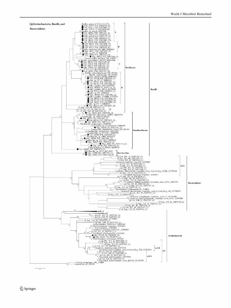
The phylogenetic analysis of the 16S rRNA gene
sequences of clones obtained from excised DGGE bands
also revealed numerous changes in bacterial community
composition among the three treatments (Table 1; Fig. 2).
Bacilli were only detected at 48 �C and they all were in the
cluster E. Actinobacteria were only found at 25 �C. In
c-Proteobacteria (Fig. 2b), the lineage gamV (Xanthomo-
nadaceae) contained clone sequences from both the 42 and
48 �C treated water, and the lineage gamIV (Pseudomo-
nadaceae) only contained sequences from the 42 �C treat-
ment. The cluster F had sequences from the control.
Sequences in a cluster G were originated from 42 �C. In
a-Proteobacteria (Fig. 2b), except the cluster H, all the
other clone sequences were from heat-treated water at both
42 and 48 �C. In b-Proteobacteria (Fig. 2b), the sequences
in the lineage betII (Burkholderiaceae) were from the
control. In Actinobacteria (Fig. 2a), all the clones, includ-
ing those in the lineage acI (Actinomycetales), were orig-
inated from the control. In Bacteroidetes (Fig. 2a), the
lineage bacI (Chitinophagaceae) was mostly at 42 and
48 �C, all other sequences were from both the control and
heat-treated water.
Discussion
This study demonstrated a bacterial community compo-
sition shift in irrigation water treated at 42 and 48 �C
as compared to the control at 25 �C. Culturable bacteria
in the genera Bacillus, Paenibacillus, and Brevibacillus
World J Microbiol Biotechnol
123

dominated the 48 �C-treated irrigation water, and cultur-
able bacteria in the family Pseudomonadaceae increased
in abundance at the 42 �C-treated irrigation water. The
DGGE-detected Bacillus also originated in irrigation
water treated at 48 �C while those Pseudomonadaceae
mostly originated from 42 �C. Likewise, bacteria in sev-
eral clusters of a-Proteobacteria and Xanthomonadaceae
of c-Proteobacteria detected via DGGE also originated
from water at 42 and 48 �C. These findings may have
practical implications.
0
10
20
30
40
50
25°C 42°C 48°C 25°C 42°C 48°C
Feb-12 May-12
Rel
ativ
e ab
unda
nce
of c
ultu
rabl
e ba
cter
ia (
%)
Sample date and temperature treatment
Actinobacteria -Proteobacteria
-Proteobacteria -ProteobacteriaBacilli
(a)
0 10 20 30 40
Others
Brevibacillus
Paenibacillaceae
Bacillaceae others
Bacillaceae Cluster D
Bacillaceae Cluster C
Bacillaceae Cluster B
Bacillaceae Cluster A
0 10 20 30 40
25°C 42°C 48°C
0 20 40 60 80 100
-Proteobacteria Cluster F
Enterobacteriales(gamII)
Pseudomonadaceae(gamIV)
0 20 40 60 80 100
(b)Bacilli
-Proteobacteria
Relative abundance of culturable bacteria (%)
Feb 2012 May 2012
Fig. 1 a Relative abundance of
culturable bacteria detected in
irrigation water samples in
February and May 2012 after
48 h temperature treatments at
25, 42 and 48 �C; b relative
abundance of culturable
bacterial families/clusters
within each of Bacilli and c-
Proteobacteria among three
temperature treatments
World J Microbiol Biotechnol
123
World J Microbiol Biotechnol
123
The increased abundance of Bacillus and Paenibacillus
species in the 48 �C-treated water, and that of Pseudomo-
nas species in the 42 �C-treated water was not unexpected.
Warth (1978) indicated that the optimum temperatures of
mesophilic Bacillus species are between 30 and 45 �C, but
some thermophiles are able to grow as high as 76 �C. The
optimal growth temperature of Paenibacillus species is
28–42 �C, and the maximum growth temperature lies
between 50 and 55 �C (Bosshard et al. 2002; Scheldeman
et al. 2004). The heat tolerance of those bacteria, in the
family Bacillaceae and Paenibacillaceae, is related to their
ability to differentiate into endospores. When heat kills the
vegetative cells, the heat-resistant spores can survive and
germinate when environmental conditions improve (Wil-
son et al. 1992; Hahn et al. 2004). The optimum temper-
atures for growth of Pseudomonas species are variable.
However, some species, such as P. aeruginosa, can grow at
temperatures as high as 42 �C (Tsuji et al. 1982).
Increased abundance of Bacillus, Paenibacillus, and
Pseudomonas species could be indicative more antagonis-
tic activities against plant pathogens in heat-treated water.
A large number of Bacillus, Paenibacillus, and Pseudo-
monas species are known to have antimicrobial activities
against various plant pathogens. For instance, Bacillus
amyloliquefaciens RC-2, B. cereus L7, B. megaterium KL
39, B. pumilus INR7, B. subtilis strains QST713 and GB03
have been used as biocontrol agents for different plant
diseases, including gray mold, damping-off, and powdery
mildews (Raupach and Kloepper 1998; Paulitz and Bel-
anger 2001; Yoshida et al. 2001; Jung and Kim 2003;
Romero et al. 2004; Santoyo et al. 2012; Zhao et al. 2012).
The underlying mechanisms include competition, parasit-
ism, antibiosis, and induced systemic resistance (ISR).
Some Paenibacillus species, for example, P. polymyxa
strains and P. koreensis, produce antibacterial (Piuri et al.
1998; Martin et al. 2003; Shaheen et al. 2011) or antifungal
compounds (Singh et al. 1999; Chung et al. 2000; Beatty
and Jensen 2002; Tupinamba et al. 2008; Deng et al. 2011).
Pseudomonas species, such as P. aeruginosa, P. fluores-
cens, P. putida, are the most extensively utilized for bio-
logical control of many seedling diseases and root rots on
various crops, due to their ability to scavenge available
ions in soil, produce antibiotics, and trigger ISR (Duijff
et al. 1993; Yang et al. 1994; Punja 1997; Maier and
Soberon-Chavez 2000; Paulitz and Belanger 2001; Viji
et al. 2003; Loper et al. 2007; Loper et al. 2008). The
increased abundance of these groups in heat treated water
likely enhances the biological control activities against
plant pathogens in this aquatic environment.
The a-Proteobacteria and Xanthomonadaceae of c-Pro-
teobacteria detected via DGGE were mainly from heat-
treated water in this study. The identity of these a-Prote-
obacteria cannot be determined at this time, because the
closest matches in GenBank have not been identified to
order/family level. Some a-Proteobacteria species have
been reported to be heat-tolerant. For instance, Rhodo-
spirillum centenum, a thermo-tolerant a-proteobacterium
producing heat-resistant cysts under certain nutritional
conditions, grows from 39 to 47 �C, and its survival cysts
can stand for 55–75 �C (Stadtwald-Demchick et al. 1990).
Rhodomicrobium vannielii also forms the exospore-like
cysts with a moderate heat resistance (Imhoff 2006). Very
few studies reported the survival temperatures of Xantho-
monadaceae. Our results showed some a-Proteobacteria
and Xanthomonadaceae are heat resistant up to 48 �C in
water, however, the practical implications of these bacteria
in heat-treated water are yet to be determined.
Lack of Actinobacteria detection from heat treated water
was expected. Antinobacteria were observed only at 25 �C
regardless of detection method (Fig. 1a). No endospores or
microcysts have been found for Actinobacteria. Hahn et al.
(2003) reported that freshwater representative Actinobac-
teria were very small (\0.1 lm3) in size with thin cell
walls. These characteristics of Actinobacteria may be
responsible for their susceptibility to extreme environ-
mental factors, such as, heat stress.
In conclusion, the new heat treatment resulted in an
increased abundance of Bacillus, Paenibacillus, and
Pseudomonas species in irrigation water. Some members
in these groups are known to be antagonistic against
numerous plant pathogens; thus this new heat treatment
may enhance the biological control activities in treated
water, providing a hidden advantage in promoting sus-
tainable disease management. Further investigations are
warranted to test this hypothesis and elucidate the eco-
logical functions of those a-Proteobacteria and Xantho-
monadaceae detected in heat-treated irrigation water via
DGGE.
bFig. 2 Phylogenetic analyses of partial 16S rDNA sequences of
bacteria detected from irrigation water samples collected in February
and May 2012 after 48 h temperature treatments at 25 �C (triangle), 42
(circle) and 48 �C (diamond) via cultures-dependent (filled) and
-independent strategies (unfilled). The frequency of each sequence
detected is listed in parentheses. The maximum likelihood method
based on the Tamura–Nei model of 1,000 replicates was used.
Bootstrap values higher than 50 are shown with lineage and clades
designated from Newton et al. (2011). a Actinobacteria, Bacilli, and
Bacteroidetes with Escherichia coli (Z83205) and Erwinia tasmanien-
sis (NR_074869) as outgroup; b a-Proteobacteria, b-Proteobacteria,
and c-Proteobacteria with Methanotorris formicicus (NR_028646) and
Thermococcus acidaminovorans (AB055120) as outgroup
World J Microbiol Biotechnol
123

Fig. 2 continued
World J Microbiol Biotechnol
123
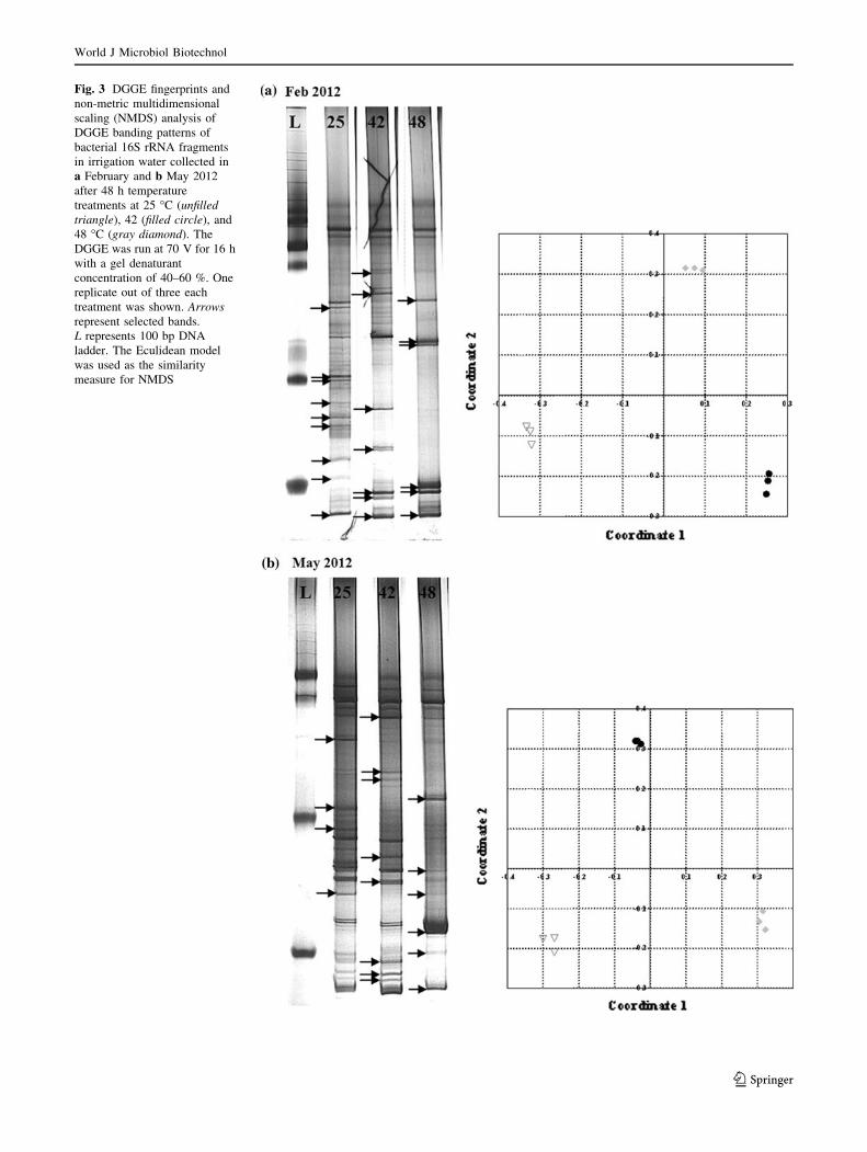
Fig. 3 DGGE fingerprints and
non-metric multidimensional
scaling (NMDS) analysis of
DGGE banding patterns of
bacterial 16S rRNA fragments
in irrigation water collected in
a February and b May 2012
after 48 h temperature
treatments at 25 �C (unfilled
triangle), 42 (filled circle), and
48 �C (gray diamond). The
DGGE was run at 70 V for 16 h
with a gel denaturant
concentration of 40–60 %. One
replicate out of three each
treatment was shown. Arrows
represent selected bands.
L represents 100 bp DNA
ladder. The Eculidean model
was used as the similarity
measure for NMDS
World J Microbiol Biotechnol
123

Acknowledgments This work was supported by a grant from the
USDA National Institute of Food and Agriculture—Specialty Crop
Research Initiative (Agreement #: 2010-51181-21140). We would
like to thank Drs. Boris Vinatzer, Anton Baudoin, Erik Stromberg,
Michael Benson, Giovanni Cafa, and Ping Kong for their valuable
advice during this study, and we also would like to thank Patricia
Richardson for assisting with water sampling and proofreading this
manuscript.
References
Baker KF (1962) Principles of heat treatment of soil and planting
material. J Aust Inst Agric Sci 28(2):118–126
Baker KF, Matkin OA (1978) Dectection and control of pathogens in
water. Ornamentals Northwest April–May:12–13
Beatty PH, Jensen SE (2002) Paenibacillus polymyxa produces
fusaricidin-type antifungal antibiotics active against Leptosp-
haeria maculans, the causative agent of blackleg disease of
canola. Can J Microbiol 48(2):159–169. doi:10.1139/w02-002
Beidler JL, Hilliard PR, Rill RL (1982) Ultrasensitive staining of
nucleic acids with silver. Anal Biochem 126:374–380
Bosshard PP, Zbinden R, Altwegg M (2002) Paenibacillus turicensis
sp. nov., a novel bacterium harbouring heterogeneities between
16S rRNA genes. Int J Syst Evol Microbiol 52:2241–2249.
doi:10.1099/ijs.0.02105-0
Cayanan DF, Dixon M, Zheng YB, Llewellyn J (2009) Response of
container-grown nursery plants to chlorine used to disinfest
irrigation water. HortScience 44(1):164–167
Chung YR, Kim CH, Hwang I, Chun J (2000) Paenibacillus koreensis
sp. nov., a new species that produces an iturin-like antifungal
compound. Int J Syst Evol Microbiol 50:1495–1500
Clarke KR, Ainsworth M (1993) A method of linking multivariate
community structure to environmental variables. Mar Ecol Prog
Ser 92:204–219
Deng Y, Lu ZX, Lu FX, Wang Y, Bie XM (2011) Study on an
antimicrobial protein produced by Paenibacillus polymyxa JSa-9
isolated from soil. World J Microbiol Biotechnol 27(8):1803–
1807. doi:10.1007/s11274-010-0638-6
Duijff BJ, Meijer JW, Bakker PAHM, Schippers B (1993) Sidero-
phore-mediated competition for iron and induced resistance in
the suppression of Fusarium wilt of carnation by fluorescent
Pseudomonas spp. Neth J Plant Path 99(5–6):277–289. doi:10.
1007/bf01974309
Geldreich EE (1996) Pathogenic agents in freshwater resources.
Hydrological Process 10(2):315–333
Hahn MW, Lunsdorf H, Wu QL, Schauer M, Hofle MG, Boenigk J,
Stadler P (2003) Isolation of novel ultramicrobacteria classified
as Actinobacteria from five freshwater habitats in Europe and
Asia. Appl Environ Microbiol 69(3):1442–1451. doi:10.1128/
aem.69.3.1442- 1451.2003
Hahn MW, Stadler P, Wu QL, Pockl M (2004) The filtration-
acclimatization method for isolation of an important fraction of
the not readily cultivable bacteria. J Microbiol Methods
57(3):379–390. doi:10.1016/j.mimet.2004.02.004
Hall TA (1999) BioEdit: a user-friendly biological sequence align-
ment editor and analysis program for Windows 95/98/NT.
Nucleic Acids Symp Ser 41:95–98
Hammer O, Harper DAT, Ryan PD (2001) PAST: paleontological
statistics software package for education and data analysis.
Palaeontologia Electronica 4(1):1–9
Hao W, Ahonsi MO, Vinatzer BA, Hong CX (2012) Inactivation of
Phytophthora and bacterial species in water by a potential
energy-saving heat treatment. Eur J Plant Pathol 134(2):357–
365. doi:10.1007/s10658-012-9994-4
Holmberg AIJ, Melin P, Levenfors JP, Sundh I (2009) Development
and evaluation of SCAR markers for a Pseudomonas brassic-
acearum strain used in biological control of snow mould. Biol
Control 48(2):181–187. doi:10.1016/j.biocontrol.2008.10.016
Hong CX, Moorman GW (2005) Plant pathogens in irrigation water:
challenges and opportunities. Crit Rev Plant Sci 24(3):189–208.
doi:10.1080/07352680591005838
Imhoff JF (2006) The phototrophic alpha-proteobacteria. Prokaryotes:
a handbook on the biology of bacteria, Vol 5, Third Edition:
Proteobacteria: alpha and beta subclasses. Springer, New York.
doi:10.1007/0-387-30745-1_2
Jung H-K, Kim S-D (2003) Purification and characterization of an
antifungal antibiotic from Bacillus megaterium KL 39, a
biocontrol agent of red-papper phytophthora blight disease.
Korean J Microbiol Biotechnol 31(3):235–241
Kawai M, Matsutera E, Kanda H, Yamaguchi N, Tani K, Nasu M
(2002) 16S ribosomal DNA-based analysis of bacterial diversity
in purified water used in pharmaceutical manufacturing pro-
cesses by PCR and denaturing gradient gel electrophoresis. Appl
Environ Microbiol 68(2):699–704
Kong P, Hong CX, Richardson PA, Gallegly ME (2003) Single-
strand-conformation polymorphism of ribosomal DNA for rapid
species differentiation in genus Phytophthora. Fungal Genet Biol
39(3):238–249. doi:10.1016/s1087-1845(03)00052-5
Loper JE, Kobayashi DY, Paulsen IT (2007) The genomic sequence
of Pseudomonas fluorescens Pf-5: insights into biological
control. Phytopathology 97(2):233–238. doi:10.1094/phyto-97-
2-0233
Loper JE, Henkels MD, Shaffer BT, Valeriote FA, Gross H (2008)
Isolation and identification of rhizoxin analogs from Pseudomonasfluorescens Pf-5 by using a genomic mining strategy. Appl
Environ Microbiol 74(10):3085–3093. doi:10.1128/aem.02848-07
Maier RM, Soberon-Chavez G (2000) Pseudomonas aeruginosa
rhamnolipids: biosynthesis and potential applications. Appl
Microbiol Biotechnol 54(5):625–633
Martin NI, Hu HJ, Moake MM, Churey JJ, Whittal R, Worobo RW,
Vederas JC (2003) Isolation, structural characterization, and
properties of mattacin (Polymyxin M), a cyclic peptide antibiotic
produced by Paenibacillus kobensis M. J Biol Chem
278(15):13124–13132. doi:10.1074/jbc.M212364200
McPherson GM, Harriman MR, Pattison D (1995) The potential for
spread of root diseases in recirculating hydroponic systems and
their control with disinfection. Meded Fac Landbouwkd Toegep
Biol Wet Univ Gent 60(28):371–379
Newton RJ, Jones SE, Eiler A, McMahon KD, Bertilsson S (2011) A
guide to the natural history of freshwater lake bacteria. Microbiol
Mol Biol Rev 75:14–49. doi:10.1128/MMBR.00028-10
Table 1 Summary of bacterial presence in irrigation water at dif-
ferent temperatures collected in February and May 2012, identified by
partial 16S rDNA sequences of excised DGGE bands
Taxonomy February 2012 May 2012
Class 25 �C 42 �C 48 �C 25 �C 42 �C 48 �C
Actinobacteria ? – – ? – –
Bacilli – – ? – – ?
Bacteroidetes ? ? ? – ? ?
a-Proteobacteria ? ? ? ? ? –
b-Proteobacteria ? ? – – – –
c-Proteobacteria – ? – ? ? ?
? Indicating the presence of the bacterial class
World J Microbiol Biotechnol
123

Nubel U, Engelen B, Felske A, Snaidr J, Wieshuber A, Amann RI,
Ludwig W, Backhaus H (1996) Sequence heterogeneities of
genes encoding 16S rRNAs in Paenibacillus polymyxa detected
by temperature gradient gel electrophoresis. J Bacteriol
178(19):5636–5643
Paulitz TC, Belanger RR (2001) Biological control in greenhouse
system. Annu Rev Phytopathol 39:103–133
Peixoto RS, Coutinho HLD, Rumjanek NG, Macrae A, Rosado AS
(2002) Use of rpo B and 16S rRNA genes to analyse bacterial
diversity of a tropical soil using PCR and DGGE. Lett Appl
Microbiol 35(4):316–320
Piuri M, Sanchez-Rivas C, Ruzal SM (1998) A novel antimicrobial
activity of a Paenibacillus polymyxa strain isolated from
regional fermented sausages. Lett Appl Microbiol 27(1):9–13
Punja ZK (1997) Comparative efficacy of bacteria, fungi, and yeasts
as biological control agents for diseases of vegetable crops. Can
J Plant Pathol 19(3):315–323
Raupach GS, Kloepper JW (1998) Mixtures of plant growth-
promoting rhizobacteria enhance biological control of multiple
cucumber pathogens. Phytopathology 88:1158–1164
Romero D, Perez-Garcia A, Rivera ME, Cazorla FM, de Vicente A
(2004) Isolation and evaluation of antagonistic bacteria towards
the cucurbit powdery mildew fungus Podosphaera fusca. Appl
Microbiol Biotechnol 64(2):263–269
Runia WT (1995) A review of possibilities for disinfection of
recirculation water from soilless cultures. Acta Horticulturae
382:221–228
Runia WT, Vanos EA, Bollen GJ (1988) Disinfection of drainwater
from soilless cultures by heat-treatment. Neth J Agric Sci 36(3):
231–238
Sambrook J, Russell DW (2001) Molecular cloning: a laboratory
manual, 3rd edn. Cold Spring Laboratory Press, Cold Spring
Harbor, NY
Santoyo G, del Carmen Orozco-Mosqueda M, Govindappa M (2012)
Mechanisms of biocontrol and plant growth-promoting activity
in soil bacterial species of Bacillus and Pseudomonas: a review.
Biocontrol Sci Technol 22(8):855–872. doi:10.1080/09583157.
2012.694413
Scheldeman P, Goossens K, Rodriguez-Diaz M, Pil A, Goris J, Herman
L, De Vos P, Logan NA, Heyndrickx M (2004) Paenibacillus
lactis sp. nov., isolated from raw and heat-treated milk. Int J Syst
Evol Microbiol 54:885–891. doi:10.1099/ijs.0.02822-0
Schumann GL (1991) Plant diseases: their biology and social impact.
APS Press, St. Paul, MN
Schumann GL, D’Arcy CJ (2006) Essential plant pathology, 1st edn.
APS Press, St. Paul, MN
Shaheen M, Li JR, Ross AC, Vederas JC, Jensen SE (2011)
Paenibacillus polymyxa PKB1 produces variants of polymyxin
B-type antibiotics. Chem Biol 18(12):1640–1648. doi:10.1016/j.
chembiol.2011.09.017
Singh PP, Shin YC, Park CS, Chung YR (1999) Biological control of
fusarium wilt of cucumber by chitinolytic bacteria. Phytopathol-
ogy 89(1):92–99. doi:10.1094/phyto.1999.89.1.92
Stadtwald-Demchick R, Turner FR, Gest H (1990) Physiological
properties of the thermotolerant photosynthetic bacterium,
Rhodospirillum centenum. FEMS Microbiol Lett 67(1–2):139–
143. doi:10.1016/0378-1097(90)90183-q
Stanghellini ME, Rasmussen SL (1994) Hydroponics—a solution for
zoosporic pathogens. Plant Dis 78(12):1129–1138
Tamura K, Nei M (1993) Estimation of the number of nucleotide
substitutions in the control region of mitochondrial-DNA in
humans and chimpanzees. Mol Biol Evol 10(3):512–526
Tamura K, Peterson D, Peterson N, Stecher G, Nei M, Kumar S
(2011) MEGA5: molecular evolutionary genetics analysis using
maximum likelihood, evolutionary distance, and maximum
parsimony methods. Mol Biol Evol 28:2731–2739
Thomson SV, Allen RM (1974) Occurrence of Phytophthora species
and other potential plant pathogens in recycled irrigation water.
Plant Dis Rep 58:945–949
Toze S (1999) PCR and the detection of microbial pathogens in water
and wastewater. Water Res 33(17):3545–3556
Toze S (2006) Water reuse and health risks—real vs. perceived.
Desalination 187(1–3):41–51. doi:10.1016/j.desal.2005.04.066
Tsuji A, Kaneko Y, Takahashi K, Ogawa M, Goto S (1982) The effect
of temperature and pH on the growth of eight enteric and nine
glucose non-fermenting species of gram-negative rods. Micro-
biol Immunol 26(1):15–24
Tupinamba G, da Silva AJR, Alviano CS, Souto-Padron T, Seldin L,
Alviano DS (2008) Antimicrobial activity of Paenibacillus
polymyxa SCE2 against some mycotoxin-producing fungi.
J Appl Microbiol 105(4):1044–1053. doi:10.1111/j.1365-2672.
2008.03844.x
van Os EA (1999) Closed soilless growing systems: a sustainable
solution for Dutch greenhouse horticulture. Water Sci Technol
39(5):105–112
Viji G, Uddin W, Romaine CP (2003) Suppression of gray leaf spot
(blast) of perennial ryegrass turf by Pseudomonas aeruginosa
from spent mushroom substrate. Biol Control 26(3):233–243.
doi:10.1016/s1049-9644(02)00170-6
Warth AD (1978) Relationship between heat-resistance of spores and
optimum and maximum growth temperatures of Bacillus species.
J Bacteriol 134(3):699–705
Wilson M, Epton HAS, Sigee DC (1992) Biological control of fire
blight of hawthore (Crataegus monogyna) with fluorescent
Pseudomonas spp under protected conditions. J Phytopathol
136(1):16–26. doi:10.1111/j.1439-0434.1992.tb01277.x
Yang C-H, Menge JA, Cooksey DA (1994) Mutations affecting
hyphal colonization and pyoverdine production in Pseudomo-
nads antagonistic toward Phytophthora parasitica. Appl Environ
Microbiol 60(2):473–481
Yoshida S, Hiradate S, Tsukamoto T, Hatakeda K, Shirata A (2001)
Antimicrobial activity of culture filtrate of Bacillus amylolique-
faciens RC-2 isolated from mulberry leaves. Phytopathology
91(2):181–187. doi:10.1094/phyto.2001.91.2.181
Zhao S, Pan WB, Ma C (2012) Stimulation and inhibition effects of
algae-lytic products from Bacillus cereus strain L7 on Anabaena
flos-aquae. J Appl Phycol 24(5):1015–1021. doi:10.1007/
s10811-011-9725-9
World J Microbiol Biotechnol
123