heart valve disease presenting in adults: investigation
TRANSCRIPT

National Institute for Health and Care Excellence
Draft
Heart valve disease presenting in adults: investigation and management [A] Evidence reviews for symptoms and signs indicating need for echocardiography or direct referral to a specialist
NICE guideline <number>
Evidence reviews underpinning recommendations 1.1.1 to 1.1.5 and 1.1.8 to 1.1.11 in the NICE guideline
March 2021
Draft for Consultation
These evidence reviews were developed by the National Guideline Centre


Heart valve disease: DRAFT FOR CONSULTATION Error! No text of specified style in document.
Heart valve disease: DRAFT FOR CONSULTATION
Disclaimer
The recommendations in this guideline represent the view of NICE, arrived at after careful consideration of the evidence available. When exercising their judgement, professionals are expected to take this guideline fully into account, alongside the individual needs, preferences and values of their patients or service users. The recommendations in this guideline are not mandatory and the guideline does not override the responsibility of healthcare professionals to make decisions appropriate to the circumstances of the individual patient, in consultation with the patient and/or their carer or guardian.
Local commissioners and/or providers have a responsibility to enable the guideline to be applied when individual health professionals and their patients or service users wish to use it. They should do so in the context of local and national priorities for funding and developing services, and in light of their duties to have due regard to the need to eliminate unlawful discrimination, to advance equality of opportunity and to reduce health inequalities. Nothing in this guideline should be interpreted in a way that would be inconsistent with compliance with those duties.
NICE guidelines cover health and care in England. Decisions on how they apply in other UK countries are made by ministers in the Welsh Government, Scottish Government, and Northern Ireland Executive. All NICE guidance is subject to regular review and may be updated or withdrawn.
Copyright
© NICE 2021. All rights reserved. Subject to Notice of rights.
ISBN:

Heart valve disease: DRAFT FOR CONSULTATION
4
Contents
1 Introduction ...................................................................................................................... 6
2 Signs and symptoms indicating echocardiography referral ......................................... 7
2.1 In adults with suspected heart valve disease what symptoms and signs indicate referral (for example from primary care) for echocardiography? ............................ 7
2.1.2 Summary of the protocol ............................................................................... 7
2.1.3 Methods and process ................................................................................... 9
2.1.4 Diagnostic evidence ..................................................................................... 9
2.1.5 Summary of studies included in the diagnostic evidence ............................ 10
2.1.6 Summary of the diagnostic evidence .......................................................... 18
2.1.7 Economic evidence .................................................................................... 37
2.1.8 Summary of included economic evidence ................................................... 37
2.1.9 Economic model ......................................................................................... 37
2.1.10 Unit costs .................................................................................................. 37
3 Signs and symptoms indicating referral to a specialist .............................................. 38
3.1 In adults with suspected heart valve disease, what symptoms and signs indicate direct referral (for example from primary care) to a specialist? ............................ 38
3.1.2 Summary of the protocol ............................................................................. 38
3.1.3 Methods and process ................................................................................. 40
3.1.4 Diagnostic evidence ................................................................................... 40
3.1.5 Summary of studies included in the diagnostic evidence ............................ 41
3.1.6 Summary of the diagnostic evidence .......................................................... 47
3.1.7 Economic evidence .................................................................................... 61
3.1.8 Summary of included economic evidence ................................................... 61
3.1.9 Economic model ......................................................................................... 61
3.1.10 Unit costs .................................................................................................. 61
4 The committee’s discussion of the evidence ............................................................... 62
4.1 Interpreting the evidence ............................................................................... 62
4.2 Cost effectiveness and resource use ............................................................. 68
4.3 Other factors the committee took into account ............................................... 68
4.2 Recommendations supported by this evidence review............................................ 69
5 Women of child bearing age and pregnancy ................................................................ 70
5.1 In women of child bearing age and women who are pregnant what issues across the review questions need to be considered? ...................................................... 70
References ......................................................................................................................... 71
Appendices ........................................................................................................................ 87
Appendix A – Review protocols ................................................................................ 87
Appendix B Literature search strategies ................................................................ 110
B.1 Clinical search literature search strategy ............................................................... 111

Heart valve disease: DRAFT FOR CONSULTATION
5
B.2 Health Economics literature search strategy ......................................................... 114
Appendix C –Diagnostic evidence study selection ................................................ 118
Appendix D – Diagnostic evidence ......................................................................... 119
D.1 Symptoms and signs indicating echocardiography referral ................................. 119
D.2 Symptoms and signs indicating direct referral to a specialist .............................. 191
Appendix E – Forest plots ....................................................................................... 235
E.1 Symptoms and signs for echocardiography referral ............................................. 235
E.1.1 Coupled sensitivity and specificity forest plots ............................................ 235
E.1.1.1 Reference standard – echocardiography ...................................................... 235
E.1.1.2 Reference standard – cardiac catheterisation............................................... 240
E.2 Symptoms and signs for direct referral to a specialist .......................................... 241
E.2.1 Coupled sensitivity and specificity forest plots ............................................ 241
E.2.2 Diagnostic association plots .......................................................................... 244
Appendix F – Economic evidence study selection ................................................ 246
Appendix G – Economic evidence tables ............................................................... 248
Appendix H – Health economic model .................................................................... 249
Appendix I – Excluded studies............................................................................... 250
I.1 Symptoms and signs indicating echocardiography referral ................................. 250
Clinical studies .................................................................................................. 250
Health Economic studies ................................................................................... 258
I.2 Symptoms and signs indicating direct referral to a specialist .............................. 258
Clinical studies .................................................................................................. 258
Health Economic studies ................................................................................... 267
Appendix J – Research recommendations – full details ....................................... 268
Appendix K – Expert witness testimony ................................................................. 269

Heart valve disease: DRAFT FOR CONSULTATION .
Heart valve disease: evidence reviews for symptoms or signs indicating referral for echocardiography or specialist assessment DRAFT [March 2021]
6
1 Introduction 1
The assessment of patients with suspected heart valve disease begins with a comprehensive 2 clinical assessment, comprising history taking and systematic physical examination including 3 cardiac auscultation to detect murmurs and associated changes in the normal heart sounds. 4 This initial clinical assessment can be performed in primary care, in hospital settings outside 5 cardiology or within cardiology, it can increase or decrease the suspicion of existence of 6 heart valve disease and it can provide indications of heart valve disease severity. However, 7 the firm diagnosis of existence and of severity of heart valve disease is made with cardiac 8 imaging, primarily with echocardiography. 9
There is access to echocardiography as a result of a referral from primary care or from 10 hospital settings outside or within cardiology. However, if cardiac auscultation is reassuring, 11 echocardiography may be unnecessary. The capacity for echocardiography is not unlimited 12 and unnecessary assessments would both inconvenience the individual assessed and delay 13 the essential assessment of another individual. Consequently, it is important to identify the 14 symptoms and signs that indicate referral for echocardiography. 15
The clinical pathway comprising referral for echocardiography and subsequent assessment 16 of the result to decide if referral to a specialist is needed, may introduce delay in the care of 17 patients with severe symptoms due to potentially severe heart valve disease. Consequently, 18 it is important to identify the symptoms and signs that indicate direct referral to a specialist to 19 avoid delay. Specialist clinics offer one stop echocardiography or echocardiography prior to 20 the clinic appointment, as such shortening the clinical pathway. 21
22
Heart valve disease: DRAFT FOR CONSULTATION .
Heart valve disease: evidence reviews for symptoms or signs indicating referral for echocardiography or specialist assessment DRAFT [March 2021]
7
2 Signs and symptoms indicating 1
echocardiography referral 2
2.1 In adults with suspected heart valve disease what 3
symptoms and signs indicate referral (for example from 4
primary care) for echocardiography? 5
2.1.2 Summary of the protocol 6
For full details see the review protocol in Appendix A. 7
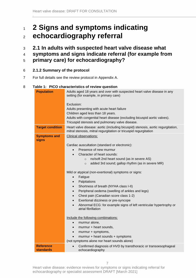
Table 1: PICO characteristics of review question 8
Population Adults aged 18 years and over with suspected heart valve disease in any setting (for example, in primary care)
Exclusion:
Adults presenting with acute heart failure
Children aged less than 18 years.
Adults with congenital heart disease (excluding bicuspid aortic valves).
Tricuspid stenosis and pulmonary valve disease.
Target condition Heart valve disease: aortic (including bicuspid) stenosis, aortic regurgitation, mitral stenosis, mitral regurgitation or tricuspid regurgitation
Symptoms and signs
Clinical observations:
Cardiac auscultation (standard or electronic):
• Presence of new murmur
• Character of heart sounds:
o no/soft 2nd heart sound (as in severe AS)
o added 3rd sound; gallop rhythm (as in severe MR)
Mild or atypical (non-exertional) symptoms or signs:
• Fatigue
• Palpitations
• Shortness of breath (NYHA class I-II)
• Peripheral oedema (swelling of ankles and legs)
• Chest pain (Canadian score class 1-2)
• Exertional dizziness or pre-syncope
• Abnormal ECG: for example signs of left ventricular hypertrophy or atrial fibrillation
Include the following combinations:
• murmur alone,
• murmur + heart sounds,
• murmur + symptoms,
• murmur + heart sounds + symptoms
(not symptoms alone nor heart sounds alone)
Reference standards
• Confirmed diagnosis of HVD by transthoracic or transoesophageal echocardiography

Heart valve disease: DRAFT FOR CONSULTATION .
Heart valve disease: evidence reviews for symptoms or signs indicating referral for echocardiography or specialist assessment DRAFT [March 2021]
8
• Confirmed diagnosis of HVD by invasive cardiac catheterisation will be considered as indirect evidence to avoid excluding older studies
Statistical measures
Diagnostic accuracy of symptoms and signs for a confirmed diagnosis of HVD of any severity.
Measured by:
Primary measures
Accuracy data
• Sensitivity
• Specificity
• Raw data to calculate 2x2 tables to calculate sensitivity and specificity (number of true positives, true negatives, false positives and false negatives).
Secondary measures
• Likelihood ratios
• Positive Predictive Value (PPV)
• Negative Predictive Value (NPV)
If insufficient accuracy data are found, diagnostic association of signs and symptoms with a confirmed diagnosis of HVD will be included.
Measured by:
Association data
• Adjusted RR or OR
For decision-making, it was agreed that sensitivity should be the primary measure taken into account as avoiding false negatives was considered to be the priority over avoiding false positives to avoid sending many people away early without further testing.
Agreed a threshold of ≥60% to represent suitable sensitivity to consider recommending a test, with emphasis on importance of follow-up on those with continuing symptoms or concerns.
Study design • Single-gate diagnostic studies (these may be called cohort studies or cross-sectional studies) will be included preferentially
• If no/insufficient diagnostic accuracy studies are identified prospective and retrospective cohort studies with multivariate analysis of the association between signs and symptoms and a confirmed diagnosis of heart valve disease will be included.
Confounding factors (if diagnostic association studies are included):
• Age (<65 years or ≥65 years)
• Type of murmur:
o Innocent murmur
o Ejection systolic murmur
o Regurgitant systolic murmur
o Diastolic murmur
• Presence/absence of anaemia
• Presence/absence of pregnancy
• Presence/absence of atrial fibrillation

Heart valve disease: DRAFT FOR CONSULTATION .
Heart valve disease: evidence reviews for symptoms or signs indicating referral for echocardiography or specialist assessment DRAFT [March 2021]
9
2.1.3 Methods and process 1
This evidence review was developed using the methods and process described in 2 Developing NICE guidelines: the manual. Methods specific to this review question are 3 described in the review protocol in appendix A and the methods document. 4
Declarations of interest were recorded according to NICE’s conflicts of interest policy. 5
2.1.4 Diagnostic evidence 6
2.1.4.1 Included studies 7
A search was conducted for cross-sectional and prospective and retrospective cohort studies 8 assessing the diagnostic test accuracy of murmur with or without other signs or symptoms 9 (heart sounds and/or symptoms) to identify whether the condition is present (as indicated by 10 the reference standard) in people under investigation for condition heart valve disease. 11
Diagnostic association studies that report data on the association between murmur with or 12 without other signs or symptoms (heart sounds and/or symptoms) and diagnosis of heart 13 valve disease were also considered if limited diagnostic studies were available. 14
Thirty studies with diagnostic accuracy data or data that could be used to calculate 15 diagnostic accuracy data were included in the review; 5, 11, 16-19, 21-23, 27, 29, 34, 51, 72, 86, 97, 103, 109, 113, 16 117, 121, 132, 137, 139, 143, 163, 171, 174, 175, 230 these are summarised in Table 2 below. Evidence from 17 these studies is summarised in the clinical evidence summary below in Tables 3-16. 18
Most of the studies investigated the accuracy of murmur alone for the diagnosis of heart 19 valve disease, with the definition of the murmur and person conducting auscultation differing 20 between studies. However, two studies23, 174 looked at murmur plus symptoms, three 21 studies174,19,132 assessed murmur plus an absent or reduced second heart sound, and one 22 study174 looked at murmur plus abnormal ECG. 23
The assessment of the evidence quality was conducted with emphasis on test sensitivity, as 24 this was identified by the committee as the primary measure in guiding decision-making as 25 the priority would be to avoid missing cases (false negatives) and not sending them for 26 further testing as a result. The committee set clinical decision thresholds as sensitivity = 0.60. 27
Reference standards 28
Of the 30 studies included in the review, 25 used the preferred reference standard of 29 echocardiography. However, a further 5 studies were included that used cardiac 30 catheterisation as the method of confirming valve disease, as this was the preferred method 31 confirming valve disease before echocardiography was available. This more invasive 32 procedure was used in older studies. 33
Populations 34
Studies that involved screening for heart valve disease and murmurs in presumably healthy 35 populations where there could be no reason for a suspicion of heart valve disease were 36 excluded, for example, where screening was performed for everyone who experienced a hip 37 fracture or in populations that were said to be healthy. However, studies where there was not 38 necessarily a suspicion of heart valve disease but had some indication for either attendance 39 at hospital or primary care, echocardiography or were experiencing cardiac symptoms were 40 included, as there was limited evidence where the populations were defined as specifically 41 being suspected of having heart valve disease. 42
Studies where the presence of a murmur was required for a participant to be included in a 43 study were also included, despite the fact that this would mean all were already known to be 44 index test positive before enrolment. Limited diagnostic accuracy data can be obtained from 45

Heart valve disease: DRAFT FOR CONSULTATION .
Heart valve disease: evidence reviews for symptoms or signs indicating referral for echocardiography or specialist assessment DRAFT [March 2021]
10
these studies, but it was agreed to include these given that murmur would be one of the main 1 reasons for suspicion of heart valve disease and these studies could still provide information 2 on the proportion of those with the murmur that actually had reference standard confirmed 3 valve disease, in the form of the positive predictive ratio. The limitations of these studies 4 were highlighted. 5
See also the study selection flow chart in Appendix C, and sensitivity and specificity forest 6 plots in Appendix E, and study evidence tables in Appendix D. 7
2.1.4.2 Excluded studies 8
See the excluded studies list in Appendix I. 9
2.1.5 Summary of studies included in the diagnostic evidence 10
Table 2: Summary of studies included in the evidence review 11
Study Population Target condition Index test
Reference standard Comments
Aggarwal 20145
n=100
India
Outpatients presenting for echocardiography at Cardiology centre
Heart valve disease: any valve disease
Detection of murmur using stethoscope and specific software
Systolic or diastolic murmurs
Echocardiography confirmed heart valve disease
Patients known to have pre-existing heart murmurs excluded
ZargisCardioscan™ software used
Amano 198611
n=55
Japan
People presenting with early or mid-systolic murmurs
Heart valve disease: mitral regurgitation
Presence of murmur (all had one to be included in study)
Apical early or mid-systolic murmurs
Echocardiography confirmed mitral regurgitation
Aronow 198916
n=450
USA
Unselected elderly patients in a long-term health care facility with echocardiography of aortic valve performed
Heart valve disease: aortic regurgitation
Murmur of aortic regurgitation
High frequency diastolic decrescendo murmur beginning with A2
Echocardiography confirmed aortic regurgitation
Potentially indirect population: unselected elderly patients in a long-term health care facility – not necessarily suspected HVD
Aronow 198717
n=75
USA
Unselected elderly patients in a long-term health care facility with echocardiography of aortic valve performed and
Heart valve disease: aortic stenosis
Aortic systolic ejection murmurs (all had one to be included in the study)
Echocardiography confirmed aortic stenosis

Heart valve disease: DRAFT FOR CONSULTATION .
Heart valve disease: evidence reviews for symptoms or signs indicating referral for echocardiography or specialist assessment DRAFT [March 2021]
11
Study Population Target condition Index test
Reference standard Comments
aortic systolic ejection murmurs
Aronow 199118
n=781
USA
Unselected elderly patients in long term health care facility
Heart valve disease: aortic stenosis
Aortic systolic ejection murmur
Echocardiography confirmed AS
Note: only sufficient information to be able to calculate sensitivity (no details of number of true negatives/false positives
Attenhofer Jost 200019
n=100
Switzerland
Those referred for echocardiography due to systolic murmur of unknown cause - no prior echo examination
Heart valve disease: aortic stenosis or valvular regurgitation (AR, MR, TR)
Systolic murmur (all had one)
Systolic murmur +diminished aortic closure sound (AS and MR only)
Echocardiography confirmed AS or valvular regurgitation (AR, TR, MR)
Reports different types of HVD separately and not possible to report as single group
Barron 198821
n=140
USA
People with suspected mitral valve prolapse referred for echocardiography
Heart valve disease: mitral regurgitation or tricuspid regurgitation
Systolic murmur on auscultation
Echocardiography confirmed mitral regurgitation or tricuspid regurgitation
Barzilai 198822
n=59
USA
Hospitalised patients with documented acute myocardial infarction
Heart valve disease: mitral regurgitation
Systolic murmur on auscultation
Echocardiography confirmed mitral regurgitation
Baur 200623
n=198
The Netherlands
Suspected heart failure or valve disease (restricted to: dyspnoea, cardiac murmur or peripheral oedema of unexplained origin)
Heart valve disease: aortic or mitral valve disease (including stenosis and regurgitation)
Cardiac murmur
Cardiac murmur + other indication (e.g. dyspnoea, peripheral oedema or other)
Echocardiography confirmed aortic or mitral valve disease
Breisblatt 198827
n=150
USA
Referred for cardiac catheterisation with known ischaemic heart disease and no previous
Heart valve disease: mitral regurgitation
Systolic murmur on physical examination
Radionuclide angiography confirmed mitral regurgitation
Reference standard indirectness: invasive cardiac catheterisation rather than

Heart valve disease: DRAFT FOR CONSULTATION .
Heart valve disease: evidence reviews for symptoms or signs indicating referral for echocardiography or specialist assessment DRAFT [March 2021]
12
Study Population Target condition Index test
Reference standard Comments
history of valvular disease
echocardiography
Cantley 199529
n=32
UK
Those with systolic murmur on clinical examination at acute assessment and rehabilitation unit of hospital
Heart valve disease: aortic stenosis, aortic regurgitation and mitral regurgitation
Systolic murmur on clinical examination (all had one to be included in the study)
Echocardiography confirmed aortic stenosis, aortic regurgitation or mitral regurgitation
Study reports data separately for each type of valve disease and not possible to combine into a single ‘HVD’ category
Chin 199234
n=42
The Netherlands
Those with diagnosed mitral valve prolapse based on echocardiography
Heart valve disease: mitral regurgitation
Late systolic murmur on auscultation
Echocardiography confirmed mitral regurgitation
Selected population that is more likely to have higher incidence of mitral regurgitation as they already have echo-confirmed mitral valve abnormality?
Decoodt 199051
n=100
Belgium
Those with mitral valve prolapse confirmed by echocardiography
Heart valve disease: mitral regurgitation
Systolic murmur on auscultation (early systolic, late systolic or holosystolic)
Echocardiography confirmed mitral regurgitation
Selected population that is more likely to have higher incidence of mitral regurgitation as they already have echo-confirmed mitral valve abnormality?
Gardezi 201872
n=251
UK
Those undergoing echocardiography at two primary care sites participating in OxVALVE study
Heart valve disease: mild or significant valve disease
Murmur (systolic or diastolic)
Echocardiography confirmed valve disease
Potential population indirectness: screening type study – part of the OxVALVE study where echocardiography performed in asymptomatic people in primary care

Heart valve disease: DRAFT FOR CONSULTATION .
Heart valve disease: evidence reviews for symptoms or signs indicating referral for echocardiography or specialist assessment DRAFT [March 2021]
13
Study Population Target condition Index test
Reference standard Comments
May not represent current practice and how suspected HVD patients would usually be identified
Separates them into mild (aortic sclerosis or any mild regurgitation) and significant (at least moderate regurgitation of any valve or at least mild stenosis of any valve)
Taken murmurs as measured by GPs rather than cardiologists, as our review is set before they have been referred to a specialist
Hoffmann 198386
n=58
Switzerland
Those undergoing right or left heart catheterisation for valvular or coronary heart disease, or both, due to an ill-defined systolic murmur
Heart valve disease: aortic stenosis or mitral regurgitation
Systolic murmur – all had one to be included in the subgroup
Cardiac catheterisation confirmed aortic stenosis or mitral regurgitation
Potential population indirectness: some already with confirmed valve disease (19%)
Reference standard indirectness: invasive cardiac catheterisation rather than echocardiography

Heart valve disease: DRAFT FOR CONSULTATION .
Heart valve disease: evidence reviews for symptoms or signs indicating referral for echocardiography or specialist assessment DRAFT [March 2021]
14
Study Population Target condition Index test
Reference standard Comments
Study reports separately for the two types of valve disease and not possible to combine
Kalinauskiene 201997
n=30
Lithuania
Obese patients referred for echocardiography due to symptoms or abnormal findings
Heart valve disease: aortic stenosis, aortic regurgitation, mitral stenosis, mitral regurgitation or tricuspid regurgitation
Murmur on electronic or acoustic stethoscope
Echocardiography confirmed valve disease
Provides data separately for stenosis and regurgitation, and also separately for electronic and acoustic stethoscopes.
Data was extracted for residents rather than cardiologists as more relevant to the setting of this review.
Kinney 1988103
n=294
USA
Patients referred for echocardiography at hospital
Heart valve disease: aortic regurgitation, mitral regurgitation or tricuspid regurgitation
Murmur on auscultation
(systolic or diastolic)
Echocardiography confirmed aortic regurgitation, mitral regurgitation or tricuspid regurgitation
Gives separately for each type of regurgitation and also for different types of examiners assessing the murmur:
student, internet, junior assistant residents, cardiology fellows – selected ‘resident’ as closest to the area we are interested in.
Labovitz 1985109
n=51
USA
Patients with mitral annular calcium detected on echocardiography
Heart valve disease: mitral stenosis or mitral regurgitation
Apical systolic murmur
Echocardiography confirmed mitral stenosis or mitral regurgitation (sufficient data to combine as ‘any mitral valve disease’
Potential population indirectness: selected population with likely increased incidence of disease as already had

Heart valve disease: DRAFT FOR CONSULTATION .
Heart valve disease: evidence reviews for symptoms or signs indicating referral for echocardiography or specialist assessment DRAFT [March 2021]
15
Study Population Target condition Index test
Reference standard Comments
echocardiography performed?
Lehmann 1992113
n=206
USA
Patients with acute myocardial infarction
Heart valve disease: mitral regurgitation
Any murmur Cardiac catheterisation/ventriculography confirmed mitral regurgitation
Limacher 1985117
n=81
USA
Pregnant women referred for evaluation of murmurs
Heart valve disease: tricuspid regurgitation
Cardiac murmur (all had one to be included in the study)
Echocardiography confirmed tricuspid regurgitation
Loperfido 1986121
n=72
Italy
Patients diagnosed with myocardial infarction 1-3 months prior to the study at the coronary care unit based on chest pain, ECG and increase and decrease of creatine kinase-MB fraction
Heart valve disease: mitral regurgitation
Systolic murmur
Echocardiography confirmed mitral regurgitation
Potential population indirectness: not necessarily suspected HVD but all have had MI with cardiac symptoms
McGee 2010132
n=376
USA
Non-intensive care unit patients undergoing echocardiography - around 16% already known to have valve disease
Heart valve disease: aortic stenosis
Systolic heart murmur
Broad apical-based systolic murmur + absence second heart sound
Echocardiography confirmed aortic stenosis
Potential population indirectness: some already known to have valve disease
Study reports details separately for different types of valve disease and not possible to combine
Meyers 1982139
n=75
USA
Patients with suspected aortic regurgitation undergoing aortograms (angiography)
Heart valve disease: aortic regurgitation
Early diastolic murmur of aortic regurgitation
Angiography confirmed aortic regurgitation
Reference standard indirectness: invasive cardiac catheterisation rather than

Heart valve disease: DRAFT FOR CONSULTATION .
Heart valve disease: evidence reviews for symptoms or signs indicating referral for echocardiography or specialist assessment DRAFT [March 2021]
16
Study Population Target condition Index test
Reference standard Comments
echocardiography
Meyers 1986137
n=35
USA
Those evaluated by Doppler echocardiography, cardiac auscultation and let ventriculography – 20% with already documented valve disease
Heart valve disease: mitral regurgitation
Systolic murmur
Left ventriculography confirmed mitral regurgitation
Potential population indirectness: some with already diagnosed valve disease.
Reference standard indirectness: invasive cardiac catheterisation rather than echocardiography
Mishra, 1992143
n=103
UK
Pregnant women referred for cardiac opinion
Heart valve disease: any type of echo abnormality – can obtain information for those relevant to our protocol
Pathological or possibly pathological murmur detected
Echocardiography confirmed valve disease
Panidis 1986163
n=80
USA
Those with mitral valve prolapse confirmed on echocardiography and signs and symptoms
Heart valve disease: mitral regurgitation
Systolic murmur
Echocardiography confirmed mitral regurgitation
Potential population indirectness: selected population that is more likely to have higher incidence of mitral regurgitation as they already have echo-confirmed mitral valve abnormality?
Rahko 1989171
n=408
USA
Patients presenting to echocardiography laboratory
Heart valve disease: aortic regurgitation, mitral regurgitation or tricuspid regurgitation
Regurgitant murmur on auscultation
Echocardiography confirmed aortic regurgitation, mitral regurgitation or tricuspid regurgitation
Potential population indirectness: not necessarily suspected HVD but some indication for echocardiography
Heart valve disease: DRAFT FOR CONSULTATION .
Heart valve disease: evidence reviews for symptoms or signs indicating referral for echocardiography or specialist assessment DRAFT [March 2021]
17
Study Population Target condition Index test
Reference standard Comments
Study reports data for each type of regurgitation separately and not possible to combine as single ‘regurgitation’ group
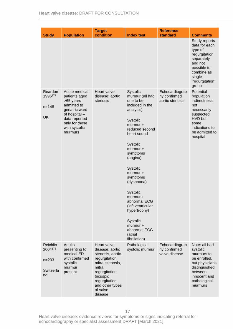
Reardon 1996174
n=148
UK
Acute medical patients aged >65 years admitted to geriatric ward of hospital – data reported only for those with systolic murmurs
Heart valve disease: aortic stenosis
Systolic murmur (all had one to be included in the analysis)
Systolic murmur + reduced second heart sound
Systolic murmur + symptoms (angina)
Systolic murmur + symptoms (dyspnoea)
Systolic murmur + abnormal ECG (left ventricular hypertrophy)
Systolic murmur + abnormal ECG (atrial fibrillation)
Echocardiography confirmed aortic stenosis
Potential population indirectness: not necessarily suspected HVD but some indications to be admitted to hospital
Reichlin 2004175
n=203
Switzerland
Adults presenting to medical ED with confirmed systolic murmur present
Heart valve disease: aortic stenosis, aortic regurgitation, mitral stenosis, mitral regurgitation, tricuspid regurgitation and other types of valve disease
Pathological systolic murmur
Echocardiography confirmed valve disease
Note: all had systolic murmurs to be enrolled, but physicians distinguished between innocent and pathological murmurs

Heart valve disease: DRAFT FOR CONSULTATION .
Heart valve disease: evidence reviews for symptoms or signs indicating referral for echocardiography or specialist assessment DRAFT [March 2021]
18
Study Population Target condition Index test
Reference standard Comments
Yamashita 2020230
n=74
Japan
Inpatients diagnosed with infective endocarditis at a single hospital in Japan between September 2007 and August 2017
Heart valve disease: aortic regurgitation, mitral regurgitation and tricuspid regurgitation (results reported separately for each of these)
Audible cardiac murmur
Echocardiography confirmed valve disease
Note: population includes 14.9% with acute heart failure as a complication of the infective endocarditis
See Appendix D for full evidence. 1
2
2.1.6 Summary of the diagnostic evidence 3
The assessment of the evidence quality was conducted with emphasis on test sensitivity and 4 specificity as this was identified by the committee as the primary measure in guiding 5 decision-making. The committee set a clinical decision threshold of 0.6 for sensitivity. 6
The populations, target conditions and index tests used across the included studies were 7 considered to be very broad and wide-ranging, and therefore no studies were pooled into a 8 diagnostic meta-analysis. Sensitivity and specificity for each individual study is given below, 9 separated into broad categories based on the population and also by whether the reference 10 standard was echocardiography or cardiac catheterisation. 11
For studies where all of those included had to be positive for murmur with/without another 12 characteristic (which was used as an index test in our review), sensitivity and specificity, as 13 well as other measures, could not be calculated, and positive predictive values are instead 14 presented. 15
Note that although all included studies detected heart valve disease as the target condition, 16 the type of heart valve disease that was included in the studies varied. For example, some 17 studies aimed to diagnose and report any type of valve disease (including aortic stenosis, 18 aortic regurgitation, mitral stenosis, mitral regurgitation and tricuspid regurgitation), while 19 others focused specifically on one or two types of valve disease, such as aortic stenosis or 20 mitral stenosis and mitral regurgitation, or any type of regurgitation but not stenosis (i.e. 21 aortic regurgitation, mitral regurgitation and tricuspid regurgitation). Where possible, results 22 have been calculated for ‘any valve disease’; however, in many cases results are reported 23 separately for each type of valve disease as it was not possible to determine how many may 24 have had more than one time of valve disease at the same time to calculate diagnostic 25 accuracy results for overall heart valve disease in each study. 26
Reference standard – echocardiography 27
Table 3: Clinical evidence summary: murmur for heart valve disease in various 28 settings in populations with various indications for assessment 29
Study population N
Risk of bias
Inconsistency
Indirectness
Imprecision
Effect size (95%CI) Quality
Other results
Systolic or diastolic murmur detected with stethoscope and specific software for the diagnosis of any valve disease – community medicine physician
PPV: 0.68

Heart valve disease: DRAFT FOR CONSULTATION .
Heart valve disease: evidence reviews for symptoms or signs indicating referral for echocardiography or specialist assessment DRAFT [March 2021]
19
Study population N
Risk of bias
Inconsistency
Indirectness
Imprecision
Effect size (95%CI) Quality
Other results
Outpatients presenting for echocardiography
100
Serious1
NA Serious2
Serious3
Sensitivity=0.41 (0.28 to 0.56)
VERY LOW
NPV: 0.57
PLR: 2.02
NLR: 0.74
Prevalence on reference standard: 0.51
Serious1
NA Serious2
Serious3
Specificity= 0.80 (0.66 to 0.90)
VERY LOW
Murmur of AR (high frequency diastolic decrescendo murmur beginning with A2) for the diagnosis of AR – experienced cardiologist
PPV: 0.93
NPV: 0.92
PLR: 31.96
NLR: 0.20
Prevalence on reference standard: 0.29
Unselected elderly patients in a long-term health care facility
450
Very serious1
NA Serious2
None Sensitivity=0.80 (0.72 to 0.87)
VERY LOW
Very serious1
NA Serious2
None Specificity= 0.97 (0.95 to 0.99)
VERY LOW
Aortic systolic ejection murmur for the diagnosis of AS – experienced cardiologist Prevalence on reference standard: 0.18
Other measures could not be calculated due to insufficient data
Unselected elderly patients in a long-term health care facility
781
Very serious1
NA Serious2
None Sensitivity=0.97 (0.93 to 0.99)
VERY LOW
NA NA NA NA Specificity could not be calculated due to insufficient data provided
NA
Systolic murmur on auscultation for the diagnosis of MR or TR – cardiologist PPV: 0.51
NPV: 0.74
PLR: 1.93
NLR: 0.65
Prevalence on reference standard: 0.35
People with suspected mitral valve prolapse referred for echocardiography
140
Serious1
NA None Serious3 Sensitivity=0.53 (0.38 to 0.67)
LOW
Serious1
NA None None Specificity= 0.73 (0.62 to 0.81)
MODERATE
Systolic murmur on auscultation for the diagnosis of MR – attending physician PPV: 0.63
NPV: 0.70
PLR: 2.61
NLR: 0.68
Prevalence on reference
Hospitalised patients with documented
59 Very serious1
NA Serious2
Very serious3
Sensitivity=0.43 (0.23 to 0.66)
VERY LOW
Very serious1
NA Serious2
Very serious3
Specificity= 0.83 (0.67 to 0.94)
VERY LOW

Heart valve disease: DRAFT FOR CONSULTATION .
Heart valve disease: evidence reviews for symptoms or signs indicating referral for echocardiography or specialist assessment DRAFT [March 2021]
20
Study population N
Risk of bias
Inconsistency
Indirectness
Imprecision
Effect size (95%CI) Quality
Other results
acute myocardial infarction
standard: 0.39
Cardiac murmur for the diagnosis of any aortic or mitral valve disease – GPs PPV: 0.19
NPV: 0.99
PLR: 2.06
NLR: 0.09
Prevalence on reference standard: 0.10
Suspected heart failure or valve disease (restricted to: dyspnoea, cardiac murmur or peripheral oedema of unexplained origin)
198
Very serious1
NA None Serious3 Sensitivity=0.95 (0.75 to 1.00)
VERY LOW
Very serious1
NA None None Specificity= 0.54 (0.46 to 0.61)
LOW
Systolic or diastolic murmur for the diagnosis of any valve disease
Those undergoing echocardiography at two primary care sites participating in OxVALVE study
251
Mild valve disease (aortic sclerosis or mild regurgitation) – GPs PPV: 0.67
NPV: 0.32
PLR: 0.97
NLR: 1.01
Prevalence on reference standard: 0.68
Serious1
NA Serious2
None Sensitivity=0.32 (0.25 to 0.40)
LOW
Serious1
NA Serious2
Serious3 Specificity= 0.67 (0.55 to 0.77)
VERY LOW
Significant valve disease (at least moderate regurgitation or at least mild stenosis of any valve) – GPs
PPV: 0.20
NPV: 0.88
PLR: 1.45
NLR: 0.80
Prevalence on reference standard: 0.14
Serious1
NA Serious2
Serious3 Sensitivity=0.44 (0.28 to 0.62)
VERY LOW
Serious1
NA Serious2
None Specificity= 0.69 (0.63 to 0.75)
LOW
Murmur on electronic or acoustic stethoscope for the diagnosis of any valve disease – 3rd year medical resident doctor
Obese patients referred for echocardiography
30 Aortic stenosis – resident using acoustic stethoscope PPV:0.25
NPV:0.92
PLR:3.00
NLR:0.75
Prevalence on reference
Serious1
NA Serious2
Very serious3
Sensitivity=0.33 (0.01 to 0.91)
VERY LOW
Serious1
NA Serious2
Serious3 Specificity= 0.89 (0.71 to 0.98)
VERY LOW

Heart valve disease: DRAFT FOR CONSULTATION .
Heart valve disease: evidence reviews for symptoms or signs indicating referral for echocardiography or specialist assessment DRAFT [March 2021]
21
Study population N
Risk of bias
Inconsistency
Indirectness
Imprecision
Effect size (95%CI) Quality
Other results
due to symptoms or abnormal findings
standard:0.10
Aortic stenosis – resident using electronic stethoscope PPV:0.25
NPV:0.92
PLR:3.00
NLR:0.75
Prevalence on reference standard:0.10
Serious1
NA Serious2
Very serious3
Sensitivity=0.33 (0.01 to 0.91)
VERY LOW
Serious1
NA Serious2
Serious3 Specificity= 0.89 (0.71 to 0.98)
VERY LOW
Aortic regurgitation – resident using acoustic stethoscope PPV:1.00
NPV:0.44
PLR: Not calculable
NLR:0.74
Prevalence on reference standard:0.63
Serious1
NA Serious2
Very serious3
Sensitivity=0.26 (0.09 to 0.51)
VERY LOW
Serious1
NA Serious2
Serious3 Specificity= 1.00 (0.72 to 1.00)
VERY LOW
Aortic regurgitation – resident using electronic stethoscope PPV:1.00
NPV:0.48
PLR: Not calculable
NLR:0.63
Prevalence on reference standard:0.63
Serious1
NA Serious2
Very serious3
Sensitivity=0.37 (0.16 to 0.62)
VERY LOW
Serious1
NA Serious2
Serious3 Specificity= 1.00 (0.72 to 1.00)
VERY LOW
Mitral stenosis – resident using acoustic stethoscope NPV: 1.00
Prevalence on reference standard: 0.00
Other values not calculable
Serious1
NA NA NA Sensitivity could not be calculated due to none being positive for MS
NA
Serious1
NA Serious2
None Specificity= 0.97 (0.83 to 1.00)
LOW
Mitral stenosis – resident using electronic stethoscope NPV: 1.00
Prevalence on reference standard: 0.00
Other values not calculable
Serious1
NA Serious2
NA Sensitivity could not be calculated due to none being positive for MS
NA
Serious1
NA Serious2
None Specificity= 0.97 (0.83 to 1.00)
LOW
Mitral regurgitation – resident using acoustic stethoscope

Heart valve disease: DRAFT FOR CONSULTATION .
Heart valve disease: evidence reviews for symptoms or signs indicating referral for echocardiography or specialist assessment DRAFT [March 2021]
22
Study population N
Risk of bias
Inconsistency
Indirectness
Imprecision
Effect size (95%CI) Quality
Other results
Serious1
NA Serious2
Serious3 Sensitivity=0.76 (0.55 to 0.91)
VERY LOW
PPV:0.90
NPV:0.33
PLR:1.90
NLR:0.40
Prevalence on reference standard:0.83
Serious1
NA Serious2
Very serious3
Specificity= 0.60 (0.15 to 0.95)
VERY LOW
Mitral regurgitation – resident using electronic stethoscope PPV:0.88
NPV:0.33
PLR:1.40
NLR:0.40
Prevalence on reference standard:0.83
Serious1
NA Serious2
Serious3 Sensitivity=0.84 (0.64 to 0.95)
VERY LOW
Serious1
NA Serious2
Very serious3
Specificity= 0.40 (0.05 to 0.85)
VERY LOW
Tricuspid regurgitation – resident using acoustic stethoscope PPV:0.91
NPV:0.47
PLR:5.00
NLR:0.56
Prevalence on reference standard:0.67
Serious1
NA Serious2
Very serious3
Sensitivity=0.50 (0.27 to 0.73)
VERY LOW
Serious1
NA Serious2
Very serious3
Specificity= 0.90 (0.55 to 1.00)
VERY LOW
Tricuspid regurgitation – resident using electronic stethoscope PPV:0.87
NPV:0.53
PLR:3.25
NLR:0.44
Prevalence on reference standard:0.67
Serious1
NA Serious2
Very serious3
Sensitivity=0.65 (0.41 to 0.85)
VERY LOW
Serious1
NA Serious2
Very serious3
Specificity= 0.80 (0.44 to 0.97)
VERY LOW
Systolic or diastolic murmur on auscultation for the diagnosis of AR, MR or TR
Patients referred for echocardiography at hospital
294
Aortic regurgitation – junior assistant residents PPV: 0.27
NPV: 0.79
PLR: 1.33
NLR: 0.99
Prevalence on reference standard: 0.214
Very serious1
NA Serious2
None Sensitivity=0.05 (0.01 to 0.13)
VERY LOW
Very serious1
NA Serious2
None Specificity= 0.97 (0.94 to 0.99)
VERY LOW
Aortic regurgitation – senior assistant residents NPV: 0.77
NLR: 1.10
Prevalence on
Very serious1
NA Serious2
None Sensitivity=0.00 (0.00 to 0.06)
VERY LOW

Heart valve disease: DRAFT FOR CONSULTATION .
Heart valve disease: evidence reviews for symptoms or signs indicating referral for echocardiography or specialist assessment DRAFT [March 2021]
23
Study population N
Risk of bias
Inconsistency
Indirectness
Imprecision
Effect size (95%CI) Quality
Other results
Very serious1
NA Serious2
None Specificity= 0.91 (0.86 to 0.94)
VERY LOW
reference standard: 0.214
Other values not calculable
Mitral regurgitation – junior assistant residents PPV: 0.48
NPV: 0.71
PLR: 1.87
NLR: 0.85
Prevalence on reference standard: 0.327
Very serious1
NA Serious2
None Sensitivity=0.28 (0.19 to 0.38)
VERY LOW
Very serious1
NA Serious2
None Specificity= 0.85 (0.79 to 0.90)
VERY LOW
Mitral regurgitation – senior assistant residents PPV: 0.39
NPV: 0.68
PLR: 1.30
NLR: 0.97
Prevalence on reference standard: 0.327
Very serious1
NA Serious2
None Sensitivity=0.13 (0.07 to 0.21)
VERY LOW
Very serious1
NA Serious2
None Specificity= 0.90 (0.85 to 0.94)
VERY LOW
Tricuspid regurgitation – junior assistant residents PPV: 1.00
NPV: 0.87
NLR: 0.73
Prevalence on reference standard: 0.167
PLR not calculable
Very serious1
NA Serious2
Serious3 Sensitivity=0.27 (0.15 to 0.41)
VERY LOW
Very serious1
NA Serious2
None Specificity= 1.00 (0.99 to 1.00)
VERY LOW
Tricuspid regurgitation – senior assistant residents PPV: 1.00
NPV: 0.88
NLR: 0.67
Prevalence on reference standard: 0.167
PLR not calculable
Very serious1
NA Serious2
Serious3 Sensitivity=0.33 (0.20 to 0.48)
VERY LOW
Very serious1
NA Serious2
None Specificity= 1.00 (0.99 to 1.00)
VERY LOW
Systolic murmur for the diagnosis of MR – cardiologist (performed at coronary care unit)
PPV: 0.81
NPV: 0.52
PLR: 3.47
NLR: 0.74
Patients diagnos
72 Serious1
NA Serious2
Serious3 Sensitivity=0.33 (0.19 to 0.49)
VERY LOW

Heart valve disease: DRAFT FOR CONSULTATION .
Heart valve disease: evidence reviews for symptoms or signs indicating referral for echocardiography or specialist assessment DRAFT [March 2021]
24
Study population N
Risk of bias
Inconsistency
Indirectness
Imprecision
Effect size (95%CI) Quality
Other results
ed with myocardial infarction 1-3 months prior to the study at the coronary care unit
Serious1
NA Serious2
Serious3 Specificity= 0.91 (0.75 to 0.98)
VERY LOW
Prevalence on reference standard: 0.56
Systolic heart murmur for the diagnosis of AS – physician in primary and specialist medical care department
PPV: 0.33
NPV: 0.99
PLR: 1.96
NLR: 0.05
Prevalence on reference standard: 0.20
Non-intensive care unit patients undergoing echocardiography
376
Very serious1
NA Serious2
None Sensitivity=0.97 (0.90 to 1.00)
VERY LOW
Very serious1
NA Serious2
None Specificity= 0.50 (0.44 to 0.56)
VERY LOW
Regurgitant murmur on auscultation for the diagnosis of AR, MR or TR – cardiologist
Patients presenting to an echocardiography laboratory
408
Aortic regurgitation PPV: 0.34
NPV: 0.99
PLR: 5.90
NLR: 0.11
Prevalence on reference standard: 0.08
Very serious1
NA Serious2
None Sensitivity=0.60 (0.52 to 0.69)
VERY LOW
Very serious1
NA Serious2
None Specificity= 0.98 (0.95 to 0.99)
VERY LOW
Mitral regurgitation PPV: 0.28
NPV: 0.98
PLR: 3.49
NLR: 0.20
Prevalence on reference standard: 0.10
Very serious1
NA Serious2
None Sensitivity=0.56 (0.48 to 0.64)
VERY LOW
Very serious1
NA Serious2
None Specificity= 0.89 (0.85 to 0.93)
VERY LOW
Tricuspid regurgitation PPV: 0.42
NPV: 0.97
PLR: 10.15
NLR: 0.41
Prevalence on reference standard: 0.07
Very serious1
NA Serious2
None Sensitivity=0.23 (0.16 to 0.31)
VERY LOW
Very serious1
NA Serious2
None Specificity= 0.98 (0.96 to 1.00)
VERY LOW

Heart valve disease: DRAFT FOR CONSULTATION .
Heart valve disease: evidence reviews for symptoms or signs indicating referral for echocardiography or specialist assessment DRAFT [March 2021]
25
Study population N
Risk of bias
Inconsistency
Indirectness
Imprecision
Effect size (95%CI) Quality
Other results
Pathological systolic murmur (as interpreted by auscultator) for the diagnosis of any valve disease – emergency department attending physician
PPV: 0.59
NPV: 0.88
PLR: 2.63
NLR: 0.27
Prevalence on reference standard: 0.35
Adults presenting to medical ED with confirmed systolic murmur present
203
Serious1
NA Serious2
None Sensitivity=0.82 (0.71 to 0.90)
LOW
Serious1
NA Serious2
None Specificity= 0.69 (0.60 to 0.77)
LOW
Audible cardiac murmur for the diagnosis of aortic regurgitation – unclear who assessed murmur
PPV: 0.47
NPV: 0.76
PLR: 1.44
NLR: 0.52
Prevalence on reference standard: 0.38
Inpatients diagnosed with infective endocarditis at a single hospital in Japan
74 Very serious1
NA Serious2
Serious3 Sensitivity=0.75 (0.55 to 0.89)
VERY LOW
Very serious1
NA Serious2
Serious3 Specificity= 0.48 (0.33 to 0.63)
VERY LOW
Audible cardiac murmur for the diagnosis of mitral regurgitation – unclear who assessed murmur
Inpatients diagnosed with infective endocarditis at a single hospital in Japan
74 Very serious1
NA Serious2
Serious3 Sensitivity=0.66 (0.51 to 0.79)
VERY LOW
PPV: 0.69
NPV: 0.45
PLR: 1.27
NLR: 0.71
Prevalence on reference standard: 0.64
Very serious1
NA Serious2
Serious3 Specificity= 0.48 (0.29 to 0.68)
VERY LOW
Audible cardiac murmur for the diagnosis of tricuspid regurgitation – unclear who assessed murmur
Inpatients diagnosed with infective endocarditis at a single hospital in Japan
74 Very serious1
NA Serious2
Very serious3
Sensitivity=0.62 (0.38 to 0.82)
VERY LOW
PPV: 0.29
NPV: 0.72
PLR: 1.03
NLR: 0.96
Prevalence on reference standard: 0.28
Very serious11
NA Serious2
Serious3 Specificity= 0.40 (0.26 to 0.54)
VERY LOW
1 Risk of bias was assessed using the QUADAS-2 checklist. The evidence was downgraded by 1 increment if the 1 majority of studies were rated at high risk of bias, and downgraded by 2 increments if the majority of 2 studies were rated at very high risk of bias. 3

Heart valve disease: DRAFT FOR CONSULTATION .
Heart valve disease: evidence reviews for symptoms or signs indicating referral for echocardiography or specialist assessment DRAFT [March 2021]
26
2 Indirectness was assessed using the QUADAS-2 checklist. The evidence was downgraded by 1 increment if the 1 majority of studies were considered to have a high degree of indirectness, and downgraded by 2 2 increments if the majority of studies were considered to have a very high degree of indirectness. 3
3 Imprecision was assessed by considering the width of the confidence intervals around the sensitivity and 4 specificity. A variation of 0-20% was considered precise, 20-40% serious imprecision, and >40% very 5 serious imprecision. 6
7
Table 4: Clinical evidence summary: murmur for heart valve disease in populations 8 with MVP that has already been diagnosed by echocardiography 9
Study population N
Risk of bias
Inconsistency
Indirectness
Imprecision
Effect size (95%CI) Quality
Other results
Late systolic murmur on auscultation for the diagnosis of MR – unclear who did examination
PPV: 0.82
NPV: 0.80
PLR: 6.23
NLR: 0.35
Prevalence on reference standard: 0.42
Those with diagnosed mitral valve prolapse based on echocardiography
42 Very serious1
NA Serious2
Very serious3
Sensitivity=0.69 (0.39 to 0.91)
VERY LOW
Very serious1
NA Serious2
Very serious3
Specificity=0.89 (0.65 to 0.99)
VERY LOW
Systolic murmur on auscultation (early, late or holosystolic included) for the diagnosis of MR – unclear who did examination
PPV: 0.90
NPV: 0.85
PLR: 8.01
NLR: 0.15
Prevalence on reference standard: 0.54
Those with mitral valve prolapse confirmed by echocardiography
100
Very serious1
NA Serious2
Serious3
Sensitivity=0.87 (0.75 to 0.95)
VERY LOW
Very serious1
NA Serious2
Serious3
Specificity=0.89 (0.76 to 0.96)
VERY LOW
Systolic murmur for the diagnosis of MR – unclear who did examination PPV: 0.81
NPV: 0.46
PLR: 1.99
NLR: 0.53
Prevalence on reference standard: 0.69
Those with mitral valve prolapse confirmed on echocardiography and signs and symptoms
80 Serious1
NA Serious2
Serious3
Sensitivity=0.64 (0.50 to 0.76)
VERY LOW
Serious1
NA Serious2
Serious3
Specificity=0.68 (0.46 to 0.85)
VERY LOW

Heart valve disease: DRAFT FOR CONSULTATION .
Heart valve disease: evidence reviews for symptoms or signs indicating referral for echocardiography or specialist assessment DRAFT [March 2021]
27
1 Risk of bias was assessed using the QUADAS-2 checklist. The evidence was downgraded by 1 increment if the 1 majority of studies were rated at high risk of bias, and downgraded by 2 increments if the majority of 2 studies were rated at very high risk of bias. 3
2 Indirectness was assessed using the QUADAS-2 checklist. The evidence was downgraded by 1 increment if the 4 majority of studies were considered to have a high degree of indirectness, and downgraded by 2 5 increments if the majority of studies were considered to have a very high degree of indirectness. 6
3 Imprecision was assessed by considering the width of the confidence intervals around the sensitivity and 7 specificity. A variation of 0-20% was considered precise, 20-40% serious imprecision, and >40% very 8 serious imprecision. 9
10
Table 5: Clinical evidence summary: murmur for heart valve disease in a population 11 with mitral annular calcium observed by echocardiography 12
Studies N Risk of bias
Inconsistency
Indirectness
Imprecision
Effect size (95%CI) Quality
Other results
Apical systolic murmur for the diagnosis of mitral stenosis or regurgitation – unclear who did examination
PPV: 0.39
NPV: 0.74
PLR: 1.29
NLR: 0.71
Prevalence on reference standard: 0.33
Patients with mitral annular calcium detected on echocardiography
51 Very serious1
NA Serious2
Serious3
Sensitivity=0.59 (0.41 to 0.76)
VERY LOW
Very serious1
NA Serious2
Very serious3
Specificity=0.53 (0.29 to 0.76)
VERY LOW
1 Risk of bias was assessed using the QUADAS-2 checklist. The evidence was downgraded by 1 increment if the 13 majority of studies were rated at high risk of bias, and downgraded by 2 increments if the majority of 14 studies were rated at very high risk of bias. 15
2 Indirectness was assessed using the QUADAS-2 checklist. The evidence was downgraded by 1 increment if the 16 majority of studies were considered to have a high degree of indirectness, and downgraded by 2 17 increments if the majority of studies were considered to have a very high degree of indirectness. 18
3 Imprecision was assessed by considering the width of the confidence intervals around the sensitivity and 19 specificity. A variation of 0-20% was considered precise, 20-40% serious imprecision, and >40% very 20 serious imprecision. 21
22
Table 6: Clinical evidence summary: murmur for heart valve disease (all with murmur 23 to be included) 24
Study population N
Risk of bias
Inconsistency
Indirectness
Imprecision Effect size 1 Quality
Apical early or mid-systolic murmur for the diagnosis of MR – prevalence 0.53 – unclear who did examination
People presenting with early or mid-systolic murmurs
55 Very serious2
NA Serious3 Could not be assessed
PPV=0.53 VERY LOW
Aortic systolic ejection murmur for the diagnosis of AS – prevalence 0.56 – experienced cardiologist

Heart valve disease: DRAFT FOR CONSULTATION .
Heart valve disease: evidence reviews for symptoms or signs indicating referral for echocardiography or specialist assessment DRAFT [March 2021]
28
Study population N
Risk of bias
Inconsistency
Indirectness
Imprecision Effect size 1 Quality
Unselected elderly patients in a long-term health care facility with aortic systolic ejection murmurs
75 Serious2 NA Serious3 Could not be assessed
PPV=0.56 VERY LOW
Systolic murmur for the diagnosis of AS, AR, MR or TR – cardiologists
Referred for echocardiography due to systolic murmur of unknown cause - no prior echo examination
100 Aortic stenosis - prevalence 0.29
Serious2
NA Serious3 Could not be assessed
PPV=0.29 VERY LOW
Aortic regurgitation- prevalence 0.28
Serious2
NA Serious3 Could not be assessed
PPV=0.28 VERY LOW
Mitral regurgitation - prevalence 0.30
Serious2
NA Serious3 Could not be assessed
PPV=0.30 VERY LOW
Tricuspid regurgitation - prevalence 0.24
Serious2
NA Serious3 Could not be assessed
PPV=0.24 VERY LOW
Systolic murmur on clinical examination for the diagnosis of AS, AR or MR – unclear who did examination
Those with systolic murmur on clinical examination at acute assessment and rehabilitation unit of hospital
32 Aortic stenosis – prevalence 0.38
Very serious2
NA Serious3 Could not be assessed
PPV=0.38 VERY LOW
Aortic regurgitation – prevalence 0.45
Very serious2
NA Serious3 Could not be assessed
PPV=0.45 VERY LOW
Mitral regurgitation – prevalence 0.55
Very serious2
NA Serious3 Could not be assessed
PPV=0.55 VERY LOW

Heart valve disease: DRAFT FOR CONSULTATION .
Heart valve disease: evidence reviews for symptoms or signs indicating referral for echocardiography or specialist assessment DRAFT [March 2021]
29
Study population N
Risk of bias
Inconsistency
Indirectness
Imprecision Effect size 1 Quality
Systolic murmur for the diagnosis of AS – prevalence 0.81 – junior hospital doctor and one of authors
Acute medical patients aged >65 years admitted to geriatric ward of hospital with confirmed systolic murmur present
148 Very serious2
NA Serious3 Could not be assessed
PPV=0.81 VERY LOW
1 In these studies, all patients had to have a murmur to be included in the study. Therefore, sensitivity and 1 specificity could not be calculated, and positive predictive values are instead presented for each 2 study.95% confidence intervals could not be calculated for this effect measure. 3
2 Risk of bias was assessed using the QUADAS-2 checklist. The evidence was downgraded by 1 increment if the 4 majority of studies were rated at high risk of bias, and downgraded by 2 increments if the majority of 5 studies were rated at very high risk of bias. 6
3 Indirectness was assessed using the QUADAS-2 checklist. The evidence was downgraded by 1 increment if the 7 majority of studies were considered to have a high degree of indirectness, and downgraded by 2 8 increments if the majority of studies were considered to have a very high degree of indirectness. 9
10
Table 7: Clinical evidence summary: murmur + dyspnoea for heart valve disease in 11 acute medical patients admitted to geriatric ward of hospital 12
Study population N
Risk of bias
Inconsistency
Indirectness
Imprecision
Effect size (95%CI) Quality
Other results
Systolic murmur + symptoms (dyspnoea) for the diagnosis of AS – junior hospital doctor and one of authors
PPV: 1.00
NPV: 0.24
PLR: could not be calculated as there were no false positives reported
NLR: 0.73
Prevalence on reference standard: 0.81
Acute medical patients aged >65 years admitted to geriatric ward of hospital with confirmed systolic murmur present
148
Very serious1
NA None Serious2
Sensitivity=0.27 (0.17 to 0.40)
VERY LOW
Very serious1
NA None Serious2
Specificity=1.00 (0.78 to 1.00)
VERY LOW

Heart valve disease: DRAFT FOR CONSULTATION .
Heart valve disease: evidence reviews for symptoms or signs indicating referral for echocardiography or specialist assessment DRAFT [March 2021]
30
1 Risk of bias was assessed using the QUADAS-2 checklist. The evidence was downgraded by 1 increment if the 1 majority of studies were rated at high risk of bias, and downgraded by 2 increments if the majority of 2 studies were rated at very high risk of bias. 3
2 Imprecision was assessed by considering the width of the confidence intervals around the sensitivity and 4 specificity. A variation of 0-20% was considered precise, 20-40% serious imprecision, and >40% very 5 serious imprecision. 6
7
Table 8: Clinical evidence summary: murmur + angina for heart valve disease in acute 8 medical patients admitted to geriatric ward of hospital 9
Study population N
Risk of bias
Inconsistency
Indirectness
Imprecision
Effect size (95%CI) Quality
Other results
Systolic murmur + symptoms (angina) for the diagnosis of AS – junior hospital doctor and one of authors
PPV: 1.00
NPV: 0.19
PLR: could not be calculated as there were no false positives reported
NLR: 0.97
Prevalence on reference standard: 0.81
Acute medical patients aged >65 years admitted to geriatric ward of hospital with confirmed systolic murmur present
148
Very serious1
NA None None Sensitivity=0.03 (0.00 to 0.11)
LOW
Very serious1
NA None Serious2
Specificity=1.00 (0.78 to 1.00)
VERY LOW
10 1 Risk of bias was assessed using the QUADAS-2 checklist. The evidence was downgraded by 1 increment if the 11
majority of studies were rated at high risk of bias, and downgraded by 2 increments if the majority of 12 studies were rated at very high risk of bias. 13
2 Imprecision was assessed by considering the width of the confidence intervals around the sensitivity and 14 specificity. A variation of 0-20% was considered precise, 20-40% serious imprecision, and >40% very 15 serious imprecision. 16
17
Table 9: Clinical evidence summary: murmur + other indication (dyspnoea, peripheral 18 oedema or other) for heart valve disease in patients with suspected heart 19 failure of heart valve disease) 20
Study population N
Risk of bias
Inconsistency
Indirectness
Imprecision
Effect size (95%CI) Quality
Other results
Cardiac murmur + other indication (e.g. dyspnoea, peripheral oedema or other) for the diagnosis of any aortic or mitral valve disease – GPs
PPV: 0.35
NPV: 0.95
Suspected heart failure
198
Very serious1
NA None Very serious2
Sensitivity=0.60 (0.36 to 0.81)
VERY LOW

Heart valve disease: DRAFT FOR CONSULTATION .
Heart valve disease: evidence reviews for symptoms or signs indicating referral for echocardiography or specialist assessment DRAFT [March 2021]
31
Study population N
Risk of bias
Inconsistency
Indirectness
Imprecision
Effect size (95%CI) Quality
Other results
or valve disease (restricted to: dyspnoea, cardiac murmur or peripheral oedema of unexplained origin)
Very serious1
NA None None Specificity=0.88 (0.82 to 0.92)
LOW PLR: 4.85
NLR: 0.46
Prevalence on reference standard: 0.10
1 Risk of bias was assessed using the QUADAS-2 checklist. The evidence was downgraded by 1 increment if the 1 majority of studies were rated at high risk of bias, and downgraded by 2 increments if the majority of 2 studies were rated at very high risk of bias. 3
2 Imprecision was assessed by considering the width of the confidence intervals around the sensitivity and 4 specificity. A variation of 0-20% was considered precise, 20-40% serious imprecision, and >40% very 5 serious imprecision. 6
Table 10: Clinical evidence summary: systolic murmur + absent/reduced second heart 7 sound for heart valve disease 8
Study population N
Risk of bias
Inconsistency
Indirectness
Imprecision
Effect size (95%CI) Quality
Other results
Systolic murmur + diminished aortic closure sound for the diagnosis of AS or MR - cardiologists
Those referred for echocardiography due to systolic murmur of unknown cause - no prior echo examination
100
Aortic stenosis Prevalence on reference standard 0.29
Other values could not be calculated
Serious1
NA Serious2
Serious3
Sensitivity=0.29 (0.13 to 0.49)
VERY LOW
NA NA NA NA Could not calculate specificity as insufficient information provided
NA
Mitral regurgitation Prevalence on reference standard 0.30
Other values could not be calculated
Serious1
NA Serious2
Serious3
Sensitivity=0.10 (0.02 to 0.27)
VERY LOW
NA NA NA NA Could not calculate specificity as insufficient information provided
NA
Heart valve disease: DRAFT FOR CONSULTATION .
Heart valve disease: evidence reviews for symptoms or signs indicating referral for echocardiography or specialist assessment DRAFT [March 2021]
32
Study population N
Risk of bias
Inconsistency
Indirectness
Imprecision
Effect size (95%CI) Quality
Other results
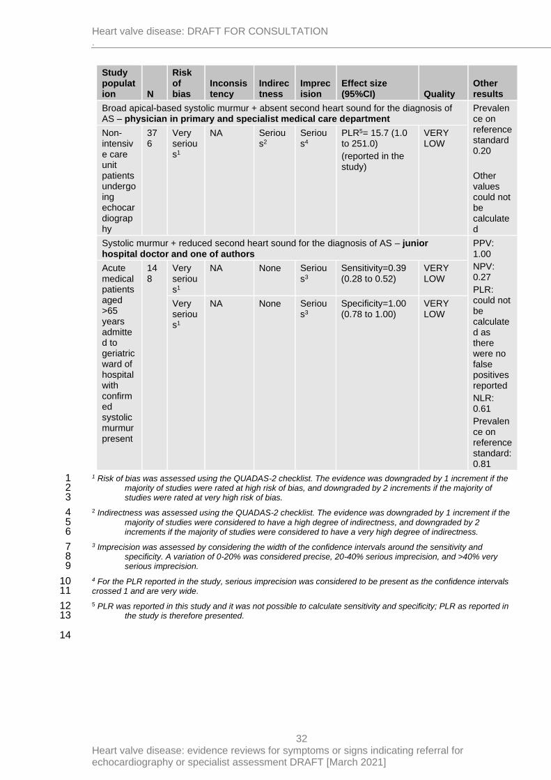
Broad apical-based systolic murmur + absent second heart sound for the diagnosis of AS – physician in primary and specialist medical care department
Prevalence on reference standard 0.20
Other values could not be calculated
Non-intensive care unit patients undergoing echocardiography
376
Very serious1
NA Serious2
Serious4
PLR5= 15.7 (1.0 to 251.0)
(reported in the study)
VERY LOW
Systolic murmur + reduced second heart sound for the diagnosis of AS – junior hospital doctor and one of authors
PPV: 1.00
NPV: 0.27
PLR: could not be calculated as there were no false positives reported
NLR: 0.61
Prevalence on reference standard: 0.81
Acute medical patients aged >65 years admitted to geriatric ward of hospital with confirmed systolic murmur present
148
Very serious1
NA None Serious3
Sensitivity=0.39 (0.28 to 0.52)
VERY LOW
Very serious1
NA None Serious3
Specificity=1.00 (0.78 to 1.00)
VERY LOW
1 Risk of bias was assessed using the QUADAS-2 checklist. The evidence was downgraded by 1 increment if the 1 majority of studies were rated at high risk of bias, and downgraded by 2 increments if the majority of 2 studies were rated at very high risk of bias. 3
2 Indirectness was assessed using the QUADAS-2 checklist. The evidence was downgraded by 1 increment if the 4 majority of studies were considered to have a high degree of indirectness, and downgraded by 2 5 increments if the majority of studies were considered to have a very high degree of indirectness. 6
3 Imprecision was assessed by considering the width of the confidence intervals around the sensitivity and 7 specificity. A variation of 0-20% was considered precise, 20-40% serious imprecision, and >40% very 8 serious imprecision. 9
4 For the PLR reported in the study, serious imprecision was considered to be present as the confidence intervals 10 crossed 1 and are very wide. 11
5 PLR was reported in this study and it was not possible to calculate sensitivity and specificity; PLR as reported in 12 the study is therefore presented. 13
14

Heart valve disease: DRAFT FOR CONSULTATION .
Heart valve disease: evidence reviews for symptoms or signs indicating referral for echocardiography or specialist assessment DRAFT [March 2021]
33
Table 11: Clinical evidence summary: non-flow murmur for heart valve disease in 1 pregnant women 2
Study population N
Risk of bias
Inconsistency
Indirectness
Imprecision
Effect size (95%CI) Quality
Other results
Pathological or possibly pathological murmur (as interpreted by auscultator) for the diagnosis of any valve disease – senior cardiologist
PPV: 0.18
NPV: 1.00
PLR: 5.50
NLR: 0.00
Prevalence on reference standard: 0.04
Pregnant women referred for cardiac opinion
103
Serious1
NA Serious2
Very serious3
Sensitivity=1.00 (0.40 to 1.00)
VERY LOW
Serious1
NA Serious2
None Specificity=0.82 (0.73 to 0.89)
LOW
1 Risk of bias was assessed using the QUADAS-2 checklist. The evidence was downgraded by 1 increment if the 3 majority of studies were rated at high risk of bias, and downgraded by 2 increments if the majority of 4 studies were rated at very high risk of bias. 5
2 Indirectness was assessed using the QUADAS-2 checklist. The evidence was downgraded by 1 increment if the 6 majority of studies were considered to have a high degree of indirectness, and downgraded by 2 7 increments if the majority of studies were considered to have a very high degree of indirectness. 8
3 Imprecision was assessed by considering the width of the confidence intervals around the sensitivity and 9 specificity. A variation of 0-20% was considered precise, 20-40% serious imprecision, and >40% very 10 serious imprecision. 11
12
Table 12: Clinical evidence summary: murmur in pregnant women for heart valve 13 disease (all with murmur to be included) 14
Study population N
Risk of bias
Inconsistency
Indirectness
Imprecision Effect size 1 Quality
Cardiac murmur for the diagnosis of TR – prevalence 0.43 – referring physician
Pregnant women referred for evaluation of murmurs
81 Very serious2
NA Serious3 Could not be assessed
PPV=0.43 VERY LOW
1 In these studies, all patients had to have a murmur to be included in the study. Therefore, sensitivity and 15 specificity could not be calculated, and positive predictive values are instead presented for each 16 study.95% confidence intervals could not be calculated for this effect measure. 17
2 Risk of bias was assessed using the QUADAS-2 checklist. The evidence was downgraded by 1 increment if the 18 majority of studies were rated at high risk of bias, and downgraded by 2 increments if the majority of 19 studies were rated at very high risk of bias. 20
3 Indirectness was assessed using the QUADAS-2 checklist. The evidence was downgraded by 1 increment if the 21 majority of studies were considered to have a high degree of indirectness, and downgraded by 2 22 increments if the majority of studies were considered to have a very high degree of indirectness. 23
24
Heart valve disease: DRAFT FOR CONSULTATION .
Heart valve disease: evidence reviews for symptoms or signs indicating referral for echocardiography or specialist assessment DRAFT [March 2021]
34
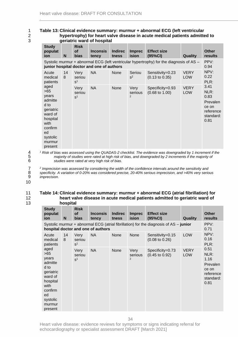
Table 13: Clinical evidence summary: murmur + abnormal ECG (left ventricular 1 hypertrophy) for heart valve disease in acute medical patients admitted to 2 geriatric ward of hospital 3
Study population N
Risk of bias
Inconsistency
Indirectness
Imprecision
Effect size (95%CI) Quality
Other results
Systolic murmur + abnormal ECG (left ventricular hypertrophy) for the diagnosis of AS – junior hospital doctor and one of authors
PPV: 0.94
NPV: 0.22
PLR: 3.41
NLR: 0.83
Prevalence on reference standard: 0.81
Acute medical patients aged >65 years admitted to geriatric ward of hospital with confirmed systolic murmur present
148
Very serious1
NA None Serious2
Sensitivity=0.23 (0.13 to 0.35)
VERY LOW
Very serious1
NA None Very serious2
Specificity=0.93 (0.68 to 1.00)
VERY LOW
1 Risk of bias was assessed using the QUADAS-2 checklist. The evidence was downgraded by 1 increment if the 4 majority of studies were rated at high risk of bias, and downgraded by 2 increments if the majority of 5 studies were rated at very high risk of bias. 6
2 Imprecision was assessed by considering the width of the confidence intervals around the sensitivity and 7 specificity. A variation of 0-20% was considered precise, 20-40% serious imprecision, and >40% very serious 8 imprecision. 9
10
Table 14: Clinical evidence summary: murmur + abnormal ECG (atrial fibrillation) for 11 heart valve disease in acute medical patients admitted to geriatric ward of 12 hospital 13
Study population N
Risk of bias
Inconsistency
Indirectness
Imprecision
Effect size (95%CI) Quality
Other results
Systolic murmur + abnormal ECG (atrial fibrillation) for the diagnosis of AS – junior hospital doctor and one of authors
PPV: 0.71
NPV: 0.16
PLR: 0.51
NLR: 1.16
Prevalence on reference standard: 0.81
Acute medical patients aged >65 years admitted to geriatric ward of hospital with confirmed systolic murmur present
148
Very serious1
NA None None Sensitivity=0.15 (0.08 to 0.26)
LOW
Very serious1
NA None Very serious2
Specificity=0.73 (0.45 to 0.92)
VERY LOW

Heart valve disease: DRAFT FOR CONSULTATION .
Heart valve disease: evidence reviews for symptoms or signs indicating referral for echocardiography or specialist assessment DRAFT [March 2021]
35
1 Risk of bias was assessed using the QUADAS-2 checklist. The evidence was downgraded by 1 increment if the 1 majority of studies were rated at high risk of bias, and downgraded by 2 increments if the majority of 2 studies were rated at very high risk of bias. 3
2 Imprecision was assessed by considering the width of the confidence intervals around the sensitivity and 4 specificity. A variation of 0-20% was considered precise, 20-40% serious imprecision, and >40% very serious 5 imprecision. 6
7
Reference standard – cardiac catheterisation 8
Table 15: Clinical evidence summary: murmur for heart valve disease in various 9 settings in populations with various indications for assessment 10
Studies N Risk of bias
Inconsistency
Indirectness
Imprecision
Effect size (95%CI) Quality
Other results
Systolic murmur on physical examination for the diagnosis of MR – cardiologists PPV: 0.42
NPV: 0.97
PLR: 3.01
NLR: 0.15
Prevalence on reference standard: 0.19
Referred for cardiac catheterisation with known ischaemic heart disease and no previous history of valvular disease
150
Serious1
NA Serious2
Serious3
Sensitivity=0.90 (0.73 to 0.98)
VERY LOW
Serious1
NA Serious2
None Specificity=0.70 (0.61 to 0.78)
LOW
Any murmur for the diagnosis of MR – cardiology attending physician or fellow PPV: 0.21
NPV: 0.88
PLR: 1.74
NLR: 0.91
Prevalence on reference standard: 0.13
Patients with acute myocardial infarction
206
Very serious1
NA Serious2
Serious3
Sensitivity=0.19 (0.06 to 0.38)
VERY LOW
Very serious1
NA Serious2
None Specificity=0.89 (0.84 to 0.93)
VERY LOW
Early diastolic murmur of AR for the diagnosis of AR – attending cardiologist PPV: 0.96
NPV: 0.28
PLR: 3.27
NLR: 0.35
Prevalence on reference standard: 0.88
Patients with suspected aortic regurgitation undergoing aortograms
75 Very serious1
NA None Serious3
Sensitivity=0.73 (0.60 to 0.83)
VERY LOW
Very serious1
NA None Very serious3
Specificity=0.78 (0.40 to 0.97)
VERY LOW

Heart valve disease: DRAFT FOR CONSULTATION .
Heart valve disease: evidence reviews for symptoms or signs indicating referral for echocardiography or specialist assessment DRAFT [March 2021]
36
Studies N Risk of bias
Inconsistency
Indirectness
Imprecision
Effect size (95%CI) Quality
Other results
Apical systolic murmur of MR for the diagnosis of MR – unclear who performed the examination
PPV: 0.93
NPV: 0.75
PLR: 11.79
NLR: 0.28
Prevalence on reference standard: 0.54
Those evaluated by Doppler echocardiography, cardiac auscultation and left ventriculography
35 Very serious1
NA Serious2
Very serious3
Sensitivity=0.74 (0.49 to 0.91)
VERY LOW
Very serious1
NA Serious2
Serious3
Specificity=0.94 (0.70 to 1.00)
VERY LOW
1 Risk of bias was assessed using the QUADAS-2 checklist. The evidence was downgraded by 1 increment if the 1 majority of studies were rated at high risk of bias, and downgraded by 2 increments if the majority of 2 studies were rated at very high risk of bias. 3
2 Indirectness was assessed using the QUADAS-2 checklist. The evidence was downgraded by 1 increment if the 4 majority of studies were considered to have a high degree of indirectness, and downgraded by 2 5 increments if the majority of studies were considered to have a very high degree of indirectness. 6
3 Imprecision was assessed by considering the width of the confidence intervals around the sensitivity and 7 specificity. A variation of 0-20% was considered precise, 20-40% serious imprecision, and >40% very 8 serious imprecision. 9
10
Table 16: Clinical evidence summary: systolic murmur for heart valve disease (all with 11 murmur to be included) 12
Study population N
Risk of bias
Inconsistency
Indirectness
Imprecision Effect size 1 Quality
Systolic murmur for the diagnosis of AS or MR – unclear who performed the examination
Those undergoing right or left heart catheterisation for valvular or coronary heart disease, or both, due to an ill-defined systolic murmur
58 Aortic stenosis – prevalence 0.38
Very serious 2
NA Serious3 Could not be assessed
PPV=0.38 VERY LOW
Mitral regurgitation – prevalence 0.62
Very serious 2
NA Serious3 Could not be assessed
PPV=0.62 VERY LOW
1 In these studies, all patients had to have a murmur to be included in the study. Therefore, sensitivity and 13 specificity could not be calculated, and positive predictive values are instead presented for each 14 study.95% confidence intervals could not be calculated for this effect measure. 15
2 Risk of bias was assessed using the QUADAS-2 checklist. The evidence was downgraded by 1 increment if the 16 majority of studies were rated at high risk of bias, and downgraded by 2 increments if the majority of 17 studies were rated at very high risk of bias. 18

Heart valve disease: DRAFT FOR CONSULTATION .
Heart valve disease: evidence reviews for symptoms or signs indicating referral for echocardiography or specialist assessment DRAFT [March 2021]
37
3 Indirectness was assessed using the QUADAS-2 checklist. The evidence was downgraded by 1 increment if the 1 majority of studies were considered to have a high degree of indirectness, and downgraded by 2 2 increments if the majority of studies were considered to have a very high degree of indirectness. 3
4
2.1.7 Economic evidence 5
2.1.7.1 Included studies 6
No health economic studies were included. 7
2.1.7.2 Excluded studies 8
No relevant health economic studies were excluded due to assessment of limited 9 applicability or methodological limitations. 10
See also the health economic study selection flow chart in Appendix F. 11
2.1.8 Summary of included economic evidence 12
None 13
2.1.9 Economic model 14
This area was not prioritised for new cost-effectiveness analysis. 15
2.1.10 Unit costs 16
Relevant unit costs are provided below to aid consideration of cost effectiveness. 17
Table 17: UK cost of echocardiogram 18
Resource Unit cost Source
Simple Echocardiogram (a) £108 NHS reference Costs 2017/18151
Complex Echocardiogram (b) £196 NHS reference Costs 2017/18151
(a) Cost code RD51A outpatient 19 (b) Cost code EY50Y outpatient 20
21
22

Heart valve disease: DRAFT FOR CONSULTATION .
Heart valve disease: evidence reviews for symptoms or signs indicating referral for echocardiography or specialist assessment DRAFT [March 2021]
38
3 Signs and symptoms indicating referral 1
to a specialist 2
3.1 In adults with suspected heart valve disease, what 3
symptoms and signs indicate direct referral (for example 4
from primary care) to a specialist? 5
3.1.2 Summary of the protocol 6
For full details see the review protocol in Appendix A. 7
Table 18: PICO characteristics of review question 8
Population Adults aged 18 years and over with suspected heart valve disease in any setting (for example, in primary care)
Exclusion:
Children aged less than 18 years.
Adults with congenital heart disease (excluding bicuspid aortic valves).
Tricuspid stenosis and pulmonary valve disease.
Adults presenting with acute heart failure
Target condition Severe heart valve disease: aortic (including bicuspid) stenosis, aortic regurgitation, mitral stenosis, mitral regurgitation or tricuspid regurgitation
Symptoms and signs
Clinical observations:
Cardiac auscultation (standard or electronic):
• Presence of new murmur
• Character of heart sounds:
o no/soft 2nd heart sound (as in severe AS)
o added 3rd sound; gallop rhythm (as in severe MR)
Signs, severe symptoms or simple investigations:
• Shortness of breath (exertional breathlessness, for example classified as NYHA class ≥2)
• Shortness of breath + elevated serum natriuretic peptides (B‑type
natriuretic peptide [BNP] or N‑terminal pro‑B‑type natriuretic peptide
[NT‑proBNP]; for example NT‑proBNP 400-2000 or >2000 ng/litre)
• Peripheral oedema (ie. swelling of ankles and legs)
• Peripheral oedema (ie. swelling of ankles and legs) + BNP or NT
proBNP (for example NT‑proBNP 400-2000 or >2000 ng/litre)
• Pulmonary oedema
• Exertional chest pain (Canadian score class 2+)
• Exertional syncope (fainting)
• Abnormal ECG: for example signs of LV hypertrophy or AF
Include the following combinations:
• murmur alone
• murmur + heart sounds
• murmur + any of the listed symptoms, signs, or investigative findings

Heart valve disease: DRAFT FOR CONSULTATION .
Heart valve disease: evidence reviews for symptoms or signs indicating referral for echocardiography or specialist assessment DRAFT [March 2021]
39
• murmur + heart sounds + any of the listed symptoms, signs, or investigative findings
• murmur + heart failure
(not symptoms alone nor heart sounds alone)
Reference standard
Confirmed diagnosis of severe HVD by transthoracic or transoesophageal echocardiography
Statistical measures
Diagnostic accuracy of symptoms and signs for a confirmed diagnosis of severe HVD.
Measured by:
Primary
measures
Accuracy data
• Sensitivity
• Specificity
• Raw data to calculate 2x2 tables to calculate sensitivity and specificity (number of true positives, true negatives, false positives and false negatives).
Secondary measures
• Likelihood ratios
• Positive Predictive Value (PPV)
• Negative Predictive Value (NPV)
• Receiver Operating Characteristic (ROC) curve or area under curve for BNP and NT pro-BNP
If insufficient accuracy data are found, diagnostic association of signs and symptoms with a confirmed diagnosis of severe HVD will be included.
Measured by:
Association data
• Adjusted RR or OR
For decision-making, it was agreed that sensitivity should be the primary measure taken into account as avoiding false negatives was considered to be the priority over avoiding false positives to avoid sending many people away early without further testing.
Agreed a threshold of ≥60% to represent suitable sensitivity to consider recommending a test, with emphasis on importance of follow-up on those with continuing symptoms or concerns.
Study design • Single-gate diagnostic studies (these may be called cohort studies or cross-sectional studies) will be included preferentially
• If no/insufficient diagnostic accuracy studies are identified prospective and retrospective cohort studies with multivariate analysis of the association between signs and symptoms and a confirmed diagnosis of severe heart valve disease will be included.
Confounding factors (if diagnostic association studies are included):
• Age (<65 years or ≥65 years)

Heart valve disease: DRAFT FOR CONSULTATION .
Heart valve disease: evidence reviews for symptoms or signs indicating referral for echocardiography or specialist assessment DRAFT [March 2021]
40
• Type of murmur:
o Innocent murmur
o Ejection systolic murmur
o Regurgitant systolic murmur
o Diastolic murmur
• Presence/absence of atrial fibrillation
3.1.3 Methods and process 1
This evidence review was developed using the methods and process described in 2 Developing NICE guidelines: the manual. Methods specific to this review question are 3 described in the review protocol in appendix A and the methods document. 4
Declarations of interest were recorded according to NICE’s conflicts of interest policy. 5
3.1.4 Diagnostic evidence 6
3.1.4.1 Included studies 7
A search was conducted for cross-sectional and prospective and retrospective cohort studies 8 assessing the diagnostic test accuracy of murmur with or without other signs or symptoms 9 (heart sounds and/or symptoms) to identify whether the condition is present (as indicated by 10 the reference standard) in people under investigation for condition severe heart valve 11 disease. 12
Diagnostic association studies that report data on the association between murmur with or 13 without other signs or symptoms (heart sounds and/or symptoms) and diagnosis of severe 14 heart valve disease were also considered if limited diagnostic accuracy studies were 15 available. 16
Nineteen studies with diagnostic accuracy data or data that could be used to calculate 17 diagnostic accuracy data were included in the review; 3, 5, 16-19, 51, 63, 89, 109, 117, 121, 131, 132, 134, 163, 18 171, 174, 191 these are summarised in Table 19 below. Most of the studies investigated the 19 accuracy of murmur alone for the diagnosis of severe heart valve disease, with the definition 20 of the murmur and person conducting auscultation differing between studies. However, two 21 studies174, 191 looked at murmur plus symptoms, three studies3, 132, 174assessed murmur plus 22 an absent or reduced second heart sound, and one study174 looked at murmur plus abnormal 23 ECG. 24
One of these studies19 also provided diagnostic association data for a particular index test 25 (murmur + diminished aortic closure sound) for the diagnosis of moderate or severe aortic 26 stenosis. 27
Evidence from these studies is summarised in the clinical evidence summary below in Tables 28 20-32 below. 29
The assessment of the evidence quality was conducted with emphasis on test sensitivity, as 30 this was identified by the committee as the primary measure in guiding decision-making as 31 the priority would be to avoid missing cases (false negatives) and not sending them for 32 further testing as a result. The committee set clinical decision thresholds as sensitivity = 0.60. 33
34
Reference standards 35
Only studies that had used echocardiography as a reference standard for confirming valve 36 disease were included in this review as the older method of confirming valve disease 37 (cardiac catheterisation) is not as good at assessing the severity of heart valve disease as 38

Heart valve disease: DRAFT FOR CONSULTATION .
Heart valve disease: evidence reviews for symptoms or signs indicating referral for echocardiography or specialist assessment DRAFT [March 2021]
41
the current gold standard of echocardiography is, and this review focuses on the diagnosis of 1 severe heart valve disease, which may differ between cardiac catheterisation and 2 echocardiography. 3
4
Populations 5
Studies that involved screening for heart valve disease and murmurs in presumably healthy 6 populations where there could be no reason for a suspicion of heart valve disease were 7 excluded, for example, where screening was performed for everyone who experienced a hip 8 fracture or in populations that were said to be healthy. However, studies where there was not 9 necessarily a suspicion of heart valve disease but had some indication for either attendance 10 at hospital or primary care, echocardiography or were experiencing cardiac symptoms were 11 included, as there was limited evidence where the populations were defined as specifically 12 being suspected of having heart valve disease. 13
Studies where the presence of a murmur was required for a participant to be included in a 14 study were also included, despite the fact that this would mean all were already known to be 15 index test positive before enrolment. Limited diagnostic accuracy data can be obtained from 16 these studies, but it was agreed to include these given that murmur would be one of the main 17 reasons for suspicion of heart valve disease and these studies could still provide information 18 on the proportion of those with the murmur that actually had reference standard confirmed 19 valve disease, in the form of the positive predictive ratio. The limitations of these studies 20 were highlighted. 21
22
See also the study selection flow chart in Appendix C, sensitivity and specificity forest plots in 23 Appendix E, and study evidence tables in Appendix D. 24
3.1.4.2 Excluded studies 25
See the excluded studies list in Appendix I. 26
3.1.5 Summary of studies included in the diagnostic evidence 27
Table 19: Summary of studies included in the evidence review 28
Study Population Target condition Index test
Reference standard Comments
Abe 20133
n=130
Japan
Patients with systolic ejection murmurs with grade ≥2 or known aortic stenosis and referred for echocardiography
Heart valve disease: severe aortic stenosis
Systolic ejection murmur + diminished second heart sound
Echocardiography confirmed aortic stenosis
(subgroup for severe AS)
Population indirectness: 35% had known AS before study so may affect accuracy data
Murmur + diminished heart sound: not clear if all had a murmur, but at least 65% did – could include some that just had a diminished

Heart valve disease: DRAFT FOR CONSULTATION .
Heart valve disease: evidence reviews for symptoms or signs indicating referral for echocardiography or specialist assessment DRAFT [March 2021]
42
Study Population Target condition Index test
Reference standard Comments
second heart sound
Aggarwal 20145
n=100
India
Outpatients presenting for echocardiography at Cardiology centre
Heart valve disease: significant valve lesion
Detection of murmur using stethoscope and specific software
Systolic or diastolic murmurs
Echocardiography confirmed heart valve disease
(subgroup for significant valve lesions)
Patients known to have pre-existing heart murmurs excluded
ZargisCardioscan™ software used
Target condition indirectness: significant lesion defined as any stenotic lesion and moderate-severe regurgitant lesions
Aronow 198717
n=75
USA
Unselected elderly patients in a long-term health care facility with echocardiography of aortic valve performed and aortic systolic ejection murmurs
Heart valve disease: severe aortic stenosis
Aortic systolic ejection murmurs (all had one to be included in the study)
Echocardiography confirmed aortic stenosis
(subgroup for severe aortic stenosis)
Aronow 198916
n=450
USA
Unselected elderly patients in a long-term health care facility with echocardiography of aortic valve performed
Heart valve disease: moderate or severe aortic regurgitation
Murmur of aortic regurgitation
High frequency diastolic decrescendo murmur beginning with A2
Echocardiography confirmed moderate or severe aortic regurgitation
Potentially indirect population: unselected elderly patients in a long-term health care facility – not necessarily suspected HVD
Aronow 199118
n=781
USA
Unselected elderly patients in long term health care facility
Heart valve disease: severe aortic stenosis
Aortic systolic ejection murmur
Echocardiography confirmed AS
(subgroup for severe disease)
Attenhofer Jost, 200019
n=100
Those referred for echocardiography due to systolic murmur of unknown cause -
Heart valve disease: moderate or severe aortic stenosis or
Systolic murmur (all had one)
Echocardiography confirmed AS or valvular regurgitation (AR, TR, MR)
Reports separately for each type of HVD and not possible to combine into

Heart valve disease: DRAFT FOR CONSULTATION .
Heart valve disease: evidence reviews for symptoms or signs indicating referral for echocardiography or specialist assessment DRAFT [March 2021]
43
Study Population Target condition Index test
Reference standard Comments
Switzerland
no prior echo examination
valvular regurgitation (AR, TR, MR)
Systolic murmur +diminished aortic closure sound (diagnostic association)
(subgroup for moderate or severe disease)
one group for severe disease.
Decoodt 199051
n=100
Belgium
Those with mitral valve prolapse confirmed by echocardiography
Heart valve disease: severe mitral regurgitation
Systolic murmur on auscultation (early systolic, late systolic or holosystolic)
Echocardiography confirmed mitral regurgitation
(subgroup for severe mitral regurgitation)
Potential population indirectness: selected population that is more likely to have higher incidence of mitral regurgitation as they already have echo-confirmed mitral valve abnormality?
Etchells 199863
n=162
Canada
Hospital inpatients referred for echocardiography. Most had cardiac symptoms.
Heart valve disease: moderate or severe aortic stenosis
Systolic murmur
Echocardiography confirmed moderate or severe aortic stenosis
Results reported separately in the study for internist rather than cardiologist as fits better with setting of review
Target condition indirectness: some had moderate rather than severe AS
Iversen 200889
n=2977
Denmark
All patients admitted to medical or surgical departments of a hospital
Heart valve disease (moderate or severe aortic stenosis, aortic regurgitation, mitral stenosis or mitral regurgitation)
Heart murmur Echocardiography confirmed moderate or severe valve disease
Potential population indirectness: those admitted to a hospital, not necessarily suspected HVD but obviously some indication for them being in hospital
Target condition indirectness: moderate or severe combined so

Heart valve disease: DRAFT FOR CONSULTATION .
Heart valve disease: evidence reviews for symptoms or signs indicating referral for echocardiography or specialist assessment DRAFT [March 2021]
44
Study Population Target condition Index test
Reference standard Comments
not all severe disease
Labovitz 1985109
n=51
USA
Patients with mitral annular calcium detected on echocardiography
Heart valve disease: moderate or severe mitral regurgitation
Apical systolic murmur
Echocardiography confirmed moderate or severe mitral regurgitation
Potential population indirectness: selected population with likely increased incidence of disease as already had echocardiography performed?
Target condition indirectness: moderate or severe combined so not all severe disease
Limacher 1985117
n=81
USA
Pregnant women referred for evaluation of murmurs
Heart valve disease: severe tricuspid regurgitation
Cardiac murmur (all had one to be included in the study)
Echocardiography confirmed tricuspid regurgitation
(data given for severe TR)
Loperfido 1986121
n=72
Italy
Patients diagnosed with myocardial infarction 1-3 months prior to the study at the coronary care unit based on chest pain, ECG and increase and decrease of creatine kinase-MB fraction
Heart valve disease: grade 3+ mitral regurgitation
Systolic murmur
Echocardiography confirmed mitral regurgitation
(subgroup for grade 3+ MR)
Potential population indirectness: not necessarily suspected HVD but all have had MI with cardiac symptoms
McClelland 2020131
n=350
USA
Consecutive patients ≥18 years referred for initial transthoracic echocardiography imaging with a heart murmur at single centre
Heart valve disease: severe heart valve disease
Unclear which types of valve disease were included under severe valve
Heart murmur – no further details
Echocardiography confirmed severe valve disease

Heart valve disease: DRAFT FOR CONSULTATION .
Heart valve disease: evidence reviews for symptoms or signs indicating referral for echocardiography or specialist assessment DRAFT [March 2021]
45
Study Population Target condition Index test
Reference standard Comments
disease, but report states moderate or severe cases of aortic regurgitation, mitral regurgitation, tricuspid regurgitation and aortic stenosis were identified.
McGee 2010132
n=376
USA
Non-intensive care unit patients undergoing echocardiography - around 16% already known to have valve disease
Heart valve disease: aortic stenosis (severe, Vmax ≥4.0 m/sec), moderate or severe mitral regurgitation or tricuspid regurgitation
Systolic heart murmur
Broad apical-based systolic murmur + absence second heart sound
Echocardiography confirmed aortic stenosis, mitral regurgitation or tricuspid regurgitation
(subgroup for severe can be obtained for AS, but moderate and severe cases combined for MR and TR)
Potential population indirectness: some already known to have valve disease
Study reports details separately for different types of valve disease and not possible to combine
Target condition indirectness:
For MR and TR, moderate and severe cases are combined, so not all had severe disease.
McKillop 1991134
n=35
UK
Elderly patients with systolic ejection murmurs
Heart valve disease: significant aortic stenosis (defined as gradient >30 mmHg) or mitral regurgitation (no definition provided for
Systolic ejection murmur (all had one to be included in the study)
Echocardiography confirmed significant aortic stenosis (defined as gradient >30 mmHg) or mitral regurgitation (no definition provided for significant disease)
Target condition indirectness: for AS, gradient of >30 mmHg doesn’t necessarily indicate severe disease, and could include some moderate cases? No definition of significant

Heart valve disease: DRAFT FOR CONSULTATION .
Heart valve disease: evidence reviews for symptoms or signs indicating referral for echocardiography or specialist assessment DRAFT [March 2021]
46
Study Population Target condition Index test
Reference standard Comments
significant disease)
disease given for MR.
Panidis 1986163
n=80
USA
Those with mitral valve prolapse confirmed on echocardiography and signs and symptoms
Heart valve disease: moderate or severe mitral regurgitation
Systolic murmur
Echocardiography confirmed mitral regurgitation
(data available for moderate or severe disease)
Potential population indirectness: selected population that is more likely to have higher incidence of mitral regurgitation as they already have echo-confirmed mitral valve abnormality?
Rahko 1989171
n=408
USA
Patients presenting to echocardiography laboratory
Heart valve disease: 3+ or 4+ (moderate-severe or severe) aortic regurgitation, mitral regurgitation or tricuspid regurgitation
Regurgitant murmur on auscultation
Echocardiography confirmed aortic regurgitation, mitral regurgitation or tricuspid regurgitation
(data available for 3+ or 4+ regurgitation - moderate-severe or severe)
Potential population indirectness: not necessarily suspected HVD but some indication for echocardiography
Study reports data for each type of regurgitation separately and not possible to combine as single ‘regurgitation’ group
Reardon 1996174
n=148
UK
Acute medical patients aged >65 years admitted to geriatric ward of hospital – data reported only for those with systolic murmurs
Heart valve disease: significant (gradient >30 mmHg) aortic stenosis
Systolic murmur (all had one to be included in the analysis)
Systolic murmur + reduced second heart sound
Systolic murmur + symptoms (syncope)
Systolic murmur + symptoms (angina)
Echocardiography confirmed aortic stenosis
(subgroup for significant AS – gradient >30 mmHg)
Potential population indirectness: not necessarily suspected HVD but some indications to be admitted to hospital
Target condition indirectness: gradient of >30 mmHg doesn’t necessarily indicate severe disease, and could include

Heart valve disease: DRAFT FOR CONSULTATION .
Heart valve disease: evidence reviews for symptoms or signs indicating referral for echocardiography or specialist assessment DRAFT [March 2021]
47
Study Population Target condition Index test
Reference standard Comments
Systolic murmur + symptoms (dyspnoea)
Systolic murmur + abnormal ECG (left ventricular hypertrophy)
Systolic murmur + abnormal ECG (atrial fibrillation)
some moderate cases?
Sarasin 2002191
n=20
Switzerland
Patients >18 years presenting with syncope in emergency department with systolic murmur
Small subgroup of those with suspected AS due to presence of murmur + syncope on exertion with/without chest pain (n=20)
Heart valve disease: severe aortic stenosis
Systolic murmur + syncope on exertion with/without chest pain (all of those within the subgroup had this as an indicator)
Echocardiography confirmed severe aortic stenosis
See Appendix D for full evidence tables. 1
2
3.1.6 Summary of the diagnostic evidence 3
The populations, target conditions and index tests used across the included studies were 4 considered to be very broad and wide-ranging, and therefore no studies were pooled into a 5 diagnostic meta-analysis. Sensitivity and specificity for each individual study is given below, 6 separated into broad categories based on the population. 7
For studies where all of those included had to be positive for murmur with/without another 8 characteristic (which was used as an index test in our review), sensitivity and specificity, as 9 well as other measures, could not be calculated, and positive predictive values are instead 10 presented. 11
Note that although all included studies detected heart valve disease as the target condition, 12 the type of heart valve disease that was included in the studies varied. For example, some 13 studies aimed to diagnose and report any type of valve disease (including aortic stenosis, 14 aortic regurgitation, mitral stenosis, mitral regurgitation and tricuspid regurgitation), while 15 others focused specifically on one or two types of valve disease, such as aortic stenosis or 16

Heart valve disease: DRAFT FOR CONSULTATION .
Heart valve disease: evidence reviews for symptoms or signs indicating referral for echocardiography or specialist assessment DRAFT [March 2021]
48
mitral stenosis and mitral regurgitation, or any type of regurgitation but not stenosis (i.e. 1 aortic regurgitation, mitral regurgitation and tricuspid regurgitation). Where possible, results 2 have been calculated for ‘any valve disease’; however, in many cases results are reported 3 separately for each type of valve disease as it was not possible to determine how many may 4 have had more than one time of valve disease at the same time to calculate diagnostic 5 accuracy results for overall heart valve disease in each study. 6
The assessment of the evidence quality was conducted with emphasis on test sensitivity and 7 specificity as this was identified by the committee as the primary measure in guiding 8 decision-making. The committee set a clinical decision threshold of 0.6 for sensitivity. 9
10
Table 20: Clinical evidence summary: murmur for moderate or severe heart valve 11 disease in various settings in populations with various indications for 12 assessment 13
Study population N
Risk of bias
Inconsistency
Indirectness
Imprecision
Effect size (95%CI) Quality
Other results
Detection of systolic or diastolic murmur on stethoscope with specific software for the diagnosis of significant valve disease – community medicine physician
Outpatients presenting for echocardiography at Cardiology centre
100
Serious1
NA Serious2
Very serious3
Sensitivity=0.64 (0.35 to 0.87)
VERY LOW
PPV: 0.29
NPV: 0.93
PLR: 2.51
NLR: 0.48
Prevalence on reference standard: 0.14
Serious1
NA Serious2
None Specificity=0.74 (0.64 to 0.83)
LOW
Murmur of AR (high frequency diastolic decrescendo murmur beginning with A2) for the diagnosis of moderate or severe AR – experienced cardiologist
Unselected elderly patients in a long-term health care facility with echocardiography
450
Very serious1
NA Very serious2
None Sensitivity=0.95 (0.87 to 0.99)
VERY LOW
PPV: 0.62
NPV: 0.99
PLR: 8.27
NLR: 0.06
Prevalence on reference standard: 0.16
Very serious1
NA Very serious2
None Specificity=0.89 (0.85 to 0.92)
VERY LOW
Aortic systolic ejection murmur for the diagnosis of severe AS – experienced cardiologist
Unselected elderly patients in long
781
Very serious1
NA Serious2
None Sensitivity=1.00 (0.82 to 1.00)
VERY LOW
Prevalence on reference standard: 0.02
NA NA NA NA Specificity could not be calculated
NA

Heart valve disease: DRAFT FOR CONSULTATION .
Heart valve disease: evidence reviews for symptoms or signs indicating referral for echocardiography or specialist assessment DRAFT [March 2021]
49
Study population N
Risk of bias
Inconsistency
Indirectness
Imprecision
Effect size (95%CI) Quality
Other results
term health care facility
as insufficient data provided
Other values could not be calculated
Systolic murmur for the diagnosis of moderate or severe AS – third year resident/staff general internist
Hospital inpatients referred for echocardiography. Most had cardiac symptoms.
112
Serious1
NA Serious2
Serious3
Sensitivity=1.00 (0.77 to 1.00)
VERY LOW
PPV: 0.20
NPV: 1.00
PLR reported in the study (95% CI): 1.60 (1.20, 2.00)
NLR reported in the study (95% CI): 0.00 (0.00, 0.71)
Prevalence on reference standard: 0.13
Serious1
NA Serious2
Serious3
Specificity=0.43 (0.33 to 0.53)
VERY LOW
Heart murmur for the diagnosis of moderate or severe AS, AR, MS or MR – unclear who performed examination
All patients admitted to medical or surgical departments of a hospital
2977
Serious1
NA Very serious2
None Sensitivity=0.81 (0.73 to 0.87)
VERY LOW
PPV: 0.18
NPV: 0.99
PLR: 4.30
NLR: 0.24
Prevalence on reference standard: 0.05
Serious1
NA Very serious2
None Specificity=0.81 (0.80 to 0.83)
VERY LOW
Systolic murmur for the diagnosis of grade 3+ MR – cardiologist (performed at coronary care unit)
Patients diagnosed with
72 Serious1
NA Serious2
Very serious3
Sensitivity=0.50 (0.07 to 0.53)
VERY LOW
PPV: 0.13

Heart valve disease: DRAFT FOR CONSULTATION .
Heart valve disease: evidence reviews for symptoms or signs indicating referral for echocardiography or specialist assessment DRAFT [March 2021]
50
Study population N
Risk of bias
Inconsistency
Indirectness
Imprecision
Effect size (95%CI) Quality
Other results
myocardial infarction 1-3 months prior to the study at the coronary care
Serious1
NA Serious2
Serious3
Specificity=0.79 (0.68 to 0.88)
VERY LOW
NPV: 0.96
PLR: 2.43
NLR: 0.63
Prevalence on reference standard: 0.06
Systolic heart murmur for the diagnosis of severe AS, or moderate or severe MR or TR – physician in primary and specialist medical care department
Non-intensive care unit patients undergoing echocardiography
376
Severe aortic stenosis PPV: 0.12
NPV: 1.0
PLR: 1.79
NLR: could not be calculated
Prevalence on reference standard: 0.07
Very serious1
NA Serious2
None Sensitivity=1.00 (0.87 to 1.00)
VERY LOW
Very serious1
NA Serious2
None Specificity=0.44 (0.39 to 0.49)
VERY LOW
Moderate or severe mitral regurgitation PPV: 0.27
NPV: 0.91
PLR: 1.52
NLR: 0.41
Prevalence on reference standard: 0.20
Very serious1
NA Very serious2
None Sensitivity=0.81 (0.70 to 0.89)
VERY LOW
Very serious1
NA Very serious2
None Specificity=0.47 (0.41 to 0.52)
VERY LOW
Moderate or severe tricuspid regurgitation PPV: 0.21
NPV: 0.88
PLR: 1.29
NLR: 0.63
Prevalence on reference standard: 0.17
Very serious1
NA Very serious2
Serious3
Sensitivity=0.72 (0.60 to 0.83)
VERY LOW
Very serious1
NA Very serious2
None Specificity=0.44 (0.38 to 0.50)
VERY LOW

Heart valve disease: DRAFT FOR CONSULTATION .
Heart valve disease: evidence reviews for symptoms or signs indicating referral for echocardiography or specialist assessment DRAFT [March 2021]
51
Study population N
Risk of bias
Inconsistency
Indirectness
Imprecision
Effect size (95%CI) Quality
Other results
Regurgitant murmur on auscultation for the diagnosis of 3+ or 4+ (moderate-severe or severe) AR, MR or TR – cardiologist
Patients presenting to an echocardiography laboratory
408
3+ or 4+ aortic regurgitation PPV: 0.34
NPV: 0.99
PLR: 5.90
NLR: 0.11
Prevalence on reference standard: 0.08
Very serious1
NA Serious2
Serious3
Sensitivity=0.91 (0.76 to 0.98)
VERY LOW
Very serious1
NA Serious2
None Specificity=0.85 (0.81 to 0.88)
VERY LOW
3+ or 4+ mitral regurgitation PPV: 0.28
NPV: 0.98
PLR: 3.49
NLR: 0.20
Prevalence on reference standard: 0.10
Very serious1
NA Serious2
Serious3
Sensitivity=0.85 (0.69 to 0.94)
VERY LOW
Very serious1
NA Serious2
None Specificity=0.76 (0.71 to 0.80)
VERY LOW
3+ or 4+ tricuspid regurgitation PPV: 0.42
NPV: 0.97
PLR: 10.15
NLR: 0.41
Prevalence on reference standard: 0.07
Very serious1
NA Serious2
Very serious3
Sensitivity=0.62 (0.38 to 0.82)
VERY LOW
Very serious1
NA Serious2
None Specificity=0.94 (0.91 to 0.96)
VERY LOW
1 Risk of bias was assessed using the QUADAS-2 checklist. The evidence was downgraded by 1 increment if the 1 majority of studies were rated at high risk of bias, and downgraded by 2 increments if the majority of 2 studies were rated at very high risk of bias. 3
2 Indirectness was assessed using the QUADAS-2 checklist. The evidence was downgraded by 1 increment if the 4 majority of studies were considered to have a high degree of indirectness, and downgraded by 2 5 increments if the majority of studies were considered to have a very high degree of indirectness. 6
3 Imprecision was assessed by considering the width of the confidence intervals around the sensitivity and 7 specificity. A variation of 0-20% was considered precise, 20-40% serious imprecision, and >40% very 8 serious imprecision. 9
10

Heart valve disease: DRAFT FOR CONSULTATION .
Heart valve disease: evidence reviews for symptoms or signs indicating referral for echocardiography or specialist assessment DRAFT [March 2021]
52
Table 21: Clinical evidence summary: murmur for moderate or severe heart valve 1 disease in populations with MVP that has already been diagnosed by 2 echocardiography 3
Study population N
Risk of bias
Inconsistency
Indirectness
Imprecision
Effect size (95%CI) Quality
Other results
Systolic murmur on auscultation (including early systolic, late systolic or holosystolic) for the diagnosis of severe MR – unclear who did examination
PPV: 0.19
NPV: 1.0
PLR: 2.14
NLR: could not be calculated
Prevalence on reference standard: 0.10
Those with mitral valve prolapse confirmed by echocardiography
100
Very serious1
NA Serious2
Serious3
Sensitivity=1.00 (0.69 to 1.00)
VERY LOW
Very serious1
NA Serious2
None Specificity=0.53 (0.43 to 0.64)
VERY LOW
Systolic murmur for the diagnosis of moderate or severe MR – unclear who did examination
PPV: 0.19
NPV: 1.00
PLR: 2.06
Prevalence on reference standard: 0.10
Those with mitral valve prolapse confirmed on echocardiography and signs and symptoms
80 Serious1
NA Serious2
Serious3
Sensitivity=1.00 (0.63 to 1.00)
VERY LOW
Serious1
NA Serious2
Serious3
Specificity=0.51 (0.39 to 0.63)
VERY LOW
1 Risk of bias was assessed using the QUADAS-2 checklist. The evidence was downgraded by 1 increment if the 4 majority of studies were rated at high risk of bias, and downgraded by 2 increments if the majority of 5 studies were rated at very high risk of bias. 6
2 Indirectness was assessed using the QUADAS-2 checklist. The evidence was downgraded by 1 increment if the 7 majority of studies were considered to have a high degree of indirectness, and downgraded by 2 8 increments if the majority of studies were considered to have a very high degree of indirectness. 9
3 Imprecision was assessed by considering the width of the confidence intervals around the sensitivity and 10 specificity. A variation of 0-20% was considered precise, 20-40% serious imprecision, and >40% very 11 serious imprecision. 12
13
Table 22: Clinical evidence summary: murmur for moderate or severe heart valve 14 disease in a population with mitral annular calcium observed by 15 echocardiography 16
Studies N Risk of bias
Inconsistency
Indirectness
Imprecision
Effect size (95%CI) Quality
Other results
Apical systolic murmur for the diagnosis of moderate or severe MR – unclear who did examination
PPV: 0.39

Heart valve disease: DRAFT FOR CONSULTATION .
Heart valve disease: evidence reviews for symptoms or signs indicating referral for echocardiography or specialist assessment DRAFT [March 2021]
53
Studies N Risk of bias
Inconsistency
Indirectness
Imprecision
Effect size (95%CI) Quality
Other results
Patients with mitral annular calcium detected on echocardiography
51 Very serious1
NA Very serious2
Very serious3
Sensitivity=0.65 (0.38 to 0.86)
VERY LOW
NPV: 0.74
PLR: 1.29
NLR: 0.71
Prevalence on reference standard: 0.33
Very serious1
NA Very serious2
Serious3
Specificity=0.50 (0.32 to 0.68)
VERY LOW
1 Risk of bias was assessed using the QUADAS-2 checklist. The evidence was downgraded by 1 increment if the 1 majority of studies were rated at high risk of bias, and downgraded by 2 increments if the majority of 2 studies were rated at very high risk of bias. 3
2 Indirectness was assessed using the QUADAS-2 checklist. The evidence was downgraded by 1 increment if the 4 majority of studies were considered to have a high degree of indirectness, and downgraded by 2 5 increments if the majority of studies were considered to have a very high degree of indirectness. 6
3 Imprecision was assessed by considering the width of the confidence intervals around the sensitivity and 7 specificity. A variation of 0-20% was considered precise, 20-40% serious imprecision, and >40% very 8 serious imprecision. 9
10
Table 23: Clinical evidence summary: murmur for heart valve disease (all had a 11 murmur to be included in the study) 12
Study population N
Risk of bias
Inconsistency
Indirectness
Imprecision Effect size 1 Quality
Aortic systolic ejection murmur for the diagnosis of severe AS – prevalence 0.05 – experienced cardiologist
Unselected elderly patients in a long-term health care facility with echocardiography
75 Serious2 NA Serious3 Could not be assessed
PPV=0.05 VERY LOW
Systolic murmur for the diagnosis of moderate or severe AS or valvular regurgitation (AR, TR, MR) – cardiologist
Those referred for echocardiography due to systolic murmur of unknown cause - no prior
100 Moderate or severe aortic stenosis – prevalence 0.15
Serious2
NA Very serious3
Could not be assessed
PPV=0.15 VERY LOW
Moderate or severe aortic regurgitation – prevalence 0.06
Serious2
NA Very serious3
Could not be assessed
PPV=0.06 VERY LOW
Moderate or severe mitral regurgitation – prevalence 0.06

Heart valve disease: DRAFT FOR CONSULTATION .
Heart valve disease: evidence reviews for symptoms or signs indicating referral for echocardiography or specialist assessment DRAFT [March 2021]
54
Study population N
Risk of bias
Inconsistency
Indirectness
Imprecision Effect size 1 Quality
echo examination
Serious2
NA Very serious3
Could not be assessed
PPV=0.06 VERY LOW
Moderate or severe tricuspid regurgitation – prevalence 0.02
Serious2
NA Very serious3
Could not be assessed
PPV=0.02 VERY LOW
Systolic ejection murmur for the diagnosis of significant AS (gradient >30 mmHg) or mitral regurgitation (no definition provided for significant MR) – prevalence 0.37 – cardiologist and geriatrician
Elderly patients with systolic ejection murmurs
35 Very serious2
NA Very serious3
Could not be assessed
PPV=0.37 VERY LOW
Systolic murmur for the diagnosis of significant AS (gradient >30 mmHg) – prevalence 0.26 – junior hospital doctor and one of authors
Acute medical patients aged >65 years admitted to geriatric ward of hospital
148 Very serious2
NA Very serious3
Could not be assessed
PPV=0.26 VERY LOW
Heart murmur for the diagnosis of severe heart valve disease – prevalence 0.04 – unclear who assessed murmur
Referred for initial transthoracic echocardiography imaging with a heart murmur at single centre.
350 Very serious2
NA Very serious3
Could not be assessed
PPV=0.04 VERY LOW
1 In these studies, all patients had to have a murmur to be included in the study. Therefore, sensitivity and 1 specificity could not be calculated, and positive predictive values are instead presented for each 2 study.95% confidence intervals could not be calculated for this effect measure. 3
2 Risk of bias was assessed using the QUADAS-2 checklist. The evidence was downgraded by 1 increment if the 4 majority of studies were rated at high risk of bias, and downgraded by 2 increments if the majority of 5 studies were rated at very high risk of bias. 6
3 Indirectness was assessed using the QUADAS-2 checklist. The evidence was downgraded by 1 increment if the 7 majority of studies were considered to have a high degree of indirectness, and downgraded by 2 8 increments if the majority of studies were considered to have a very high degree of indirectness. 9
10

Heart valve disease: DRAFT FOR CONSULTATION .
Heart valve disease: evidence reviews for symptoms or signs indicating referral for echocardiography or specialist assessment DRAFT [March 2021]
55
Table 24: Clinical evidence summary: murmur in pregnant women for heart valve 1 disease (all with murmur to be included) 2
Study population N
Risk of bias
Inconsistency
Indirectness
Imprecision Effect size 1 Quality
Cardiac murmur for the diagnosis of severe TR – prevalence 0.0 – referring physician
Pregnant women referred for evaluation of murmurs
81 Very serious2
NA Very serious3
Could not be assessed
In all 81 patients with a murmur, none of them had severe TR.
VERY LOW
1 In this study, all patients had to have a murmur to be included in the study. Therefore, sensitivity and specificity 3 could not be calculated. PPV could also not be calculated as all patients in the study were negative for 4 severe TR. 5
2 Risk of bias was assessed using the QUADAS-2 checklist. The evidence was downgraded by 1 increment if the 6 majority of studies were rated at high risk of bias, and downgraded by 2 increments if the majority of 7 studies were rated at very high risk of bias. 8
3 Indirectness was assessed using the QUADAS-2 checklist. The evidence was downgraded by 1 increment if the 9 majority of studies were considered to have a high degree of indirectness, and downgraded by 2 10 increments if the majority of studies were considered to have a very high degree of indirectness. 11
12
13
Table 25: Clinical evidence summary: murmur + syncope for significant heart valve 14 disease in acute medical patients admitted to geriatric ward of hospital 15
Studies N Risk of bias
Inconsistency
Indirectness
Imprecision
Effect size (95%CI) Quality
Other results
Systolic murmur + symptoms (syncope) for the diagnosis of significant (gradient >30 mmHg) AS – junior hospital doctor and one of authors
PPV: 0.60
NPV: 0.76
PLR: 4.29
NLR: 0.89
Prevalence on reference standard: 0.26
Acute medical patients aged >65 years admitted to geriatric ward of hospital with confirmed systolic murmur present
148
Very serious1
NA Serious2
Serious3
Sensitivity=0.14 (0.03 to 0.36)
VERY LOW
Very serious1
NA Serious2
None Specificity=0.97 (0.88 to 1.00)
VERY LOW
1 Risk of bias was assessed using the QUADAS-2 checklist. The evidence was downgraded by 1 increment if the 16 majority of studies were rated at high risk of bias, and downgraded by 2 increments if the majority of 17 studies were rated at very high risk of bias. 18
2 Indirectness was assessed using the QUADAS-2 checklist. The evidence was downgraded by 1 increment if the 19 majority of studies were considered to have a high degree of indirectness, and downgraded by 2 20 increments if the majority of studies were considered to have a very high degree of indirectness. 21

Heart valve disease: DRAFT FOR CONSULTATION .
Heart valve disease: evidence reviews for symptoms or signs indicating referral for echocardiography or specialist assessment DRAFT [March 2021]
56
3 Imprecision was assessed by considering the width of the confidence intervals around the sensitivity and 1 specificity. A variation of 0-20% was considered precise, 20-40% serious imprecision, and >40% very 2 serious imprecision. 3
4
Table 26: Clinical evidence summary: murmur + dyspnoea for significant heart valve 5 disease in acute medical patients admitted to geriatric ward of hospital 6
Studies N Risk of bias
Inconsistency
Indirectness
Imprecision
Effect size (95%CI) Quality
Systolic murmur + symptoms (dyspnoea) for the diagnosis of significant (gradient >30 mmHg) AS – junior hospital doctor and one of authors
PPV: 0.50
NPV: 0.81
PLR: 2.86
NLR: 0.67
Prevalence on reference standard: 0.26
Acute medical patients aged >65 years admitted to geriatric ward of hospital with confirmed systolic murmur present
148
Very serious1
NA Serious2
Very serious3
Sensitivity=0.43 (0.22 to 0.66)
VERY LOW
Very serious1
NA Serious2
Serious3
Specificity=0.85 (0.73 to 0.93)
VERY LOW
1 Risk of bias was assessed using the QUADAS-2 checklist. The evidence was downgraded by 1 increment if the 7 majority of studies were rated at high risk of bias, and downgraded by 2 increments if the majority of 8 studies were rated at very high risk of bias. 9
2 Indirectness was assessed using the QUADAS-2 checklist. The evidence was downgraded by 1 increment if the 10 majority of studies were considered to have a high degree of indirectness, and downgraded by 2 11 increments if the majority of studies were considered to have a very high degree of indirectness. 12
3 Imprecision was assessed by considering the width of the confidence intervals around the sensitivity and 13 specificity. A variation of 0-20% was considered precise, 20-40% serious imprecision, and >40% very 14 serious imprecision. 15
16
Table 27: Clinical evidence summary: murmur + angina for significant heart valve 17 disease in acute medical patients admitted to geriatric ward of hospital 18
Studies N Risk of bias
Inconsistency
Indirectness
Imprecision
Effect size (95%CI) Quality
Other results
Systolic murmur + symptoms (angina) for the diagnosis of significant (gradient >30 mmHg) AS – junior hospital doctor and one of authors
PPV: could not calculate as there were no true positives reported on index test
NPV: 0.73
PLR: could not
Acute medical patients aged >65 years admitted to geriatric ward of hospital with
148
Very serious1
NA Serious2
None Sensitivity=0.0 (0.0 to 0.16)
VERY LOW
Very serious1
NA Serious2
None Specificity=0.97 (0.88 to 1.00)
VERY LOW

Heart valve disease: DRAFT FOR CONSULTATION .
Heart valve disease: evidence reviews for symptoms or signs indicating referral for echocardiography or specialist assessment DRAFT [March 2021]
57
Studies N Risk of bias
Inconsistency
Indirectness
Imprecision
Effect size (95%CI) Quality
Other results
confirmed systolic murmur present
be calculated as there were true positives reported on index test
NLR: 1.03
Prevalence on reference standard: 0.26
1 Risk of bias was assessed using the QUADAS-2 checklist. The evidence was downgraded by 1 increment if the 1 majority of studies were rated at high risk of bias, and downgraded by 2 increments if the majority of 2 studies were rated at very high risk of bias. 3
2 Indirectness was assessed using the QUADAS-2 checklist. The evidence was downgraded by 1 increment if the 4 majority of studies were considered to have a high degree of indirectness, and downgraded by 2 5 increments if the majority of studies were considered to have a very high degree of indirectness. 6
7
Table 28: Clinical evidence summary: systolic murmur + syncope on exertion 8 with/without chest pain for heart valve disease (all had this combination to 9 be included in the subgroup) 10
Study population N
Risk of bias
Inconsistency
Indirectness
Imprecision Effect size 1 Quality
Systolic murmur + syncope on exertion with/without chest pain for the diagnosis of severe AS – prevalence 0.40 – research physician in emergency department
Those presenting to emergency department with suspected AS due to presence of systolic murmur + syncope on exertion with/without chest pain
20 Very serious2
NA Serious3 Could not be assessed
PPV=0.40 VERY LOW
1 In this study, all patients had a murmur + syncope on exertion with/without chest pain to be included in the 11 analysis. Therefore, sensitivity and specificity could not be calculated, and the positive predictive value is 12 instead presented.95% confidence intervals could not be calculated for this effect measure. 13

Heart valve disease: DRAFT FOR CONSULTATION .
Heart valve disease: evidence reviews for symptoms or signs indicating referral for echocardiography or specialist assessment DRAFT [March 2021]
58
2 Risk of bias was assessed using the QUADAS-2 checklist. The evidence was downgraded by 1 increment if the 1 majority of studies were rated at high risk of bias, and downgraded by 2 increments if the majority of 2 studies were rated at very high risk of bias. 3
3 Indirectness was assessed using the QUADAS-2 checklist. The evidence was downgraded by 1 increment if the 4 majority of studies were considered to have a high degree of indirectness, and downgraded by 2 5 increments if the majority of studies were considered to have a very high degree of indirectness. 6
7
Table 29: Clinical evidence summary: systolic murmur +absent/reduced second heart 8 sound for heart valve disease 9
Study population N
Risk of bias
Inconsistency
Indirectness
Imprecision
Effect size (95%CI) Quality
Other results
Systolic ejection murmur + diminished second heart sound for the diagnosis of severe AS – experienced cardiologist
PPV: 0.73
NPV: 0.91
PLR: 10.50
NLR: 0.39
Prevalence on reference standard: 0.21
Patients with systolic ejection murmurs with grade ≥2 or known aortic stenosis and referred for echocardiography
130
Very serious1
NA Serious2
Serious3
Sensitivity=0.63 (0.42 to 0.81)
VERY LOW
Very serious1
NA Serious2
None Specificity=0.94 (0.88 to 0.98)
VERY LOW
Broad apical-based systolic heart murmur + absence of second heart sound for the diagnosis of moderate or severe MR – physician in primary and specialist medical care department
Non-intensive care unit patients undergoing echocardiography
376
Very serious1
NA Very serious2
Serious4
PLR5= 0.2 (0.0 to 1.5)
(reported in the study)
VERY LOW
Prevalence on reference standard: 0.20
Other values could not be calculated
Systolic murmur + reduced second heart sound for the diagnosis of significant (gradient >30 mmHg) AS – junior hospital doctor and one of authors
PPV: 0.73
NPV: 0.96
PLR: 7.76
NLR: 0.11
Prevalence on
Acute medical patients aged >65 years admitted to geriatric
148
Very serious1
NA Serious2
Serious3
Sensitivity=0.90 (0.70 to 0.99)
VERY LOW
Very serious1
NA Serious2
None Specificity=0.88 (0.77 to 0.95)
VERY LOW

Heart valve disease: DRAFT FOR CONSULTATION .
Heart valve disease: evidence reviews for symptoms or signs indicating referral for echocardiography or specialist assessment DRAFT [March 2021]
59
Study population N
Risk of bias
Inconsistency
Indirectness
Imprecision
Effect size (95%CI) Quality
Other results
ward of hospital with confirmed systolic murmur present
reference standard: 0.26
1 Risk of bias was assessed using the QUADAS-2 checklist. The evidence was downgraded by 1 increment if the 1 majority of studies were rated at high risk of bias, and downgraded by 2 increments if the majority of 2 studies were rated at very high risk of bias. 3
2 Indirectness was assessed using the QUADAS-2 checklist. The evidence was downgraded by 1 increment if the 4 majority of studies were considered to have a high degree of indirectness, and downgraded by 2 5 increments if the majority of studies were considered to have a very high degree of indirectness. 6
3 Imprecision was assessed by considering the width of the confidence intervals around the sensitivity and 7 specificity. A variation of 0-20% was considered precise, 20-40% serious imprecision, and >40% very 8 serious imprecision. 9
4 For the PLR reported in the study, serious imprecision was considered to be present as the confidence intervals 10 crossed 1. 11
5 PLR was reported in this study and it was not possible to calculate sensitivity and specificity; PLR as reported in 12 the study is therefore presented. 13
14
Table 30: Clinical evidence summary: murmur + abnormal ECG (left ventricular 15 hypertrophy) for heart valve disease in acute medical patients admitted to 16 geriatric ward of hospital 17
Studies N Risk of bias
Inconsistency
Indirectness
Imprecision
Effect size (95%CI) Quality
Other results
Systolic murmur + abnormal ECG (left ventricular hypertrophy) significant (gradient >30 mmHg) AS – junior hospital doctor and one of authors
PPV: 0.50
NPV: 0.80
PLR: 2.86
NLR: 0.71
Prevalence on reference standard: 0.26
Acute medical patients aged >65 years admitted to geriatric ward of hospital
148
Very serious1
NA Serious2
Very serious3
Sensitivity=0.38 (0.18 to 0.62)
VERY LOW
Very serious1
NA Serious2
None Specificity=0.87 (0.75 to 0.94)
VERY LOW
1 Risk of bias was assessed using the QUADAS-2 checklist. The evidence was downgraded by 1 increment if the 18 majority of studies were rated at high risk of bias, and downgraded by 2 increments if the majority of 19 studies were rated at very high risk of bias. 20
2 Indirectness was assessed using the QUADAS-2 checklist. The evidence was downgraded by 1 increment if the 21 majority of studies were considered to have a high degree of indirectness, and downgraded by 2 22 increments if the majority of studies were considered to have a very high degree of indirectness. 23
3 Imprecision was assessed by considering the width of the confidence intervals around the sensitivity and 24 specificity. A variation of 0-20% was considered precise, 20-40% serious imprecision, and >40% very 25 serious imprecision. 26
27

Heart valve disease: DRAFT FOR CONSULTATION .
Heart valve disease: evidence reviews for symptoms or signs indicating referral for echocardiography or specialist assessment DRAFT [March 2021]
60
Table 31: Clinical evidence summary: murmur + abnormal ECG (atrial fibrillation) for 1 heart valve disease in acute medical patients admitted to geriatric ward of 2 hospital 3
Studies N Risk of bias
Inconsistency
Indirectness
Imprecision
Effect size (95%CI) Quality
Other results
Systolic murmur + abnormal ECG (atrial fibrillation) significant (gradient >30 mmHg) AS – junior hospital doctor and one of authors
PPV: 0.21
NPV: 0.73
PLR: 0.78
NLR: 1.05
Prevalence on reference standard: 0.26
Acute medical patients aged >65 years admitted to geriatric ward of hospital
148
Very serious1
NA Serious2
Serious3
Sensitivity=0.14 (0.03 to 0.36)
VERY LOW
Very serious1
NA Serious2
Serious3
Specificity=0.82 (0.70 to 0.90)
VERY LOW
1 Risk of bias was assessed using the QUADAS-2 checklist. The evidence was downgraded by 1 increment if the 4 majority of studies were rated at high risk of bias, and downgraded by 2 increments if the majority of 5 studies were rated at very high risk of bias. 6
2 Indirectness was assessed using the QUADAS-2 checklist. The evidence was downgraded by 1 increment if the 7 majority of studies were considered to have a high degree of indirectness, and downgraded by 2 8 increments if the majority of studies were considered to have a very high degree of indirectness. 9
3 Imprecision was assessed by considering the width of the confidence intervals around the sensitivity and 10 specificity. A variation of 0-20% was considered precise, 20-40% serious imprecision, and >40% very 11 serious imprecision. 12
13
Table 32: Clinical evidence summary: Systolic murmur + diminished aortic closure 14 sound vs. systolic murmur without diminished aortic closure sound – 15 association with a diagnosis of moderate or severe AS 16
Risk factor and outcome
(population)
Number of studies Effect (95% CI)
Imprecision
GRADE Quality
Systolic murmur + diminished aortic closure sound vs. systolic murmur without diminished aortic closure sound (adjusted OR for the diagnosis of moderate or severe AS)
(those referred for echocardiography due to systolic murmur of unknown cause)
1 Adjusted OR 14 (2.5-79.0)1
None2 ⊕⊝⊝⊝
VERY LOW3,4
1. Methods: multivariable analysis, covariates included are unclear, but may have included age (pre-specified in the 17 protocol). Atrial fibrillation, also listed in the protocol, was not mentioned and therefore likely hasn’t been 18 adjusted for this. The other key confounder listed in the protocol was the type of murmur, and all participants in 19 this study had the same type (systolic). 20
2. Imprecision was considered to be present if the 95% CI around the effect size crossed the null line. 21 3. Risk of bias was assessed using QUIPS and the study was considered to be at very high risk of bias, resulting in 22
downgrading by 2 increments 23 4. Indirectness was considered to be present as the target condition was moderate or severe aortic stenosis grouped 24
together, so not all are severe cases. The study was downgraded by 1 increment as a result. 25
26

Heart valve disease: DRAFT FOR CONSULTATION .
Heart valve disease: evidence reviews for symptoms or signs indicating referral for echocardiography or specialist assessment DRAFT [March 2021]
61
3.1.7 Economic evidence 1
3.1.7.1 Included studies 2
No health economic studies were included. 3
3.1.7.2 Excluded studies 4
No relevant health economic studies were excluded due to assessment of limited 5 applicability or methodological limitations. 6
See also the health economic study selection flow chart in Appendix F. 7
3.1.8 Summary of included economic evidence 8
None 9
3.1.9 Economic model 10
This area was not prioritised for new cost-effectiveness analysis. 11
3.1.10 Unit costs 12
Table 33: UK appointment costs 13
Resource No. of attendances Unit cost Source
Consultant led
Non-Admitted Face-to-Face Attendance, First (a)
883,741 £172
NHS reference Costs 2018/19150
Non-consultant led
Non-Admitted Face-to-Face Attendance, First (b)
303,851 £104
NHS reference Costs 2018/19150
(a) Currency code WF01B, was used to cost the consultant led appointments 14 (b) Currency code WF01B, was used to cost the non-consultant led appointments 15
16
17
18
19
20
21
22
23
24

Heart valve disease: DRAFT FOR CONSULTATION .
Heart valve disease: evidence reviews for symptoms or signs indicating referral for echocardiography or specialist assessment DRAFT [March 2021]
62
4 The committee’s discussion of the 1
evidence 2
4.1 Interpreting the evidence 3
4.1.1 The diagnostic measures that matter most 4
Symptoms and signs indicating referral for echocardiography and direct referral to a 5 specialist 6
For decision-making, it was agreed that sensitivity should be the primary measure taken into 7 account, as avoiding false negatives was considered to be the priority over avoiding false 8 positives, to avoid sending many people away early without further testing. This was because 9 missing potentially severe cases of heart valve disease that may require intervention at the 10 time of evaluation or further down the line, or non-severe heart valve disease that may 11 progress to severe disease and requires monitoring, may result in negative consequences 12 for patients. 13
A threshold of ≥60% was selected to represent suitable sensitivity to consider recommending 14 a symptom or sign as an indicator for echocardiography or specialist referral, as although this 15 is fairly low for sensitivity, the committee considered this to be a reasonable threshold for the 16 heart valve disease population, as sensitivity of symptoms and signs for heart valve disease 17 in general was considered to be low. 18
The specificity was still considered to be important and was considered alongside sensitivity 19 to ensure that any recommendations made would not lead to a large proportion of people 20 without heart valve disease being referred and to avoid an unnecessary strain on 21 echocardiography and specialist services. 22
In studies where the inclusion criteria required all participants to have a particular symptom 23 or sign, for example all with a murmur, the positive predictive value was the only diagnostic 24 accuracy measure that could be obtained and was equivalent to the prevalence of heart 25 valve disease in the population (for example, the prevalence of heart valve disease in those 26 that present with a murmur). This gave useful information on the proportion with a murmur 27 that would actually have echocardiography-confirmed heart valve disease or severe heart 28 valve disease and helped guide the decision on when echocardiography or specialist referral 29 should be offered or considered, alongside specificity values from other studies. 30
Women of childbearing age and pregnancy 31
The evidence is in the form of expert testimony and can be found in Appendix K. This 32 testimony was further discussed at a committee meeting and used to inform 33 recommendations in this area that were aimed at cardiologists. Expert testimony for 34 recommendations in pregnant women or women considering pregnancy was agreed to be 35 important by the committee across the guideline as it was a population where limited or no 36 evidence was expected and identified depending on the individual review question and the 37 committee did not feel able to make consensus recommendations for this population without 38 expert testimony. 39
4.1.2 The quality of the evidence 40
The issues with the quality of the evidence were the same for both evidence reviews covered 41 by this discussion document and are summarised below. 42
The characteristics of the included studies were very varied. The differences between the 43 studies included: 44

Heart valve disease: DRAFT FOR CONSULTATION .
Heart valve disease: evidence reviews for symptoms or signs indicating referral for echocardiography or specialist assessment DRAFT [March 2021]
63
• Different populations (e.g. some had to have a murmur to be included while others 1 looked at a broader population of anyone that was referred for echocardiography 2 evaluation) 3
• Type of heart valve disease they aimed to detect (e.g. some studies reported any 4 detected heart valve disease while others were focused on a specific type, such as 5 mitral regurgitation) 6
• The definition of symptoms and signs used (e.g. some studies defined murmur as any 7 cardiac murmur while others focused on specific type of murmur, such as the high 8 frequency diastolic decrescendo murmur beginning with A2 for the detection of aortic 9 regurgitation) 10
• The type of clinician performing the clinical examination for the detection of the 11 murmur (some were performed by the equivalent of primary care practitioners, but 12 many were performed by experienced cardiologists) 13
The differences discussed above meant that for both reviews, no pooling of the studies was 14 possible, and the committee had to consider each study separately, which made 15 interpretation difficult. 16
In addition, the majority of the evidence for both reviews was of very low quality based on the 17 assessment of risk of bias using the QUADAS-2 checklist, indirectness in relation to the 18 protocols and a measure of imprecision for sensitivity and specificity. 19
• The main reasons that studies were downgraded for risk of bias were a lack of or no 20 reporting of blinding between the index symptoms/signs and the reference standard 21 used to confirm the presence of heart valve disease, as well as an unclear time 22 interval between the two methods of evaluation. 23
• The main source of indirectness was the inclusion of people in whom heart valve 24 disease may not have been suspected prior to the study. 25
• Studies where all had to have a particular symptom or sign to be included (e.g. 26 murmur) were also downgraded for indirectness as this is not representative of the 27 population presenting with suspected heart valve disease. 28
• A further issue with indirectness was the fact that in many of the studies the clinical 29 examination for the detection of a murmur was performed by an experienced 30 cardiologist rather than a primary care physician. The committee agreed that the 31 experience of cardiologists means they should be able to determine whether a 32 murmur is present, and whether it is pathological or not, with improved accuracy 33 compared with primary care physicians. Therefore, the sensitivity and specificity 34 values obtained from these studies may be indirect in relation to the protocols as both 35 reviews are designed to cover the population that have not yet been referred to a 36 cardiologist. 37
• Moderate or severe heart valve disease indirectness – direct referral to a specialist 38 review: In addition to the factors described above, there was also indirectness for 39 various studies included in the review on direct referral to a specialist, as some 40 studies only gave information for the number of moderate or severe cases combined, 41 rather than for only severe cases. This means that the sensitivity and specificity 42 values obtained from these studies are indirect in relation to the protocol-defined 43 diagnosis of severe heart valve disease. 44
The majority of the studies were considered to be small in size and many were not designed 45 as diagnostic accuracy studies but had sufficient information available to be able to produce 46 2x2 tables and calculate sensitivity, specificity and other diagnostic accuracy measures. 47
Despite the limitations described above and the differences between the studies, the 48 committee did feel able to make recommendations by carefully considering all the evidence 49 presented and the impact any changes would have on current practice, while acknowledging 50 the limitations associated with the evidence reviewed. These factors were also taken into 51 account when deciding on the strength of recommendations. 52

Heart valve disease: DRAFT FOR CONSULTATION .
Heart valve disease: evidence reviews for symptoms or signs indicating referral for echocardiography or specialist assessment DRAFT [March 2021]
64
4.1.3 Benefits and harms 1
The recommendations were based on evidence from both reviews listed above. Therefore, 2 the discussion of the evidence from both reviews has been presented as single discussion 3 document. 4
Referral for echocardiography 5
Murmur alone 6
The committee discussed that in current practice, not everyone with a murmur detected 7 incidentally in primary care, in the absence of other symptoms or signs, would be referred for 8 echocardiography. This is because murmurs can be pathological or innocent and in many 9 cases primary care would not be able to distinguish between pathological and innocent 10 murmurs based on clinical examination. Innocent murmurs are common in particular groups 11 of people, for example in teenagers / young adults and during pregnancy. Referring anyone 12 with a murmur in primary care would therefore include these groups with innocent murmurs 13 and lead to a considerable proportion of those with innocent murmurs being referred for 14 echocardiography. The committee agreed that in current practice those with a murmur and 15 some suspicion of cardiac pathology would usually be referred for echocardiography. 16
On review of the evidence presented, the committee agreed that the sensitivity values 17 obtained for the detection of heart valve disease of any severity varied substantially due to 18 differences in study population, murmur definition, type of heart valve disease covered and 19 the individual performing the clinical examination for detection of murmur. Most studies had a 20 sensitivity value falling under the threshold of 60% specified in the protocol. 21
However, in general the specificity values reported across studies were reasonably good, 22 with most being ≥80%. Despite these results suggesting that the presence of a murmur is a 23 fairly specific indicator of heart valve disease being present, with a low proportion of false 24 positives, results from studies where all had to have a murmur to be included reported a low 25 prevalence of heart valve disease in those included in the study (all but one of the six studies 26 reported prevalence <60%, including one study in pregnant women), suggesting that at least 27 40% of people with a murmur would not subsequently be confirmed to have heart valve 28 disease on echocardiography. 29
Based on a discussion of sensitivity and specificity as described above, the committee 30 agreed that in those that have a murmur alone and no other symptoms or signs, referral for 31 echocardiography should be considered only if there is some suspicion that heart valve 32 disease may be present, for example based on the nature of the murmur, family history or 33 patient characteristics, such as age or medical history. This is because the evidence was not 34 considered to be strong enough, as some studies suggested that a large proportion of false 35 positives would be identified and sent for unnecessary further testing, to support referring 36 everyone with a murmur for echocardiography, considering that this would represent a 37 change in current practice and would increase pressure on echocardiography services. The 38 committee also agreed that patient preferences should be taken into account regarding 39 referral for echocardiography and future intervention. For example, it was highlighted that if a 40 patient does not wish to undergo an intervention in the future then referring for 41 echocardiography may not be necessary, but this should be discussed with the patient. 42
The committee noted that the aim was not to recommend screening for a murmur but that if a 43 murmur was detected in those already presenting with suspected heart valve disease then 44 echocardiography referral should be considered. The committee also acknowledged that, 45 although the nature of the murmur may be the key factor that indicates a likely heart valve 46 disease diagnosis, it may be difficult on auscultation to determine whether the nature of a 47 murmur indicates heart valve disease. Typical examples of murmurs associated with heart 48 valve disease are mid-systolic ejectional murmurs for aortic stenosis and holo-systolic (pan-49 systolic) regurgitant murmurs due to regurgitation of the mitral or tricuspid valve. 50

Heart valve disease: DRAFT FOR CONSULTATION .
Heart valve disease: evidence reviews for symptoms or signs indicating referral for echocardiography or specialist assessment DRAFT [March 2021]
65
Systolic murmur with a reduced second heart sound 1
The committee agreed that there was evidence from two studies that few false positives are 2 identified in terms of echocardiography-confirmed aortic stenosis when the presence of a 3 systolic murmur + reduced second heart sound is detected, with one study reporting 100% 4 specificity and the other reporting a positive likelihood ratio of 15.7. A recommendation 5 involving this combination was therefore made. . The recommendation specifies ejection 6 systolic murmur as this combined with a reduced second heart sound is a classic indicator of 7 aortic stenosis and is most often present in severe aortic stenosis. Although information on 8 false positives was only available from two studies, the committee agreed that people with 9 these features should be referred for echocardiography, in line with current practice, but 10 based on the limitations of the evidence this was also limited to those in whom heart valve 11 disease was considered to be a possible explanation of these signs. The committee noted 12 that sensitivity values of systolic murmur + reduced second heart sound were poorer than 13 when murmur alone was used. This was explained by the fact that a systolic murmur with a 14 reduced second heart sound is usually a sign of severe aortic stenosis, meaning mild and 15 moderate cases would not usually present with this sign. As the aim of the review focusing 16 on referral for echocardiography was to diagnose heart valve disease of any severity, this 17 observation added to the importance of a consider recommendation for those with suspected 18 heart valve disease and only a murmur, as detailed above under ‘murmur alone’. 19
20
Murmur with other symptoms or signs 21
The definition of other symptoms and signs varied between studies but included abnormal 22 ECG (atrial fibrillation or left ventricular hypertrophy) or symptoms such as angina, dyspnoea 23 (breathlessness) or peripheral oedema. The committee agreed that based on the evidence 24 presented, the specificity values for heart valve disease detection when murmur + other 25 symptoms or signs (including atrial fibrillation or left ventricular hypertrophy on ECG, or 26 symptoms or signs of heart failure such as angina, dyspnoea and peripheral oedema) was 27 detected were generally higher than those for murmur alone, suggesting a stronger argument 28 for echocardiography referral in this group of people. However, these observations were 29 only based on a few studies. Therefore, a recommendation was made that echocardiography 30 referral should be offered in individuals with a murmur and other symptoms or signs in line 31 with current practice, but based on the limitations of the evidence this was also limited to 32 those in whom heart valve disease was considered to be a possible explanation of these 33 signs and symptoms. Peripheral oedema was recognised to be a very common presenting 34 symptom in primary care that would not usually indicate the need for an echocardiogram, 35 and so the recommendation specifies peripheral oedema consistent with heart failure. The 36 committee noted that sensitivity values of murmur + other symptoms or signs for heart valve 37 disease of any severity were poorer than when murmur alone was used, which added to the 38 importance of a consider recommendation for those with suspected heart valve disease and 39 only a murmur, as detailed above under ‘murmur alone’. 40
41
No murmur 42
Although the sensitivity of a murmur alone or with other symptoms or signs was poor for 43 detection of heart valve disease of any severity in many of the studies included in the review 44 (most <60% for murmur alone and <40% for murmur in combination with other symptoms or 45 signs), the presence of other symptoms or signs alone, without a murmur being present, was 46 not covered by the review protocol and therefore recommendations for those with symptoms 47 and signs but no murmur could not be made. However, the committee agreed that for adults 48 with breathlessness and suspected valve disease but no murmur, recommendations in the 49 NICE guideline on chronic heart failure should be followed. The committee agreed not to 50 prioritise this area for research recommendations due to the difficulties in carry this out. 51

Heart valve disease: DRAFT FOR CONSULTATION .
Heart valve disease: evidence reviews for symptoms or signs indicating referral for echocardiography or specialist assessment DRAFT [March 2021]
66
1
Direct referral to a specialist 2
Further information to support echocardiography referral recommendations 3
The sensitivity values obtained for murmur alone or murmur with a reduced or absent second 4 heart sound for the diagnosis of severe heart valve disease were generally higher compared 5 with the same signs for the detection of any heart valve disease severity. This means that the 6 poorer sensitivity values observed for any heart valve disease severity may partially be a 7 result of mild or moderate heart valve disease not presenting with these signs, including 8 murmur, and therefore being missed, and that more cases of severe heart valve disease do 9 present with these signs and are likely to be referred for echocardiography based on the 10 recommendations the committee made. This information obtained from the direct referral to 11 a specialist review added to the evidence obtained from the echocardiography referral review 12 and contributed to the recommendations the committee made on echocardiography referral 13 for murmur alone and systolic murmur with a reduced or absent second heart sound. 14
Recommendations on direct specialist referral 15
Despite improved sensitivity values for the diagnosis of severe heart valve disease, 16 specificity values were in general poorer compared with for the diagnosis of heart valve 17 disease of any severity because the signs and symptoms were not only present in those with 18 severe heart valve disease and some with mild and moderate cases of heart valve disease 19 presented with the same signs or symptoms. This included when murmur alone was used as 20 a sign and also combinations of murmur and other symptoms or signs, such as murmur + 21 dyspnoea and murmur + abnormal ECG. Similarly, in those studies where all participants had 22 to have a particular sign or combination of signs and symptoms to be included, such as 23 murmur alone or murmur + another indication, the positive predictive values as a measure of 24 prevalence of severe heart valve disease were poorer for severe heart valve disease than 25 any severity of heart valve disease covered in the previous review. 26
As a result of this, and the limitations associated with the evidence presented, 27 recommendations concerning urgent assessment were limited to those with severe 28 symptoms that limit daily activities (angina: Canadian Cardiovascular Society score≥3 or 29 breathlessness: NYHA class ≥3 or more on minimal exertion or at rest, or exertional 30 syncope), a murmur and a suspicion of heart valve disease. These thresholds of ≥3 on the 31 mentioned scales were based on committee experience as they were considered to 32 represent severe angina and breathlessness, respectively. This was to avoid unnecessary 33 referrals to specialists, as specificity of the signs and symptoms investigated for diagnosis of 34 severe heart valve disease was lower than for any heart valve disease severity, and severe 35 heart valve disease is an indication for specialist referral in current practice as it is likely that 36 intervention may be required. 37
The committee recommended that in people with suspected heart valve disease, exertional 38 syncope and a systolic murmur urgent specialist assessment or urgent echocardiogram 39 should be offered as in some cases an echocardiogram may be faster than direct specialist 40 referral and the decision between these should be made based on the opinion of the 41 examiner. This was made based on consensus as although there was some evidence to 42 suggest a good specificity (97%) for the combination of syncope with a murmur for 43 echocardiography-confirmed ‘significant’ aortic stenosis (gradient ≥30 mmHg), the evidence 44 for exertional syncope with a systolic murmur was more limited as sensitivity and specificity 45 values could not be calculated; the positive predictive value from this study was available 46 and suggested that a large proportion of those with this combination would not have 47 echocardiography-confirmed severe aortic stenosis. The strong offer recommendation was 48 made in this group because, based on committee experience, if exertional syncope is caused 49 by severe aortic stenosis it represents a high risk for poor outcome. Therefore, the diagnosis 50 needs to be made quickly to allow appropriate management, which would likely include 51

Heart valve disease: DRAFT FOR CONSULTATION .
Heart valve disease: evidence reviews for symptoms or signs indicating referral for echocardiography or specialist assessment DRAFT [March 2021]
67
intervention if severe aortic stenosis is confirmed. This was considered to be in line with 1 current practice as usually anyone with a systolic murmur and exertional syncope is offered 2 echocardiography or specialist review. 3
For people with suspected heart valve disease, severe angina or breathlessness (≥3 on 4 Canadian Cardiovascular Society score or NYHA class, respectively) on minimal exertion or 5 at rest and a murmur, urgent specialist assessment, which would include access to 6 echocardiogram, should be considered. This was considered to be in line with current 7 practice as this group of patients are usually referred for echocardiography first and then the 8 urgency of a specialist review is decided upon. 9
The committee discussed whether the timeframe for urgent referral could be specified. The 10 time frame of four weeks is consistent with current practice and should be before the disease 11 progress significantly. The committee noted that non-exertional syncope is covered by the 12 transient loss of consciousness guideline in terms of referral to a specialist, and therefore 13 cross referral to this guideline should be made. 14
Similar to the review on referral for echocardiography, the presence of other symptoms or 15 signs alone, without a murmur being present, was not covered by the review protocol and 16 therefore recommendations for those with symptoms and signs but no murmur could not be 17 made. The sensitivity values for severe disease in this review when murmur alone was used 18 as the sign appeared in general to be better than the sensitivity values when any severity of 19 valve disease was being detected with this sign; however, fewer studies reported data for the 20 severe heart valve disease which was the focus of this direct referral to a specialist review 21 and sensitivity values for murmur with another sign or symptom were still poor in this review 22 (most <50%). However, the committee highlighted that recommendations in the NICE 23 guideline on chronic heart failure should be followed for adults with breathlessness and 24 suspected valve disease but no murmur, as recommendations for those without a murmur 25 could not be made as part of this guideline. 26
Women of childbearing age and pregnancy Although recommendations in this specific 27 population were made based on the discussion of expert testimony and consensus, some 28 evidence in the evidence reviews was identified on the use of murmur as a sign of any heart 29 valve disease (n=1 study) or tricuspid regurgitation in pregnant women (n=1 study). The 30 latter study also provided results for severe tricuspid regurgitation as well as any severity of 31 tricuspid regurgitation. The evidence from these studies was limited as in one study all of 32 those included had a murmur, which meant only the sensitivity and specificity values could 33 not be calculated. The other study allowed calculation of sensitivity and specificity for 34 murmurs considered to be pathological by the senior cardiologist performing the assessment 35 in terms of any valve disease confirmed on echocardiography, demonstrating good sensitivity 36 (100%) and specificity values (82%). However, the committee noted that in practice 37 assessments to detect valve disease would be done in primary care and not by senior 38 cardiologists, meaning the evidence was too limited to base recommendations on. The 39 committee noted that in their experience flow murmurs were common in many pregnant 40 women that do not have echocardiography-confirmed valve disease, which was supported by 41 the expert testimony discussed below. 42
As the evidence identified and discussed above was limited for this population, the 43 committee made recommendations based on the discussion of the expert testimony. The 44 committee recognised that the proportion of women who are pregnant and who have heart 45 valve disease is small compared with the number of women of childbearing age who may be 46 considering pregnancy. It was agreed that it was important that these women are given 47 advice before making a treatment decision as they need to carefully consider the impact of 48 treatment on any future pregnancy. It was noted that factors to consider should include the 49 type of valve they receive if surgery is performed and that to inform this decision it may be 50 appropriate for their clinician to seek specialist advice from a cardiologist with expertise in the 51

Heart valve disease: DRAFT FOR CONSULTATION .
Heart valve disease: evidence reviews for symptoms or signs indicating referral for echocardiography or specialist assessment DRAFT [March 2021]
68
care of pregnant women. A recommendation was therefore made to consider seeking 1 specialist advice on the choice of replacement valve in women of childbearing potential. 2
The committee noted that women may be inappropriately advised against becoming 3 pregnant by health professionals who lack specialist expertise. The committee agreed that a 4 woman diagnosed with heart valve disease who may wish to become pregnant or who is 5 pregnant should be referred to a cardiologist with specialist expertise. The committee 6 highlighted and recommended that it is only women with moderate or severe heart valve 7 disease, bicuspid aortic valve disease with associated aortopathy or those with a mechanical 8 valve that need to referred, as mild heart valve disease, for example, regurgitation secondary 9 to mitral valve prolapse, is very common, haemodynamically insignificant and very unlikely to 10 confer any additional risk or require any specific management in pregnancy. On balance the 11 committee felt that these women could be safely and appropriately managed by general 12 cardiology and obstetric services, though it should be emphasised that in any cases of doubt 13 specialist advice should always be sought. The committee noted that there is no national 14 accreditation for cardiologists with a specialist interest in pregnancy. The committee also 15 acknowledged that an ejection systolic flow murmur is present in most pregnant women and 16 is not a cause for concern. A recommendation highlighting that most women with valve 17 disease can have a pregnancy without complications was made to acknowledge these 18 points. 19
For guidance on intrapartum care in this population, the committee agreed to cross-refer to 20 the NICE guideline on intrapartum care for women with existing medical conditions or 21 obstetric complications and their babies. 22
4.2 Cost effectiveness and resource use 23
There was no published evidence of cost effectiveness. The committee were presented with 24 unit costs for echocardiography and cardiology outpatient appointments. The cost 25 effectiveness for specific symptoms for determining referral is uncertain. However, the 26 recommendations do not represent a significant change from current practice and they imply 27 that referral should not take place unless relevant symptoms are present. 28
4.3 Other factors the committee took into account 29
The recommendations are consistent with the NHS long term plan which refers to greater 30 access to echocardiography in primary care to improve the investigation of those with 31 breathlessness, and the early detection of heart failure and heart valve disease. 32
The committee agreed that the recommendations drafted should apply to anyone with a 33 suspicion of heart valve disease and the signs or symptoms specified, including pregnant 34 women if there is still a suspicion that heart valve disease may be present. Therefore, no 35 separate recommendation is needed for pregnant women, although mitral stenosis is known 36 to be a particular concern in pregnancy. 37
The committee acknowledged that informing patients that they have a murmur but that no 38 further investigations, such as echocardiography, are needed because there are no reasons 39 to suspect heart valve disease can cause anxiety in some patients and confirming the 40 absence of heart valve disease on echocardiography could relieve this anxiety. However, 41 recommendations for echocardiography referral were focused on those where there may be 42 a suspicion of heart valve disease to avoid overwhelming echocardiography services with 43 referrals that would subsequently be negative on echocardiography for heart valve disease. 44
The committee also noted that even if no murmur is heard, heart valve disease could still be 45 present, and referral may be appropriate if, for example, severe symptoms are present. A 46 recommendation on this could not however be made as the review protocols focused on 47 looking at murmur with or without other signs or symptoms and did not allow evidence on 48

Heart valve disease: DRAFT FOR CONSULTATION .
Heart valve disease: evidence reviews for symptoms or signs indicating referral for echocardiography or specialist assessment DRAFT [March 2021]
69
symptoms or signs on their own, without a murmur being present, to be included. As the 1 evidence was not reviewed recommendations could not be made for those without a murmur. 2
3
The committee prioritised areas for research recommendations that were most practical to 4 carry out. 5
6
4.2 Recommendations supported by this evidence review 7
This evidence review supports recommendations 1.1.1-1.1.5. 8
9 10

Heart valve disease: DRAFT FOR CONSULTATION .
Heart valve disease: evidence reviews for symptoms or signs indicating referral for echocardiography or specialist assessment DRAFT [March 2021]
70
5 Women of child bearing age and 1
pregnancy 2
5.1 In women of child bearing age and women who are 3
pregnant what issues across the review questions need to 4
be considered? 5
5.1.1 Introduction 6
More women with valvular heart disease are reaching child-bearing age and considering 7 pregnancy. The need for pre-conceptual advice is an important component of supporting the 8 person to make informed decisions but access is highly varied. In addition, many women 9 with significant valve disease are often are not aware of their diagnosis prior to pregnancy, 10 without an opportunity for preconception advice and timely treatment before pregnancy. 11
Expert witness testimony was sought to inform recommendations in this population with heart 12 valve disease as there was expected and confirmed to be a lack of evidence specifically in 13 this population and there are important factors to be considered when managing heart valve 14 disease in pregnant women or women of childbearing age. The expert witness testimony can 15 be found in Appendix K. An expert was invited to attend a committee meeting to provide 16 evidence from their experience and specific expertise. They answered questions from 17 committee members and were invited to present evidence in the form of expert testimony. 18
This evidence review supports recommendations 1.1.8-1.1.11. A discussion of how this 19 expert testimony was used to inform recommendations is provided in the benefits and harms 20 section above, under ‘women of childbearing age and pregnancy’. 21
22

Heart valve disease: DRAFT FOR CONSULTATION .
Heart valve disease: evidence reviews for symptoms or signs indicating referral for echocardiography or specialist assessment DRAFT [March 2021]
71
References 1
2
1. Abbasi AS, DeCristofaro D, Anabtawi J, Irwin L. Mitral valve prolapse: comparative 3 value of m-mode, two-dimensional and doppler echocardiography. Journal of the 4 American College of Cardiology. 1983; 2(6):1219-1223 5
2. Abdulla AM, Frank MJ, Erdin RA, Jr., Canedo MI. Clinical significance and 6 hemodynamic correlates of the third heart sound gallop in aortic regurgitation. A 7 guide to optimal timing of cardiac catheterization. Circulation. 1981; 64(3):464-471 8
3. Abe Y, Ito M, Tanaka C, Ito K, Naruko T, Itoh A et al. A novel and simple method 9 using pocket-sized echocardiography to screen for aortic stenosis. Journal of the 10 American Society of Echocardiography. 2013; 26(6):589-596 11
4. Abernethy M, Bass N, Sharpe N, Grant C, Neutze J, Clarkson P et al. Doppler 12 echocardiography and the early diagnosis of carditis in acute rheumatic fever. 13 Australian and New Zealand Journal of Medicine. 1994; 24(5):530-535 14
5. Aggarwal P, Kumar R, Kandpal SD, Gupta D, Rai T. ZargisCardioscanTM aided heart 15 murmurs recognition has high negative predictive values when correlated and 16 validated with echocardiography: A potential dawn for valvular heart disease 17 prevalence studies in rural India. Nepal Journal of Epidemiology. 2014; 4(3):363-369 18
6. Ahlstrom C, Ask P, Rask P, Karlsson JE, Nylander E, Dahlstrom U et al. Assessment 19 of suspected aortic stenosis by auto mutual information analysis of murmurs. Annual 20 International Conference of the IEEE Engineering in Medicine & Biology Society. 21 2007; 2007:1945-1948 22
7. Ahlstrom C, Hult P, Rask P, Karlsson JE, Nylander E, Dahlstrom U et al. Feature 23 extraction for systolic heart murmur classification. Annals of Biomedical Engineering. 24 2006; 34(11):1666-1677 25
8. Ahmad MS, Mir J, Ullah MO, Shahid MLUR, Syed MA. An efficient heart murmur 26 recognition and cardiovascular disorders classification system. Australasian Physical 27 and Engineering Sciences in Medicine. 2019; 42(3):733-743 28
9. Ahmed MI, Sanagala T, Denney T, Inusah S, McGiffin D, Knowlan D et al. Mitral 29 valve prolapse with a late-systolic regurgitant murmur may be associated with 30 significant hemodynamic consequences. American Journal of the Medical Sciences. 31 2009; 338(2):113-115 32
10. Ahuja IM. Functional systolic murmurs. Indian Heart Journal. 1982; 34(4):241-244 33
11. Amano K, Sakamoto T, Hada Y, Takahashi H, Hasegawa I, Takahashi T et al. 34 Clinical significance of early or mid-systolic apical murmurs: analysis by 35 phonocardiography, two-dimensional echocardiography and pulsed Doppler 36 echocardiography. Journal of Cardiography - Supplement. 1986; 16(2):433-443 37
12. Anjorin FI, Julian DG. A clinical and electrocardiographic method of assessing the 38 severity of aortic stenosis. Transactions of the Royal Society of Tropical Medicine and 39 Hygiene. 1984; 78(1):69-72 40
13. Ansari A. M-mode echocardiography in supine and standing position in control 41 subjects and patients with auscultatory evidence of mitral valve prolapse but negative 42 supine echocardiography: does sensitivity improve? Clinical Cardiology. 1985; 43 8(11):591-596 44

Heart valve disease: DRAFT FOR CONSULTATION .
Heart valve disease: evidence reviews for symptoms or signs indicating referral for echocardiography or specialist assessment DRAFT [March 2021]
72
14. Ari S, Kumar P, Saha G. A robust heart sound segmentation algorithm for commonly 1 occurring heart valve diseases. Journal of Medical Engineering and Technology. 2 2008; 32(6):456-465 3
15. Ari S, Sensharma K, Saha G. DSP implementation of a heart valve disorder detection 4 system from a phonocardiogram signal. Journal of Medical Engineering and 5 Technology. 2008; 32(2):122-132 6
16. Aronow WS, Kronzon I. Correlation of prevalence and severity of aortic regurgitation 7 detected by pulsed Doppler echocardiography with the murmur of aortic regurgitation 8 in elderly patients in a long-term health care facility. American Journal of Cardiology. 9 1989; 63(1):128-129 10
17. Aronow WS, Kronzon I. Correlation of prevalence and severity of valvular aortic 11 stenosis determined by continuous-wave Doppler echocardiography with physical 12 signs of aortic stenosis in patients aged 62 to 100 years with aortic systolic ejection 13 murmurs. American Journal of Cardiology. 1987; 60(4):399-401 14
18. Aronow WS, Kronzon I. Prevalence and severity of valvular aortic stenosis 15 determined by Doppler echocardiography and its association with echocardiographic 16 and electrocardiographic left ventricular hypertrophy and physical signs of aortic 17 stenosis in elderly patients. American Journal of Cardiology. 1991; 67(8):776-777 18
19. Attenhofer Jost CH, Turina J, Mayer K, Seifert B, Amann FW, Buechi M et al. 19 Echocardiography in the evaluation of systolic murmurs of unknown cause. American 20 Journal of Medicine. 2000; 108(8):614-620 21
20. Babaei S, Geranmayeh A. Heart sound reproduction based on neural network 22 classification of cardiac valve disorders using wavelet transforms of PCG signals. 23 Computers in Biology and Medicine. 2009; 39(1):8-15 24
21. Barron JT, Manrose DL, Liebson PR. Comparison of auscultation with two-25 dimensional and Doppler echocardiography in patients with suspected mitral valve 26 prolapse. Clinical Cardiology. 1988; 11(6):401-406 27
22. Barzilai B, Gessler C, Jr., Perez JE, Schaab C, Jaffe AS. Significance of Doppler-28 detected mitral regurgitation in acute myocardial infarction. American Journal of 29 Cardiology. 1988; 61(4):220-223 30
23. Baur LH, Veenstra L, Lenderink T, der Bolt CL, Winkens RA, Soomers FL et al. Open 31 access echocardiography is feasible in the Netherlands. Netherlands Heart Journal. 32 2006; 14(11):361-365 33
24. Betriu A, Wigle ED, Felderhof CH, McLoughlin MJ. Prolapse of the posterior leaflet of 34 the mitral valve associated with secundum atrial septal defect. American Journal of 35 Cardiology. 1975; 35(3):363-369 36
25. Bloch A, Crittin J, Jaussi A. Should functional cardiac murmurs be diagnosed by 37 auscultation or by Doppler echocardiography? Clinical Cardiology. 2001; 24(12):767-38 769 39
26. Bodegard J, Skretteberg PT, Gjesdal K, Pyorala K, Kjeldsen SE, Liestol K et al. Low-40 grade systolic murmurs in healthy middle-aged individuals: innocent or clinically 41 significant? A 35-year follow-up study of 2014 Norwegian men. Journal of Internal 42 Medicine. 2012; 271(6):581-588 43
27. Breisblatt WM, Cerqueira M, Francis CK, Plankey M, Zaret BL, Berger HJ. Left 44 ventricular function in ischemic mitral regurgitation--a precatheterization assessment. 45 American Heart Journal. 1988; 115(1 Pt 1):77-82 46

Heart valve disease: DRAFT FOR CONSULTATION .
Heart valve disease: evidence reviews for symptoms or signs indicating referral for echocardiography or specialist assessment DRAFT [March 2021]
73
28. Brusco M, Nazeran H. Development of an Intelligent PDA-based Wearable Digital 1 Phonocardiograph. Annual International Conference of the IEEE Engineering in 2 Medicine & Biology Society. 2005; 4:3506-3509 3
29. Cantley PM, Hardwick DJ, Norris CA. Stand-alone Doppler echocardiography in the 4 assessment of elderly patients with possible aortic stenosis. Cardiology in the Elderly. 5 1995; 3(3):213-216 6
30. Cha SD, Gooch AS, Maranhao V. Intracardiac phonocardiography in tricuspid 7 regurgitation: relation to clinical and angiographic findings. American Journal of 8 Cardiology. 1981; 48(3):578-583 9
31. Chabchoub S, Mansouri S, Ben Salah R. Detection of valvular heart diseases using 10 impedance cardiography ICG. Biocybernetics and Biomedical Engineering. 2018; 11 38(2):251-261 12
32. Chambers J, Kabir S, Cajeat E. Detection of heart disease by open access 13 echocardiography: a retrospective analysis of general practice referrals. British 14 Journal of General Practice. 2014; 64(619):e105-111 15
33. Chen Y, Wang S, Shen CH, Choy FK. Matrix decomposition based feature extraction 16 for murmur classification. Medical Engineering and Physics. 2012; 34(6):756-761 17
34. Chin JGJ, Van Herpen G, Vermarien H, Wang J, Koops J, Scheerlinck R et al. Mitral 18 valve prolapse: A comparative study with two-dimensional and Doppler 19 echocardiography, auscultation, conventional and esophageal phonocardiography. 20 American Journal of Noninvasive Cardiology. 1992; 6(3):147-153 21
35. Choi S, Cho SH, Park CW, Shin JH. A novel cardiac spectral envelope extraction 22 algorithm using a single-degree-of-freedom vibration model. Biomedical Signal 23 Processing and Control. 2015; 18:169-173 24
36. Choi S, Jiang Z. Cardiac sound murmurs classification with autoregressive spectral 25 analysis and multi-support vector machine technique. Computers in Biology and 26 Medicine. 2010; 40(1):8-20 27
37. Choudhry NK, Etchells EE. Does this patient have aortic regurgitation? JAMA. 1999; 28 281(23):2231-2238 29
38. Cohen IS. Two-dimensional echocardiographic mitral valve prolapse: evidence for a 30 relationship of echocardiographic morphology to clinical findings and to mitral annular 31 size. American Heart Journal. 1987; 113(4):859-868 32
39. Cohen MV, Shah PK, Spindola-Franco H. Angiographic-echocardiographic 33 correlation in mitral valve prolapse. American Heart Journal. 1979; 97(1):43-52 34
40. Cohen MV, Spindola-Franco H. Correlation between left ventriculography, 35 auscultation, and M-mode and two-dimensional echocardiography in mitral valve 36 prolapse. Herz. 1988; 13(5):293-308 37
41. Comak E, Arslan A. A biomedical decision support system using LS-SVM classifier 38 with an efficient and new parameter regularization procedure for diagnosis of heart 39 valve diseases. Journal of Medical Systems. 2012; 36(2):549-556 40
42. Comak E, Arslan A, Turkoglu I. A decision support system based on support vector 41 machines for diagnosis of the heart valve diseases. Computers in Biology and 42 Medicine. 2007; 37(1):21-27 43
43. Come PC, Fortuin NJ, White RI, Jr., McKusick VA. Echocardiographic assessment of 44 cardiovascular abnormalities in the Marfan syndrome. Comparison with clinical 45

Heart valve disease: DRAFT FOR CONSULTATION .
Heart valve disease: evidence reviews for symptoms or signs indicating referral for echocardiography or specialist assessment DRAFT [March 2021]
74
findings and with roentgenographic estimation of aortic root size. American Journal of 1 Medicine. 1983; 74(3):465-474 2
44. Come PC, Riley MF, Carl LV, Nakao S. Pulsed Doppler echocardiographic evaluation 3 of valvular regurgitation in patients with mitral valve prolapse: comparison with normal 4 subjects. Journal of the American College of Cardiology. 1986; 8(6):1355-1364 5
45. Danielsen R, Nordrehaug JE, Vik-Mo H. Clinical and haemodynamic features in 6 relation to severity of aortic stenosis in adults. European Heart Journal. 1991; 7 12(7):791-795 8
46. Darsee JR, Mikolich JR, Nicoloff NB, Lesser LE. Prevalence of mitral valve prolapse 9 in presumably healthy young men. Circulation. 1979; 59(4):619-622 10
47. Das P, Pocock C, Chambers J. The patient with a systolic murmur: severe aortic 11 stenosis may be missed during cardiovascular examination. QJM. 2000; 93(10):685-12 688 13
48. Das R, Turkoglu I, Sengur A. Diagnosis of valvular heart disease through neural 14 networks ensembles. Computer Methods and Programs in Biomedicine. 2009; 15 93(2):185-191 16
49. De Panfilis S, Moroni C, Peccianti M, Chiru OM, Vashkevich V, Parisi G et al. Multi-17 point accelerometric detection and principal component analysis of heart sounds. 18 Physiological Measurement. 2013; 34(3):L1-9 19
50. Debbal SM, Bereksi-Reguig F. Time-frequency analysis of the second cardiac sound 20 in phonocardiogram signals. Medical Physics. 2005; 32(9):2911-2917 21
51. Decoodt P, Peperstraete B, Kacenelenbogen R, Verbeet T, Bar JP, Telerman M. The 22 spectrum of mitral regurgitation in idiopathic mitral valve prolapse: a color Doppler 23 study. International Journal of Cardiac Imaging. 1990; 6(1):47-56 24
52. Deng YB, Takenaka K, Sakamoto T, Hada Y, Suzuki J, Shiota T et al. Follow-up in 25 mitral valve prolapse by phonocardiography, M-mode and two-dimensional 26 echocardiography and Doppler echocardiography. American Journal of Cardiology. 27 1990; 65(5):349-354 28
53. Denham MJ, Pomerance A, Hodkinson HM. Pathological validation of auscultation of 29 the elderly heart. Postgraduate Medical Journal. 1977; 53(616):66-68 30
54. Desjardins VA, Enriquez-Sarano M, Tajik AJ, Bailey KR, Seward JB. Intensity of 31 murmurs correlates with severity of valvular regurgitation. American Journal of 32 Medicine. 1996; 100(2):149-156 33
55. Devereux RB, Kramer-Fox R, Brown WT, Shear MK, Hartman N, Kligfield P et al. 34 Relation between clinical features of the mitral prolapse syndrome and 35 echocardiographically documented mitral valve prolapse. Journal of the American 36 College of Cardiology. 1986; 8(4):763-772 37
56. Devereux RB, Kramer-Fox R, Kligfield P. Mitral valve prolapse: causes, clinical 38 manifestations, and management. Annals of Internal Medicine. 1989; 111(4):305-317 39
57. Devereux RB, Kramer-Fox R, Shear MK, Kligfield P. Relation of panic attacks and 40 midsystolic murmurs to over-diagnosis of mitral valve prolapse. Cardiovascular 41 Reviews and Reports. 1994; 15(4):11-15 42
58. Dittmann H, Karsch KR, Seipel L. Diagnosis and quantification of aortic regurgitation 43 by pulsed Doppler echocardiography in patients with mitral valve disease. European 44 Heart Journal. 1987; 8 (Suppl C):53-57 45

Heart valve disease: DRAFT FOR CONSULTATION .
Heart valve disease: evidence reviews for symptoms or signs indicating referral for echocardiography or specialist assessment DRAFT [March 2021]
75
59. Draper J, Subbiah S, Bailey R, Chambers JB. Murmur clinic: validation of a new 1 model for detecting heart valve disease. Heart. 2019; 105(1):56-59 2
60. Ellison RC, Wagner HR, Weidman WH, Miettinen OS. Congenital valvular aortic 3 stenosis: clinical detection of small pressure gradient. Prepared for the joint study on 4 the joint study on the natural history of congenital heart defects. American Journal of 5 Cardiology. 1976; 37(5):757-761 6
61. Esper RJ. Detection of mild aortic regurgitation by range-gated pulsed Doppler 7 echocardiograhy. American Journal of Cardiology. 1982; 50(5):1037-1043 8
62. Etchells E, Bell C, Robb K. Does this patient have an abnormal systolic murmur? 9 JAMA. 1997; 277(7):564-571 10
63. Etchells E, Glenns V, Shadowitz S, Bell C, Siu S. A bedside clinical prediction rule for 11 detecting moderate or severe aortic stenosis. Journal of General Internal Medicine. 12 1998; 13(10):699-704 13
64. Fabich NC, Harrar H, Chambers JB. 'Quick-scan' cardiac ultrasound in a high-risk 14 general practice population. British Journal of Cardiology. 2016; 23(1) 15
65. Fahad HM, Ghani Khan MU, Saba T, Rehman A, Iqbal S. Microscopic abnormality 16 classification of cardiac murmurs using ANFIS and HMM. Microscopy Research and 17 Technique. 2018; 81(5):449-457 18
66. Figueroa FE, Valdes P, Carrion F, Valdes F, Soledad Fernandez M, Wilson C et al. 19 Prospective comparison of clinical and echocardiographic diagnosis of rheumatic 20 carditis: Long term follow up of patients with subclinical disease. Heart. 2001; 21 85(4):407-410 22
67. Fink JC, Schmid CH, Selker HP. A decision aid for referring patients with systolic 23 murmurs for echocardiography. Journal of General Internal Medicine. 1994; 9(9):479-24 484 25
68. Forssell G, Jonasson R, Orinius E. Identifying severe aortic valvular stenosis by 26 bedside examination. Acta Medica Scandinavica. 1985; 218(4):397-400 27
69. Fukuda N, Oki T, Iuchi A, Tabata T, Manabe K, Kageji Y et al. Predisposing factors 28 for severe mitral regurgitation in idiopathic mitral valve prolapse. American Journal of 29 Cardiology. 1995; 76(7):503-507 30
70. Gahl K, Sutton R, Pearson M, Caspari P, Lairet A, McDonald L. Mitral regurgitation in 31 coronary heart disease. British Heart Journal. 1977; 39(1):13-18 32
71. Gamaza-Chulian S, Serrano-Munoz B, Diaz-Retamino E, Giraldez A, Leon J, 33 Carmona R et al. Physical examination in aortic stenosis. Correlation with 34 echocardiographic and peripheral Doppler echocardiography findings. REC: 35 CardioClinics. 2020; 55(3):139-146 36
72. Gardezi SKM, Myerson SG, Chambers J, Coffey S, d'Arcy J, Hobbs FDR et al. 37 Cardiac auscultation poorly predicts the presence of valvular heart disease in 38 asymptomatic primary care patients. Heart. 2018; 104(22):1832-1835 39
73. Gardin JM, Isner JM, Ronan JA, Jr., Fox SM, 3rd. Pseudoischemic "false positive" S-40 T segment changes induced by hyperventilation in patients with mitral valve prolapse. 41 American Journal of Cardiology. 1980; 45(5):952-958 42
74. Gharehbaghi A, Borga M, Sjoberg BJ, Ask P. A novel method for discrimination 43 between innocent and pathological heart murmurs. Medical Engineering and Physics. 44 2015; 37(7):674-682 45

Heart valve disease: DRAFT FOR CONSULTATION .
Heart valve disease: evidence reviews for symptoms or signs indicating referral for echocardiography or specialist assessment DRAFT [March 2021]
76
75. Goli VD, Teague SM, Jamidar H, Prasad R, Thadani U. Acute aortic regurgitation in 1 critically III patients: Importance of echo-Doppler studies. American Journal of 2 Noninvasive Cardiology. 1993; 7(3):160-167 3
76. Grayburn PA, Smith MD, Handshoe R, Friedman BJ, DeMaria AN. Detection of aortic 4 insufficiency by standard echocardiography, pulsed Doppler echocardiography, and 5 auscultation. A comparison of accuracies. Annals of Internal Medicine. 1986; 6 104(5):599-605 7
77. Griffiths RA, Sheldon MG. The clinical significance of systolic murmurs in the elderly. 8 Age and Ageing. 1975; 4(2):99-104 9
78. Guillermo JE, Ricalde Castellanos LJ, Sanchez EN, Alanis AY. Detection of heart 10 murmurs based on radial wavelet neural network with Kalman learning. 11 Neurocomputing. 2015; 164:307-317 12
79. Haikal M, Alpert MA, Whiting RB, Ahmad M, Kelly D. Sensitivity and specificity of M 13 mode echocardiographic signs of mitral valve prolapse. American Journal of 14 Cardiology. 1982; 50(1):185-190 15
80. Heidenreich PA, Schnittger I, Hancock SL, Atwood JE. A systolic murmur is a 16 common presentation of aortic regurgitation detected by echocardiography. Clinical 17 Cardiology. 2004; 27(9):502-506 18
81. Herold J, Schroeder R, Nasticzky F, Baier V, Mix A, Huebner T et al. Diagnosing 19 aortic valve stenosis by correlation analysis of wavelet filtered heart sounds. Medical 20 and Biological Engineering and Computing. 2005; 43(4):451-456 21
82. Hershman WY, Balady GJ, Moskowitz MA. Relationship between clinical and 22 echocardiographic criteria for mitral valve prolapse: reevaluation employing the 23 parasternal long-axis view1. American Journal of Noninvasive Cardiology. 1990; 24 4(2):91-96 25
83. Higuchi K, Sato K, Makuuchi H, Furuse A, Takamoto S, Takeda H. Automated 26 diagnosis of heart disease in patients with heart murmurs: application of a neural 27 network technique. Journal of Medical Engineering and Technology. 2006; 30(2):61-28 68 29
84. Hirata K, Triposkiadis F, Sparks E, Bowen J, Boudoulas H, Wooley CF. The Marfan 30 syndrome: cardiovascular physical findings and diagnostic correlates. American Heart 31 Journal. 1992; 123(3):743-752 32
85. Hoagland PM, Cook EF, Wynne J, Goldman L. Value of noninvasive testing in adults 33 with suspected aortic stenosis. American Journal of Medicine. 1986; 80(6):1041-1050 34
86. Hoffmann A, Burckhardt D. Evaluation of systolic murmurs by Doppler 35 ultrasonography. British Heart Journal. 1983; 50(4):337-342 36
87. Homaeinezhad MR, Atyabi SA, Daneshvar E, Ghaffari A, Tahmasebi M. Discrete 37 wavelet-aided delineation of PCG signal events via analysis of an area curve length-38 based decision statistic. Cardiovascular Engineering. 2010; 10(4):218-234 39
88. Ilmurzynska K. The protomesosystolic murmur as a sign of mitral incompetence. 40 Polish Medical Science & History Bulletin. 1966; 9(4):167-169 41
89. Iversen K, Nielsen OW, Kirk V, Bay M, Hassager C, Boesgaard S et al. Heart murmur 42 and N-terminal pro-brain natriuretic peptide as predictors of death in 2977 43 consecutive hospitalized patients. American Journal of the Medical Sciences. 2008; 44 335(6):444-450 45

Heart valve disease: DRAFT FOR CONSULTATION .
Heart valve disease: evidence reviews for symptoms or signs indicating referral for echocardiography or specialist assessment DRAFT [March 2021]
77
90. Iversen K, Sogaard Teisner A, Dalsgaard M, Greibe R, Timm HB, Skovgaard LT et al. 1 Effect of teaching and type of stethoscope on cardiac auscultatory performance. 2 American Heart Journal. 2006; 152(1):85.e81-87 3
91. Jaffe WM, Roche AH, Coverdale HA, McAlister HF, Ormiston JA, Greene ER. Clinical 4 evaluation versus Doppler echocardiography in the quantitative assessment of 5 valvular heart disease. Circulation. 1988; 78(2):267-275 6
92. Jeyaseelan S, Goudie BM, Pringle SD, Donnan PT, Sullivan FM, Struthers AD. A 7 critical re-appraisal of different ways of selecting ambulatory patients with suspected 8 heart failure for echocardiography. European Journal of Heart Failure. 2007; 9(1):55-9 61 10
93. Jick H, Vasilakis C, Weinrauch LA, Meier CR, Jick SS, Derby LE. A population-based 11 study of appetite-suppressant drugs and the risk of cardiac-valve regurgitation. New 12 England Journal of Medicine. 1998; 339(11):719-724 13
94. Johnson GL, Humphries LL, Shirley PB, Mazzoleni A, Noonan JA. Mitral valve 14 prolapse in patients with anorexia nervosa and bulimia. Archives of Internal Medicine. 15 1986; 146(8):1525-1529 16
95. Johnson GR, Adolph RJ, Campbell DJ. Estimation of the severity of aortic valve 17 stenosis by frequency analysis of the murmur. Journal of the American College of 18 Cardiology. 1983; 1(5):1315-1323 19
96. Johnson GR, Myers GS, Lees RS. Evaluation of aortic stenosis by spectral analysis 20 of the murmur. Journal of the American College of Cardiology. 1985; 6(1):55-65 21
97. Kalinauskiene E, Razvadauskas H, Morse DJ, Maxey GE, Naudziunas A. A 22 comparison of electronic and traditional stethoscopes in the heart auscultation of 23 obese patients. Medicina. 2019; 55:94 24
98. Kambe T, Tada H, Miwa A, Nishimura K, Arakawa T, Fukui Y et al. Clinical study on 25 the acoustic phenomena in coronary venous system with intracardiac 26 phonocardiography. Japanese Heart Journal. 1977; 18(6):789-797 27
99. Karar ME, El-Khafif SH, El-Brawany MA. Automated diagnosis of heart sounds using 28 rule-based classification tree. Journal of Medical Systems. 2017; 41(4):60 29
100. Kavalier MA, Stewart J, Tavel ME. The apical A wave versus the fourth heart sound 30 in assessing the severity of aortic stenosis. Circulation. 1975; 51(2):324-327 31
101. Kay E, Agarwal A. DropConnected neural networks trained on time-frequency and 32 inter-beat features for classifying heart sounds. Physiological Measurement. 2017; 33 38(8):1645-1657 34
102. Kim D, Tavel ME. Assessment of severity of aortic stenosis through time-frequency 35 analysis of murmur. Chest. 2003; 124(5):1638-1644 36
103. Kinney EL. Causes of false-negative auscultation of regurgitant lesions: a Doppler 37 echocardiographic study of 294 patients. Journal of General Internal Medicine. 1988; 38 3(5):429-434 39
104. Kinney EL, Wright RJ. The natural history of unexpected Doppler mitral regurgitation. 40 Angiology. 1989; 40(5):484-488 41
105. Koegelenberg S, Scheffer C, Blanckenberg MM, Doubell AF. Application of laser 42 doppler vibrometery for human heart auscultation. Annual International Conference of 43 the IEEE Engineering in Medicine & Biology Society. 2014; 2014:4479-4482 44

Heart valve disease: DRAFT FOR CONSULTATION .
Heart valve disease: evidence reviews for symptoms or signs indicating referral for echocardiography or specialist assessment DRAFT [March 2021]
78
106. Kolibash AJ, Bush CA, Fontana MB, Ryan JM, Kilman J, Wooley CF. Mitral valve 1 prolapse syndrome: analysis of 62 patients aged 60 years and older. American 2 Journal of Cardiology. 1983; 52(5):534-539 3
107. Krivokapich J, Child JS, Dadourian BJ, Perloff JK. Reassessment of 4 echocardiographic criteria for diagnosis of mitral valve prolapse. American Journal of 5 Cardiology. 1988; 61(1):131-135 6
108. Kumar D, Carvalho P, Antunes M, Henriques J, Sa e Melo A, Habetha J. Heart 7 murmur recognition and segmentation by complexity signatures. Annual International 8 Conference of the IEEE Engineering in Medicine and Biology Society. 2008; 9 2008:2128-2132 10
109. Labovitz AJ, Nelson JG, Windhorst DM, Kennedy HL, Williams GA. Frequency of 11 mitral valve dysfunction from mitral anular calcium as detected by Doppler 12 echocardiography. American Journal of Cardiology. 1985; 55(1):133-137 13
110. Landau DA, Grossman A, Sherer Y, Harpaz D, Azaria B, Carter D et al. Physical 14 examination and ECG screening in relation to echocardiography findings in young 15 healthy adults. Cardiology. 2008; 109(3):202-207 16
111. Lee D, Chen CH, Hsu TL, Chiang CE, Wang SP, Chang MS. Reappraisal of cardiac 17 murmurs related to aortic regurgitation. Chinese Medical Journal. 1995; 56(3):152-18 158 19
112. Leech G, Mills P, Leatham A. The diagnosis of a non-stenotic bicuspid aortic valve. 20 British Heart Journal. 1978; 40(9):941-950 21
113. Lehmann KG, Francis CK, Dodge HT. Mitral regurgitation in early myocardial 22 infarction. Incidence, clinical detection, and prognostic implications. TIMI Study 23 Group. Annals of Internal Medicine. 1992; 117(1):10-17 24
114. Lembo NJ, Dell'Italia LJ, Crawford MH, O'Rourke RA. Bedside diagnosis of systolic 25 murmurs. New England Journal of Medicine. 1988; 318(24):1572-1578 26
115. Liberfarb RM, Goldblatt A. Prevalence of mitral-valve prolapse in the Stickler 27 syndrome. American Journal of Medical Genetics. 1986; 24(3):387-392 28
116. Liberthson R, Sheehan DV, King ME, Weyman AE. The prevalence of mitral valve 29 prolapse in patients with panic disorders. American Journal of Psychiatry. 1986; 30 143(4):511-515 31
117. Limacher MC, Ware JA, O'Meara ME, Fernandez GC, Young JB. Tricuspid 32 regurgitation during pregnancy: two-dimensional and pulsed Doppler 33 echocardiographic observations. American Journal of Cardiology. 1985; 55(8):1059-34 1062 35
118. Lingamneni R, Cha SD, Maranhao V, Gooch AS, Goldberg H. Tricuspid regurgitation: 36 clinical and angiographic assessment. Catheterization and Cardiovascular Diagnosis. 37 1979; 5(1):7-17 38
119. Lippman SM, Ginzton LE, Thigpen T, Tanaka KR, Laks MM. Mitral valve prolapse in 39 sickle cell disease. Presumptive evidence for a linked connective tissue disorder. 40 Archives of Internal Medicine. 1985; 145(3):435-438 41
120. Lockhart PB, Crist D, Stone PH. The reliability of the medical history in the 42 identification of patients at risk for infective endocarditis. Journal of the American 43 Dental Association. 1989; 119(3):417-418, 421-412 44
Heart valve disease: DRAFT FOR CONSULTATION .
Heart valve disease: evidence reviews for symptoms or signs indicating referral for echocardiography or specialist assessment DRAFT [March 2021]
79
121. Loperfido F, Biasucci LM, Pennestri F, Laurenzi F, Gimigliano F, Vigna C et al. 1 Pulsed doppler echocardiographic analysis of mitral regurgitation after myocardial 2 infarction. American Journal of Cardiology. 1986; 58(9):692-697 3
122. Lopez JF, Hanson S, Orchard RC, Tan L. Quantification of mitral valvular 4 incompetence. Catheterization and Cardiovascular Diagnosis. 1985; 11(2):139-152 5
123. Loxdale SJ, Sneyd JR, Donovan A, Werrett G, Viira DJ. The role of routine pre-6 operative bedside echocardiography in detecting aortic stenosis in patients with a hip 7 fracture. Anaesthesia. 2012; 67(1):51-54 8
124. Luisada AA, Portaluppi F, Knighten V. Evaluation of aortic systolic murmurs in the 9 aged. Practical Cardiology. 1980; 6(11):61-68 10
125. Maglogiannis I, Loukis E, Zafiropoulos E, Stasis A. Support Vectors Machine-based 11 identification of heart valve diseases using heart sounds. Computer Methods and 12 Programs in Biomedicine. 2009; 95(1):47-61 13
126. Maisel AS, Atwood JE, Goldberger AL. Hepatojugular reflux: useful in the bedside 14 diagnosis of tricuspid regurgitation. Annals of Internal Medicine. 1984; 101(6):781-15 782 16
127. Markiewicz W, Stoner J, London E, Hunt SA, Popp RL. Mitral valve prolapse in one 17 hundred presumably healthy young females. Circulation. 1976; 53(3):464-473 18
128. Marsalese DL, Moodie DS, Vacante M, Lytle BW, Gill CC, Sterba R et al. Marfan's 19 syndrome: natural history and long-term follow-up of cardiovascular involvement. 20 Journal of the American College of Cardiology. 1989; 14(2):422-428; discussion 429-21 431 22
129. Martin LD, Howell EE, Ziegelstein RC, Martire C, Whiting-O'Keefe QE, Shapiro EP et 23 al. Hand-carried ultrasound performed by hospitalists: does it improve the cardiac 24 physical examination? American Journal of Medicine. 2009; 122(1):35-41 25
130. McBrien ME, Heyburn G, Stevenson M, McDonald S, Johnston NJ, Elliott JR et al. 26 Previously undiagnosed aortic stenosis revealed by auscultation in the hip fracture 27 population--echocardiographic findings, management and outcome. Anaesthesia. 28 2009; 64(8):863-870 29
131. McClelland I, Mor-Avi V, Lang RM, Ward RP. Prevalence of clinically important 30 abnormalities found on transthoracic echocardiography ordered for indication of heart 31 murmur found on physical examination. Journal of the American Society of 32 Echocardiography. 2020; 33(7):900-901 33
132. McGee S. Etiology and diagnosis of systolic murmurs in adults. American Journal of 34 Medicine. 2010; 123(10):913-921.e911 35
133. McGee S. Physical examination and classification of murmur findings into patterns 36 improved diagnosis of systolic murmurs in adult inpatients. Annals of Internal 37 Medicine. 2011; 154(6):JC3-12 38
134. McKillop GM, Stewart DA, Burns JM, Ballantyne D. Doppler echocardiography in 39 elderly patients with ejection systolic murmurs. Postgraduate Medical Journal. 1991; 40 67(794):1059-1061 41
135. Mehta M, Jacobson T, Peters D, Le E, Chadderdon S, Allen AJ et al. Handheld 42 ultrasound versus physical examination in patients referred for transthoracic 43 echocardiography for a suspected cardiac condition. JACC: Cardiovascular Imaging. 44 2014; 7(10):983-990 45

Heart valve disease: DRAFT FOR CONSULTATION .
Heart valve disease: evidence reviews for symptoms or signs indicating referral for echocardiography or specialist assessment DRAFT [March 2021]
80
136. Menahem S, Johns JA, del Torso S, Goh TH, Venables AW. Evaluation of aortic 1 valve prolapse in ventricular septal defect. British Heart Journal. 1986; 56(3):242-249 2
137. Meyers DG, McCall D, Sears TD, Olson TS, Felix GL. Duplex pulsed Doppler 3 echocardiography in mitral regurgitation. Journal of Clinical Ultrasound. 1986; 4 14(2):117-121 5
138. Meyers DG, Olson TS, Hansen DA. Auscultation, M-mode echocardiography and 6 pulsed Doppler echocardiography compared with angiography for diagnosis of 7 chronic aortic regurgitation. American Journal of Cardiology. 1985; 56(12):811-812 8
139. Meyers DG, Sagar KB, Ingram RF, Paulsen WJ, Romhilt DW. Diagnosis of aortic 9 insufficiency: comparison of auscultation and M-mode echocardiography to 10 angiography. Southern Medical Journal. 1982; 75(10):1192-1194 11
140. Meziani F, Debbal SM. The packet wavelet transform in the analysis of 12 phonocardiogram's signals (aortic stenosis and mitral stenosis). International Journal 13 of Medical Engineering and Informatics. 2018; 10(2):103-134 14
141. Meziani F, Debbal SM, Atbi A. Analysis of the pathological severity degree of aortic 15 stenosis (AS) and mitral stenosis (MS) using the discrete wavelet transform (DWT). 16 Journal of Medical Engineering and Technology. 2013; 37(1):61-74 17
142. Minich LL, Tani LY, Pagotto LT, Shaddy RE, Veasy LG. Doppler echocardiography 18 distinguishes between physiologic and pathologic "silent" mitral regurgitation in 19 patients with rheumatic fever. Clinical Cardiology. 1997; 20(11):924-926 20
143. Mishra M, Chambers JB, Jackson G. Murmurs in pregnancy: an audit of 21 echocardiography. BMJ. 1992; 304(6839):1413-1414 22
144. Missri J, Agnarsson U, Sverrisson J. The clinical spectrum of tricuspid regurgitation 23 detected by pulsed Doppler echocardiography. Angiology. 1985; 36(10):746-753 24
145. Movahed MR, Ebrahimi R. The prevalence of valvular abnormalities in patients who 25 were referred for echocardiographic examination with a primary diagnosis of "heart 26 murmur". Echocardiography. 2007; 24(5):447-451 27
146. Munt B, Legget ME, Kraft CD, Miyake-Hull CY, Fujioka M, Otto CM. Physical 28 examination in valvular aortic stenosis: correlation with stenosis severity and 29 prediction of clinical outcome. American Heart Journal. 1999; 137(2):298-306 30
147. Nakamura T, Hultgren HN, Shettigar UR, Fowles RE. Noninvasive evaluation of the 31 severity of aortic stenosis in adult patients. American Heart Journal. 1984; 107(5 Pt 32 1):959-966 33
148. Naseri H, Homaeinezhad MR. Detection and boundary identification of 34 phonocardiogram sounds using an expert frequency-energy based metric. Annals of 35 Biomedical Engineering. 2013; 41(2):279-292 36
149. National Institute for Health and Care Excellence. Developing NICE guidelines: the 37 manual [updated 2020]. London. National Institute for Health and Care Excellence, 38 2014. Available from: 39 http://www.nice.org.uk/article/PMG20/chapter/1%20Introduction%20and%20overview 40
150. NHS England and NHS Improvement. 2018/19 National Cost Collection data. 2020. 41 Available from: https://www.england.nhs.uk/national-cost-collection/#ncc1819 Last 42 accessed: 01/12/2020. 43
151. NHS Improvement. 2017/18 Reference costs and guidance. 2018. Available from: 44 https://improvement.nhs.uk/resources/reference-costs/ Last accessed: 01/12/2020. 45

Heart valve disease: DRAFT FOR CONSULTATION .
Heart valve disease: evidence reviews for symptoms or signs indicating referral for echocardiography or specialist assessment DRAFT [March 2021]
81
152. Nienaber CA, von Kodolitsch Y, Nicolas V, Siglow V, Piepho A, Brockhoff C et al. The 1 diagnosis of thoracic aortic dissection by noninvasive imaging procedures. New 2 England Journal of Medicine. 1993; 328(1):1-9 3
153. Nitta M, Nakamura T, Hultgren HN, Bilisoly J, Marquess B. Noninvasive evaluation of 4 the severity of aortic stenosis in adults. Chest. 1987; 91(5):682-687 5
154. Noah MS, Alharthy SS, Joharjy IA, Alsedairy RM. Prevalence of mitral valve prolapse 6 in healthy Saudi women. International Journal of Cardiology. 1987; 14(1):65-69 7
155. Noble LM, Dabestani A, Child JS, Krivokapich J. Mitral valve prolapse. Cross 8 sectional and provocative M-mode echocardiography. Chest. 1982; 82(2):158-163 9
156. Nygaard H, Thuesen L, Hasenkam JM, Pedersen EM, Paulsen PK. Assessing the 10 severity of aortic valve stenosis by spectral analysis of cardiac murmurs (spectral 11 vibrocardiography). Part I: Technical aspects. Journal of Heart Valve Disease. 1993; 12 2(4):454-467 13
157. Nygaard H, Thuesen L, Terp K, Hasenkam JM, Paulsens PK. Assessing the severity 14 of aortic valve stenosis by spectral analysis of cardiac murmurs (spectral 15 vibrocardiography). Part II: Clinical aspects. Journal of Heart Valve Disease. 1993; 16 2(4):468-475 17
158. Nylander E, Ekman I, Marklund T, Sinnerstad B, Karlsson E, Wranne B. Severe aortic 18 stenosis in elderly patients. British Heart Journal. 1986; 55(5):480-487 19
159. Oh SL, Jahmunah V, Ooi CP, Tan RS, Ciaccio EJ, Yamakawa T et al. Classification 20 of heart sound signals using a novel deep WaveNet model. Computer Methods and 21 Programs in Biomedicine. 2020; 196:105604 22
160. Oladapo OO, Falase AO. Prevalence of mitral valve prolapse in healthy adult 23 Nigerians as diagnosed by echocardiography. African Journal of Medicine and 24 Medical Sciences. 2001; 30(1-2):13-16 25
161. Olive KE, Grassman ED. Mitral valve prolapse: comparison of diagnosis by physical 26 examination and echocardiography. Southern Medical Journal. 1990; 83(11):1266-27 1269 28
162. Oweis RJ, Hamad H, Shammout M. Heart sounds segmentation utilizing teager 29 energy operator. Journal of Medical Imaging and Health Informatics. 2014; 4(4):488-30 499 31
163. Panidis IP, McAllister M, Ross J, Mintz GS. Prevalence and severity of mitral 32 regurgitation in the mitral valve prolapse syndrome: a Doppler echocardiographic 33 study of 80 patients. Journal of the American College of Cardiology. 1986; 7(5):975-34 981 35
164. Papadaniil CD, Hadjileontiadis LJ. Efficient heart sound segmentation and extraction 36 using ensemble empirical mode decomposition and kurtosis features. IEEE Journal of 37 Biomedical & Health Informatics. 2014; 18(4):1138-1152 38
165. Parras JI, Escalante JM, Lange JM. Diagnostic ability of physical examination in 39 aortic valve stenosis. Revista Argentina de Cardiología. 2015; 83(3):204-209 40
166. Patel A, Tomar NS, Bharani A. Utility of physical examination and comparison to 41 echocardiography for cardiac diagnosis. Indian Heart Journal. 2017; 69(2):141-145 42
167. Patidar S, Pachori RB. Segmentation of cardiac sound signals by removing murmurs 43 using constrained tunable-Q wavelet transform. Biomedical Signal Processing and 44 Control. 2013; 8(6):559-567 45

Heart valve disease: DRAFT FOR CONSULTATION .
Heart valve disease: evidence reviews for symptoms or signs indicating referral for echocardiography or specialist assessment DRAFT [March 2021]
82
168. Patnaik AN. The diastolic murmurs. Indian Journal of Cardiovascular Disease in 1 Women - WINCARS. 2019; 4(4):228-232 2
169. Phoon CK. Estimation of pressure gradients by auscultation: an innovative and 3 accurate physical examination technique. American Heart Journal. 2001; 141(3):500-4 506 5
170. Procacci PM, Savran SV, Schreiter SL, Bryson AL. Prevalence of clinical mitral-valve 6 prolapse in 1169 young women. New England Journal of Medicine. 1976; 7 294(20):1086-1088 8
171. Rahko PS. Prevalence of regurgitant murmurs in patients with valvular regurgitation 9 detected by Doppler echocardiography. Annals of Internal Medicine. 1989; 10 111(6):466-472 11
172. Rama BN, Mohiuddin SM, Esterbrooks DJ, Lynch JD, Holmberg MJ, Mooss AN et al. 12 Correlation of intensity of aortic stenosis murmur by auscultation with 13 echocardiographically determined transvalvular gradients and valve area. Journal of 14 Noninvasive Cardiology. 1999; 3(1):25-31 15
173. Ranganathan N, Silver MD, Robinson TI, Wilson JK. Idiopathic prolapsed mitral 16 leaflet syndrome. Angiographic-clinical correlations. Circulation. 1976; 54(5):707-716 17
174. Reardon M, Hyland CM, Twomey C. The significance of basal systolic murmurs in the 18 elderly. Irish Medical Journal. 1996; 89(6):230-231 19
175. Reichlin S, Dieterle T, Camli C, Leimenstoll B, Schoenenberger RA, Martina B. Initial 20 clinical evaluation of cardiac systolic murmurs in the ED by noncardiologists. 21 American Journal of Emergency Medicine. 2004; 22(2):71-75 22
176. Rispler S, Rinkevich D, Markiewicz W, Reisner SA. Missed diagnosis of severe 23 symptomatic aortic stenosis. American Journal of Cardiology. 1995; 76(10):728-730 24
177. Roldan CA, Crawford MH. How valuable is the physical examination for detecting 25 valvular heart disease? Cardiology Review. 1997; 14(4):51-54 26
178. Roldan CA, Gill EA, Shively BK. Prevalence and diagnostic value of precordial 27 murmurs for valvular regurgitation in obese patients treated with dexfenfluramine. 28 American Journal of Cardiology. 2000; 86(5):535-539 29
179. Roldan CA, Shively BK, Crawford MH. Value of the cardiovascular physical 30 examination for detecting valvular heart disease in asymptomatic subjects. American 31 Journal of Cardiology. 1996; 77(15):1327-1331 32
180. Rouhani M, Abdoli R. A comparison of different feature extraction methods for 33 diagnosis of valvular heart diseases using PCG signals. Journal of Medical 34 Engineering and Technology. 2012; 36(1):42-49 35
181. Rueda B, Arvan S. The relationship between clinical and echocardiographic findings 36 in mitral valve prolapse. Herz. 1988; 13(5):277-283 37
182. Rujoie A, Fallah A, Rashidi S, Rafiei Khoshnood E, Seifi Ala T. Classification and 38 evaluation of the severity of tricuspid regurgitation using phonocardiogram. 39 Biomedical Signal Processing and Control. 2020; 57:101688 40
183. Saal AK, Gross BW, Franklin DW, Pearlman AS. Noninvasive detection of aortic 41 insufficiency in patients with mitral stenosis by pulsed Doppler echocardiography. 42 Journal of the American College of Cardiology. 1985; 5(1):176-181 43

Heart valve disease: DRAFT FOR CONSULTATION .
Heart valve disease: evidence reviews for symptoms or signs indicating referral for echocardiography or specialist assessment DRAFT [March 2021]
83
184. Saeidi A, Almasganj F. Cardiac valves disorder classification based on active valves 1 appearance periodic sequences tree of murmurs. Biomedical Signal Processing and 2 Control. 2020; 57:101775 3
185. Saeidi A, Almasganj F, Shojaeifard M. Automatic cardiac phase detection of mitral 4 and aortic valves stenosis and regurgitation via localization of active valves. 5 Biomedical Signal Processing and Control. 2017; 36:11-19 6
186. Safara F. Cumulant-based trapezoidal basis selection for heart sound classification. 7 Medical and Biological Engineering and Computing. 2015; 53(11):1153-1164 8
187. Safara F, Doraisamy S, Azman A, Jantan A, Abdullah Ramaiah AR. Multi-level basis 9 selection of wavelet packet decomposition tree for heart sound classification. 10 Computers in Biology and Medicine. 2013; 43(10):1407-1414 11
188. Safara F, Doraisamy S, Azman A, Jantan A, Ranga S. Wavelet packet entropy for 12 heart murmurs classification. Advances in Bioinformatics Print. 2012; 2012:327269 13
189. Salah IB, De la Rosa R, Ouni K, Salah RB. Automatic diagnosis of valvular heart 14 diseases by impedance cardiography signal processing. Biomedical Signal 15 Processing and Control. 2020; 57:101758 16
190. Saraf K, Baek CI, Wasko MH, Zhang X, Zheng Y, Borgstrom PH et al. Fully-17 automated diagnosis of aortic stenosis using phonocardiogram-based features. 18 Conference Proceedings: Annual International Conference of the IEEE Engineering 19 in Medicine & Biology Society. 2019; 2019:6673-6676 20
191. Sarasin FP, Junod AF, Carballo D, Slama S, Unger PF, Louis-Simonet M. Role of 21 echocardiography in the evaluation of syncope: a prospective study. Heart. 2002; 22 88(4):363-367 23
192. Sathesh K, Rajkumar S, Goyal NK. Least Mean Square (LMS) based neural design 24 and metric evaluation for auscultation signal separation. Biomedical Signal 25 Processing and Control. 2020; 59(101784) 26
193. Sbarbaro JA, Mehlman DJ, Wu L, Brooks HL. A prospective study of mitral valvular 27 prolapse in young men. Chest. 1979; 75(5):555-559 28
194. Schnittger I, Appleton CP, Hatle LK, Popp RL. Diastolic mitral and tricuspid 29 regurgitation by Doppler echocardiography in patients with atrioventricular block: new 30 insight into the mechanism of atrioventricular valve closure. Journal of the American 31 College of Cardiology. 1988; 11(1):83-88 32
195. Sengur A. An expert system based on principal component analysis, artificial immune 33 system and fuzzy k-NN for diagnosis of valvular heart diseases. Computers in Biology 34 and Medicine. 2008; 38(3):329-338 35
196. Shry EA, Smithers MA, Mascette AM. Auscultation versus echocardiography in a 36 healthy population with precordial murmur. American Journal of Cardiology. 2001; 37 87(12):1428-1430 38
197. Shub C. Echocardiography or auscultation? How to evaluate systolic murmurs. 39 Canadian Family Physician. 2003; 49(FEB.):163-167 40
198. Sinha RK, Aggarwal Y, Das BN. Backpropagation artificial neural network classifier to 41 detect changes in heart sound due to mitral valve regurgitation. Journal of Medical 42 Systems. 2007; 31(3):205-209 43

Heart valve disease: DRAFT FOR CONSULTATION .
Heart valve disease: evidence reviews for symptoms or signs indicating referral for echocardiography or specialist assessment DRAFT [March 2021]
84
199. Smith ER, Fraser DB, Purdy JW, Anderson RN. Angiographic diagnosis of mitral 1 valve prolapse: correlation with echocardiography. American Journal of Cardiology. 2 1977; 40(2):165-170 3
200. Spencer KT, Anderson AS, Bhargava A, Bales AC, Sorrentino M, Furlong K et al. 4 Physician-performed point-of-care echocardiography using a laptop platform 5 compared with physical examination in the cardiovascular patient. Journal of the 6 American College of Cardiology. 2001; 37(8):2013-2018 7
201. Stanger D, Wan D, Moghaddam N, Elahi N, Argulian E, Narula J et al. Insonation 8 versus auscultation in valvular disorders: Is aortic stenosis the exception? A 9 systematic review. Annals of Global Health. 2019; 85(1):11 10
202. Strauss RH, Stevenson LW, Dadourian BA, Child JS. Predictability of mitral 11 regurgitation detected by Doppler echocardiography in patients referred for cardiac 12 transplantation. American Journal of Cardiology. 1987; 59(8):892-894 13
203. Streib EW, Meyers DG, Sun SF. Mitral valve prolapse in myotonic dystrophy. Muscle 14 and Nerve. 1985; 8(8):650-653 15
204. Sun Z, Poh KK, Ling LH, Hong GS, Chew CH. Acoustic diagnosis of aortic stenosis. 16 Journal of Heart Valve Disease. 2005; 14(2):186-194 17
205. Sztajzel JM, Picard-Kossovsky M, Lerch R, Vuille C, Sarasin FP. Accuracy of cardiac 18 auscultation in the era of Doppler-echocardiography: A comparison between 19 cardiologists and internists. International Journal of Cardiology. 2010; 138(3):308-310 20
206. Thiyagaraja SR, Dantu R, Shrestha PL, Chitnis A, Thompson MA, Anumandla PT et 21 al. A novel heart-mobile interface for detection and classification of heart sounds. 22 Biomedical Signal Processing and Control. 2018; 45:313-324 23
207. Thomas F, Flint N, Setareh-Shenas S, Rader F, Kobal SL, Siegel RJ. Accuracy and 24 efficacy of hand-held echocardiography in diagnosing valve disease: A systematic 25 review. American Journal of Medicine. 2018; 131(10):1155-1160 26
208. Thomas R, Ling Lieng H, Soh Cheong B, Gunawan E. Classification of severity of 27 mitral regurgitation patients using multifractal analysis. Annual International 28 Conference of the IEEE Engineering in Medicine & Biology Society. 2016; 2016:6226-29 6229 30
209. Thompson WR, Hayek CS, Tuchinda C, Telford JK, Lombardo JS. Automated cardiac 31 auscultation for detection of pathologic heart murmurs. Pediatric Cardiology. 2001; 32 22(5):373-379 33
210. Thompson WR, Reinisch AJ, Unterberger MJ, Schriefl AJ. Artificial intelligence-34 assisted auscultation of heart murmurs: Validation by virtual clinical trial. Pediatric 35 Cardiology. 2019; 40(3):623-629 36
211. Tofler OB, Tofler GH. Use of auscultation to follow patients with mitral systolic clicks 37 and murmurs. American Journal of Cardiology. 1990; 66(19):1355-1358 38
212. Tokuda Y, Matayoshi T, Nakama Y, Kurihara M, Suzuki T, Kitahara Y et al. Cardiac 39 auscultation skills among junior doctors: effects of sound simulation lesson. 40 International Journal of Medical Education. 2020; 11:107-110 41
213. Tribouilloy CM, Enriquez-Sarano M, Mohty D, Horn RA, Bailey KR, Seward JB et al. 42 Pathophysiologic determinants of third heart sounds: a prospective clinical and 43 Doppler echocardiographic study. American Journal of Medicine. 2001; 111(2):96-44 102 45

Heart valve disease: DRAFT FOR CONSULTATION .
Heart valve disease: evidence reviews for symptoms or signs indicating referral for echocardiography or specialist assessment DRAFT [March 2021]
85
214. Turkoglu I, Arslan A, Ilkay E. An intelligent system for diagnosis of the heart valve 1 diseases with wavelet packet neural networks. Computers in Biology and Medicine. 2 2003; 33(4):319-331 3
215. Tutar HE, Ozcelik N, Atalay S, Derelli E, Ekici F, Imamoglu A. Clinical and 4 echocardiography correlations in rheumatic fever: Evaluation of the diagnostic role of 5 auscultation. Turk Kardiyoloji Dernegi Arsivi. 2005; 33(8):460-466 6
216. Uguz H. A biomedical system based on artificial neural network and principal 7 component analysis for diagnosis of the heart valve diseases. Journal of Medical 8 Systems. 2012; 36(1):61-72 9
217. Uretsky BF. Does mitral valve prolapse cause nonspecific symptoms? International 10 Journal of Cardiology. 1982; 1(5-6):435-442 11
218. van Klei WA, Kalkman CJ, Tolsma M, Rutten CL, Moons KG. Pre-operative detection 12 of valvular heart disease by anaesthetists. Anaesthesia. 2006; 61(2):127-132 13
219. Varadarajan P, Sharma S, Heywood JT, Pai RG. High prevalence of clinically silent 14 severe mitral regurgitation in patients with heart failure: role for echocardiography. 15 Journal of the American Society of Echocardiography. 2006; 19(12):1458-1461 16
220. Vargas-Barron J, Bialostozky D, Attie F, Pop G, Keirns C, Gil-Moreno M et al. 17 Differential diagnosis of various causes of systolic-diastolic murmurs using pulsed 18 Doppler echocardiography. American Heart Journal. 1984; 108(6):1507-1513 19
221. Voelkel AG, Kendrick M, Pietro DA, Parisi AF, Voelkel V, Greenfield D et al. 20 Noninvasive tests to evaluate the severity of aortic stenosis. Limitations and reliability. 21 Chest. 1980; 77(2):155-160 22
222. Voss A, Mix A, Hubner T. Diagnosing aortic valve stenosis by parameter extraction of 23 heart sound signals. Annals of Biomedical Engineering. 2005; 33(9):1167-1174 24
223. Vourvouri EC, Poldermans D, Deckers JW, Parharidis GE, Roelandt JR. Evaluation 25 of a hand carried cardiac ultrasound device in an outpatient cardiology clinic. Heart. 26 2005; 91(2):171-176 27
224. Wang WQ, Zhu HS, Yo GF. A noninvasive diagnostic method for aortic regurgitation 28 by detecting carotid blood flow with bidirectional Doppler ultrasound. Ultrasound in 29 Medicine and Biology. 1984; 10(5):597-600 30
225. Wann LS, Grove JR, Hess TR, Glisch L, Ptacin MJ, Hughes CV et al. Prevalence of 31 mitral prolapse by two dimensional echocardiography in healthy young women. 32 British Heart Journal. 1983; 49(4):334-340 33
226. Ward JM, Baker DW, Rubenstein SA, Johnson SL. Detection of aortic insufficiency by 34 pulse Doppler echocardiography. Journal of Clinical Ultrasound. 1977; 5(1):5-10 35
227. Weis AJ, Salcedo EE, Stewart WJ, Lever HM, Klein AL, Thomas JD. Anatomic 36 explanation of mobile systolic clicks: implications for the clinical and 37 echocardiographic diagnosis of mitral valve prolapse. American Heart Journal. 1995; 38 129(2):314-320 39
228. Wong M, Tei C, Shah PM. Degenerative calcific valvular disease and systolic 40 murmurs in the elderly. Journal of the American Geriatrics Society. 1983; 31(3):156-41 163 42
229. Xu M, McHaffie DJ. Nonspecific systolic murmurs: an audit of the clinical value of 43 echocardiography. New Zealand Medical Journal. 1993; 106(950):54-56 44

Heart valve disease: DRAFT FOR CONSULTATION .
Heart valve disease: evidence reviews for symptoms or signs indicating referral for echocardiography or specialist assessment DRAFT [March 2021]
86
230. Yamashita S, Tokushima M, Nakashima T, Katsuki NE, Tago M, Yamashita SI. 1 Clinical status quo of infective endocarditis in a university hospital in Japan: A single-2 hospital-based retrospective cohort study. Internal Medicine. 2020; 59(12):1497-1507 3
4
5

Heart valve disease: DRAFT FOR CONSULTATION .
Heart valve disease: evidence reviews for symptoms or signs indicating referral for echocardiography or specialist assessment DRAFT [March 2021] 87
Appendices 1
Appendix A – Review protocols 2
Review protocol for symptoms and signs indicating echocardiography referral 3
ID Field Content
0. PROSPERO registration number CRD42020168662
1. Review title In adults with suspected heart valve disease what symptoms and signs indicate referral (for example from primary care) for echocardiography?
2. Review question In adults with suspected heart valve disease what symptoms and signs indicate referral (for example from primary care) for echocardiography?
3. Objective To determine the accuracy of presenting symptoms and signs to diagnose heart valve disease. This will inform a decision on which presenting factors indicate that referral for echocardiography is required to confirm the diagnosis in people with suspected heart valve disease.
4. Searches The following databases (from inception) will be searched:
• Cochrane Database of Systematic Reviews (CDSR)
• Embase
• MEDLINE
Searches will be restricted by:
• English language
• Human studies
• Letters and comments are excluded

Heart valve disease: DRAFT FOR CONSULTATION .
Heart valve disease: evidence reviews for symptoms or signs indicating referral for echocardiography or specialist assessment DRAFT [March 2021] 88
Other searches:
• Inclusion lists of systematic reviews will be checked by the reviewer
The searches may be re-run 6 weeks before the final committee meeting and further studies retrieved for inclusion if relevant.
The full search strategies will be published in the final review.
5. Condition or domain being studied
Suspected heart valve disease in adults aged 18 years and over: Aortic (including bicuspid) stenosis, aortic regurgitation, mitral stenosis, mitral regurgitation, and tricuspid regurgitation.
6. Population Inclusion:
Adults aged 18 years and over with suspected heart valve disease in any setting (for example, in primary care)
Exclusion:
Adults presenting with acute heart failure
Children aged less than 18 years.
Adults with congenital heart disease (excluding bicuspid aortic valves).
Tricuspid stenosis and pulmonary valve disease.
7. Symptoms and signs Clinical observations:
• Cardiac auscultation (standard or electronic):
o Presence of new murmur
o Character of heart sounds: – no/soft 2nd heart sound (as in severe AS)
– added 3rd sound; gallop rhythm (as in severe MR)

Heart valve disease: DRAFT FOR CONSULTATION .
Heart valve disease: evidence reviews for symptoms or signs indicating referral for echocardiography or specialist assessment DRAFT [March 2021] 89
• Mild or atypical (non-exertional) symptoms or signs:
o Fatigue
o Palpitations
o Shortness of breath (NYHA class I-II)
o Peripheral oedema (swelling of ankles and legs)
o Chest pain (Canadian score class 1-2)
o Exertional dizziness or pre-syncope
o Abnormal ECG: for example signs of left ventricular hypertrophy or atrial fibrillation
Include the following combinations:
• murmur alone,
• murmur + heart sounds,
• murmur + symptoms,
• murmur + heart sounds + symptoms
(not symptoms alone nor heart sounds alone)
8. Reference standard / Confounding factors
Reference (gold) standard:
• Confirmed diagnosis of HVD by transthoracic or transoesophageal echocardiography
• Confirmed diagnosis of HVD by invasive cardiac catheterisation will be considered as indirect evidence to avoid excluding older studies
Confounding factors (if diagnostic association studies are included):
• Age (<65 years or ≥65 years)
• Type of murmur:
o Innocent murmur
o Ejection systolic murmur
o Regurgitant systolic murmur
o Diastolic murmur

Heart valve disease: DRAFT FOR CONSULTATION .
Heart valve disease: evidence reviews for symptoms or signs indicating referral for echocardiography or specialist assessment DRAFT [March 2021] 90
• Presence/absence of anaemia
• Presence/absence of pregnancy
• Presence/absence of atrial fibrillation
9. Types of study to be included • Single-gate diagnostic studies (these may be called cohort studies or cross-sectional studies) will be included preferentially
• If no/insufficient1 diagnostic accuracy studies are identified prospective and retrospective cohort studies with multivariate analysis of the association between signs and symptoms and a confirmed diagnosis of heart valve disease will be included.
10. Other exclusion criteria
Exclusion criteria:
• Conference abstracts will be excluded because they are unlikely to contain enough information to assess whether the population matches the review question in terms of previous medication use, or enough detail on outcome definitions, or on the methodology to assess the risk of bias of the study.
• Case-control or ‘two-gate’ diagnostic studies
• Non-English language studies
11. Context
In clinical practice a number of symptoms and signs might indicate that a person has heart valve disease. An understanding of which symptoms and signs better indicate HVD as a cause can facilitate further investigations to confirm diagnosis and guide management.
12. Primary outcomes (critical outcomes)
Diagnostic accuracy of symptoms and signs for a confirmed diagnosis of HVD of any severity.
Measured by:
• Accuracy data
o Sensitivity
o Specificity
1 This will be assessed for the review as a whole. There is no strict definition, but in discussion with the GC we will consider whether we have enough to form the basis for a
recommendation.

Heart valve disease: DRAFT FOR CONSULTATION .
Heart valve disease: evidence reviews for symptoms or signs indicating referral for echocardiography or specialist assessment DRAFT [March 2021] 91
o Raw data to calculate 2x2 tables to calculate sensitivity and specificity (number of true positives, true negatives, false positives and false negatives).
13. Secondary outcomes (important outcomes) • Likelihood ratios
• Positive Predictive Value (PPV)
• Negative Predictive Value (NPV)
If insufficient2 accuracy data are found, diagnostic association of signs and symptoms with a confirmed diagnosis of HVD will be included.
Measured by:
• Association data
o Adjusted RR or OR
14. Data extraction (selection and coding)
EndNote will be used for reference management, sifting, citations and bibliographies. All references identified by the searches and from other sources will be screened for inclusion. 10% of the abstracts will be reviewed by two reviewers, with any disagreements resolved by discussion or, if necessary, a third independent reviewer.
The full text of potentially eligible studies will be retrieved and will be assessed in line with the criteria outlined above.
A standardised form will be used to extract data from studies (see Developing NICE guidelines: the manual section 6.4).
15. Risk of bias (quality) assessment
Risk of bias will be assessed using QUADAS-2 for diagnostic accuracy.
QUIPS will be used to assess diagnostic association reviews.
10% of all evidence reviews are quality assured by a senior research fellow. This includes checking:
2 This will be assessed for the review as a whole. There is no strict definition, but in discussion with the GC we will consider whether we have enough to form the basis for a
recommendation.

Heart valve disease: DRAFT FOR CONSULTATION .
Heart valve disease: evidence reviews for symptoms or signs indicating referral for echocardiography or specialist assessment DRAFT [March 2021] 92
• papers were included /excluded appropriately
• a sample of the data extractions
• correct methods are used to synthesise data
• a sample of the risk of bias assessments
Disagreements between the review authors over the risk of bias in particular studies will be resolved by discussion, with involvement of a third review author where necessary.
16. Strategy for data synthesis Diagnostic accuracy studies
Where possible data will be meta-analysed in WinBUGS (if at least 3 studies reporting data at the same diagnostic threshold). Summary diagnostic outcomes will be reported from the meta-analyses with their 95% confidence intervals in adapted GRADE tables.
Assessment of the quality of evidence for each outcome will take into account individual study quality and the meta-analysis results. The 4 main quality elements (risk of bias, indirectness, inconsistency and imprecision) will be appraised for each outcome.
Heterogeneity will be assessed by visual inspection of the sensitivity and specificity plots and summary area under the curve (AUC) plots. Particular attention will be placed on the measure determined by the committee to be the primary outcome for decision making.
Diagnostic association studies
Aggregate data on diagnostic association of signs and symptoms will be collected and synthesised in a quantitative data analysis.
If more than one study covered the same combination of population, sign/symptom, outcome and confounding factors accounted for then meta-analysis will be used to pool results. Meta-analysis will be carried out using the generic inverse variance function on Review Manager using fixed effect model.
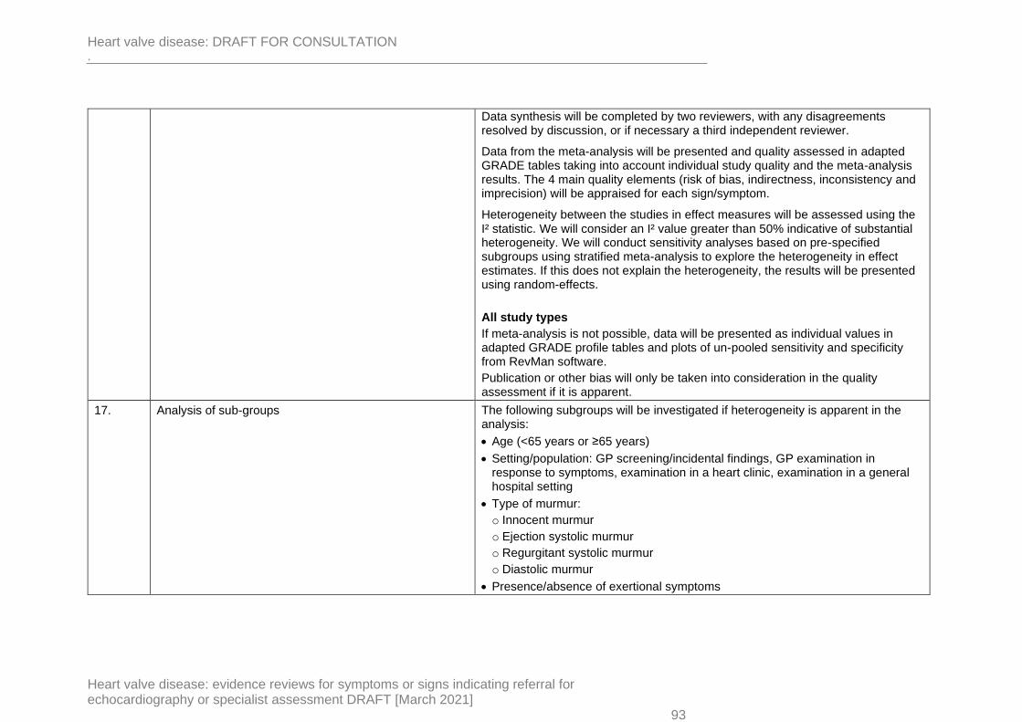
Heart valve disease: DRAFT FOR CONSULTATION .
Heart valve disease: evidence reviews for symptoms or signs indicating referral for echocardiography or specialist assessment DRAFT [March 2021] 93
Data synthesis will be completed by two reviewers, with any disagreements resolved by discussion, or if necessary a third independent reviewer.
Data from the meta-analysis will be presented and quality assessed in adapted GRADE tables taking into account individual study quality and the meta-analysis results. The 4 main quality elements (risk of bias, indirectness, inconsistency and imprecision) will be appraised for each sign/symptom.
Heterogeneity between the studies in effect measures will be assessed using the I² statistic. We will consider an I² value greater than 50% indicative of substantial heterogeneity. We will conduct sensitivity analyses based on pre-specified subgroups using stratified meta-analysis to explore the heterogeneity in effect estimates. If this does not explain the heterogeneity, the results will be presented using random-effects.
All study types
If meta-analysis is not possible, data will be presented as individual values in adapted GRADE profile tables and plots of un-pooled sensitivity and specificity from RevMan software.
Publication or other bias will only be taken into consideration in the quality assessment if it is apparent.
17. Analysis of sub-groups
The following subgroups will be investigated if heterogeneity is apparent in the analysis:
• Age (<65 years or ≥65 years)
• Setting/population: GP screening/incidental findings, GP examination in response to symptoms, examination in a heart clinic, examination in a general hospital setting
• Type of murmur:
o Innocent murmur
o Ejection systolic murmur
o Regurgitant systolic murmur
o Diastolic murmur
• Presence/absence of exertional symptoms

Heart valve disease: DRAFT FOR CONSULTATION .
Heart valve disease: evidence reviews for symptoms or signs indicating referral for echocardiography or specialist assessment DRAFT [March 2021] 94
• Type of valve disease diagnosed (aortic stenosis [including bicuspid], aortic regurgitation, mitral stenosis, mitral regurgitation, tricuspid regurgitation)
• Presence/absence of anaemia
• Presence/absence of pregnancy
• Presence/absence of atrial fibrillation
18. Type and method of review
☐ Intervention
☒ Diagnostic
☐ Prognostic
☐ Qualitative
☐ Epidemiologic
☐ Service Delivery
☐ Other (please specify)
19. Language English
20. Country England
21. Anticipated or actual start date 09/05/2019
22. Anticipated completion date 17/06/2021
23. Stage of review at time of this submission Review stage Started Completed
Preliminary searches
Piloting of the study selection process
Formal screening of search results against eligibility criteria

Heart valve disease: DRAFT FOR CONSULTATION .
Heart valve disease: evidence reviews for symptoms or signs indicating referral for echocardiography or specialist assessment DRAFT [March 2021] 95
Data extraction
Risk of bias (quality) assessment
Data analysis
24. Named contact 5a. Named contact
National Guideline Centre
5b Named contact e-mail
5e Organisational affiliation of the review
National Institute for Health and Care Excellence (NICE) and the National Guideline Centre
25. Review team members From the National Guideline Centre:
Sharon Swain [Guideline lead]
Eleanor Samarasekera [Senior systematic reviewer]
Nicole Downes [Systematic reviewer]
George Wood [Systematic reviewer]
Robert King [Health economist]
Jill Cobb [Information specialist]
Katie Broomfield [Project manager]
26. Funding sources/sponsor
This systematic review is being completed by the National Guideline Centre which receives funding from NICE.

Heart valve disease: DRAFT FOR CONSULTATION .
Heart valve disease: evidence reviews for symptoms or signs indicating referral for echocardiography or specialist assessment DRAFT [March 2021] 96
27. Conflicts of interest All guideline committee members and anyone who has direct input into NICE guidelines (including the evidence review team and expert witnesses) must declare any potential conflicts of interest in line with NICE's code of practice for declaring and dealing with conflicts of interest. Any relevant interests, or changes to interests, will also be declared publicly at the start of each guideline committee meeting. Before each meeting, any potential conflicts of interest will be considered by the guideline committee Chair and a senior member of the development team. Any decisions to exclude a person from all or part of a meeting will be documented. Any changes to a member's declaration of interests will be recorded in the minutes of the meeting. Declarations of interests will be published with the final guideline.
28. Collaborators
Development of this systematic review will be overseen by an advisory committee who will use the review to inform the development of evidence-based recommendations in line with section 3 of Developing NICE guidelines: the manual. Members of the guideline committee are available on the NICE website: https://www.nice.org.uk/guidance/indevelopment/gid-ng10122
29. Other registration details None
30. Reference/URL for published protocol
31. Dissemination plans NICE may use a range of different methods to raise awareness of the guideline. These include standard approaches such as:
• notifying registered stakeholders of publication
• publicising the guideline through NICE's newsletter and alerts
• issuing a press release or briefing as appropriate, posting news articles on the NICE website, using social media channels, and publicising the guideline within NICE.
32. Keywords Aortic regurgitation; aortic stenosis; diagnosis; echocardiography; heart valve disease; mitral regurgitation; mitral stenosis; primary care; referral; tricuspid regurgitation
33. Details of existing review of same topic by same authors N/A

Heart valve disease: DRAFT FOR CONSULTATION .
Heart valve disease: evidence reviews for symptoms or signs indicating referral for echocardiography or specialist assessment DRAFT [March 2021] 97
34. Current review status ☒ Ongoing
☐ Completed but not published
☐ Completed and published
☐ Completed, published and being updated
☐ Discontinued
35. Additional information N/A
36. Details of final publication www.nice.org.uk
1
Review protocol for symptoms and signs indicating direct referral to a specialist 2
ID Field Content
0. PROSPERO registration number CRD42020168665
1. Review title In adults with suspected heart valve disease, what symptoms and signs indicate direct referral (for example from primary care) to a specialist?
2. Review question In adults with suspected heart valve disease, what symptoms and signs indicate direct referral (for example from primary care) to a specialist?
3. Objective To determine the accuracy of presenting symptoms and signs to diagnose severe heart valve disease. This will inform a decision on which presenting factors indicate that direct referral to a specialist is required in people with suspected heart valve disease.
4. Searches The following databases (from inception) will be searched:
• Cochrane Database of Systematic Reviews (CDSR)
o Embase

Heart valve disease: DRAFT FOR CONSULTATION .
Heart valve disease: evidence reviews for symptoms or signs indicating referral for echocardiography or specialist assessment DRAFT [March 2021] 98
o MEDLINE
Searches will be restricted by:
• English language
• Human studies
• Letters and comments are excluded
Other searches:
• Inclusion lists of relevant systematic reviews will be checked by the reviewer.
The searches may be re-run 6 weeks before the final committee meeting and further studies retrieved for inclusion if relevant
The full search strategies will be published in the final review.
5. Condition or domain being studied
Suspected heart valve disease in adults aged 18 years and over: Aortic (including bicuspid) stenosis, aortic regurgitation, mitral stenosis, mitral regurgitation and tricuspid regurgitation.
6. Population Inclusion:
Adults aged 18 years and over with suspected heart valve disease in any setting (for example, in primary care)
Exclusion:
Children aged less than 18 years.
Adults with congenital heart disease (excluding bicuspid aortic valves).
Tricuspid stenosis and pulmonary valve disease.
Adults presenting with acute heart failure
7. Symptoms and signs Clinical observations:
• Cardiac auscultation (standard or electronic):

Heart valve disease: DRAFT FOR CONSULTATION .
Heart valve disease: evidence reviews for symptoms or signs indicating referral for echocardiography or specialist assessment DRAFT [March 2021] 99
o Presence of new murmur
o Character of heart sounds: – no/soft 2nd heart sound (as in severe AS)
– added 3rd sound; gallop rhythm (as in severe MR)
• Signs, severe symptoms or simple investigations:
o Shortness of breath (exertional breathlessness, for example classified as NYHA class ≥2)
o Shortness of breath + elevated serum natriuretic peptides (B‑type natriuretic
peptide [BNP] or N‑terminal pro‑B‑type natriuretic peptide [NT‑proBNP]; for
example NT‑proBNP 400-2000 or >2000 ng/litre)
o Peripheral oedema (ie. swelling of ankles and legs)
o Peripheral oedema (ie. swelling of ankles and legs) + BNP or NT proBNP (for
example NT‑proBNP 400-2000 or >2000 ng/litre)
o Pulmonary oedema
o Exertional chest pain (Canadian score class 2+)
o Exertional syncope (fainting)
o Abnormal ECG: for example signs of LV hypertrophy or AF
Include the following combinations:
• murmur alone
• murmur + heart sounds
• murmur + any of the listed symptoms, signs, or investigative findings
• murmur + heart sounds + any of the listed symptoms, signs, or investigative findings
• murmur + heart failure
(not symptoms alone nor heart sounds alone)

Heart valve disease: DRAFT FOR CONSULTATION .
Heart valve disease: evidence reviews for symptoms or signs indicating referral for echocardiography or specialist assessment DRAFT [March 2021] 100
8. Reference standard / Confounding factors
Reference (gold) standard:
• Confirmed diagnosis of severe HVD by transthoracic or transoesophageal echocardiography
Confounding factors (if diagnostic association studies are included):
• Age (<65 years or ≥65 years)
• Type of murmur:
o Innocent murmur
o Ejection systolic murmur
o Regurgitant systolic murmur
o Diastolic murmur
• Presence/absence of atrial fibrillation
9. Types of study to be included • Single-gate diagnostic studies (these may be called cohort studies or cross-sectional studies) will be included preferentially
• If no/insufficient3 diagnostic accuracy studies are identified prospective and retrospective cohort studies with multivariate analysis of the association between signs and symptoms and a confirmed diagnosis of severe heart valve disease will be included.
10. Other exclusion criteria
Exclusion criteria:
• Conference abstracts will be excluded because they are unlikely to contain enough information to assess whether the population matches the review question in terms of previous medication use, or enough detail on outcome definitions, or on the methodology to assess the risk of bias of the study.
• Case-control or ‘two-gate’ diagnostic studies
• Non-English language studies
11. Context
In clinical practice a number of symptoms and signs might indicate that a person has severe heart valve disease. An understanding of which symptoms and signs
3 This will be assessed for the review as a whole. There is no strict definition, but in discussion with the GC we will consider whether we have enough to form the basis for a
recommendation.

Heart valve disease: DRAFT FOR CONSULTATION .
Heart valve disease: evidence reviews for symptoms or signs indicating referral for echocardiography or specialist assessment DRAFT [March 2021] 101
better indicate severe HVD as a cause can facilitate further investigations to confirm diagnosis and guide management.
12. Primary outcomes (critical outcomes)
Diagnostic accuracy of symptoms and signs for severe HVD.
Measured by:
• Accuracy data
o Sensitivity
o Specificity
o Raw data to calculate 2x2 tables to calculate sensitivity and specificity (number of true positives, true negatives, false positives and false negatives).
13. Secondary outcomes (important outcomes) • Likelihood ratios
• Positive Predictive Value (PPV)
• Negative Predictive Value (NPV)
• Receiver Operating Characteristic (ROC) curve or area under curve for BNP and NT pro-BNP
If insufficient4 accuracy data are found, diagnostic association of signs and symptoms with a confirmed diagnosis of severe HVD will be included.
Measured by:
• Association data
o Adjusted RR or OR.
14. Data extraction (selection and coding)
EndNote will be used for reference management, sifting, citations and bibliographies. All references identified by the searches and from other sources will be screened for inclusion. 10% of the abstracts will be reviewed by two reviewers, with any disagreements resolved by discussion or, if necessary, a third independent reviewer.
4 This will be assessed for the review as a whole. There is no strict definition, but in discussion with the GC we will consider whether we have enough to form the basis for a
recommendation.

Heart valve disease: DRAFT FOR CONSULTATION .
Heart valve disease: evidence reviews for symptoms or signs indicating referral for echocardiography or specialist assessment DRAFT [March 2021] 102
The full text of potentially eligible studies will be retrieved and will be assessed in line with the criteria outlined above.
A standardised form will be used to extract data from studies (see Developing NICE guidelines: the manual section 6.4).
15. Risk of bias (quality) assessment
Risk of bias will be assessed using QUADAS-2 for diagnostic accuracy studies.
QUIPS will be used to assess diagnostic association reviews.
A 10% sample of the risk of bias assessments will be independently quality assured by a second reviewer. Disagreements between the review authors over the risk of bias in particular studies will be resolved by discussion, with involvement of a third review author where necessary.
16. Strategy for data synthesis Diagnostic accuracy studies
Where possible data will be meta-analysed in WinBUGS (if at least 3 studies reporting data at the same diagnostic threshold). Summary diagnostic outcomes will be reported from the meta-analyses with their 95% confidence intervals in adapted GRADE tables. Assessment of the quality of evidence for each outcome will take into account individual study quality and the meta-analysis results. The 4 main quality elements (risk of bias, indirectness, inconsistency and imprecision) will be appraised for each outcome.
Heterogeneity will be assessed by visual inspection of the sensitivity and specificity plots and summary area under the curve (AUC) plots. Particular attention will be placed on the measure determined by the committee to be the primary outcome for decision making.
If meta-analysis is not possible, data will be presented as individual values in adapted GRADE profile tables and plots of un-pooled sensitivity and specificity from RevMan software.
Diagnostic association studies
Aggregate data on diagnostic association of signs and symptoms will be collected and synthesised in a quantitative data analysis.

Heart valve disease: DRAFT FOR CONSULTATION .
Heart valve disease: evidence reviews for symptoms or signs indicating referral for echocardiography or specialist assessment DRAFT [March 2021] 103
If more than one study covered the same combination of population, sign/symptom, outcome and confounding factors accounted for then meta-analysis will be used to pool results. Meta-analysis will be carried out using the generic inverse variance function on Review Manager using fixed effect model. Data synthesis will be completed by two reviewers, with any disagreements resolved by discussion, or if necessary a third independent reviewer.
Data from the meta-analysis will be presented and quality assessed in adapted GRADE tables taking into account individual study quality and the meta-analysis results. The 4 main quality elements (risk of bias, indirectness, inconsistency and imprecision) will be appraised for each sign/symptom.
Heterogeneity between the studies in effect measures will be assessed using the I² statistic. We will consider an I² value greater than 50% indicative of substantial heterogeneity. We will conduct sensitivity analyses based on pre-specified subgroups using stratified meta-analysis to explore the heterogeneity in effect estimates. If this does not explain the heterogeneity, the results will be presented using random-effects.
All study types
A second reviewer will quality assure 10% of the data analyses. Discrepancies will be identified and resolved through discussion (with a third party where necessary).
Publication or other bias will only be taken into consideration in the quality assessment if it is apparent.
17. Analysis of sub-groups
The following subgroups will be investigated if heterogeneity is apparent in the analysis:
• Age (<65 years or ≥65 years)
• Setting/population: GP screening/incidental findings, GP examination in response to symptoms, examination in a heart clinic, examination in a general hospital setting
• Type of valve disease diagnosed (aortic stenosis [including bicuspid], aortic regurgitation, mitral stenosis, mitral regurgitation, tricuspid regurgitation)?
• Presence/absence of atrial fibrillation

Heart valve disease: DRAFT FOR CONSULTATION .
Heart valve disease: evidence reviews for symptoms or signs indicating referral for echocardiography or specialist assessment DRAFT [March 2021] 104
• Type of murmur:
o Innocent murmur
o Ejection systolic murmur
o Regurgitant systolic murmur
o Diastolic murmur
18. Type and method of review
☐ Intervention
☒ Diagnostic
☐ Prognostic
☐ Qualitative
☐ Epidemiologic
☐ Service Delivery
☐ Other (please specify)
19. Language English
20. Country England
21. Anticipated or actual start date 09/05/2019
22. Anticipated completion date 17/06/2021
23. Stage of review at time of this submission Review stage Started Completed
Preliminary searches
Piloting of the study selection process

Heart valve disease: DRAFT FOR CONSULTATION .
Heart valve disease: evidence reviews for symptoms or signs indicating referral for echocardiography or specialist assessment DRAFT [March 2021] 105
Formal screening of search results against eligibility criteria
Data extraction
Risk of bias (quality) assessment
Data analysis
24. Named contact 5a. Named contact
National Guideline Centre
5b Named contact e-mail
5e Organisational affiliation of the review
National Institute for Health and Care Excellence (NICE) and the National Guideline Centre
25. Review team members From the National Guideline Centre:
Sharon Swain [Guideline lead]
Eleanor Samarasekera [Senior systematic reviewer]
Nicole Downes [Systematic reviewer]
George Wood [Systematic reviewer]
Robert King [Health economist]
Jill Cobb [Information specialist]
Katie Broomfield [Project manager]

Heart valve disease: DRAFT FOR CONSULTATION .
Heart valve disease: evidence reviews for symptoms or signs indicating referral for echocardiography or specialist assessment DRAFT [March 2021] 106
26. Funding sources/sponsor
This systematic review is being completed by the National Guideline Centre which receives funding from NICE.
27. Conflicts of interest All guideline committee members and anyone who has direct input into NICE guidelines (including the evidence review team and expert witnesses) must declare any potential conflicts of interest in line with NICE's code of practice for declaring and dealing with conflicts of interest. Any relevant interests, or changes to interests, will also be declared publicly at the start of each guideline committee meeting. Before each meeting, any potential conflicts of interest will be considered by the guideline committee Chair and a senior member of the development team. Any decisions to exclude a person from all or part of a meeting will be documented. Any changes to a member's declaration of interests will be recorded in the minutes of the meeting. Declarations of interests will be published with the final guideline.
28. Collaborators
Development of this systematic review will be overseen by an advisory committee who will use the review to inform the development of evidence-based recommendations in line with section 3 of Developing NICE guidelines: the manual. Members of the guideline committee are available on the NICE website: https://www.nice.org.uk/guidance/indevelopment/gid-ng10122
29. Other registration details None
30. Reference/URL for published protocol
31. Dissemination plans NICE may use a range of different methods to raise awareness of the guideline. These include standard approaches such as:
• notifying registered stakeholders of publication
• publicising the guideline through NICE's newsletter and alerts
• issuing a press release or briefing as appropriate, posting news articles on the NICE website, using social media channels, and publicising the guideline within NICE.

Heart valve disease: DRAFT FOR CONSULTATION .
Heart valve disease: evidence reviews for symptoms or signs indicating referral for echocardiography or specialist assessment DRAFT [March 2021] 107
32. Keywords Aortic regurgitation; aortic stenosis; clinical assessment; diagnosis; heart valve disease; mitral regurgitation; mitral stenosis; primary care; referral; tricuspid regurgitation
33. Details of existing review of same topic by same authors
N/A
34. Current review status ☐ Ongoing
☒ Completed but not published
☐ Completed and published
☐ Completed, published and being updated
☐ Discontinued
35. Additional information N/A
36. Details of final publication www.nice.org.uk
1
2
3
4
5
6
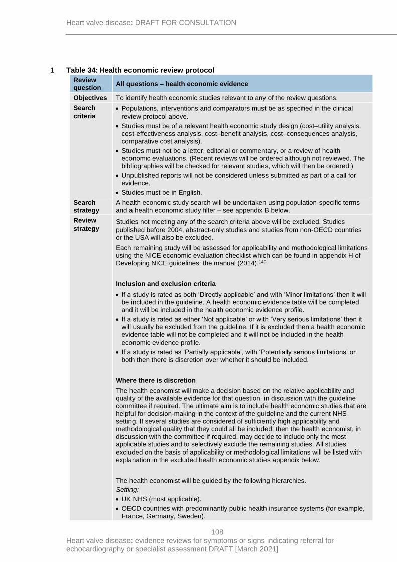
Heart valve disease: DRAFT FOR CONSULTATION
Heart valve disease: evidence reviews for symptoms or signs indicating referral for echocardiography or specialist assessment DRAFT [March 2021]
108
Table 34: Health economic review protocol 1
Review question
All questions – health economic evidence
Objectives To identify health economic studies relevant to any of the review questions.
Search criteria
• Populations, interventions and comparators must be as specified in the clinical review protocol above.
• Studies must be of a relevant health economic study design (cost–utility analysis, cost-effectiveness analysis, cost–benefit analysis, cost–consequences analysis, comparative cost analysis).
• Studies must not be a letter, editorial or commentary, or a review of health economic evaluations. (Recent reviews will be ordered although not reviewed. The bibliographies will be checked for relevant studies, which will then be ordered.)
• Unpublished reports will not be considered unless submitted as part of a call for evidence.
• Studies must be in English.
Search strategy
A health economic study search will be undertaken using population-specific terms and a health economic study filter – see appendix B below.
Review strategy
Studies not meeting any of the search criteria above will be excluded. Studies published before 2004, abstract-only studies and studies from non-OECD countries or the USA will also be excluded.
Each remaining study will be assessed for applicability and methodological limitations using the NICE economic evaluation checklist which can be found in appendix H of Developing NICE guidelines: the manual (2014).149
Inclusion and exclusion criteria
• If a study is rated as both ‘Directly applicable’ and with ‘Minor limitations’ then it will be included in the guideline. A health economic evidence table will be completed and it will be included in the health economic evidence profile.
• If a study is rated as either ‘Not applicable’ or with ‘Very serious limitations’ then it will usually be excluded from the guideline. If it is excluded then a health economic evidence table will not be completed and it will not be included in the health economic evidence profile.
• If a study is rated as ‘Partially applicable’, with ‘Potentially serious limitations’ or both then there is discretion over whether it should be included.
Where there is discretion
The health economist will make a decision based on the relative applicability and quality of the available evidence for that question, in discussion with the guideline committee if required. The ultimate aim is to include health economic studies that are helpful for decision-making in the context of the guideline and the current NHS setting. If several studies are considered of sufficiently high applicability and methodological quality that they could all be included, then the health economist, in discussion with the committee if required, may decide to include only the most applicable studies and to selectively exclude the remaining studies. All studies excluded on the basis of applicability or methodological limitations will be listed with explanation in the excluded health economic studies appendix below.
The health economist will be guided by the following hierarchies.
Setting:
• UK NHS (most applicable).
• OECD countries with predominantly public health insurance systems (for example, France, Germany, Sweden).

Heart valve disease: DRAFT FOR CONSULTATION
Heart valve disease: evidence reviews for symptoms or signs indicating referral for echocardiography or specialist assessment DRAFT [March 2021]
109
• OECD countries with predominantly private health insurance systems (for example, Switzerland).
• Studies set in non-OECD countries or in the USA will be excluded before being assessed for applicability and methodological limitations.
Health economic study type:
• Cost–utility analysis (most applicable).
• Other type of full economic evaluation (cost–benefit analysis, cost-effectiveness analysis, cost–consequences analysis).
• Comparative cost analysis.
• Non-comparative cost analyses including cost-of-illness studies will be excluded before being assessed for applicability and methodological limitations.
Year of analysis:
• The more recent the study, the more applicable it will be.
• Studies published in 2004 or later that depend on unit costs and resource data entirely or predominantly from before 2004 will be rated as ‘Not applicable’.
• Studies published before 2004 will be excluded before being assessed for applicability and methodological limitations.
Quality and relevance of effectiveness data used in the health economic analysis:
• The more closely the clinical effectiveness data used in the health economic analysis match with the outcomes of the studies included in the clinical review the more useful the analysis will be for decision-making in the guideline.
1
2

Heart valve disease: DRAFT FOR CONSULTATION
Heart valve disease: evidence reviews for symptoms or signs indicating referral for echocardiography or specialist assessment DRAFT [March 2021]
110
Appendix B Literature search strategies 1

Heart valve disease: DRAFT FOR CONSULTATION
Heart valve disease: evidence reviews for symptoms or signs indicating referral for echocardiography or specialist assessment DRAFT [March 2021]
111
Heart valve disease – search strategy 1 – signs and symptoms 1
This literature search strategy was used for the following review questions: 2
• In adults with suspected heart valve disease what symptoms and signs indicate referral 3 (for example from primary care) for echocardiography? 4
• In adults with suspected heart valve disease, what symptoms and signs indicate direct 5 referral (for example from primary care) to a specialist? 6
7
The literature searches for this review are detailed below and complied with the methodology 8 outlined in Developing NICE guidelines: the manual.149 9
For more information, please see the Methodology review published as part of the 10 accompanying documents for this guideline. 11
B.1 Clinical search literature search strategy 12
Searches were constructed using a PICO framework where population (P) terms were 13 combined with Intervention (I) and in some cases Comparison (C) terms. Outcomes (O) are 14 rarely used in search strategies for interventions as these concepts may not be well 15 described in title, abstract or indexes and therefore difficult to retrieve. Search filters were 16 applied to the search where appropriate. 17
Table 35: Database date parameters and filters used 18
Database Dates searched Search filter used
Medline (OVID) 1946 – 14 October 2020 Exclusions
Embase (OVID) 1974 – 14 October 2020 Exclusions
The Cochrane Library (Wiley) Cochrane Reviews to 2020 Issue 10 of 12
None
Medline (Ovid) search terms 19
1. exp Heart Valve Diseases/
2. exp heart valves/
3. ((primary or secondary) adj valv* disease*).ti,ab.
4. ((valv* or flap* or leaflet*) adj1 (heart or cardiac) adj (disease* or disorder* or failure or failed or dysfunction* or insufficien* or repair* or replace* or damage* or leak*)).ti,ab.
5. ((mitral or aortic or tricuspid or pulmon*) adj (valv* or flap* or leaflet*) adj (disease* or disorder* or failure or failed or dysfunction* or insufficien* or repair* or replace* or damage* or leak*)).ti,ab.
6. ((mitral or aortic or tricuspid or pulmon*) adj3 (prolapse or regurgitation or stenos?s or atresia or insufficienc*)).ti,ab.
7. or/1-6
8. letter/
9. editorial/
10. news/
11. exp historical article/
12. Anecdotes as Topic/
13. comment/
14. case report/
15. (letter or comment*).ti.
<Click this field on the first page and insert footer text if required>

Heart valve disease: DRAFT FOR CONSULTATION
Heart valve disease: evidence reviews for symptoms or signs indicating referral for echocardiography or specialist assessment DRAFT [March 2021]
112
16. or/8-15
17. randomized controlled trial/ or random*.ti,ab.
18. 16 not 17
19. animals/ not humans/
20. exp Animals, Laboratory/
21. exp Animal Experimentation/
22. exp Models, Animal/
23. exp Rodentia/
24. (rat or rats or mouse or mice).ti.
25. or/18-24
26. 7 not 25
27. limit 26 to English language
28. (exp child/ or exp pediatrics/ or exp infant/) not (exp adolescent/ or exp adult/ or exp middle age/ or exp aged/)
29. 27 not 28
30. exp Heart Murmurs/
31. murm*r*.ti,ab.
32. Heart Sounds/
33. ((heart or cardiac) adj sound*).ti,ab.
34. (wooshing or blowing or flutter* or rasping).ti,ab.
35. (heart beat* or heartbeat*).ti,ab.
36. turbulent blood flow.ti,ab.
37. Heart Auscultation/
38. auscultation*.ti,ab.
39. or/30-38
40. 29 and 39
Embase (Ovid) search terms 1
1. exp valvular heart disease/
2. exp heart valve/
3. ((primary or secondary) adj valv* disease*).ti,ab.
4. ((valv* or flap* or leaflet*) adj1 (heart or cardiac) adj (disease* or disorder* or failure or failed or dysfunction* or insufficien* or repair* or replace* or damage* or leak*)).ti,ab.
5. ((mitral or aortic or tricuspid or pulmon*) adj (valv* or flap* or leaflet*) adj (disease* or disorder* or failure or failed or dysfunction* or insufficien* or repair* or replace* or damage* or leak*)).ti,ab.
6. ((mitral or aortic or tricuspid or pulmon*) adj3 (prolapse or regurgitation or stenos?s or atresia or insufficienc*)).ti,ab.
7. or/1-6
8. letter.pt. or letter/
9. note.pt.
10. editorial.pt.
11. Case report/ or Case study/
12. (letter or comment*).ti.
13. or/8-12
14. randomized controlled trial/ or random*.ti,ab.

Heart valve disease: DRAFT FOR CONSULTATION
Heart valve disease: evidence reviews for symptoms or signs indicating referral for echocardiography or specialist assessment DRAFT [March 2021]
113
15. 13 not 14
16. animal/ not human/
17. Nonhuman/
18. exp Animal Experiment/
19. exp Experimental animal/
20. Animal model/
21. exp Rodent/
22. (rat or rats or mouse or mice).ti.
23. or/15-22
24. 7 not 23
25. (exp child/ or exp pediatrics/) not (exp adult/ or exp adolescent/)
26. 24 not 25
27. limit 26 to English language
28. exp heart murmur/
29. murm*r*.ti,ab.
30. ((heart or cardiac) adj sound*).ti,ab.
31. (wooshing or blowing or flutter* or rasping).ti,ab.
32. (heart beat* or heartbeat*).ti,ab.
33. turbulent blood flow.ti,ab.
34. heart sound/
35. heart auscultation/
36. auscultation*.ti,ab.
37. or/28-36
38. 27 and 37
Cochrane Library (Wiley) search terms 1
#1. MeSH descriptor: [Heart Valve Diseases] explode all trees
#2. MeSH descriptor: [Heart Valves] explode all trees
#3. ((primary or secondary) NEXT valv* disease*):ti,ab
#4. ((valv* or flap* or leaflet*) near/1 (heart or cardiac) NEXT (disease* or disorder* or failure or failed or dysfunction* or insufficien* or repair* or replace* or damage* or leak*)):ti,ab
#5. ((mitral or aortic or tricuspid or pulmon*) NEXT (valv* or flap* or leaflet*) NEXT (disease* or disorder* or failure or failed or dysfunction* or insufficien* or repair* or replace* or damage* or leak*)):ti,ab
#6. ((mitral or aortic or tricuspid or pulmon*) NEAR/3 (prolapse or regurgitation or stenos?s or atresia or insufficienc*)):ti,ab
#7. (or #1-#6)
#8. MeSH descriptor: [Heart Murmurs] explode all trees
#9. murm*r*:ti,ab
#10. MeSH descriptor: [Heart Sounds] this term only
#11. ((heart or cardiac) next sound*):ti,ab
#12. (wooshing or blowing or flutter* or rasping):ti,ab
#13. (heart next beat* or heartbeat*).ti,ab.
#14. turbulent blood flow:ti,ab
#15. MeSH descriptor: [Heart Auscultation] explode all trees

Heart valve disease: DRAFT FOR CONSULTATION
Heart valve disease: evidence reviews for symptoms or signs indicating referral for echocardiography or specialist assessment DRAFT [March 2021]
114
#16. auscultation*:ti,ab
#17. (or #8-#16)
#18. #7 and #17
B.2 Health Economics literature search strategy 1
Health economic evidence was identified by conducting a broad search relating to heart 2 valve disease population in NHS Economic Evaluation Database (NHS EED) – (this ceased 3 to be updated after March 2015) and the Health Technology Assessment database (HTA) – 4 (this ceased to be updated after March 2018) with no date restrictions. NHS EED and HTA 5 databases are hosted by the Centre for Research and Dissemination (CRD). Additional 6 searches were run on Medline and Embase for health economics. 7
Table 36: Database date parameters and filters used 8
Database Dates searched Search filter used
Medline 01 January 2014 – 15 October 2020
Exclusions
Health economics studies
Embase 01 January 2014 – 15 October 2020
Exclusions
Health economics studies
Centre for Research and Dissemination (CRD)
HTA - Inception – 31 March 2018
NHSEED - Inception to 31 March 2015
None
Medline (Ovid) search terms 9
1. exp Heart Valve Diseases/
2. exp heart valves/
3. ((primary or secondary) adj valv* disease*).ti,ab.
4. ((valv* or flap* or leaflet*) adj1 (heart or cardiac) adj (disease* or disorder* or failure or failed or dysfunction* or insufficien* or repair* or replace* or damage* or leak*)).ti,ab.
5. ((mitral or aortic or tricuspid or pulmon*) adj (valv* or flap* or leaflet*) adj (disease* or disorder* or failure or failed or dysfunction* or insufficien* or repair* or replace* or damage* or leak*)).ti,ab.
6. ((mitral or aortic or tricuspid or pulmon*) adj3 (prolapse or regurgitation or stenos?s or atresia or insufficienc*)).ti,ab.
7. Heart Valve Prosthesis/
8. ((mechanical or artificial or prosthe* or bioprosthe* or biological or tissue) adj (valv* or flap* or leaflet*)).ti,ab.
9. valve-in-valve.ti,ab.
10. (transcatheter adj2 (valve or valves)).ti,ab.
11. exp Heart Murmurs/
12. ((heart or cardiac) adj murmur*).ti,ab.
13. or/1-12
14. letter/
15. editorial/
16. news/
17. exp historical article/

Heart valve disease: DRAFT FOR CONSULTATION
Heart valve disease: evidence reviews for symptoms or signs indicating referral for echocardiography or specialist assessment DRAFT [March 2021]
115
18. Anecdotes as Topic/
19. comment/
20. case report/
21. (letter or comment*).ti.
22. or/14-21
23. randomized controlled trial/ or random*.ti,ab.
24. 22 not 23
25. animals/ not humans/
26. exp Animals, Laboratory/
27. exp Animal Experimentation/
28. exp Models, Animal/
29. exp Rodentia/
30. (rat or rats or mouse or mice).ti.
31. or/24-30
32. 13 not 31
33. limit 32 to english language
34. (exp child/ or exp pediatrics/ or exp infant/) not (exp adolescent/ or exp adult/ or exp middle age/ or exp aged/)
35. 33 not 34
36. Economics/
37. Value of life/
38. exp "Costs and Cost Analysis"/
39. exp Economics, Hospital/
40. exp Economics, Medical/
41. Economics, Nursing/
42. Economics, Pharmaceutical/
43. exp "Fees and Charges"/
44. exp Budgets/
45. budget*.ti,ab.
46. cost*.ti.
47. (economic* or pharmaco?economic*).ti.
48. (price* or pricing*).ti,ab.
49. (cost* adj2 (effective* or utilit* or benefit* or minimi* or unit* or estimat* or variable*)).ab.
50. (financ* or fee or fees).ti,ab.
51. (value adj2 (money or monetary)).ti,ab.
52. or/36-51
53. 35 and 52
Embase (Ovid) search terms 1
1. exp valvular heart disease/
2. exp heart valve/
3. ((primary or secondary) adj valv* disease*).ti,ab.

Heart valve disease: DRAFT FOR CONSULTATION
Heart valve disease: evidence reviews for symptoms or signs indicating referral for echocardiography or specialist assessment DRAFT [March 2021]
116
4. ((valv* or flap* or leaflet*) adj1 (heart or cardiac) adj (disease* or disorder* or failure or failed or dysfunction* or insufficien* or repair* or replace* or damage* or leak*)).ti,ab.
5. ((mitral or aortic or tricuspid or pulmon*) adj (valv* or flap* or leaflet*) adj (disease* or disorder* or failure or failed or dysfunction* or insufficien* or repair* or replace* or damage* or leak*)).ti,ab.
6. ((mitral or aortic or tricuspid or pulmon*) adj3 (prolapse or regurgitation or stenos?s or atresia or insufficienc*)).ti,ab.
7. exp heart valve prosthesis/
8. ((mechanical or artificial or prosthe* or bioprosthe* or biological or tissue) adj (valv* or flap* or leaflet*)).ti,ab.
9. valve-in-valve.ti,ab.
10. (transcatheter adj2 (valve or valves)).ti,ab.
11. exp heart murmur/
12. ((heart or cardiac) adj murmur*).ti,ab.
13. or/1-12
14. letter.pt. or letter/
15. note.pt.
16. editorial.pt.
17. Case report/ or Case study/
18. (letter or comment*).ti.
19. or/14-18
20. randomized controlled trial/ or random*.ti,ab.
21. 19 not 20
22. animal/ not human/
23. Nonhuman/
24. exp Animal Experiment/
25. exp Experimental animal/
26. Animal model/
27. exp Rodent/
28. (rat or rats or mouse or mice).ti.
29. or/21-28
30. 13 not 29
31. limit 30 to English language
32. (exp child/ or exp pediatrics/) not (exp adult/ or exp adolescent/)
33. 31 not 32
34. health economics/
35. exp economic evaluation/
36. exp health care cost/
37. exp fee/
38. budget/
39. funding/
40. budget*.ti,ab.
41. cost*.ti.
42. (economic* or pharmaco?economic*).ti.
43. (price* or pricing*).ti,ab.

Heart valve disease: DRAFT FOR CONSULTATION
Heart valve disease: evidence reviews for symptoms or signs indicating referral for echocardiography or specialist assessment DRAFT [March 2021]
117
44. (cost* adj2 (effective* or utilit* or benefit* or minimi* or unit* or estimat* or variable*)).ab.
45. (financ* or fee or fees).ti,ab.
46. (value adj2 (money or monetary)).ti,ab.
47. or/34-46
48. 33 and 47
NHS EED and HTA (CRD) search terms 1
#1. MeSH DESCRIPTOR Heart Valve Diseases EXPLODE ALL TREES
#2. MeSH DESCRIPTOR Heart Valves EXPLODE ALL TREES
#3. (((primary or secondary) adj Valv* adj disease*))
#4. (((valv* or flap* or leaflet*) adj (heart or cardiac) adj (disease* or disorder* or failure or failed or dysfunction* or insufficien* or repair* or replace* or damage* or leak*)))
#5. ((heart or cardiac) adj (valv* or flap* or leaflet*) adj (disease* or disorder* or failure or failed or dysfunction* or insufficien* or repair* or replace* or damage* or leak*))
#6. (((mitral or aortic or tricuspid or pulmon*) adj (valv* or flap* or leaflet*) adj (disease* or disorder* or failure or failed or dysfunction* or insufficien* or repair* or replace* or damage* or leak*)))
#7. (((mitral or aortic or tricuspid or pulmon*) adj3 (prolapse or regurgitation or stenos?s or atresia or insufficienc*)))
#8. MeSH DESCRIPTOR Heart Valve Prosthesis EXPLODE ALL TREES
#9. (((mechanical or artificial or prosthe* or bioprosthe* or biological or tissue) adj (valv* or flap* or leaflet*)))
#10. (valve-in-valve)
#11. ((transcatheter adj2 (valve or valves)))
#12. #1 or #2 or #3 or #4 or #5 or #6 or #7 or #8 or #9 or #10 or #11
2
3

Heart valve disease: DRAFT FOR CONSULTATION
Heart valve disease: evidence reviews for symptoms or signs indicating referral for echocardiography or specialist assessment DRAFT [March 2021]
118
Appendix C –Diagnostic evidence study selection 1
2
Figure 1: Flow chart of clinical study selection for the reviews of symptoms and signs 3 indicating referral for echocardiography (1.1) and direct referral to a 4 specialist (1.2) 5
6
Records screened, n=5176
Records excluded, n=4949
Papers included in review
• Q1.1 – echocardiography referral, n=30
• Q1.2 – direct specialist referral, n=19
Papers excluded from review
• Q1.1 – echocardiography referral, n=197
• Q1.2 – direct specialist referral, n=208
Reasons for exclusion: see appendix I
Records identified through database searching, n=5164
Additional records identified through other sources, n=12
Full-text papers assessed for eligibility, n=227 for both reviews

Heart valve disease: DRAFT FOR CONSULTATION
Heart valve disease: evidence reviews for symptoms or signs indicating referral for echocardiography or specialist assessment DRAFT [March 2021] 119
Appendix D – Diagnostic evidence 1
D.1 Symptoms and signs indicating echocardiography referral 2
Reference Aggarwal 20145
Study type Double blind prospective correlation study
Study methodology
Recruitment: consecutive (first 100 patients with inclusion criteria visiting the clinic)
Number of patients
n = 100
Patient characteristics
Age, mean (SD): 54.6 (sd not calculable) Gender (male to female ratio): 61:39 Ethnicity: Not reported Setting: Cardiology centre of an academic university hospital, in a rural area Country: India Inclusion criteria: Patients advised to undergo echocardiography when visiting the clinic Exclusion criteria: Known pre-existing heart murmurs No other characteristics provided
Target condition(s)
Any valve disease: stenosis or regurgitation
Index test(s) and reference standard
Index test Detection of murmur using stethoscope and specific software (ZargisCardioscan software) After taking informed consent, the principal investigator, a community medicine physician performed the auscultation of patients’ hearts in sitting position. Subsequently, the ZargisCardioscan™ software was used to analyse the heart sounds auscultated by the 3M™ Littmann® Model 3200 stethoscope. The heart auscultation was performed at all four auscultation sites on the chest: aortic area in second intercostal space on right parasternal line, pulmonary area in second intercostal space on left parasternal line, tricuspid area in fourth intercostal space on left parasternal line and cardiac apex (mitral area) in fifth intercostal space on midclavicular line. The analysis by ZargisCardioscan™ about presence and absence of systolic and/or diastolic murmurs were recorded. Sub-analysis by ZargisCardioscan™ further confirmed whether the auscultated systolic murmur was Class I murmur based on its grade and occurrence-time in cardiac cycle; all auscultated diastolic murmurs were considered Class I murmur based on the American College of

Heart valve disease: DRAFT FOR CONSULTATION
Heart valve disease: evidence reviews for symptoms or signs indicating referral for echocardiography or specialist assessment DRAFT [March 2021] 120
Reference Aggarwal 20145
Cardiology/American Heart Association (ACC/AHA) Practice Guidelines for the Management of Patients with Valvular Heart Disease that classify murmurs in asymptomatic patients as Class I murmurs if they are diastolic or continuous or holosystolic or late systolic or mid-systolic (grade 3 or higher).
It appears that the index test categories were no murmur (-ve) and murmur [Class 1 and above] (+ve) but this is unclear from the methodological description.
Reference standard Echocardiography confirmed valve disease, by blinded cardiologist. All except minimal-mild regurgitant valvular lesions were considered significant echocardiographic findings. Results have been extracted to include both minimal-mild regurgitant valvular lesions and significant valvular lesions as positives on gold standard for this review, as it covers heart valve disease regardless of severity.
Time between measurement of index test and reference standard: unclear
2×2 table
Reference standard + Reference standard − Total Results have been extracted to include both minimal-mild regurgitant valvular lesions and significant valvular lesions as positives on gold standard for this review, as it covers heart valve disease regardless of severity. Though only a 2x2 table for significant regurgitant lesions was emphasised in the paper, sufficient data was available to be able to construct the 2x2 table ourselves for any severity of regurgitant lesion, which included significant regurgitant lesions as well as minimal-mild regurgitant lesions as positives on the reference standard.
Index test + 21 10 31
Index test − 30 39 69
Total
51 49 100
Statistical measures
Index text: detection of murmur using stethoscope and specific software Sensitivity: 0.41 Specificity: 0.80 PPV: 0.68 NPV: 0.57 PLR: 2.02 NLR: 0.74 Prevalence on reference standard: 0.51

Heart valve disease: DRAFT FOR CONSULTATION
Heart valve disease: evidence reviews for symptoms or signs indicating referral for echocardiography or specialist assessment DRAFT [March 2021] 121
Reference Aggarwal 20145
Source of funding
No funding was received from any agency for carrying out this research work. However, ZargisCardioscan™ software
and 3M™ Littmann® Model 3200 stethoscope were provided by Deepak Gupta, MD, Anaesthesiologist, Detroit Medical Center/Wayne State University, Detroit, Michigan, United States from his personally owned equipments’ inventory on loan basis (academic / research purposes only) only as a gesture of supporting medical research under the principal investigator at the institution. There was no
competing interest between the authors of this research study.
Limitations Risk of bias: serious (unclear duration between index and gold standard tests)
Indirectness: serious – population is not necessarily those with suspected heart valve disease, but they have an indication for echocardiography
Comments
1 Reference Amano 198611
Study type Retrospective cross-sectional study
Study methodology
Recruitment: consecutive patients that were examined by auscultation, phonocardiography, 2D-echocardiography and pulsed Doppler echocardiography were reviewed – those with apical early or mid-systolic murmurs were included in the study.
Number of patients
n = 55
Patient characteristics
Age, mean (SD): not reported. Does appear to include some under the age of 18 but proportion unclear. Gender (male to female ratio): not reported Ethnicity: not reported Setting: unclear Country: Japan Inclusion criteria: had been examined by auscultation, phonocardiography, 2D-echocardiography and pulsed Doppler echocardiography; had apical early or mid-systolic murmurs Exclusion criteria: none reported No patient characteristics reported.
Target condition(s)
Heart valve disease: mitral regurgitation

Heart valve disease: DRAFT FOR CONSULTATION
Heart valve disease: evidence reviews for symptoms or signs indicating referral for echocardiography or specialist assessment DRAFT [March 2021] 122
Reference Amano 198611
Index test(s) and reference standard
Index test Presence of murmur – all had to have one to be included. These were apical early or mid-systolic murmurs. Murmurs appear to have been identified using auscultation and then analysed by phonocardiography. Prior to phonocardiography, careful auscultation was made with special attention to the point of the maximum intensity, area of transmission, timing, duration, intensity, pitch (high or medium), quality (blowing, harsh, rough, musical or vibratory and scratchy or clicky), and respiratory changes of murmurs. Reference standard Echocardiography confirmed mitral regurgitation. Pulsed Doppler echocardiography – Doppler signal recorded simultaneously with the M-mode echocardiogram, phonocardiogram and electrocardiogram using a strip chart recorder. Mitral regurgitation was estimated on basis of the location and area of distribution of abnormal systolic flow detected within the left atrium. 2D echocardiography – performed in supine or slightly left lateral position using commercially available, real-time scanner. Four cross-sectional images: long-axis, short-axis, apical four-chamber and subxiphoid views. Valve motion was carefully assessed to determine the lesions. Time between measurement of index test and reference standard: unclear – performance of phonocardiography and echocardiography was described as simultaneous, but delay between hearing murmur on auscultation and phonocardiography and echocardiography is unclear. Likely to be short time period as described it as ‘before’, suggesting shortly before rather than there being a long time delay.
2×2 table
Reference standard + Reference standard − Total
Index test + 29 26 55
Index test − 0 0 0
Total
29 26 55
Statistical measures
Index test: Presence of murmur – all had to have one to be included Sensitivity: could not calculate as all were index + to be included Specificity: could not calculate as all were index + to be included PPV: 0.53 NPV: could not calculate as all were index + to be included PLR: could not calculate as all were index + to be included NLR: could not calculate as all were index + to be included Prevalence on reference standard: 0.53
Source of funding
Not reported
Limitations Risk of bias: very serious – no reporting of blinding to index results; time interval between index test and reference standard unclear and exclusion criteria not listed. Indirectness: serious – all had to have a murmur to be included, which is the index test for this review and limits the use of accuracy data

Heart valve disease: DRAFT FOR CONSULTATION
Heart valve disease: evidence reviews for symptoms or signs indicating referral for echocardiography or specialist assessment DRAFT [March 2021] 123
Reference Amano 198611
Comments
1 Reference Aronow 198916
Study type Prospective cross-sectional study
Study methodology
Recruitment: unselected elderly patients in a long-term health care facility
Number of patients
n = 450
Patient characteristics
Age, mean (SD): 82 (8) years, range 61-100 years Gender (male to female ratio): 114:336 Ethnicity: not reported Setting: long-term health care facility Country: USA Inclusion criteria: elderly patients in a long-term health care facility; had technically adequate M-mode and 2D echocardiograms and pulsed Doppler recordings of the aortic valve Exclusion criteria: not reported. No other characteristics of patients reported.
Target condition(s)
Heart valve disease: aortic regurgitation
Index test(s) and reference standard
Index test Murmur of aortic regurgitation. A high frequency decrescendo murmur beginning with A2 was classified as an aortic regurgitation murmur. Cardiovascular examination was performed by an experienced cardiologist. Reference standard Echocardiography confirmed aortic regurgitation. M-mode and 2D echocardiograms and pulsed Doppler recordings of the aortic valve were obtained. Aortic regurgitation was diagnosed when an abnormal, high-velocity turbulent diastolic flow was detected in the left ventricular outflow tract. AR was considered mild when the signal was limited to the first centimetre proximal to the aortic valve, moderate

Heart valve disease: DRAFT FOR CONSULTATION
Heart valve disease: evidence reviews for symptoms or signs indicating referral for echocardiography or specialist assessment DRAFT [March 2021] 124
Reference Aronow 198916
when signal was detected in the left ventricular outflow tract in the area beyond the first centimetre but not beyond the tip of the anterior mitral leaflet, and severe when the abnormal signal persisted to a distance beyond the tip of the anterior mitral leaflet and could be detected in the left ventricle. Echocardiograms and Doppler recordings were interpreted by an experienced echocardiographer. Time between measurement of index test and reference standard: unclear – all patients underwent a cardiovascular examination by an experienced cardiologist before interpretation of the echocardiograms and Doppler recordings.
2×2 table
Reference standard + Reference standard − Total
Index test + 105 8 113
Index test − 26 311 337
Total
131 319 450
Statistical measures
Index text: murmur of aortic regurgitation Sensitivity: 0.80 Specificity: 0.97 PPV: 0.93 NPV: 0.92 PLR: 31.96 NLR: 0.20 Prevalence on reference standard: 0.29
Source of funding
Not reported
Limitations Risk of bias: very serious – no reporting of blinding to index results; time interval between index test and reference standard unclear Indirectness: serious – population may not necessarily be suspected HVD and may be in long-term health care facility for other reasons
Comments
1 Reference Aronow 198717
Study type Prospective cross-sectional study
Study methodology
Recruitment: unselected elderly patients with aortic systolic ejection murmurs in a long-term health care facility
Number of patients
n = 75
Patient characteristics
Age, mean (SD): 83 (8) years (range, 62-100)

Heart valve disease: DRAFT FOR CONSULTATION
Heart valve disease: evidence reviews for symptoms or signs indicating referral for echocardiography or specialist assessment DRAFT [March 2021] 125
Reference Aronow 198717
Gender (male to female ratio): 16:59 Ethnicity: not reported Setting: long-term health care facility Country: USA Inclusion criteria: elderly patients in a long-term health care facility; had technically adequate M-mode and 2D echocardiograms and pulsed Doppler recordings of the aortic valve Exclusion criteria: patients with more than mild aortic regurgitation as determined clinically or by Doppler echocardiography; patients with subvalvular stenosis. No other patient characteristics reported.
Target condition(s)
Heart valve disease: aortic stenosis
Index test(s) and reference standard
Index test Aortic systolic ejection murmur – all had to have one to be included in the study. All patients underwent a cardiovascular examination performed by an experienced cardiologist before interpretation of echocardiograms and Doppler recordings. A systolic ejection murmur heard in the second right intercostal space, down the left sternal border toward the apex or at the apex was classified as aortic systolic ejection murmur. Reference standard Echocardiography confirmed aortic stenosis. M-mode and 2D echocardiograms, and continuous wave Doppler measurement of aortic valve flow, were obtained. Valve flow velocities were assessed in multiple views, including apical, suprasternal and right parasternal views. Peak flow velocity across the aortic valve of 1.5 m/s or less was defined as normal. Peak aortic flow velocity 1.6-2.5 m/sec (peak gradient 10-25 mmHg), 2.6-3.5 m/sec (peak gradient 26-49 mmHg) and ≥3.6 m/sec (peak gradient ≥50 mmHg) were defined as mild, moderate and severe aortic stenosis, respectively. Echocardiographic and Doppler studies were interpreted by an experienced echocardiographer without knowledge of the cardiovascular findings. Time between measurement of index test and reference standard: unclear - cardiovascular examination was performed prior to interpretation of echocardiograms and Doppler recordings, but time interval unclear.
2×2 table
Reference standard + Reference standard − Total
Index test + 42 33 75
Index test − 0 0 0

Heart valve disease: DRAFT FOR CONSULTATION
Heart valve disease: evidence reviews for symptoms or signs indicating referral for echocardiography or specialist assessment DRAFT [March 2021] 126
Reference Aronow 198717
Total
42 33 75
Statistical measures
Index text: aortic systolic ejection murmur – all had to have one to be included in the study Sensitivity: could not calculate as all were index + to be included Specificity: could not calculate as all were index + to be included PPV: 0.56 NPV: could not calculate as all were index + to be included PLR: could not calculate as all were index + to be included NLR: could not calculate as all were index + to be included Prevalence on reference standard: 0.56
Source of funding
Not reported
Limitations Risk of bias: serious – time interval between index test and reference standard unclear Indirectness: serious – all had to have a murmur to be included, which is the index test for this review and limits the use of accuracy data
Comments
1 Reference Aronow 199118
Study type Prospective cross-sectional study
Study methodology
Recruitment: unselected elderly patients in a long-term health care facility
Number of patients
n = 781
Patient characteristics
Age, mean (SD): 82 (8) years (range, 62-100 years) Gender (male to female ratio): 223:558 Ethnicity: not reported Setting: long-term health care facility Country: USA Inclusion criteria: elderly patients in a long-term health care facility with technical Exclusion criteria: not reported. No other patient characteristics reported.

Heart valve disease: DRAFT FOR CONSULTATION
Heart valve disease: evidence reviews for symptoms or signs indicating referral for echocardiography or specialist assessment DRAFT [March 2021] 127
Reference Aronow 199118
Target condition(s)
Heart valve disease: aortic stenosis
Index test(s) and reference standard
Index test Aortic systolic ejection murmur. All patients underwent a cardiovascular examination performed by an experienced cardiologist before interpretation of echocardiograms and Doppler recordings. A systolic ejection murmur heard in the second right intercostal space, down the left sternal border toward the apex or at the apex was classified as aortic systolic ejection murmur. Reference standard Echocardiography confirmed aortic stenosis. M-mode and 2D echocardiograms, and continuous wave Doppler measurement of aortic valve flow, were obtained. Valve flow velocities were assessed in multiple views, including apical, suprasternal and right parasternal views. Peak flow velocity across the aortic valve of 1.5 m/s or less was defined as normal. Peak aortic flow velocity 1.6-2.5 m/sec (peak gradient 10-25 mmHg), 2.6-3.5 m/sec (peak gradient 26-49 mmHg) and ≥3.6 m/sec (peak gradient ≥50 mmHg) were defined as mild, moderate and severe aortic stenosis, respectively. Echocardiographic and Doppler studies were interpreted by an echocardiographer. Time between measurement of index test and reference standard: unclear - cardiovascular examination was performed prior to interpretation of echocardiograms and Doppler recordings, but time interval unclear.
2×2 table
Reference standard + Reference standard − Total
Index test + 138 Not reported Not reported
Index test − 4 Not reported Not reported
Total
142 639 781
Statistical measures
Index text: aortic systolic ejection murmur Sensitivity: 0.97 Specificity: could not calculate as no information regarding number of true negatives or false positives. PPV: could not calculate as no information regarding number of true negatives or false positives. NPV: could not calculate as no information regarding number of true negatives or false positives. PLR: could not calculate as no information regarding number of true negatives or false positives. NLR: could not calculate as no information regarding number of true negatives or false positives. Prevalence on reference standard: 0.18
Source of funding
Not reported
Limitations Risk of bias: very serious – no reporting of blinding to index results; time interval between index test and reference standard unclear
Indirectness: serious – population may not necessarily be suspected HVD and may be in long-term health care facility for other reasons
Comments
1

Heart valve disease: DRAFT FOR CONSULTATION
Heart valve disease: evidence reviews for symptoms or signs indicating referral for echocardiography or specialist assessment DRAFT [March 2021] 128
Reference Attenhofer Jost 200019
Study type Prospective cross-sectional study
Study methodology
Recruitment: consecutive patients referred to echocardiography laboratory because of a systolic murmur of unknown cause and who had not had previous echocardiographic examination
Number of patients
n = 100
Patient characteristics
Age, mean (SD): 58 (22) years (range, 17-92 years) Gender (male to female ratio): 43:57 Ethnicity: not reported Setting: echocardiography laboratory of a hospital Country: Switzerland Inclusion criteria: referred for echocardiography due to systolic murmur of unknown cause; no previous echocardiographic examination Exclusion criteria: not reported No other patient characteristics reported.
Target condition(s)
Heart valve disease: aortic stenosis or valvular regurgitation (AR, MR, TR) – reports separately for each type of valve disease
Index test(s) and reference standard
Index test 1. Systolic murmur – all had one to be included in the study 2. Systolic murmur + diminished aortic closure sound (AS and MR only)
Immediately before echocardiography, patients were examined by two cardiologists blinded to the patient’s history, electrocardiogram and other medical data. Clinical examination included estimate of jugular venous pressure, assessment of apical impulse and carotid artery upstroke, and auscultation at rest during quiet respiration, with assessment of heart sounds and murmurs and their radiation. Associated findings, such as thrills and systolic clicks, were noted. The Valsava manoeuvre was done in every patient and other dynamic manoeuvres were added if thought necessary. Characteristics of murmurs were classified at point of maximal intensity. Murmurs were located in aortic area, pulmonic area, base of the heart or apex. Timing and duration of murmurs were classified as early systolic, late systolic or holosystolic. The examiner had to state if the murmur was functional or organic. If thought to be organic, the examiner had to classify the underlying heart disease as significant or insignificant. Significant disease was defined as moderate or severe valvular heart disease, congenital shunts or intraventricular gradients. An isolated valvular lesion was defined if there was no clinical evidence of other types of heart disease – for the purposes of this review, information only on valve disease was extracted.

Heart valve disease: DRAFT FOR CONSULTATION
Heart valve disease: evidence reviews for symptoms or signs indicating referral for echocardiography or specialist assessment DRAFT [March 2021] 129
Reference Attenhofer Jost 200019
Reference standard Echocardiography confirmed aortic stenosis or valvular regurgitation (AR, MR, TR). Transthoracic 2D and Doppler echocardiography in the left supine position. Valvular regurgitation was graded as trivial, mild, moderate or severe based on a combination of factors, especially the vena contracta for the atrioventricular valves and the ratio of the regurgitant jet height to the outflow tract height for the semilunar valves. Aortic stenosis was classified as severe (mean systolic gradient ≥50 mmHg or aortic valve area ≤0.8 cm2), moderate (mean systolic gradient 30-49 mmHg or aortic valve area 0.8-1.0 cm2), mild (mean systolic gradient 10-29 mmHg or aortic valve area 1.1-1.9 cm2) or trivial (mean systolic gradient <10 mmHg or aortic valve area ≥2.0 cm2, but with thickening of bicuspid or tricuspid aortic valve). An intraventricular gradient (left or right ventricle) was defined as a peak systolic gradient ≥10 mmHg at rest or with Valsava within the left ventricular outflow tract or midventricular by continuous-wave Doppler with the typical shape (left convex) and the peak velocity occurring late in systole. Time between measurement of index test and reference standard: index test was performed immediately before echocardiography.
2×2 tables
Systolic murmur - AS
Reference standard + Reference standard − Total
Index test + 29 71 100
Index test − 0 0 0
Total
29 71 100
Systolic murmur - AR
Reference standard + Reference standard − Total
Index test + 28 72 100
Index test − 0 0 0
Total
28 72 100
Systolic murmur - MR
Reference standard + Reference standard − Total
Index test + 30 70 100
Index test − 0 0 0
Total
30 70 100
Systolic murmur - TR
Reference standard + Reference standard − Total
Index test + 24 76 100

Heart valve disease: DRAFT FOR CONSULTATION
Heart valve disease: evidence reviews for symptoms or signs indicating referral for echocardiography or specialist assessment DRAFT [March 2021] 130
Reference Attenhofer Jost 200019
Index test − 0 0 0
Total
24 76 100
Systolic murmur + diminished aortic closure sound - AS
Reference standard + Reference standard − Total
Index test + 8 Not reported Not reported
Index test − 20 Not reported Not reported
Total
28 72 100
Systolic murmur + diminished aortic closure sound - MR
Reference standard + Reference standard − Total
Index test + 3 Not reported Not reported
Index test − 27 Not reported Not reported
Total
30 70 100
Statistical measures
Index text: AS - systolic murmur – all had one to be included in the study Sensitivity: could not calculate as all were index + to be included Specificity: could not calculate as all were index + to be included PPV: 0.29 NPV: could not calculate as all were index + to be included PLR: could not calculate as all were index + to be included
NLR: could not calculate as all were index + to be included Prevalence on reference standard: 0.29 Index text: AR - systolic murmur – all had one to be included in the study Sensitivity: could not calculate as all were index + to be included Specificity: could not calculate as all were index + to be included

Heart valve disease: DRAFT FOR CONSULTATION
Heart valve disease: evidence reviews for symptoms or signs indicating referral for echocardiography or specialist assessment DRAFT [March 2021] 131
Reference Attenhofer Jost 200019
PPV: 0.28 NPV: could not calculate as all were index + to be included PLR: could not calculate as all were index + to be included NLR: could not calculate as all were index + to be included Prevalence on reference standard: 0.28 Index text: MR - systolic murmur – all had one to be included in the study Sensitivity: could not calculate as all were index + to be included Specificity: could not calculate as all were index + to be included PPV: 0.30 NPV: could not calculate as all were index + to be included PLR: could not calculate as all were index + to be included
NLR: could not calculate as all were index + to be included Prevalence on reference standard: 0.30 Index text: TR - systolic murmur – all had one to be included in the study Sensitivity: could not calculate as all were index + to be included Specificity: could not calculate as all were index + to be included PPV: 0.24 NPV: could not calculate as all were index + to be included r PLR: could not calculate as all were index + to be included NLR: could not calculate as all were index + to be included Prevalence on reference standard: 0.24 Index text: AS systolic murmur + diminished aortic closure sound Sensitivity: 0.29 Specificity: could not calculate as no information regarding number of true negatives or false positives. PPV: could not calculate as no information regarding number of true negatives or false positives. NPV: could not calculate as no information regarding number of true negatives or false positives. PLR: could not calculate as no information regarding number of true negatives or false positives. NLR: could not calculate as no information regarding number of true negatives or false positives. Prevalence on reference standard: 0.29 Index text: MR systolic murmur + diminished aortic closure sound

Heart valve disease: DRAFT FOR CONSULTATION
Heart valve disease: evidence reviews for symptoms or signs indicating referral for echocardiography or specialist assessment DRAFT [March 2021] 132
Reference Attenhofer Jost 200019
Sensitivity: 0.10 Specificity: could not calculate as no information regarding number of true negatives or false positives. PPV: could not calculate as no information regarding number of true negatives or false positives. NPV: could not calculate as no information regarding number of true negatives or false positives. PLR: could not calculate as no information regarding number of true negatives or false positives. NLR: could not calculate as no information regarding number of true negatives or false positives. Prevalence on reference standard: 0.30
Source of funding
Not reported
Limitations Risk of bias: serious – certain manoeuvres may have been used for auscultation in some patients and not others Indirectness: serious – for the use of murmur alone as a diagnostic feature, all had to have a murmur to be included, which is the index test for this review and limits the use of accuracy data; for murmur + diminished aortic closure sound, insufficient information to calculate full diagnostic accuracy measures
Comments
1 Reference Barron 198821
Study type Cross-sectional study
Study methodology
Recruitment: consecutive patients referred to echocardiography for evaluation of suspected mitral valve prolapse
Number of patients
n = 140
Patient characteristics
Age, mean (SD): 36.8 (12.6) years Gender (male to female ratio): 23:117 Ethnicity: not reported Setting: 125 patients were outpatients and 15 were inpatients Country: USA Inclusion criteria: patients referred to echocardiography for evaluation of suspected mitral valve prolapse Exclusion criteria: not reported No other patient characteristics were reported.

Heart valve disease: DRAFT FOR CONSULTATION
Heart valve disease: evidence reviews for symptoms or signs indicating referral for echocardiography or specialist assessment DRAFT [March 2021] 133
Reference Barron 198821
Target condition(s)
Heart valve disease: mitral or tricuspid regurgitation
Index test(s) and reference standard
Index test Systolic murmur. Auscultation was performed by one investigator either immediately prior to or after echocardiographic and Doppler studies. Patients were examined in the supine left lateral decubitus, standing and squatting positions. Physical examination was positive for mitral valve prolapse if a midsystolic click was heard I supine position and if it moved toward the first heart sound with standing and toward the second heart sound with squatting. Clicks elicited only with standing or squatting were also deemed consistent with mitral valve prolapse. Presence of a systolic murmur heard in the left lower sternal border or apex, with or without radiation to the axilla, was noted in each position. Systolic murmurs heard loudest in the aortic or pulmonic areas were not consistent with mitral or tricuspid origin and therefore discounted. In the absence of a click, a systolic murmur alone was not considered indicative of mitral valve prolapse. For the purposes of this review, those with a murmur on auscultation were considered to be index positive – those with only clicks, and not a murmur, were not considered to be index test positive. Reference standard Echocardiography confirmed mitral or tricuspid regurgitation. 2D echocardiographs and Doppler studies were performed. The presence of mitral valve prolapse and tricuspid valve prolapse was assessed. Doppler flow studies were performed using pulsed Doppler sample volumes. The left atrium and right atrium above the valve leaflets were interrogated for valvular regurgitation. In the event of transmitral flow, the parasternal long-axis and apical four-chamber and two-chamber views were used. For the tricuspid valve, the parasternal right ventricular inflow tract view and apical four chamber views were used. Echocardiograms were interpreted by one investigator without knowledge of the auscultatory findings. Time between measurement of index test and reference standard: index test was performed by one investigator either immediately prior to or after echocardiographic and Doppler studies.
2×2 table
Reference standard + Reference standard − Total
Index test + 26 25 51
Index test − 23 66 89
Total
49 91 140
Statistical measures
Index text: systolic murmur Sensitivity: 0.53 Specificity: 0.73 PPV: 0.51 NPV: 0.74 PLR: 1.93 NLR: 0.65
Prevalence on reference standard: 0.35

Heart valve disease: DRAFT FOR CONSULTATION
Heart valve disease: evidence reviews for symptoms or signs indicating referral for echocardiography or specialist assessment DRAFT [March 2021] 134
Reference Barron 198821
Source of funding
Not reported.
Limitations Risk of bias: serious – in some where index test was performed after reference standard it is unclear whether they were blinded to reference standard results Indirectness: none
Comments
1 Reference Barzilai 198822
Study type Prospective cross-sectional study
Study methodology
Recruitment: consecutive patients with documented acute myocardial infarction admitted to hospital cardiac care unit
Number of patients
n = 59
Patient characteristics
Age, mean (SD): 65 (2) years Gender (male to female ratio): 34:25 Ethnicity: not reported Setting: secondary care – Barnes Hospital Cardiac Care Unit Country: USA Inclusion criteria: admitted to hospital cardiac care unit between September 1985 and March 1986 with documented acute myocardial infarction and who could be examined within 48 h of the onset of infarction Exclusion criteria: not reported

Heart valve disease: DRAFT FOR CONSULTATION
Heart valve disease: evidence reviews for symptoms or signs indicating referral for echocardiography or specialist assessment DRAFT [March 2021] 135
Reference Barzilai 198822
Type of myocardial infarction: n=35 with Q-wave acute MI and n=24 with non-Q-wave acute MI. The acute MI was anterior (Q-wave =18) in 24, inferior (Q-wave =16) in 34 and the locus could not be determined in 1 patient.
Target condition(s)
Heart valve disease: mitral regurgitation
Index test(s) and reference standard
Index test Systolic murmur. Relevant data from the history, an attending physician’s physical examination, laboratory findings, including peak total and MB creatine kinase, and electrocardiograms were collected prospectively in all patients. Reference standard Echocardiography confirmed mitral regurgitation. Pulsed Doppler echocardiography was performed usually on the morning after admission. The presence of mitral regurgitation was determined from the apical 4 chamber and parasternal long-axis views with pulsed Doppler. Mitral regurgitation was diagnosed by the presence of a high pitched audio signal accompanied by turbulent systolic flow when the sample volume was placed in the left atrium. Only patients with flow velocities >150 cm/s were considered to have mitral regurgitation. The results of Doppler examination were not routinely revealed to physicians caring for the patients unless requested or unless unanticipated findings were documented. Time between measurement of index test and reference standard: unclear – potentially up to/longer than 24 h, as Doppler studies usually performed on the morning after admission.
2×2 table
Reference standard + Reference standard − Total
Index test + 10 6 16
Index test − 13 30 43
Total
23 36 59
Statistical measures
Index text: systolic murmur Sensitivity: 0.43 Specificity: 0.83 PPV: 0.63 NPV: 0.70 PLR: 2.61 NLR: 0.68
Prevalence on reference standard: 0.39 Source of funding
Not reported
Limitations Risk of bias: very serious – no mention of blinding to index results when reference standard interpreted; time interval between the index test and reference standard being performed unclear and could have been up to/longer than 24 h

Heart valve disease: DRAFT FOR CONSULTATION
Heart valve disease: evidence reviews for symptoms or signs indicating referral for echocardiography or specialist assessment DRAFT [March 2021] 136
Reference Barzilai 198822
Indirectness: serious – population is those admitted for acute myocardial infarction so may not necessarily have been suspicion of heart valve disease, but rather assessing its onset after acute myocardial infarction
Comments
1 Reference Baur 200623
Study type Prospective cohort study
Study methodology
Recruitment: 43 general practices were recruited, and each then referred any eligible patients to the study
Number of patients
n = 198 (43 general practices, covering 130,000 people; the 198 were those who were referred for echocardiography on the basis of approved indications)
Patient characteristics
Age, mean (SD): 64.5 (18.1)
Gender (male to female ratio): 86:112
Ethnicity: Not reported
Setting: Urban primary care
Country: Netherlands
Inclusion criteria: Patients at any of 43 general practices; suspected of having heart failure or valve disease, as shown by the following approved indications: shortness of breath, cardiac murmur and peripheral oedema of otherwise unexplained origin.
Exclusion criteria: Other indications 34% cardiac murmur only; 28% dyspnea only; 3% peripheral oedema only; 13% cardiac murmur and dyspnea; 2% cardiac murmur and peripheral oedema; 1% cardiac murmur and dyspnea and peripheral oedema
Target condition(s)
Heart valve disease: aortic or mitral valve disease (including stenosis and regurgitation)
Index test(s) and reference standard
I Index test 1. Cardiac murmur 2. Cardiac murmur + other indication (e.g. dyspnoea, peripheral oedema or other)
Reference standard

Heart valve disease: DRAFT FOR CONSULTATION
Heart valve disease: evidence reviews for symptoms or signs indicating referral for echocardiography or specialist assessment DRAFT [March 2021] 137
Reference Baur 200623
Echocardiography confirmed aortic or mitral valve disease (including stenosis and regurgitation). Results were interpreted by the cardiologist according to the criteria of the American and European Societies of Echocardiography. Systolic left ventricular dysfunction was defined as a left ventricular ejection fraction <40% measured by 2D echocardiography in the apical four-chamber and two-chamber view. Quantification of the echocardiograms was performed according to Simpson's rule. Diastolic dysfunction was defined as an abnormal flow pattern across the mitral valve and an abnormal flow pattern across the pulmonary vein. Because diastolic mitral flow is age-dependent, flow patterns were considered abnormal if the flow was beyond the mean and once the standard deviation of the normal flow of that age group. Left ventricular hypertrophy was defined as a mean wall thickness of >12 mm. Pulmonary hypertension was considered present if the measured systolic pulmonary pressure was >35 mmHg. Measurement of the systolic pulmonary pressure was done by measuring the maximal velocity of the tricuspid regurgitation and calculation of the systolic pressure gradient between the right ventricle and right atrium according to the Bernoulli equation. Right atrial pressure was estimated by looking to the diameter and the collapse of the inferior cava vein. Aortic insufficiency was measured using the criteria of Perry and Reynolds. Aortic valve insufficiency was assumed to be important if it was more than grade 2. Aortic valve stenosis was considered important if the maximal gradient was >30 mmHg and the mean gradient >20 mmHg. If one or more criteria were present the patient was assumed to have significant aortic valve disease. Mitral valve insufficiency was graded by measurement of the jet area and proximal jet width at the vena contracta in addition to measurement of the continuous wave flow and the pulsed wave flow in the pulmonary veins. Mitral valve regurgitation was assumed to be important if leakage was more than grade 2. Mitral valve stenosis was considered haemodynamically relevant when the maximal gradient was .10 mmHg. If one or both criteria were present, the patient was assumed to have significant mitral valve disease.
Time between measurement of index test and reference standard: approximately 1 week
2×2 tables
Cardiac murmur
Reference standard + Reference standard − Total
Index test + 19 82 101
Index test − 1 96 97
Total
20 178 198
Cardiac murmur + other indication (e.g. dyspnoea, peripheral oedema or other)
Reference standard + Reference standard − Total
Index test + 12 22 34
Index test − 8 156 164

Heart valve disease: DRAFT FOR CONSULTATION
Heart valve disease: evidence reviews for symptoms or signs indicating referral for echocardiography or specialist assessment DRAFT [March 2021] 138
Reference Baur 200623
Total
20 178 198
Statistical measures
Index text: cardiac murmur Sensitivity: 0.95 Specificity: 0.54 PPV: 0.19 NPV: 0.99 PLR: 2.06 NLR: 0.09
Prevalence on reference standard: 0.10 Index text: cardiac murmur + other indication (e.g. dyspnoea, peripheral oedema or other) Sensitivity: 0.60 Specificity: 0.88 PPV: 0.35 NPV: 0.95 PLR: 4.85 NLR: 0.46
Prevalence on reference standard: 0.10
Source of funding
Not reported
Limitations Risk of bias: Very serious: 1 week duration between index and gold standard tests; no reporting of echo assessor blinding Indirectness: None
Comments
1 Reference Breisblatt 198827
Study type Prospective cross-sectional study
Study methodology
Recruitment: patients referred for cardiac catheterisation with known ischaemic heart disease and no previous history of valvular disease, being assessed for the presence of mitral regurgitation. Unclear if consecutive.
Number of patients
n = 150
Patient characteristics
Age, mean (SD): 62 years (SD not reported), range 34-80 years Gender (male to female ratio): 112:38

Heart valve disease: DRAFT FOR CONSULTATION
Heart valve disease: evidence reviews for symptoms or signs indicating referral for echocardiography or specialist assessment DRAFT [March 2021] 139
Reference Breisblatt 198827
Ethnicity: not reported Setting: 75% were evaluated in a coronary care unit or post-coronary care unit setting for unstable angina or myocardial infarction. Remaining patients were referred to radionuclide laboratory as patients who had previously been unstable and whose physicians wanted a pre-catheterisation assessment of left ventricular function. Country: USA Inclusion criteria: patients referred for cardiac catheterisation with known ischaemic heart disease; no previous history of valvular disease Exclusion criteria: not reported All patients had known or suspected coronary artery disease Previous transmural myocardial infarction, 52% Previous subendocardial myocardial infarction, 15% Three-vessel disease, 68% Two-vessel disease, 21% One-vessel disease, 11%
Target condition(s)
Heart valve disease: mitral regurgitation
Index test(s) and reference standard
Index test Systolic murmur. In pre-catheterisation assessment, patients were examined by two cardiologists who were blinded to radionuclide data. The presence of absence of a systolic murmur was noted, as well as whether the murmur was characteristic of mitral regurgitation – defined as a holosystolic apical murmur. Other systolic murmurs identified during the examination were assessed as suggestive of mitral regurgitation. Reference standard Radionuclide angiography confirmed mitral regurgitation. Right-sided catheterisation was performed in 85% patients with flow-directed balloon-tipped catheter and left ventricular end-diastolic pressure was recorded in all patients. Left ventriculography was performed. Mitral regurgitation was graded on a scale of 1+ to 4+. Post-extra systolic beats were excluded and only normal beats were evaluated. The degree of mitral regurgitation was based on consensus of two blinded angiographers. Equilibrium radionuclide angiocardiography performed at rest in all patients. A four-view study was performed in all patients (best septal view, anterior, left lateral and left posterior oblique). Both lateral views were obtained in the right-sided down decubitus position. Radionuclide interpretation was the consensus of two observers who were blinded to data from angiographic studies and physical examination. Regurgitant index was determined by the stroke volume method.

Heart valve disease: DRAFT FOR CONSULTATION
Heart valve disease: evidence reviews for symptoms or signs indicating referral for echocardiography or specialist assessment DRAFT [March 2021] 140
Reference Breisblatt 198827
Time between measurement of index test and reference standard: unclear.
2×2 table
Reference standard + Reference standard − Total
Index test + 26 36 62
Index test − 3 85 88
Total
29 121 150
Statistical measures
Index text: systolic murmur Sensitivity: 0.90 Specificity: 0.70 PPV: 0.42 NPV: 0.97 PLR: 3.01 NLR: 0.15
Prevalence on reference standard: 0.19 Source of funding
Not reported
Limitations Risk of bias: serious – time interval between index test and reference standard unclear Indirectness: serious – population with known ischaemic heart disease, likely to have a different incidence to a more general population presenting for first time with a murmur with/without symptoms
Comments
1 Reference Cantley 199529
Study type Prospective cross-sectional study
Study methodology
Recruitment: newly admitted patients in an acute assessment and rehabilitation unit at a general hospital who were found to have systolic murmurs on clinical examination. Recruited over an 8-week period.
Number of patients
n = 32
Patient characteristics
Age, mean (SD): 81.8 years (SD not reported), range 56-95 years Gender (male to female ratio): 9:23 Ethnicity: not reported Setting: those referred to acute assessment and rehabilitation unit at a general hospital

Heart valve disease: DRAFT FOR CONSULTATION
Heart valve disease: evidence reviews for symptoms or signs indicating referral for echocardiography or specialist assessment DRAFT [March 2021] 141
Reference Cantley 199529
Country: Scotland, UK Inclusion criteria: referred to acute assessment and rehabilitation unit at general hospital; presence of systolic murmur on clinical examination Exclusion criteria: not reported
Target condition(s)
Heart valve disease: aortic stenosis, aortic regurgitation or mitral regurgitation (reported separately)
Index test(s) and reference standard
Index test Systolic murmur – all had to have one to be included. Systolic murmur noted on clinical examination – unclear who the clinical examination was performed by. Reference standard Echocardiography confirmed aortic stenosis, aortic regurgitation or mitral regurgitation. Each patient assessed independently by a registrar operator using mobile stand-alone continuous wave Doppler machine and by a consultant radiologist using full echocardiographic assessment, including Doppler techniques. The results from full echocardiographic assessment with Doppler were used as the reference standard results in terms of this review. The presence or absence of aortic stenosis was recorded and if present the pressure gradient across the valve was noted. A gradient >20 mmHg was considered to indicate aortic stenosis. The presence or absence of aortic and mitral regurgitation was also noted. Time between measurement of index test and reference standard: unclear.
2×2 tables
Aortic stenosis Reference standard + Reference standard − Total
Index test + 12 20 32
Index test − 0 0 0
Total
12 20 32
Aortic regurgitation
Reference standard + Reference standard − Total
Index test + 14 17 31
Index test − 0 0 0
Total
14 17 31

Heart valve disease: DRAFT FOR CONSULTATION
Heart valve disease: evidence reviews for symptoms or signs indicating referral for echocardiography or specialist assessment DRAFT [March 2021] 142
Reference Cantley 199529
Mitral regurgitation
Reference standard + Reference standard − Total
Index test + 17 14 31
Index test − 0 0 0
Total
17 14 31
Statistical measures
Aortic stenosis Index text Systolic murmur – all had to have one to be included Sensitivity: could not calculate as all were index + to be included Specificity: could not calculate as all were index + to be included PPV: 0.38 NPV: could not calculate as all were index + to be included PLR: could not calculate as all were index + to be included NLR: could not calculate as all were index + to be included
Prevalence on reference standard: 0.38 Aortic regurgitation Index text Systolic murmur – all had to have one to be included Sensitivity: could not calculate as all were index + to be included Specificity: could not calculate as all were index + to be included PPV: 0.45 NPV: could not calculate as all were index + to be included PLR: could not calculate as all were index + to be included NLR: could not calculate as all were index + to be included
Prevalence on reference standard: 0.45 Mitral regurgitation Index text Systolic murmur – all had to have one to be included Sensitivity: could not calculate as all were index + to be included Specificity: could not calculate as all were index + to be included PPV: 0.55 NPV: could not calculate as all were index + to be included PLR: could not calculate as all were index + to be included
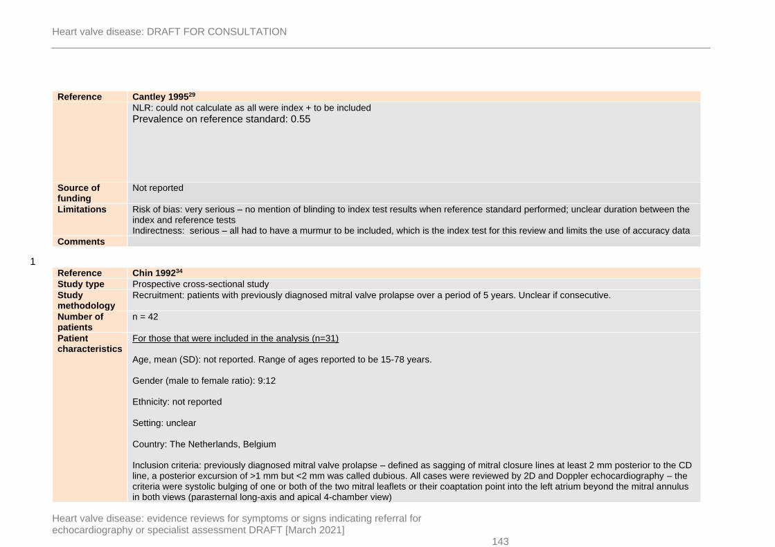
Heart valve disease: DRAFT FOR CONSULTATION
Heart valve disease: evidence reviews for symptoms or signs indicating referral for echocardiography or specialist assessment DRAFT [March 2021] 143
Reference Cantley 199529
NLR: could not calculate as all were index + to be included
Prevalence on reference standard: 0.55
Source of funding
Not reported
Limitations Risk of bias: very serious – no mention of blinding to index test results when reference standard performed; unclear duration between the index and reference tests Indirectness: serious – all had to have a murmur to be included, which is the index test for this review and limits the use of accuracy data
Comments
1 Reference Chin 199234
Study type Prospective cross-sectional study
Study methodology
Recruitment: patients with previously diagnosed mitral valve prolapse over a period of 5 years. Unclear if consecutive.
Number of patients
n = 42
Patient characteristics
For those that were included in the analysis (n=31) Age, mean (SD): not reported. Range of ages reported to be 15-78 years. Gender (male to female ratio): 9:12 Ethnicity: not reported Setting: unclear Country: The Netherlands, Belgium Inclusion criteria: previously diagnosed mitral valve prolapse – defined as sagging of mitral closure lines at least 2 mm posterior to the CD line, a posterior excursion of >1 mm but <2 mm was called dubious. All cases were reviewed by 2D and Doppler echocardiography – the criteria were systolic bulging of one or both of the two mitral leaflets or their coaptation point into the left atrium beyond the mitral annulus in both views (parasternal long-axis and apical 4-chamber view)

Heart valve disease: DRAFT FOR CONSULTATION
Heart valve disease: evidence reviews for symptoms or signs indicating referral for echocardiography or specialist assessment DRAFT [March 2021] 144
Reference Chin 199234
Exclusion criteria: not reported. No additional patient characteristics reported
Target condition(s)
Heart valve disease: mitral regurgitation
Index test(s) and reference standard
Index test Late systolic murmur. Immediately following ultrasound studies auscultation was performed in supine and left decubitus positions. Criteria for the diagnosis of MVP were a midsystolic click and/or late systolic murmur at the apex. The late systolic murmur was considered to be a sign of mitral regurgitation. Phonocardiographic recordings were also performed but results on auscultation reported separately. Reference standard Echocardiography confirmed mitral regurgitation. 2D and Doppler echocardiography used to confirm mitral valve prolapse. Detection of mitral regurgitation was performed using continuous and pulsed-wave Doppler recordings. In latter half of study, recordings could be made using colour-coded Doppler observations. Time between measurement of index test and reference standard: index test of murmur performed immediately after echocardiography.
2×2 table
Reference standard + Reference standard − Total
Index test + 9 2 11
Index test − 4 16 20
Total
13 18 31
Statistical measures
Index text: late systolic murmur Sensitivity: 0.69 Specificity: 0.89 PPV: 0.82 NPV: 0.80 PLR: 6.23 NLR: 0.35 Prevalence on reference standard: 0.42
Source of funding
Not reported
Limitations Risk of bias: very serious – index test performed after reference standard and unclear if blinded; ~25% were not included in the analysis as not able to perform full assessment with reference standard or dubious results on the reference standard Indirectness: serious – already had confirmed mitral valve prolapse on echocardiography, may differ to the more general population with suspected HVD based on a murmur with/without symptoms only and no confirmation of existing structural problems

Heart valve disease: DRAFT FOR CONSULTATION
Heart valve disease: evidence reviews for symptoms or signs indicating referral for echocardiography or specialist assessment DRAFT [March 2021] 145
Reference Chin 199234
Comments
1 Reference Decoodt 199051
Study type Prospective cross-sectional study
Study methodology
Recruitment: patients with idiopathic mitral valve prolapse confirmed on echocardiography during 1 year period. Unclear if consecutive.
Number of patients
n = 100
Patient characteristics
Age, mean (SD): 53.5 years (SD not reported), range 18-83 years Gender (male to female ratio): 37:63 Ethnicity: not reported Setting: referred to a cardiology laboratory for echocardiography Country: Belgium Inclusion criteria: patients with idiopathic mitral valve prolapse – criteria based on M-mode and 2D echocardiography findings: late or holosystolic posterior movement of the valve of >2 mm below the CD line of coaptation of the mitral leaflets during systole (M-mode) and mitral coaptation of type 2-3. Prolapse criteria based on an apical view were avoided. Exclusion criteria: concomitant major cardiac abnormalities (hypertrophic cardiomyopathy, major aortic insufficiency, uncorrected atrial septal defect, surgically corrected atrial septal defects, corrected patent ductus arteriosus, dilated cardiomyopathy, ischaemic cardiopathies requiring transluminal dilatation and prior myocardial infarction). No other patient characteristics reported.
Target condition(s)
Heart valve disease: mitral regurgitation
Index test(s) and reference standard
Index test Systolic murmur – this included some with early systolic murmurs, late systolic murmurs and holosystolic murmurs. Auscultatory features were reported by another observer than the person performing the echocardiography. Reference standard Echocardiography confirmed mitral regurgitation. A 2.5 Mhz multi-element transducer was used for colour flow mapping study. Pulse repetition frequencies of 4, 6 or 8 KHz were available. Diagnostic range of 12 or 15 cm routinely used for mitral valve prolapse. When

Heart valve disease: DRAFT FOR CONSULTATION
Heart valve disease: evidence reviews for symptoms or signs indicating referral for echocardiography or specialist assessment DRAFT [March 2021] 146
Reference Decoodt 199051
mitral regurgitation was found, the grade, direction of the jet and systolic timing were determined. For the grade, regurgitant flow on left atrial area ratio was obtained in the plane at which it appeared greatest. Same Doppler colour gain setting algorithm was used. Mitral regurgitation was classified as mild (ratio <20%), moderate (ratio 20-40%) and severe (ratio >40%). Time between measurement of index test and reference standard: unclear
2×2 table
Reference standard + Reference standard − Total
Index test + 47 5 52
Index test − 7 41 48
Total
54 46 100
Statistical measures
Index text: systolic murmur Sensitivity: 0.87 Specificity: 0.89 PPV: 0.90 NPV: 0.85 PLR: 8.01 NLR: 0.15
Prevalence on reference standard: 0.54 Source of funding
Not reported
Limitations Risk of bias: very serious – some of those with major concomitant heart abnormalities excluded; no blinding mentioned for performance of index test and reference standard; unclear time interval between index test and references standard Indirectness: serious – already had confirmed mitral valve prolapse on echocardiography, may differ to the more general population with suspected HVD based on a murmur with/without symptoms only and no confirmation of existing structural problems
Comments
1 Reference Gardezi 201872
Study type Prospective cross-sectional study
Study methodology
Recruitment: people undergoing echocardiography at two primary care sites participating in OxVALVE – prospective screening study to identify prevalence of undiagnosed valvular heart disease in asymptomatic subjects aged >65 years
Number of patients
n = 251
Patient characteristics
Age, mean (SD): 75 (6) years

Heart valve disease: DRAFT FOR CONSULTATION
Heart valve disease: evidence reviews for symptoms or signs indicating referral for echocardiography or specialist assessment DRAFT [March 2021] 147
Reference Gardezi 201872
Gender (male to female ratio): 128:123 Ethnicity: not reported Setting: primary care sites Country: UK Inclusion criteria: asymptomatic subjects >65 years undergoing screening for undiagnosed heart valve disease at two primary care sites enrolled in the OxVALVE study Exclusion criteria: history of valvular heart disease No other patient characteristics reported
Target condition(s)
Heart valve disease: mild or significant valve disease
Index test(s) and reference standard
Index test Murmur (systolic or diastolic) – as assessed by GPs. Systematic cardiac auscultation, incorporating assessment of pulse character and murmur radiation (where appropriate), was undertaken by one of two fully trained primary care/family doctors. Each participating primary care/family doctors had >10 years of clinical experience but had not received specialist cardiology training. They used an acoustic stethoscope under ‘real world conditions’ without the knowledge of the echocardiography results. Heart sounds were recorded using an electronic stethoscope and were analysed at a later date by two consultant cardiologists. Reference standard Echocardiography confirmed valve disease – reports separately for mild (aortic sclerosis or any mild regurgitation) and significant (at least moderate regurgitation or at least mild stenosis of any valve) valve disease. An investigating physician or sonographer performed detailed echocardiography immediately following auscultation using standard views according to the British Society of Echocardiography guidelines. Unclear if blinded to the index test results. Time between measurement of index test and reference standard: reference standard performed immediately after auscultation.
2×2 table
Mild valve disease (aortic sclerosis or mild regurgitation)
Reference standard + Reference standard − Total
Index test + 55 27 82

Heart valve disease: DRAFT FOR CONSULTATION
Heart valve disease: evidence reviews for symptoms or signs indicating referral for echocardiography or specialist assessment DRAFT [March 2021] 148
Reference Gardezi 201872
Index test − 115 54 169
Total
170 81 251
Significant valve disease (at least moderate regurgitation or at least mild stenosis of any valve)
Reference standard + Reference standard − Total
Index test + 16 66 82
Index test − 20 149 169
Total
36 215 251
Statistical measures
Index test: murmur (systolic or diastolic) – mild valve disease (sclerosis or mild regurgitation) Sensitivity: 0.32 Specificity: 0.67 PPV: 0.67 NPV: 0.32 PLR: 0.97 NLR: 1.01 Prevalence on reference standard: 0.68 Index test: murmur (systolic or diastolic) – significant valve disease (at least moderate regurgitation or at least mild stenosis of any valve) Sensitivity: 0.44 Specificity: 0.69 PPV: 0.20 NPV: 0.88 PLR: 1.45 NLR: 0.80 Prevalence on reference standard: 0.14

Heart valve disease: DRAFT FOR CONSULTATION
Heart valve disease: evidence reviews for symptoms or signs indicating referral for echocardiography or specialist assessment DRAFT [March 2021] 149
Reference Gardezi 201872
Source of funding
Supported by the National Institute for Health Research (NIHR) Biomedical Research Centre, Oxford, UK Reported to be no competing interests
Limitations Risk of bias: serious – no mention of blinding to index test results when reference standard performed Indirectness: serious – appears to be a screening study in those over a certain age, and did not necessarily have any symptoms or signs that would lead to a suspicion of heart valve disease
Comments
1 Reference Hoffmann 198386
Study type Cross-sectional study
Study methodology
Recruitment: Consecutive patients
Number of patients
n = 102 in whole study (n=58 analysed as remaining 67 had Doppler ultrasound to assess aortic valve pressure gradient only, rather than to assess clinically ill-defined systolic murmur)
Patient characteristics
Age, range: 20-79 years Gender (male to female ratio): 57:45 Ethnicity: Not reported Note: above patient characteristics based on the whole cohort of n=102 patients, not limited to n=58 included in analysis of ill-defined systolic murmur. Setting: Cardiac catheter clinic (secondary care) Country: Switzerland Inclusion criteria: people undergoing right or left heart catheterisation for valvular or coronary heart disease, or both. Exclusion criteria: None reported Aortic stenosis: 22/102; mitral regurgitation 36/102; ventricular septal defect 8/102
Target condition(s)
Heart valve disease: aortic stenosis or mitral regurgitation. Aortic stenosis defined as pressure gradient >20 mmHg.

Heart valve disease: DRAFT FOR CONSULTATION
Heart valve disease: evidence reviews for symptoms or signs indicating referral for echocardiography or specialist assessment DRAFT [March 2021] 150
Reference Hoffmann 198386
Index test(s) and reference standard
Index test Systolic murmur – all had one to be included Doppler US was the index test in the study but for the purpose of this review, data used to obtain information on the number of those with murmur that went on to have confirmed valve disease by cardiac catheterisation. Reference standard Cardiac catheterisation confirmed aortic stenosis or mitral regurgitation Time between measurement of index test and reference standard: unclear
2×2 tables
Aortic stenosis Reference standard + Reference standard − Total Though flow murmurs are mentioned in the paper, they do not give numbers assessed as having flow murmurs on auscultation – the accuracy data given in the paper in terms of distinguishing between valve disease and flow murmurs appears to be for Doppler ultrasonography, not auscultation.
Index test + 22 36 58
Index test − 0 0 0
Total
22 36 58
Mitral regurgitation
Reference standard + Reference standard − Total Though flow murmurs are mentioned in the paper, they do not give numbers assessed as having flow murmurs on auscultation – the accuracy data given in the paper in terms of distinguishing between valve disease and flow murmurs appears to be for Doppler ultrasonography, not auscultation.
Index test + 36 22 58
Index test − 0 0 0
Total
36 22 58
Statistical measures
Index text: systolic murmur – AS Sensitivity: could not calculate as all were index + to be included Specificity: could not calculate as all were index + to be included PPV: 0.38 NPV: could not calculate as all were index + to be included PLR: could not calculate as all were index + to be included NLR: could not calculate as all were index + to be included Prevalence on reference standard: 0.38 Index text: systolic murmur – MR Sensitivity: could not calculate as all were index + to be included Specificity: could not calculate as all were index + to be included

Heart valve disease: DRAFT FOR CONSULTATION
Heart valve disease: evidence reviews for symptoms or signs indicating referral for echocardiography or specialist assessment DRAFT [March 2021] 151
Reference Hoffmann 198386
PPV: 0.62 NPV: could not calculate as all were index + to be included PLR: could not calculate as all were index + to be included NLR: could not calculate as all were index + to be included Prevalence on reference standard: 0.62
Source of funding
None reported
Limitations Risk of bias: very serious: unclear duration between index and gold standard tests; no reporting of gold standard assessor blinding Indirectness: serious – all had to have a murmur to be included, which is the index test for this review and limits the use of accuracy data
Comments
1 Reference Kalinauskiene 201997
Study type Cross-sectional study
Study methodology Recruitment: consecutive patients arriving at Kaunas Clinical Hospital meeting inclusion criteria
Number of patients n = 30
Patient characteristics
Age, mean (SD): 68.7 (12.09) Gender (male to female ratio): 2:1 Ethnicity: Not reported Setting: ‘Clinical Hospital’ – unclear if primary or secondary care Country: Lithuania Inclusion criteria: BMI >30; aged >18 years; referred for echocardiogram Exclusion criteria: ‘Severe’ status; deemed unsuitable for inclusion Other characteristics: shortness of breath 83.33%; chest pain 76.67%; leg oedema 36.67%; fatigue 30%; echocardiography findings: mitral regurgitation 83.33%, tricuspid regurgitation 66.67%; aortic regurgitation 63.33%; pulmonary regurgitation 6.67%; aortic stenosis 3.33%.

Heart valve disease: DRAFT FOR CONSULTATION
Heart valve disease: evidence reviews for symptoms or signs indicating referral for echocardiography or specialist assessment DRAFT [March 2021] 152
Reference Kalinauskiene 201997
Target condition(s) Heart valve disease: aortic stenosis, aortic regurgitation, mitral stenosis, mitral regurgitation or tricuspid regurgitation
Index test(s) and reference standard
Index test Murmur via acoustic and electronic auscultation.
Each subject received 4 auscultation examinations (Figure 1). Two auscultations were done by a cardiologist with about 20 years of experience using both an acoustic traditional stethoscope (3M Littman Cardiology III Mechanical Stethoscope, 3M Health Care, St. Paul, MN, USA) and an electronic stethoscope (3M Littmann 3200 Electronic Stethoscope,3MHealthCare, St. Paul, MN, USA). Two additional auscultations were done by a 3rd-year medical resident doctor also using both an acoustic traditional stethoscope and an electronic stethoscope. Half of the patients were auscultated by one cardiologist and one resident, and half, by another cardiologist and another resident. Based on the randomization for each subject, the auscultation may begin with either the 3M Littmann 3200 Electronic or 3M Littmann Cardiology III Mechanical stethoscope. Physicians had a 2-week period to gain experience in using the electronic stethoscope before the commencement of the study. Each auscultation consisted of heart murmurs being listened to in the following sites: mitral (apex), aortic (right second intercostal space), pulmonary (left second intercostal space), tricuspid (lower left sternal border), and Erb’s (left third intercostal space). All physicians used the same ordinary methodology of Lithuanian University of Health Sciences: All physicians used the same ordinary methodology of Lithuanian University of Health (1) Using the bell listen first to the apex (mitral area) just above the apex beat (palpate the apex beat), then at the second right interspace parasternally (aortic area), at the second left interspace parasternally (pulmonic area), at the third interspace adjacent to the left sternal border (Erb‘s area), and, finally, at the left parasternal area at the lower part of the sternum (tricuspid area) in the supine position. (2) Shift to the diaphragm and return to all these areas. (3) Ask the patient to exhale completely and stop breathing, listen at the apex and aortic areas, and Erb‘s area with the bell and the diaphragm; ask the patient to inhale completely and stop breathing, listen at the pulmonic and tricuspid areas with the bell and the diaphragm. (4) Ask the patient to roll partly onto the left side, listen at the apex with the bell and the diaphragm, then also ask the patient to exhale completely and stop breathing, and again listen with the bell and the diaphragm. (5) Ask the patient to sit up, lean forward and put his/her arms on the head, listen at the aortic and Erb‘s areas only with the diaphragm, also repeat the auscultation at these areas in full held expiration. Physical conditions for all listeners were as in a real-life: all auscultations were performed in Sciences: 1) Using the bell listen first to the apex (mitral area) just above the apex beat (palpate the apex beat), then at the second right interspace parasternally (aortic area), at the second left interspace parasternally (pulmonic area), at the third interspace adjacent to the left sternal border (Erb‘s area), and, finally, at the left parasternal area at the lower part of the sternum (tricuspid area) in the supine position. 2) Shift to the diaphragm and return to all these areas. 3) Ask the patient to exhale completely and stop breathing, listen at the apex and aortic areas, and Erb‘s area with the bell and the diaphragm; ask the patient to inhale completely and stop breathing, listen at the pulmonic and tricuspid areas with the bell and the diaphragm. 4) Ask the patient to roll partly onto the left side, listen at the apex with the bell and the diaphragm, then also ask the patient to exhale completely and stop breathing, and again listen with the bell and the diaphragm. 5) Ask the patient to sit up, lean forward and put his/her arms on the head, listen at the aortic and Erb‘s areas only with the diaphragm, also repeat the auscultation at these areas in full held expiration.
Reference standard Echocardiogram, carried after index tests who were blinded to index test results.

Heart valve disease: DRAFT FOR CONSULTATION
Heart valve disease: evidence reviews for symptoms or signs indicating referral for echocardiography or specialist assessment DRAFT [March 2021] 153
Reference Kalinauskiene 201997
Time between measurement of index test and reference standard: unclear
2×2 table
Resident/acoustic/mitral regurgitation
Reference standard +
Reference standard −
Total
Index test + 19 2 21
Index test − 6 3 9
Total
25 5 30
Resident/electronic/mitral regurgitation
Reference standard +
Reference standard −
Total
Index test + 21 3 24
Index test − 4 2 6
Total
25 5 30
Resident/acoustic/aortic regurgitation
Reference standard +
Reference standard −
Total
Index test + 5 0 5
Index test − 14 11 25
Total
19 11 30
Resident/electronic/aortic regurgitation
Reference standard +
Reference standard −
Total
Index test + 7 0 7
Index test − 12 11 23
Total
19 11 30
Resident/acoustic/tricuspid regurgitation
Reference standard +
Reference standard −
Total
Index test + 10 1 11
Index test − 10 9 19
Total
20 10 30

Heart valve disease: DRAFT FOR CONSULTATION
Heart valve disease: evidence reviews for symptoms or signs indicating referral for echocardiography or specialist assessment DRAFT [March 2021] 154
Reference Kalinauskiene 201997
Resident/electronic/tricuspid regurgitation
Reference standard +
Reference standard −
Total
Index test + 13 2 15
Index test − 7 8 15
Total
20 10 30
Resident/acoustic/mitral stenosis
Reference standard +
Reference standard −
Total
Index test + 0 1 1
Index test − 0 29 29
Total
0 30 30
Resident/electronic/mitral stenosis
Reference standard +
Reference standard −
Total
Index test + 0 1 1
Index test − 0 29 29
Total
0 30 30
Resident/acoustic/aortic stenosis
Reference standard +
Reference standard −
Total
Index test + 1 3 4
Index test − 2 24 26
Total
3 27 30
Resident/electronic/aortic stenosis
Reference standard +
Reference standard −
Total
Index test + 1 3 4
Index test − 2 24 26
Total
3 27 30

Heart valve disease: DRAFT FOR CONSULTATION
Heart valve disease: evidence reviews for symptoms or signs indicating referral for echocardiography or specialist assessment DRAFT [March 2021] 155
Reference Kalinauskiene 201997
Statistical measures
Index text: Resident/acoustic/mitral regurgitation Sensitivity: 0.76 Specificity:0.60 PPV:0.90 NPV:0.33 PLR:1.90 NLR:0.40
Prevalence on reference standard:0.83 Index text Resident/electronic/mitral regurgitation Sensitivity: 0.84 Specificity:0.40 PPV:0.88 NPV:0.33 PLR:1.40 NLR:0.40
Prevalence on reference standard:0.83 Index text: Resident/acoustic/aortic regurgitation Sensitivity: 0.26 Specificity:1.00 PPV:1.00 NPV:0.44 PLR: Not calculable NLR:0.74
Prevalence on reference standard:0.63 Index text Resident/electronic/aortic regurgitation Sensitivity: 0.37 Specificity:1.00 PPV:1.00 NPV:0.48 PLR: Not calculable NLR:0.63
Prevalence on reference standard:0.63 Index text: Resident/acoustic/tricuspid regurgitation

Heart valve disease: DRAFT FOR CONSULTATION
Heart valve disease: evidence reviews for symptoms or signs indicating referral for echocardiography or specialist assessment DRAFT [March 2021] 156
Reference Kalinauskiene 201997
Sensitivity: 0.50 Specificity:0.90 PPV:0.91 NPV:0.47 PLR:5.00 NLR:0.56
Prevalence on reference standard:0.67 Index text Resident/electronic/tricuspid regurgitation Sensitivity: 0.65 Specificity:0.80 PPV:0.87 NPV:0.53 PLR:3.25 NLR:0.44
Prevalence on reference standard:0.67 Index text: Resident/acoustic/mitral stenosis Sensitivity: not calculable Specificity: 0.97 PPV: 0.00 NPV: 1.00 PLR: not calculable NLR: not calculable
Prevalence on reference standard: 0.00 Index text Resident/electronic/mitral stenosis Sensitivity: not calculable Specificity: 0.97 PPV: 0.00 NPV: 1.00 PLR: not calculable NLR: not calculable
Prevalence on reference standard: 0.00 Index text: Resident/acoustic/aortic stenosis Sensitivity: 0.33

Heart valve disease: DRAFT FOR CONSULTATION
Heart valve disease: evidence reviews for symptoms or signs indicating referral for echocardiography or specialist assessment DRAFT [March 2021] 157
Reference Kalinauskiene 201997
Specificity:0.89 PPV:0.25 NPV:0.92 PLR:3.00 NLR:0.75
Prevalence on reference standard:0.10 Index text Resident/electronic/aortic stenosis Sensitivity: 0.33 Specificity:0.89 PPV:0.25 NPV:0.92 PLR:3.00 NLR:0.75
Prevalence on reference standard:0.10 Source of funding Sponsored by 3M, the manufacturers of the electronic and acoustic stethoscopes used in the study.
Limitations Risk of bias: serious: unclear time interval between index tests and gold standard. No other limitations: index tests carried out before gold standard so these were effectively blinded from gold standard results; gold standard measured by different clinicians who were blinded to index test results; no attrition; consecutive sample. Indirectness: serious: these patients were those referred for an echo, but were from a sub-group with BMI >30. This sub-group may not be representative of the review population.
Comments
1 Reference Kinney 1988103
Study type Retrospective review of hospital records
Study methodology
Recruitment: retrospective review of records of patients who had echocardiography performed between July 1982 and June 1985. Inpatients and outpatients included.
Number of patients
n = 294
Patient characteristics
Age, mean (SD): 59 (14) years Gender (male to female ratio): 100:0 – all patients were male Ethnicity: white, 70%; black, 30%

Heart valve disease: DRAFT FOR CONSULTATION
Heart valve disease: evidence reviews for symptoms or signs indicating referral for echocardiography or specialist assessment DRAFT [March 2021] 158
Reference Kinney 1988103
Setting: data from inpatients and outpatients included Country: USA Inclusion criteria: patients that had echocardiography performed between July 1982 and June 1985; data on self-reported heights and weights just before echocardiography Exclusion criteria: hospital charts too fragmentary to be useful; patients without Doppler study; equivocal Doppler results; Doppler studies of poor quality Most patients had one or more of the following conditions:
• Hypertension, n=136
• Coronary artery disease, n=118. Of these, n=9 were within a week of acute myocardial infarction at time of Doppler echocardiography.
• Alcoholism now or in the past, n=107
• Congestive heart failure now or in the past, n=70
• Symptomatic arrhythmias, n=16
• Prosthetic heart valves, n=5 (n=4 aortic prosthesis and n=1 mitral prosthesis)
• Acute endocarditis, n=5 There were n=35 patients without apparent heart disease.
Target condition(s)
Heart valve disease: aortic regurgitation, mitral regurgitation or tricuspid regurgitation
Index test(s) and reference standard
Index test Murmur detected on auscultation. Auscultation performed as part of routine examination. Minimal requirement for using written examination notes in this study was that a cardiac examination had been recorded, and that the note was dated and signed. Information about each murmur was coded – whether it was systolic or diastolic, the location on the chest wall where it was heard best and its radiation, shape, loudness, duration and tonal quality. Auscultation was performed for various different examiners, of different experience levels, including students, interns, junior assistant residents, senior assistant residents and cardiology fellows. For the purpose of this review, data for junior and senior assistant residents were reported to match the setting of this review. Reference standard Echocardiography confirmed aortic regurgitation, mitral regurgitation or tricuspid regurgitation. M-mode, 2D and pulsed Doppler echocardiography were performed at the echocardiography laboratory unless there was equipment malfunction or a shortage of personnel. Presence or absence of aortic regurgitation was determined by pulsed Doppler by searching in the left ventricular outflow tract just below aortic valve in the apical 5-chamber plane. Aortic regurgitation was present if holodiastolic turbulence observed in left ventricular outflow tract. For mitral and tricuspid regurgitation, sample volume was placed below the mitral and tricuspid valves in the left

Heart valve disease: DRAFT FOR CONSULTATION
Heart valve disease: evidence reviews for symptoms or signs indicating referral for echocardiography or specialist assessment DRAFT [March 2021] 159
Reference Kinney 1988103
and right atrium, respectively. Mitral regurgitation was diagnosed when holosystolic turbulence was observed in the left atrium, which was best recorded adjacent to the mitral valve. Tricuspid regurgitation was diagnosed when holosystolic turbulence was observed in the right atrium, which was best recorded adjacent to the tricuspid valve. When some but not all of the criteria for AR, MR and TR were present, the study was considered to be equivocal and the patients were not included in the study. The results of auscultation and cardiac catheterisation were not known at time of Doppler interpretation. Time between measurement of index test and reference standard: average time between index test and reference standard was 58 days – varied as some were inpatients while others were outpatients. However, interval was strongly skewed towards 1 day. Half of examinations done within 1 week of each other, and 65% of those were within 1 day.
2×2 tables
Aortic regurgitation – junior assistant residents
Reference standard + Reference standard − Total
Index test + 3 7 9
Index test − 60 224 285
Total
63 231 294
Aortic regurgitation – senior assistant residents
Reference standard + Reference standard − Total
Index test + 0 21 21
Index test − 63 210 273
Total
63 231 294
Mitral regurgitation – junior assistant residents
Reference standard + Reference standard − Total
Index test + 27 30 57
Index test − 69 168 237

Heart valve disease: DRAFT FOR CONSULTATION
Heart valve disease: evidence reviews for symptoms or signs indicating referral for echocardiography or specialist assessment DRAFT [March 2021] 160
Reference Kinney 1988103
Total
96 198 294
Mitral regurgitation – senior assistant residents
Reference standard + Reference standard − Total
Index test + 12 20 32
Index test − 84 178 262
Total
96 198 294
Tricuspid regurgitation – junior assistant residents
Reference standard + Reference standard − Total
Index test + 13 0 13
Index test − 36 245 281
Total
49 245 294
Tricuspid regurgitation – senior assistant residents
Reference standard + Reference standard − Total
Index test + 16 0 16
Index test − 33 245 278
Total
49 245 294

Heart valve disease: DRAFT FOR CONSULTATION
Heart valve disease: evidence reviews for symptoms or signs indicating referral for echocardiography or specialist assessment DRAFT [March 2021] 161
Reference Kinney 1988103
Statistical measures
Index text: AR junior assistant residents - murmur Sensitivity: 0.04 Specificity: 0.97 PPV: 0.27 NPV: 0.79 PLR: 1.33 NLR: 0.99 Prevalence on reference standard: 0.214 Index text: AR senior assistant residents - murmur Sensitivity: 0.00 Specificity: 0.91 PPV: could not calculate as there were no true positives reported NPV: 0.77 PLR: could not calculate as there were no true positives reported NLR: 1.10 Prevalence on reference standard: 0.214 Index text: MR junior assistant residents - murmur Sensitivity: 0.28 Specificity: 0.85 PPV: 0.48 NPV: 0.71 PLR: 1.87 NLR: 0.85 Prevalence on reference standard: 0.327 Index text: MR senior assistant residents - murmur Sensitivity: 0.13 Specificity: 0.90 PPV: 0.39 NPV: 0.68 PLR: 1.30 NLR: 0.97 Prevalence on reference standard: 0.327 Index text: TR junior assistant residents - murmur

Heart valve disease: DRAFT FOR CONSULTATION
Heart valve disease: evidence reviews for symptoms or signs indicating referral for echocardiography or specialist assessment DRAFT [March 2021] 162
Reference Kinney 1988103
Sensitivity: 0.27 Specificity: 1.00 PPV: 1.00 NPV: 0.87 PLR: could not calculate as there were no false positives NLR: 0.73 Prevalence on reference standard: 0.167 Index text: TR senior assistant residents - murmur Sensitivity: 0.33 Specificity: 1.00 PPV: 1.00 NPV: 0.88 PLR: could not calculate as there were no false positives NLR: 0.67 Prevalence on reference standard: 0.167
Source of funding
Not reported
Limitations Risk of bias: very serious – unclear whether blinded to index test results when reference standard performed; duration between index test and reference standard varied between patients and was >24 in a proportion of cases Indirectness: serious – population includes all patients referred for echocardiography and may not be limited to those with suspicion of heart valve disease, though all had some reason for referral for echocardiography
Comments
1 Reference Labovitz 1985109
Study type Prospective cross-sectional study
Study methodology
Recruitment: consecutive series of patients with mitral annular calcium on echocardiography
Number of patients
n = 51
Patient characteristics
Age, mean (SD): 70 years (SD not reported), range 54-91 years Gender (male to female ratio): 21:30 Ethnicity: not reported

Heart valve disease: DRAFT FOR CONSULTATION
Heart valve disease: evidence reviews for symptoms or signs indicating referral for echocardiography or specialist assessment DRAFT [March 2021] 163
Reference Labovitz 1985109
Setting: those that were originally referred for echocardiography Country: USA Inclusion criteria: echocardiographic diagnosis of mitral annular calcium – mitral annular calcium was diagnosed by echocardiography findings using standard criteria. Exclusion criteria: patients with calcified mitral valve leaflets Most patients were referred for symptoms of chest pain, congestive heart failure, dyspnoea or evaluation of a cardiac murmur. Hypertension, n=10 Coronary artery disease, n=7 Aortic valve replacement, n=4 Aortic stenosis, n=2 Cardiomyopathy, n=1 Chronic renal failure, n=3 Other patients had no associated cardiovascular abnormalities
Target condition(s)
Heart valve disease: mitral stenosis or mitral regurgitation
Index test(s) and reference standard
Index test Apical systolic murmur detected on clinical examination. No further details about the methods used. Reference standard Echocardiography confirmed mitral stenosis or mitral regurgitation. M-mode and 2D echocardiography and cardiac Doppler studies were performed. Mitral annular calcium was diagnosed by echocardiography findings using standard criteria. Doppler studies in pulsed or continuous- wave mode. Transmitral flow was sampled by placing the transducer at the cardiac apex and aligning the Doppler cursor parallel to flow using the 2D image from the 4-chamber view. The valve was scanned in continuous mode to determine maximal velocities of left ventricular inflow as well as to detect the presence of mitral regurgitation. Mitral regurgitation was defined as a holosystolic jet moving away from the transducer with velocity of >2 m/sec. If mitral regurgitation was present, it was quantified in pulsed mode and the extent of the regurgitant jet was mapped in the left atrium. Systolic flow away from the transducer seen 2 cm or more into the left atrium was considered significant (moderate to severe) mitral regurgitation. Jets that were <2 cm into the left atrium were considered mild mitral regurgitation. The mitral valve orifice area was determined by the pressure half-time method. Valve areas <2 cm2 were classified as functional mitral stenosis. Time between measurement of index test and reference standard: unclear
2×2 table Reference standard + Reference standard − Total

Heart valve disease: DRAFT FOR CONSULTATION
Heart valve disease: evidence reviews for symptoms or signs indicating referral for echocardiography or specialist assessment DRAFT [March 2021] 164
Reference Labovitz 1985109
Index test + 19 9 28
Index test − 13 10 23
Total
32 19 51
Statistical measures
Index text: apical systolic murmur Sensitivity: 0.59 Specificity: 0.53 PPV: 0.68 NPV: 0.43 PLR: 1.25 NLR: 0.77
Prevalence on reference standard: 0.63 Source of funding
Not reported
Limitations Risk of bias: very serious –murmur assessment poorly reported and unclear whether reference standard was performed with blinding to index test results; time interval between index test and reference standard unclear Indirectness: serious – some already had known valve disease or had a prosthetic valve replacement (<10%) and all had echocardiography confirmed mitral annular calcium, which may mean the population differs from a more general one where heart valve disease may be suspected based on a murmur with/without symptoms
Comments
1 Reference Lehmann 1992113
Study type Prospective cross-sectional study
Study methodology
Recruitment: selected from those participating in phase I of the Thrombolysis in Myocardial Infarction (TIMI) trial. Unclear if consecutive and method of selection not described.
Number of patients
n = 206
Patient characteristics
Age, median (range): no MR, 57 (21-75) years; mild MR, 60 (26-74) years; moderate-severe MR, 68 (66-71) years Gender (male to female ratio): 170: 36 Ethnicity: not reported Setting: secondary care – acute presentation with myocardial infarction

Heart valve disease: DRAFT FOR CONSULTATION
Heart valve disease: evidence reviews for symptoms or signs indicating referral for echocardiography or specialist assessment DRAFT [March 2021] 165
Reference Lehmann 1992113
Country: USA Inclusion criteria: absence of previous myocardial infarction, cardiac surgery or dilated cardiomyopathy to help exclude pre-existent mitral regurgitation; good quality ventriculogram suitable for accurate quantification. In addition, the inclusion criteria of the TIMI trial were: age <76 years; severe ischaemic pain of at least 30 min duration; new or presumably new ST-segment elevation of at least 0.1 mV in two or more electrocardiographic leads; interval of <7 h between onset of symptoms and ventriculography; ability and willingness to grant informed consent Exclusion criteria: cardiogenic shock, uncontrolled hypertension or left bundle-branch block at presentation Site of infarction: anterior, 47%; inferior, 52%; uncertain, 1.9% Ejection fraction, mean (SD): 49.4 (10.1)%
Target condition(s)
Heart valve disease: mitral regurgitation
Index test(s) and reference standard
Index test Any murmur on auscultation. Auscultation performed by cardiology attending or fellow at presentation and the presence and characteristics of any murmur noted were recorded. Reference standard Cardiac catheterisation/ventriculography confirmed mitral regurgitation. Left heart catheterisation used to record intracardiac pressures. A contrast ventriculogram obtained in the 30 degree right anterior oblique position. After coronary angiography and attempted intravenous thrombolysis, the patient was transferred to cardiac care unit and standard care provided. Using a non-post extrasystolic beat, mitral regurgitation was graded ‘none’ if no contrast appeared in the left atrium, ‘mild’ if contrast did appear but was of insufficient quantity to completely fill the left atrium and moderate-severe if complete atrial opacification occurred. Artifactual regurgitation caused by catheter malposition was not included. Time between measurement of index test and reference standard: unclear, possibly short duration as presently within acute myocardial infarction but unclear.
2×2 table
Reference standard + Reference standard − Total
Index test + 5 19 24
Index test − 22 160 182
Total
27 179 206

Heart valve disease: DRAFT FOR CONSULTATION
Heart valve disease: evidence reviews for symptoms or signs indicating referral for echocardiography or specialist assessment DRAFT [March 2021] 166
Reference Lehmann 1992113
Statistical measures
Index text: murmur Sensitivity: 0.19 Specificity: 0.89 PPV: 0.21 NPV: 0.88 PLR: 1.74 NLR: 0.91 Prevalence on reference standard: 0.13
Source of funding
TIMI study was sponsored by National Heart, Lung and Blood Institute
Limitations Risk of bias: very serious – unclear how patients were selected from the original trial and excluded those with history that may have meant mitral regurgitation was present before the acute myocardial infarction; no mention of blinding to index results when reference standard performed; time interval between index test and reference standard unclear Indirectness: serious – population is those admitted for acute myocardial infarction so may not necessarily have been suspicion of heart valve disease, but rather assessing its onset after acute myocardial infarction
Comments
1 Reference Limacher 1985117
Study type Prospective cross-sectional study
Study methodology
Recruitment: pregnant women referred to echocardiography laboratory for evaluation of cardiac murmurs. Unclear if consecutive.
Number of patients
n = 81
Patient characteristics
Age, mean (SD): 22 (4) years Gender (male to female ratio): 0:81 – all were women Ethnicity: not reported Setting: those referred for echocardiography Country: USA Inclusion criteria: pregnant women referred to echocardiography laboratory for evaluation of cardiac murmurs Exclusion criteria: history of murmur or congenital or rheumatic heart disease

Heart valve disease: DRAFT FOR CONSULTATION
Heart valve disease: evidence reviews for symptoms or signs indicating referral for echocardiography or specialist assessment DRAFT [March 2021] 167
Reference Limacher 1985117
Pregnant women were in the 11th to 39th week of gestation (average, 30 weeks)
Target condition(s)
Heart valve disease: tricuspid regurgitation
Index test(s) and reference standard
Index test Murmur – all had one to be included in the study. Murmurs detected by the referring physician were described as early to midsystolic, best heard at the left sternal border, of grade I or II intensity. Reference standard Echocardiography confirmed tricuspid regurgitation. M-mode and 2D echocardiography were performed using standard views (parasternal long- and short-axis, apical 4-chamber, apical 2-chamber and subcostal). In the apical 4-chamber view, measurements of right atrial length and width were made at end-systole and the widest right ventricular diameter was measured at end-diastole. Tricuspid annular diameter was measured in the apical 4-chamber view during early diastole. Doppler studies performed with sampling in multiple views proximal and distal to all 3 valves and along the atrial and ventricular septae. Tricuspid regurgitation was diagnosed by holosystolic spectral dispersion of blood flow velocities (turbulence) with or without respiratory variation and by the presence of the typical harsh quality of the audio signal in systole. Time between measurement of index test and reference standard: unclear.
2×2 table
Reference standard + Reference standard − Total
Index test + 35 46 81
Index test − 0 0 0
Total
35 46 81
Statistical measures
Index text: murmur Sensitivity: could not calculate as all were index + to be included Specificity: could not calculate as all were index + to be included PPV: 0.43 NPV: could not calculate as all were index + to be included PLR: could not calculate as all were index + to be included NLR: could not calculate as all were index + to be included
Prevalence on reference standard: 0.43 Source of funding
Not reported
Limitations Risk of bias: very serious – some potentially inappropriate exclusions (those with a history of murmur and rheumatic/congenital heart disease); unclear whether blinded to index results when reference standard performed; time interval between index test and reference standard unclear Indirectness: serious – all had to have a murmur to be included, which is the index test for this review and limits the use of accuracy data

Heart valve disease: DRAFT FOR CONSULTATION
Heart valve disease: evidence reviews for symptoms or signs indicating referral for echocardiography or specialist assessment DRAFT [March 2021] 168
Reference Limacher 1985117
Comments
1 Reference Loperfido 1986121
Study type Prospective cross-sectional study
Study methodology
Recruitment: consecutive patients with myocardial infarction diagnosed 1-3 months before at coronary care unit on basis of chest pain, electrocardiogram and increase and decrease of creatine kinase-MB fraction
Number of patients
n = 72
Patient characteristics
Age, mean (SD): 53 (14) years, range 31-70 years Gender (male to female ratio): 62: 10 Ethnicity: not reported Setting: echocardiography performed in those who had had myocardial infarction 1-3 months prior Country: Italy Inclusion criteria: patients with myocardial infarction diagnosed 1-3 months before at coronary care unit on basis of chest pain, electrocardiogram and increase and decrease of creatine kinase-MB fraction; 2D echocardiography performed during acute myocardial infarction had excluded mitral leaflet abnormalities such as prolapse, vegetation or fibrosis Exclusion criteria: patients in clinical unstable condition at the time of Doppler study; complete bundle branch block; technically inadequate Doppler or echocardiographic studies Electrocardiogram:
• Anterior myocardial infarction, n=42
• Inferior myocardial infarction, n=30
Target condition(s)
Heart valve disease: mitral regurgitation
Index test(s) and reference standard
Index test Systolic murmur. Cardiac auscultation was done at the time of Doppler examination with the patient in the left lateral decubitus position. An apical holosystolic or late systolic murmur was considered indicative of mitral regurgitation. A midsystolic murmur loudest at the apex was considered suggestive of mitral regurgitation.

Heart valve disease: DRAFT FOR CONSULTATION
Heart valve disease: evidence reviews for symptoms or signs indicating referral for echocardiography or specialist assessment DRAFT [March 2021] 169
Reference Loperfido 1986121
Reference standard Echocardiography confirmed mitral regurgitation. Doppler was performed at discharge (34±8 days following myocardial infarction) in 33 patients and during follow-up (101±6 days following myocardial infarction) in 39 patients. In 15 patients a Doppler study was obtained either at discharge or during follow-up. To assess mitral regurgitation, systolic turbulence was mapped within the left atrium using parasternal and apical approaches with the patient in the left lateral decubitus position. Mitral regurgitation diagnosed by presence of a high-pitched, whistling audio signal and confirmed by recording left atrial holosystolic turbulence in 5 consecutive cycles, excluding premature ventricular contractions. In the apical approach, care was taken to exclude the left ventricular outflow signal. Mitral regurgitation was semi-quantitatively graded according to extension of systolic turbulence below the mitral plane: 1+, up to 1 cm below the valve; 2+, up to half the superoinferior diameter of left atrium; and 3+, turbulence spreading even further. Time between measurement of index test and reference standard: not clear, but state auscultation was performed at the time of Doppler examination so possibly short time interval between them.
2×2 table
Reference standard + Reference standard − Total
Index test + 13 3 16
Index test − 27 29 56
Total
40 32 72
Statistical measures
Index text: systolic murmur Sensitivity: 0.33 Specificity: 0.91 PPV: 0.81 NPV: 0.52 PLR: 3.47 NLR: 0.74
Prevalence on reference standard: 0.56 Source of funding
Not reported
Limitations Risk of bias: serious – unclear whether index test or reference standard performed first and no mention of any blinding to the results of the other Indirectness: serious – population is those previously admitted for acute myocardial infarction so may not necessarily have been suspicion of heart valve disease, but rather assessing its onset after myocardial infarction
Comments
1 Reference McGee 2010132
Study type Prospective cross-sectional study

Heart valve disease: DRAFT FOR CONSULTATION
Heart valve disease: evidence reviews for symptoms or signs indicating referral for echocardiography or specialist assessment DRAFT [March 2021] 170
Reference McGee 2010132
Study methodology
Recruitment: convenience sample of non-intensive care unit patients undergoing echocardiography during their hospital stay between 2001 and 2006
Number of patients
n = 376
Patient characteristics
Age, mean (SD): 69 (12) years, range 22-94 years (reported for the number assessed during the time period and includes those excluded for various reasons) Gender (male to female ratio): 399:10 (reported for the number assessed during the time period and includes those excluded for various reasons) Ethnicity: not reported Setting: hospitalised patients referred for echocardiography Country: USA Inclusion criteria: hospitalised non-intensive care unit patients undergoing echocardiography during their hospital stay between 2001 and 2006 Exclusion criteria: those with diastolic or systolic/diastolic murmurs; lacking complete echocardiogram Indications for echocardiography: assessment for structural heart disease, 59%; progression of pre-existing valvular disease, 16%; source of arterial emboli, 8%; suspected endocarditis, 7%; suspected pericardial disease, 2%. Only 7% of echocardiograms were to diagnosed unexplained murmurs.
Target condition(s)
Heart valve disease: aortic stenosis (mild, moderate or severe)
Index test(s) and reference standard
Index test 1. Systolic heart murmur 2. Broad apical-based systolic murmur + absent second heart sound
With the exception of 14 cases, author unaware of patient diagnosis, indication for echocardiography or echocardiography results. Author recorded patient vital signs, arterial and venous pulsations, precordial pulsations, heart tones (first, second, third, fourth, and extra heart sounds and their characteristics) and murmurs (systolic, diastolic or both). Examination of the arteries, veins and precordium was performed prior to auscultation. The anterior chest from apex to clavicles was examined and radiation of murmurs completely described. Most patients examined in three positions (supine, left lateral decubitus and upright positions), but reported findings only refer to those in supine position. Murmurs defined as continuous sounds persisting during inspiration and expiration, though intensity could vary during

Heart valve disease: DRAFT FOR CONSULTATION
Heart valve disease: evidence reviews for symptoms or signs indicating referral for echocardiography or specialist assessment DRAFT [March 2021] 171
Reference McGee 2010132
respiratory cycle. Continuous sounds that completely disappeared during inspiration or expiration were termed ‘rubs’. All murmurs characterised using onomatopoeia and conventional grading. Reference standard Echocardiography confirmed aortic stenosis (mild, moderate or severe). All echocardiograms were interpreted by a cardiologist independent from bedside examination. Aortic stenosis was defined as peak aortic velocity ≥2.5 m/sec, with mild, moderate and severe aortic stenosis defined as peak aortic velocity 2.5-2.9 m/sec, 3.0-3.9 m/sec and ≥4.0 m/sec, respectively. Mitral regurgitation and tricuspid regurgitation were also assessed by echocardiography, but was only significant if moderate or severe regurgitation detected. No definition of this provided. No description of how mitral and tricuspid regurgitation confirmed on echocardiography. Time between measurement of index test and reference standard: unclear
2×2 table
Systolic heart murmur - AS
Reference standard + Reference standard − Total
Index test + 71 146 217
Index test − 2 148 150
Total
73 294 367
Broad apical-based systolic murmur + absent second heart sound - AS
Reference standard + Reference standard − Total
Index test + Not reported Not reported 22
Index test − Not reported Not reported 354
Total
73 303 376

Heart valve disease: DRAFT FOR CONSULTATION
Heart valve disease: evidence reviews for symptoms or signs indicating referral for echocardiography or specialist assessment DRAFT [March 2021] 172
Reference McGee 2010132
Statistical measures
Index text: systolic heart murmur - AS Sensitivity: 0.97 Specificity: 0.50 PPV: 0.33 NPV: 0.99 PLR: 1.96 NLR: 0.05 Prevalence on reference standard: 0.20 Index text: broad apical-based systolic murmur + absent second heart sound - AS Sensitivity: could not be calculated Specificity: could not be calculated PPV: could not be calculated NPV: could not be calculated PLR (95% CI): 15.7 (1.0, 251) – reported in study. NLR(95% CI): not reported in study Prevalence on reference standard: 0.20
Source of funding
Not reported Reported to be no financial or personal relationships that could have biased the study
Limitations Risk of bias: very serious – potentially inappropriate exclusions (those with diastolic murmurs or systolic/diastolic murmurs); unclear time interval between index test and reference standard being performed Indirectness: serious – not necessarily all suspected heart valve disease as some referred for echocardiography for other reasons, including 16% for evaluation of pre-existing heart valve disease
Comments
1 Reference Meyers 1982139
Study type Prospective cross-sectional study
Study methodology
Recruitment: patients that had supravalvular aortogram to evaluate aortic valve disease during a two-year period. Unclear if consecutive.
Number of patients
n = 75
Patient characteristics
Age, mean (SD): not reported Gender (male to female ratio): not reported Ethnicity: not reported

Heart valve disease: DRAFT FOR CONSULTATION
Heart valve disease: evidence reviews for symptoms or signs indicating referral for echocardiography or specialist assessment DRAFT [March 2021] 173
Reference Meyers 1982139
Setting: referred for supravalvular aortogram to evaluate valve disease Country: USA Inclusion criteria: patients that had supravalvular aortogram to evaluate aortic valve disease during a two-year period Exclusion criteria: those without echocardiograms available to compare with angiograms; suboptimal echocardiograms; Starr-Edwards prosthetic valve in the mitral position Study population consists of a group in which there was a high pre-angiography clinical suspicion of aortic regurgitation.
Target condition(s)
Heart valve disease: aortic regurgitation
Index test(s) and reference standard
Index test Early diastolic murmur of aortic regurgitation. Presence or absence of the early diastolic murmur of aortic regurgitation on auscultation was noted by an attending cardiologist. Reference standard Angiography confirmed aortic regurgitation. Angiographic diagnosis of aortic regurgitation was made, with care taken to position the catheter correctly in the ascending aorta and to maintain this position, 4-6 cm above the aortic valve, during the injection to prevent spurious regurgitation due to catheter proximity to the valve and avoided missing a true regurgitation as a result of the catheter being too far from the valve. The presence or absence of regurgitation was also assessed on M-mode echocardiography by two experienced echocardiographers, independently and without the knowledge of the clinical and angiographic findings. Evidence of aortic regurgitation on echocardiography was defined by fine diastolic fluttering of anterior leaflet, posterior mitral leaflet or left ventricular surface of the septum, alone or in combination. Time between measurement of index test and reference standard: unclear
2×2 table
Reference standard + Reference standard − Total
Index test + 48 2 50
Index test − 18 7 25
Total
66 9 75

Heart valve disease: DRAFT FOR CONSULTATION
Heart valve disease: evidence reviews for symptoms or signs indicating referral for echocardiography or specialist assessment DRAFT [March 2021] 174
Reference Meyers 1982139
Statistical measures
Index text: early diastolic murmur of aortic regurgitation Sensitivity: 0.73 Specificity: 0.78 PPV: 0.96 NPV: 0.28 PLR: 3.27 NLR: 0.35
Prevalence on reference standard: 0.88 Source of funding
Not reported
Limitations Risk of bias: very serious – unclear whether blinded to results of index test when reference standard performed; unclear time interval between index test and reference standard Indirectness: none
Comments
1 Reference Meyers 1986137
Study type Prospective cross-sectional study
Study methodology
Recruitment: patients evaluated by pulsed Doppler echocardiography, cardiac auscultation and left ventriculography – selection other than this unclear.
Number of patients
n = 35
Patient characteristics
Age, mean (SD): 55.4 (12.7) years Gender (male to female ratio): 16:19 Ethnicity: not reported Setting: undergoing echocardiography and left ventriculography – cardiology department Country: USA Inclusion criteria: patients evaluated by pulsed Doppler echocardiography, cardiac auscultation and left ventriculography Exclusion criteria: not reported

Heart valve disease: DRAFT FOR CONSULTATION
Heart valve disease: evidence reviews for symptoms or signs indicating referral for echocardiography or specialist assessment DRAFT [March 2021] 175
Reference Meyers 1986137
No other patient characteristics reported
Target condition(s)
Heart valve disease: mitral regurgitation
Index test(s) and reference standard
Index test Systolic murmur. The presence of absence of a characteristic apical systolic murmur of mitral regurgitation was noted during the precatheterisation evaluation. Reference standard Left ventriculography confirmed mitral regurgitation. All ventriculograms were evaluated for the presence or absence of mitral regurgitation by an investigator blinded to the Doppler findings. Semiquantitative estimates of severity were made angiographically on a scale of 1+ (mild) to 4+ (severe). Pulsed Doppler echocardiography also performed, by technicians blinded to the results of auscultation and angiography – described in detail in the paper but not used as the reference standard in this study. Time between measurement of index test and reference standard: all underwent diagnostic left ventriculography and Doppler echocardiography with a maximum interval between them of 10 days
2×2 table
Reference standard + Reference standard − Total
Index test + 14 1 15
Index test − 5 15 20
Total
19 16 35
Statistical measures
Index text: Systolic murmur Sensitivity: 0.74 Specificity: 0.94 PPV: 0.93 NPV: 0.75 PLR: 11.79 NLR: 0.28
Prevalence on reference standard: 0.54 Source of funding
Not reported
Limitations Risk of bias: very serious – selection of patients for the study poorly described; unclear if blinded to index test results when reference standard performed; unclear time interval between index test and reference standard Indirectness: serious – population may not necessarily be those with suspected heart valve disease, but are all undergoing cardiac assessment for some indication
Comments

Heart valve disease: DRAFT FOR CONSULTATION
Heart valve disease: evidence reviews for symptoms or signs indicating referral for echocardiography or specialist assessment DRAFT [March 2021] 176
1 Reference Mishra, 1992 143
Study type Cross-sectional – audit of use of echocardiography between July 1989 and August 1991
Study methodology
Recruitment: consecutive attendees referred for a cardiac opinion from the 4680 women attending an antenatal clinic in the period between July 1989 and August 1991.
Number of patients
n = 103
Patient characteristics
Age, mean (SD): no data Gender (male to female ratio): all women Ethnicity: no data Setting: Antenatal clinic Country: UK Inclusion criteria: pregnant women referred for a cardiac opinion Exclusion criteria: Known history of cardiac problems. No characteristics of patients reported.
Target condition(s)
Heart valve disease: any type of echo abnormality – can obtain information for those relevant to our protocol
Index test(s) and reference standard
Index test Murmur – three categorisations used, based on examination by a senior cardiologist. The first categorisation was ‘flow murmur’ and the second were ‘possibly pathological ‘ and ‘pathological’. We can regard the first category as non-pathological, based on the logic that if it is not ‘possibly pathological’ or ‘pathological’ it must be non-pathological. Hence anyone placed in the ‘possibly pathological’ and ‘pathological’ classes was index test + and anyone in the ‘flow murmur’ was index test -ve. Reference standard Echocardiography confirmed valve disease Time between measurement of index test and reference standard: unclear
2×2 table
Reference standard + Reference standard − Total Some of the echo ‘positives’ were not positives in the context of this review. So out of the 10 abnormal gold standard findings, only 4 of them
Index test + 4 18 22
Index test − 0 81 81

Heart valve disease: DRAFT FOR CONSULTATION
Heart valve disease: evidence reviews for symptoms or signs indicating referral for echocardiography or specialist assessment DRAFT [March 2021] 177
Reference Mishra, 1992 143
Total
4 99 103 were related to valve disease (the rest were cardiomyopathy findings, septal defect findings etc). Hence only these 4 cases were counted as gold standard positive.
Statistical measures
Index text: pathological or possibly pathological murmur Sensitivity: 1.00 Specificity: 0.82 PPV: 0.18 NPV: 1.00 PLR: 5.50 NLR: 0.00
Prevalence on reference standard: 0.04 Source of funding
Not reported
1
2
3 4

Heart valve disease: DRAFT FOR CONSULTATION
Heart valve disease: evidence reviews for symptoms or signs indicating referral for echocardiography or specialist assessment DRAFT [March 2021] 178
Limitations Risk of bias: serious - unclear duration between index and gold standard test; otherwise no other limitations. Indirectness: serious – pregnant women sub-group may not be representative of the overall population
Comments
1 Reference Panidis 1986163
Study type Cross-sectional study
Study methodology
Recruitment: consecutive patients referred by primary physician meeting inclusion criteria.
Number of patients
n = 80
Patient characteristics
Age, mean (SD): 38(16) Gender (male to female ratio): 22:58 Ethnicity: Not reported Setting: Secondary care – echocardiography laboratory Country: USA Inclusion criteria: Definite mitral valve prolapse on 2D echocardiography Exclusion criteria: Patients with potential causes of secondary mitral valve prolapse (such as rheumatic mitral valve disease, atrial septal defect, CAD with prior MI, significant pericardial effusion or cardiomyopathy) Chest pain 43/80; shortness of breath 28/80; palpitations 22/80; dizziness/near syncope 12/80; asymptomatic 16/80; AF 2/80
Target condition(s)
Heart valve disease: mitral regurgitation
Index test(s) and reference standard
Index test Systolic murmur on auscultation. Little information provided Reference standard Echocardiography confirmed mitral regurgitation, performed after index tests with blinding. Time between measurement of index test and reference standard: unclear

Heart valve disease: DRAFT FOR CONSULTATION
Heart valve disease: evidence reviews for symptoms or signs indicating referral for echocardiography or specialist assessment DRAFT [March 2021] 179
Reference Panidis 1986163
2×2 table
Reference standard + Reference standard − Total
Index test + 35 8 43
Index test − 20 17 37
Total
55 25 80
Statistical measures
Index text: Systolic murmur Sensitivity: 0.64 Specificity: 0.68 PPV: 0.81 NPV: 0.46 PLR: 1.99 NLR: 0.53
Prevalence on reference standard: 0.69 Source of funding
Not reported
Limitations Risk of bias: Serious: Unclear duration between index and gold standard tests Indirectness: Serious: all patients had mitral valve prolapse, which makes them a sub-group of the population in this review.
Comments
1 Reference Rahko 1989171
Study type Prospective cross-sectional study
Study methodology
Recruitment: consecutive series of patients who presented for clinical studies at echocardiography laboratory
Number of patients
n = 408
Patient characteristics
Age, mean (SD): 52 years (SD not reported), range 17-94 years Gender (male to female ratio): 210:198 Ethnicity: Setting: echocardiography laboratory Country: USA Inclusion criteria: echocardiogram of sufficient quality to analyse two valves completely; patient available for full auscultatory examination

Heart valve disease: DRAFT FOR CONSULTATION
Heart valve disease: evidence reviews for symptoms or signs indicating referral for echocardiography or specialist assessment DRAFT [March 2021] 180
Reference Rahko 1989171
Exclusion criteria: not reported No other patient characteristics reported.
Target condition(s)
Heart valve disease: aortic regurgitation, mitral regurgitation or tricuspid regurgitation
Index test(s) and reference standard
Index test Regurgitant murmur on auscultation. Auscultation done in a quiet room after completion of the echocardiogram and after had had reviewed the study for technical adequacy. Patients were examined in the supine, left lateral and upright positions and the results were recorded on a standard form and coded for subsequent analysis. Clinical criteria were used to classify murmurs as a regurgitant murmur of one of the four valves, a systolic ejection murmur or a murmur of another type. Murmur intensity was graded on a scale of 1-6. Reference standard Echocardiography confirmed aortic regurgitation, mitral regurgitation or tricuspid regurgitation. Echocardiography was done by two experienced technologists. No special manoeuvres or agents were used to enhance the ability to detect valve regurgitation. Heart examined in multiple parasternal long-axis, parasternal shot-axis, apical and subcostal imaging planes using M-mode, 2D pulsed Doppler and continuous-wave Doppler modalities. Each valve interrogated using pulsed-Doppler mapping starting at the annular plane and moving forward until the full extent of any regurgitant jet was characterised fully. Mitral valve examined in parasternal long-axis, apical 4-chaamber, apical long-axis and apical 2-chamber views. Aortic valve examined in parasternal long-axis, apical 5-chamber and apical long-axis views. Tricuspid valve imaged using parasternal long-axis, parasternal short-axis and apical 4-chamber views. Doppler study was positive for valve regurgitation if an audio and spectral signal clearly present, if the spectral signal displayed turbulent flow and if the spectral signal was present for the duration of >50% of either systole or diastole for a particular valve. Severity of valve regurgitation was graded from 0 to 4+ for all valves but the pulmonary valve: 0, none; 1+, mild; 2+, moderate; 3+, moderate-severe; 4+, severe. For mitral and tricuspid valves, regurgitation was mild if turbulence confined to area within 1 cm of the valve plane, moderate if turbulence was confined to 1-2.5 cm from the valve plane, moderately severe if turbulence detected beyond the moderate zone but within the proximal half of the atrial chamber, and severe if turbulence extended into distal half of the atrial chamber. For the aortic valve, regurgitation was mild if turbulence confined to 1 cm of valve plane, moderate if turbulence was beyond 1 cm but no further than the tip of the anterior mitral leaflet in diastole, moderately severe if turbulence beyond mitral leaflet tip but confined to the proximal half of the left ventricle, and severe if turbulence extended into the distal half of the left ventricle. Each imaging plane graded separately and final regurgitation severity assigned based on view showing most severe regurgitation. Time between measurement of index test and reference standard: not clear, but seem to have been performed quite close together. Analysis of the results of Doppler studies were performed several months after completion and blinded to the results of auscultation.
2×2 table
3+ or 4+ aortic regurgitation
Reference standard + Reference standard − Total
Index test + 30 57 87

Heart valve disease: DRAFT FOR CONSULTATION
Heart valve disease: evidence reviews for symptoms or signs indicating referral for echocardiography or specialist assessment DRAFT [March 2021] 181
Reference Rahko 1989171
Index test − 3 313 316
Total
33 370 403
3+ or 4+ mitral regurgitation
Reference standard + Reference standard − Total
Index test + 33 86 119
Index test − 6 269 275
Total 39 355 394
3+ or 4+ tricuspid regurgitation
Reference standard + Reference standard − Total
Index test + 13 18 31
Index test − 8 277 285
Total 21 295 316
Statistical measures
Index text: regurgitant murmur - AR Sensitivity: 0.91 Specificity: 0.85 PPV: 0.34 NPV: 0.99 PLR: 5.90 NLR: 0.11 Prevalence on reference standard: 0.08
Index text: regurgitant murmur - MR Sensitivity: 0.85 Specificity: 0.76 PPV: 0.28 NPV: 0.98 PLR: 3.49 NLR: 0.20 Prevalence on reference standard: 0.10
Index text: regurgitant murmur - TR Sensitivity: 0.62 Specificity: 0.94

Heart valve disease: DRAFT FOR CONSULTATION
Heart valve disease: evidence reviews for symptoms or signs indicating referral for echocardiography or specialist assessment DRAFT [March 2021] 182
Reference Rahko 1989171
PPV: 0.42 NPV: 0.97 PLR: 10.15 NLR: 0.41 Prevalence on reference standard: 0.07
Source of funding
Not reported
Limitations Risk of bias: very serious – index test performed after reference standard assessed for technical adequacy by same physician so could have affected index test; some attrition and numbers in different tables within the paper do not match so possibly very slight errors in diagnostic accuracy measures Indirectness: serious – population consists of anyone referred for echocardiography, not necessarily suspected heart valve disease but some indication for heart examination
Comments
1 Reference Reardon 1996174
Study type Prospective cross-sectional study
Study methodology
Recruitment: acute medical patients >65 years admitted to acute geriatric ward of a hospital during a 5 month period, with a basal systolic murmur detected
Number of patients
n = 148
Patient characteristics
Age, mean (SD): not reported for the subgroup with murmurs investigated in this study Gender (male to female ratio): not reported for the subgroup with murmurs investigated in this study Ethnicity: not reported Setting: acute medical patients admitted to hospital Country: UK Inclusion criteria: acute medical patient admitted to acute geriatric ward of a hospital during 5 month period; basal systolic murmur detected; >65 years of age

Heart valve disease: DRAFT FOR CONSULTATION
Heart valve disease: evidence reviews for symptoms or signs indicating referral for echocardiography or specialist assessment DRAFT [March 2021] 183
Reference Reardon 1996174
Exclusion criteria: inability to complete echocardiography (patient refusal, patients being too ill to echocardiograph or deaths prior to echocardiography); unsatisfactory quality of a complete echocardiogram No other patient characteristics reported
Target condition(s)
Heart valve disease: aortic stenosis
Index test(s) and reference standard
Index test 1. Systolic murmur - all had one to be included in the analysis 2. Systolic murmur + reduced second heart sound 3. Systolic murmur + symptoms (angina – severity unclear) 4. Systolic murmur + symptoms (dyspnoea) 5. Systolic murmur + abnormal ECG (left ventricular hypertrophy) 6. Systolic murmur + abnormal ECG (atrial fibrillation)
Patients examined by junior hospital doctor and one of the authors and if a basal systolic murmur was detected, the patient was asked about a history of rheumatic fever, stroke, angina, syncope or dyspnoea. They were also asked if they had known about the murmur previously and when it was first detected. Blood pressure was recorded and on auscultation the intensity of the murmur and their second heart sound was noted. If a palpable thrill was detected, it was recorded as was any radiation to the neck. Any aortic regurgitation that was audible was also noted. A standard 12-lead ECG performed and left ventricular hypertrophy assessed. The presence of atrial fibrillation and haemoglobin levels were also noted. Reference standard Echocardiography confirmed aortic stenosis. Echocardiography performed by one of the authors. Echocardiographs and Doppler studies were used to estimate the gradient across the aortic valve. Presence of calcification in the aortic valve was noted, as was mitral regurgitation and aortic regurgitation. Significant aortic stenosis defined as aortic gradient >30 mmHg Time between measurement of index test and reference standard: unclear
2×2 tables
Systolic murmur
Reference standard + Reference standard − Total
Index test + 66 15 81
Index test − 0 0 0
Total
66 15 81
Systolic murmur + reduced
Reference standard + Reference standard − Total

Heart valve disease: DRAFT FOR CONSULTATION
Heart valve disease: evidence reviews for symptoms or signs indicating referral for echocardiography or specialist assessment DRAFT [March 2021] 184
Reference Reardon 1996174
second heart sound
Index test + 26 0 26
Index test − 40 15 55
Total
66 15 81
Systolic murmur + symptoms (angina)
Reference standard + Reference standard − Total
Index test + 2 0 2
Index test − 64 15 79
Total
66 15 81
Systolic murmur + symptoms (dyspnoea)
Reference standard + Reference standard − Total
Index test + 18 0 18
Index test − 48 15 63
Total
66 15 81
Systolic murmur + abnormal ECG (left ventricular hypertrophy)
Reference standard + Reference standard − Total
Index test + 15 1 16
Index test − 51 14 65
Total 66 15 81
Systolic murmur +
Reference standard + Reference standard − Total

Heart valve disease: DRAFT FOR CONSULTATION
Heart valve disease: evidence reviews for symptoms or signs indicating referral for echocardiography or specialist assessment DRAFT [March 2021] 185
Reference Reardon 1996174
abnormal ECG (atrial fibrillation)
Index test + 10 4 14
Index test − 56 11 67
Total 66 15 81
Statistical measures
Index text: systolic murmur - all had one to be included in the analysis Sensitivity: could not calculate as all were index + to be included Specificity: could not calculate as all were index + to be included PPV: 0.81 NPV: could not calculate as all were index + to be included PLR: could not calculate as all were index + to be included NLR: could not calculate as all were index + to be included Prevalence on reference standard: 0.81 Index text: systolic murmur + reduced second heart sound Sensitivity: 0.39 Specificity: 1.00 PPV: 1.00 NPV: 0.27 PLR: could not be calculated as there were no false positives reported NLR: 0.61 Prevalence on reference standard: 0.81 Index text: systolic murmur + symptoms (angina) Sensitivity: 0.03 Specificity: 1.00 PPV: 1.00 NPV: 0.19 PLR: could not be calculated as there were no false positives reported NLR: 0.97 Prevalence on reference standard: 0.81 Index text: systolic murmur + symptoms (dyspnoea) Sensitivity: 0.27 Specificity: 1.00 PPV: 1.00

Heart valve disease: DRAFT FOR CONSULTATION
Heart valve disease: evidence reviews for symptoms or signs indicating referral for echocardiography or specialist assessment DRAFT [March 2021] 186
Reference Reardon 1996174
NPV: 0.24 PLR: could not be calculated as there were no false positives reported NLR: 0.73 Prevalence on reference standard: 0.81 Index text: systolic murmur + abnormal ECG (left ventricular hypertrophy) Sensitivity: 0.23 Specificity: 0.93 PPV: 0.94 NPV: 0.22 PLR: 3.41 NLR: 0.83 Prevalence on reference standard: 0.81 Index text: systolic murmur + abnormal ECG (atrial fibrillation) Sensitivity: 0.15 Specificity: 0.73 PPV: 0.71 NPV: 0.16 PLR: 0.51 NLR: 1.16 Prevalence on reference standard: 0.81
Source of funding
Not reported
Limitations Risk of bias: very serious – no mention of blinding to index test results when reference standard performed; unclear time interval between index test and reference standard being performed Indirectness: none to serious – for the use of murmur alone as a diagnostic feature, all had to have a murmur to be included, which is the index test for this review and limits the use of accuracy data
Comments
1 Reference Reichlin 2004175
Study type Prospective cohort study
Study methodology
Recruitment: consecutive adult medical patients presenting to the medical ED
Number of patients
n = 203

Heart valve disease: DRAFT FOR CONSULTATION
Heart valve disease: evidence reviews for symptoms or signs indicating referral for echocardiography or specialist assessment DRAFT [March 2021] 187
Reference Reichlin 2004175
Patient characteristics
Age, mean (SD): 63.7 (22.3) Gender (male to female ratio): 85:118 Ethnicity: Not reported Setting: University Hospital Country: Switzerland Inclusion criteria: Not stated explicitly; appears that all patients recruited from the emergency department needed to have a systolic murmur confirmed by at least 2/3 examiners, and to have had an echo performed. Exclusion criteria: Not stated Other characteristics: bp> 160/100 mmHg 32%; current smoker 25%; chest pain 22%; pulse>100bpm 22%; pathologic CXR 53%; fever 18%; pathologic ECG 61%
Target condition(s)
Heart valve disease: non-innocent murmurs indicating heart valve disease
Index test(s) and reference standard
Index test Cardiac auscultation (almost certainly acoustic as not reported to be electronic) by emergency department attending physician. Examiner graded the murmur in loudness from 1 to 6 out of 6 and stated in writing if the murmur was innocent or indicated valvular heart disease. No other details provided. Reference standard 2-colour Doppler transthoracic echocardiography studies using Toshiba sonolayer SSH 140 A, performed independently by 2 experienced cardiologists within 24 hours of ED presentation. Blinded to history and index test results. Explicit criteria for ‘valvular heart disease’ on echo were: 1) aortic stenosis with maximal valvular pressure gradient >20 mmHg, 2) mitral regurgitation if jet >2mm width at base and crossed valve insertion, 3) other relevant valve abnormalities as defined by current guidelines. Carried out by 2 cardiologists, with a third cardiologist adjudicating if there was discordancy. Time between measurement of index test and reference standard: unclear but <24 hours
2×2 table
Reference standard + Reference standard − Total In text of paper it is stated that of the 71 true cases, 14 were missed by the examiner (14 false negatives). However that number does not tally with the calculated diagnostic accuracy data
Index test + 58 41 99
Index test − 13 91 104
Total 71 132 203

Heart valve disease: DRAFT FOR CONSULTATION
Heart valve disease: evidence reviews for symptoms or signs indicating referral for echocardiography or specialist assessment DRAFT [March 2021] 188
Reference Reichlin 2004175
given in the paper, that is all consistent with13 false negatives and 41 false positives. A value of 14 false negatives would change the sensitivity, specificity, PPV and NPV to values that are not those reported in the paper. On the balance of the evidence it was decided to go with the most consistent result.
Statistical measures
Index text: murmur indicating heart valve disease Sensitivity: 0.82 Specificity: 0.69 PPV: 0.59 NPV: 0.88 PLR: 2.63 NLR: 0.27
Prevalence on reference standard: 0.35 Source of funding
Not reported
Limitations Risk of bias: serious: time between index and gold standard tests up to 24 hours; no other limitations: index test before gold standard and gold standard tests blinded from index tests; no attrition reported. Indirectness: serious: only people with murmurs included, but investigator distinguished between innocent murmur and one indicating heart valve disease
Comments
1 Reference Yamashita 2020230
Study type Retrospective cohort study
Study methodology
Recruitment: inpatients diagnosed with infective endocarditis at a single hospital in Japan between September 2007 and August 2017
Number of patients
n = 74
Patient characteristics
Age, median (IQR): 66.5 (53.8-76.0) years; ≥60 years old, 68.9% Gender (male to female ratio): 42/32 (56.8%/43.2%) Ethnicity: not reported Setting: Secondary care – Saga University Hospital

Heart valve disease: DRAFT FOR CONSULTATION
Heart valve disease: evidence reviews for symptoms or signs indicating referral for echocardiography or specialist assessment DRAFT [March 2021] 189
Reference Yamashita 2020230
Country: Japan Inclusion criteria: in-patients diagnosed with “definite infective endocarditis” according to the modified Duke’s clinical criteria or pathological criteria between September 2007 and August 2017 Exclusion criteria: not reported Infective endocarditis hospitalisation: Mean duration of hospitalisation: 41.0 days (range, 28.8-60.5) days Transported by ambulance, 48.6% Nosocomial infection, 5.4% Valvular surgery performed, 47.3% Antibiotics administered prior to blood culture, 43.2% Comorbidities/history: Diabetes mellitus, 20.3% History of prosthetic valve replacement, 14.9% Presence of intravascular device (e.g. pacemaker or central intravenous catheter), 14.9% Administration of steroids or immunosuppressants, 12.2% Chronic dermatological disorder (e.g. atopic dermatitis), 10.8% Haemodialysis, 8.1% Dental disease, 39.2% Acute heart failure, 14.9% Visited dental clinical within past 6 months, 34.9% Invasive dental care within past 6 months, 17.6%
Target condition(s)
Any valve disease: including aortic regurgitation, mitral regurgitation and tricuspid regurgitation. Data reported separately for each of these three types as combined data also includes pulmonary regurgitation, which is excluded from this review.
Index test(s) and reference standard
Index test Audible cardiac murmur – method used to determine this unclear. Assessed at admission and obtained from medical charts. Data said to be available for 73/74 in the report but table suggests data for all 74 – may have assumed the patient without data did not have a murmur. No definition of cardiac murmurs considered to be positive for this murmur (e.g. innocent or pathological types). Reference standard Echocardiography (transthoracic in all and some also receiving transoesophageal) confirmed valve disease. Mild valve disease was defined as that below grade I. Transthoracic echocardiography was performed in all 74 patients and transoesophageal echocardiography

Heart valve disease: DRAFT FOR CONSULTATION
Heart valve disease: evidence reviews for symptoms or signs indicating referral for echocardiography or specialist assessment DRAFT [March 2021] 190
Reference Yamashita 2020230
was performed in 26 patients (35.1%). For this review, this includes aortic regurgitation, mitral regurgitation and tricuspid regurgitation (pulmonary regurgitation also reported but is excluded from this review).
Time between measurement of index test and reference standard: unclear. Cardiac murmur said to be assessed at admission and echocardiography appears to have been performed during the same admission, but timing between the two is unclear.
2×2 tables
Aortic regurgitation Reference standard +
Reference standard −
Total Data includes aortic regurgitation below grade 1 in severity as well as grade 1 and above. Index test + 21 24 45
Index test − 7 22 29
Total
28 46 74
Mitral regurgitation Reference standard +
Reference standard −
Total Data includes mitral regurgitation below grade 1 in severity as well as grade 1 and above. Index test + 31 14 45
Index test − 16 13 29
Total
47 27 74
Tricuspid regurgitation Reference standard +
Reference standard −
Total Data includes tricuspid regurgitation below grade 1 in severity as well as grade 1 and above. Index test + 13 32 45
Index test − 8 21 29 Total
21 53 74
Statistical measures
Index text: audible cardiac murmur – method used to determine this unclear. Aortic regurgitation Sensitivity: 0.75 Specificity: 0.48 PPV: 0.47 NPV: 0.76 PLR: 1.44 NLR: 0.52 Prevalence on reference standard: 0.38

Heart valve disease: DRAFT FOR CONSULTATION
Heart valve disease: evidence reviews for symptoms or signs indicating referral for echocardiography or specialist assessment DRAFT [March 2021] 191
Reference Yamashita 2020230
Mitral regurgitation
Sensitivity: 0.66 Specificity: 0.48 PPV: 0.69 NPV: 0.45 PLR: 1.27 NLR: 0.71 Prevalence on reference standard: 0.64
Tricuspid regurgitation
Sensitivity: 0.62 Specificity: 0.40 PPV: 0.29 NPV: 0.72 PLR: 1.03 NLR: 0.96 Prevalence on reference standard: 0.28
Source of funding
Funding not reported.
Limitations Risk of bias: very serious – definition and method of measuring index text unclear; no mention of blinding to index results when reference standard interpreted; time interval between the index test and reference standard being performed unclear; and some patients received transthoracic and transoesophageal echocardiography while others only received transthoracic echocardiography as the reference test
Indirectness: serious – population includes 14.9% with acute heart failure as a complication of the infective endocarditis; and thresholds used to define valve disease on echocardiography not reported
Comments
1
2
3
D.2 Symptoms and signs indicating direct referral to a specialist 4
Reference Abe 20133
Study type Prospective cross-sectional study

Heart valve disease: DRAFT FOR CONSULTATION
Heart valve disease: evidence reviews for symptoms or signs indicating referral for echocardiography or specialist assessment DRAFT [March 2021] 192
Reference Abe 20133
Study methodology
Recruitment: consecutive patients >20 years of age with a systolic ejection murmur ≥ grade 2 or known aortic stenosis referred for echocardiography
Number of patients
n = 147
Patient characteristics
Age, mean (SD): 74 (10) years Gender (male to female ratio): 55:75 Ethnicity: not reported Setting: those referred for echocardiography – 59%, 16%, 14% and 12% referred from cardiology outpatient department, cardiology ward, non-cardiology outpatient department and non-cardiology ward, respectively. Country: Japan Inclusion criteria: >20 years of age; systolic ejection murmur ≥ grade 2 or known aortic stenosis referred for echocardiography Exclusion criteria: patents with atrial fibrillation; any other significant murmurs louder than the systolic ejection murmur; technical difficulty observing aortic valve cusps on pocket-sized echocardiography or in evaluating aortic valve area using continuity equation with high-end echocardiography; bicuspid aortic valves; any other significant disease leading to systolic ejection murmur such as left ventricular outflow tract obstruction; severe mitral regurgitation or tricuspid regurgitation on pocket echocardiography as holosytolic regurgitant murmurs had been misdiagnosed as systolic ejection murmurs in these patients Hypertension, 81% Dyslipidaemia, 62% Diabetes mellitus, 31% Smoking, 28% Dialysis, 12% Known coronary artery disease, 38% Known aortic stenosis, 35% Some symptoms (dyspnoea, palpitations, angina or syncope), 35% NYHA class III or IV, 5% Mean (SD) body surface area, 1.54 (0.17) m2

Heart valve disease: DRAFT FOR CONSULTATION
Heart valve disease: evidence reviews for symptoms or signs indicating referral for echocardiography or specialist assessment DRAFT [March 2021] 193
Reference Abe 20133
Target condition(s)
Heart valve disease: severe aortic stenosis
Index test(s) and reference standard
Index test Murmur + diminished second heart sound (unclear whether all had a murmur but at least 65% did as this was indication for echocardiography in that proportion – may include some with diminished heart sound and no murmur, which is outside of protocol). Cardiac physical examination in supine position. Performed by experienced cardiologist, blinded to all other patient information. Presence or absence of following physical examination findings assessed: transmission of systolic ejection murmur to the neck; late peaking of systolic ejection murmur; diminished second heart sound; delayed carotid artery upstroke; carotid artery shudder. A diminished second heart sound was considered present if the aortic component of the second heart sound was significantly smaller than the first heart sound at the second or third left intercostal space. During normal breathing, the aortic component of the second heart sound was identified by its temporal relation to the pulmonary component that occurs later in the inspiratory period. Reference standard Echocardiography confirmed severe aortic stenosis. Complete examination with high-end echocardiography performed by a level 3 sonographer, who was blinded to all other patient information. LV diastolic dimension, systolic dimension, mass index and ejection fraction were evaluated. Doppler flow data obtained from LV outflow tract region in pulsed-wave mode using multiple transducer positions to obtain maximal velocity. Aortic valve area calculated using continuity equation. Aortic valve area index obtained by dividing aortic valve area by body surface area – indexed aortic valve area <0.60 cm2 and 0.60 to 0.85 cm2 considered to indicate severe and moderate aortic stenosis, respectively. Pocked echocardiography use also described, but this was not the reference standard for our review. Time between measurement of index test and reference standard: states that all tests were performed in sequence, but time interval unclear
2×2 table
Reference standard + Reference standard − Total
Index test + 17 6 23
Index test − 10 97 107
Total
27 103 130
Statistical measures
Index text: murmur + diminished second heart sound Sensitivity: 0.63 Specificity: 0.94 PPV: 0.73 NPV: 0.91 PLR: 10.50 NLR: 0.39 Prevalence on reference standard: 0.21

Heart valve disease: DRAFT FOR CONSULTATION
Heart valve disease: evidence reviews for symptoms or signs indicating referral for echocardiography or specialist assessment DRAFT [March 2021] 194
Reference Abe 20133
Source of funding
Not reported
Limitations Risk of bias: very serious – some potentially inappropriate exclusions, including those that were misclassified as having systolic ejection murmurs but actually had regurgitant murmurs once reference standard had been performed; though sequentially, time interval between index test and echocardiography unclear Indirectness: serious – population included 35% that had known aortic stenosis before the study, but included in review due to lack of other information regarding a reduced second heart sound
Comments
1 Reference Aggarwal 20145
Study type Double blind prospective correlation study
Study methodology
Recruitment: consecutive (first 100 patients with inclusion criteria visiting the clinic)
Number of patients
n = 100
Patient characteristics
Age, mean (SD): 54.6 (sd not calculable) Gender (male to female ratio): 61:39 Ethnicity: Not reported Setting: Cardiology centre of an academic university hospital, in a rural area Country: India Inclusion criteria: Patients advised to undergo echocardiography when visiting the clinic Exclusion criteria: Known pre-existing heart murmurs No other characteristics provided
Target condition(s)
Significant valve disease
Index test(s) and reference standard
Index test Detection of murmur using stethoscope and specific software (ZargisCardioscan software) After taking informed consent, the principal investigator, a community medicine physician performed the auscultation of patients’ hearts in sitting position. Subsequently, the ZargisCardioscan™ software was used to analyse the heart sounds auscultated by the 3M™ Littmann® Model 3200 stethoscope. The heart auscultation was performed at all four auscultation sites on the chest: aortic area in second intercostal

Heart valve disease: DRAFT FOR CONSULTATION
Heart valve disease: evidence reviews for symptoms or signs indicating referral for echocardiography or specialist assessment DRAFT [March 2021] 195
Reference Aggarwal 20145
space on right parasternal line, pulmonary area in second intercostal space on left parasternal line, tricuspid area in fourth intercostal space on left parasternal line and cardiac apex (mitral area) in fifth intercostal space on midclavicular line. The analysis by ZargisCardioscan™ about presence and absence of systolic and/or diastolic murmurs were recorded. Sub-analysis by ZargisCardioscan™ further confirmed whether the auscultated systolic murmur was Class I murmur based on its grade and occurrence-time in cardiac cycle; all auscultated diastolic murmurs were considered Class I murmur based on the American College of Cardiology/American Heart Association (ACC/AHA) Practice Guidelines for the Management of Patients with Valvular Heart Disease that classify murmurs in asymptomatic patients as Class I murmurs if they are diastolic or continuous or holosystolic or late systolic or mid-systolic (grade 3 or higher).
It appears that the index test categories were no murmur (-ve) and murmur [Class 1 and above] (+ve) but this is unclear from the methodological description.
Reference standard Echocardiography confirmed significant valve disease (significant defined as any stenotic lesion or as anything other than minimal/mild for regurgitation), by blinded cardiologist . Time between measurement of index test and reference standard: unclear
2×2 table
Reference standard + Reference standard − Total Results have been extracted to include only significant valvular lesions as positives on gold standard for this review, as it covers severe heart valve disease.
Index test + 9 22 31
Index test − 5 64 69
Total
14 86 100
Statistical measures
Index text: detection of murmur using stethoscope and specific software Sensitivity: 0.64 Specificity: 0.74 PPV: 0.29 NPV: 0.93 PLR: 2.51 NLR: 0.48 Prevalence on reference standard: 0.14
Source of funding
No funding was received from any agency for carrying out this research work. However, ZargisCardioscan™ software
and 3M™ Littmann® Model 3200 stethoscope were provided by Deepak Gupta, MD, Anaesthesiologist, Detroit Medical Center/Wayne State University, Detroit, Michigan, United States from his personally owned equipments’ inventory on loan basis (academic / research purposes only) only as a gesture of supporting medical research under the principal investigator at the institution. There was no

Heart valve disease: DRAFT FOR CONSULTATION
Heart valve disease: evidence reviews for symptoms or signs indicating referral for echocardiography or specialist assessment DRAFT [March 2021] 196
Reference Aggarwal 20145
competing interest between the authors of this research study.
Limitations Risk of bias: serious (unclear duration between index and gold standard tests)
Indirectness: serious – even mild stenosis was included under the term ‘significant’ disease, so not all severe valve disease
Comments
1 Reference Aronow 198717
Study type Prospective cross-sectional study
Study methodology
Recruitment: unselected elderly patients with aortic systolic ejection murmurs in a long-term health care facility
Number of patients
n = 75
Patient characteristics
Age, mean (SD): 83 (8) years (range, 62-100) Gender (male to female ratio): 16:59 Ethnicity: not reported Setting: long-term health care facility Country: USA Inclusion criteria: elderly patients with aortic systolic ejection murmurs who had technically adequate M-mode and 2D echocardiograms of the aortic valve and continuous wave Doppler recordings of the aortic valve. Exclusion criteria: patients with more than mild aortic regurgitation as determined clinically or by Doppler echocardiography; patients with subvalvular stenosis. No other patient characteristics reported.
Target condition(s)
Heart valve disease: severe aortic stenosis
Index test(s) and reference standard
Index test Aortic systolic ejection murmur – all had to have one to be included in the study. All patients underwent a cardiovascular examination performed by an experienced cardiologist before interpretation of echocardiograms and Doppler recordings. A systolic ejection murmur heard in the second right intercostal space, down the left sternal border toward the apex or at the apex was classified as aortic systolic ejection murmur.

Heart valve disease: DRAFT FOR CONSULTATION
Heart valve disease: evidence reviews for symptoms or signs indicating referral for echocardiography or specialist assessment DRAFT [March 2021] 197
Reference Aronow 198717
Reference standard Echocardiography confirmed severe aortic stenosis. M-mode and 2D echocardiograms, and continuous wave Doppler measurement of aortic valve flow, were obtained. Valve flow velocities were assessed in multiple views, including apical, suprasternal and right parasternal views. Peak flow velocity across the aortic valve of 1.5 m/s or less was defined as normal. Peak aortic flow velocity 1.6-2.5 m/sec (peak gradient 10-25 mmHg), 2.6-3.5 m/sec (peak gradient 26-49 mmHg) and ≥3.6 m/sec (peak gradient ≥50 mmHg) were defined as mild, moderate and severe aortic stenosis, respectively. Echocardiographic and Doppler studies were interpreted by an experienced echocardiographer without knowledge of the cardiovascular findings. Time between measurement of index test and reference standard: unclear - cardiovascular examination was performed prior to interpretation of echocardiograms and Doppler recordings, but time interval unclear.
2×2 table
Reference standard + Reference standard − Total
Index test + 4 71 75
Index test − 0 0 0
Total
4 71 75
Statistical measures
Index text: aortic systolic ejection murmur – all had to have one to be included in the study Sensitivity: could not calculate as all were index + to be included Specificity: could not calculate as all were index + to be included PPV: 0.05 NPV: could not calculate as all were index + to be included PLR: could not calculate as all were index + to be included NLR: could not calculate as all were index + to be included Prevalence on reference standard: 0.05
Source of funding
Not reported
Limitations Risk of bias: serious – time interval between index test and reference standard unclear Indirectness: serious – all had to have a murmur to be included, which is the index test for this review and limits the use of accuracy data
Comments
1
2 Reference Aronow 198916
Study type Prospective cross-sectional study

Heart valve disease: DRAFT FOR CONSULTATION
Heart valve disease: evidence reviews for symptoms or signs indicating referral for echocardiography or specialist assessment DRAFT [March 2021] 198
Reference Aronow 198916
Study methodology
Recruitment: unselected elderly patients in a long-term health care facility
Number of patients
n = 450
Patient characteristics
Age, mean (SD): 82 (8) years, range 61-100 years Gender (male to female ratio): 114:336 Ethnicity: not reported Setting: long-term health care facility Country: USA Inclusion criteria: had technically adequate M-mode and 2D echocardiograms and pulsed Doppler recordings of the aortic valve Exclusion criteria: not reported. No other characteristics of patients reported.
Target condition(s)
Heart valve disease: moderate or severe aortic regurgitation
Index test(s) and reference standard
Index test Murmur of aortic regurgitation. A high frequency decrescendo murmur beginning with A2 was classified as an aortic regurgitation murmur. Cardiovascular examination was performed by an experienced cardiologist. Reference standard Echocardiography confirmed moderate or severe aortic regurgitation. M-mode and 2D echocardiograms and pulsed Doppler recordings of the aortic valve were obtained. Aortic regurgitation was diagnosed when an abnormal, high-velocity turbulent diastolic flow was detected in the left ventricular outflow tract. AR was considered mild when the signal was limited to the first centimetre proximal to the aortic valve, moderate when signal was detected in the left ventricular outflow tract in the area beyond the first centimetre but not beyond the tip of the anterior mitral leaflet, and severe when the abnormal signal persisted to a distance beyond the tip of the anterior mitral leaflet and could be detected in the left ventricle. Echocardiograms and Doppler recordings were interpreted by an experienced echocardiographer.

Heart valve disease: DRAFT FOR CONSULTATION
Heart valve disease: evidence reviews for symptoms or signs indicating referral for echocardiography or specialist assessment DRAFT [March 2021] 199
Reference Aronow 198916
Time between measurement of index test and reference standard: unclear – all patients underwent a cardiovascular examination by an experienced cardiologist before interpretation of the echocardiograms and Doppler recordings.
2×2 table
Reference standard + Reference standard − Total
Index test + 70 43 113
Index test − 4 333 337
Total
74 376 450
Statistical measures
Index text: murmur of aortic regurgitation Sensitivity: 0.95
Specificity: 0.89
PPV: 0.62
NPV: 0.99
PLR: 8.27
NLR: 0.06
Prevalence on reference standard: 0.16 Source of funding
Not reported
Limitations Risk of bias: very serious – no reporting of blinding to index results; time interval between index test and reference standard unclear Indirectness: very serious – population may not necessarily be suspected HVD and may be in long-term health care facility for other reasons; moderate or severe aortic regurgitation grouped together so not all are severe cases.
Comments
1 Reference Aronow 199118
Study type Prospective cross-sectional study
Study methodology
Recruitment: unselected elderly patients in a long-term health care facility
Number of patients
n = 781
Patient characteristics
Age, mean (SD): 82 (8) years (range, 62-100 years) Gender (male to female ratio): 223:558

Heart valve disease: DRAFT FOR CONSULTATION
Heart valve disease: evidence reviews for symptoms or signs indicating referral for echocardiography or specialist assessment DRAFT [March 2021] 200
Reference Aronow 199118
Ethnicity: not reported Setting: long-term health care facility Country: USA Inclusion criteria: elderly patients in a long-term health care facility with technical Exclusion criteria: not reported. No other patient characteristics reported.
Target condition(s)
Heart valve disease: severe aortic stenosis
Index test(s) and reference standard
Index test Aortic systolic ejection murmur. All patients underwent a cardiovascular examination performed by an experienced cardiologist before interpretation of echocardiograms and Doppler recordings. A systolic ejection murmur heard in the second right intercostal space, down the left sternal border toward the apex or at the apex was classified as aortic systolic ejection murmur. Reference standard Echocardiography confirmed severe aortic stenosis. M-mode and 2D echocardiograms, and continuous wave Doppler measurement of aortic valve flow, were obtained. Valve flow velocities were assessed in multiple views, including apical, suprasternal and right parasternal views. Peak flow velocity across the aortic valve of 1.5 m/s or less was defined as normal. Peak aortic flow velocity 1.6-2.5 m/sec (peak gradient 10-25 mmHg), 2.6-3.5 m/sec (peak gradient 26-49 mmHg) and ≥3.6 m/sec (peak gradient ≥50 mmHg) were defined as mild, moderate and severe aortic stenosis, respectively. Echocardiographic and Doppler studies were interpreted by an echocardiographer. Time between measurement of index test and reference standard: unclear - cardiovascular examination was performed prior to interpretation of echocardiograms and Doppler recordings, but time interval unclear.
2×2 table
Reference standard + Reference standard − Total
Index test + 19 Not reported Not reported
Index test − 0 Not reported Not reported
Total
19 762 781

Heart valve disease: DRAFT FOR CONSULTATION
Heart valve disease: evidence reviews for symptoms or signs indicating referral for echocardiography or specialist assessment DRAFT [March 2021] 201
Reference Aronow 199118
Statistical measures
Index text: aortic systolic ejection murmur Sensitivity: 1.00 Specificity: could not calculate as no information regarding number of true negatives or false positives. PPV: could not calculate as no information regarding number of true negatives or false positives. NPV: could not calculate as no information regarding number of true negatives or false positives. PLR: could not calculate as no information regarding number of true negatives or false positives. NLR: could not calculate as no information regarding number of true negatives or false positives. Prevalence on reference standard: 0.02
Source of funding
Not reported.
Limitations Risk of bias: very serious – no reporting of blinding to index results; time interval between index test and reference standard unclear
Indirectness: serious – population may not necessarily be suspected HVD and may be in long-term health care facility for other reasons
Comments
1
2 Reference Attenhofer Jost 200019
Study type Prospective cross-sectional study
Study methodology
Recruitment: consecutive patients referred to echocardiography laboratory because of a systolic murmur of unknown cause and who had not had previous echocardiographic examination
Number of patients
n = 100
Patient characteristics
Age, mean (SD): 58 (22) years (range, 17-92 years) Gender (male to female ratio): 43:57 Ethnicity: not reported Setting: echocardiography laboratory of a hospital Country: Switzerland Inclusion criteria: referred for echocardiography due to systolic murmur of unknown cause; no previous echocardiographic examination Exclusion criteria: not reported

Heart valve disease: DRAFT FOR CONSULTATION
Heart valve disease: evidence reviews for symptoms or signs indicating referral for echocardiography or specialist assessment DRAFT [March 2021] 202
Reference Attenhofer Jost 200019
No other patient characteristics reported.
Target condition(s)
Heart valve disease: moderate or severe aortic stenosis or valvular regurgitation (AR, MR, TR) – reports separately for each type of valve disease
Index test(s) and reference standard
Index test 1. Systolic murmur – all had one to be included in the study 2. Systolic murmur + diminished aortic closure sound (AS only) – given as adjusted odds ratio (diagnostic association)
Immediately before echocardiography, patients were examined by two cardiologists blinded to the patient’s history, electrocardiogram and other medical data. Clinical examination included estimate of jugular venous pressure, assessment of apical impulse and carotid artery upstroke, and auscultation at rest during quiet respiration, with assessment of heart sounds and murmurs and their radiation. Associated findings, such as thrills and systolic clicks, were noted. The Valsava manoeuvre was done in every patient and other dynamic manoeuvres were added if thought necessary. Characteristics of murmurs were classified at point of maximal intensity. Murmurs were located in aortic area, pulmonic area, base of the heart or apex. Timing and duration of murmurs were classified as early systolic, late systolic or holosystolic. The examiner had to state if the murmur was functional or organic. If thought to be organic, the examiner had to classify the underlying heart disease as significant or insignificant. Significant disease was defined as moderate or severe valvular heart disease, congenital shunts or intraventricular gradients. An isolated valvular lesion was defined if there was no clinical evidence of other types of heart disease – for the purposes of this review, information only on valve disease was extracted. Reference standard Echocardiography confirmed moderate or severe aortic stenosis or valvular regurgitation (AR, MR, TR). Transthoracic 2D and Doppler echocardiography in the left supine position. Valvular regurgitation was graded as trivial, mild, moderate or severe based on a combination of factors, especially the vena contracta for the atrioventricular valves and the ratio of the regurgitant jet height to the outflow tract height for the semilunar valves. Aortic stenosis was classified as severe (mean systolic gradient ≥50 mmHg or aortic valve area ≤0.8 cm2), moderate (mean systolic gradient 30-49 mmHg or aortic valve area 0.8-1.0 cm2), mild (mean systolic gradient 10-29 mmHg or aortic valve area 1.1-1.9 cm2) or trivial (mean systolic gradient <10 mmHg or aortic valve area ≥2.0 cm2, but with thickening of bicuspid or tricuspid aortic valve). An intraventricular gradient (left or right ventricle) was defined as a peak systolic gradient ≥10 mmHg at rest or with Valsalva within the left ventricular outflow tract or midventricular by continuous-wave Doppler with the typical shape (left convex) and the peak velocity occurring late in systole. Time between measurement of index test and reference standard: index test was performed immediately before echocardiography.
2×2 tables
Systolic murmur - AS
Reference standard + Reference standard − Total
Index test + 15 85 100
Index test − 0 0 0
Total
15 85 100

Heart valve disease: DRAFT FOR CONSULTATION
Heart valve disease: evidence reviews for symptoms or signs indicating referral for echocardiography or specialist assessment DRAFT [March 2021] 203
Reference Attenhofer Jost 200019
Systolic murmur - AR
Reference standard + Reference standard − Total
Index test + 6 94 100
Index test − 0 0 0
Total
6 94 100
Systolic murmur - MR
Reference standard + Reference standard − Total
Index test + 6 94 100
Index test − 0 0 0
Total
6 94 100
Systolic murmur - TR
Reference standard + Reference standard − Total
Index test + 2 98 100
Index test − 0 0 0
Total
2 98 100
Statistical measures
Index text: Moderate or severe AS - systolic murmur – all had one to be included in the study Sensitivity: could not calculate as all were index + to be included Specificity: could not calculate as all were index + to be included PPV: 0.15 NPV: could not calculate as all were index + to be included PLR: could not calculate as all were index + to be included NLR: could not calculate as all were index + to be included Prevalence on reference standard: 0.15 Index text: Moderate or severe AR - systolic murmur – all had one to be included in the study Sensitivity: could not calculate as all were index + to be included Specificity: could not calculate as all were index + to be included PPV: 0.06 NPV: could not calculate as all were index + to be included PLR: could not calculate as all were index + to be included

Heart valve disease: DRAFT FOR CONSULTATION
Heart valve disease: evidence reviews for symptoms or signs indicating referral for echocardiography or specialist assessment DRAFT [March 2021] 204
Reference Attenhofer Jost 200019
NLR: could not calculate as all were index + to be included Prevalence on reference standard: 0.06 Index text: Moderate or severe MR - systolic murmur Sensitivity: could not calculate as all were index + to be included Specificity: could not calculate as all were index + to be included PPV: 0.06 NPV: could not calculate as all were index + to be included PLR: could not calculate as all were index + to be included NLR: could not calculate as all were index + to be included Prevalence on reference standard: 0.06 Index text: Moderate or severe TR – systolic murmur Sensitivity: could not calculate as all were index + to be included Specificity: could not calculate as all were index + to be included PPV: 0.02 NPV: could not calculate as all were index + to be included PLR: could not calculate as all were index + to be included NLR: could not calculate as all were index + to be included Prevalence on reference standard: 0.02 Index text: Moderate or severe AS – systolic murmur + absent/diminished aortic closure sound No diagnostic accuracy data reported, but instead provides odds ratio obtained from multivariate analysis: OR 14 (2.5-79.0). Clear that MVA performed, but list of factors adjusted for is unclear. Interpretation is that absent/diminished aortic closure sound is a predictor of moderate or severe AS.
Source of funding
Not reported
Limitations Risk of bias: serious – certain manoeuvres may have been used for auscultation in some patients and not others
Indirectness: serious to very serious – all had to have a murmur to be included, which is the index test for this review and limits the use of accuracy data; moderate or severe valve disease grouped together so not all are severe cases.
Comments
1

Heart valve disease: DRAFT FOR CONSULTATION
Heart valve disease: evidence reviews for symptoms or signs indicating referral for echocardiography or specialist assessment DRAFT [March 2021] 205
1 Reference Decoodt 199051
Study type Prospective cross-sectional study
Study methodology
Recruitment: patients with idiopathic mitral valve prolapse confirmed on echocardiography during 1 year period. Unclear if consecutive.
Number of patients
n = 100
Patient characteristics
Age, mean (SD): 53.5 years (SD not reported), range 18-83 years Gender (male to female ratio): 37:63 Ethnicity: not reported Setting: referred to a cardiology laboratory for echocardiography Country: Belgium Inclusion criteria: patients with idiopathic mitral valve prolapse – criteria based on M-mode and 2D echocardiography findings: late or holosystolic posterior movement of the valve of >2 mm below the CD line of coaptation of the mitral leaflets during systole (M-mode) and mitral coaptation of type 2-3. Prolapse criteria based on an apical view were avoided. Exclusion criteria: concomitant major cardiac abnormalities (hypertrophic cardiomyopathy, major aortic insufficiency, uncorrected atrial septal defect, surgically corrected atrial septal defects, corrected patent ductus arteriosus, dilated cardiomyopathy, ischaemic cardiopathies requiring transluminal dilatation and prior myocardial infarction). No other patient characteristics reported.
Target condition(s)
Heart valve disease: severe mitral regurgitation
Index test(s) and reference standard
Index test Systolic murmur – this included some with early systolic murmurs, late systolic murmurs and holosystolic murmurs. Auscultatory features were reported by another observer than the person performing the echocardiography. Reference standard Echocardiography confirmed severe mitral regurgitation. A 2.5 Mhz multi-element transducer was used for colour flow mapping study. Pulse repetition frequencies of 4, 6 or 8 KHz were available. Diagnostic range of 12 or 15 cm routinely used for mitral valve prolapse. When mitral regurgitation was found, the grade, direction of the jet and systolic timing were determined. For the grade, regurgitant flow on left atrial area ratio was obtained in the plane at which it appeared greatest. Same Doppler colour gain setting algorithm was used. Mitral regurgitation was classified as mild (ratio <20%), moderate (ratio 20-40%) and severe (ratio >40%).

Heart valve disease: DRAFT FOR CONSULTATION
Heart valve disease: evidence reviews for symptoms or signs indicating referral for echocardiography or specialist assessment DRAFT [March 2021] 206
Reference Decoodt 199051
Time between measurement of index test and reference standard: unclear
2×2 table
Reference standard + Reference standard − Total
Index test + 10 42 52
Index test − 0 48 48
Total
10 90 100
Statistical measures
Index text: systolic murmur Sensitivity: 1.0 Specificity: 0.53 PPV: 0.19 NPV: 1.0 PLR: 2.14 NLR: could not be calculated as there were no false negatives (sensitivity was 1.0)
Prevalence on reference standard: 0.10 Source of funding
Not reported
Limitations Risk of bias: very serious – some of those with major concomitant heart abnormalities excluded; no blinding mentioned for performance of index test and reference standard; unclear time interval between index test and references standard Indirectness: serious – already had confirmed mitral valve prolapse on echocardiography, may differ to the more general population with suspected HVD based on a murmur with/without symptoms only and no confirmation of existing structural problems
Comments
1 Reference Etchells 199863
Study type Prospective cross-sectional study
Study methodology
Recruitment: consecutive hospital inpatients who had been referred for echocardiography by their treating physicians during September 1994 (reliability study) and September-October 1995 (accuracy study)
Number of patients
n = 162 (n=124 in accuracy study and 38 in reliability study)
Patient characteristics
Age, median (IQR): 68 (60-75) years (for n=123 patients) Gender (male to female ratio): 71:52 (for n=123 patients) Ethnicity: not reported

Heart valve disease: DRAFT FOR CONSULTATION
Heart valve disease: evidence reviews for symptoms or signs indicating referral for echocardiography or specialist assessment DRAFT [March 2021] 207
Reference Etchells 199863
Setting: inpatients of hospital referred from treating physician – majority referred from general medical wards, providing secondary level generalist care, and cardiology wards, which provide tertiary/quaternary-level cardiology care Country: Canada Inclusion criteria: consecutive hospital inpatients who had been referred for echocardiography by their treating physicians Exclusion criteria: age <50 years; already discharged from hospital; admitted to the coronary care or intensive care unit; unstable angina within 48 hours; myocardial infarction within 6 weeks; recovering from cardiothoracic surgery; previous valve replacement; severe dyspnoea at rest; unable to provide informed consent.
Patient history: Angina, 53% Congestive heart failure, 63% Myocardial infarction, 56% No patients had exertional syncope Functional NYHA class: I, 56%; II, 21%; III, 16%; IV, 7% 95% patients had at least one cardinal symptom or sign of aortic stenosis (history of angina, congestive heart failure or systolic murmur)
Target condition(s)
Heart valve disease: moderate or severe aortic stenosis
Index test(s) and reference standard
Index test Systolic murmur on physical examination. The examination included assessment of carotid artery volume and upstroke, second heart sound intensity, murmur intensity, location, and radiation. For the accuracy study, there were two study physicians: a third-year resident and a staff general internist. Each physician also obtained a focused clinical history from the patient prior to the physical examination. For the purposes of this review, only information related to the accuracy study has been extracted. All study physicians were unaware of the participants’ diagnoses, echocardiographic data, and results of the examinations by other study physicians.
Reference standard Echocardiography confirmed moderate or severe aortic stenosis. Echocardiograms were analysed by echocardiographers unaware of the results of the study physician examinations. Moderate to severe aortic stenosis was defined as either a calculated aortic valve area of 1.2 cm2 or less, or a peak instantaneous transvalvular gradient of 25 mmHg or greater. An independent echocardiographer reviewed a subset (20%) of echocardiograms, with perfect agreement regarding the presence of moderate or severe aortic stenosis.
Time between measurement of index test and reference standard: unclear

Heart valve disease: DRAFT FOR CONSULTATION
Heart valve disease: evidence reviews for symptoms or signs indicating referral for echocardiography or specialist assessment DRAFT [March 2021] 208
Reference Etchells 199863
2×2 table
Resident Reference standard + Reference standard − Total
Index test + 14 56 70
Index test − 0 42 42
Total
14 98 112
Statistical measures
Resident Index text: systolic murmur Sensitivity: 1.0 - reported in study Specificity: 0.43 - reported in study PPV: 0.20 - calculated NPV: 1.00 - calculated PLR: 1.75 – PLR reported in the study (95% CI): 1.60 (1.20, 2.00) NLR: could not be calculated as no false negatives – NLR reported in the study (95% CI): 0.00 (0.00, 0.71)
Prevalence on reference standard: 0.125 – calculated
Source of funding
Not reported
Limitations Risk of bias: serious - ~10% not included in the analysis as both index test and reference standard couldn’t be completed (main reasons for this were patient being discharged or unavailability of study physician) and unclear time interval between index test and reference standard Indirectness: serious – target condition was moderate or severe aortic stenosis, so is not limited to severe valve disease
Comments
1
2 Reference Iversen 200889
Study type Retrospective review of data from patients included in the Copenhagen Hospital Heart Failure study (CHHF)
Study methodology
Recruitment: CHHF included all patients ≥40 years of age admitted to medical or surgical departments of local hospital between 1st April 1998 and 31st March 1999. Consecutive patients that agreed to participate.
Number of patients
n = 2977

Heart valve disease: DRAFT FOR CONSULTATION
Heart valve disease: evidence reviews for symptoms or signs indicating referral for echocardiography or specialist assessment DRAFT [March 2021] 209
Reference Iversen 200889
Patient characteristics
Age, mean (SD): 70.6 (14.3) years Gender (male to female ratio): 1215: 1762 Ethnicity: not reported Setting: inpatients of hospital Country: Denmark Inclusion criteria: CHHF included all patients ≥40 years of age admitted to medical or surgical departments of local hospital between 1st April 1998 and 31st March 1999; patients examined by both auscultation and echocardiography included in the current study; Exclusion criteria: not reported. Diabetes, 12.4% Previous myocardial infarction, 11.1% Previous lung disease, 18.0% Previous congestive heart failure, 12.5% Anaemia, 8.3% Mean (SD) ejection fraction, 59.1 (10.9)% Elevated NT-pro-BNP, 33.5%
Target condition(s)
Heart valve disease: moderate or severe aortic stenosis, aortic regurgitation, mitral stenosis or mitral regurgitation
Index test(s) and reference standard
Index test Murmur on physical examination. Within 24 h of hospital admission, patients underwent structured, comprehensive clinical examination including heart auscultation to determine whether a murmur was present. Structured medical history with focus on heart-related symptoms, without knowledge of the echocardiography results, was recorded. Information about hypertension, diabetes, dyslipidaemia, NYHA class, previous myocardial infarction, previously diagnosed congestive heart failure, angina pectoris, lung and liver disease obtained from self-reported medical history. Reference standard Echocardiography confirmed moderate or severe valve disease. Echocardiography was performed by one of two experience doctors. Echocardiograms performed without knowledge of results of clinical examination and patient history. Valvular disease was diagnosed on basis of echocardiographic evidence of valvular pathology in conjunction with structural changes of the left atrium of ventricle. Aortic stenosis defined as peak gradient >50 mmHg and left ventricular septum or posterior wall thicker than 11 mm on continuous-wave Doppler. Aortic regurgitation defined as presence of a moderate or severe regurgitant jet in the colour-flow echocardiogram as judged

Heart valve disease: DRAFT FOR CONSULTATION
Heart valve disease: evidence reviews for symptoms or signs indicating referral for echocardiography or specialist assessment DRAFT [March 2021] 210
Reference Iversen 200889
visually by the examiner in conjunction with dilatation of the left ventricle (end diastolic diameter >60 mm for women and >63 mm for men). Mitral regurgitation defined as presence of moderate or severe regurgitant jet visually judged by examiner in conjunction with dilatation of the left atrium to >45 mm. Mitral stenosis defined at the discretion of the examiner. Time between measurement of index test and reference standard: unclear
2×2 table
Murmur Reference standard + Reference standard − Total
Index test + 117 532 649
Index test − 28 2300 2328
Total
145 2832 2977
Statistical measures
Index text: murmur Sensitivity: 0.81 Specificity: 0.81 PPV: 0.18 NPV: 0.99 PLR: 4.30 NLR: 0.24 Prevalence on reference standard: 0.05
Source of funding
Study supported by unrestricted research grants from AstraZeneca (Copenhagen, Denmark), Roche (Basel, Switzerland) and the Danish Heart Foundation (Copenhagen, Denmark)
Limitations Risk of bias: serious – time interval between index test and references standard unclear Indirectness: very serious – population not necessarily suspected heart valve disease as includes anyone hospitalised >40 years of age; target condition defined as moderate or severe valve disease, so not limited to severe disease
Comments
1 Reference Labovitz 1985109
Study type Prospective cross-sectional study
Study methodology
Recruitment: consecutive series of patients with mitral annular calcium on echocardiography
Number of patients
n = 51
Patient characteristics
Age, mean (SD): 70 years (SD not reported), range 54-91 years Gender (male to female ratio): 21:30

Heart valve disease: DRAFT FOR CONSULTATION
Heart valve disease: evidence reviews for symptoms or signs indicating referral for echocardiography or specialist assessment DRAFT [March 2021] 211
Reference Labovitz 1985109
Ethnicity: not reported Setting: those that were originally referred for echocardiography Country: USA Inclusion criteria: echocardiographic diagnosis of mitral annular calcium – mitral annular calcium was diagnosed by echocardiography findings using standard criteria. Exclusion criteria: patients with calcified mitral valve leaflets Most patients were referred for symptoms of chest pain, congestive heart failure, dyspnoea or evaluation of a cardiac murmur. Hypertension, n=10 Coronary artery disease, n=7 Aortic valve replacement, n=4 Aortic stenosis, n=2 Cardiomyopathy, n=1 Chronic renal failure, n=3 Other patients had no associated cardiovascular abnormalities
Target condition(s)
Heart valve disease: moderate or severe mitral regurgitation
Index test(s) and reference standard
Index test Apical systolic murmur detected on clinical examination. No further details about the methods used. Reference standard Echocardiography confirmed moderate or severe mitral regurgitation. M-mode and 2D echocardiography and cardiac Doppler studies were performed. Mitral annular calcium was diagnosed by echocardiography findings using standard criteria. Doppler studies in pulsed or continuous- wave mode. Transmitral flow was sampled by placing the transducer at the cardiac apex and aligning the Doppler cursor parallel to flow using the 2D image from the 4-chamber view. The valve was scanned in continuous mode to determine maximal velocities of left ventricular inflow as well as to detect the presence of mitral regurgitation. Mitral regurgitation was defined as a holosystolic jet moving away from the transducer with velocity of >2 m/sec. If mitral regurgitation was present, it was quantified in pulsed mode and the extent of the regurgitant jet was mapped in the left atrium. Systolic flow away from the transducer seen 2 cm or more into the left atrium was considered significant (moderate to severe) mitral regurgitation. Jets that were <2 cm into the left atrium were considered mild mitral regurgitation. The mitral valve orifice area was determined by the pressure half-time method. Time between measurement of index test and reference standard: unclear
2×2 table Reference standard + Reference standard − Total

Heart valve disease: DRAFT FOR CONSULTATION
Heart valve disease: evidence reviews for symptoms or signs indicating referral for echocardiography or specialist assessment DRAFT [March 2021] 212
Reference Labovitz 1985109
Index test + 11 17 28
Index test − 6 17 23
Total 17 34 51
Statistical measures
Index text: apical systolic murmur Sensitivity: 0.65 Specificity: 0.50 PPV: 0.39 NPV: 0.74 PLR: 1.29 NLR: 0.71
Prevalence on reference standard: 0.33 Source of funding
Not reported
Limitations Risk of bias: very serious –murmur assessment poorly reported and unclear whether reference standard was performed with blinding to index test results; time interval between index test and reference standard unclear Indirectness: very serious – some already had known valve disease or had a prosthetic valve replacement (<10%) and all had echocardiography confirmed mitral annular calcium, which may mean the population differs from a more general one where heart valve disease may be suspected based on a murmur with/without symptoms; target condition in this case is moderate or severe disease, so not limited to severe valve disease
Comments
1 Reference Limacher 1985117
Study type Prospective cross-sectional study
Study methodology
Recruitment: pregnant women referred to echocardiography laboratory for evaluation of cardiac murmurs. Unclear if consecutive.
Number of patients
n = 81
Patient characteristics
Age, mean (SD): 22 (4) years Gender (male to female ratio): 0:81 – all were women Ethnicity: not reported

Heart valve disease: DRAFT FOR CONSULTATION
Heart valve disease: evidence reviews for symptoms or signs indicating referral for echocardiography or specialist assessment DRAFT [March 2021] 213
Reference Limacher 1985117
Setting: those referred for echocardiography Country: USA Inclusion criteria: pregnant women referred to echocardiography laboratory for evaluation of cardiac murmurs Exclusion criteria: history of murmur or congenital or rheumatic heart disease Pregnant women were in the 11th to 39th week of gestation (average, 30 weeks)
Target condition(s)
Heart valve disease: severe tricuspid regurgitation
Index test(s) and reference standard
Index test Murmur – all had one to be included in the study. Murmurs detected by the referring physician were described as early to midsystolic, best heard at the left sternal border, of grade I or II intensity. Reference standard Echocardiography confirmed severe tricuspid regurgitation. M-mode and 2D echocardiography were performed using standard views (parasternal long- and short-axis, apical 4-chamber, apical 2-chamber and subcostal). In the apical 4-chamber view, measurements of right atrial length and width were made at end-systole and the widest right ventricular diameter was measured at end-diastole. Tricuspid annular diameter was measured in the apical 4-chamber view during early diastole. Doppler studies performed with sampling in multiple views proximal and distal to all 3 valves and along the atrial and ventricular septae. Tricuspid regurgitation was diagnosed by holosystolic spectral dispersion of blood flow velocities (turbulence) with or without respiratory variation and by the presence of the typical harsh quality of the audio signal in systole. Definition of severe tricuspid regurgitation not provided. Time between measurement of index test and reference standard: unclear
2×2 table
Reference standard + Reference standard − Total
Index test + 0 81 81
Index test − 0 0 0
Total
0 81 81

Heart valve disease: DRAFT FOR CONSULTATION
Heart valve disease: evidence reviews for symptoms or signs indicating referral for echocardiography or specialist assessment DRAFT [March 2021] 214
Reference Limacher 1985117
Statistical measures
Index text: murmur Sensitivity: could not be calculated as there were none with severe TR in the study Specificity: could not be calculated as none were negatives for murmur PPV: could not be calculated as there were none with severe TR in the study NPV: could not be calculated as none were negatives for murmur PLR: could not be calculated as there were none with severe TR in the study NLR: could not be calculated as none were negatives for murmur Prevalence on reference standard: 0.00
Source of funding
Not reported
Limitations Risk of bias: very serious – some potentially inappropriate exclusions (those with a history of murmur and rheumatic/congenital heart disease); unclear whether blinded to index results when reference standard performed; time interval between index test and reference standard unclear Indirectness: very serious – all had to have a murmur to be included, which is the index test for this review and limits the use of accuracy data; no definition of severe tricuspid regurgitation provided.
Comments
1
2 Reference Loperfido 1986121
Study type Prospective cross-sectional study
Study methodology
Recruitment: consecutive patients with myocardial infarction diagnosed 1-3 months before at coronary care unit on basis of chest pain, electrocardiogram and increase and decrease of creatine kinase-MB fraction
Number of patients
n = 72
Patient characteristics
Age, mean (SD): 53 (14) years, range 31-70 years Gender (male to female ratio): 62: 10 Ethnicity: not reported Setting: echocardiography performed in those who had had myocardial infarction 1-3 months prior Country: Italy

Heart valve disease: DRAFT FOR CONSULTATION
Heart valve disease: evidence reviews for symptoms or signs indicating referral for echocardiography or specialist assessment DRAFT [March 2021] 215
Reference Loperfido 1986121
Inclusion criteria: patients with myocardial infarction diagnosed 1-3 months before at coronary care unit on basis of chest pain, electrocardiogram and increase and decrease of creatine kinase-MB fraction; 2D echocardiography performed during acute myocardial infarction had excluded mitral leaflet abnormalities such as prolapse, vegetation or fibrosis Exclusion criteria: patients in clinical unstable condition at the time of Doppler study; complete bundle branch block; technically inadequate Doppler or echocardiographic studies Electrocardiogram:
• Anterior myocardial infarction, n=42
• Inferior myocardial infarction, n=30
Target condition(s)
Heart valve disease: grade 3+ (moderate-severe or severe) mitral regurgitation
Index test(s) and reference standard
Index test Systolic murmur. Cardiac auscultation was done at the time of Doppler examination with the patient in the left lateral decubitus position. An apical holosystolic or late systolic murmur was considered indicative of mitral regurgitation. A midsystolic murmur loudest at the apex was considered suggestive of mitral regurgitation. Reference standard Echocardiography confirmed grade 3+ (moderate-severe or severe) mitral regurgitation. Doppler was performed at discharge (34±8 days following myocardial infarction) in 33 patients and during follow-up (101±6 days following myocardial infarction) in 39 patients. In 15 patients a Doppler study was obtained either at discharge or during follow-up. To assess mitral regurgitation, systolic turbulence was mapped within the left atrium using parasternal and apical approaches with the patient in the left lateral decubitus position. Mitral regurgitation diagnosed by presence of a high-pitched, whistling audio signal and confirmed by recording left atrial holosystolic turbulence in 5 consecutive cycles, excluding premature ventricular contractions. In the apical approach, care was taken to exclude the left ventricular outflow signal. Mitral regurgitation was semi-quantitatively graded according to extension of systolic turbulence below the mitral plane: 1+, up to 1 cm below the valve; 2+, up to half the superoinferior diameter of left atrium; and 3+, turbulence spreading even further. Time between measurement of index test and reference standard: not clear, but state auscultation was performed at the time of Doppler examination so possibly short time interval between them.
2×2 table
Reference standard + Reference standard − Total
Index test + 2 14 16
Index test − 2 54 56
Total
4 68 72

Heart valve disease: DRAFT FOR CONSULTATION
Heart valve disease: evidence reviews for symptoms or signs indicating referral for echocardiography or specialist assessment DRAFT [March 2021] 216
Reference Loperfido 1986121
Statistical measures
Index text: systolic murmur Sensitivity: 0.50 Specificity: 0.79 PPV: 0.13 NPV: 0.96 PLR: 2.43 NLR: 0.63 Prevalence on reference standard: 0.06
Source of funding
Not reported
Limitations Risk of bias: serious – unclear whether index test or reference standard performed first and no mention of any blinding to the results of the other Indirectness: serious – population is those previously admitted for acute myocardial infarction so may not necessarily have been suspicion of heart valve disease, but rather assessing its onset after myocardial infarction
Comments
1 Reference McClelland 2020131
Study type Cohort, possibly retrospective
Study methodology
Recruitment: consecutive patients ≥18 years referred for initial transthoracic echocardiography imaging with a heart murmur at single centre. Unclear time period.
Number of patients
n = 350
Patient characteristics
Age, mean (no SD reported): 62.3 years Gender (male to female ratio): 126/224 (36%/64%) Ethnicity: not reported Setting: referred for transthoracic echocardiography at University of Chicago Medicine. Of these, 86% were reported to be outpatients. Country: USA Inclusion criteria: aged ≥18 years and referred for transthoracic echocardiography due to a heart murmur being present Exclusion criteria: not reported

Heart valve disease: DRAFT FOR CONSULTATION
Heart valve disease: evidence reviews for symptoms or signs indicating referral for echocardiography or specialist assessment DRAFT [March 2021] 217
Reference McClelland 2020131
No other patient characteristics reported
Target condition(s)
Heart valve disease: severe heart valve disease. Unclear which types of valve disease were included under severe valve disease, but report states moderate or severe cases of aortic regurgitation, mitral regurgitation, tricuspid regurgitation and aortic stenosis were identified.
Index test(s) and reference standard
Index test Heart murmur – method used to determine this unclear. Murmur appears to have been detected elsewhere and a referral for echocardiography at this centre then set up. Types of murmurs included unclear. Reference standard Transthoracic echocardiography confirmed severe valve disease. Unclear definition of severe valve disease. Study reports that 4% of those included had severe valve disease detected on echocardiography. While the types of valve disease these severe cases included were unclear, the report states moderate or severe cases of aortic regurgitation, mitral regurgitation, tricuspid regurgitation and aortic stenosis were identified. Unclear whether other types (e.g. mitral stenosis) were searched for but no cases identified. Threshold used for severe valve disease is unclear.
Time between measurement of index test and reference standard: unclear. Cardiac murmur appears to have been assessed elsewhere initially and then a referral for echocardiography at this centre set up.
2×2 table
Severe valve disease Reference standard +
Reference standard −
Total Study reports that severe valve disease was identified in 4% of those in the study. Index test + 14 336 350
Index test − 0 0 0
Total
14 336 350
Statistical measures
Index text: heart murmur – method used to determine this unclear. Severe valve disease Sensitivity: could not calculate as all were index + to be included Specificity: could not calculate as all were index + to be included PPV: 0.04 NPV: could not calculate as all were index + to be included PLR: could not calculate as all were index + to be included NLR: could not calculate as all were index + to be included Prevalence on reference standard: 0.04
Source of funding
Funding not reported.
Limitations Risk of bias: very serious – definition and method of measuring index text unclear; no mention of blinding to index results when reference standard interpreted; and time interval between the index test and reference standard being performed unclear

Heart valve disease: DRAFT FOR CONSULTATION
Heart valve disease: evidence reviews for symptoms or signs indicating referral for echocardiography or specialist assessment DRAFT [March 2021] 218
Reference McClelland 2020131
Indirectness: very serious – all had to have a murmur to be included, which is the index test for this review and limits the use of accuracy data; and definition of severe valve disease unclear (e.g. thresholds used not reported)
Comments
1
2 Reference McGee 2010132
Study type Prospective cross-sectional study
Study methodology
Recruitment: convenience sample of non-intensive care unit patients undergoing echocardiography during their hospital stay between 2001 and 2006
Number of patients
n = 376
Patient characteristics
Age, mean (SD): 69 (12) years, range 22-94 years (reported for the number assessed during the time period and includes those excluded for various reasons) Gender (male to female ratio): 399:10 (reported for the number assessed during the time period and includes those excluded for various reasons) Ethnicity: not reported Setting: hospitalised patients referred for echocardiography Country: USA Inclusion criteria: hospitalised non-intensive care unit patients undergoing echocardiography during their hospital stay between 2001 and 2006 Exclusion criteria: those with diastolic or systolic/diastolic murmurs; lacking complete echocardiogram Indications for echocardiography: assessment for structural heart disease, 59%; progression of pre-existing valvular disease, 16%; source of arterial emboli, 8%; suspected endocarditis, 7%; suspected pericardial disease, 2%. Only 7% of echocardiograms were to diagnosed unexplained murmurs.
Target condition(s)
Heart valve disease: severe (Vmax ≥4.0 m/sec) aortic stenosis, moderate or severe mitral regurgitation or moderate or severe tricuspid regurgitation

Heart valve disease: DRAFT FOR CONSULTATION
Heart valve disease: evidence reviews for symptoms or signs indicating referral for echocardiography or specialist assessment DRAFT [March 2021] 219
Reference McGee 2010132
Index test(s) and reference standard
Index test 1. Systolic heart murmur 2. Broad apical-based systolic murmur + absent second heart sound
With the exception of 14 cases, author unaware of patient diagnosis, indication for echocardiography or echocardiography results. Author recorded patient vital signs, arterial and venous pulsations, precordial pulsations, heart tones (first, second, third, fourth, and extra heart sounds and their characteristics) and murmurs (systolic, diastolic or both). Examination of the arteries, veins and precordium was performed prior to auscultation. The anterior chest from apex to clavicles was examined and radiation of murmurs completely described. Most patients examined in three positions (supine, left lateral decubitus and upright positions), but reported findings only refer to those in supine position. Murmurs defined as continuous sounds persisting during inspiration and expiration, though intensity could vary during respiratory cycle. Continuous sounds that completely disappeared during inspiration or expiration were termed ‘rubs’. All murmurs characterised using onomatopoeia and conventional grading. Reference standard Echocardiography confirmed severe (Vmax ≥4.0 m/sec) aortic stenosis, moderate or severe mitral regurgitation or moderate or severe tricuspid regurgitation. All echocardiograms were interpreted by a cardiologist independent from bedside examination. Aortic stenosis was defined as peak aortic velocity ≥2.5 m/sec, with mild, moderate and severe aortic stenosis defined as peak aortic velocity 2.5-2.9 m/sec, 3.0-3.9 m/sec and ≥4.0 m/sec, respectively. Mitral regurgitation and tricuspid regurgitation were also assessed by echocardiography, but was only significant if moderate or severe regurgitation detected. No definition of this provided. No description of how mitral and tricuspid regurgitation confirmed on echocardiography. Time between measurement of index test and reference standard: unclear
2×2 table
Systolic heart murmur – severe AS
Reference standard + Reference standard − Total
Index test + 26 191 217
Index test − 0 150 150
Total
26 341 367
Systolic heart murmur – moderate or severe MR
Reference standard + Reference standard − Total
Index test + 60 161 221
Index test − 14 141 155
Total 74 302 376

Heart valve disease: DRAFT FOR CONSULTATION
Heart valve disease: evidence reviews for symptoms or signs indicating referral for echocardiography or specialist assessment DRAFT [March 2021] 220
Reference McGee 2010132
Broad apical-based systolic murmur + absent second heart sound – moderate or severe MR
Reference standard + Reference standard − Total
Index test + Not reported Not reported 22
Index test − Not reported Not reported 354
Total
73 303 376
Systolic heart murmur – moderate or severe TR
Reference standard + Reference standard − Total
Index test + 47 174 221
Index test − 18 137 155
Total
65 311 376
Statistical measures
Index text: systolic heart murmur – severe AS Sensitivity: 1.00 Specificity: 0.44 PPV: 0.12 NPV: 1.0 PLR: 1.79 NLR: could not be calculated as there were no false negatives reported Prevalence on reference standard: 0.07
Index text: systolic heart murmur – moderate or severe MR Sensitivity: 0.81 Specificity: 0.47 PPV: 0.27 NPV: 0.91

Heart valve disease: DRAFT FOR CONSULTATION
Heart valve disease: evidence reviews for symptoms or signs indicating referral for echocardiography or specialist assessment DRAFT [March 2021] 221
Reference McGee 2010132
PLR: 1.52 NLR: 0.41 Prevalence on reference standard: 0.20
Index text: broad apical-based systolic murmur + absent second heart sound – moderate or severe MR Sensitivity: could not be calculated Specificity: could not be calculated PPV: could not be calculated NPV: could not be calculated PLR (95% CI): 0.2 (0, 1.5) – reported in study. NLR(95% CI): not reported in study Prevalence on reference standard: 0.20
Index text: systolic heart murmur – moderate or severe TR Sensitivity: 0.72 Specificity: 0.44 PPV: 0.21 NPV: 0.88 PLR: 1.29 NLR: 0.63 Prevalence on reference standard: 0.17
Source of funding
Not reported Reported to be no financial or personal relationships that could have biased the study
Limitations Risk of bias: very serious – potentially inappropriate exclusions (those with diastolic murmurs or systolic/diastolic murmurs); unclear time interval between index test and reference standard being performed Indirectness: serious-very serious – not necessarily all suspected heart valve disease as some referred for echocardiography for other reasons, including 16% for evaluation of pre-existing heart valve disease; for mitral and tricuspid regurgitation, moderate and severe cases combined and no definition of what was considered to be moderate or worse
Comments
1 Reference McKillop 1991134
Study type Prospective cross-sectional study
Study methodology
Recruitment: Recruitment was from among both general medical and geriatric inpatients and outpatients and from attenders at a geriatric day hospital. Consecutive patients with ejection systolic murmurs of any grade as judged by attending medical staff were recruited regardless of medical symptoms.

Heart valve disease: DRAFT FOR CONSULTATION
Heart valve disease: evidence reviews for symptoms or signs indicating referral for echocardiography or specialist assessment DRAFT [March 2021] 222
Reference McKillop 1991134
Number of patients
n = 35 (39 recruited but 4 not analysed due to poor quality Doppler echography)
Patient characteristics
Age, mean (range): 77 (65-96) Gender (male to female ratio): Not reported Ethnicity: not reported Setting: General medical and geriatric inpatients and outpatients and from attenders at a geriatric day hospital. Country: UK Inclusion criteria: Ejection systolic murmurs of any grade as judged by attending medical staff Exclusion criteria: Overt cardiac failure; those unable to cooperate with echocardiography No other information on patient characteristics given.
Target condition(s)
Heart valve disease: aortic stenosis or mitral regurgitation
Index test(s) and reference standard
Index test Systolic ejection murmur – all had to have one to be included in study. Patients were assessed by a cardiologist and by a geriatrician. Grade of murmur, quality of the aortic second sound (reduced, normal, or increased), the presence or absence of aortic regurgitation, and finally a verdict on the presence or absence of significant aortic stenosis were recorded independently by the two clinicians, along with blood pressure and pulse pressure.
Reference standard Echocardiography confirmed aortic stenosis or mitral regurgitation. Echocardiograms were recorded on VHS videotape and analysed later for valvular abnormalities (aortic stenosis or aortic regurgitation), aortic valve calcification and cusp separation on 2D and aortic valve gradient in systole and the presence of aortic regurgitation on Doppler. All echocardiographic and Doppler data was averaged over 3 cardiac cycles. No reporting of blinding. Doppler gradients of 30 mmHg were regarded as representing significant stenosis and those under 20 mmHg as not significant. Definition of significant mitral regurgitation not provided, but may include moderate and severe disease.
Time between measurement of index test and reference standard: unclear
2×2 table
Reference standard + Reference standard − Total
Index test + 13 22 35
Index test − 0 0 0
Total 13 22 35

Heart valve disease: DRAFT FOR CONSULTATION
Heart valve disease: evidence reviews for symptoms or signs indicating referral for echocardiography or specialist assessment DRAFT [March 2021] 223
Reference McKillop 1991134
Statistical measures
Index text: systolic ejection murmur Sensitivity: could not calculate as all were index + to be included Specificity: could not calculate as all were index + to be included PPV: 0.37 NPV: could not calculate as all were index + to be included PLR: could not calculate as all were index + to be included NLR: could not calculate as all were index + to be included
Prevalence on reference standard: 0.37 Source of funding
Not reported
Limitations Risk of bias: very serious: no reporting of blinding; unclear duration between index and gold standard test; attrition of >10% which may have been of patients whose index tests would be systematically different to the average. Indirectness: very serious: all had to have a murmur to be included, which is the index test for this review and limits the use of accuracy data; threshold of >30 mmHg aortic valve gradient used for significant aortic stenosis, which may include moderate as well as severe valve disease (similarly, only ‘significant’ mitral regurgitation reported and definition unclear)
Comments
1 Reference Panidis 1986163
Study type Cross-sectional study
Study methodology
Recruitment: consecutive patients referred by primary physician meeting inclusion criteria.
Number of patients
n = 80
Patient characteristics
Age, mean (SD): 38(16) Gender (male to female ratio): 22:58 Ethnicity: Not reported Setting: Secondary care – echocardiography laboratory Country: USA Inclusion criteria: Definite mitral valve prolapse on 2D echocardiography

Heart valve disease: DRAFT FOR CONSULTATION
Heart valve disease: evidence reviews for symptoms or signs indicating referral for echocardiography or specialist assessment DRAFT [March 2021] 224
Reference Panidis 1986163
Exclusion criteria: Patients with potential causes of secondary mitral valve prolapse (such as rheumatic mitral valve disease, atrial septal defect, CAD with prior MI, significant pericardial effusion or cardiomyopathy) Chest pain 43/80; shortness of breath 28/80; palpitations 22/80; dizziness/near syncope 12/80; asymptomatic 16/80; AF 2/80
Target condition(s)
Heart valve disease: moderate or severe mitral regurgitation
Index test(s) and reference standard
Index test Systolic murmur on auscultation. Little information provided Reference standard Echocardiography confirmed moderate or severe mitral regurgitation, performed after index tests with blinding. Mitral regurgitation was defined as minimal or mild when the regurgitant spectral signal was recorded just below the mitral valve or less than 2 cm from the mitral valve into the left atrium. Significant mitral regurgitation was considered to be present when the regurgitant jet was recorded
in the mid- (moderate) or distal (severe) left atrial cavity.
Time between measurement of index test and reference standard: unclear
2×2 table
Reference standard + Reference standard − Total
Index test + 8 35 43
Index test − 0 37 37
Total
8 72 80
Statistical measures
Index text: Systolic murmur Sensitivity: 1.00 Specificity: 0.51 PPV: 0.19 NPV: 1.00 PLR: 2.06 NLR: could not calculate as there were no false negatives reported Prevalence on reference standard: 0.10
Source of funding
Not reported
Limitations Risk of bias: Serious: Unclear duration between index and gold standard tests Indirectness: Serious: already had confirmed mitral valve prolapse on echocardiography, may differ to the more general population with suspected HVD based on a murmur with/without symptoms only and no confirmation of existing structural problems
Comments

Heart valve disease: DRAFT FOR CONSULTATION
Heart valve disease: evidence reviews for symptoms or signs indicating referral for echocardiography or specialist assessment DRAFT [March 2021] 225
1 Reference Rahko 1989171
Study type Prospective cross-sectional study
Study methodology
Recruitment: consecutive series of patients who presented for clinical studies at echocardiography laboratory
Number of patients
n = 408
Patient characteristics
Age, mean (SD): 52 years (SD not reported), range 17-94 years Gender (male to female ratio): 210:198 Ethnicity: Setting: echocardiography laboratory Country: USA Inclusion criteria: echocardiogram of sufficient quality to analyse two valves completely; patient available for full auscultatory examination Exclusion criteria: not reported No other patient characteristics reported.
Target condition(s)
Heart valve disease: 3+ or 4+ (moderate-severe or severe) aortic regurgitation, mitral regurgitation or tricuspid regurgitation
Index test(s) and reference standard
Index test Regurgitant murmur on auscultation. Auscultation done in a quiet room after completion of the echocardiogram and after had had reviewed the study for technical adequacy. Patients were examined in the supine, left lateral and upright positions and the results were recorded on a standard form and coded for subsequent analysis. Clinical criteria were used to classify murmurs as a regurgitant murmur of one of the four valves, a systolic ejection murmur or a murmur of another type. Murmur intensity was graded on a scale of 1-6. Reference standard Echocardiography confirmed 3+ or 4+ (moderate-severe or severe) aortic regurgitation, mitral regurgitation or tricuspid regurgitation. Echocardiography was done by two experienced technologists. No special manoeuvres or agents were used to enhance the ability to detect valve regurgitation. Heart examined in multiple parasternal long-axis, parasternal shot-axis, apical and subcostal imaging planes using M-mode, 2D pulsed Doppler and continuous-wave Doppler modalities. Each valve interrogated using pulsed-Doppler mapping starting at the annular plane and moving forward until the full extent of any regurgitant jet was characterised fully. Mitral valve examined in parasternal long-axis, apical 4-chaamber, apical long-axis and apical 2-chamber views. Aortic valve examined in parasternal long-axis, apical 5-chamber and apical long-axis views. Tricuspid valve imaged using parasternal long-axis, parasternal short-axis and apical 4-

Heart valve disease: DRAFT FOR CONSULTATION
Heart valve disease: evidence reviews for symptoms or signs indicating referral for echocardiography or specialist assessment DRAFT [March 2021] 226
Reference Rahko 1989171
chamber views. Doppler study was positive for valve regurgitation if an audio and spectral signal clearly present, if the spectral signal displayed turbulent flow and if the spectral signal was present for the duration of >50% of either systole or diastole for a particular valve. Severity of valve regurgitation was graded from 0 to 4+ for all valves but the pulmonary valve: 0, none; 1+, mild; 2+, moderate; 3+, moderate-severe; 4+, severe. For mitral and tricuspid valves, regurgitation was mild if turbulence confined to area within 1 cm of the valve plane, moderate if turbulence was confined to 1-2.5 cm from the valve plane, moderately severe if turbulence detected beyond the moderate zone but within the proximal half of the atrial chamber, and severe if turbulence extended into distal half of the atrial chamber. For the aortic valve, regurgitation was mild if turbulence confined to 1 cm of valve plane, moderate if turbulence was beyond 1 cm but no further than the tip of the anterior mitral leaflet in diastole, moderately severe if turbulence beyond mitral leaflet tip but confined to the proximal half of the left ventricle, and severe if turbulence extended into the distal half of the left ventricle. Each imaging plane graded separately and final regurgitation severity assigned based on view showing most severe regurgitation. Time between measurement of index test and reference standard: not clear, but seem to have been performed quite close together. Analysis of the results of Doppler studies were performed several months after completion and blinded to the results of auscultation.
2×2 table
3+ or 4+ aortic regurgitation
Reference standard + Reference standard − Total
Index test + 30 57 87
Index test − 3 313 316
Total
33 370 403
3+ or 4+ mitral regurgitation
Reference standard + Reference standard − Total
Index test + 33 86 119
Index test − 6 269 275
Total 39 355 394
3+ or 4+ tricuspid regurgitation
Reference standard + Reference standard − Total
Index test + 13 18 31
Index test − 8 277 285
Total 21 295 316

Heart valve disease: DRAFT FOR CONSULTATION
Heart valve disease: evidence reviews for symptoms or signs indicating referral for echocardiography or specialist assessment DRAFT [March 2021] 227
Reference Rahko 1989171
Statistical measures
Index text: regurgitant murmur - 3+ or 4+ AR Sensitivity: 0.91 Specificity: 0.85 PPV: 0.34 NPV: 0.99 PLR: 5.90 NLR: 0.11 Prevalence on reference standard: 0.08
Index text: regurgitant murmur - 3+ or 4+ MR Sensitivity: 0.85 Specificity: 0.76 PPV: 0.28 NPV: 0.98 PLR: 3.49 NLR: 0.20 Prevalence on reference standard: 0.10
Index text: regurgitant murmur - 3+ or 4+ TR Sensitivity: 0.62 Specificity: 0.94 PPV: 0.42 NPV: 0.97 PLR: 10.15 NLR: 0.41 Prevalence on reference standard: 0.07
Source of funding
Not reported
Limitations Risk of bias: very serious – index test performed after reference standard assessed for technical adequacy by same physician so could have affected index test; some attrition and numbers in different tables within the paper do not match so possibly very slight errors in diagnostic accuracy measures Indirectness: serious – population consists of anyone referred for echocardiography, not necessarily suspected heart valve disease but some indication for heart examination
Comments
1

Heart valve disease: DRAFT FOR CONSULTATION
Heart valve disease: evidence reviews for symptoms or signs indicating referral for echocardiography or specialist assessment DRAFT [March 2021] 228
1 Reference Reardon 1996174
Study type Prospective cross-sectional study
Study methodology
Recruitment: acute medical patients >65 years admitted to acute geriatric ward of a hospital during a 5 month period, with a basal systolic murmur detected
Number of patients
n = 148
Patient characteristics
Age, mean (SD): not reported for the subgroup with murmurs investigated in this study Gender (male to female ratio): not reported for the subgroup with murmurs investigated in this study Ethnicity: not reported Setting: acute medical patients admitted to hospital Country: UK Inclusion criteria: acute medical patient admitted to acute geriatric ward of a hospital during 5 month period; basal systolic murmur detected; >65 years of age Exclusion criteria: inability to complete echocardiography (patient refusal, patients being too ill to echocardiograph or deaths prior to echocardiography); unsatisfactory quality of a complete echocardiogram No other patient characteristics reported
Target condition(s)
Heart valve disease: significant (gradient >30 mmHg) aortic stenosis
Index test(s) and reference standard
Index test 1. Systolic murmur - all had one to be included in the analysis 2. Systolic murmur + reduced second heart sound 3. Systolic murmur + symptoms (angina – severity unclear) 4. Systolic murmur + symptoms (dyspnoea) 5. Systolic murmur + abnormal ECG (left ventricular hypertrophy) 6. Systolic murmur + abnormal ECG (atrial fibrillation)
Patients examined by junior hospital doctor and one of the authors and if a basal systolic murmur was detected, the patient was asked about a history of rheumatic fever, stroke, angina, syncope or dyspnoea. They were also asked if they had known about the murmur previously and when it was first detected. Blood pressure was recorded and on auscultation the intensity of the murmur and their second heart sound was noted. If a palpable thrill was detected, it was recorded as was any radiation to the neck. Any aortic regurgitation that was

Heart valve disease: DRAFT FOR CONSULTATION
Heart valve disease: evidence reviews for symptoms or signs indicating referral for echocardiography or specialist assessment DRAFT [March 2021] 229
Reference Reardon 1996174
audible was also noted. A standard 12-lead ECG performed and left ventricular hypertrophy assessed. The presence of atrial fibrillation and haemoglobin levels were also noted. Reference standard Echocardiography confirmed significant (gradient >30 mmHg) aortic stenosis. Echocardiography performed by one of the authors. Echocardiographs and Doppler studies were used to estimate the gradient across the aortic valve. Presence of calcification in the aortic valve was noted, as was mitral regurgitation and aortic regurgitation. Significant aortic stenosis defined as aortic gradient >30 mmHg Time between measurement of index test and reference standard: unclear
2×2 tables
Systolic murmur
Reference standard + Reference standard − Total
Index test + 21 60 81
Index test − 0 0 0
Total
21 60 81
Systolic murmur + reduced second heart sound
Reference standard + Reference standard − Total
Index test + 19 7 26
Index test − 2 53 55
Total
21 60 81
Systolic murmur + symptoms (syncope)
Reference standard + Reference standard − Total
Index test + 3 2 5
Index test − 18 58 76
Total
21 60 81

Heart valve disease: DRAFT FOR CONSULTATION
Heart valve disease: evidence reviews for symptoms or signs indicating referral for echocardiography or specialist assessment DRAFT [March 2021] 230
Reference Reardon 1996174
Systolic murmur + symptoms (angina)
Reference standard + Reference standard − Total
Index test + 0 2 2
Index test − 21 58 79
Total
21 60 81
Systolic murmur + symptoms (dyspnoea)
Reference standard + Reference standard − Total
Index test + 9 9 18
Index test − 12 51 63
Total
21 60 81
Systolic murmur + abnormal ECG (left ventricular hypertrophy)
Reference standard + Reference standard − Total
Index test + 8 8 16
Index test − 13 52 65
Total 21 60 81
Systolic murmur + abnormal ECG (atrial fibrillation)
Reference standard + Reference standard − Total
Index test + 3 11 14
Index test − 18 49 67
Total 21 60 81

Heart valve disease: DRAFT FOR CONSULTATION
Heart valve disease: evidence reviews for symptoms or signs indicating referral for echocardiography or specialist assessment DRAFT [March 2021] 231
Reference Reardon 1996174
Statistical measures
Index text: systolic murmur - all had one to be included in the analysis Sensitivity: could not calculate as all were index + to be included Specificity: could not calculate as all were index + to be included PPV: 0.26 NPV: could not calculate as all were index + to be included PLR: could not calculate as all were index + to be included NLR: could not calculate as all were index + to be included Prevalence on reference standard: 0.26 Index text: systolic murmur + reduced second heart sound Sensitivity: 0.90 Specificity: 0.88 PPV: 0.73 NPV: 0.96 PLR: 7.76 NLR: 0.11 Prevalence on reference standard: 0.26 Index text: systolic murmur + symptoms (syncope) Sensitivity: 0.14 Specificity: 0.97 PPV: 0.60 NPV: 0.76 PLR: 4.29 NLR: 0.89 Prevalence on reference standard: 0.26 Index text: systolic murmur + symptoms (angina) Sensitivity: 0.00 Specificity: 0.97 PPV: could not calculate as there were no true positives reported on index test NPV: 0.73 PLR: could not be calculated as there were true positives reported on index test NLR: 1.03 Prevalence on reference standard: 0.26

Heart valve disease: DRAFT FOR CONSULTATION
Heart valve disease: evidence reviews for symptoms or signs indicating referral for echocardiography or specialist assessment DRAFT [March 2021] 232
Reference Reardon 1996174
Index text: systolic murmur + symptoms (dyspnoea) Sensitivity: 0.43 Specificity: 0.85 PPV: 0.50 NPV: 0.81 PLR: 2.86 NLR: 0.67 Prevalence on reference standard: 0.26 Index text: systolic murmur + abnormal ECG (left ventricular hypertrophy) Sensitivity: 0.38 Specificity: 0.87 PPV: 0.50 NPV: 0.80 PLR: 2.86 NLR: 0.71 Prevalence on reference standard: 0.26 Index text: systolic murmur + abnormal ECG (atrial fibrillation) Sensitivity: 0.14 Specificity: 0.82 PPV: 0.21 NPV: 0.73 PLR: 0.78 NLR: 1.05 Prevalence on reference standard: 0.26
Source of funding
Not reported
Limitations Risk of bias: very serious – no mention of blinding to index test results when reference standard performed; unclear time interval between index test and reference standard being performed Indirectness: serious to very serious – for the use of murmur alone as a diagnostic feature, all had to have a murmur to be included, which is the index test for this review and limits the use of accuracy data; definition of significant aortic stenosis is aortic valve gradient >30 mmHg, which may include moderate disease as well as severe disease
Comments
1

Heart valve disease: DRAFT FOR CONSULTATION
Heart valve disease: evidence reviews for symptoms or signs indicating referral for echocardiography or specialist assessment DRAFT [March 2021] 233
1
2 Reference Sarasin 2002191
Study type Prospective cohort study
Study methodology
Recruitment: patients were identified from daily visits to the departments (emergency department and inpatient services) by one of the investigators
Number of patients
n = 20 (subgroup of larger population within the study)
Patient characteristics
Age, mean (SD): not reported for the subgroup with suspected AS Gender (male to female ratio): not reported for the subgroup with suspected AS Ethnicity: not reported Setting: emergency department and the inpatient services of a hospital
Country: Switzerland Inclusion criteria: >18 years of age; presenting with syncope as main complaint (defined as sudden transient loss of consciousness with an inability to maintain postural tone, and with spontaneous recovery).
Exclusion criteria: not reported No other patient characteristics reported for the subgroup with suspected AS
Target condition(s)
Heart valve disease: severe aortic stenosis
Index test(s) and reference standard
Index test Murmur + syncope on exertion with/without chest pain – all within this subgroup had these features. Note that this is a much small proportion of the total population in the study, but is the population that is relevant to this review. Research physician collected
baseline data on clinical and physical examination, current drug treatment, cardiovascular risk factors, and the results of all the tests. At admission, the patients were questioned using a standardised protocol recording the number of syncopal episodes, precipitating factors, and the occurrence and duration of prodromal and recovery symptoms, such as those suggesting aortic stenosis (aortic systolic murmur and syncope on exertion with or without chest pain), seizures, stroke or transient ischaemic attacks, and pulmonary embolism.
Reference standard

Heart valve disease: DRAFT FOR CONSULTATION
Heart valve disease: evidence reviews for symptoms or signs indicating referral for echocardiography or specialist assessment DRAFT [March 2021] 234
Reference Sarasin 2002191
Echocardiography confirmed severe aortic stenosis. Transthoracic echocardiographic examination was performed with cross-sectional Doppler ultrasound. Severe aortic stenosis was defied as mean aortic gradient >50mmHg and valvular area < 0.9 cm2.
Time between measurement of index test and reference standard: unclear
2×2 table Reference standard + Reference standard − Total
Index test + 8 12 20
Index test − 0 0 0
Total
8 12 20
Statistical measures
Index text: murmur + syncope on exertion with/without chest pain – all within this subgroup had these features Sensitivity: could not calculate as all were index + to be included Specificity: could not calculate as all were index + to be included PPV: 0.40
NPV: could not calculate as all were index + to be included PLR: could not calculate as all were index + to be included NLR: could not calculate as all were index + to be included Prevalence on reference standard: 0.40
Source of funding
Supported by grant 32-49853.96 from the Swiss National Research Foundation.
Limitations Risk of bias: very serious – unclear whether blinded to index test results when reference standard performed; unclear time interval between index test and reference standard Indirectness: serious – only a small subgroup of the study was those with suspected aortic stenosis, all based on having the same indication (murmur + syncope on exertion with/without chest pain) – when used as the index test for this review it limits the use of accuracy data
Comments
1

Heart valve disease: DRAFT FOR CONSULTATION
Heart valve disease: evidence reviews for symptoms or signs indicating referral for echocardiography or specialist assessment DRAFT [March 2021] 235
Appendix E – Forest plots 1
E.1 Symptoms and signs for echocardiography referral 2
E.1.1 Coupled sensitivity and specificity forest plots 3
E.1.1.1 Reference standard – echocardiography 4
Note that sensitivity and specificity for studies where all had participants had to have a murmur to be included could not be calculated due to them 5 all being index test positive. Positive predictive values have instead been reported for these studies to provide information on the proportion with a 6 murmur that may actually go on to have echocardiography-confirmed heart valve disease. 7
8

Heart valve disease: DRAFT FOR CONSULTATION
Heart valve disease: evidence reviews for symptoms or signs indicating referral for echocardiography or specialist assessment DRAFT [March 2021] 236
Figure 2: Sensitivity and specificity of murmur for heart valve disease in various settings in populations with various indications for assessment
Study
Aggarwal 2014 any HVD
Aronow 1989 AR
Aronow 1991 AS
Barron 1988 MR or TR
Barzilai 1988 MR
Baur 2006 AS/R, MS/R
Gardezi 2018 - mild (sclerosis or mild regurg)
Gardezi 2018 - signif. (mod regurg or mild steno)
Kalinauskiene 2019 AR aco
Kalinauskiene 2019 AR electronic
Kalinauskiene 2019 AS aco
Kalinauskiene 2019 AS electronic
Kalinauskiene 2019 MR aco
Kalinauskiene 2019 MR electronic
Kalinauskiene 2019 MS aco
Kalinauskiene 2019 MS electronic
Kalinauskiene 2019 TR aco
Kalinauskiene 2019 TR electronic
Kinney 1988 AR junior assistant residents
Kinney 1988 AR senior assistant residents
Kinney 1988 MR junior assistant residents
Kinney 1988 MR senior assistant residents
Kinney 1988 TR junior assistant residents
Kinney 1988 TR senior assistant residents
Loperfido 1986 MR
McGee 2010 AS
Rahko 1989 AR
Rahko 1989 MR
Rahko 1989 TR
Reichlin 2004 any HVD
Yamashita 2020 AR
Yamashita 2020 MR
Yamashita 2020 TR
TP
21
105
138
26
10
19
55
16
5
7
1
1
19
21
0
0
10
13
3
0
27
12
13
16
13
71
81
95
28
58
21
31
13
FP
10
8
0
25
6
82
27
66
0
0
3
3
2
3
1
1
1
2
7
21
30
20
0
0
3
146
6
24
3
41
24
14
32
FN
30
26
4
23
13
1
115
20
14
12
2
2
6
4
0
0
10
7
60
63
69
84
36
33
27
2
53
74
95
13
7
16
8
TN
39
311
0
66
30
96
54
149
11
11
24
24
3
2
29
29
9
8
224
210
168
178
245
245
29
148
263
201
190
91
22
13
21
Sensitivity (95% CI)
0.41 [0.28, 0.56]
0.80 [0.72, 0.87]
0.97 [0.93, 0.99]
0.53 [0.38, 0.67]
0.43 [0.23, 0.66]
0.95 [0.75, 1.00]
0.32 [0.25, 0.40]
0.44 [0.28, 0.62]
0.26 [0.09, 0.51]
0.37 [0.16, 0.62]
0.33 [0.01, 0.91]
0.33 [0.01, 0.91]
0.76 [0.55, 0.91]
0.84 [0.64, 0.95]
Not estimable
Not estimable
0.50 [0.27, 0.73]
0.65 [0.41, 0.85]
0.05 [0.01, 0.13]
0.00 [0.00, 0.06]
0.28 [0.19, 0.38]
0.13 [0.07, 0.21]
0.27 [0.15, 0.41]
0.33 [0.20, 0.48]
0.33 [0.19, 0.49]
0.97 [0.90, 1.00]
0.60 [0.52, 0.69]
0.56 [0.48, 0.64]
0.23 [0.16, 0.31]
0.82 [0.71, 0.90]
0.75 [0.55, 0.89]
0.66 [0.51, 0.79]
0.62 [0.38, 0.82]
Specificity (95% CI)
0.80 [0.66, 0.90]
0.97 [0.95, 0.99]
Not estimable
0.73 [0.62, 0.81]
0.83 [0.67, 0.94]
0.54 [0.46, 0.61]
0.67 [0.55, 0.77]
0.69 [0.63, 0.75]
1.00 [0.72, 1.00]
1.00 [0.72, 1.00]
0.89 [0.71, 0.98]
0.89 [0.71, 0.98]
0.60 [0.15, 0.95]
0.40 [0.05, 0.85]
0.97 [0.83, 1.00]
0.97 [0.83, 1.00]
0.90 [0.55, 1.00]
0.80 [0.44, 0.97]
0.97 [0.94, 0.99]
0.91 [0.86, 0.94]
0.85 [0.79, 0.90]
0.90 [0.85, 0.94]
1.00 [0.99, 1.00]
1.00 [0.99, 1.00]
0.91 [0.75, 0.98]
0.50 [0.44, 0.56]
0.98 [0.95, 0.99]
0.89 [0.85, 0.93]
0.98 [0.96, 1.00]
0.69 [0.60, 0.77]
0.48 [0.33, 0.63]
0.48 [0.29, 0.68]
0.40 [0.26, 0.54]
Sensitivity (95% CI)
0 0.2 0.4 0.6 0.8 1
Specificity (95% CI)
0 0.2 0.4 0.6 0.8 1
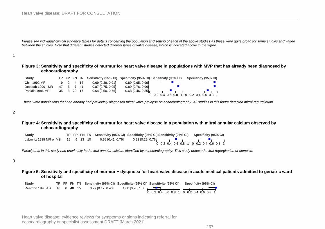
Heart valve disease: DRAFT FOR CONSULTATION
Heart valve disease: evidence reviews for symptoms or signs indicating referral for echocardiography or specialist assessment DRAFT [March 2021] 237
Please see individual clinical evidence tables for details concerning the population and setting of each of the above studies as these were quite broad for some studies and varied between the studies. Note that different studies detected different types of valve disease, which is indicated above in the figure.
1
Figure 3: Sensitivity and specificity of murmur for heart valve disease in populations with MVP that has already been diagnosed by echocardiography
These were populations that had already had previously diagnosed mitral valve prolapse on echocardiography. All studies in this figure detected mitral regurgitation.
2
Figure 4: Sensitivity and specificity of murmur for heart valve disease in a population with mitral annular calcium observed by echocardiography
Participants in this study had previously had mitral annular calcium identified by echocardiography. This study detected mitral regurgitation or stenosis.
3
Figure 5: Sensitivity and specificity of murmur + dyspnoea for heart valve disease in acute medical patients admitted to geriatric ward of hospital
Study
Chin 1992 MR
Decoodt 1990 - MR
Panidis 1986 MR
TP
9
47
35
FP
2
5
8
FN
4
7
20
TN
16
41
17
Sensitivity (95% CI)
0.69 [0.39, 0.91]
0.87 [0.75, 0.95]
0.64 [0.50, 0.76]
Specificity (95% CI)
0.89 [0.65, 0.99]
0.89 [0.76, 0.96]
0.68 [0.46, 0.85]
Sensitivity (95% CI)
0 0.2 0.4 0.6 0.8 1
Specificity (95% CI)
0 0.2 0.4 0.6 0.8 1
Study
Labovitz 1985 MR or MS
TP
19
FP
9
FN
13
TN
10
Sensitivity (95% CI)
0.59 [0.41, 0.76]
Specificity (95% CI)
0.53 [0.29, 0.76]
Sensitivity (95% CI)
0 0.2 0.4 0.6 0.8 1
Specificity (95% CI)
0 0.2 0.4 0.6 0.8 1
Study
Reardon 1996 AS
TP
18
FP
0
FN
48
TN
15
Sensitivity (95% CI)
0.27 [0.17, 0.40]
Specificity (95% CI)
1.00 [0.78, 1.00]
Sensitivity (95% CI)
0 0.2 0.4 0.6 0.8 1
Specificity (95% CI)
0 0.2 0.4 0.6 0.8 1

Heart valve disease: DRAFT FOR CONSULTATION
Heart valve disease: evidence reviews for symptoms or signs indicating referral for echocardiography or specialist assessment DRAFT [March 2021] 238
Participants in this study were >65 years of age and had to have systolic murmurs to be included. This study detected the presence of aortic stenosis.
1
Figure 6: Sensitivity and specificity of murmur + angina for heart valve disease in acute medical patients admitted to geriatric ward of hospital
Participants in this study were >65 years of age and had to have systolic murmurs to be included. This study detected the presence of aortic stenosis.
2
Figure 7: Sensitivity and specificity of murmur + other indication (dyspnoea, peripheral oedema or other) for heart valve disease in patients with suspected heart failure of heart valve disease)
Participants in this study were those with suspected heart failure or valve disease (restricted to: dyspnoea, cardiac murmur or peripheral oedema of unexplained origin). This study detected the presence of aortic stenosis, aortic regurgitation, mitral stenosis or mitral regurgitation.
3
Figure 8: Sensitivity and specificity of systolic murmur + absent/reduced second heart sound for heart valve disease
Study
Reardon 1996 AS
TP
2
FP
0
FN
64
TN
15
Sensitivity (95% CI)
0.03 [0.00, 0.11]
Specificity (95% CI)
1.00 [0.78, 1.00]
Sensitivity (95% CI)
0 0.2 0.4 0.6 0.8 1
Specificity (95% CI)
0 0.2 0.4 0.6 0.8 1
Study
Baur 2006 AS/R, MS/R
TP
12
FP
22
FN
8
TN
156
Sensitivity (95% CI)
0.60 [0.36, 0.81]
Specificity (95% CI)
0.88 [0.82, 0.92]
Sensitivity (95% CI)
0 0.2 0.4 0.6 0.8 1
Specificity (95% CI)
0 0.2 0.4 0.6 0.8 1
Study
Attenhofer Jost 2000 AS
Attenhofer Jost 2000 MR
McGee 2010 AS
Reardon 1996 AS
TP
8
3
0
26
FP
0
0
0
0
FN
20
27
0
40
TN
0
0
0
15
Sensitivity (95% CI)
0.29 [0.13, 0.49]
0.10 [0.02, 0.27]
Not estimable
0.39 [0.28, 0.52]
Specificity (95% CI)
Not estimable
Not estimable
Not estimable
1.00 [0.78, 1.00]
Sensitivity (95% CI)
0 0.2 0.4 0.6 0.8 1
Specificity (95% CI)
0 0.2 0.4 0.6 0.8 1

Heart valve disease: DRAFT FOR CONSULTATION
Heart valve disease: evidence reviews for symptoms or signs indicating referral for echocardiography or specialist assessment DRAFT [March 2021] 239
Please see individual clinical evidence tables for details concerning the population and setting of each of the above studies as these were quite broad for some studies and varied between the studies. Note that for McGee, the study reported a PLR value but did not provide sufficient information to calculate sensitivity or specificity. For Attenhofer Jost, sufficient information was available to calculate sensitivity, but there was insufficient data to calculate specificity.
1
Figure 9: Sensitivity and specificity of a non-flow murmur for heart valve disease in pregnant women
This study reported any valve abnormalities, and those included under our protocol were extracted as being reference standard positive. Any murmur that was considered to be pathological or possibly pathological by the auscultator in this study was considered to be index test positive, and flow murmurs were considered to be index test negative.
2
Figure 10: Sensitivity and specificity of murmur + abnormal ECG (left ventricular hypertrophy) for heart valve disease in acute medical patients admitted to geriatric ward of hospital
Participants in this study were >65 years of age and had to have systolic murmurs to be included. This study detected the presence of aortic stenosis.
3
Figure 11: Sensitivity and specificity of murmur + abnormal ECG (atrial fibrillation) for heart valve disease in acute medical patients admitted to geriatric ward of hospital
Participants in this study were >65 years of age and had to have systolic murmurs to be included. This study detected the presence of aortic stenosis.
4
Study
Mishra 1992 any valve
TP
4
FP
18
FN
0
TN
81
Sensitivity (95% CI)
1.00 [0.40, 1.00]
Specificity (95% CI)
0.82 [0.73, 0.89]
Sensitivity (95% CI)
0 0.2 0.4 0.6 0.8 1
Specificity (95% CI)
0 0.2 0.4 0.6 0.8 1
Study
Reardon 1996 AS
TP
15
FP
1
FN
51
TN
14
Sensitivity (95% CI)
0.23 [0.13, 0.35]
Specificity (95% CI)
0.93 [0.68, 1.00]
Sensitivity (95% CI)
0 0.2 0.4 0.6 0.8 1
Specificity (95% CI)
0 0.2 0.4 0.6 0.8 1
Study
Reardon 1996 AS
TP
10
FP
4
FN
56
TN
11
Sensitivity (95% CI)
0.15 [0.08, 0.26]
Specificity (95% CI)
0.73 [0.45, 0.92]
Sensitivity (95% CI)
0 0.2 0.4 0.6 0.8 1
Specificity (95% CI)
0 0.2 0.4 0.6 0.8 1

Heart valve disease: DRAFT FOR CONSULTATION
Heart valve disease: evidence reviews for symptoms or signs indicating referral for echocardiography or specialist assessment DRAFT [March 2021] 240
E.1.1.2 Reference standard – cardiac catheterisation 1
Note that sensitivity and specificity for studies where all had participants had to have a murmur to be included could not be calculated due to them 2 all being index test positive. Positive predictive values have instead been reported for these studies to provide information on the proportion with a 3 murmur that may actually go on to have echocardiography-confirmed heart valve disease. 4
Figure 12: Sensitivity and specificity of murmur for heart valve disease in various settings in populations with various indications for assessment
Please see individual clinical evidence tables for details concerning the population and setting of each of the above studies as these were quite broad for some studies and varied between the studies. Note that the type of murmur varied between studies.
5
Study
Breisblatt 1988 MR
Lehmann 1992 MR
Meyers 1982 AR
Meyers 1986 MR
TP
26
5
48
14
FP
36
19
2
1
FN
3
22
18
5
TN
85
160
7
15
Sensitivity (95% CI)
0.90 [0.73, 0.98]
0.19 [0.06, 0.38]
0.73 [0.60, 0.83]
0.74 [0.49, 0.91]
Specificity (95% CI)
0.70 [0.61, 0.78]
0.89 [0.84, 0.93]
0.78 [0.40, 0.97]
0.94 [0.70, 1.00]
Sensitivity (95% CI)
0 0.2 0.4 0.6 0.8 1
Specificity (95% CI)
0 0.2 0.4 0.6 0.8 1

Heart valve disease: DRAFT FOR CONSULTATION
Heart valve disease: evidence reviews for symptoms or signs indicating referral for echocardiography or specialist assessment DRAFT [March 2021] 241
E.2 Symptoms and signs for direct referral to a specialist 1
E.2.1 Coupled sensitivity and specificity forest plots 2
Note that sensitivity and specificity from one study where all had participants had a murmur + syncope on exertion with or without chest pain to be 3 included in the analysis could not be calculated due to them all being index test positive. Positive predictive values have instead been reported for 4 this study to provide information on the proportion with this indication that may actually go on to have echocardiography-confirmed heart valve 5 disease. 6
7
Figure 13: Sensitivity and specificity of murmur for moderate or severe heart valve disease in various settings in populations with various indications for assessment
Please see individual clinical evidence tables for details concerning the population and setting of each of the above studies as these were quite broad for some studies and varied between the studies. Note that different studies detected different types of valve disease, which is indicated above in the figure. Note that for some data was available for only severe disease, whereas others reported moderate or severe disease.
8
9
Study
Aggarwal 2014 significant HVD
Aronow 1989 moderate or severe AR
Aronow 1991 severe AS
Etchells 1998 moderate or severe MR
Iversen 2008 moderate or severe AS, AR, MS or MR
Loperfido 1986 grade 3+ MR
McGee 2010 moderate or severe MR
McGee 2010 moderate or severe TR
McGee 2010 severe AS
Rahko 1989 3+ or 4+ AR
Rahko 1989 3+ or 4+ MR
Rahko 1989 3+ or 4+ TR
TP
9
70
19
14
117
2
60
47
26
30
33
13
FP
22
43
0
56
532
14
161
174
191
57
86
18
FN
5
4
0
0
28
2
14
18
0
3
6
8
TN
64
333
0
42
2300
54
141
137
150
313
269
277
Sensitivity (95% CI)
0.64 [0.35, 0.87]
0.95 [0.87, 0.99]
1.00 [0.82, 1.00]
1.00 [0.77, 1.00]
0.81 [0.73, 0.87]
0.50 [0.07, 0.93]
0.81 [0.70, 0.89]
0.72 [0.60, 0.83]
1.00 [0.87, 1.00]
0.91 [0.76, 0.98]
0.85 [0.69, 0.94]
0.62 [0.38, 0.82]
Specificity (95% CI)
0.74 [0.64, 0.83]
0.89 [0.85, 0.92]
Not estimable
0.43 [0.33, 0.53]
0.81 [0.80, 0.83]
0.79 [0.68, 0.88]
0.47 [0.41, 0.52]
0.44 [0.38, 0.50]
0.44 [0.39, 0.49]
0.85 [0.81, 0.88]
0.76 [0.71, 0.80]
0.94 [0.91, 0.96]
Sensitivity (95% CI)
0 0.2 0.4 0.6 0.8 1
Specificity (95% CI)
0 0.2 0.4 0.6 0.8 1

Heart valve disease: DRAFT FOR CONSULTATION
Heart valve disease: evidence reviews for symptoms or signs indicating referral for echocardiography or specialist assessment DRAFT [March 2021] 242
Figure 14: Sensitivity and specificity of murmur for moderate or severe heart valve disease in populations with MVP that has already been diagnosed by echocardiography
These were populations that had already had previously diagnosed mitral valve prolapse on echocardiography. All studies in this figure detected mitral regurgitation.
1
2
Figure 15: Sensitivity and specificity of murmur for moderate or severe heart valve disease in a population with mitral annular calcium observed by echocardiography
Participants in this study had previously had mitral annular calcium identified by echocardiography. For moderate or severe disease, data were only provided for mitral regurgitation and not mitral stenosis.
3
Figure 16: Sensitivity and specificity of murmur + syncope for significant heart valve disease in acute medical patients admitted to geriatric ward of hospital
Note that it was unclear whether the peak or mean gradient was being referred to in the study to define significant aortic stenosis. Participants in this study were >65 years of age and had to have systolic murmurs to be included.
4
Study
Decoodt 1990 - severe MR
Panidis 1986 moderate or severe MR
TP
10
8
FP
42
35
FN
0
0
TN
48
37
Sensitivity (95% CI)
1.00 [0.69, 1.00]
1.00 [0.63, 1.00]
Specificity (95% CI)
0.53 [0.43, 0.64]
0.51 [0.39, 0.63]
Sensitivity (95% CI)
0 0.2 0.4 0.6 0.8 1
Specificity (95% CI)
0 0.2 0.4 0.6 0.8 1
Study
Labovitz 1985 moderate or severe MR
TP
11
FP
17
FN
6
TN
17
Sensitivity (95% CI)
0.65 [0.38, 0.86]
Specificity (95% CI)
0.50 [0.32, 0.68]
Sensitivity (95% CI)
0 0.2 0.4 0.6 0.8 1
Specificity (95% CI)
0 0.2 0.4 0.6 0.8 1
Study
Reardon 1996 significant AS (gradient 30 mmHg)
TP
3
FP
2
FN
18
TN
58
Sensitivity (95% CI)
0.14 [0.03, 0.36]
Specificity (95% CI)
0.97 [0.88, 1.00]
Sensitivity (95% CI)
0 0.2 0.4 0.6 0.8 1
Specificity (95% CI)
0 0.2 0.4 0.6 0.8 1

Heart valve disease: DRAFT FOR CONSULTATION
Heart valve disease: evidence reviews for symptoms or signs indicating referral for echocardiography or specialist assessment DRAFT [March 2021] 243
Figure 17: Sensitivity and specificity of murmur + dyspnoea for significant heart valve disease in acute medical patients admitted to geriatric ward of hospital
Note that it was unclear whether the peak or mean gradient was being referred to in the study to define significant aortic stenosis. Participants in this study were >65 years of age and had to have systolic murmurs to be included.
1
Figure 18: Sensitivity and specificity of murmur + angina for significant heart valve disease in acute medical patients admitted to geriatric ward of hospital
Note that it was unclear whether the peak or mean gradient was being referred to in the study to define significant aortic stenosis. Participants in this study were >65 years of age and had to have systolic murmurs to be included.
2
Figure 19: Sensitivity and specificity of systolic murmur +absent/reduced second heart sound for heart valve disease
Please see individual clinical evidence tables for details concerning the population and setting of each of the above studies as these were quite broad for some studies and varied between the studies. Note that for McGee, the study reported a PLR value for moderate or severe MR but did not provide sufficient information to calculate sensitivity or specificity.
3
Study
Reardon 1996 significant AS (gradient 30 mmHg)
TP
9
FP
9
FN
12
TN
51
Sensitivity (95% CI)
0.43 [0.22, 0.66]
Specificity (95% CI)
0.85 [0.73, 0.93]
Sensitivity (95% CI)
0 0.2 0.4 0.6 0.8 1
Specificity (95% CI)
0 0.2 0.4 0.6 0.8 1
Study
Reardon 1996 significant AS (gradient 30 mmHg)
TP
0
FP
2
FN
21
TN
58
Sensitivity (95% CI)
0.00 [0.00, 0.16]
Specificity (95% CI)
0.97 [0.88, 1.00]
Sensitivity (95% CI)
0 0.2 0.4 0.6 0.8 1
Specificity (95% CI)
0 0.2 0.4 0.6 0.8 1
Study
Abe 2013 severe AS
McGee 2010 moderate or severe MR
Reardon 1996 significant AS (gradient 30 mmHg)
TP
17
0
19
FP
6
0
7
FN
10
0
2
TN
97
0
53
Sensitivity (95% CI)
0.63 [0.42, 0.81]
Not estimable
0.90 [0.70, 0.99]
Specificity (95% CI)
0.94 [0.88, 0.98]
Not estimable
0.88 [0.77, 0.95]
Sensitivity (95% CI)
0 0.2 0.4 0.6 0.8 1
Specificity (95% CI)
0 0.2 0.4 0.6 0.8 1

Heart valve disease: DRAFT FOR CONSULTATION
Heart valve disease: evidence reviews for symptoms or signs indicating referral for echocardiography or specialist assessment DRAFT [March 2021] 244
Figure 20: Sensitivity and specificity of murmur + abnormal ECG (left ventricular hypertrophy) for heart valve disease in acute medical patients admitted to geriatric ward of hospital
Note that it was unclear whether the peak or mean gradient was being referred to in the study to define significant aortic stenosis. Participants in this study were >65 years of age and had to have systolic murmurs to be included.
1
Figure 21: Sensitivity and specificity of murmur + abnormal ECG (atrial fibrillation) for heart valve disease in acute medical patients admitted to geriatric ward of hospital
Note that it was unclear whether the peak or mean gradient was being referred to in the study to define significant aortic stenosis. Participants in this study were >65 years of age and had to have systolic murmurs to be included.
E.2.2 Diagnostic association plots 2
3
One study reported diagnostic association data, instead of diagnostic accuracy data, for the association of systolic murmur + diminished aortic 4 closure sound with a subsequent diagnosis of moderate or severe aortic stenosis. 5
6
Study
Reardon 1996 significant AS (gradient 30 mmHg)
TP
8
FP
8
FN
13
TN
52
Sensitivity (95% CI)
0.38 [0.18, 0.62]
Specificity (95% CI)
0.87 [0.75, 0.94]
Sensitivity (95% CI)
0 0.2 0.4 0.6 0.8 1
Specificity (95% CI)
0 0.2 0.4 0.6 0.8 1
Study
Reardon 1996 significant AS (gradient 30 mmHg)
TP
3
FP
11
FN
18
TN
49
Sensitivity (95% CI)
0.14 [0.03, 0.36]
Specificity (95% CI)
0.82 [0.70, 0.90]
Sensitivity (95% CI)
0 0.2 0.4 0.6 0.8 1
Specificity (95% CI)
0 0.2 0.4 0.6 0.8 1

Heart valve disease: DRAFT FOR CONSULTATION
Heart valve disease: evidence reviews for symptoms or signs indicating referral for echocardiography or specialist assessment DRAFT [March 2021] 245
Figure 22: Diagnostic association OR for systolic murmur + diminished aortic closure sound and diagnosis of moderate or severe aortic stenosis
1
Study or Subgroup
Attenhofer Jost 2000
Total (95% CI)
Heterogeneity: Not applicable
Test for overall effect: Z = 3.00 (P = 0.003)
log[Odds Ratio]
2.6391
SE
0.879
Weight
100.0%
100.0%
IV, Fixed, 95% CI
14.00 [2.50, 78.41]
14.00 [2.50, 78.41]
Odds Ratio Odds Ratio
IV, Fixed, 95% CI
0.01 0.1 1 10 100-ve diagnos. association +ve diagnos. association

Heart valve disease: DRAFT FOR CONSULTATION
Heart valve disease: evidence reviews for symptoms or signs indicating referral for echocardiography or specialist assessment DRAFT [March 2021]
246
Appendix F – Economic evidence study selection

Heart valve disease: DRAFT FOR CONSULTATION
Heart valve disease: evidence reviews for symptoms or signs indicating referral for echocardiography or specialist assessment DRAFT [March 2021]
247
1
Records screened in 1st sift, n=1260
Full-text papers assessed for eligibility in 2nd sift, n=195
Records excluded* in 1st sift, n=1065
Papers excluded* in 2nd sift, n=154
Papers included n=14 (0 studies) Studies included by review:
• 1.1 and 1.2, Signs and symptoms: n=0
• 1.3, Indications for specialist referral: n=0
• 1.4 Stress testing and stress ECG: n=0
• 1.5, Cardiac MRI and CT: n=0
• 2.1, Pharmacological management: n=0
• 2.2, Pharmacological management no HF: n=0
• 3.1, Indications for intervention: n=0
• 4.1, Interventions: n=14
• 4.2, Repeat intervention: n=0
• 5.1, Antithrombotic: n=0
• 6.1, Monitoring before an intervention: n=0
• 6.2, Monitoring after an intervention: n=0
Papers selectively excluded, n=27 (0 studies) Studies selectively excluded by review:
• 1.1 and 1.2, Signs and symptoms: n=0
• 1.3, Indications for specialist referral: n=0
• 1.4 Stress testing and stress ECG: n=0
• 1.5, Cardiac MRI and CT: n=0
• 2.1, Pharmacological management: n=0
• 2.2, Pharmacological management no HF: n=0
• 3.1, Indications for intervention: n=0
• 4.1, Interventions: n=27
• 4.2, Repeat intervention: n=0
• 5.1, Antithrombotic: n=0
• 6.1, Monitoring before an intervention: n=0
• 6.2, Monitoring after an intervention: n=0
Records identified through database searching, n=1258
Additional records identified through other sources: n=2
Full-text papers assessed for applicability and quality of methodology, n=41
Papers excluded, n=0 (0 studies) Studies excluded by review:
• 1.1 and 1.2, Signs and symptoms: n=0
• 1.3, Indications for specialist referral: n=0
• 1.4 Stress testing and stress ECG: n=0
• 1.5, Cardiac MRI and CT: n=0
• 2.1, Pharmacological management: n=0
• 2.2, Pharmacological management no HF: n=0
• 3.1, Indications for intervention: n=0
• 4.1, Interventions: n=0
• 4.2, Repeat intervention: n=0
• 5.1, Antithrombotic: n=0
• 6.1, Monitoring before an intervention: n=0
• 6.2, Monitoring after an intervention: n=0
* Non-relevant population, intervention, comparison, design or setting; non-English language

Heart valve disease: DRAFT FOR CONSULTATION
Heart valve disease: evidence reviews for symptoms or signs indicating referral for echocardiography or specialist assessment DRAFT [March 2021] 248
Appendix G – Economic evidence tables 1
2
None.3

Heart valve disease: DRAFT FOR CONSULTATION
Heart valve disease: evidence reviews for symptoms or signs indicating referral for echocardiography or specialist assessment DRAFT [March 2021]
249
Appendix H – Health economic model 1
No original economic modelling was undertaken. 2
3

Heart valve disease: DRAFT FOR CONSULTATION
Heart valve disease: evidence reviews for symptoms or signs indicating referral for echocardiography or specialist assessment DRAFT [March 2021]
250
Appendix I – Excluded studies 1
I.1 Symptoms and signs indicating echocardiography referral 2
Clinical studies 3
Table 37: Studies excluded from the clinical review 4
Reference Reason for exclusion
Abbasi 19831 Incorrect target condition - mitral valve prolapse.
Incorrect index test/reference standard - echo as the index test. Incorrect study design - case-control.
Abdulla 19812 Incorrect study design – two-gate (separate populations included - those with confirmed AR and those with absence of heart disease on catheterisation). Incorrect index test (heart sounds alone). Incorrect outcome/analysis - not diagnosis of HVD.
Abe 20133 Incorrect target condition: limits to moderate or severe disease. Considered for inclusion in another review focusing on severe disease.
Abernethy 19944 Incorrect population: all under 18 years of age
Ahlstrom 20067 Incorrect index test - algorithms for classification of different types of valve disease.
Ahlstrom 20076 Incorrect study design: not a diagnostic accuracy study
Ahmad 20198 Incorrect index test: algorithm for classifying types of heart sounds/murmurs
Ahmed 20099 Incorrect study design- two-gate, case control (all had murmur and MR already diagnosed).
Ahuja 198210 Incorrect index test - ability of physician to distinguish between innocent and pathological murmur rather than presence or absence of murmur
Anjorin 198412 Incorrect population: all already diagnosed with AS. Incorrect diagnosis: assessing severity in those with established AS rather than diagnosing AS.
Ansari 198513 Incorrect population: suspected MVP and already had negative echo
Ari 200815 Incorrect study design
Ari 200814 Incorrect study design
Babaei 200920 Incorrect index test - various algorithms for classification of different types of valve disease.
Betriu 197524 Incorrect target condition: mitral valve prolapse
Bloch 200125 Insufficient information to calculate diagnostic accuracy data.
Bodegard 201226
Incorrect population: healthy cohort rather than suspected valve disease
Brusco 200528 Incorrect index test: algorithm for classifying types of heart sounds/murmurs
Cha 198130 Incorrect population, index test and reference standard
Chabchoub 201831
Incorrect study design: known HVD vs control and classification algorithm not physician assessment
Chambers 201432
Incorrect target condition: regurgitation/stenosis mixed in with other valve abnormalities that may just be structural

Heart valve disease: DRAFT FOR CONSULTATION
Heart valve disease: evidence reviews for symptoms or signs indicating referral for echocardiography or specialist assessment DRAFT [March 2021]
251
Reference Reason for exclusion
Chen 201233 Incorrect index test - algorithm used to distinguish between innocent and organic murmurs. No assessment of whether murmur diagnostic for HVD.
Choi 201036 Incorrect index test: assessing accuracy of algorithm to classify different heart sounds
Choi 201535 Incorrect study design: Known HVD vs normal heart sounds
Choudhry 199937
Incorrect study design: narrative review. References checked.
Cohen 198738 Incorrect population: known mitral valve prolapse Incorrect target condition: severity of MVP
Cohen 198840 Incorrect study design: narrative review. References checked.
Cohen 197939 Incorrect target condition: mitral valve prolapse rather than regurgitation/stenosis.
Comak 200742 Incorrect index test: assessing accuracy of algorithm to classify different heart sounds
Comak 201241 Incorrect index test: assessing accuracy of algorithm to classify different heart sounds
Come 198343 Incorrect outcome/analysis: insufficient information to calculate accuracy data for mitral regurgitation
Come 198644 Incorrect index test – auscultation or phonocardiogram Insufficient information to calculate accuracy data for TR and AR
Danielsen 199145
Incorrect index test: no information on number with/without murmur. Incorrect diagnosis: no information on number with/without AS - only compares those with significant AS vs. those without significant AS, looking for predictors of more severe AS.
Darsee 197946 Incorrect population: healthy young men Incorrect diagnosis: mitral valve prolapse rather than stenosis/regurgitation specifically.
Das 200047 Incorrect study design - narrative review. References checked.
Das 200948 Incorrect population and study design and unclear tests used
De Panfilis 201349
Incorrect index test - algorithm used to distinguish between innocent and organic heart sounds.
Debbal 200550 Incorrect study design/index test - no assessment of diagnostic accuracy of any signs/symptoms.
Deng 199052 Incorrect target condition: MVP and incorrect index test
Denham 197753 Incorrect diagnosis: valve abnormalities (structural) rather than presence of stenosis/regurgitation specifically. Incorrect reference standard/population: findings on post-mortem
Desjardins 199654
Incorrect study design - two-gate (case-control) as separate groups of those with confirmed VD and those without VD.
Devereux 198956
Incorrect study design: narrative review - references checked.
Devereux 199457
Incorrect diagnosis: diagnosis of mitral valve prolapse, with no information on the number that also had mitral regurgitation/stenosis
Devereux 198655
Incorrect target condition: mitral valve prolapse rather than stenosis/regurgitation specifically.
Dittmann 198758 Incorrect index test: auscultation, but not clear whether this involved detection of murmur or whether other signs could also mean auscultation was positive.

Heart valve disease: DRAFT FOR CONSULTATION
Heart valve disease: evidence reviews for symptoms or signs indicating referral for echocardiography or specialist assessment DRAFT [March 2021]
252
Reference Reason for exclusion
Draper 201959 Incorrect diagnosis: some were diagnosed with non-valve disease conditions and included in the values reported – insufficient information to work out values for heart valve disease alone
Ellison 197660 Incorrect population: congenital AS in children and young adults
Esper 198261 Incorrect study design: predefined groups with and without murmur, and with and without AR
Etchells 199762 Incorrect study design: systematic review and insufficient quality assessment. References checked.
Etchells 199863 Incorrect target condition: limits to moderate or severe disease. Considered for inclusion in another review focusing on severe disease.
Fabich 201664 Incorrect comparison: handheld vs standard echo
Fahad 201865 Incorrect index test - algorithm used to distinguish between different types of heart sounds automatically.
Figueroa 200166 Incorrect index test: clinical examination, unclear now positive test was defined
Fink 199467 Incorrect target population: cardiac lesions (40% non-HVD diagnoses)
Forssell 198568 Incorrect population: known HVD
Fukuda 199569 Incorrect analysis / insufficient reporting
Gahl 197770 Incorrect population: those undergoing cardiac surgery - not group that would be under consideration for echocardiography referral as they would have full cardiac assessment
Gamaza-Chulian 202071
Incorrect population: all already diagnosed with valve disease
Gardin 198073 Incorrect target condition: mitral valve prolapse rather than existence of stenosis/regurgitation specifically.
Gharehbaghi 201574
Incorrect index test - algorithm used to distinguish between innocent and organic murmurs.
Goli 199375 Incorrect population: all included had diagnosed aortic regurgitation. Incorrect diagnosis: aim was to assess severity of AR rather than diagnose it.
Grayburn 198676 Incorrect population: already diagnosed with or without valve disease
Griffiths 197577 Incorrect study design: no use of a reference standard to assess accuracy of murmur in diagnosis of valve disease
Guillermo 201578
Incorrect study design: no accuracy or association data
Haikal 198279 Incorrect target condition: diagnosis of mitral valve prolapse rather than regurgitation/stenosis.
Heidenreich 200480
Incorrect population: Screening of a population at increased risk of HVD due to treatment received.
Herold 200581 Incorrect index test: algorithm/features of the algorithm to diagnose/classify valve disease.
Hershman 199082
Incorrect outcome/analysis: insufficient information to work out diagnostic accuracy for presence of murmur in HVD
Higuchi 200683 Incorrect study design: no accuracy or association data
Hirata 199284 Incorrect population: patients have a condition that means they will already be seeing specialists and being monitored by echocardiography
Hoagland 198685
Incorrect target condition: limits to severe/surgical valve disease. Considered for inclusion in another review focusing on severe disease.

Heart valve disease: DRAFT FOR CONSULTATION
Heart valve disease: evidence reviews for symptoms or signs indicating referral for echocardiography or specialist assessment DRAFT [March 2021]
253
Reference Reason for exclusion
Homaeinezhad 201087
Incorrect index test: assessing accuracy of algorithm to classify different heart sounds.
Ilmurzynska 196688
Incorrect population: valve disease already diagnosed in all patients.
Iversen 200889 Incorrect target condition: limits to moderate or severe disease. Considered for inclusion in another review focusing on severe disease.
Iversen 200690 Incorrect population: valve disease already diagnosed prior to study
Jaffe 198891 Incorrect index test: no information provided for the presence/absence of murmur in those diagnosed with/without valve disease by the reference standard. Only gives diagnostic accuracy results for significant valve disease and this was based on clinical measures other than a murmur.
Jeyaseelan 200792
Incorrect index test: insufficient information to calculate accuracy data for murmur as diagnostic factor
Jick 199893 Incorrect study design: retrospective review of records and echo not performed on all patients to confirm diagnosis
Johnson 198395 Incorrect population: already diagnosed AS.
Johnson 198596 Incorrect population: already diagnosed valve disease.
Johnson 198694 Incorrect target condition: looking at diagnosis of mitral valve prolapse rather than regurgitation/stenosis specifically.
Kambe 197798 Incorrect index test: murmur on phonocardiogram rather than auscultation
Karar 201799 Incorrect index test: use of algorithm developed to automatically classify normal and various abnormal heart sounds.
Kavalier 1975100 Incorrect population: all had been diagnosed prior to the study. Incorrect study design: inclusion of cases and controls, 2-gate design. Incorrect index test and diagnosis: 4th heart sound alone and predicting severity of disease.
Kay 2017101 Incorrect index test - algorithm used to distinguish between different types of heart sounds automatically.
Kim 2003102 Incorrect population: all with previously diagnosed AS. Incorrect outcomes: comparing correlation between murmur features on auscultation and certain Doppler measurements.
Kinney 1989104 Incorrect study design: case series - all had confirmed MR.
Koegelenberg 2014105
Incorrect index test - algorithm used to distinguish between different types of heart sounds automatically.
Kolibash 1983106 Incorrect index test: auscultatory abnormalities could include clicks alone, without a murmur. Incorrect target condition: focus is on mitral valve prolapse rather than stenosis/regurgitation.
Krivokapich 1988107
Incorrect target condition: mitral valve prolapse.
Kumar 2008108 Incorrect index test - algorithm used to distinguish between different types of heart sounds automatically.
Landau 2008110 Incorrect population: screening of presumably healthy population - not suspected HVD
Lee 1995111 Not available: not in English language
Leech 1978112 Incorrect reference standard: stenosis diagnosed at operation/necroscopy in some and may have been time gap between this and when murmur first detected.
Lembo 1988114 Incorrect study design: known valve disease prior to study

Heart valve disease: DRAFT FOR CONSULTATION
Heart valve disease: evidence reviews for symptoms or signs indicating referral for echocardiography or specialist assessment DRAFT [March 2021]
254
Reference Reason for exclusion
Liberfarb 1986115
Incorrect target condition: mitral valve prolapse rather than stenosis/regurgitation specifically.
Liberthson 1986116
Incorrect target condition: mitral valve prolapse.
Lingamneni 1979118
Incorrect population: valve disease already diagnosed
Lippman 1985119 Incorrect target condition: mitral valve prolapse rather than stenosis/regurgitation specifically.
Lockhart 1989120 Incorrect target condition: diagnosis of mitral valve prolapse rather than stenosis/regurgitation
Lopez 1985122 Incorrect study design: case-control, 2-gate. Includes group with confirmed HVD and a normal control group.
Loxdale 2012123 Incorrect population: Those with hip fractures - no further reasons to suspect HVD so is more of a screening study.
Luisada 1980124 Incorrect study design: case control, 2-gate.
Maglogiannis 2009125
Incorrect population and study design and unclear tests used
Maisel 1984126 Incorrect study design: case control two-gate - all known to have/not have valve disease prior to study.
Markiewicz 1976127
Incorrect population - presumably healthy individuals so is a screening study - not those with suspected HVD. Incorrect target condition: prolapse rather than stenosis/regurgitation being present.
Marsalese 1989128
Incorrect study design - compares outcomes for different interventions rather than accuracy of different diagnostic factors for valve disease. Insufficient information to calculate diagnostic accuracy of murmur for HVD.
Martin 2009129 Incorrect index test: physical exam in general which included physician interpretation of their findings. No information regarding presence/absence of murmur and diagnosis of HVD.
McBrien 2009130 Incorrect population: hip fractures, more of a screening study as no reason to suspect HVD
McClelland 2020131
Incorrect target conditions: combines valve disease with other abnormalities
McGee 2011133 Incorrect study design - abstract only
McKillop 1991134 Incorrect target condition: limits to severe/surgical valve disease. Considered for inclusion in another review focusing on severe disease.
Mehta 2014135 Incorrect index test: physical examination, not specifically the presence/absence of murmur.
Menahem 1986136
Incorrect population: all under 18 years of age
Meyers 1985138 Incorrect index test: auscultation with no mention of whether murmur was considered to be positive test - could have also referred to heart sounds alone, or may have been on physician interpretation as to whether a murmur was innocent or pathological.
Meziani 2013141 Incorrect study design: no accuracy or association data
Meziani 2018140 Incorrect study design: not a diagnostic accuracy study
Minich 1997142 Incorrect population: children
Missri 1985144 Incorrect study design: two gate, case control design. Confirmed TR and a control group with no disease.

Heart valve disease: DRAFT FOR CONSULTATION
Heart valve disease: evidence reviews for symptoms or signs indicating referral for echocardiography or specialist assessment DRAFT [March 2021]
255
Reference Reason for exclusion
Movahed 2007145
Incorrect study design/population: retrospective analysis of all who had echo. May be many with murmur detected who were not sent for echo and so not included in this analysis
Munt 1999146 Incorrect population: known HVD
Nakamura 1984147
Incorrect population: known AS (diagnosing severity)
Naseri 2013148 Incorrect index test: assesses accuracy of an algorithm
Nienaber 1993 152
Incorrect index test: no information for number with murmur and subsequent AR detected
Nitta 1987153 Incorrect population: all with known AS. Incorrect index test: no use of murmur alone to detect presence/absence of HVD.
Noah 1987154 Incorrect population: healthy sample rather than those with suspected valve disease - differs from population likely to be used in. Incorrect target condition: mitral valve prolapse
Noble 1982155 Incorrect study design: different groups with or without evidence of prolapse on both auscultation and echo enrolled - different cohorts. Incorrect target condition: mitral valve prolapse
Nygaard 1993156 Incorrect population - all with diagnosed aortic stenosis. Incorrect target condition: assessing severity in those with established AS rather than diagnosing AS.
Nygaard 1993157 Incorrect outcome/analysis: cannot calculate diagnostic accuracy.
Nylander 1986158
Incorrect population: all had established AS before the study. Incorrect target condition: assessing severity of AS rather than presence/absence of it.
Oh 2020159 Incorrect index test: algorithm used to classify different types of HVD
Oladapo 2001160 Incorrect population: presumably healthy volunteers. Screening study rather than those with suspected HVD.
Olive 1990161 Incorrect target condition: diagnosis of mitral valve prolapse (structural) rather than stenosis/regurgitation specifically.
Oweis 2014162 Incorrect index test - algorithm used to distinguish between different types of heart sounds automatically.
Papadaniil 2014164
Incorrect study design and index test (heart sounds alone)
Parras 2015165 Incorrect population: all previously diagnosed with valve disease
Patel 2017166 Incorrect index test - auscultation rather than detection of murmur specifically. No information with regards to those with/without diagnosis of HVD and number with/without murmur in each case
Patidar 2013167 Incorrect index test - algorithm used to distinguish between different types of heart sounds automatically.
Patnaik 2019168 Incorrect study design: narrative review – references checked.
Phoon 2001169 Incorrect population: known HVD. Incorrect index test and analysis (correlation only)
Procacci 1976170 Incorrect population: healthy young women rather than those with suspected valve disease. Incorrect target condition: mitral valve prolapse
Rama 1999172 Incorrect population: all had diagnosed AS. Incorrect target condition: aim is to assess correlation of murmur intensity and other physical findings with severity of AS.
Ranganathan 1976173
Incorrect outcomes/analysis: insufficient information to calculate diagnostic accuracy for HVD

Heart valve disease: DRAFT FOR CONSULTATION
Heart valve disease: evidence reviews for symptoms or signs indicating referral for echocardiography or specialist assessment DRAFT [March 2021]
256
Reference Reason for exclusion
Rispler 1995176 Incorrect study design - case series. All had diagnosed AS and clinical characteristics were reviewed to compare between those where it was suspected and those where it was not suspected prior to echo.
Roldan 1996179 Incorrect population - large proportion (48%) were presumably healthy and remaining population were those with connective tissue diseases but no further symptoms of heart disease. Does not represent population with suspected HVD.
Roldan 1997177 Incorrect population - large proportion (48%) were presumably healthy, and remaining population were those with connective tissue diseases but no further symptoms of heart disease. Does not represent population with suspected HVD.
Roldan 2000178 Incorrect population: Screening of a population at increased risk of it due to treatment received.
Rouhani 2012180 Incorrect study design: no accuracy or association data
Rueda 1988181 Incorrect study design: narrative review. References checked
Rujoie 2020182 Incorrect index test - algorithm used to distinguish between different types of heart sounds automatically.
Saal 1985183 Incorrect population: valve disease already diagnosed
Saeidi 2017185 Incorrect index test - algorithm used to distinguish between different types of heart sounds automatically.
Saeidi 2020184 Incorrect study design: deriving an algorithm using data from known HVD and controls
Safara 2012188 Incorrect index test - algorithm used to automatically classify heart sounds into different pathologies.
Safara 2013187 Incorrect index test - algorithm used to automatically classify heart sounds into different pathologies.
Safara 2015186 Incorrect index test - algorithm used to automatically classify heart sounds into different pathologies.
Salah 2020189 Incorrect index test - algorithm used to distinguish between different types of heart sounds and diagnose HVD automatically.
Saraf 2019190 Incorrect index test – algorithm used to diagnose HVD
Sarasin 2002191 Incorrect target condition: limits to moderate or severe disease. Considered for inclusion in another review focusing on severe disease.
Sathesh 2020192 Incorrect index test – algorithm used to classify auscultation sounds
Sbarbaro 1979193
Incorrect population: sample of healthy individuals, not suspected HVD (more like a screening study). Incorrect target condition: mitral valve prolapse (structural feature) rather than stenosis/regurgitation specifically.
Schnittger 1988194
Incorrect outcome/analysis: insufficient information to be able to calculate diagnostic accuracy measures
Sengur 2008195 Incorrect index test: algorithm used to automatically classify heart sounds.
Shry 2001196 Incorrect population: healthy individuals screened for presence of murmur. No other indication for suspicion of HVD.
Shub 2003197 Incorrect study design: narrative review. References checked
Sinha 2007198 Incorrect index test: algorithm used to automatically classify heart sounds.
Smith 1977 199 Incorrect target condition: mitral valve prolapse rather than stenosis or regurgitation
Spencer 2001 200
Incorrect study design: diagnosis already known

Heart valve disease: DRAFT FOR CONSULTATION
Heart valve disease: evidence reviews for symptoms or signs indicating referral for echocardiography or specialist assessment DRAFT [March 2021]
257
Reference Reason for exclusion
Stanger 2019201 Incorrect index test: SR of studies looking at diagnostic accuracy of handheld echo in valve disease - references checked
Strauss 1987202 Incorrect population: acute heart failure
Streib 1985203 Incorrect target condition: mitral valve prolapse
Sun 2005204 Incorrect study design: case control, 2-gate - presence/absence of AS already known on enrolment. Incorrect target condition: aim was to identify factors associated with different AS severity, not diagnosis of AS
Sztajzel 2010205 Incorrect index test: no info regarding presence/absence of murmur in the patients and results may be due to physician interpretation.
Thiyagaraja 2018206
Incorrect index test: classification model
Thomas 2016208 Incorrect study design: no accuracy or association data
Thomas 2018207 Incorrect reference test: hand-held echo
Thompson 2001209
Incorrect population: under 18 years of age. Incorrect index test: algorithm developed and accuracy for diagnosing different heart sounds assessed.
Thompson 2019210
Incorrect index test: algorithm used to automatically classify abnormal heart sounds/murmurs.
Tofler 1990211 Incorrect target condition: mitral valve prolapse Incorrect outcome/analysis: insufficient to be able to calculate diagnostic accuracy
Tokuda 2020212 Incorrect study design – assessing improvement in auscultation skills after a training session
Tribouilloy 2001213
Incorrect study design: case-control; and index test (3rd heart sound alone)
Turkoglu 2003214
Incorrect index test: classification model
Tutar 2005215 Incorrect population: children
Uguz 2012216 Incorrect index test: algorithm used to automatically classify heart sounds.
Uretsky 1982217 Incorrect study design: not all of the participants had echo or an alternative reference standard performed - some classified just based on auscultatory findings. Incorrect target condition: mitral valve prolapse
van Klei 2006218 Incorrect population: screening all of those undergoing non-cardiac surgery. Not representative of the population that would usually be considered for referral in current practice and is more of a screening study.
Varadarajan 2006219
Incorrect population: congestive heart failure monitoring by echocardiography already covered by NICE chronic heart failure guideline
Vargas-Barron 1984220
Incorrect population: children
Voelkel 1980221 Incorrect index test: no use of murmur as an index test/sign. Incorrect study design: all included had known and confirmed aortic stenosis, divided into severities.
Voss 2005222 Incorrect index test: algorithm used to automatically classify heart sounds.
Vourvouri 2005223
Incorrect index test: no indication of number with murmur in study and number subsequently diagnosed with valve disease

Heart valve disease: DRAFT FOR CONSULTATION
Heart valve disease: evidence reviews for symptoms or signs indicating referral for echocardiography or specialist assessment DRAFT [March 2021]
258
Reference Reason for exclusion
Wang 1984224 Incorrect study design - 2-gate case control, aortic regurgitation and controls already diagnosed before study. Incorrect outcome/analysis: insufficient to be able to calculate any accuracy measures.
Wann 1983225 Incorrect population: healthy young women so represents a screening study - not suspected HVD. Incorrect target condition: mitral valve prolapse
Ward 1977226 Incorrect population: includes children
Weis 1995227 Incorrect index test: heart sounds alone. Incorrect target condition: mitral valve prolapse
Wong 1983228 Incorrect target condition: Valve abnormalities rather than specifically stenosis or regurgitation.
Xu 1993229 Incorrect population: uses number of scans rather than number of patients to report results, meaning certain patients would be included more than once. Also appears to be large proportion <18 years of age included.
1
Health Economic studies 2
Published health economic studies that met the inclusion criteria (relevant population, 3 comparators, economic study design, published 2004 or later and not from non-OECD 4 country or USA) but that were excluded following appraisal of applicability and 5 methodological quality are listed below. See the health economic protocol for more details. 6
None. 7
I.2 Symptoms and signs indicating direct referral to a 8
specialist 9
Clinical studies 10
Table 38: Studies excluded from the clinical review 11
Reference Reason for exclusion
Abbasi 19831 Incorrect target condition - mitral valve prolapse.
Incorrect index test/reference standard - echo as the index test. Incorrect study design - case-control.
Abdulla 19812 Incorrect study design – two-gate (separate populations included - those with confirmed AR and those with absence of heart disease on catheterisation). Incorrect index test (heart sounds alone). Incorrect outcome/analysis - not diagnosis of HVD.
Abernethy 19944 Incorrect population: all under 18 years of age
Ahlstrom 20067 Incorrect index test - algorithms for classification of different types of valve disease.
Ahlstrom 20076 Incorrect study design: not a diagnostic accuracy study
Ahmad 20198 Incorrect index test: algorithm for classifying types of heart sounds/murmurs
Ahmed 20099 Incorrect study design- two-gate, case control (all had murmur and MR already diagnosed). Not able to calculate ay accuracy data as all had the disease and diagnostic feature (murmur)

Heart valve disease: DRAFT FOR CONSULTATION
Heart valve disease: evidence reviews for symptoms or signs indicating referral for echocardiography or specialist assessment DRAFT [March 2021]
259
Reference Reason for exclusion
Ahuja 198210 Incorrect index test - ability of physician to distinguish between innocent and pathological murmur rather than presence or absence of murmur
Amano 198611 Incorrect target condition: no information for severe valve disease specifically
Anjorin 198412 Incorrect population: all already diagnosed with AS. Incorrect diagnosis: assessing severity in those with established AS rather than diagnosing AS.
Ansari 198513 Incorrect population: suspected MVP and already had negative echo
Ari 200815 Incorrect study design
Ari 200814 Incorrect study design
Babaei 200920 Incorrect index test - various algorithms for classification of different types of valve disease.
Barron 198821 Incorrect target condition: no information for severe valve disease specifically
Barzilai 198822 Incorrect target condition: no information for severe valve disease specifically
Baur 200623 Incorrect target condition: no information for severe valve disease specifically
Betriu 197524 Incorrect target condition: mitral valve prolapse
Bloch 200125 Insufficient information to calculate diagnostic accuracy data.
Bodegard 201226
Incorrect population: healthy cohort rather than suspected valve disease
Breisblatt 198827 Incorrect reference standard: cardiac catheterisation not as good as echocardiography at quantifying severity of valve disease
Brusco 200528 Incorrect index test: algorithm for classifying types of heart sounds/murmurs
Cantley 199529 Incorrect target condition: no information for severe valve disease specifically
Cha 198130 Incorrect population, index test and reference standard
Chabchoub 201831
Incorrect study design: known HVD vs control and classification algorithm not physician assessment
Chambers 201432
Incorrect target condition: regurgitation/stenosis mixed in with other valve abnormalities that may just be structural
Chen 201233 Incorrect index test - algorithm used to distinguish between innocent and organic murmurs.
Chin 199234 Incorrect target condition: no information for severe valve disease specifically
Choi 201036 Incorrect index test: assessing accuracy of algorithm to classify different heart sounds
Choi 201535 Incorrect study design: Known HVD vs normal heart sounds
Choudhry 199937
Incorrect study design: narrative review. References checked.
Cohen 198738 Incorrect population: known mitral valve prolapse Incorrect target condition: severity of MVP
Cohen 198840 Incorrect study design: narrative review. References checked.
Cohen, 1979 39 Incorrect target condition: mitral valve prolapse rather than regurgitation/stenosis.
Comak 200742 Incorrect index test: assessing accuracy of algorithm to classify different heart sounds

Heart valve disease: DRAFT FOR CONSULTATION
Heart valve disease: evidence reviews for symptoms or signs indicating referral for echocardiography or specialist assessment DRAFT [March 2021]
260
Reference Reason for exclusion
Comak 201241 Incorrect index test: assessing accuracy of algorithm to classify different heart sounds
Come 198343 Incorrect outcome/analysis: insufficient information to calculate accuracy data for mitral regurgitation
Come 198644 Incorrect index test – auscultation or phonocardiogram Insufficient information to calculate accuracy data for TR and AR
Danielsen 199145
Incorrect index test: no information on number with/without murmur. Incorrect diagnosis: no information on number with/without AS - only compares those with significant AS vs. those without significant AS, looking for predictors of more severe AS.
Darsee 197946 Incorrect population: healthy young men. Incorrect diagnosis: mitral valve prolapse rather than stenosis/regurgitation specifically.
Das 200047 Incorrect study design - narrative review. References checked.
Das 200948 Incorrect population and study design and unclear tests used
De Panfilis 201349
Incorrect index test - algorithm used to distinguish between innocent and organic heart sounds.
Debbal 200550 Incorrect study design/index test - no assessment of diagnostic accuracy of any signs/symptoms.
Deng 199052 Incorrect target condition: MVP and incorrect index test
Denham 197753 Incorrect diagnosis: valve abnormalities (structural) rather than presence of stenosis/regurgitation specifically. Incorrect reference standard/population: findings on post-mortem
Desjardins 199654
Incorrect study design - two-gate (case-control) as separate groups of those with confirmed VD and those without VD.
Devereux 198956
Incorrect study design: narrative review - references checked.
Devereux 199457
Incorrect diagnosis: diagnosis of mitral valve prolapse, with no information on the number that also had mitral regurgitation/stenosis
Devereux 198655
Incorrect target condition: mitral valve prolapse rather than stenosis/regurgitation specifically.
Dittmann 198758 Incorrect index test: auscultation, but not clear whether this involved detection of murmur or whether other signs could also mean auscultation was positive.
Draper 201959 Incorrect diagnosis: some were diagnosed with non-valve disease conditions and included in the values reported – insufficient information to work out values for heart valve disease alone
Ellison 197660 Incorrect population: congenital AS in children and young adults
Esper 198261 Incorrect study design: predefined groups with and without murmur, and with and without AR
Etchells 199762 Incorrect study design: systematic review and insufficient quality assessment. References checked.
Fabich 201664 Incorrect comparison: handheld vs standard echo
Fahad 201865 Incorrect index test - algorithm used to distinguish between different types of heart sounds automatically.
Figueroa 200166 Incorrect index test: clinical examination, unclear now positive test was defined
Fink 199467 Incorrect target population
Forssell 198568 Incorrect population: known HVD
Fukuda 199569 Incorrect analysis / insufficient reporting

Heart valve disease: DRAFT FOR CONSULTATION
Heart valve disease: evidence reviews for symptoms or signs indicating referral for echocardiography or specialist assessment DRAFT [March 2021]
261
Reference Reason for exclusion
Gahl 197770 Incorrect population: those undergoing cardiac surgery - not group that would be under consideration for echocardiography referral as they would have full cardiac assessment
Gamaza-Chulian 202071
Incorrect population: all already diagnosed with valve disease
Gardezi 201872 Incorrect target condition: no information for severe valve disease specifically
Gardin 198073 Incorrect target condition: mitral valve prolapse rather than existence of stenosis/regurgitation specifically.
Gharehbaghi 201574
Incorrect index test - algorithm used to distinguish between innocent and organic murmurs.
Goli 199375 Incorrect population: all included had diagnosed aortic regurgitation. Incorrect diagnosis: aim was to assess severity of AR rather than diagnose it.
Grayburn 198676 Incorrect population: already diagnosed with or without valve disease
Griffiths 197577 Incorrect study design: no use of a reference standard to assess accuracy of murmur in diagnosis of valve disease
Guillermo 201578
Incorrect study design: no accuracy or association data
Haikal 198279 Incorrect target condition: diagnosis of mitral valve prolapse rather than regurgitation/stenosis.
Heidenreich 200480
Incorrect population: Screening of a population at increased risk of HVD due to treatment received.
Herold 200581 Incorrect index test: algorithm/features of the algorithm to diagnose/classify valve disease.
Hershman 199082
Incorrect outcome/analysis: insufficient information to work out diagnostic accuracy for presence of murmur in HVD
Higuchi 200683 Incorrect study design: no accuracy or association data
Hirata 199284 Incorrect population: patients have a condition that means they will already be seeing specialists and being monitored by echocardiography
Hoagland 198685
Incorrect target condition: limits to severe/surgical valve disease. Considered for inclusion in another review focusing on severe disease.
Hoffmann 198386
Incorrect target condition: no information for severe valve disease specifically. Incorrect reference standard: cardiac catheterisation not as good as echocardiography at quantifying severity of valve disease
Homaeinezhad 201087
incorrect index test: assessing accuracy of algorithm to classify different heart sounds
Ilmurzynska 196688
Incorrect population: valve disease already diagnosed in all patients.
Iversen 2006 90 Incorrect population: valve disease already diagnosed prior to study
Jaffe 198891 Incorrect index test: no information provided for the presence/absence of murmur in those diagnosed with/without valve disease by the reference standard. Only gives diagnostic accuracy results for significant valve disease and this was based on clinical measures other than a murmur.
Jeyaseelan 200792
Incorrect index test: insufficient information to calculate accuracy data for murmur as diagnostic factor
Jick 1998 93 Incorrect study design: retrospective review of records and echo not performed on all patients to confirm diagnosis
Johnson 198395 Incorrect population: already diagnosed AS.

Heart valve disease: DRAFT FOR CONSULTATION
Heart valve disease: evidence reviews for symptoms or signs indicating referral for echocardiography or specialist assessment DRAFT [March 2021]
262
Reference Reason for exclusion
Johnson 198596 Incorrect population: already diagnosed valve disease.
Johnson 198694 Incorrect target condition: looking at diagnosis of mitral valve prolapse rather than regurgitation/stenosis specifically.
Kalinauskiene 201997
Incorrect target condition: no information for severe valve disease specifically
Kambe 197798 Incorrect reference standard: cardiac catheterisation not as good as echocardiography at quantifying severity of valve disease. Incorrect index test: murmur on phonocardiogram rather than auscultation.
Karar 201799 Incorrect index test: use of algorithm developed to automatically classify normal and various abnormal heart sounds.
Kavalier 1975100 Incorrect population: all had been diagnosed prior to the study. Incorrect study design: inclusion of cases and controls, 2-gate design. Incorrect index test and diagnosis: 4th heart sound alone and predicting severity of disease.
Kay 2017101 Incorrect index test - algorithm used to distinguish between different types of heart sounds automatically.
Kim 2003102 Incorrect population: all with previously diagnosed AS. Incorrect outcomes: comparing correlation between murmur features on auscultation and certain Doppler measurements.
Kinney 1988103 Incorrect target condition: no information for severe valve disease specifically
Kinney 1989104 Incorrect study design: case series - all had confirmed MR.
Koegelenberg 2014105
Incorrect index test - algorithm used to distinguish between different types of heart sounds automatically.
Kolibash 1983106 Incorrect index test: auscultatory abnormalities could include clicks alone, without a murmur. Incorrect target condition: focus is on mitral valve prolapse rather than stenosis/regurgitation.
Krivokapich 1988107
Incorrect target condition: mitral valve prolapse
Kumar 2008108 Incorrect index test - algorithm used to distinguish between different types of heart sounds automatically.
Landau 2008110 Incorrect population: screening of presumably healthy population - not suspected HVD
Lee 1995111 Not available: not in English language
Leech 1978112 Incorrect reference standard: stenosis diagnosed at operation/necroscopy in some and may have been time gap between this and when murmur first detected.
Lehmann 1992113
Incorrect study design: known valve disease prior to study
Lembo 1988114 Incorrect target condition: mitral valve prolapse rather than stenosis/regurgitation specifically.
Liberfarb 1986115
Incorrect target condition: mitral valve prolapse.
Liberthson 1986116
Incorrect population: screening of presumably healthy population - not suspected HVD
Lingamneni 1979118
Incorrect population: valve disease already diagnosed
Lippman 1985119 Incorrect target condition: mitral valve prolapse rather than stenosis/regurgitation specifically
Lockhart 1989120 Incorrect target condition: diagnosis of mitral valve prolapse rather than stenosis/regurgitation

Heart valve disease: DRAFT FOR CONSULTATION
Heart valve disease: evidence reviews for symptoms or signs indicating referral for echocardiography or specialist assessment DRAFT [March 2021]
263
Reference Reason for exclusion
Lopez 1985122 Incorrect study design: case-control, 2-gate. Includes group with confirmed HVD and a normal control group.
Loxdale 2012123 Incorrect population: Those with hip fractures - no further reasons to suspect HVD so is more of a screening study.
Luisada 1980124 Incorrect study design: case control, 2-gate.
Maglogiannis 2009125
Incorrect population and study design and unclear tests used
Maisel 1984126 Incorrect study design: case control two-gate - all known to have/not have valve disease prior to study.
Markiewicz 1976127
Incorrect population - presumably healthy individuals so is a screening study - not those with suspected HVD. Incorrect target condition: prolapse rather than stenosis/regurgitation being present.
Marsalese 1989128
Incorrect study design - compares outcomes for different interventions rather than accuracy of different diagnostic factors for valve disease. Insufficient information to calculate diagnostic accuracy of murmur for HVD.
Martin 2009129 Incorrect index test: physical exam in general which included physician interpretation of their findings. No information regarding presence/absence of murmur and diagnosis of HVD.
McBrien 2009130 Incorrect population: hip fractures, more of a screening study as no reason to suspect HVD
McGee 2011133 Incorrect study design - abstract only
Mehta 2014135 Incorrect index test: physical examination, not specifically the presence/absence of murmur.
Menahem 1986136
Incorrect population: all under 18 years of age
Meyers 1982139 Incorrect reference standard: cardiac catheterisation not as good as echocardiography at quantifying severity of valve disease
Meyers 1985138 Incorrect index test: auscultation with no mention of whether murmur was considered to be positive test - could have also referred to heart sounds alone, or may have been on physician interpretation as to whether a murmur was innocent or pathological.
Meyers 1986137 Incorrect reference standard: cardiac catheterisation not as good as echocardiography at quantifying severity of valve disease
Meziani 2013141 Incorrect study design: no accuracy or association data
Meziani 2018140 Incorrect study design: not a diagnostic accuracy study
Minich 1997142 Incorrect population: children
Mishra 1992143 Incorrect target condition: no information for severe valve disease specifically
Missri 1985144 Incorrect study design: two gate, case control design. Confirmed TR and a control group with no disease.
Movahed 2007145
Incorrect study design/population: retrospective analysis of all who had echo. May be many with murmur detected who were not sent for echo and so not included in this analysis
Munt 1999146 Incorrect population: known HVD
Nakamura 1984147
Incorrect population: known AS (diagnosing severity)
Naseri 2013148 Incorrect index test: assesses accuracy of an algorithm
Nienaber 1993152
Incorrect index test: no information for number with murmur and subsequent AR detected

Heart valve disease: DRAFT FOR CONSULTATION
Heart valve disease: evidence reviews for symptoms or signs indicating referral for echocardiography or specialist assessment DRAFT [March 2021]
264
Reference Reason for exclusion
Nitta 1987153 Incorrect population: all with known AS. Incorrect index test: no use of murmur alone to detect presence/absence of HVD.
Noah 1987154 Incorrect population: healthy sample rather than those with suspected valve disease - differs from population likely to be used in. Incorrect target condition: mitral valve prolapse
Noble 1982155 Incorrect study design: different groups with or without evidence of prolapse on both auscultation and echo enrolled - different cohorts. Incorrect target condition: mitral valve prolapse
Nygaard 1993156 Incorrect population - all with diagnosed aortic stenosis. Incorrect target condition: assessing severity in those with established AS rather than diagnosing AS.
Nygaard 1993157 Incorrect outcome/analysis: cannot calculate diagnostic accuracy.
Nylander 1986158
Incorrect population: all had established AS before the study. Incorrect target condition: assessing severity of AS rather than presence/absence of it.
Oh 2020159 Incorrect index test: algorithm used to classify different types of HVD
Oladapo 2001160 Incorrect population: presumably healthy volunteers. Screening study rather than those with suspected HVD.
Olive 1990161 Incorrect target condition: diagnosis of mitral valve prolapse (structural) rather than stenosis/regurgitation specifically.
Oweis 2014162 Incorrect index test - algorithm used to distinguish between different types of heart sounds automatically.
Papadaniil 2014164
Incorrect study design and index test (heart sounds alone)
Parras 2015165 Incorrect population: all previously diagnosed with valve disease
Patel 2017166 Incorrect index test - auscultation rather than detection of murmur specifically. No information with regards to those with/without diagnosis of HVD and number with/without murmur in each case
Patidar 2013167 Incorrect index test - algorithm used to distinguish between different types of heart sounds automatically.
Patnaik 2019168 Incorrect study design: narrative review – references checked.
Phoon 2001169 Incorrect population: known HVD. Incorrect index test and analysis (correlation only)
Procacci 1976170 Incorrect population: healthy young women rather than those with suspected valve disease. Incorrect target condition: mitral valve prolapse
Rama 1999172 Incorrect population: all had diagnosed AS. Incorrect target condition: aim is to assess correlation of murmur intensity and other physical findings with severity of AS.
Ranganathan 1976173
Incorrect outcomes/analysis: insufficient information to calculate diagnostic accuracy for HVD
Reichlin 2004175 Incorrect target condition: no information for severe valve disease specifically
Rispler 1995176 Incorrect population: known HVD. Incorrect index test and analysis (correlation only)
Roldan 1996179 Incorrect population - large proportion (48%) were presumably healthy and remaining population were those with connective tissue diseases but no further symptoms of heart disease. Does not represent population with suspected HVD.
Roldan 1997177 Incorrect population - large proportion (48%) were presumably healthy, and remaining population were those with connective tissue diseases

Heart valve disease: DRAFT FOR CONSULTATION
Heart valve disease: evidence reviews for symptoms or signs indicating referral for echocardiography or specialist assessment DRAFT [March 2021]
265
Reference Reason for exclusion
but no further symptoms of heart disease. Does not represent population with suspected HVD.
Roldan 2000178 Incorrect population: Screening of a population at increased risk of it due to treatment received.
Rouhani 2012180 Incorrect study design: no accuracy or association data
Rueda 1988181 Incorrect study design: narrative review. References checked
Rujoie 2020182 Incorrect index test - algorithm used to distinguish between different types of heart sounds automatically.
Saal 1985183 Incorrect population: valve disease already diagnosed
Saeidi 2017185 Incorrect index test - algorithm used to distinguish between different types of heart sounds automatically.
Saeidi 2020184 Incorrect study design: deriving an algorithm using data from known HVD and controls
Safara 2012188 Incorrect index test - algorithm used to automatically classify heart sounds into different pathologies.
Safara 2013187 Incorrect index test - algorithm used to automatically classify heart sounds into different pathologies.
Safara 2015186 Incorrect index test - algorithm used to automatically classify heart sounds into different pathologies.
Salah 2020189 Incorrect index test - algorithm used to distinguish between different types of heart sounds and diagnose HVD automatically.
Saraf 2019190 Incorrect index test – algorithm used to diagnose HVD
Sathesh 2020192 Incorrect index test – algorithm used to classify auscultation sounds
Sbarbaro 1979193
Incorrect population: sample of healthy individuals, not suspected HVD (more like a screening study). Incorrect target condition: mitral valve prolapse (structural feature) rather than stenosis/regurgitation specifically.
Schnittger 1988194
Incorrect outcome/analysis: insufficient information to be able to calculate diagnostic accuracy measures
Sengur 2008195 Incorrect index test: algorithm used to automatically classify heart sounds.
Shry 2001196 Incorrect population: healthy individuals screened for presence of murmur. No other indication for suspicion of HVD.
Shub 2003197 Incorrect study design: narrative review. References checked
Sinha 2007198 Incorrect index test: algorithm used to automatically classify heart sounds.
Smith 1977199 Incorrect target condition: mitral valve prolapse rather than stenosis or regurgitation
Spencer 2001200 Incorrect study design: diagnosis already known
Stanger 2019201 Incorrect index test: SR of studies looking at diagnostic accuracy of handheld echo in valve disease -– references checked
Strauss 1987202 Incorrect population: acute heart failure
Streib 1985203 Incorrect target condition: mitral valve prolapse
Sun 2005204 Incorrect study design: case control, 2-gate - presence/absence of AS already known on enrolment. Incorrect target condition: aim was to identify factors associated with different AS severity, not diagnosis of AS
Sztajzel 2010205 Incorrect index test: no info regarding presence/absence of murmur in the patients and results may be due to physician interpretation.

Heart valve disease: DRAFT FOR CONSULTATION
Heart valve disease: evidence reviews for symptoms or signs indicating referral for echocardiography or specialist assessment DRAFT [March 2021]
266
Reference Reason for exclusion
Thiyagaraja 2018206
Incorrect index test: classification model
Thomas 2016208 Incorrect study design: no accuracy or association data
Thomas 2018207 Incorrect reference test: hand-held echo
Thompson 2001209
Incorrect population: under 18 years of age. Incorrect index test: algorithm developed and accuracy for diagnosing different heart sounds assessed.
Thompson 2019210
Incorrect index test: algorithm used to automatically classify abnormal heart sounds/murmurs.
Tofler 1990211 Incorrect target condition: mitral valve prolapse Incorrect outcome/analysis: insufficient to be able to calculate diagnostic accuracy
Tokuda 2020212 Incorrect study design – assessing improvement in auscultation skills after a training session
Tribouilloy 2001213
Incorrect study design: case-control; and index test (3rd heart sound alone)
Turkoglu 2003214
Incorrect index test: classification model
Tutar 2005215 Incorrect population: children
Uguz 2012216 Incorrect index test: algorithm used to automatically classify heart sounds.
Uretsky 1982217 Incorrect study design: not all of the participants had echo or an alternative reference standard performed - some classified just based on auscultatory findings. Incorrect target condition: mitral valve prolapse
van Klei 2006218 Incorrect population: screening all of those undergoing non-cardiac surgery. Not representative of the population that would usually be considered for referral in current practice and is more of a screening study.
Varadarajan 2006219
Incorrect population: congestive heart failure monitoring by echocardiography already covered by NICE chronic heart failure guideline
Vargas-Barron 1984220
Incorrect population: children
Voelkel 1980221 Incorrect index test: no use of murmur as an index test/sign. Incorrect study design: all included had known and confirmed aortic stenosis, divided into severities.
Voss 2005222 Incorrect index test: algorithm used to automatically classify heart sounds.
Vourvouri, 2005 223
Incorrect index test: no indication of number with murmur in study and number subsequently diagnosed with valve disease
Wang 1984224 Incorrect study design - 2-gate case control, aortic regurgitation and controls already diagnosed before study. Incorrect outcome/analysis: insufficient to be able to calculate any accuracy measures.
Wann 1983225 Incorrect population: healthy young women so represents a screening study - not suspected HVD. Incorrect target condition: mitral valve prolapse
Ward 1977226 Incorrect population: includes children
Weis 1995227 Incorrect index test: heart sounds alone. Incorrect target condition: mitral valve prolapse

Heart valve disease: DRAFT FOR CONSULTATION
Heart valve disease: evidence reviews for symptoms or signs indicating referral for echocardiography or specialist assessment DRAFT [March 2021]
267
Reference Reason for exclusion
Wong 1983228 Incorrect target condition: Valve abnormalities rather than specifically stenosis or regurgitation.
Xu 1993229 Incorrect population: uses number of scans rather than number of patients to report results, meaning certain patients would be included more than once. Also appears to be large proportion <18 years of age included.
Yamashita 2020230
Incorrect target condition: no information for severe valve disease specifically
1
Health Economic studies 2
Published health economic studies that met the inclusion criteria (relevant population, 3 comparators, economic study design, published 2004 or later and not from non-OECD 4 country or USA) but that were excluded following appraisal of applicability and 5 methodological quality are listed below. See the health economic protocol for more details. 6
None. 7
8

Heart valve disease: DRAFT FOR CONSULTATION
Heart valve disease: evidence reviews for symptoms or signs indicating referral for echocardiography or specialist assessment DRAFT [March 2021]
268
Appendix J – Research recommendations – full details 1
None 2
3

Heart valve disease: DRAFT FOR CONSULTATION
Heart valve disease: evidence reviews for symptoms or signs indicating referral for echocardiography or specialist assessment DRAFT [March 2021]
269
Appendix K – Expert witness testimony 1
Name: Dr Catherine Head
Role: Consultant Cardiologist
Institution/Organisation
(where applicable):
Contact information
To be redacted prior to publication
Norfolk and Norwich University Hospital
Norwich NR4 7UY
Guideline title: Heart valve disease
Guideline committee: Heart valve disease
Subject of expert testimony: Pregnant women and women considering pregnancy
Evidence gaps or uncertainties: There is limited information across the guideline for the population including pregnant women and women of childbearing age and information relating to this population in terms of the review questions listed below would be useful
No. Review question
1 In adults with suspected heart valve disease, what symptoms and signs indicate referral (for example from primary care) for echocardiography?
2 In adults with suspected heart valve disease, what symptoms and signs indicate direct referral (for example from primary care) to a specialist?
3 In adults who have had echocardiography, what are the indications for referral to a specialist?
4 In adults with heart valve disease, what is the predictive accuracy and cost effectiveness of stress testing and stress echocardiography to determine the need for intervention?
5 In adults with heart valve disease, what is the predictive accuracy and cost effectiveness of cardiac MRI and cardiac CT to determine the need for intervention?
6 In adults with heart failure and concomitant heart valve disease, what is the clinical and cost effectiveness of angiotensin-converting enzyme (ACE) inhibitors, angiotensin II receptor blockers (ARBs), beta-blockers, calcium channel blockers, digoxin, diuretics and nitrates to improve clinical outcome?

Heart valve disease: DRAFT FOR CONSULTATION
Heart valve disease: evidence reviews for symptoms or signs indicating referral for echocardiography or specialist assessment DRAFT [March 2021]
270
7 In adults with heart valve disease without concomitant heart failure, what is the clinical and cost effectiveness of ACE inhibitors, ARBs, alpha-blockers, beta blockers, calcium channel blockers, digoxin, diuretics, statins and nitrates to improve clinical outcome?
8 What are the indications that interventions should be offered to adults with asymptomatic, severe heart valve disease?
9 What is the clinical and cost effectiveness of transcatheter intervention, surgery (with mechanical or biological valves) and conservative management compared with each other for adults with heart valve disease?
10 What is the clinical and cost effectiveness of transcatheter or surgical repeat valve intervention for prosthetic biological valve degeneration?
11 What is the clinical and cost effectiveness of anticoagulant and/or antiplatelet therapy for adults with transcatheter or surgical biological prosthetic valves or after valve repair?
12 Where there is no current indication for intervention, what is the most clinically and cost-effective type and frequency of test for monitoring in adults with heart valve disease?
13 What is the most clinically and cost-effective frequency of echocardiography or clinical review for monitoring in adults with repaired or replaced heart valves?
14 What information and advice should adults with heart valve disease and their family and carers receive?
Summary testimony
Please use the space below to summarise your testimony in 250–1000 words. Continue over page if necessary ]
Indications for referral for echo
Echo systolic murmur is present in approximately 90% of women and therefore murmur alone should not be used as an indication for referral.
Pharmacological management
The interventions should be used in accordance with the BNF, for example statins are contraindicated in pregnancy
Indications for referral for intervention
Any women with heart valve disease should be referred to a cardiologist with specialist expertise
Diagnosis
Exercise testing is safe in pregnancy and pre-pregnancy. Low flow low gradient stenosis is not relevant for pregnancy

Heart valve disease: DRAFT FOR CONSULTATION
Heart valve disease: evidence reviews for symptoms or signs indicating referral for echocardiography or specialist assessment DRAFT [March 2021]
271
Monitoring before an intervention
A woman who is pregnancy needs a different frequency and type of monitoring. This needs to be determined by a multi-disciplinary team (MDT)
Interventions
The choice of a mechanical or biological valve needs to take into account the possibility of a future pregnancy. The decision should be taken in consultation with a MDT. The choice between TAVI and surgery is the same as for all people with heart valve disease.
Monitoring after an intervention
Whilst the women is pregnant monitoring needs to take place within the context of an MDT. After pregnancy monitoring is the same as for all people with heart valve disease
Anticoagulation
This is a highly specialised area and needs to be individualised to the type of valve, site of valve and risk factors
References to other work or publications to support your testimony (if applicable):
1
2