heart rate variability in cirrhosis liverrepository-tnmgrmu.ac.in/1580/1/160400209aravindh.pdf ·...
TRANSCRIPT
HEART RATE VARIABILITY IN CIRRHOSIS LIVER
Dissertation Submitted to
THE TAMIL NADU DR MGR MEDICAL UNIVERSITY
in partial fulfillment of the regulations
for the award of the degree of
DM (GASTROENTEROLOGY) BRANCH ndash IV
DEPARTMENT OF MEDICAL GASTROENTEROLOGY
GOVT STANLEY MEDICAL COLLEGE amp HOSPITAL
THE TAMIL NADU DR MGR MEDICAL UNIVERSITY
CHENNAI INDIA
AUGUST 2009
CERTIFICATE
This is to certify that the dissertation entitled ldquoHEART RATE VARIABILITY IN CIRRHOSIS
LIVERrdquo is the bonafide original work of Dr S ARAVINDH in partial fulfillment of the requirements
for DM (GASTROENTEROLOGY) BRANCH ndash IV Examination of the Tamilnadu Dr MGR
Medical University to be held in August 2009 The period of study was from June 2007 to December
2008
Dr J MOHANASUNDARAM MD PhD DNBDEAN
Govt Stanley Medical College amp HospitalChennai-600 001
Prof V JAYANTHI MDDMProfessor amp Head
Department of Medical GastroenterologyGovt Stanley Medical College
Chennai-600 001
ACKNOWLEDGEMENT
I express my profound gratitude to Dr J MOHANASUNDARAM MD PhD DNB Dean of
Government Stanley Medical College and Hospital Chennaindash600001 for permitting me to use all the
needed resources for this dissertation work
I sincerely express my grateful thanks to Prof V JAYANTHI MD DM Professor and
Head Department of Medical Gastroenterology Stanley Medical College for her unstinted support and
advice rendered throughout my study I thank her for being a constant source of encouragement
inspiration not only in this study but in all my professional endeavors
I express my gratitude to Prof Balasubramaniam MD Head of Dept and Assistant
Professor Dr K MURALIKRISHNAN MD Department of Physiology Stanley Medical College
conducting the tests in the Electrophysiology Lab Physiology dept
I express my sincere thanks to all the Assistant Professors Dr A Murali Dr T Rajkumar
Solomon Dr M S Revathy and Dr R Ravi Department of Medical Gastroenterology SMC
Chennai
My sincere thanks to Mr Xavier Lecturer in statistics Dept of Statistics Loyola college
Chennai for statistical analysis in preparation of this study
I also sincerely thank Ethical Committee SMC Chennai for approving my study
I extend my sincere thanks to my subjects but for them the project would not have been
possible
I am greatly indebted to all my friends Postgraduate colleagues who have been the greatest
source of encouragement support enthusiasms criticism friendly concern and timely help
CONTENTS
Serial No
Title Page No
1 INTRODUCTION 1
2 AIM OF THE STUDY 3
3 REVIEW OF LITERATURE 4
4 MATERIALS AND METHODS 38
5 RESULTS 41
6 DISCUSSION 49
7 SUMMARY amp CONCLUSION 52
8 REFERENCES 54
9 ANNEXURES 62
Proforma
Photographs
Master chart
Introduction
Hyperdynamic circulatory state is a common and long-recognized feature of patients with
decompensated chronic liver disease (1) It is characterized by elevated cardiac rate and output and
reduced peripheral vascular resistance with pooling of blood in the splanchnic and peripheral
circulation and effective central hypovolemia
The autonomic nervous system plays a central role in modulating cardiac performance and the
vasomotor activity in the hyperdynamic circulatory state (2)
The presence of an autonomic dysfunction (AD) in cirrhosis has been clearly shown through different
experimental approaches including the evaluation of the cardiovascular and sudomotor responses to
physiological and pharmacological stimulation (3-6) and by showing a hyperproduction of weak
adrenergic neurotransmitters (7)
It has also been reported that the severity of AD is proportional to the severity of cirrhosis (58) and its
presence is an indicator of poor prognosis in patients with both early and advanced liver disease (910)
In an earlier study from our department the presence of AD correlated with the occurrence of variceal
bleed (11)
Heart rate variability implies variations in the interval between consecutive heart beats as well as
between consecutive instantaneous heart rate ie it describes the variations of both instantaneous heart
rate and RR intervals (12) It is a simple test to assess the function of the autonomic nervous system
and its modulation on the heart rate
Measurement of AD in chronic liver disease using Heart rate variability (HRV) has been shown to
correlate with the underlying severity of the liver disease (13) But there are no studies from India on
the impact of Heart rate variability in predicting the severity prognosis and bleed pattern in patients
with cirrhosis liver
Aim of the study
bull To assess Heart rate variability (HRV) as a test for Autonomic nervous system (ANS) function
in patients with liver cirrhosis comparing it with healthy controls
bull To correlate HRV results with the severity of the underlying liver disease
bull To compare HRV outcome between
ndash Bleeders and non-bleeders
ndash Survivors and non-survivors
Review of literature
Autonomic Nervous System (ANS)
The ANS is predominantly an efferent system transmitting impulses from the central nervous system
(CNS) to peripheral organs Its effects include control of Heart rate (HR) and force of heart contraction
constriction and dilatation of blood vessels contraction and relaxation of smooth muscle in various
organs and glandular secretions Autonomic nerves constitute all of the efferent fibers that leave the
CNS except for those that innervate skeletal muscle (14 15) There are some afferent autonomic fibers
(ie from the periphery to the CNS) that innervate the baroreceptors and chemoreceptors in the carotid
sinus and aortic arch which are important in the control of HR blood pressure and respiratory activity
The ANS is divided into two separate divisions parasympathetic and sympathetic based on anatomical
and functional differences (Table 1)
Parasympathetic Nervous System
The preganglionic outflow of the parasympathetic nervous system (PNS) arises from the brain stem and
is known as the craniosacral outflow The vagus nerve (or Xth cranial nerve) carries fibers to the heart
and lungs (as well as other organs) and is the primary parasympathetic innervation of these organs The
PNS is largely concerned with conservation and restoration of energy by causing a reduction in HR and
blood pressure and by facilitating digestion and absorption of nutrients and discharge of wastes The
chemical transmitter at synapses in the PNS is acetylcholine (Ach) thus nerve fibers that release Ach
from their endings are described as cholinergic The specific Ach receptors have been further
subdivided pharmacologically by the actions of the alkaloids muscarine and nicotine on these receptors
Postganglionic parasympathetic nerve endings the response of which to Ach is mimicked by
muscarine are referred to as muscarinic Ach receptors and postganglionic receptors the response of
which to Ach is mimicked by nicotine are termed nicotinic Ach receptors Vagal tone declines with
aging and the only physiological stimulus that has been found to increase vagal tone is regular
dynamic exercise
Table 1 Functional consequences of autonomic innervation
Physiologic function Sympathetic ParasympatheticHeart rate Increased DecreasedBlood pressure Increased Mildly decreasedBladder Increased sphincter tone Voiding (decreased tone)Bowel motility Decreased motility IncreasedLung Bronchodilatation BronchoconstrictionSweat glands Sweating -Pupils Dilatation ConstrictionAdrenal glands Catecholamine release -Sexual function Ejaculation orgasm Erection
Sympathetic Nervous System
The cell bodies of the sympathetic preganglionic fibers are in the lateral horns of spinal segments T1
through L2 which comprise the thoracolumbar outflow of the sympathetic ganglionic chains The
adrenal medulla is innervated by preganglionic fibers and therefore adrenaline is released from the
gland by stimulation of nicotinic Ach receptors At most postganglionic sympathetic endings the
chemical transmitter is noradrenaline which is present in the presynaptic terminal as well as in the
adrenal medulla The synthesis and storage of the catecholamines adrenaline and noradrenaline (which
are synthesized from the essential amino acid phenylalanine) in the adrenal medulla is similar to that of
sympathetic postganglionic nerve endings but most noradrenaline in the adrenal medulla is converted
to adrenaline The adrenal medulla responds to nervous impulses in the sympathetic cholinergic
preganglionic fibers by hormonal secretion In situations involving physical or psychological stress
much larger quantities are released In contrast to the parasympathetic system the sympathetic system
enables the body to respond to challenges to survival (fight or flight) or situations of hemodynamic
collapse or respiratory failure Sympathetic responses include an increase in HR blood pressure and
cardiac output a diversion of blood flow from the skin and splanchnic vessels to those supplying
skeletal muscle bronchiolar dilation and a decline in metabolic activity The actions of catecholamines
are mediated by α and szlig receptors szlig1-adrenoceptorndashmediated effects in the heart which include
increased force and rate of contraction are differentiated from those producing smooth muscle
relaxation in the bronchi and blood vessels which are szlig2-mediated effects
Hemodynamic changes in Chronic liver disease
Hyperdynamic circulation
Hyperdynamic circulation is a feature of patients with advanced cirrhosis consisting of elevated
cardiac rate and output and reduced peripheral vascular resistance so that arterial pressure is
tendentially or frankly reduced (116)
An increase in cardiac output can be attributed to an increase in venous return heart rate and
myocardial contractility all of which are controlled by the autonomic nervous system Vasodilatation
(low systemic vascular resistance) the presence of arteriovenous communications expanded blood
volume and increased sympathetic nervous activity may further raise the cardiac output most of these
pathophysiological mechanisms are active in advanced cirrhosis (16) In the early stages the presence
of a hyperdynamic circulation is often not apparent However with the progression of the liver disease
there is an overall association between the severity of the cirrhosis and the degree of hyperdynamic
circulation Studies on circulatory changes with posture suggest that the patients are mostly
hyperdynamic in the supine position (2)
Blood and plasma volumes are raised in advanced cirrhosis but the distribution between central and
non-central vascular areas is unequal (17) Thus by different techniques it has been established that the
central and arterial blood volume- that is the blood volume in the heart lungs and central arterial tree
is most often decreased whereas the non-central blood volume in particular the splanchnic blood
volume is increased in animals and patients with cirrhosis (Table 2) (16-18) The effective arterial
blood volume (ie the circulatory compartment sensed by baroreceptors) and the central circulation time
(ie central blood volume relative to cardiac output) are substantially reduced and bear a significant
relationship to poorer survival in advanced cirrhosis (19)
Total vascular compliance as well as arterial compliance (ie an increase in intravascular volume
relative to an increase in transmural blood pressure) are increased in cirrhosis with the degree of
decompensation (20)
Pathophysiology of splanchnic arteriolar vasodilatation
Arteriolar vasodilatation in cirrhosis and portal hypertension may be brought about by a combination of
overproduction of circulating vasodilators vasodilators of intestinal or systemic origin vasodilators
that escape degradation in the diseased liver or bypass the liver through portosystemic collaterals
reduced resistance to vasoconstrictors and increased sensitivity to vasodilators (16)
According to lsquolsquothe arterial vasodilation hypothesisrsquorsquo splanchnic arteriolar vasodilation leads to
reduction of the systemic vascular resistance central arterial underfilling with effective hypovolaemia
activation of vasoconstrictor systems such as the sympathetic nervous system (SNS) the reninndash
angiotensinndashaldosterone system (RAAS) vasopressin endothelins (ETs) and neuropeptide Y (20)
This leads to the development of a hyperkinetic circulatory state The predominantly splanchnic
vasodilation in cirrhosis precedes the increase in cardiac output and heart rate With the progression of
the disease the splanchnic vasodilatation becomes more pronounced and the hyperdynamic circulation
may no longer be sufficient to correct the effective hypovolaemia (21) The splanchnic circulation is
less sensitive to the effects of angiotensin II noradrenaline and vasopressin because of the surplus of
vasodilators which may play a role in the development of the vascular hyporesponsiveness to
vasoconstrictors (22) The arterial blood pressure is mainly maintained by vasoconstriction in the renal
cerebral and hepatic vascular beds where there seems to be a diminished release of nitric oxide (NO)
from endothelial cells (23) To explain the vasodilatation in the systemic circulation recent research
has focused especially on substances such as NO CGRP and adrenomedullin but natriuretic peptides
interleukins hydrogen sulphide ETs and endocannabinoids have also been implicated (20) Systemic
NO production is increased and precedes the development of the hyperdynamic circulation in cirrhosis
thereby playing a major role in the arteriolar and splanchnic
Table 2 Circulatory changes in specific vascular beds in cirrhosis
Systemic circulation ChangesPlasma volume Total blood volume Non-central blood volume Central and arterial blood volume Arterial blood pressure Systemic vascular resistance
HeartHeart rate Cardiac output Left atrial volume Left ventricular volume Total vascular complianceArterial compliance
Renal circulationRenal blood flowGlomerular filtration rate
Hepatic and splanchnic circulationHepatic blood flowHepatic venous pressure gradient Postsinusoidal resistance
vasodilation and vascular hyporeactivity (23)
Thus the excess of vasodilators combined with an inadequate haemodynamic response to
vasoconstrictors may explain the vasodilatatory state and vascular hyporeactivity in cirrhosis combined
with a hyperdynamic circulation but the pathophysiological mechanisms behind the development of
the hyperdynamic circulation in cirrhosis may be multifarious (Table 3)
Hepatic circulation
In healthy subjects the hepatic blood flow equals the splanchnic blood flow but patients with portal
hypertension have a substantial portosystemic collateral circulation and an increased mesenteric inflow
of up to several litres per minute has been reported (table 2) Thus a large part of the increased cardiac
output is returned through portosystemic collaterals The azygous blood flow is especially important as
the azygous vein drains oesophageal varices and an increase in azygous flow is associated with an
increased risk of variceal bleeding there seems to be a defective sinusoidal eNOS-derived production
of
NO In addition recent investigations of endogenous vasoactive substances have focused especially on
ET-1 angiotensin II catecholamines and leukotrienes in the increased hepatic-sinusoidal resistance
(22) The haemodynamic imbalance with a predominant sinusoidal constriction may contribute
significantly to the development of portal hypertension
Volume distribution and circulatory dysfunction
Imbalance between vasodilating and vasoconstricting forces in cirrhosis contributes to an abnormal
distribution of volume vascular resistance and flow Although the cardiac output is increased thereby
reflecting substantial vasodilatation it covers hyperperfused Table 3 Pathophysiological
components in the hyperdynamic circulation and cardiovascular dysfunction in cirrhosis
bull Peripheral and splanchnic arterial vasodilatation
Baroreceptor-induced increase in heart rate
bull Autonomic dysfunction
Increased sympathetic nervous activity
Vagal impairment
bull Alterations in cardiac preload
Increased portosystemic shunting
Increased blood volume
Effects of posture
Decreased blood viscosity
bull Alterations in oxygen exchange
Anaemia
Hypoxaemia
Hepatopulmonary syndrome
Portopulmonary hypertension
normoperfused and hypoperfused vascular beds Thus in the kidney vasoconstriction prevails and
plays a pivotal role along with the development of hepatic decompensation
Liver dysfunction central hypovolaemia arterial hypotension and neurohumoral activation of
particularly the RAAS and SNS with renal vasoconstriction is of major importance (20 21) The
increased plasma volume in cirrhosis should therefore be considered secondary to the activation of
neurohumoral mechanisms consequent on mainly splanchnic vasodilatation low arterial blood pressure
and reduced central and arterial blood volume (Table 3)
Central hypovolaemia and arterial hypotension may be ameliorated by infusion of plasma expanders
Irrespective of severity volume expansion produces a rise in stroke volume and cardiac output In early
cirrhosis there is a proportional expansion of the central and noncentral parts of the blood volume
whereas in late cirrhosis expansion is mainly confined to the noncentral part with a proportionally
smaller increase in cardiac output probably because of cardiac dysfunction and abnormal vascular
compliance (20)
When therapeutic paracentesis is done in decompensated cirrhosis without administration of plasma
expanders about 75 of patients will develop what is termed paracentesis induced circulatory
dysfunction This condition is characterized by a pronounced activation of the RAAS and SNS which
reflects central hypovolaemia It is mainly caused by a paracentesis-induced splanchnic arteriolar
vasodilatation and brings about a further reduction in the systemic vascular resistance Intravenous
infusion of albumin has been shown to prevent complications caused by circulatory dysfunction and
may prevent development of renal failure and rapid occurrence of ascites and prolong survival (24)
Recent studies have shown however that administration of vasoconstrictors such as terlipressin or
noradrenaline may be effective alone or especially in combination with albumin Paracentesis-induced
circulatory dysfunction is thus an example of a cirrhotic condition where complications attributable to a
potentially reduced effective blood volume can be prevented by a specific volume expansion
The deterioration of the liver function is followed by a decreased renal blood flow and glomerular
filtration rate and increased sodium and water reabsorption and may progress into the hepatorenal
syndrome a functional and potentially reversible renal impairment in severely ill patients (22) Studies
in non-azotaemic cirrhotic patients suggest that circulatory dysfunction with a decrease in cardiac
output combined with splanchnic arterial vasodilatation and activation of the RAAS contribute to renal
dysfunction and the hepatorenal syndrome (21 24) Angiotensin II mainly acts on the efferent arteriole
and a low dose of an ACE inhibitor may induce a significant reduction in glomerular filtration and a
further reduction in sodium excretion even in the absence of a change in arterial blood pressure This
suggests that the integrity of the RAAS is important for the maintenance of renal function in cirrhotic
patients and that RAAS overactivity does not solely contribute to the adverse renal vasoconstriction
The circulation of the extremities
The cutaneous and muscular circulations may be increased in patients with cirrhosis (20) Palmar
erythema spider naevi and potatory face were early recognised as clinical signs of cutaneous
hyperperfusion These types of circulatory abnormalities illustrate capillary hyperperfusion and the
presence of arteriovenous fistulae
Abnormalities in the regulation of the circulation
Autonomic dysfunction
Cirrhosis is often associated with autonomic neuropathy which has become evident from studies of
haemodynamic responses to standard cardiovascular reflex tests such as heart rate variability and
isometric exercise (2 11 25) Most studies on these issues have found a high prevalence of autonomic
dysfunction in cirrhosis with associations with liver dysfunction and survival (13 26) Most studies
have focused on defects in the SNS but the importance of vagal impairment for sodium and fluid
retention has been shown (2 25 26) Sympathetic responses to exercise are clearly impaired (27 28)
Similarly blood pressure responses to orthostasis are impaired probably because of a blunted
baroreflex function in advanced cirrhosis (29) Abnormal cardiovascular responses to vasoconstrictors
have been reported in patients with cirrhosis (20) and there is experimental evidence that haem
oxygenase mediates hyporeactivity to phenylephrine in the mesenteric vessels of cirrhotic rats with
ascites (30) Administration of captopril partly corrects the parasympathetic dysfunction in cirrhosis
which indicates that the vagal component is to a certain extent caused by neuromodulation with
angiotensin II (26) Involvement of the RAAS is also supported by data that show normalisation of
cardiac responses to postural changes after administration of canrenone an aldosterone antagonist to
compensated cirrhotic patients (31) Interestingly the vasoconstrictor hyporeactivity seems to be
reversible by such antioxidants as vitamin C which indicates that oxidative stress plays a role in
vascular hyporeactivity and that antioxidant therapy could possibly have a role in these complications
in cirrhosis (32) The pathophysiological basis underlying the autonomic dysfunction in cirrhosis is
unknown but relationships to the severity of the liver disease mortality and reversibility after liver
transplantation point to hepatic metabolism and increased NO production and reduced vasoconstrictor
sensitivity with postreceptor defects This provides some explanation for the vascular hyporeactivity in
cirrhosis
Figure 1 Schematic flow chart representing the hemodynamic changes in Cirrhosis
Central blood volume (CBV) cardiac output (CO) heart rate (HR) plasma volume (PV) Blood
volumes (BV) renal vascular resistance (RVR) renal blood flow (RBF) systemic Vascular resistance
(SVR) Mean arterial pressure (MAP) SNS sympathetic nervous system RAAS reninndashangiotensinndash
aldosterone system AVP arginine vasopressin ET endothelin
Arterial blood pressure and baroreceptor function in cirrhosis
The level of the arterial blood pressure which depends on the cardiac output and the systemic vascular
Hepatocellular dysfunctionPortosystemic shunting
uarr Vasodilator production
Splanchnic arteriolar vasodilatation
Enhanced sensitivity to vasodilators
Resistance to vasoconstrictors
CO uarr SVR darr MAP darr
Activation of baroreceptors HR uarr CO uarr
CirrhosisPortal hypertension
RVR uarr RBF darr HR uarr CO uarr
Activation of SNS RAAS AVP ET
PV uarr BV uarr CBV uarrRenal vasoconstrictionSodium and water retention
resistance is kept low normal in cirrhosis as a circulatory compromise between the vasodilatating and
counter-regulatory vasoconstricting forces affecting both vascular resistance and arterial compliance
There is a relationship between the degree of arterial hypotension in cirrhosis and the severity of
disease signs of decompensation and survival (19 20) SNS RAAS vasopressin and ET-1 are all
important vasoconstrictors involved in the maintenance of the arterial blood pressure in cirrhosis (20)
Some of the potent vasodilators including Nitric oxide (NO) endocannabinoids endotoxins and
andamide may contribute to the hyperkinetic state and arterial hypotension in cirrhosis (33 34) The
arterial blood pressure possesses a circadian variation In cirrhosis the arterial blood pressures are
reduced during the day whereas at night the values are normal which indicates an abnormal blood
pressure regulation (35) Whereas the baroreflex sensitivity (BRS) may be normal in early cirrhosis
(36) there is substantial evidence that BRS is impaired in patients with advanced disease (37 38)
Together with a flat blood pressureheart rate slope as found during 24 h ambulatory blood pressure
monitoring the low BRS contributes to the dysregulation of the arterial blood pressure although the
precise mechanism is unknown (35 38)
Evaluation of the Autonomic function system
Failure or dysfunction of some portion of the ANS can affect various bodily functions and a variety of
symptoms may be attributable to autonomic dysfunction Common symptoms of autonomic
dysfunction are listed in the table The autonomic symptoms recognized by clinicians most commonly
are those which relate to failure of cardiovascular control (41)
Table 4 Common Symptoms of Autonomic Dysfunction
bull Postural hypotension with symptoms (dizziness or fainting on standing)
bull Gastric symptoms gastric retention nausea nd vomiting postprandial epigastric bloating early
satiety
bull Bowel disorders nocturnal diarrhea altered bowel habits fecal incontinence
bull Sweating disorders Hypohidrosis hyperhidrosis gustatory diaphoresis
bull Impotence (males)
bull Dry eyesmouth
bull Failure of papillary accommodation (blurring of vision)
bull Bladder dysfunction urinary retention impairment of bladder sensation
Assessment of ANS function
Since many of the disorders of ANS function are manifested by effects on heart rate and blood pressure several techniques
have been devised for assessment of these functions
Autonomic function testing laboratories have evolved over the years and equipments have been developed that allow for
detailed and rather elaborate analysis techniques The testing modalities available most widely are those that record heart
rate and blood pressure Testing protocols which allow for evaluation of both the sympathetic and parasympathetic arms of
baroreflexes have been devised and many are now relatively standardized (39 40 41)
Physiological tests that assess the SNS function
1) BP response to sustained handgrip
The blood pressure of the patient is taken three times before the manoeuvre with the help of a modified
sphygmomanometer The patient is asked to grip the inflatable rubber bag and apply maximum
voluntary pressure possible A reading from the attached mercury manometer is taken during
maximum voluntary contraction Thereafter the patient is asked to maintain 30 of maximum
voluntary contraction for as long as possible up to five minutes Blood pressure is measured at one
minute intervals during the handgrip The result is expressed as the difference between the highest
diastolic blood pressure during the handgrip exercise and the mean of the three diastolic blood pressure
readings before the handgrip began
Normal response is an increase in diastolic BP by ge 16 mm Hg SNS dysfunction is said to be present
if the raise in diastolic BP is lt 10 mm Hg
2) BP response to standing
This test measures the subjectrsquos blood pressure with a sphygmomanometer while he was lying quietly
and one minute after he is made to stand up The postural fall in blood pressure is taken as the
difference between the systolic pressure lying and the systolic blood pressure standing The test is
repeated three times and the mean is calculated
An abnormal response is defined as a fall in systolic BP by atleast 20 mm Hg and in diastolic BP by at
least 10 mm Hg
3) BP response to immersion of hand in cold water
The patient is asked to immerse the hand in ice water (1-4ordmC) for one minute Blood pressure is
recorded in the other arm at the end of one minute Systolic and diastolic BP rise by 10-20 mm
normally Afferent for this pathway is the spinothalamic tract
5) Sweat response
QSART Quantitative Sudomotor Axon Reflex test is a measure of the regional autonomic function
mediated by acetyl choline induced sweating A reduced or absent response indicates a lesion of the
post ganglionic sudomotor neuron
TST Thermoregulatory sweat test is a qualitative measure of regional sweat production in response to
an elevation in body temperature An indicator powder placed on the anterior body surface changes
colour with sweat production during temperature elevation
Combining QSART and TST results will determine the site of lesion A postganglionic lesion is present
if both QSART and TST show absent sweating In a preganglionic lesion QSART is intact but TST
shows anhidrosis
Physiological tests that assess the PNS function
1) Heart rate response to long valsalva maneuver
The subject is seated quietly and then asked to blow into a mouthpiece attached to a manometer
holding it at a pressure of 40 mm Hg for 15 seconds while a continuous electrocardiogram (ECG) is
recorded The manoeuvre is repeated three times with one minute interval in between and results are
expressed as
Valsalva ratio = longest R-R interval after the manoeuvre divide shortest R-R interval during the
manoeuvre
The mean of the three Valsalva ratios is taken as the final value Normal ratio is ge 121 and PNS
dysfunction is said to be present if the ratio is lt 110
2) Heart rate variation during deep breathing
The subject is asked to breathe deeply at six breathsmin (five seconds ldquoinrdquo and five seconds ldquooutrdquo) for
one minute An ECG is recorded throughout the period of deep breathing and onset of each inspiration
and expiration is marked on ECG paper The maximum and minimum R-R intervals during each
breathing cycle are measured with a ruler and converted to beatsmin The results of the test were
expressed as the mean of the difference between maximum and minimum heart rates for the six
measured cycles in beatsmin
Normal response is gt 15 beats per minute PNS dysfunction is present if the difference is lt 10 beats per
minute
3) Immediate heart rate response to standing
The test is performed with the subject lying quietly on a couch while the heart rate is recorded
continuously on an electrocardiograph The patient was then asked to stand unaided and the point at
starting to stand was marked on ECG paper The shortest R-R interval at or around the 15th beat and
the longest R-R interval at around the 30th beat after starting to stand are measured with a ruler The
characteristic heart rate response is expressed by 3015 ratios
Normal ratio is gt 104 and PNS dysfunction is present if the ratio is lt 100
Pharmacological tests
Pharmacological assessments can help localize an autonomic defect to the central or peripheral nervous
system
1) Measurement of plasma Norepinephrine (NE) level in supine position and 5 min after standing
Supine values are reduced in postganglionic disorders (eg autonomic neuropathy) and may fail to
raise on standing in pre or postganglionic disorders
2) Administration of Tyramine (releases NE from postganglionic terminals) and phenylephrine
(denervation supersensitivity- directly acting α1- agonist) is often used to evaluate postganglionic
adrenergic function
Disadvantages of the standard tests
bull Some of the tests are provocative that require a stimulus (cold pressor)
bull Cumbersome to perform and interpret (eg valsalva ratio)
bull Difficult to reproduce
Advantage of the standard tests
bull Can help localize the site of the autonomic neuropathy (whether peripheral or central)
The latest development in the assessment of ANS function includes the analysis of heart rate
variability
HEART RATE VARIABILITY
Introduction
The heart rate and rhythm are largely under the control of autonomic nervous system Heart
rate variability (HRV) is the oscillations in the interval between consecutive heart beats as well as
oscillations between consecutive instantaneous heart rate ie it describes the variations of both
instantaneous heart rate and RR intervals (from the electrocardiogram tracing) Other terms have been
used in the literature include cycle length variability heart period variability RR variability and
RR interval tachogram and they more appropriately emphasize the fact that it is the interval between
consecutive beats that is being analyzed rather than the heart rate per se (12)
Back ground
The clinical use of HRV was well recognized when Hon and Lee in 1965 (42) showed that fetal
distress was preceded by alterations in HRV before any notable change occurred in the heart rate itself
During 1970rsquos Ewing et al (39) made simple bedside test of short term RR difference to detect a
neuropathy in diabetic patients The association of higher post-infarction mortality with reduced HRV
was later shown by Wolf et al (43) In 1981 Akselrod et al (44) introduced first power spectral analysis
of heart rate fluctuations to quantitative evaluation
The clinical importance of HRV became apparent in the late 1980s when it was confirmed that HRV
was a strong and independent predictor of mortality following an acute myocardial infarction (45)
With the availability of new digital high frequency 24-h multi-channel electrocardiographic
recorders HRV has the potential to provide additional valuable insight into physiological and
pathological conditions and to enhance risk stratification
The European society of Cardiology and North American society of pacing and electrophysiology have
contributed a Task force to develop appropriate standards to measure HRV which has been followed in
this study (12)
Figure 2 Flow chart summarizing individual steps used when recording and processing the ECG
signal in order to obtain data for HRV analysis
Measurement of heart rate variability
Time domain methods
Variations in heart rate may be evaluated by a number of methods Perhaps the simplest to perform are
the time domain measures With these methods either the heart rate at any point in time or the intervals
between successive normal complexes are determined In a continuous electrocardiographic (ECG)
record each QRS complex is detected and the so-called normal-to-normal (NN) intervals (that is all
intervals between adjacent QRS complexes resulting from sinus node depolarizations) or the
instantaneous heart rate is determined Simple time-domain variables that can be calculated include the
mean NN interval the mean heart rate the difference between the longest and shortest NN interval
the difference between night and day heart rate etc
Other time domain measurements that can be used are variations in instantaneous heart rate secondary
to respiration tilt Valsalva manoeuvre or secondary to phenylephrine infusion These differences can
be described as either differences in heart rate or cycle length The important time domain measures
are
bull Mean R-R (msec) Mean of the normal-to-normal (N-N) interbeat interval
bull SDNN Standard deviation (SD) of all the R-R intervals in a given period expressed in
milliseconds
bull SDANN (msec) Standard deviation of the average of N-N intervals for each 5-min period over
24 hr SDANN is mainly accepted to represent the sympathetic component of autonomic
function
bull r-MSSD (msec) Root mean square successive differences (the square root of the mean of the
sum of the squares of differences between adjacent N-N intervals)
bull pNN50 () Percentage of adjacent N-N intervals that are gt50 msec apart (pNN50) NN50 is
the number of interval differences of successive NN intervals gt50 msec and pNN50 is the
proportion derived by dividing the NN50 by the total number of NN intervals pNN50 and r-
MSSD are mainly accepted to represent the parasympathetic component of autonomic function
Frequency domain methods
The second method of assessing HRV is by the use of power spectral density analysis or frequency
domain analysis PSD analysis provides the basic information of how power (ie variance) distributes as
a function of frequency Power spectral analysis plots the distribution (spectra) of HR oscillations in the
frequency domain by mathematically transforming sequential R-R intervals on the electrocardiogram
into specific frequency components
Methods for the calculation of PSD may be generally classified as non-parametric and parametric In
most instances both methods provide comparable results The advantages of the non-parametric
methods are (a) the simplicity of the algorithm employed (Fast Fourier Transform -FFT- in most of the
cases) and (b) the high processing speed
Fast Fourier transform algorithm can plot the relative energy of different frequency components of
HRV The Fourier transform is based on the Fourier theorem which states that any periodic signal can
be expressed as a sum of an infinite set of sine and cosine functions with different characteristic periods
of oscillation and different weighting coefficients The Fourier transform projects the complex periodic
oscillation in R-R interval onto each of these periodic basis functions in much the same way that
vectors in 3 dimensional space are projected onto the 3 basis vectors that define our visual space The
relative power of each point in the frequency domain subsequently can be obtained
Spectral components
The three main components that are distinguished in a spectral calculation from short term ECG
recording are (44)
VLF - Very low frequency
LF - Low frequency
HF - High frequency
The distribution of power and central frequency of LF and HF are not fixed but may vary in relation to
changes in autonomic modulations of the heart period (46) The physiological explanation for VLF
component is much less defined and the existence of a specific physiological process attributable to
these heart period changes might even be questioned Thus VLF assessed from short-term recordings
(eg 5 min) is a dubious measure and should be avoided when interpreting the Power spectral density
(PSD) of short-term ECGs
The measurement of VLF LF and HF power are usually made in absolute values of power (millisecond
squared) Their frequency range is given as follows
Range (Hz)
VLF 00 - 004
LF 004 - 015
HF 015 - 04
LF and HF are also measured in normalized units (47) which represent the relative value of each
power component in proportion to the total power minus the VLF component So the representation of
LF and HF in normalized units emphasizes the controlled and balanced behavior of the two branches of
ANS Moreover normalization tends to minimize the effect on the values of LF and HF components of
the changes in total power Nevertheless nu should be always quoted with absolute values of LF and
HF power in order to describe in total the distribution of power in spectral components In this study
we have quoted absolute value for LF and HF and also the normalized units for LF and HF The ratio
of LF to HF gives the autonomic modulation If the modulations are constant the interpretations of the
results of frequency analysis are well defined In our physiological mechanism the heart period
modulations are taking place every movement and so these changes are seen in LF and HF
components
Figure 3 HRV analysis with the Power Spectral Density (PSD) of a healthy person in supine
(left) and upright (right) posture showing the VLF LF and HF components LF dark shaded
area HF light shaded area VLF white area
Thus spectral analysis are taken on the entire 24-hrs period as well as spectral results are obtained from
shorter segments (5 min) averaged over entire 24hrs also (48) In the present study we have used short
term (5 min) HRV analysis It should be remembered that the components of HRV provide
measurement of the degree of autonomic modulation rather than of the level of autonomic tone
Physiological correlations of HRV
An understanding of the modulatory effects of neural mechanisms on the sinus node has been enhanced
by spectral analysis of HRV (47) The efferent vagal activity is a major contributor to the HF
component as seen in clinical and experimental observations of autonomic maneuvers such as
electrical vagal stimulation muscarinic receptor blockade and vagotomy More controversial is the
interpretation the LF component which is considered by some (49 50 51) as a marker of sympathetic
modulation (especially when expressed in normalized units) and by others (52) as a parameter that
includes both sympathetic and vagal influence This discrepancy is due to the fact that in some
conditions associated with sympathetic excitation a decrease in the absolute power of the LF
component is observed It is important to recall that during sympathetic activation the resulting
tachycardia is usually accompanied by a marked reduction in total power whereas the reverse occurs
during vagal activation When the spectral components are expressed in absolute units (milliseconds
squared) the changes in total power influence LF and HF in the same direction and prevent the
appreciation of the fractional distribution of the energy This concept is exemplified in showing the
spectral analysis of HRV in a normal subject during control supine condition and 900 head-up
tilt(Montano et al 1994) Because of the reduction in total power LF appears as unchanged if
considered in absolute units However after normalization an increase in LF becomes evident Similar
results apply to the LFHF ratio also
LF and HF can increase under different conditions An increased LF (expressed in normalized units) is
observed during 900 tilt standing mental stress and moderate exercise in healthy subjects and during
moderate hypotension physical activity and occlusion of a coronary artery or common carotid arteries
in conscious dogs Conversely an increase in HF is induced by controlled respiration cold stimulation
of the face and rotational stimuli To conclude HRV analysis has got wide application in clinical
medicine such as diabetic neuropathy Post Myocardial infarction (sudden death malignant
arrhythmias) autonomic function disturbances myocardial dysfunctions CVS drug therapy vagal
dominance identification exercise training and many other clinical disorders
Advantages of HRV analysis over standard autonomic function tests
bull Single test gives information on both the sympathetic and the parasympathetic nervous system
function
bull Easy to perform and to interpret for the clinician
bull Patient friendly (no need for provocative stimuli)
bull Easily reproducible
Disadvantage of HRV analysis over standard autonomic function tests
bull Can not localize the site of autonomic neuropathy (whether central or peripheral)
Definitions and Interpretations
bull Mean RR interval Mean of all the R-R intervals
bull Parasympathetic function
ndash SDNN SD of N to N (R-R) intervals
ndash HF power (ms2150-400 mHz)
ndash HF (normalized units) HF (power) divide [LF + HF (power) ]
bull Sympathetic and parasympathetic function LF power (ms2 4-150mHz) Indicates sympathetic
more than parasympathetic function
bull Sympathetic function LF (normalized units) LF (power) divide [LF + HF (power)]
Effects of ANS dysfunction on the natural history of cirrhosis
The prevalence autonomic dysfunction (AD) in cirrhosis has varied from 8 - 80 in different series
(9 53-57) depending on the underlying severity of liver disease
ANS dysfunction and the etiology of liver disease
Autonomic nervous dysfunction is a known complication of diabetes (39) and alcohol abuse (58)
Autonomic damage is expected in some patients with alcohol related cirrhosis since autonomic
neuropathy especially of vagal origin is seen in chronic alcoholics (58) However it is well
established from studies that AD occurs irrespective of the etiology of liver disease ethanol or non-
ethanol related (10 40 55)
ANS dysfunction and severity of liver disease
Hendrickse et al in 1993 (59) described that chronic liver disease is accompanied by a number of
circulatory changes including impairment of cardiovascular autonomic reflexes This occurs
irrespective of the aetiology of liver disease increases in prevalence and severity with worsening
hepatic function and is related at least in part to an autonomic neuropathy Parasympathetic
abnormalities predominate and although largely subclinical they may play a role in the altered fluid
homeostasis and neurohumoral disturbances associated with cirrhosis On prospective follow up the
presence of autonomic impairment was associated with a five-fold increased mortality largely from
sepsis and variceal haemorrhage Defective responses to such stressful events as a result of an afferent
defect could possibly explain these findings
Fleckenstein et al (10) showed that the prevalence of AD was 143 in Childrsquos A 313 in
Childrsquos B and 60Childrsquos C patients Six patients died during a median observation period of
10months and all had AD On the basis of this observation they suggested that
consideration should be given for early liver transplantation in patients with advance liver
disease and autonomic neuropathy
Fleisher et al (60) in a longitudinal study to determine whether the severity of liver disease correlated with measures of
heart rate variability studied 21 patients being evaluated for liver transplantation Heart rate variability was determined for a
series of 500 consecutive R-R intervals during quiet breathing Standard deviation pNN50 a marker of parasympathetic
function and approximate entropy (ApEn) a recently described measure of regularity were calculated Four standard tests
of autonomic function were also performed pNN50 was significantly reduced in all liver disease patients compared to
controls (P lt 005) Both standard deviation and ApEn were significantly reduced in Childs class C patients suggesting a
generalized dysfunction in cardiovascular homeostasis ApEn was significantly lower in the nonsurvivors during follow-up
than the survivors (P lt 005) The authors concluded that increasing severity of liver failure is associated with a reduction
in total heart rate variability and regularity amp measurement of heart rate variability offers a simple noninvasive means of
assessing the cardiovascular and autonomic effects of liver disease particularly in those awaiting liver transplantation
Coelho et al (61) evaluated autonomic function in 22 cirrhotic patients (55 alcoholic cirrhosis) by using the 24 hour
Heart Rate Variability study The cirrhotic patients showed severe decrease in Heart Rate Variability when compared to
healthy volunteers The spectral analysis revealed marked decrease of average total power with reduction of all components
(VLF LF HF) in the absence of significant difference in LFHF ratio Ascites had relationship with more pronounced
autonomic impairment On the other hand alcohol related etiology did not influence Heart Rate Variability parameters The
authors found significant positive correlations between SDNN (dependent variable) and Prothrombin activity (r = 064 p =
0001) as well as with Serum Albumin (r = 040 p = 005) but not with Total Bilirubin (r = -014 p = 051) Prothrombin
activity was the only independent predictor of autonomic dysfunction The authors concluded that the greater the
hepatopathy severity the greater the heart rate variability impairment Hepatocellular dysfunction indicators have more
accuracy to demonstrate autonomic disturbances than cholestasis indicators
Iga et al (62) evaluated abnormalities of autonomic nervous function in 50 patients with liver cirrhosis
by 123I-metaiodobenzylguanidine (MIBG) myocardial scintigraphy and heart-rate variability
Echocardiogram urinary nitrite and nitrate and cathecholamines were examined Washout rate of
MIBG LFHF and blood levels of norepinephrine increased and HF power decreased with the
progression of Liver Cirrhosis However the urinary secretion of nitrite and nitrate were significantly
increased only in cirrhotic patients with Child C The authors concluded that autonomic abnormalities
appear early in cirrhotics and that these abnormalities can be detected by MIBG myocardial
scintigraphy and analysis of heart-rate variability
Ates et al (12) analyzed HRV using 24-hr ECG recording in 30 cirrhotic patients and 28 normal
controls The changes in HRV parameters involving the time domain measures were analyzed The
time-domain measures of HRV in cirrhotic patients were significantly reduced compared with those in
the control group (for all parameters P lt 0001) The severity of disease was associated with reduced
HRV measures (for all parameters P lt 0001) After the 2-year follow-up periods HRV measurements
in cirrhotic patients were significantly much lower in nonsurvivors than in survivors (Plt0001 for all)
The authors concluded that increasing severity of cirrhosis is associated with a reduction in HRV
which may be an indicator of poor prognosis and mortality for cirrhosis
Thus various studies have shown that AD does occur in cirrhosis and has a positive correlation with
increasing severity of the underlying liver disease With increasing liver disease there is an over
activity of the sympathetic tone accompanied with a decrease in the parasympathetic tone Also there
is reduced heart rate variability with increasing severity of cirrhosis liver
ANS dysfunction and cholelithiasis
Chawla et al studied the contribution of autonomic neuropathy to the formation of gallstones or
gallbladder disease (due to impaired gallbladder emptying) in 123 patients (Child classes A 40 B 45
C 35) with cirrhosis liver In all 54 patients had gallstones and an additional 22 patients had other
gallbladder disease (cholecystitis common bile duct stones or debris) Autonomic neuropathy was
seen in 97 patients (one abnormal test in 48 and two or more in 49) The prevalence of gallstones was
similar in Child A (57) Child B (64) and Child C (63) cirrhosis The gallstones or gallbladder
disease was not increased in women blacks diabetics or alcoholic cirrhotics The prevalence of
gallbladder disease was increased in patients with autonomic neuropathy (51 vs 35 P = 008) in
patients with Child C cirrhosis gallstones (P = 0018) and gallbladder disease (P = 003) were seen
more commonly in patients with autonomic neuropathy Thus the authors concluded that autonomic
neuropathy may contribute to the formation of gallstones in patients with advanced cirrhosis perhaps
by impairing gallbladder and sphincter of Oddi dysmotility
ANS dysfunction and variceal bleed
Varghese at al (11) studied the influence of autonomic dysfunction on the variceal bleed (using
standard ANS function tests) in fifty cirrhotics (variceal bleeders 34) belonging to either sex and
Childs B class True AD was noted in 10 patients ie 20 Parasympathetic dysfunction alone was
positive in 19 (38) and sympathetic in 10 patients (20) The EI ratio (p lt 0001) and an increase in
diastolic BP during sustained hand grip (p lt 004) were significantly positive amongst variceal
bleeders They concluded that lsquoTruersquo autonomic dysfunction can predispose a cirrhotic to variceal
bleed It was postulated that in presence of AD a variceal bleed may fail to elicit the compensatory
cardiovascular reflex response and may cause an inappropriate pooling of blood in the splanchnic
circulation thereby increasing the risk of further variceal bleed
Effect of postural variation on cardiovascular autonomic responses
Laffi et al (37) performed a study on the effects of passive head tilting on the cardiovascular
autonomic responses in 15 cirrhotic patients with ascites and in 13 healthy subjects using heart rate
variability analysis and measurement of plasma norepinephrine levels In the supine position no
significant differences in the Power Spectral Analysis data (LF HF) were observed between the control
subjects and cirrhotic patients who had higher plasma norepinephrine levels In healthy subjects tilting
was associated with an increase in the LF and arterial pressure and a decrease in the HF In contrast
patients with cirrhosis showed a decrease of both LF and HF Consequently the LFHF ratio
significantly increased in healthy subjects whereas it was unchanged in cirrhotic patients The LF
component of the diastolic pressure also decreased during tilting in cirrhotic patients Plasma
norepinephrine increased after tilting in both groups They concluded that the autonomic responnse to
passive tilting is impaired in cirrhotic patients with ascites at both the cardiac and vascular levels as a
result of an altered sympatho-vagal balance with reduced sympathetic predominance They also
postulated that since these alterations occurred despite an appropriate response to the tilting of plasma
norepinephrine it pointed to a receptorial or postreceptorial site of the autonomic impairment
More recently Moller et al (64) studied the humoral and central hemodynamic responses to changes
with posture in 23 patients with cirrhosis (Child-Pugh class BC 21) and 14 healthy controls in the
supine position and after 60deg passive head-up tilting for a maximum of 20 minutes After the head-up
tilting the central blood volume (CBV) decreased in both groups but the decrease was significantly
smaller in patients than in controls The absolute increases in circulating norepinephrine and renin after
head-up tilting were significantly higher in the patients than in the controls They concluded that head-
up tilting decreases the central blood volume less in patients with cirrhosis and it might be because of a
differential regulation of central hemodynamics in patients with cirrhosis with a higher activity of the
RAAS
ANS function after liver tansplantation
Though autonomic dysfunction is a well recognized complication of end-stage liver disease (ESLD) but there is little
information on how liver transplantation (LT) affects this problem Carey et al (65) prospectively evaluated autonomic
function in patients with ESLD before and after LT Autonomic reflex screen (ARS) was performed on 30 patients with
ESLD prior to transplantation A 10-point composite autonomic score (CAS) was calculated from these data ARS was
repeated after LT and these scores were compared with the pre-LT ARS Thirty patients (25 male 5 female) with cirrhosis
that were listed for LT were enrolled in the study and underwent ARS prior to LT The average age was 554 plusmn 91 years
Indications for LT included hepatitis C virus (14) cryptogenic cirrhosis (5) alcoholic cirrhosis (4) and other (7) The mean
native Model for End-Stage Liver Disease (MELD) score at ARS was 170 plusmn 50 Prior to LT 867 of patients had
evidence of autonomic dysfunction Mean CAS was 27 plusmn 22 Sudomotor function was disturbed in 66 parasympathetic
function was disturbed in 57 and adrenergic function was disturbed in 37 There was no relationship between pre-LT
CAS and age gender diabetes etiology of liver disease or MELD score Twenty-one patients (17 male 4 female) had
repeat ARS a mean of 9 plusmn 62 months after LT The mean native MELD score at the time of ARS testing was 181 plusmn 43
Mean pre-LT CAS in this group was 30 plusmn 24 Pretransplant CAS was not related to age gender diabetes or MELD score
Autonomic dysfunction improved after LT (CAS pre-LT 30 versus CAS post-LT 19 P = 002) There was no
relationship between post-LT CAS and age gender diabetes etiology of liver disease immunosuppression or type of
transplant The authors concluded that autonomic dysfunction is common in patients with ESLD with over 86 having
abnormal testing Sixty-three percent of patients with cirrhosis with autonomic dysfunction show improvement after LT
Materials and Methods
Patients with a diagnosis of cirrhosis of the liver were included in the study group The diagnosis of
cirrhosis was made by clinical biochemical and ultrasound criteria The patients were grouped
according to the modified Child-Pugh classification and also according to their bleed status (Variceal
bleeders or non-bleeders)
Exclusion criteria were
bull Use of Alcohol or tobacco products
bull Diabetes hypertension coronary artery disease rhythm or conduction disturbances
bull Severe anemia (Hb lt 7 gmdl) electrolyte disorders
bull Recent bleed (lt 4 wk) thyroid disease renal or cardiac failure
bull Known neurological disorder
bull Hepatocellular carcinoma
Study period was from June 2007 to December 2008 HRV analysis was performed in the subjects
between June and December 2007 and the patients were further followed up for a period of one year
during outpatient visits or by telephonic calls at frequent intervals or till death The cause of death was
ascertained in each patient
Controls were age and sex matched healthy adults The control group consisted of consorts or relatives
of the patients
Ethical committee approval was obtained prior to the commencement of the study
Study protocol
The tests were carried out in the Electrophysiology lab Department of Physiology Stanley Medical
College Chennai
All the recordings were done between 1000 am and 100 pm The laboratory environment was quiet
with an ambient temperature between 25 - 28 degrees Celsius and the lighting subdued Subjects were
asked to empty their bladder before the tests The tests did not involve intravascular instrumentation or
administration of any drugs at any stage Patients on Beta blockers were asked to withhold the drug for
1 week prior to testing Diuretics were stopped on the day of the testing No patient underwent
paracentesis in the three weeks preceding the study
Equipment
ECG was acquired using RMS Polyrite D hardware 22 (India) and instantaneous heart rate amp RR
intervals were continuously plotted using RMS 22 software on a Microsoft Window-based PC (The
RMS polyrite software 22 helps to save multiple records and is provided with additional filter settings
calculation tools automated analysis and auto report generation) (Figures 4 amp 5) Respiratory
movements were recorded using a respiratory belt which detected both inspiration and expiration
Measurement of HRV
The subjects were made to sit in the lab for 10min to get accustomed to the new environment after
emptying the urinary bladder with a prior instruction of not to have anything by mouth for 1frac12 hours
before test They were also instructed not to have tea or coffee on the morning of the test After
obtaining an informed consent and explaining about the procedure the test was carried out as follows -
1) Electrodes were placed in the left and right shoulder and in the right leg
2) Respiratory belt was tied around the chest at the level of nipple to record respiratory movement
3) The electrodes and the respiratory belt were connected to RMS polyrite D equipment
4) ECG was recorded for 10 minutes to determine the HRV at supine rest with the eyes closed with
normal quite respiratory movement (12-16min)
5) After recording in the supine position the subjects were asked to stand without support on a wooden
plank and the recordings were repeated
Technique of Heart Rate Variability Analysis
The ECG recording was checked for artifacts and ectopics Recordings which showed such conduction
disturbances were excluded A 5-minute interval in the recording for selected for analysis of the data in
the time domain (Mean R-R interval amp Standard deviation of N-N intervals) and frequency domain
[Low Frequency (LF) amp High frequency (HF) in power and normalized units (nu) and LFHF ratio]
analysis (Fig 6)
All the derivations complied with the standards recommended by the Task force of the European
Society of Cardiology and the North American Society for Pacing and Electrophysiology (12)
Statistical analysis
Data were expressed as Mean plusmn standard deviation Statistical analyses were carried out using
Independent sample t test or one-way ANOVA test where appropriate P value lt 005 was considered
as significant
Observation and Results
51 patients were enrolled in the study 13 patients were later excluded (Not co-operative for HRV
analysis 3 Alcohol consumption 5 Ectopic beatsMovement artifacts 3)
38 cirrhotic men with an age range between 20-62 years were finally included for analysis
The etiology of cirrhosis was Hepatitis B 15 Hepatitis C 11 Wilson disease 2 and Cryptogenic
cirrhosis 10 (Table 5)
Table 5 Characteristics of the study population
Variable NumberNo of patients 38Variceal bleed 19Etiology Hepatitis B 15 Hepatitis C 11 Wilson disease 2 Cryptogenic 10Child Class ABC 61913Survivors 26Cause of death Variceal bleed 5 Spontaneous Bacterial Peritonitis 4 Hepatoma 2 Leptospirosis with renal failure 16 patients belonged to Child-Pugh (CP) class A 9 to CP class B and 13 to CP class C 18 patients
(belonging to CP class B amp C) had ascites requiring diuretics for control 19 patients each were variceal
bleeders and non-bleeders
All patients were followed up till the end of the study period 12 patients passed away on follow up
The causes of death were- Variceal bleed 5 patients Spontaneous Bacterial Peritonitis 4 patients
Hepatoma 2 patients and Leptospirosis with renal failure 1 patient (Table 5) Symptoms of autonomic
dysfunction (postural dizziness) were present in 6 patients (Child Class B 3 Class C 3)
HRV analysis
Cases vs Controls
Table 6 Cases vs Controls (Supine position)
Cases (38) Controls (22) P valueMean RR 082plusmn 014 088 plusmn013 0088SDNN 2242plusmn 136 4231 plusmn1907 0000LF (power) 190 plusmn156 1406 plusmn905 0181HF (power) 1264 plusmn1569 384 plusmn1595 0853LF (nu) 6786 plusmn1638 5791 plusmn1725 003HF (nu) 335 plusmn 1782 4197 plusmn1724 062LFHF 225 plusmn307 191 plusmn156 0627
Cirrhotics have a significantly lower SDNN (implying reduced HRV and a decreased parasympathetic
tone) higher LF (nu) amp LFHF ratio in supine (Increased sympathetic tone) posture
Table 7 Cases vs Controls (Upright position)
Cases (38) Control (22) P valueMean RR 077 plusmn015 071plusmn 01 0062SDNN 2023 plusmn1408 3115plusmn 1092 0003LF (power) 1719 plusmn136 2847 plusmn1544 0005HF (power) 1085 plusmn1125 85 plusmn591 0368LF (nu) 6634 plusmn1739 7637 plusmn1352 0058HF (nu) 3228 plusmn162 2113 plusmn 939 0355LFHF 16 plusmn112 448 plusmn262 0000
Cirrhotics have a significantly lower LF (power) in upright position ie lowered sympathetic tone
compared to controls in upright posture Also they have a lower SDNN (implying reduced HRV)
compared to controls in the upright position
Table 8 Controls (Supine vs Upright posture)
Control (22) Supine Upright P valueMean RR 088plusmn 013 071plusmn 010 000SDNN 4231plusmn 1970 3115plusmn 1092 0022LF (power) 1406 plusmn905 2847 plusmn1544 0000HF (power) 1343 plusmn1595 850 plusmn591 0182LF (nu) 579 plusmn1725 7637 plusmn1352 0001HF (nu) 4197 plusmn1724 2113 plusmn 939 0001LFHF 191 plusmn156 448 plusmn262 0000As the control subject assumes an upright posture from the supine position an increase LF (power and
nu) amp LFHF ratio with a decrease in SDNN Mean R-R interval amp HF (nu) is observed These
changes imply the physiological compensatory increase in sympathetic tone associated with a decrease
in parasympathetic tone as one assumes an erect posture
Table 9 Cases (Supine vs Upright posture)
Cases (38) Supine Standing P valueMean RR 081 plusmn 014 077 plusmn 013 015SDNN 2242 plusmn 136 2022 plusmn 1408 049LF (power) 190 plusmn 156 1719 plusmn 136 059HF (power) 1264 plusmn 1569 1085 plusmn 1125 057LF (nu) 6786 plusmn 1638 6634 plusmn 1739 070HF (nu) 335 plusmn 1782 3228 plusmn 162 032LFHF 225 plusmn 307 16 plusmn 112 022
On changing from the supine to the upright posture the decrease in SDNN accompanied with an
increase in LF power observed in controls was not seen in cirrhotics
Child-Pugh Class A vs B vs C
Table 10 Child-Pugh Class A vs B vs C (Supine posture)
CP class A (6) CP class B (9) CP class C (13) P Value
Mean RR 083 plusmn 015 083 plusmn 015 079 plusmn 0 13 065
SDNN3090 plusmn 1608 2116 plusmn 1132 2034 plusmn 1508 025
LF (power) 3863 plusmn 1660 3093 plusmn 1368 2997 plusmn 1074 035
HF (power)1882 plusmn 1093 1499 plusmn 1860 636 plusmn 1093 018
LF (nu) 5078 plusmn 2084 6903 plusmn 1405 726 plusmn 1765 005
HF (nu) 3235 plusmn 1626 3585 plusmn 1928 306 plusmn1712 0596
LFHF248 plusmn 190 221 plusmn 334 22 plusmn328 0982
In the supine posture there was an increase in LF (nu) with increasing severity of cirrhosis ie
increased sympathetic tone with advanced liver disease (CP class A vs BC)
Table 11 Child-Pugh Class A vs B vs C (Upright posture)
CP class A (6) CP class B (9) CP class C (13) P Value
Mean RR 075 plusmn 012 08 plusmn 013 074 plusmn 013 042
SDNN 2042 plusmn 839 2131 plusmn 1591 1856 plusmn 1405 087
LF (power) 2042 plusmn 648 1933 plusmn 1705 1258 plusmn 901 032
HF (power) 162 plusmn 652 1108 plusmn 124 805 plusmn 109 035
LF (nu) 6765 plusmn1626 6678 plusmn 1670 6954 plusmn1716 090
HF (nu) 396 plusmn 2031 3082 plusmn 1409 3102 plusmn 1754 049
LFHF 117 plusmn 041 192 plusmn 126 132 plusmn 104 020
No significant difference in the HRV parameters with increasing severity of cirrhosis (CP class A vs B
vs C) in the upright posture (both SNS amp PNS function)
Survivors vs non-survivors
Table 12 Survivors vs non-survivors (Supine position)
Survivors (26) Non Survivors (12) P value
Mean RR 083 plusmn 015 079 plusmn 012 0524
SDNN 2198 plusmn 1442 2335 plusmn 1218 0777
LF (power) 1730 plusmn1422 2268 plusmn 1836 0329
HF (power) 943 plusmn 1125
1961 plusmn 2152
0145
LF (nu) 6999 plusmn 1449 6325 plusmn 1979 0244
HF (nu) 2991 plusmn 1452 3585 plusmn 2220 0300
LFHF226 plusmn 291 223 plusmn 353
0978
No significant difference in the HRV parameters (both sympathetic and parasympathetic function) was
observed between and survivors and non-survivors in the supine position
Table 13 Survivors vs non-survivors (Upright position)
Survivors (26) Non Survivors (12) P value
Mean RR 0781 plusmn 013 0746 plusmn 0129 045
SDNN2034 plusmn 1541 1890 plusmn 1115
070
LF (power) 1689 plusmn 1473 1785 plusmn 1131 084
HF (power) 1006 plusmn 1024 1256 plusmn 1352 053
LF (nu) 6788 plusmn 1592 6301 plusmn 2058 043
HF (nu)324 plusmn 1583 3202 plusmn 1769
095
LFHF167 plusmn 122 143 plusmn 089
055
No significant difference in the HRV parameters (both sympathetic and parasympathetic function) was
observed between and survivors and non-survivors in the upright position
Bleeders vs Non-bleeders
Table 14 Bleeders vs Non-bleeders (Supine position)
Supine Non-Bleeders (19) Bleeders (19) P valueMean RR 080 plusmn 016 083 plusmn 0121 0488SDNN 2325 plusmn 1445 2158 plusmn 1304 0711LF (power) 2217 plusmn 1415 1583 plusmn 1669 0214HF (power) 1221 plusmn 1386 1308 plusmn 1770 0866LF (nu) 6918 plusmn 1597 6654 plusmn 1712 0626HF (nu) 307947 plusmn 1599 3621 plusmn 1928 0300LFHF 307 plusmn 399 143 plusmn 144 0102
No significant difference in the HRV parameters (both sympathetic and parasympathetic function) was
observed between bleeders and non-bleeders in the supine postion
Table 15 Bleeders vs Non-bleeders (Upright position)
Standing Non-Bleeders (19) Bleeders (19) P value
Mean RR 076 plusmn 015 078 plusmn 011 060
SDNN 2173 plusmn 1672 189 plusmn 118 055
LF (power) 1841 plusmn 1312 1589 plusmn 1487 059
HF (power)1379 plusmn 132 846 plusmn 881
016
LF (nu) 6397 plusmn 183 6756 plusmn 1665 054
HF (nu) 3616 plusmn 1821 2956 plusmn 1361 022
LFHF14 plusmn 114 178 plusmn 115
032
No significant difference in the HRV parameters (both sympathetic and parasympathetic function) was
observed between bleeders and non-bleeders in the upright position
Discussion
Autonomic dysfunction is commonly noted in patients with chronic liver disease and increases with
worsening liver dysfunction (10 59)
Autonomic dysfunction has been correlated with mortality (9 10 13 59) and variceal bleed (11) in
patients with cirrhosis liver Rangari et al (53) in the only study on heart rate variability in patients
with liver cirrhosis from India used time domain methods for HRV analysis They showed that four
out of the five time domain measures were abnormal in patients with liver cirrhosis
The present study based on both time domain (Mean R-R amp SDNN) and frequency domain measures
(LF amp HF) of HRV analysis has shown that autonomic function is significantly impaired in patients
with cirrhosis of the liver
In the supine position cirrhotics had a significant increase in their sympathetic tone (compared to
controls) as evidenced by an increase in the LF (nu) They also had a reduced heart rate variability
(reduced SDNN) implying reduced parasympathetic tone Thus at rest in supine position cirrhotics
have a relatively increased sympathetic and a decreased parasympathetic tone This is consistent with
the observation of a hyperdynamic circulation in cirrhosis liver with an activation of the RAAS (Renin-
Angiotensin-Aldosterone system) and the SNS (Sympathetic nervous system) pathway (20) This also
correlates with other studies on heart rate variability in patients with cirrhosis of the liver (60 62)
As the cirrhotic patient assumes an upright position the compensatory changes in the autonomic
function in the form of a raise in sympathetic and a relative fall in parasympathetic tone (observed in
controls) is not evident
In a similar study by Laffi et al (37) on the effect of passive head tilting in patients with liver cirrhosis
it was observed that there was no difference in the power spectral analysis data (LF and HF) between
controls and cirrhotics in the supine position However cirrhotics had a higher plasma norepinephrine
levels in the supine position On passive head tilting observations similar to the present study were
made
With worsening liver dysfunction (CP class A vs BC) an increase in the LF (nu) was observed in the
supine position implying an increase in the sympathetic tone This is consistent with the observation
that the splanchnic arteriolar vasodilatation and hyperdynamic circulation seen in advanced cirrhosis
leads to the activation of the SNS and the RAAS pathway (20)
Varghese et al (11) assessed the ANS function in 2 groups of cirrhotics those with and without a
history of variceal bleed It was found that AD was significantly present in cirrhotics with a history of
variceal bleed However assessment of the ANS function was using the standard tests (39) and heart
rate variability analysis was not employed In our study there was no significant difference in the HRV
parameters between variceal bleeders and non-bleeders
Presence of autonomic neuropathy has been regarded as a poor prognostic indicator with increased
mortality noted in patients with AD (10 12 59) In the present study however there was no difference
in the HRV parameters between survivors amp non-survivors and AD did not predict mortality in the
study group
The causes of death in our study group were- Variceal bleed 5 patients Spontaneous Bacterial
Peritonitis (SBP) 4 patients Hepatoma 2 patients and Leptospirosis with renal failure 1 patient
Variceal bleed was the most common cause of death and as mentioned above there was no difference
in the HRV parameters between bleeders amp non-bleeders in our study Hepatitis B was the most
common etiology in our study group and it is known that hepatocellular carcinoma can occur in such
patients at a less advanced stage of chronic liver disease (66) Leptospirosis endemic in Chennai city
(68) was the cause of death in one patient SBP a complication of advanced chronic liver disease was
the cause of death in only 4 out of the 12 patients Hence not all deaths in the study group were related
to end stage liver disease which could explain the reason for no significant difference in the HRV
parameters between survivors and non-survivors Also our study group consisted of only 38 patients
with a follow up period of one year Further studies with a larger study group and with a longer
duration of follow up might probably yield a positive correlation between autonomic dysfunction and
mortality in patients with chronic liver disease
Summary
Figure 4 Parasympathetic and Sympathetic tone in controls and patients with cirrhosis (Childrsquos
class ABC)
Parasympathetic tone Sympathetic toneControls supine positionControls upright positionCirrhotics supine positionCirrhotics upright positionChild class A vs BC
Controls in the supine position have a balanced sympathetic and parasympathetic tone On assuming an
upright posture there is a physiological increase in the sympathetic and a decrease in the
parasympathetic tone in the control group
Cirrhotics in the supine position have a relatively increased sympathetic compared to the
parasympathetic tone On assuming an upright posture the physiological increase in the sympathetic
and a decrease in the parasympathetic tone (observed in controls) is not seen in cirrhotics
With worsening liver dysfunction (Child class A vs BC) there is an increase in the sympathetic tone
Conclusion
HRV analysis is a simple noninvasive test to assess the cardiovascular autonomic function in patients
with chronic liver disease
Patients with liver cirrhosis have significantly reduced heart rate variability (decreased parasympathetic
activity) and an increased sympathetic tone in supine posture
Cirrhotics have an abnormal homeostatic response to standing with no increase in sympathetic tone in
the upright posture
With worsening Child Pugh class (A vs BC) there is an increase in autonomic dysfunction with an
increased activity of the sympathetic component
There were no significant differences in HRV parameters between bleeders versus non-bleeders and
survivors versus non-survivors
References
1) Kowalski HJ Abelmann WH The cardiac output at rest in Laennecrsquos cirrhosis J Clin Invest
1953321025-1033
2) Trevisani F Sica G Mainqua P et al Autonomic dysfunction and hyperdynamic circulation in
cirrhosis with ascites Hepatology 1999301387ndash92
3) Thuluvath PJ Triger DR Autonomic neuropathy and chronic liver disease Q J Med
198972737-747
4) MacGilchrist AJ Reid JL Impairment of autonomic reflexes in cirrhosis Am J Gastroenterol
199085288-292
5) Giancane S Bernardi M Trevisani F DrsquoIntino PE Tame` MR Gasbarrini A Rimondi A et al
Autonomic neuropathy in liver cirrhosis prevalence and association with clinical and laboratory
features In Costa M Surrenti C Gorini S Maggi CA Meli A eds Sensory Nerves and Neuropeptides
in Gastroenterology From basic science to clinical perspectives New York Plenum Press 1991
295-300
6) Dillon JF Plevris JN Nolan J Ewing DJ Neilson JM Bouchier IA Hayes PC Autonomic function
in cirrhosis assessed by cardiovascular reflex tests and 24-hour heart rate variability AmJ
Gastroenterol 1994891544-1547
7) Bernardi M Trevisani F Santini C Zoli G Baraldini M Ligabue A Gasbarrini G Plasma
norepinephrine weak neurotransmitters and rennin activity during active tilting in liver cirrhosis
Relationship with cardiovascular homeostasis and renal function Hepatology 1983356-64
8) Hendrickse MT Triger DR Peripheral and cardiovascular autonomic impairment in chronic liver
disease prevalence and relation to hepatic function J Hepatol 199216177-183
9)Hendrickse MT Thuluvath PJ Triger DR Natural history of autonomic neuropathy in chronic liver
disease Lancet 1992 3391462-1464
10) Fleckenstein JF Frank SM Thuluvath PJ Presence of autonomic neuropathy is a poor prognostic
indicator in patients with advanced liver disease Hepatology 199623471-475
11) Joye Varghese S Balan N Naveen B Caroline Selvi K Jayapalan K Jayanthi V Does autonomic
dysfunction in cirrhosis liver influence variceal bleed Annals of Hepatology 2007 6(2) April-June
104-107
12) Heart rate variability Standards of measurement physiological interpretation and clinical use
Task Force of the European Society of Cardiology and The North American Society of Pacing and
Electrophysiology European Heart Journal (1996) 17 354ndash381
13) Fehmi Ates Ergun Topal Feridun Kosar Melih Karincaoglu Bulent Yildirim Yuksel Aksoy et al
The relationship of Heart rate variability with severity and prognosis of cirrhosis Dig Dis Sci 2006
511614ndash1618
14) William F Ganong Review of Medical Physiology Lange Medical publishers 23rd edition
15) James V Freeman Frederick E Dewey David M Hadley Jonathan Myers and Victor F
Froelicher Autonomic nervous system interaction with the cardiovascular system during exercise
Progress in Cardiovascular Diseases Vol 48 No 5 (MarchApril) 2006 pp 342-362
16) Iwakiri Y Groszmann RJ The hyperdynamic circulation of chronic liver diseases from the patient
to the molecule Hepatology 200643S121ndash31
17) Brinch K Moslashller S Bendtsen F et al Plasma volume expansion by albumin in cirrhosis Relation
to blood volume distribution arterial compliance and severity of disease J Hepatol 2003 3924ndash 31
18) Moslashller S Bendtsen F Henriksen JH Effect of volume expansion on systemic hemodynamics and
central and arterial blood volume in cirrhosis Gastroenterology 1995 1091917ndash25
19) Moslashller S Bendtsen F Christensen E et al Prognostic variables in patients with cirrhosis and
oesophageal varices without prior bleeding J Hepatol 1994 21 940ndash6
20) S Moslashller J H Henriksen Cardiovascular complications of cirrhosis Gut 2008 57268ndash278
21) Arroyo V Terra C Gines P Advances in the pathogenesis and treatment of type-1 and type-2
hepatorenal syndrome J Hepatol 2007 46 935ndash46
22) Helmy A Newby DE Jalan R et al Enhanced vasodilatation to endothelin antagonism in patients
with compensated cirrhosis and the role of nitric oxide Gut 200352410ndash5
23) Wiest R Groszmann RJ The paradox of nitric oxide in cirrhosis and portal hypertension too
much not enough Hepatology 200235 478ndash91
24) Ruiz-Del-Arbol L Monescillo A Arocena C et al Circulatory function and hepatorenal syndrome
in cirrhosis Hepatology 200542439ndash47
25) Hendrickse MT Triger DR Vagal dysfunction and impaired urinary sodium and water excretion in
cirrhosis Am J Gastroenterol 199489750ndash7
26) Dillon JF Nolan J Thomas H et al The correction of autonomic dysfunction in cirrhosis by
captopril J Hepatol 199726331ndash5
27) Grose RD Nolan J Dillon JF et al Exercise-induced left ventricular dysfunction in alcoholic and
non-alcoholic cirrhosis J Hepatol 199522326ndash32
28) Epstein SK Ciubotaru RL Zilberberg MD et al Analysis of impaired exercise capacity in patients
with cirrhosis Dig Dis Sci 1998431701ndash7
29) Laffi G Barletta G Lavilla G et al Altered cardiovascular responsiveness to active tilting in
nonalcoholic cirrhosis Gastroenterology 1997113891ndash8
30) Bolognesi M Sacerdoti D Di Pascoli M et al Haeme oxygenase mediates hyporeactivity to
phenylephrine in the mesenteric vessels of cirrhotic rats with ascites Gut 2005541630ndash6
31) Villa GL Barletta G Romanelli RG et al Cardiovascular effects of canrenone in patients with
preascitic cirrhosis Hepatology 2002351441ndash8
32) Ferlitsch A Pleiner J Mittermayer F et al Vasoconstrictor hyporeactivity can be reversed by
antioxidants in patients with advanced alcoholic cirrhosis of the liver and ascites Crit Care Med
2005332028ndash33
33) Ros J Claria J To-Figueras J et al Endogenous cannabinoids a new system involved in the
homeostasis of arterial pressure in experimental cirrhosis in the rat Gastroenterology 200212285ndash93
34) Moezi L Gaskari SA Liu H et al Anandamide mediates hyperdynamic circulation in cirrhotic rats
via CB(1) and VR(1) receptors Br J Pharmacol 2006149898ndash908
35) Moslashller S Wiinberg N Henriksen JH Noninvasive 24-hour ambulatory arterial blood pressure
monitoring in cirrhosis Hepatology 19952288ndash95
36) Wong F Logan A Blendis L Systemic hemodynamic forearm vascular renal and humoral
responses to sustained cardiopulmonary baroreceptor deactivation in well-compensated cirrhosis
Hepatology 199521717ndash24
37) Laffi G Lagi A Cipriani M et al Impaired cardiovascular autonomic response to passive tilting in
cirrhosis with ascites Hepatology 1996241063ndash7
38) Moslashller S Iversen JS Henriksen JH et al Reduced baroreflex sensitivity in alcoholic cirrhosis
relations tohemodynamics and humoral systems Am J Physiol Heart Circ Physiol 2007292G2966ndash
72
39) Ewing DJ Martin CN Young RJ Clarke BF The value of cardiovascular autonomic function
tests 10 years experience in diabetes Diabetic Care 1985 8 491ndash8
40) B K Bajaj M P Agarwal and B Krishna Ram Autonomic neuropathy in patients with hepatic
cirrhosis Postgrad Med J 200379408-411
41) AS Fauci et al Harrisonrsquos principle of Internal medicine 17th edition McGraw Hill Medical
42) Hon EH Lee ST Electronic evaluations of the fetal heart rate patterns preceding fetal death
further observations Am J Obstet Gynec 1965 87 814ndash26
43) Wolf MM Varigos GA Hunt D Sloman JG Sinus arrhythmia in acute myocardial infarction Med
J Australia 1978 2 52ndash3
44) Akselrod S Gordon D Ubel FA Shannon DC Barger AC Cohen RJ Power spectrum analysis of
heart rate fluctuation a quantitative probe of beat to beat cardiovascular control Science 1981 213
220ndash2
45) Bigger JT Fleiss JL Steinman RC Rolnitzky LM Kleiger RE Rottman JN Frequency domain
measures of heart period variability and mortality after myocardial infarction Circulation 1992 85
164ndash71
46) Pagani M Lombardi F Guzzetti S et al Power spectral analysis of heart rate and arterial pressure
variabilities as a marker of sympatho-vagal interaction in man and conscious dog Circ Res 1986 59
178ndash93
47) Malliani A Pagani M Lombardi F Cerutti S Cardiovascular neural regulation explored in the
frequency domain Circulation 1991 84 1482ndash92
48) Furlan R Guzetti S Crivellaro W et al Continuous 24-hour assessment of the neural regulation of
systemic arterial pressure and RR variabilities in ambulant subjects Circulation 1990 81 537ndash47
49) Kamath MV Fallen EL Power spectral analysis of heart rate variability a noninvasive signature of
cardiac autonomic function Crit Revs Biomed Eng 1993 21 245ndash311
50) Rimoldi O Pierini S Ferrari A Cerutti S Pagani M Malliani A Analysis of short-term
oscillations of R-R and arterial pressure in conscious dogs Am J Physiol 1990 258 H967ndash H976
51) Montano N Gnecchi Ruscone T Porta A Lombardi F Pagani M Malliani A Power spectrum
analysis of heart rate variability to assess the changes in sympathovagal balance
during graded orthostatic tilt Circulation 1994 90 1826ndash31
52) Appel ML Berger RD Saul JP Smith JM Cohen RJ Beat to beat variability in cardiovascular
variables Noise or music J Am Coll Cardiol 1989 14 1139ndash1148
53) Rangari M Sinha S Kapoor D Mohan JC Sarin SK Prevalence of autonomic dysfunction in
cirrhotic and noncirrhotic portal hypertension Am J Gastroenterol 2002 97 707-13
54) Khosla SN Sanyal S Nand N Autonomic function tests and clinical significance of dysautonomia
in chronic liver disease J Assoc Physicians India 1991 39 924-6
55) Gentile S Marmo R Peduto A Montella F Coltori M Autonomic neuropathy in liver cirrhosis
relationship with alcoholic aetiology and severity of the disease Ital J Gastroenterol 1994 26(2) 53-8
56) Nicholas VG Consuelo SP Michael ER Kenneth MS Adrian R Autonomic dysfunction and
gastroparesis in cirrhosis J Clin Gastroenterol 2004 38 72-766
57) Lewis FW Cohen JA Rector WG Jr Autonomic dysfunction in alcoholic cirrhosis relationship to
indicators of synthetic activation and the occurrence of renal sodium retention Am J Gastroenterol
1991 86 553-9
58) Johnson RH Robinson BJ Mortality in alcoholics with autonomic neuropathy J Neurol Neurosurg
Psychiatry 198851476ndash80
59) M T Hendrickse and D R Triger Autonomic dysfunction in chronic liver disease Clinical
Autonomic Research Vol3 Number 4 August 1993 227-231
60) Fleisher LA Fleckenstein JF Frank SM Thuluvath PJ Heart rate variability as a predictor of
autonomic dysfunction in patients awaiting liver transplantation Dig Dis Sci 2000 Feb45(2)340-4
61) Coelho L Saraiva S Guimaraumles H Freitas D Providecircncia LA Autonomic function in chronic liver
disease assessed by Heart Rate Variability Study Rev Port Cardiol 2001 Jan20(1)25-36
62) Iga A Nomura M Sawada Y Ito S Nakaya Y Autonomic nervous dysfunction in patients with
liver cirrhosis using 123 Iodine-metaiodobenzylguanidine myocardial scintigraphy and spectrum
analysis of heart-rate variability J Gastroenterol Hepatol 2003 Jun18(6)651-9
63) Chawla A Puthumana L and Thuluvath PJ Autonomic Dysfunction and Cholelithiasis in Patients
with Cirrhosis Dig Dis Sci Volume 46 Number 3 March 2001 495-498
64) Moslashller S Norgaard A Henriksen JH Frandsen E and Bendtsen F Effects of tilting on central
hemodynamics and homeostatic mechanisms in cirrhosis Hepatology 2004 40 811-819
65) Carey EJ Gautam M Ingall T Douglas DD The effect of liver transplantation on autonomic
dysfunction in patients with end-stage liver disease Liver Transpl 14235-239 2008
66) Omata M Takano S Occurrence of hepatocellular carcinoma in chronic viral hepatitis Cancer
Chemother Pharmacol (1994) 33 (Suppl) S153-S154
68) Shivkumar SLeptospirosis in Chennai-changing clinical profileJAPI Vol 54December 2006
964-965
Proforma
Name AgeSex
Address Contact No MGE No
Etiology HBVHCV Wilsonrsquos disease Others (Specify) Child Pugh Score
Clinical features
Symptoms YesNo Symptoms YesNoAscites Pedal Edema Altered sensoriumPain Abdomen GI bleedJaundice CoagulopathyOliguria Others (Specify)Autonomic symptoms
(postural dizziness)
Co-morbidity (DM HTN
CAD others specify)
Examination
Jaundice Pallor Pedal edema Spider naevi
Vital Signs PR BP Temp
Abdomen Liver Spleen Ascites
CVSRS
CNS Asterixis Hep Encephalopathy
Investigations
Hb B Urea
TC S Creatinine
DC S Electrolytes
ESR B Sugar
Platelets PT (TC)
MCVMCHMCHC INR
S Bilirubin (TD) USG Abdomen PV Doppler
ASTALT
SAP GGT CXR
Alb Globulin Ascitic fluid cell count
OGD Esophageal varices Gastric varices
HRV analysis
Mean RR interval
SDNN
LF (power)
HF (power)
LF (nu)
HF (nu)
LF HF ratio
Follow Up
Alive Dead
Cause of death (when applicable)
APPENDIX D
Consent Form
I Mr ______________________________ understand that Dr xxxxxxx a postgraduate
student in Stanley medical college and hospital Chennai is doing a study on heart rate variability I am
given to understand that these tests will assess the functioning of my heart and blood vessels and the
nerves regulating them These tests are simple involve taking ECGs blood pressure and respiratory
movements in lying and standing position They do not involve injections or taking any medicines and
are risk free I have been familiarized with the testing procedures I am participating in this study
willingly I have not been forced to do so I have also been told clearly that I could withdraw from this
study without any prejudice
Date
Signature
Figure 4 Electrophysiology Lab with HRV recording software
Figure 5 Computerized digital recording of HRV
- CERTIFICATE
- DEAN
-
- Assessment of ANS function
-
Introduction
Hyperdynamic circulatory state is a common and long-recognized feature of patients with
decompensated chronic liver disease (1) It is characterized by elevated cardiac rate and output and
reduced peripheral vascular resistance with pooling of blood in the splanchnic and peripheral
circulation and effective central hypovolemia
The autonomic nervous system plays a central role in modulating cardiac performance and the
vasomotor activity in the hyperdynamic circulatory state (2)
The presence of an autonomic dysfunction (AD) in cirrhosis has been clearly shown through different
experimental approaches including the evaluation of the cardiovascular and sudomotor responses to
physiological and pharmacological stimulation (3-6) and by showing a hyperproduction of weak
adrenergic neurotransmitters (7)
It has also been reported that the severity of AD is proportional to the severity of cirrhosis (58) and its
presence is an indicator of poor prognosis in patients with both early and advanced liver disease (910)
In an earlier study from our department the presence of AD correlated with the occurrence of variceal
bleed (11)
Heart rate variability implies variations in the interval between consecutive heart beats as well as
between consecutive instantaneous heart rate ie it describes the variations of both instantaneous heart
rate and RR intervals (12) It is a simple test to assess the function of the autonomic nervous system
and its modulation on the heart rate
Measurement of AD in chronic liver disease using Heart rate variability (HRV) has been shown to
correlate with the underlying severity of the liver disease (13) But there are no studies from India on
the impact of Heart rate variability in predicting the severity prognosis and bleed pattern in patients
with cirrhosis liver
Aim of the study
bull To assess Heart rate variability (HRV) as a test for Autonomic nervous system (ANS) function
in patients with liver cirrhosis comparing it with healthy controls
bull To correlate HRV results with the severity of the underlying liver disease
bull To compare HRV outcome between
ndash Bleeders and non-bleeders
ndash Survivors and non-survivors
Review of literature
Autonomic Nervous System (ANS)
The ANS is predominantly an efferent system transmitting impulses from the central nervous system
(CNS) to peripheral organs Its effects include control of Heart rate (HR) and force of heart contraction
constriction and dilatation of blood vessels contraction and relaxation of smooth muscle in various
organs and glandular secretions Autonomic nerves constitute all of the efferent fibers that leave the
CNS except for those that innervate skeletal muscle (14 15) There are some afferent autonomic fibers
(ie from the periphery to the CNS) that innervate the baroreceptors and chemoreceptors in the carotid
sinus and aortic arch which are important in the control of HR blood pressure and respiratory activity
The ANS is divided into two separate divisions parasympathetic and sympathetic based on anatomical
and functional differences (Table 1)
Parasympathetic Nervous System
The preganglionic outflow of the parasympathetic nervous system (PNS) arises from the brain stem and
is known as the craniosacral outflow The vagus nerve (or Xth cranial nerve) carries fibers to the heart
and lungs (as well as other organs) and is the primary parasympathetic innervation of these organs The
PNS is largely concerned with conservation and restoration of energy by causing a reduction in HR and
blood pressure and by facilitating digestion and absorption of nutrients and discharge of wastes The
chemical transmitter at synapses in the PNS is acetylcholine (Ach) thus nerve fibers that release Ach
from their endings are described as cholinergic The specific Ach receptors have been further
subdivided pharmacologically by the actions of the alkaloids muscarine and nicotine on these receptors
Postganglionic parasympathetic nerve endings the response of which to Ach is mimicked by
muscarine are referred to as muscarinic Ach receptors and postganglionic receptors the response of
which to Ach is mimicked by nicotine are termed nicotinic Ach receptors Vagal tone declines with
aging and the only physiological stimulus that has been found to increase vagal tone is regular
dynamic exercise
Table 1 Functional consequences of autonomic innervation
Physiologic function Sympathetic ParasympatheticHeart rate Increased DecreasedBlood pressure Increased Mildly decreasedBladder Increased sphincter tone Voiding (decreased tone)Bowel motility Decreased motility IncreasedLung Bronchodilatation BronchoconstrictionSweat glands Sweating -Pupils Dilatation ConstrictionAdrenal glands Catecholamine release -Sexual function Ejaculation orgasm Erection
Sympathetic Nervous System
The cell bodies of the sympathetic preganglionic fibers are in the lateral horns of spinal segments T1
through L2 which comprise the thoracolumbar outflow of the sympathetic ganglionic chains The
adrenal medulla is innervated by preganglionic fibers and therefore adrenaline is released from the
gland by stimulation of nicotinic Ach receptors At most postganglionic sympathetic endings the
chemical transmitter is noradrenaline which is present in the presynaptic terminal as well as in the
adrenal medulla The synthesis and storage of the catecholamines adrenaline and noradrenaline (which
are synthesized from the essential amino acid phenylalanine) in the adrenal medulla is similar to that of
sympathetic postganglionic nerve endings but most noradrenaline in the adrenal medulla is converted
to adrenaline The adrenal medulla responds to nervous impulses in the sympathetic cholinergic
preganglionic fibers by hormonal secretion In situations involving physical or psychological stress
much larger quantities are released In contrast to the parasympathetic system the sympathetic system
enables the body to respond to challenges to survival (fight or flight) or situations of hemodynamic
collapse or respiratory failure Sympathetic responses include an increase in HR blood pressure and
cardiac output a diversion of blood flow from the skin and splanchnic vessels to those supplying
skeletal muscle bronchiolar dilation and a decline in metabolic activity The actions of catecholamines
are mediated by α and szlig receptors szlig1-adrenoceptorndashmediated effects in the heart which include
increased force and rate of contraction are differentiated from those producing smooth muscle
relaxation in the bronchi and blood vessels which are szlig2-mediated effects
Hemodynamic changes in Chronic liver disease
Hyperdynamic circulation
Hyperdynamic circulation is a feature of patients with advanced cirrhosis consisting of elevated
cardiac rate and output and reduced peripheral vascular resistance so that arterial pressure is
tendentially or frankly reduced (116)
An increase in cardiac output can be attributed to an increase in venous return heart rate and
myocardial contractility all of which are controlled by the autonomic nervous system Vasodilatation
(low systemic vascular resistance) the presence of arteriovenous communications expanded blood
volume and increased sympathetic nervous activity may further raise the cardiac output most of these
pathophysiological mechanisms are active in advanced cirrhosis (16) In the early stages the presence
of a hyperdynamic circulation is often not apparent However with the progression of the liver disease
there is an overall association between the severity of the cirrhosis and the degree of hyperdynamic
circulation Studies on circulatory changes with posture suggest that the patients are mostly
hyperdynamic in the supine position (2)
Blood and plasma volumes are raised in advanced cirrhosis but the distribution between central and
non-central vascular areas is unequal (17) Thus by different techniques it has been established that the
central and arterial blood volume- that is the blood volume in the heart lungs and central arterial tree
is most often decreased whereas the non-central blood volume in particular the splanchnic blood
volume is increased in animals and patients with cirrhosis (Table 2) (16-18) The effective arterial
blood volume (ie the circulatory compartment sensed by baroreceptors) and the central circulation time
(ie central blood volume relative to cardiac output) are substantially reduced and bear a significant
relationship to poorer survival in advanced cirrhosis (19)
Total vascular compliance as well as arterial compliance (ie an increase in intravascular volume
relative to an increase in transmural blood pressure) are increased in cirrhosis with the degree of
decompensation (20)
Pathophysiology of splanchnic arteriolar vasodilatation
Arteriolar vasodilatation in cirrhosis and portal hypertension may be brought about by a combination of
overproduction of circulating vasodilators vasodilators of intestinal or systemic origin vasodilators
that escape degradation in the diseased liver or bypass the liver through portosystemic collaterals
reduced resistance to vasoconstrictors and increased sensitivity to vasodilators (16)
According to lsquolsquothe arterial vasodilation hypothesisrsquorsquo splanchnic arteriolar vasodilation leads to
reduction of the systemic vascular resistance central arterial underfilling with effective hypovolaemia
activation of vasoconstrictor systems such as the sympathetic nervous system (SNS) the reninndash
angiotensinndashaldosterone system (RAAS) vasopressin endothelins (ETs) and neuropeptide Y (20)
This leads to the development of a hyperkinetic circulatory state The predominantly splanchnic
vasodilation in cirrhosis precedes the increase in cardiac output and heart rate With the progression of
the disease the splanchnic vasodilatation becomes more pronounced and the hyperdynamic circulation
may no longer be sufficient to correct the effective hypovolaemia (21) The splanchnic circulation is
less sensitive to the effects of angiotensin II noradrenaline and vasopressin because of the surplus of
vasodilators which may play a role in the development of the vascular hyporesponsiveness to
vasoconstrictors (22) The arterial blood pressure is mainly maintained by vasoconstriction in the renal
cerebral and hepatic vascular beds where there seems to be a diminished release of nitric oxide (NO)
from endothelial cells (23) To explain the vasodilatation in the systemic circulation recent research
has focused especially on substances such as NO CGRP and adrenomedullin but natriuretic peptides
interleukins hydrogen sulphide ETs and endocannabinoids have also been implicated (20) Systemic
NO production is increased and precedes the development of the hyperdynamic circulation in cirrhosis
thereby playing a major role in the arteriolar and splanchnic
Table 2 Circulatory changes in specific vascular beds in cirrhosis
Systemic circulation ChangesPlasma volume Total blood volume Non-central blood volume Central and arterial blood volume Arterial blood pressure Systemic vascular resistance
HeartHeart rate Cardiac output Left atrial volume Left ventricular volume Total vascular complianceArterial compliance
Renal circulationRenal blood flowGlomerular filtration rate
Hepatic and splanchnic circulationHepatic blood flowHepatic venous pressure gradient Postsinusoidal resistance
vasodilation and vascular hyporeactivity (23)
Thus the excess of vasodilators combined with an inadequate haemodynamic response to
vasoconstrictors may explain the vasodilatatory state and vascular hyporeactivity in cirrhosis combined
with a hyperdynamic circulation but the pathophysiological mechanisms behind the development of
the hyperdynamic circulation in cirrhosis may be multifarious (Table 3)
Hepatic circulation
In healthy subjects the hepatic blood flow equals the splanchnic blood flow but patients with portal
hypertension have a substantial portosystemic collateral circulation and an increased mesenteric inflow
of up to several litres per minute has been reported (table 2) Thus a large part of the increased cardiac
output is returned through portosystemic collaterals The azygous blood flow is especially important as
the azygous vein drains oesophageal varices and an increase in azygous flow is associated with an
increased risk of variceal bleeding there seems to be a defective sinusoidal eNOS-derived production
of
NO In addition recent investigations of endogenous vasoactive substances have focused especially on
ET-1 angiotensin II catecholamines and leukotrienes in the increased hepatic-sinusoidal resistance
(22) The haemodynamic imbalance with a predominant sinusoidal constriction may contribute
significantly to the development of portal hypertension
Volume distribution and circulatory dysfunction
Imbalance between vasodilating and vasoconstricting forces in cirrhosis contributes to an abnormal
distribution of volume vascular resistance and flow Although the cardiac output is increased thereby
reflecting substantial vasodilatation it covers hyperperfused Table 3 Pathophysiological
components in the hyperdynamic circulation and cardiovascular dysfunction in cirrhosis
bull Peripheral and splanchnic arterial vasodilatation
Baroreceptor-induced increase in heart rate
bull Autonomic dysfunction
Increased sympathetic nervous activity
Vagal impairment
bull Alterations in cardiac preload
Increased portosystemic shunting
Increased blood volume
Effects of posture
Decreased blood viscosity
bull Alterations in oxygen exchange
Anaemia
Hypoxaemia
Hepatopulmonary syndrome
Portopulmonary hypertension
normoperfused and hypoperfused vascular beds Thus in the kidney vasoconstriction prevails and
plays a pivotal role along with the development of hepatic decompensation
Liver dysfunction central hypovolaemia arterial hypotension and neurohumoral activation of
particularly the RAAS and SNS with renal vasoconstriction is of major importance (20 21) The
increased plasma volume in cirrhosis should therefore be considered secondary to the activation of
neurohumoral mechanisms consequent on mainly splanchnic vasodilatation low arterial blood pressure
and reduced central and arterial blood volume (Table 3)
Central hypovolaemia and arterial hypotension may be ameliorated by infusion of plasma expanders
Irrespective of severity volume expansion produces a rise in stroke volume and cardiac output In early
cirrhosis there is a proportional expansion of the central and noncentral parts of the blood volume
whereas in late cirrhosis expansion is mainly confined to the noncentral part with a proportionally
smaller increase in cardiac output probably because of cardiac dysfunction and abnormal vascular
compliance (20)
When therapeutic paracentesis is done in decompensated cirrhosis without administration of plasma
expanders about 75 of patients will develop what is termed paracentesis induced circulatory
dysfunction This condition is characterized by a pronounced activation of the RAAS and SNS which
reflects central hypovolaemia It is mainly caused by a paracentesis-induced splanchnic arteriolar
vasodilatation and brings about a further reduction in the systemic vascular resistance Intravenous
infusion of albumin has been shown to prevent complications caused by circulatory dysfunction and
may prevent development of renal failure and rapid occurrence of ascites and prolong survival (24)
Recent studies have shown however that administration of vasoconstrictors such as terlipressin or
noradrenaline may be effective alone or especially in combination with albumin Paracentesis-induced
circulatory dysfunction is thus an example of a cirrhotic condition where complications attributable to a
potentially reduced effective blood volume can be prevented by a specific volume expansion
The deterioration of the liver function is followed by a decreased renal blood flow and glomerular
filtration rate and increased sodium and water reabsorption and may progress into the hepatorenal
syndrome a functional and potentially reversible renal impairment in severely ill patients (22) Studies
in non-azotaemic cirrhotic patients suggest that circulatory dysfunction with a decrease in cardiac
output combined with splanchnic arterial vasodilatation and activation of the RAAS contribute to renal
dysfunction and the hepatorenal syndrome (21 24) Angiotensin II mainly acts on the efferent arteriole
and a low dose of an ACE inhibitor may induce a significant reduction in glomerular filtration and a
further reduction in sodium excretion even in the absence of a change in arterial blood pressure This
suggests that the integrity of the RAAS is important for the maintenance of renal function in cirrhotic
patients and that RAAS overactivity does not solely contribute to the adverse renal vasoconstriction
The circulation of the extremities
The cutaneous and muscular circulations may be increased in patients with cirrhosis (20) Palmar
erythema spider naevi and potatory face were early recognised as clinical signs of cutaneous
hyperperfusion These types of circulatory abnormalities illustrate capillary hyperperfusion and the
presence of arteriovenous fistulae
Abnormalities in the regulation of the circulation
Autonomic dysfunction
Cirrhosis is often associated with autonomic neuropathy which has become evident from studies of
haemodynamic responses to standard cardiovascular reflex tests such as heart rate variability and
isometric exercise (2 11 25) Most studies on these issues have found a high prevalence of autonomic
dysfunction in cirrhosis with associations with liver dysfunction and survival (13 26) Most studies
have focused on defects in the SNS but the importance of vagal impairment for sodium and fluid
retention has been shown (2 25 26) Sympathetic responses to exercise are clearly impaired (27 28)
Similarly blood pressure responses to orthostasis are impaired probably because of a blunted
baroreflex function in advanced cirrhosis (29) Abnormal cardiovascular responses to vasoconstrictors
have been reported in patients with cirrhosis (20) and there is experimental evidence that haem
oxygenase mediates hyporeactivity to phenylephrine in the mesenteric vessels of cirrhotic rats with
ascites (30) Administration of captopril partly corrects the parasympathetic dysfunction in cirrhosis
which indicates that the vagal component is to a certain extent caused by neuromodulation with
angiotensin II (26) Involvement of the RAAS is also supported by data that show normalisation of
cardiac responses to postural changes after administration of canrenone an aldosterone antagonist to
compensated cirrhotic patients (31) Interestingly the vasoconstrictor hyporeactivity seems to be
reversible by such antioxidants as vitamin C which indicates that oxidative stress plays a role in
vascular hyporeactivity and that antioxidant therapy could possibly have a role in these complications
in cirrhosis (32) The pathophysiological basis underlying the autonomic dysfunction in cirrhosis is
unknown but relationships to the severity of the liver disease mortality and reversibility after liver
transplantation point to hepatic metabolism and increased NO production and reduced vasoconstrictor
sensitivity with postreceptor defects This provides some explanation for the vascular hyporeactivity in
cirrhosis
Figure 1 Schematic flow chart representing the hemodynamic changes in Cirrhosis
Central blood volume (CBV) cardiac output (CO) heart rate (HR) plasma volume (PV) Blood
volumes (BV) renal vascular resistance (RVR) renal blood flow (RBF) systemic Vascular resistance
(SVR) Mean arterial pressure (MAP) SNS sympathetic nervous system RAAS reninndashangiotensinndash
aldosterone system AVP arginine vasopressin ET endothelin
Arterial blood pressure and baroreceptor function in cirrhosis
The level of the arterial blood pressure which depends on the cardiac output and the systemic vascular
Hepatocellular dysfunctionPortosystemic shunting
uarr Vasodilator production
Splanchnic arteriolar vasodilatation
Enhanced sensitivity to vasodilators
Resistance to vasoconstrictors
CO uarr SVR darr MAP darr
Activation of baroreceptors HR uarr CO uarr
CirrhosisPortal hypertension
RVR uarr RBF darr HR uarr CO uarr
Activation of SNS RAAS AVP ET
PV uarr BV uarr CBV uarrRenal vasoconstrictionSodium and water retention
resistance is kept low normal in cirrhosis as a circulatory compromise between the vasodilatating and
counter-regulatory vasoconstricting forces affecting both vascular resistance and arterial compliance
There is a relationship between the degree of arterial hypotension in cirrhosis and the severity of
disease signs of decompensation and survival (19 20) SNS RAAS vasopressin and ET-1 are all
important vasoconstrictors involved in the maintenance of the arterial blood pressure in cirrhosis (20)
Some of the potent vasodilators including Nitric oxide (NO) endocannabinoids endotoxins and
andamide may contribute to the hyperkinetic state and arterial hypotension in cirrhosis (33 34) The
arterial blood pressure possesses a circadian variation In cirrhosis the arterial blood pressures are
reduced during the day whereas at night the values are normal which indicates an abnormal blood
pressure regulation (35) Whereas the baroreflex sensitivity (BRS) may be normal in early cirrhosis
(36) there is substantial evidence that BRS is impaired in patients with advanced disease (37 38)
Together with a flat blood pressureheart rate slope as found during 24 h ambulatory blood pressure
monitoring the low BRS contributes to the dysregulation of the arterial blood pressure although the
precise mechanism is unknown (35 38)
Evaluation of the Autonomic function system
Failure or dysfunction of some portion of the ANS can affect various bodily functions and a variety of
symptoms may be attributable to autonomic dysfunction Common symptoms of autonomic
dysfunction are listed in the table The autonomic symptoms recognized by clinicians most commonly
are those which relate to failure of cardiovascular control (41)
Table 4 Common Symptoms of Autonomic Dysfunction
bull Postural hypotension with symptoms (dizziness or fainting on standing)
bull Gastric symptoms gastric retention nausea nd vomiting postprandial epigastric bloating early
satiety
bull Bowel disorders nocturnal diarrhea altered bowel habits fecal incontinence
bull Sweating disorders Hypohidrosis hyperhidrosis gustatory diaphoresis
bull Impotence (males)
bull Dry eyesmouth
bull Failure of papillary accommodation (blurring of vision)
bull Bladder dysfunction urinary retention impairment of bladder sensation
Assessment of ANS function
Since many of the disorders of ANS function are manifested by effects on heart rate and blood pressure several techniques
have been devised for assessment of these functions
Autonomic function testing laboratories have evolved over the years and equipments have been developed that allow for
detailed and rather elaborate analysis techniques The testing modalities available most widely are those that record heart
rate and blood pressure Testing protocols which allow for evaluation of both the sympathetic and parasympathetic arms of
baroreflexes have been devised and many are now relatively standardized (39 40 41)
Physiological tests that assess the SNS function
1) BP response to sustained handgrip
The blood pressure of the patient is taken three times before the manoeuvre with the help of a modified
sphygmomanometer The patient is asked to grip the inflatable rubber bag and apply maximum
voluntary pressure possible A reading from the attached mercury manometer is taken during
maximum voluntary contraction Thereafter the patient is asked to maintain 30 of maximum
voluntary contraction for as long as possible up to five minutes Blood pressure is measured at one
minute intervals during the handgrip The result is expressed as the difference between the highest
diastolic blood pressure during the handgrip exercise and the mean of the three diastolic blood pressure
readings before the handgrip began
Normal response is an increase in diastolic BP by ge 16 mm Hg SNS dysfunction is said to be present
if the raise in diastolic BP is lt 10 mm Hg
2) BP response to standing
This test measures the subjectrsquos blood pressure with a sphygmomanometer while he was lying quietly
and one minute after he is made to stand up The postural fall in blood pressure is taken as the
difference between the systolic pressure lying and the systolic blood pressure standing The test is
repeated three times and the mean is calculated
An abnormal response is defined as a fall in systolic BP by atleast 20 mm Hg and in diastolic BP by at
least 10 mm Hg
3) BP response to immersion of hand in cold water
The patient is asked to immerse the hand in ice water (1-4ordmC) for one minute Blood pressure is
recorded in the other arm at the end of one minute Systolic and diastolic BP rise by 10-20 mm
normally Afferent for this pathway is the spinothalamic tract
5) Sweat response
QSART Quantitative Sudomotor Axon Reflex test is a measure of the regional autonomic function
mediated by acetyl choline induced sweating A reduced or absent response indicates a lesion of the
post ganglionic sudomotor neuron
TST Thermoregulatory sweat test is a qualitative measure of regional sweat production in response to
an elevation in body temperature An indicator powder placed on the anterior body surface changes
colour with sweat production during temperature elevation
Combining QSART and TST results will determine the site of lesion A postganglionic lesion is present
if both QSART and TST show absent sweating In a preganglionic lesion QSART is intact but TST
shows anhidrosis
Physiological tests that assess the PNS function
1) Heart rate response to long valsalva maneuver
The subject is seated quietly and then asked to blow into a mouthpiece attached to a manometer
holding it at a pressure of 40 mm Hg for 15 seconds while a continuous electrocardiogram (ECG) is
recorded The manoeuvre is repeated three times with one minute interval in between and results are
expressed as
Valsalva ratio = longest R-R interval after the manoeuvre divide shortest R-R interval during the
manoeuvre
The mean of the three Valsalva ratios is taken as the final value Normal ratio is ge 121 and PNS
dysfunction is said to be present if the ratio is lt 110
2) Heart rate variation during deep breathing
The subject is asked to breathe deeply at six breathsmin (five seconds ldquoinrdquo and five seconds ldquooutrdquo) for
one minute An ECG is recorded throughout the period of deep breathing and onset of each inspiration
and expiration is marked on ECG paper The maximum and minimum R-R intervals during each
breathing cycle are measured with a ruler and converted to beatsmin The results of the test were
expressed as the mean of the difference between maximum and minimum heart rates for the six
measured cycles in beatsmin
Normal response is gt 15 beats per minute PNS dysfunction is present if the difference is lt 10 beats per
minute
3) Immediate heart rate response to standing
The test is performed with the subject lying quietly on a couch while the heart rate is recorded
continuously on an electrocardiograph The patient was then asked to stand unaided and the point at
starting to stand was marked on ECG paper The shortest R-R interval at or around the 15th beat and
the longest R-R interval at around the 30th beat after starting to stand are measured with a ruler The
characteristic heart rate response is expressed by 3015 ratios
Normal ratio is gt 104 and PNS dysfunction is present if the ratio is lt 100
Pharmacological tests
Pharmacological assessments can help localize an autonomic defect to the central or peripheral nervous
system
1) Measurement of plasma Norepinephrine (NE) level in supine position and 5 min after standing
Supine values are reduced in postganglionic disorders (eg autonomic neuropathy) and may fail to
raise on standing in pre or postganglionic disorders
2) Administration of Tyramine (releases NE from postganglionic terminals) and phenylephrine
(denervation supersensitivity- directly acting α1- agonist) is often used to evaluate postganglionic
adrenergic function
Disadvantages of the standard tests
bull Some of the tests are provocative that require a stimulus (cold pressor)
bull Cumbersome to perform and interpret (eg valsalva ratio)
bull Difficult to reproduce
Advantage of the standard tests
bull Can help localize the site of the autonomic neuropathy (whether peripheral or central)
The latest development in the assessment of ANS function includes the analysis of heart rate
variability
HEART RATE VARIABILITY
Introduction
The heart rate and rhythm are largely under the control of autonomic nervous system Heart
rate variability (HRV) is the oscillations in the interval between consecutive heart beats as well as
oscillations between consecutive instantaneous heart rate ie it describes the variations of both
instantaneous heart rate and RR intervals (from the electrocardiogram tracing) Other terms have been
used in the literature include cycle length variability heart period variability RR variability and
RR interval tachogram and they more appropriately emphasize the fact that it is the interval between
consecutive beats that is being analyzed rather than the heart rate per se (12)
Back ground
The clinical use of HRV was well recognized when Hon and Lee in 1965 (42) showed that fetal
distress was preceded by alterations in HRV before any notable change occurred in the heart rate itself
During 1970rsquos Ewing et al (39) made simple bedside test of short term RR difference to detect a
neuropathy in diabetic patients The association of higher post-infarction mortality with reduced HRV
was later shown by Wolf et al (43) In 1981 Akselrod et al (44) introduced first power spectral analysis
of heart rate fluctuations to quantitative evaluation
The clinical importance of HRV became apparent in the late 1980s when it was confirmed that HRV
was a strong and independent predictor of mortality following an acute myocardial infarction (45)
With the availability of new digital high frequency 24-h multi-channel electrocardiographic
recorders HRV has the potential to provide additional valuable insight into physiological and
pathological conditions and to enhance risk stratification
The European society of Cardiology and North American society of pacing and electrophysiology have
contributed a Task force to develop appropriate standards to measure HRV which has been followed in
this study (12)
Figure 2 Flow chart summarizing individual steps used when recording and processing the ECG
signal in order to obtain data for HRV analysis
Measurement of heart rate variability
Time domain methods
Variations in heart rate may be evaluated by a number of methods Perhaps the simplest to perform are
the time domain measures With these methods either the heart rate at any point in time or the intervals
between successive normal complexes are determined In a continuous electrocardiographic (ECG)
record each QRS complex is detected and the so-called normal-to-normal (NN) intervals (that is all
intervals between adjacent QRS complexes resulting from sinus node depolarizations) or the
instantaneous heart rate is determined Simple time-domain variables that can be calculated include the
mean NN interval the mean heart rate the difference between the longest and shortest NN interval
the difference between night and day heart rate etc
Other time domain measurements that can be used are variations in instantaneous heart rate secondary
to respiration tilt Valsalva manoeuvre or secondary to phenylephrine infusion These differences can
be described as either differences in heart rate or cycle length The important time domain measures
are
bull Mean R-R (msec) Mean of the normal-to-normal (N-N) interbeat interval
bull SDNN Standard deviation (SD) of all the R-R intervals in a given period expressed in
milliseconds
bull SDANN (msec) Standard deviation of the average of N-N intervals for each 5-min period over
24 hr SDANN is mainly accepted to represent the sympathetic component of autonomic
function
bull r-MSSD (msec) Root mean square successive differences (the square root of the mean of the
sum of the squares of differences between adjacent N-N intervals)
bull pNN50 () Percentage of adjacent N-N intervals that are gt50 msec apart (pNN50) NN50 is
the number of interval differences of successive NN intervals gt50 msec and pNN50 is the
proportion derived by dividing the NN50 by the total number of NN intervals pNN50 and r-
MSSD are mainly accepted to represent the parasympathetic component of autonomic function
Frequency domain methods
The second method of assessing HRV is by the use of power spectral density analysis or frequency
domain analysis PSD analysis provides the basic information of how power (ie variance) distributes as
a function of frequency Power spectral analysis plots the distribution (spectra) of HR oscillations in the
frequency domain by mathematically transforming sequential R-R intervals on the electrocardiogram
into specific frequency components
Methods for the calculation of PSD may be generally classified as non-parametric and parametric In
most instances both methods provide comparable results The advantages of the non-parametric
methods are (a) the simplicity of the algorithm employed (Fast Fourier Transform -FFT- in most of the
cases) and (b) the high processing speed
Fast Fourier transform algorithm can plot the relative energy of different frequency components of
HRV The Fourier transform is based on the Fourier theorem which states that any periodic signal can
be expressed as a sum of an infinite set of sine and cosine functions with different characteristic periods
of oscillation and different weighting coefficients The Fourier transform projects the complex periodic
oscillation in R-R interval onto each of these periodic basis functions in much the same way that
vectors in 3 dimensional space are projected onto the 3 basis vectors that define our visual space The
relative power of each point in the frequency domain subsequently can be obtained
Spectral components
The three main components that are distinguished in a spectral calculation from short term ECG
recording are (44)
VLF - Very low frequency
LF - Low frequency
HF - High frequency
The distribution of power and central frequency of LF and HF are not fixed but may vary in relation to
changes in autonomic modulations of the heart period (46) The physiological explanation for VLF
component is much less defined and the existence of a specific physiological process attributable to
these heart period changes might even be questioned Thus VLF assessed from short-term recordings
(eg 5 min) is a dubious measure and should be avoided when interpreting the Power spectral density
(PSD) of short-term ECGs
The measurement of VLF LF and HF power are usually made in absolute values of power (millisecond
squared) Their frequency range is given as follows
Range (Hz)
VLF 00 - 004
LF 004 - 015
HF 015 - 04
LF and HF are also measured in normalized units (47) which represent the relative value of each
power component in proportion to the total power minus the VLF component So the representation of
LF and HF in normalized units emphasizes the controlled and balanced behavior of the two branches of
ANS Moreover normalization tends to minimize the effect on the values of LF and HF components of
the changes in total power Nevertheless nu should be always quoted with absolute values of LF and
HF power in order to describe in total the distribution of power in spectral components In this study
we have quoted absolute value for LF and HF and also the normalized units for LF and HF The ratio
of LF to HF gives the autonomic modulation If the modulations are constant the interpretations of the
results of frequency analysis are well defined In our physiological mechanism the heart period
modulations are taking place every movement and so these changes are seen in LF and HF
components
Figure 3 HRV analysis with the Power Spectral Density (PSD) of a healthy person in supine
(left) and upright (right) posture showing the VLF LF and HF components LF dark shaded
area HF light shaded area VLF white area
Thus spectral analysis are taken on the entire 24-hrs period as well as spectral results are obtained from
shorter segments (5 min) averaged over entire 24hrs also (48) In the present study we have used short
term (5 min) HRV analysis It should be remembered that the components of HRV provide
measurement of the degree of autonomic modulation rather than of the level of autonomic tone
Physiological correlations of HRV
An understanding of the modulatory effects of neural mechanisms on the sinus node has been enhanced
by spectral analysis of HRV (47) The efferent vagal activity is a major contributor to the HF
component as seen in clinical and experimental observations of autonomic maneuvers such as
electrical vagal stimulation muscarinic receptor blockade and vagotomy More controversial is the
interpretation the LF component which is considered by some (49 50 51) as a marker of sympathetic
modulation (especially when expressed in normalized units) and by others (52) as a parameter that
includes both sympathetic and vagal influence This discrepancy is due to the fact that in some
conditions associated with sympathetic excitation a decrease in the absolute power of the LF
component is observed It is important to recall that during sympathetic activation the resulting
tachycardia is usually accompanied by a marked reduction in total power whereas the reverse occurs
during vagal activation When the spectral components are expressed in absolute units (milliseconds
squared) the changes in total power influence LF and HF in the same direction and prevent the
appreciation of the fractional distribution of the energy This concept is exemplified in showing the
spectral analysis of HRV in a normal subject during control supine condition and 900 head-up
tilt(Montano et al 1994) Because of the reduction in total power LF appears as unchanged if
considered in absolute units However after normalization an increase in LF becomes evident Similar
results apply to the LFHF ratio also
LF and HF can increase under different conditions An increased LF (expressed in normalized units) is
observed during 900 tilt standing mental stress and moderate exercise in healthy subjects and during
moderate hypotension physical activity and occlusion of a coronary artery or common carotid arteries
in conscious dogs Conversely an increase in HF is induced by controlled respiration cold stimulation
of the face and rotational stimuli To conclude HRV analysis has got wide application in clinical
medicine such as diabetic neuropathy Post Myocardial infarction (sudden death malignant
arrhythmias) autonomic function disturbances myocardial dysfunctions CVS drug therapy vagal
dominance identification exercise training and many other clinical disorders
Advantages of HRV analysis over standard autonomic function tests
bull Single test gives information on both the sympathetic and the parasympathetic nervous system
function
bull Easy to perform and to interpret for the clinician
bull Patient friendly (no need for provocative stimuli)
bull Easily reproducible
Disadvantage of HRV analysis over standard autonomic function tests
bull Can not localize the site of autonomic neuropathy (whether central or peripheral)
Definitions and Interpretations
bull Mean RR interval Mean of all the R-R intervals
bull Parasympathetic function
ndash SDNN SD of N to N (R-R) intervals
ndash HF power (ms2150-400 mHz)
ndash HF (normalized units) HF (power) divide [LF + HF (power) ]
bull Sympathetic and parasympathetic function LF power (ms2 4-150mHz) Indicates sympathetic
more than parasympathetic function
bull Sympathetic function LF (normalized units) LF (power) divide [LF + HF (power)]
Effects of ANS dysfunction on the natural history of cirrhosis
The prevalence autonomic dysfunction (AD) in cirrhosis has varied from 8 - 80 in different series
(9 53-57) depending on the underlying severity of liver disease
ANS dysfunction and the etiology of liver disease
Autonomic nervous dysfunction is a known complication of diabetes (39) and alcohol abuse (58)
Autonomic damage is expected in some patients with alcohol related cirrhosis since autonomic
neuropathy especially of vagal origin is seen in chronic alcoholics (58) However it is well
established from studies that AD occurs irrespective of the etiology of liver disease ethanol or non-
ethanol related (10 40 55)
ANS dysfunction and severity of liver disease
Hendrickse et al in 1993 (59) described that chronic liver disease is accompanied by a number of
circulatory changes including impairment of cardiovascular autonomic reflexes This occurs
irrespective of the aetiology of liver disease increases in prevalence and severity with worsening
hepatic function and is related at least in part to an autonomic neuropathy Parasympathetic
abnormalities predominate and although largely subclinical they may play a role in the altered fluid
homeostasis and neurohumoral disturbances associated with cirrhosis On prospective follow up the
presence of autonomic impairment was associated with a five-fold increased mortality largely from
sepsis and variceal haemorrhage Defective responses to such stressful events as a result of an afferent
defect could possibly explain these findings
Fleckenstein et al (10) showed that the prevalence of AD was 143 in Childrsquos A 313 in
Childrsquos B and 60Childrsquos C patients Six patients died during a median observation period of
10months and all had AD On the basis of this observation they suggested that
consideration should be given for early liver transplantation in patients with advance liver
disease and autonomic neuropathy
Fleisher et al (60) in a longitudinal study to determine whether the severity of liver disease correlated with measures of
heart rate variability studied 21 patients being evaluated for liver transplantation Heart rate variability was determined for a
series of 500 consecutive R-R intervals during quiet breathing Standard deviation pNN50 a marker of parasympathetic
function and approximate entropy (ApEn) a recently described measure of regularity were calculated Four standard tests
of autonomic function were also performed pNN50 was significantly reduced in all liver disease patients compared to
controls (P lt 005) Both standard deviation and ApEn were significantly reduced in Childs class C patients suggesting a
generalized dysfunction in cardiovascular homeostasis ApEn was significantly lower in the nonsurvivors during follow-up
than the survivors (P lt 005) The authors concluded that increasing severity of liver failure is associated with a reduction
in total heart rate variability and regularity amp measurement of heart rate variability offers a simple noninvasive means of
assessing the cardiovascular and autonomic effects of liver disease particularly in those awaiting liver transplantation
Coelho et al (61) evaluated autonomic function in 22 cirrhotic patients (55 alcoholic cirrhosis) by using the 24 hour
Heart Rate Variability study The cirrhotic patients showed severe decrease in Heart Rate Variability when compared to
healthy volunteers The spectral analysis revealed marked decrease of average total power with reduction of all components
(VLF LF HF) in the absence of significant difference in LFHF ratio Ascites had relationship with more pronounced
autonomic impairment On the other hand alcohol related etiology did not influence Heart Rate Variability parameters The
authors found significant positive correlations between SDNN (dependent variable) and Prothrombin activity (r = 064 p =
0001) as well as with Serum Albumin (r = 040 p = 005) but not with Total Bilirubin (r = -014 p = 051) Prothrombin
activity was the only independent predictor of autonomic dysfunction The authors concluded that the greater the
hepatopathy severity the greater the heart rate variability impairment Hepatocellular dysfunction indicators have more
accuracy to demonstrate autonomic disturbances than cholestasis indicators
Iga et al (62) evaluated abnormalities of autonomic nervous function in 50 patients with liver cirrhosis
by 123I-metaiodobenzylguanidine (MIBG) myocardial scintigraphy and heart-rate variability
Echocardiogram urinary nitrite and nitrate and cathecholamines were examined Washout rate of
MIBG LFHF and blood levels of norepinephrine increased and HF power decreased with the
progression of Liver Cirrhosis However the urinary secretion of nitrite and nitrate were significantly
increased only in cirrhotic patients with Child C The authors concluded that autonomic abnormalities
appear early in cirrhotics and that these abnormalities can be detected by MIBG myocardial
scintigraphy and analysis of heart-rate variability
Ates et al (12) analyzed HRV using 24-hr ECG recording in 30 cirrhotic patients and 28 normal
controls The changes in HRV parameters involving the time domain measures were analyzed The
time-domain measures of HRV in cirrhotic patients were significantly reduced compared with those in
the control group (for all parameters P lt 0001) The severity of disease was associated with reduced
HRV measures (for all parameters P lt 0001) After the 2-year follow-up periods HRV measurements
in cirrhotic patients were significantly much lower in nonsurvivors than in survivors (Plt0001 for all)
The authors concluded that increasing severity of cirrhosis is associated with a reduction in HRV
which may be an indicator of poor prognosis and mortality for cirrhosis
Thus various studies have shown that AD does occur in cirrhosis and has a positive correlation with
increasing severity of the underlying liver disease With increasing liver disease there is an over
activity of the sympathetic tone accompanied with a decrease in the parasympathetic tone Also there
is reduced heart rate variability with increasing severity of cirrhosis liver
ANS dysfunction and cholelithiasis
Chawla et al studied the contribution of autonomic neuropathy to the formation of gallstones or
gallbladder disease (due to impaired gallbladder emptying) in 123 patients (Child classes A 40 B 45
C 35) with cirrhosis liver In all 54 patients had gallstones and an additional 22 patients had other
gallbladder disease (cholecystitis common bile duct stones or debris) Autonomic neuropathy was
seen in 97 patients (one abnormal test in 48 and two or more in 49) The prevalence of gallstones was
similar in Child A (57) Child B (64) and Child C (63) cirrhosis The gallstones or gallbladder
disease was not increased in women blacks diabetics or alcoholic cirrhotics The prevalence of
gallbladder disease was increased in patients with autonomic neuropathy (51 vs 35 P = 008) in
patients with Child C cirrhosis gallstones (P = 0018) and gallbladder disease (P = 003) were seen
more commonly in patients with autonomic neuropathy Thus the authors concluded that autonomic
neuropathy may contribute to the formation of gallstones in patients with advanced cirrhosis perhaps
by impairing gallbladder and sphincter of Oddi dysmotility
ANS dysfunction and variceal bleed
Varghese at al (11) studied the influence of autonomic dysfunction on the variceal bleed (using
standard ANS function tests) in fifty cirrhotics (variceal bleeders 34) belonging to either sex and
Childs B class True AD was noted in 10 patients ie 20 Parasympathetic dysfunction alone was
positive in 19 (38) and sympathetic in 10 patients (20) The EI ratio (p lt 0001) and an increase in
diastolic BP during sustained hand grip (p lt 004) were significantly positive amongst variceal
bleeders They concluded that lsquoTruersquo autonomic dysfunction can predispose a cirrhotic to variceal
bleed It was postulated that in presence of AD a variceal bleed may fail to elicit the compensatory
cardiovascular reflex response and may cause an inappropriate pooling of blood in the splanchnic
circulation thereby increasing the risk of further variceal bleed
Effect of postural variation on cardiovascular autonomic responses
Laffi et al (37) performed a study on the effects of passive head tilting on the cardiovascular
autonomic responses in 15 cirrhotic patients with ascites and in 13 healthy subjects using heart rate
variability analysis and measurement of plasma norepinephrine levels In the supine position no
significant differences in the Power Spectral Analysis data (LF HF) were observed between the control
subjects and cirrhotic patients who had higher plasma norepinephrine levels In healthy subjects tilting
was associated with an increase in the LF and arterial pressure and a decrease in the HF In contrast
patients with cirrhosis showed a decrease of both LF and HF Consequently the LFHF ratio
significantly increased in healthy subjects whereas it was unchanged in cirrhotic patients The LF
component of the diastolic pressure also decreased during tilting in cirrhotic patients Plasma
norepinephrine increased after tilting in both groups They concluded that the autonomic responnse to
passive tilting is impaired in cirrhotic patients with ascites at both the cardiac and vascular levels as a
result of an altered sympatho-vagal balance with reduced sympathetic predominance They also
postulated that since these alterations occurred despite an appropriate response to the tilting of plasma
norepinephrine it pointed to a receptorial or postreceptorial site of the autonomic impairment
More recently Moller et al (64) studied the humoral and central hemodynamic responses to changes
with posture in 23 patients with cirrhosis (Child-Pugh class BC 21) and 14 healthy controls in the
supine position and after 60deg passive head-up tilting for a maximum of 20 minutes After the head-up
tilting the central blood volume (CBV) decreased in both groups but the decrease was significantly
smaller in patients than in controls The absolute increases in circulating norepinephrine and renin after
head-up tilting were significantly higher in the patients than in the controls They concluded that head-
up tilting decreases the central blood volume less in patients with cirrhosis and it might be because of a
differential regulation of central hemodynamics in patients with cirrhosis with a higher activity of the
RAAS
ANS function after liver tansplantation
Though autonomic dysfunction is a well recognized complication of end-stage liver disease (ESLD) but there is little
information on how liver transplantation (LT) affects this problem Carey et al (65) prospectively evaluated autonomic
function in patients with ESLD before and after LT Autonomic reflex screen (ARS) was performed on 30 patients with
ESLD prior to transplantation A 10-point composite autonomic score (CAS) was calculated from these data ARS was
repeated after LT and these scores were compared with the pre-LT ARS Thirty patients (25 male 5 female) with cirrhosis
that were listed for LT were enrolled in the study and underwent ARS prior to LT The average age was 554 plusmn 91 years
Indications for LT included hepatitis C virus (14) cryptogenic cirrhosis (5) alcoholic cirrhosis (4) and other (7) The mean
native Model for End-Stage Liver Disease (MELD) score at ARS was 170 plusmn 50 Prior to LT 867 of patients had
evidence of autonomic dysfunction Mean CAS was 27 plusmn 22 Sudomotor function was disturbed in 66 parasympathetic
function was disturbed in 57 and adrenergic function was disturbed in 37 There was no relationship between pre-LT
CAS and age gender diabetes etiology of liver disease or MELD score Twenty-one patients (17 male 4 female) had
repeat ARS a mean of 9 plusmn 62 months after LT The mean native MELD score at the time of ARS testing was 181 plusmn 43
Mean pre-LT CAS in this group was 30 plusmn 24 Pretransplant CAS was not related to age gender diabetes or MELD score
Autonomic dysfunction improved after LT (CAS pre-LT 30 versus CAS post-LT 19 P = 002) There was no
relationship between post-LT CAS and age gender diabetes etiology of liver disease immunosuppression or type of
transplant The authors concluded that autonomic dysfunction is common in patients with ESLD with over 86 having
abnormal testing Sixty-three percent of patients with cirrhosis with autonomic dysfunction show improvement after LT
Materials and Methods
Patients with a diagnosis of cirrhosis of the liver were included in the study group The diagnosis of
cirrhosis was made by clinical biochemical and ultrasound criteria The patients were grouped
according to the modified Child-Pugh classification and also according to their bleed status (Variceal
bleeders or non-bleeders)
Exclusion criteria were
bull Use of Alcohol or tobacco products
bull Diabetes hypertension coronary artery disease rhythm or conduction disturbances
bull Severe anemia (Hb lt 7 gmdl) electrolyte disorders
bull Recent bleed (lt 4 wk) thyroid disease renal or cardiac failure
bull Known neurological disorder
bull Hepatocellular carcinoma
Study period was from June 2007 to December 2008 HRV analysis was performed in the subjects
between June and December 2007 and the patients were further followed up for a period of one year
during outpatient visits or by telephonic calls at frequent intervals or till death The cause of death was
ascertained in each patient
Controls were age and sex matched healthy adults The control group consisted of consorts or relatives
of the patients
Ethical committee approval was obtained prior to the commencement of the study
Study protocol
The tests were carried out in the Electrophysiology lab Department of Physiology Stanley Medical
College Chennai
All the recordings were done between 1000 am and 100 pm The laboratory environment was quiet
with an ambient temperature between 25 - 28 degrees Celsius and the lighting subdued Subjects were
asked to empty their bladder before the tests The tests did not involve intravascular instrumentation or
administration of any drugs at any stage Patients on Beta blockers were asked to withhold the drug for
1 week prior to testing Diuretics were stopped on the day of the testing No patient underwent
paracentesis in the three weeks preceding the study
Equipment
ECG was acquired using RMS Polyrite D hardware 22 (India) and instantaneous heart rate amp RR
intervals were continuously plotted using RMS 22 software on a Microsoft Window-based PC (The
RMS polyrite software 22 helps to save multiple records and is provided with additional filter settings
calculation tools automated analysis and auto report generation) (Figures 4 amp 5) Respiratory
movements were recorded using a respiratory belt which detected both inspiration and expiration
Measurement of HRV
The subjects were made to sit in the lab for 10min to get accustomed to the new environment after
emptying the urinary bladder with a prior instruction of not to have anything by mouth for 1frac12 hours
before test They were also instructed not to have tea or coffee on the morning of the test After
obtaining an informed consent and explaining about the procedure the test was carried out as follows -
1) Electrodes were placed in the left and right shoulder and in the right leg
2) Respiratory belt was tied around the chest at the level of nipple to record respiratory movement
3) The electrodes and the respiratory belt were connected to RMS polyrite D equipment
4) ECG was recorded for 10 minutes to determine the HRV at supine rest with the eyes closed with
normal quite respiratory movement (12-16min)
5) After recording in the supine position the subjects were asked to stand without support on a wooden
plank and the recordings were repeated
Technique of Heart Rate Variability Analysis
The ECG recording was checked for artifacts and ectopics Recordings which showed such conduction
disturbances were excluded A 5-minute interval in the recording for selected for analysis of the data in
the time domain (Mean R-R interval amp Standard deviation of N-N intervals) and frequency domain
[Low Frequency (LF) amp High frequency (HF) in power and normalized units (nu) and LFHF ratio]
analysis (Fig 6)
All the derivations complied with the standards recommended by the Task force of the European
Society of Cardiology and the North American Society for Pacing and Electrophysiology (12)
Statistical analysis
Data were expressed as Mean plusmn standard deviation Statistical analyses were carried out using
Independent sample t test or one-way ANOVA test where appropriate P value lt 005 was considered
as significant
Observation and Results
51 patients were enrolled in the study 13 patients were later excluded (Not co-operative for HRV
analysis 3 Alcohol consumption 5 Ectopic beatsMovement artifacts 3)
38 cirrhotic men with an age range between 20-62 years were finally included for analysis
The etiology of cirrhosis was Hepatitis B 15 Hepatitis C 11 Wilson disease 2 and Cryptogenic
cirrhosis 10 (Table 5)
Table 5 Characteristics of the study population
Variable NumberNo of patients 38Variceal bleed 19Etiology Hepatitis B 15 Hepatitis C 11 Wilson disease 2 Cryptogenic 10Child Class ABC 61913Survivors 26Cause of death Variceal bleed 5 Spontaneous Bacterial Peritonitis 4 Hepatoma 2 Leptospirosis with renal failure 16 patients belonged to Child-Pugh (CP) class A 9 to CP class B and 13 to CP class C 18 patients
(belonging to CP class B amp C) had ascites requiring diuretics for control 19 patients each were variceal
bleeders and non-bleeders
All patients were followed up till the end of the study period 12 patients passed away on follow up
The causes of death were- Variceal bleed 5 patients Spontaneous Bacterial Peritonitis 4 patients
Hepatoma 2 patients and Leptospirosis with renal failure 1 patient (Table 5) Symptoms of autonomic
dysfunction (postural dizziness) were present in 6 patients (Child Class B 3 Class C 3)
HRV analysis
Cases vs Controls
Table 6 Cases vs Controls (Supine position)
Cases (38) Controls (22) P valueMean RR 082plusmn 014 088 plusmn013 0088SDNN 2242plusmn 136 4231 plusmn1907 0000LF (power) 190 plusmn156 1406 plusmn905 0181HF (power) 1264 plusmn1569 384 plusmn1595 0853LF (nu) 6786 plusmn1638 5791 plusmn1725 003HF (nu) 335 plusmn 1782 4197 plusmn1724 062LFHF 225 plusmn307 191 plusmn156 0627
Cirrhotics have a significantly lower SDNN (implying reduced HRV and a decreased parasympathetic
tone) higher LF (nu) amp LFHF ratio in supine (Increased sympathetic tone) posture
Table 7 Cases vs Controls (Upright position)
Cases (38) Control (22) P valueMean RR 077 plusmn015 071plusmn 01 0062SDNN 2023 plusmn1408 3115plusmn 1092 0003LF (power) 1719 plusmn136 2847 plusmn1544 0005HF (power) 1085 plusmn1125 85 plusmn591 0368LF (nu) 6634 plusmn1739 7637 plusmn1352 0058HF (nu) 3228 plusmn162 2113 plusmn 939 0355LFHF 16 plusmn112 448 plusmn262 0000
Cirrhotics have a significantly lower LF (power) in upright position ie lowered sympathetic tone
compared to controls in upright posture Also they have a lower SDNN (implying reduced HRV)
compared to controls in the upright position
Table 8 Controls (Supine vs Upright posture)
Control (22) Supine Upright P valueMean RR 088plusmn 013 071plusmn 010 000SDNN 4231plusmn 1970 3115plusmn 1092 0022LF (power) 1406 plusmn905 2847 plusmn1544 0000HF (power) 1343 plusmn1595 850 plusmn591 0182LF (nu) 579 plusmn1725 7637 plusmn1352 0001HF (nu) 4197 plusmn1724 2113 plusmn 939 0001LFHF 191 plusmn156 448 plusmn262 0000As the control subject assumes an upright posture from the supine position an increase LF (power and
nu) amp LFHF ratio with a decrease in SDNN Mean R-R interval amp HF (nu) is observed These
changes imply the physiological compensatory increase in sympathetic tone associated with a decrease
in parasympathetic tone as one assumes an erect posture
Table 9 Cases (Supine vs Upright posture)
Cases (38) Supine Standing P valueMean RR 081 plusmn 014 077 plusmn 013 015SDNN 2242 plusmn 136 2022 plusmn 1408 049LF (power) 190 plusmn 156 1719 plusmn 136 059HF (power) 1264 plusmn 1569 1085 plusmn 1125 057LF (nu) 6786 plusmn 1638 6634 plusmn 1739 070HF (nu) 335 plusmn 1782 3228 plusmn 162 032LFHF 225 plusmn 307 16 plusmn 112 022
On changing from the supine to the upright posture the decrease in SDNN accompanied with an
increase in LF power observed in controls was not seen in cirrhotics
Child-Pugh Class A vs B vs C
Table 10 Child-Pugh Class A vs B vs C (Supine posture)
CP class A (6) CP class B (9) CP class C (13) P Value
Mean RR 083 plusmn 015 083 plusmn 015 079 plusmn 0 13 065
SDNN3090 plusmn 1608 2116 plusmn 1132 2034 plusmn 1508 025
LF (power) 3863 plusmn 1660 3093 plusmn 1368 2997 plusmn 1074 035
HF (power)1882 plusmn 1093 1499 plusmn 1860 636 plusmn 1093 018
LF (nu) 5078 plusmn 2084 6903 plusmn 1405 726 plusmn 1765 005
HF (nu) 3235 plusmn 1626 3585 plusmn 1928 306 plusmn1712 0596
LFHF248 plusmn 190 221 plusmn 334 22 plusmn328 0982
In the supine posture there was an increase in LF (nu) with increasing severity of cirrhosis ie
increased sympathetic tone with advanced liver disease (CP class A vs BC)
Table 11 Child-Pugh Class A vs B vs C (Upright posture)
CP class A (6) CP class B (9) CP class C (13) P Value
Mean RR 075 plusmn 012 08 plusmn 013 074 plusmn 013 042
SDNN 2042 plusmn 839 2131 plusmn 1591 1856 plusmn 1405 087
LF (power) 2042 plusmn 648 1933 plusmn 1705 1258 plusmn 901 032
HF (power) 162 plusmn 652 1108 plusmn 124 805 plusmn 109 035
LF (nu) 6765 plusmn1626 6678 plusmn 1670 6954 plusmn1716 090
HF (nu) 396 plusmn 2031 3082 plusmn 1409 3102 plusmn 1754 049
LFHF 117 plusmn 041 192 plusmn 126 132 plusmn 104 020
No significant difference in the HRV parameters with increasing severity of cirrhosis (CP class A vs B
vs C) in the upright posture (both SNS amp PNS function)
Survivors vs non-survivors
Table 12 Survivors vs non-survivors (Supine position)
Survivors (26) Non Survivors (12) P value
Mean RR 083 plusmn 015 079 plusmn 012 0524
SDNN 2198 plusmn 1442 2335 plusmn 1218 0777
LF (power) 1730 plusmn1422 2268 plusmn 1836 0329
HF (power) 943 plusmn 1125
1961 plusmn 2152
0145
LF (nu) 6999 plusmn 1449 6325 plusmn 1979 0244
HF (nu) 2991 plusmn 1452 3585 plusmn 2220 0300
LFHF226 plusmn 291 223 plusmn 353
0978
No significant difference in the HRV parameters (both sympathetic and parasympathetic function) was
observed between and survivors and non-survivors in the supine position
Table 13 Survivors vs non-survivors (Upright position)
Survivors (26) Non Survivors (12) P value
Mean RR 0781 plusmn 013 0746 plusmn 0129 045
SDNN2034 plusmn 1541 1890 plusmn 1115
070
LF (power) 1689 plusmn 1473 1785 plusmn 1131 084
HF (power) 1006 plusmn 1024 1256 plusmn 1352 053
LF (nu) 6788 plusmn 1592 6301 plusmn 2058 043
HF (nu)324 plusmn 1583 3202 plusmn 1769
095
LFHF167 plusmn 122 143 plusmn 089
055
No significant difference in the HRV parameters (both sympathetic and parasympathetic function) was
observed between and survivors and non-survivors in the upright position
Bleeders vs Non-bleeders
Table 14 Bleeders vs Non-bleeders (Supine position)
Supine Non-Bleeders (19) Bleeders (19) P valueMean RR 080 plusmn 016 083 plusmn 0121 0488SDNN 2325 plusmn 1445 2158 plusmn 1304 0711LF (power) 2217 plusmn 1415 1583 plusmn 1669 0214HF (power) 1221 plusmn 1386 1308 plusmn 1770 0866LF (nu) 6918 plusmn 1597 6654 plusmn 1712 0626HF (nu) 307947 plusmn 1599 3621 plusmn 1928 0300LFHF 307 plusmn 399 143 plusmn 144 0102
No significant difference in the HRV parameters (both sympathetic and parasympathetic function) was
observed between bleeders and non-bleeders in the supine postion
Table 15 Bleeders vs Non-bleeders (Upright position)
Standing Non-Bleeders (19) Bleeders (19) P value
Mean RR 076 plusmn 015 078 plusmn 011 060
SDNN 2173 plusmn 1672 189 plusmn 118 055
LF (power) 1841 plusmn 1312 1589 plusmn 1487 059
HF (power)1379 plusmn 132 846 plusmn 881
016
LF (nu) 6397 plusmn 183 6756 plusmn 1665 054
HF (nu) 3616 plusmn 1821 2956 plusmn 1361 022
LFHF14 plusmn 114 178 plusmn 115
032
No significant difference in the HRV parameters (both sympathetic and parasympathetic function) was
observed between bleeders and non-bleeders in the upright position
Discussion
Autonomic dysfunction is commonly noted in patients with chronic liver disease and increases with
worsening liver dysfunction (10 59)
Autonomic dysfunction has been correlated with mortality (9 10 13 59) and variceal bleed (11) in
patients with cirrhosis liver Rangari et al (53) in the only study on heart rate variability in patients
with liver cirrhosis from India used time domain methods for HRV analysis They showed that four
out of the five time domain measures were abnormal in patients with liver cirrhosis
The present study based on both time domain (Mean R-R amp SDNN) and frequency domain measures
(LF amp HF) of HRV analysis has shown that autonomic function is significantly impaired in patients
with cirrhosis of the liver
In the supine position cirrhotics had a significant increase in their sympathetic tone (compared to
controls) as evidenced by an increase in the LF (nu) They also had a reduced heart rate variability
(reduced SDNN) implying reduced parasympathetic tone Thus at rest in supine position cirrhotics
have a relatively increased sympathetic and a decreased parasympathetic tone This is consistent with
the observation of a hyperdynamic circulation in cirrhosis liver with an activation of the RAAS (Renin-
Angiotensin-Aldosterone system) and the SNS (Sympathetic nervous system) pathway (20) This also
correlates with other studies on heart rate variability in patients with cirrhosis of the liver (60 62)
As the cirrhotic patient assumes an upright position the compensatory changes in the autonomic
function in the form of a raise in sympathetic and a relative fall in parasympathetic tone (observed in
controls) is not evident
In a similar study by Laffi et al (37) on the effect of passive head tilting in patients with liver cirrhosis
it was observed that there was no difference in the power spectral analysis data (LF and HF) between
controls and cirrhotics in the supine position However cirrhotics had a higher plasma norepinephrine
levels in the supine position On passive head tilting observations similar to the present study were
made
With worsening liver dysfunction (CP class A vs BC) an increase in the LF (nu) was observed in the
supine position implying an increase in the sympathetic tone This is consistent with the observation
that the splanchnic arteriolar vasodilatation and hyperdynamic circulation seen in advanced cirrhosis
leads to the activation of the SNS and the RAAS pathway (20)
Varghese et al (11) assessed the ANS function in 2 groups of cirrhotics those with and without a
history of variceal bleed It was found that AD was significantly present in cirrhotics with a history of
variceal bleed However assessment of the ANS function was using the standard tests (39) and heart
rate variability analysis was not employed In our study there was no significant difference in the HRV
parameters between variceal bleeders and non-bleeders
Presence of autonomic neuropathy has been regarded as a poor prognostic indicator with increased
mortality noted in patients with AD (10 12 59) In the present study however there was no difference
in the HRV parameters between survivors amp non-survivors and AD did not predict mortality in the
study group
The causes of death in our study group were- Variceal bleed 5 patients Spontaneous Bacterial
Peritonitis (SBP) 4 patients Hepatoma 2 patients and Leptospirosis with renal failure 1 patient
Variceal bleed was the most common cause of death and as mentioned above there was no difference
in the HRV parameters between bleeders amp non-bleeders in our study Hepatitis B was the most
common etiology in our study group and it is known that hepatocellular carcinoma can occur in such
patients at a less advanced stage of chronic liver disease (66) Leptospirosis endemic in Chennai city
(68) was the cause of death in one patient SBP a complication of advanced chronic liver disease was
the cause of death in only 4 out of the 12 patients Hence not all deaths in the study group were related
to end stage liver disease which could explain the reason for no significant difference in the HRV
parameters between survivors and non-survivors Also our study group consisted of only 38 patients
with a follow up period of one year Further studies with a larger study group and with a longer
duration of follow up might probably yield a positive correlation between autonomic dysfunction and
mortality in patients with chronic liver disease
Summary
Figure 4 Parasympathetic and Sympathetic tone in controls and patients with cirrhosis (Childrsquos
class ABC)
Parasympathetic tone Sympathetic toneControls supine positionControls upright positionCirrhotics supine positionCirrhotics upright positionChild class A vs BC
Controls in the supine position have a balanced sympathetic and parasympathetic tone On assuming an
upright posture there is a physiological increase in the sympathetic and a decrease in the
parasympathetic tone in the control group
Cirrhotics in the supine position have a relatively increased sympathetic compared to the
parasympathetic tone On assuming an upright posture the physiological increase in the sympathetic
and a decrease in the parasympathetic tone (observed in controls) is not seen in cirrhotics
With worsening liver dysfunction (Child class A vs BC) there is an increase in the sympathetic tone
Conclusion
HRV analysis is a simple noninvasive test to assess the cardiovascular autonomic function in patients
with chronic liver disease
Patients with liver cirrhosis have significantly reduced heart rate variability (decreased parasympathetic
activity) and an increased sympathetic tone in supine posture
Cirrhotics have an abnormal homeostatic response to standing with no increase in sympathetic tone in
the upright posture
With worsening Child Pugh class (A vs BC) there is an increase in autonomic dysfunction with an
increased activity of the sympathetic component
There were no significant differences in HRV parameters between bleeders versus non-bleeders and
survivors versus non-survivors
References
1) Kowalski HJ Abelmann WH The cardiac output at rest in Laennecrsquos cirrhosis J Clin Invest
1953321025-1033
2) Trevisani F Sica G Mainqua P et al Autonomic dysfunction and hyperdynamic circulation in
cirrhosis with ascites Hepatology 1999301387ndash92
3) Thuluvath PJ Triger DR Autonomic neuropathy and chronic liver disease Q J Med
198972737-747
4) MacGilchrist AJ Reid JL Impairment of autonomic reflexes in cirrhosis Am J Gastroenterol
199085288-292
5) Giancane S Bernardi M Trevisani F DrsquoIntino PE Tame` MR Gasbarrini A Rimondi A et al
Autonomic neuropathy in liver cirrhosis prevalence and association with clinical and laboratory
features In Costa M Surrenti C Gorini S Maggi CA Meli A eds Sensory Nerves and Neuropeptides
in Gastroenterology From basic science to clinical perspectives New York Plenum Press 1991
295-300
6) Dillon JF Plevris JN Nolan J Ewing DJ Neilson JM Bouchier IA Hayes PC Autonomic function
in cirrhosis assessed by cardiovascular reflex tests and 24-hour heart rate variability AmJ
Gastroenterol 1994891544-1547
7) Bernardi M Trevisani F Santini C Zoli G Baraldini M Ligabue A Gasbarrini G Plasma
norepinephrine weak neurotransmitters and rennin activity during active tilting in liver cirrhosis
Relationship with cardiovascular homeostasis and renal function Hepatology 1983356-64
8) Hendrickse MT Triger DR Peripheral and cardiovascular autonomic impairment in chronic liver
disease prevalence and relation to hepatic function J Hepatol 199216177-183
9)Hendrickse MT Thuluvath PJ Triger DR Natural history of autonomic neuropathy in chronic liver
disease Lancet 1992 3391462-1464
10) Fleckenstein JF Frank SM Thuluvath PJ Presence of autonomic neuropathy is a poor prognostic
indicator in patients with advanced liver disease Hepatology 199623471-475
11) Joye Varghese S Balan N Naveen B Caroline Selvi K Jayapalan K Jayanthi V Does autonomic
dysfunction in cirrhosis liver influence variceal bleed Annals of Hepatology 2007 6(2) April-June
104-107
12) Heart rate variability Standards of measurement physiological interpretation and clinical use
Task Force of the European Society of Cardiology and The North American Society of Pacing and
Electrophysiology European Heart Journal (1996) 17 354ndash381
13) Fehmi Ates Ergun Topal Feridun Kosar Melih Karincaoglu Bulent Yildirim Yuksel Aksoy et al
The relationship of Heart rate variability with severity and prognosis of cirrhosis Dig Dis Sci 2006
511614ndash1618
14) William F Ganong Review of Medical Physiology Lange Medical publishers 23rd edition
15) James V Freeman Frederick E Dewey David M Hadley Jonathan Myers and Victor F
Froelicher Autonomic nervous system interaction with the cardiovascular system during exercise
Progress in Cardiovascular Diseases Vol 48 No 5 (MarchApril) 2006 pp 342-362
16) Iwakiri Y Groszmann RJ The hyperdynamic circulation of chronic liver diseases from the patient
to the molecule Hepatology 200643S121ndash31
17) Brinch K Moslashller S Bendtsen F et al Plasma volume expansion by albumin in cirrhosis Relation
to blood volume distribution arterial compliance and severity of disease J Hepatol 2003 3924ndash 31
18) Moslashller S Bendtsen F Henriksen JH Effect of volume expansion on systemic hemodynamics and
central and arterial blood volume in cirrhosis Gastroenterology 1995 1091917ndash25
19) Moslashller S Bendtsen F Christensen E et al Prognostic variables in patients with cirrhosis and
oesophageal varices without prior bleeding J Hepatol 1994 21 940ndash6
20) S Moslashller J H Henriksen Cardiovascular complications of cirrhosis Gut 2008 57268ndash278
21) Arroyo V Terra C Gines P Advances in the pathogenesis and treatment of type-1 and type-2
hepatorenal syndrome J Hepatol 2007 46 935ndash46
22) Helmy A Newby DE Jalan R et al Enhanced vasodilatation to endothelin antagonism in patients
with compensated cirrhosis and the role of nitric oxide Gut 200352410ndash5
23) Wiest R Groszmann RJ The paradox of nitric oxide in cirrhosis and portal hypertension too
much not enough Hepatology 200235 478ndash91
24) Ruiz-Del-Arbol L Monescillo A Arocena C et al Circulatory function and hepatorenal syndrome
in cirrhosis Hepatology 200542439ndash47
25) Hendrickse MT Triger DR Vagal dysfunction and impaired urinary sodium and water excretion in
cirrhosis Am J Gastroenterol 199489750ndash7
26) Dillon JF Nolan J Thomas H et al The correction of autonomic dysfunction in cirrhosis by
captopril J Hepatol 199726331ndash5
27) Grose RD Nolan J Dillon JF et al Exercise-induced left ventricular dysfunction in alcoholic and
non-alcoholic cirrhosis J Hepatol 199522326ndash32
28) Epstein SK Ciubotaru RL Zilberberg MD et al Analysis of impaired exercise capacity in patients
with cirrhosis Dig Dis Sci 1998431701ndash7
29) Laffi G Barletta G Lavilla G et al Altered cardiovascular responsiveness to active tilting in
nonalcoholic cirrhosis Gastroenterology 1997113891ndash8
30) Bolognesi M Sacerdoti D Di Pascoli M et al Haeme oxygenase mediates hyporeactivity to
phenylephrine in the mesenteric vessels of cirrhotic rats with ascites Gut 2005541630ndash6
31) Villa GL Barletta G Romanelli RG et al Cardiovascular effects of canrenone in patients with
preascitic cirrhosis Hepatology 2002351441ndash8
32) Ferlitsch A Pleiner J Mittermayer F et al Vasoconstrictor hyporeactivity can be reversed by
antioxidants in patients with advanced alcoholic cirrhosis of the liver and ascites Crit Care Med
2005332028ndash33
33) Ros J Claria J To-Figueras J et al Endogenous cannabinoids a new system involved in the
homeostasis of arterial pressure in experimental cirrhosis in the rat Gastroenterology 200212285ndash93
34) Moezi L Gaskari SA Liu H et al Anandamide mediates hyperdynamic circulation in cirrhotic rats
via CB(1) and VR(1) receptors Br J Pharmacol 2006149898ndash908
35) Moslashller S Wiinberg N Henriksen JH Noninvasive 24-hour ambulatory arterial blood pressure
monitoring in cirrhosis Hepatology 19952288ndash95
36) Wong F Logan A Blendis L Systemic hemodynamic forearm vascular renal and humoral
responses to sustained cardiopulmonary baroreceptor deactivation in well-compensated cirrhosis
Hepatology 199521717ndash24
37) Laffi G Lagi A Cipriani M et al Impaired cardiovascular autonomic response to passive tilting in
cirrhosis with ascites Hepatology 1996241063ndash7
38) Moslashller S Iversen JS Henriksen JH et al Reduced baroreflex sensitivity in alcoholic cirrhosis
relations tohemodynamics and humoral systems Am J Physiol Heart Circ Physiol 2007292G2966ndash
72
39) Ewing DJ Martin CN Young RJ Clarke BF The value of cardiovascular autonomic function
tests 10 years experience in diabetes Diabetic Care 1985 8 491ndash8
40) B K Bajaj M P Agarwal and B Krishna Ram Autonomic neuropathy in patients with hepatic
cirrhosis Postgrad Med J 200379408-411
41) AS Fauci et al Harrisonrsquos principle of Internal medicine 17th edition McGraw Hill Medical
42) Hon EH Lee ST Electronic evaluations of the fetal heart rate patterns preceding fetal death
further observations Am J Obstet Gynec 1965 87 814ndash26
43) Wolf MM Varigos GA Hunt D Sloman JG Sinus arrhythmia in acute myocardial infarction Med
J Australia 1978 2 52ndash3
44) Akselrod S Gordon D Ubel FA Shannon DC Barger AC Cohen RJ Power spectrum analysis of
heart rate fluctuation a quantitative probe of beat to beat cardiovascular control Science 1981 213
220ndash2
45) Bigger JT Fleiss JL Steinman RC Rolnitzky LM Kleiger RE Rottman JN Frequency domain
measures of heart period variability and mortality after myocardial infarction Circulation 1992 85
164ndash71
46) Pagani M Lombardi F Guzzetti S et al Power spectral analysis of heart rate and arterial pressure
variabilities as a marker of sympatho-vagal interaction in man and conscious dog Circ Res 1986 59
178ndash93
47) Malliani A Pagani M Lombardi F Cerutti S Cardiovascular neural regulation explored in the
frequency domain Circulation 1991 84 1482ndash92
48) Furlan R Guzetti S Crivellaro W et al Continuous 24-hour assessment of the neural regulation of
systemic arterial pressure and RR variabilities in ambulant subjects Circulation 1990 81 537ndash47
49) Kamath MV Fallen EL Power spectral analysis of heart rate variability a noninvasive signature of
cardiac autonomic function Crit Revs Biomed Eng 1993 21 245ndash311
50) Rimoldi O Pierini S Ferrari A Cerutti S Pagani M Malliani A Analysis of short-term
oscillations of R-R and arterial pressure in conscious dogs Am J Physiol 1990 258 H967ndash H976
51) Montano N Gnecchi Ruscone T Porta A Lombardi F Pagani M Malliani A Power spectrum
analysis of heart rate variability to assess the changes in sympathovagal balance
during graded orthostatic tilt Circulation 1994 90 1826ndash31
52) Appel ML Berger RD Saul JP Smith JM Cohen RJ Beat to beat variability in cardiovascular
variables Noise or music J Am Coll Cardiol 1989 14 1139ndash1148
53) Rangari M Sinha S Kapoor D Mohan JC Sarin SK Prevalence of autonomic dysfunction in
cirrhotic and noncirrhotic portal hypertension Am J Gastroenterol 2002 97 707-13
54) Khosla SN Sanyal S Nand N Autonomic function tests and clinical significance of dysautonomia
in chronic liver disease J Assoc Physicians India 1991 39 924-6
55) Gentile S Marmo R Peduto A Montella F Coltori M Autonomic neuropathy in liver cirrhosis
relationship with alcoholic aetiology and severity of the disease Ital J Gastroenterol 1994 26(2) 53-8
56) Nicholas VG Consuelo SP Michael ER Kenneth MS Adrian R Autonomic dysfunction and
gastroparesis in cirrhosis J Clin Gastroenterol 2004 38 72-766
57) Lewis FW Cohen JA Rector WG Jr Autonomic dysfunction in alcoholic cirrhosis relationship to
indicators of synthetic activation and the occurrence of renal sodium retention Am J Gastroenterol
1991 86 553-9
58) Johnson RH Robinson BJ Mortality in alcoholics with autonomic neuropathy J Neurol Neurosurg
Psychiatry 198851476ndash80
59) M T Hendrickse and D R Triger Autonomic dysfunction in chronic liver disease Clinical
Autonomic Research Vol3 Number 4 August 1993 227-231
60) Fleisher LA Fleckenstein JF Frank SM Thuluvath PJ Heart rate variability as a predictor of
autonomic dysfunction in patients awaiting liver transplantation Dig Dis Sci 2000 Feb45(2)340-4
61) Coelho L Saraiva S Guimaraumles H Freitas D Providecircncia LA Autonomic function in chronic liver
disease assessed by Heart Rate Variability Study Rev Port Cardiol 2001 Jan20(1)25-36
62) Iga A Nomura M Sawada Y Ito S Nakaya Y Autonomic nervous dysfunction in patients with
liver cirrhosis using 123 Iodine-metaiodobenzylguanidine myocardial scintigraphy and spectrum
analysis of heart-rate variability J Gastroenterol Hepatol 2003 Jun18(6)651-9
63) Chawla A Puthumana L and Thuluvath PJ Autonomic Dysfunction and Cholelithiasis in Patients
with Cirrhosis Dig Dis Sci Volume 46 Number 3 March 2001 495-498
64) Moslashller S Norgaard A Henriksen JH Frandsen E and Bendtsen F Effects of tilting on central
hemodynamics and homeostatic mechanisms in cirrhosis Hepatology 2004 40 811-819
65) Carey EJ Gautam M Ingall T Douglas DD The effect of liver transplantation on autonomic
dysfunction in patients with end-stage liver disease Liver Transpl 14235-239 2008
66) Omata M Takano S Occurrence of hepatocellular carcinoma in chronic viral hepatitis Cancer
Chemother Pharmacol (1994) 33 (Suppl) S153-S154
68) Shivkumar SLeptospirosis in Chennai-changing clinical profileJAPI Vol 54December 2006
964-965
Proforma
Name AgeSex
Address Contact No MGE No
Etiology HBVHCV Wilsonrsquos disease Others (Specify) Child Pugh Score
Clinical features
Symptoms YesNo Symptoms YesNoAscites Pedal Edema Altered sensoriumPain Abdomen GI bleedJaundice CoagulopathyOliguria Others (Specify)Autonomic symptoms
(postural dizziness)
Co-morbidity (DM HTN
CAD others specify)
Examination
Jaundice Pallor Pedal edema Spider naevi
Vital Signs PR BP Temp
Abdomen Liver Spleen Ascites
CVSRS
CNS Asterixis Hep Encephalopathy
Investigations
Hb B Urea
TC S Creatinine
DC S Electrolytes
ESR B Sugar
Platelets PT (TC)
MCVMCHMCHC INR
S Bilirubin (TD) USG Abdomen PV Doppler
ASTALT
SAP GGT CXR
Alb Globulin Ascitic fluid cell count
OGD Esophageal varices Gastric varices
HRV analysis
Mean RR interval
SDNN
LF (power)
HF (power)
LF (nu)
HF (nu)
LF HF ratio
Follow Up
Alive Dead
Cause of death (when applicable)
APPENDIX D
Consent Form
I Mr ______________________________ understand that Dr xxxxxxx a postgraduate
student in Stanley medical college and hospital Chennai is doing a study on heart rate variability I am
given to understand that these tests will assess the functioning of my heart and blood vessels and the
nerves regulating them These tests are simple involve taking ECGs blood pressure and respiratory
movements in lying and standing position They do not involve injections or taking any medicines and
are risk free I have been familiarized with the testing procedures I am participating in this study
willingly I have not been forced to do so I have also been told clearly that I could withdraw from this
study without any prejudice
Date
Signature
Figure 4 Electrophysiology Lab with HRV recording software
Figure 5 Computerized digital recording of HRV
- CERTIFICATE
- DEAN
-
- Assessment of ANS function
-
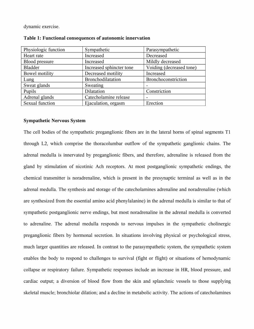
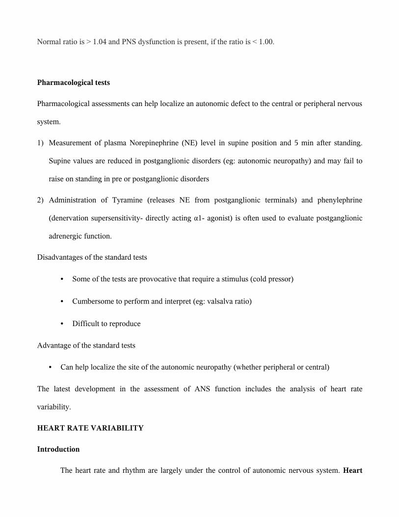
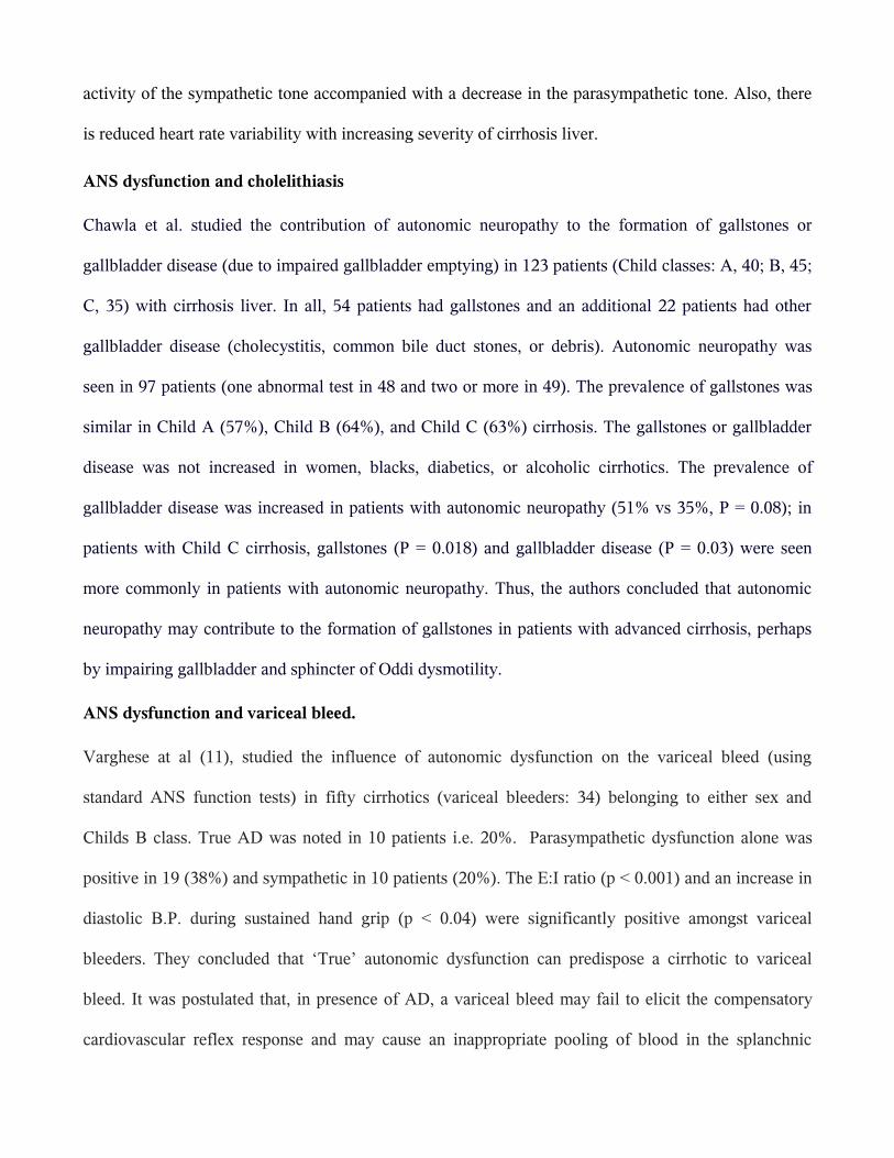
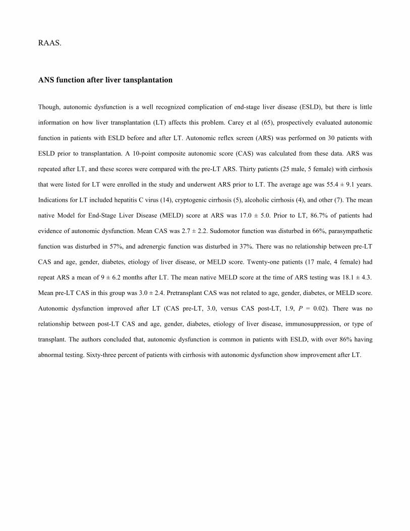
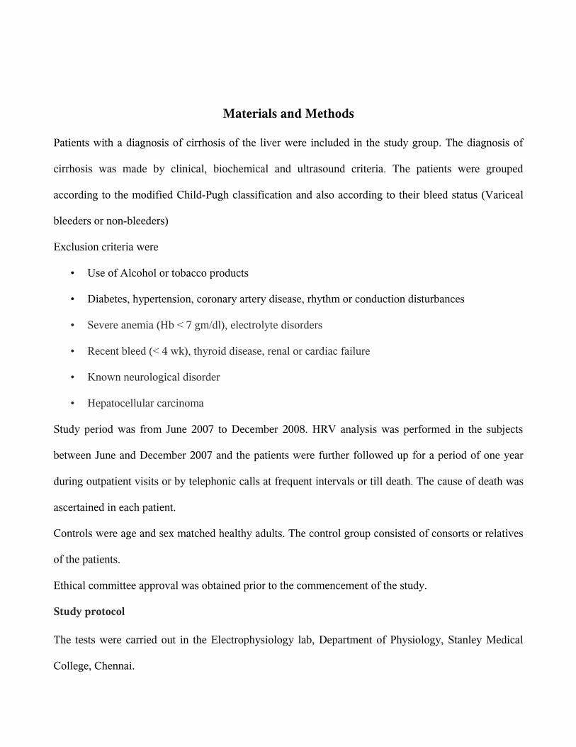
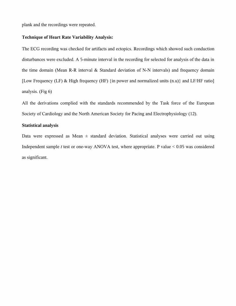
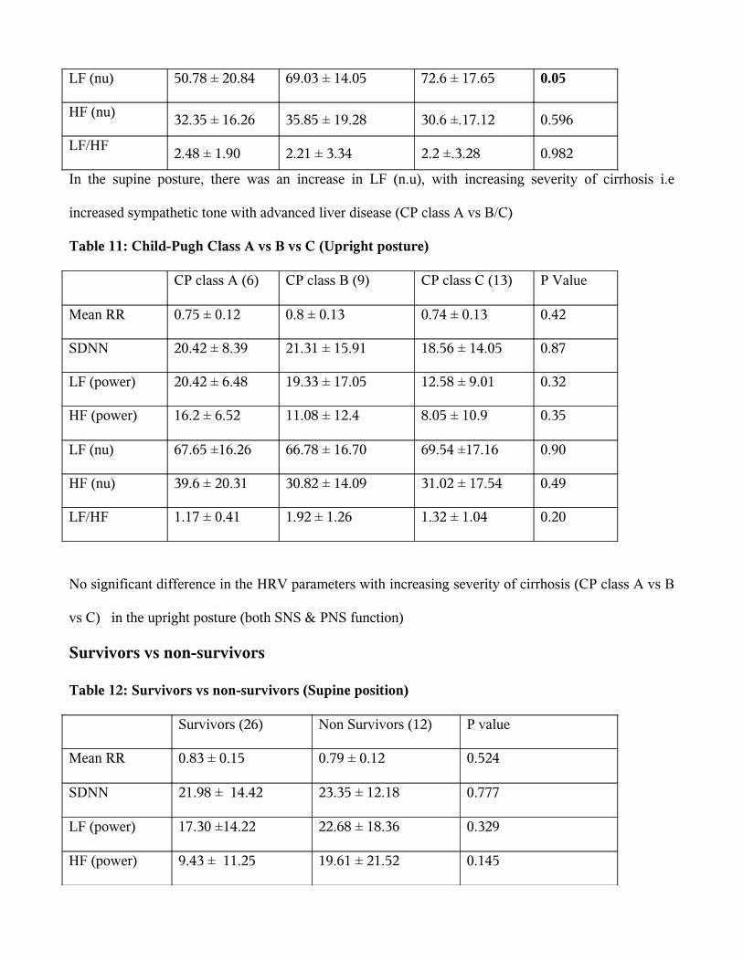
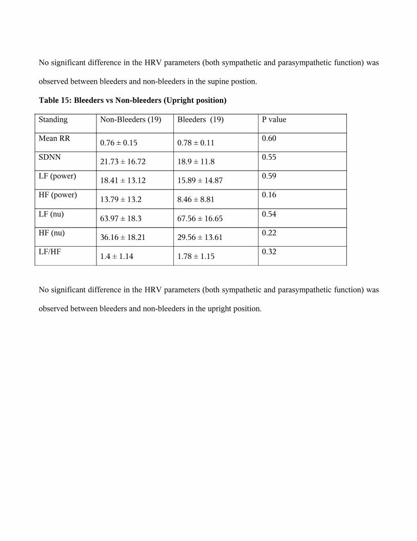
No significant difference in the HRV parameters (both sympathetic and parasympathetic function) was
observed between bleeders and non-bleeders in the supine postion
Table 15 Bleeders vs Non-bleeders (Upright position)
Standing Non-Bleeders (19) Bleeders (19) P value
Mean RR 076 plusmn 015 078 plusmn 011 060
SDNN 2173 plusmn 1672 189 plusmn 118 055
LF (power) 1841 plusmn 1312 1589 plusmn 1487 059
HF (power)1379 plusmn 132 846 plusmn 881
016
LF (nu) 6397 plusmn 183 6756 plusmn 1665 054
HF (nu) 3616 plusmn 1821 2956 plusmn 1361 022
LFHF14 plusmn 114 178 plusmn 115
032
No significant difference in the HRV parameters (both sympathetic and parasympathetic function) was
observed between bleeders and non-bleeders in the upright position
Discussion
Autonomic dysfunction is commonly noted in patients with chronic liver disease and increases with
worsening liver dysfunction (10 59)
Autonomic dysfunction has been correlated with mortality (9 10 13 59) and variceal bleed (11) in
patients with cirrhosis liver Rangari et al (53) in the only study on heart rate variability in patients
with liver cirrhosis from India used time domain methods for HRV analysis They showed that four
out of the five time domain measures were abnormal in patients with liver cirrhosis
The present study based on both time domain (Mean R-R amp SDNN) and frequency domain measures
(LF amp HF) of HRV analysis has shown that autonomic function is significantly impaired in patients
with cirrhosis of the liver
In the supine position cirrhotics had a significant increase in their sympathetic tone (compared to
controls) as evidenced by an increase in the LF (nu) They also had a reduced heart rate variability
(reduced SDNN) implying reduced parasympathetic tone Thus at rest in supine position cirrhotics
have a relatively increased sympathetic and a decreased parasympathetic tone This is consistent with
the observation of a hyperdynamic circulation in cirrhosis liver with an activation of the RAAS (Renin-
Angiotensin-Aldosterone system) and the SNS (Sympathetic nervous system) pathway (20) This also
correlates with other studies on heart rate variability in patients with cirrhosis of the liver (60 62)
As the cirrhotic patient assumes an upright position the compensatory changes in the autonomic
function in the form of a raise in sympathetic and a relative fall in parasympathetic tone (observed in
controls) is not evident
In a similar study by Laffi et al (37) on the effect of passive head tilting in patients with liver cirrhosis
it was observed that there was no difference in the power spectral analysis data (LF and HF) between
controls and cirrhotics in the supine position However cirrhotics had a higher plasma norepinephrine
levels in the supine position On passive head tilting observations similar to the present study were
made
With worsening liver dysfunction (CP class A vs BC) an increase in the LF (nu) was observed in the
supine position implying an increase in the sympathetic tone This is consistent with the observation
that the splanchnic arteriolar vasodilatation and hyperdynamic circulation seen in advanced cirrhosis
leads to the activation of the SNS and the RAAS pathway (20)
Varghese et al (11) assessed the ANS function in 2 groups of cirrhotics those with and without a
history of variceal bleed It was found that AD was significantly present in cirrhotics with a history of
variceal bleed However assessment of the ANS function was using the standard tests (39) and heart
rate variability analysis was not employed In our study there was no significant difference in the HRV
parameters between variceal bleeders and non-bleeders
Presence of autonomic neuropathy has been regarded as a poor prognostic indicator with increased
mortality noted in patients with AD (10 12 59) In the present study however there was no difference
in the HRV parameters between survivors amp non-survivors and AD did not predict mortality in the
study group
The causes of death in our study group were- Variceal bleed 5 patients Spontaneous Bacterial
Peritonitis (SBP) 4 patients Hepatoma 2 patients and Leptospirosis with renal failure 1 patient
Variceal bleed was the most common cause of death and as mentioned above there was no difference
in the HRV parameters between bleeders amp non-bleeders in our study Hepatitis B was the most
common etiology in our study group and it is known that hepatocellular carcinoma can occur in such
patients at a less advanced stage of chronic liver disease (66) Leptospirosis endemic in Chennai city
(68) was the cause of death in one patient SBP a complication of advanced chronic liver disease was
the cause of death in only 4 out of the 12 patients Hence not all deaths in the study group were related
to end stage liver disease which could explain the reason for no significant difference in the HRV
parameters between survivors and non-survivors Also our study group consisted of only 38 patients
with a follow up period of one year Further studies with a larger study group and with a longer
duration of follow up might probably yield a positive correlation between autonomic dysfunction and
mortality in patients with chronic liver disease
Summary
Figure 4 Parasympathetic and Sympathetic tone in controls and patients with cirrhosis (Childrsquos
class ABC)
Parasympathetic tone Sympathetic toneControls supine positionControls upright positionCirrhotics supine positionCirrhotics upright positionChild class A vs BC
Controls in the supine position have a balanced sympathetic and parasympathetic tone On assuming an
upright posture there is a physiological increase in the sympathetic and a decrease in the
parasympathetic tone in the control group
Cirrhotics in the supine position have a relatively increased sympathetic compared to the
parasympathetic tone On assuming an upright posture the physiological increase in the sympathetic
and a decrease in the parasympathetic tone (observed in controls) is not seen in cirrhotics
With worsening liver dysfunction (Child class A vs BC) there is an increase in the sympathetic tone
Conclusion
HRV analysis is a simple noninvasive test to assess the cardiovascular autonomic function in patients
with chronic liver disease
Patients with liver cirrhosis have significantly reduced heart rate variability (decreased parasympathetic
activity) and an increased sympathetic tone in supine posture
Cirrhotics have an abnormal homeostatic response to standing with no increase in sympathetic tone in
the upright posture
With worsening Child Pugh class (A vs BC) there is an increase in autonomic dysfunction with an
increased activity of the sympathetic component
There were no significant differences in HRV parameters between bleeders versus non-bleeders and
survivors versus non-survivors
References
1) Kowalski HJ Abelmann WH The cardiac output at rest in Laennecrsquos cirrhosis J Clin Invest
1953321025-1033
2) Trevisani F Sica G Mainqua P et al Autonomic dysfunction and hyperdynamic circulation in
cirrhosis with ascites Hepatology 1999301387ndash92
3) Thuluvath PJ Triger DR Autonomic neuropathy and chronic liver disease Q J Med
198972737-747
4) MacGilchrist AJ Reid JL Impairment of autonomic reflexes in cirrhosis Am J Gastroenterol
199085288-292
5) Giancane S Bernardi M Trevisani F DrsquoIntino PE Tame` MR Gasbarrini A Rimondi A et al
Autonomic neuropathy in liver cirrhosis prevalence and association with clinical and laboratory
features In Costa M Surrenti C Gorini S Maggi CA Meli A eds Sensory Nerves and Neuropeptides
in Gastroenterology From basic science to clinical perspectives New York Plenum Press 1991
295-300
6) Dillon JF Plevris JN Nolan J Ewing DJ Neilson JM Bouchier IA Hayes PC Autonomic function
in cirrhosis assessed by cardiovascular reflex tests and 24-hour heart rate variability AmJ
Gastroenterol 1994891544-1547
7) Bernardi M Trevisani F Santini C Zoli G Baraldini M Ligabue A Gasbarrini G Plasma
norepinephrine weak neurotransmitters and rennin activity during active tilting in liver cirrhosis
Relationship with cardiovascular homeostasis and renal function Hepatology 1983356-64
8) Hendrickse MT Triger DR Peripheral and cardiovascular autonomic impairment in chronic liver
disease prevalence and relation to hepatic function J Hepatol 199216177-183
9)Hendrickse MT Thuluvath PJ Triger DR Natural history of autonomic neuropathy in chronic liver
disease Lancet 1992 3391462-1464
10) Fleckenstein JF Frank SM Thuluvath PJ Presence of autonomic neuropathy is a poor prognostic
indicator in patients with advanced liver disease Hepatology 199623471-475
11) Joye Varghese S Balan N Naveen B Caroline Selvi K Jayapalan K Jayanthi V Does autonomic
dysfunction in cirrhosis liver influence variceal bleed Annals of Hepatology 2007 6(2) April-June
104-107
12) Heart rate variability Standards of measurement physiological interpretation and clinical use
Task Force of the European Society of Cardiology and The North American Society of Pacing and
Electrophysiology European Heart Journal (1996) 17 354ndash381
13) Fehmi Ates Ergun Topal Feridun Kosar Melih Karincaoglu Bulent Yildirim Yuksel Aksoy et al
The relationship of Heart rate variability with severity and prognosis of cirrhosis Dig Dis Sci 2006
511614ndash1618
14) William F Ganong Review of Medical Physiology Lange Medical publishers 23rd edition
15) James V Freeman Frederick E Dewey David M Hadley Jonathan Myers and Victor F
Froelicher Autonomic nervous system interaction with the cardiovascular system during exercise
Progress in Cardiovascular Diseases Vol 48 No 5 (MarchApril) 2006 pp 342-362
16) Iwakiri Y Groszmann RJ The hyperdynamic circulation of chronic liver diseases from the patient
to the molecule Hepatology 200643S121ndash31
17) Brinch K Moslashller S Bendtsen F et al Plasma volume expansion by albumin in cirrhosis Relation
to blood volume distribution arterial compliance and severity of disease J Hepatol 2003 3924ndash 31
18) Moslashller S Bendtsen F Henriksen JH Effect of volume expansion on systemic hemodynamics and
central and arterial blood volume in cirrhosis Gastroenterology 1995 1091917ndash25
19) Moslashller S Bendtsen F Christensen E et al Prognostic variables in patients with cirrhosis and
oesophageal varices without prior bleeding J Hepatol 1994 21 940ndash6
20) S Moslashller J H Henriksen Cardiovascular complications of cirrhosis Gut 2008 57268ndash278
21) Arroyo V Terra C Gines P Advances in the pathogenesis and treatment of type-1 and type-2
hepatorenal syndrome J Hepatol 2007 46 935ndash46
22) Helmy A Newby DE Jalan R et al Enhanced vasodilatation to endothelin antagonism in patients
with compensated cirrhosis and the role of nitric oxide Gut 200352410ndash5
23) Wiest R Groszmann RJ The paradox of nitric oxide in cirrhosis and portal hypertension too
much not enough Hepatology 200235 478ndash91
24) Ruiz-Del-Arbol L Monescillo A Arocena C et al Circulatory function and hepatorenal syndrome
in cirrhosis Hepatology 200542439ndash47
25) Hendrickse MT Triger DR Vagal dysfunction and impaired urinary sodium and water excretion in
cirrhosis Am J Gastroenterol 199489750ndash7
26) Dillon JF Nolan J Thomas H et al The correction of autonomic dysfunction in cirrhosis by
captopril J Hepatol 199726331ndash5
27) Grose RD Nolan J Dillon JF et al Exercise-induced left ventricular dysfunction in alcoholic and
non-alcoholic cirrhosis J Hepatol 199522326ndash32
28) Epstein SK Ciubotaru RL Zilberberg MD et al Analysis of impaired exercise capacity in patients
with cirrhosis Dig Dis Sci 1998431701ndash7
29) Laffi G Barletta G Lavilla G et al Altered cardiovascular responsiveness to active tilting in
nonalcoholic cirrhosis Gastroenterology 1997113891ndash8
30) Bolognesi M Sacerdoti D Di Pascoli M et al Haeme oxygenase mediates hyporeactivity to
phenylephrine in the mesenteric vessels of cirrhotic rats with ascites Gut 2005541630ndash6
31) Villa GL Barletta G Romanelli RG et al Cardiovascular effects of canrenone in patients with
preascitic cirrhosis Hepatology 2002351441ndash8
32) Ferlitsch A Pleiner J Mittermayer F et al Vasoconstrictor hyporeactivity can be reversed by
antioxidants in patients with advanced alcoholic cirrhosis of the liver and ascites Crit Care Med
2005332028ndash33
33) Ros J Claria J To-Figueras J et al Endogenous cannabinoids a new system involved in the
homeostasis of arterial pressure in experimental cirrhosis in the rat Gastroenterology 200212285ndash93
34) Moezi L Gaskari SA Liu H et al Anandamide mediates hyperdynamic circulation in cirrhotic rats
via CB(1) and VR(1) receptors Br J Pharmacol 2006149898ndash908
35) Moslashller S Wiinberg N Henriksen JH Noninvasive 24-hour ambulatory arterial blood pressure
monitoring in cirrhosis Hepatology 19952288ndash95
36) Wong F Logan A Blendis L Systemic hemodynamic forearm vascular renal and humoral
responses to sustained cardiopulmonary baroreceptor deactivation in well-compensated cirrhosis
Hepatology 199521717ndash24
37) Laffi G Lagi A Cipriani M et al Impaired cardiovascular autonomic response to passive tilting in
cirrhosis with ascites Hepatology 1996241063ndash7
38) Moslashller S Iversen JS Henriksen JH et al Reduced baroreflex sensitivity in alcoholic cirrhosis
relations tohemodynamics and humoral systems Am J Physiol Heart Circ Physiol 2007292G2966ndash
72
39) Ewing DJ Martin CN Young RJ Clarke BF The value of cardiovascular autonomic function
tests 10 years experience in diabetes Diabetic Care 1985 8 491ndash8
40) B K Bajaj M P Agarwal and B Krishna Ram Autonomic neuropathy in patients with hepatic
cirrhosis Postgrad Med J 200379408-411
41) AS Fauci et al Harrisonrsquos principle of Internal medicine 17th edition McGraw Hill Medical
42) Hon EH Lee ST Electronic evaluations of the fetal heart rate patterns preceding fetal death
further observations Am J Obstet Gynec 1965 87 814ndash26
43) Wolf MM Varigos GA Hunt D Sloman JG Sinus arrhythmia in acute myocardial infarction Med
J Australia 1978 2 52ndash3
44) Akselrod S Gordon D Ubel FA Shannon DC Barger AC Cohen RJ Power spectrum analysis of
heart rate fluctuation a quantitative probe of beat to beat cardiovascular control Science 1981 213
220ndash2
45) Bigger JT Fleiss JL Steinman RC Rolnitzky LM Kleiger RE Rottman JN Frequency domain
measures of heart period variability and mortality after myocardial infarction Circulation 1992 85
164ndash71
46) Pagani M Lombardi F Guzzetti S et al Power spectral analysis of heart rate and arterial pressure
variabilities as a marker of sympatho-vagal interaction in man and conscious dog Circ Res 1986 59
178ndash93
47) Malliani A Pagani M Lombardi F Cerutti S Cardiovascular neural regulation explored in the
frequency domain Circulation 1991 84 1482ndash92
48) Furlan R Guzetti S Crivellaro W et al Continuous 24-hour assessment of the neural regulation of
systemic arterial pressure and RR variabilities in ambulant subjects Circulation 1990 81 537ndash47
49) Kamath MV Fallen EL Power spectral analysis of heart rate variability a noninvasive signature of
cardiac autonomic function Crit Revs Biomed Eng 1993 21 245ndash311
50) Rimoldi O Pierini S Ferrari A Cerutti S Pagani M Malliani A Analysis of short-term
oscillations of R-R and arterial pressure in conscious dogs Am J Physiol 1990 258 H967ndash H976
51) Montano N Gnecchi Ruscone T Porta A Lombardi F Pagani M Malliani A Power spectrum
analysis of heart rate variability to assess the changes in sympathovagal balance
during graded orthostatic tilt Circulation 1994 90 1826ndash31
52) Appel ML Berger RD Saul JP Smith JM Cohen RJ Beat to beat variability in cardiovascular
variables Noise or music J Am Coll Cardiol 1989 14 1139ndash1148
53) Rangari M Sinha S Kapoor D Mohan JC Sarin SK Prevalence of autonomic dysfunction in
cirrhotic and noncirrhotic portal hypertension Am J Gastroenterol 2002 97 707-13
54) Khosla SN Sanyal S Nand N Autonomic function tests and clinical significance of dysautonomia
in chronic liver disease J Assoc Physicians India 1991 39 924-6
55) Gentile S Marmo R Peduto A Montella F Coltori M Autonomic neuropathy in liver cirrhosis
relationship with alcoholic aetiology and severity of the disease Ital J Gastroenterol 1994 26(2) 53-8
56) Nicholas VG Consuelo SP Michael ER Kenneth MS Adrian R Autonomic dysfunction and
gastroparesis in cirrhosis J Clin Gastroenterol 2004 38 72-766
57) Lewis FW Cohen JA Rector WG Jr Autonomic dysfunction in alcoholic cirrhosis relationship to
indicators of synthetic activation and the occurrence of renal sodium retention Am J Gastroenterol
1991 86 553-9
58) Johnson RH Robinson BJ Mortality in alcoholics with autonomic neuropathy J Neurol Neurosurg
Psychiatry 198851476ndash80
59) M T Hendrickse and D R Triger Autonomic dysfunction in chronic liver disease Clinical
Autonomic Research Vol3 Number 4 August 1993 227-231
60) Fleisher LA Fleckenstein JF Frank SM Thuluvath PJ Heart rate variability as a predictor of
autonomic dysfunction in patients awaiting liver transplantation Dig Dis Sci 2000 Feb45(2)340-4
61) Coelho L Saraiva S Guimaraumles H Freitas D Providecircncia LA Autonomic function in chronic liver
disease assessed by Heart Rate Variability Study Rev Port Cardiol 2001 Jan20(1)25-36
62) Iga A Nomura M Sawada Y Ito S Nakaya Y Autonomic nervous dysfunction in patients with
liver cirrhosis using 123 Iodine-metaiodobenzylguanidine myocardial scintigraphy and spectrum
analysis of heart-rate variability J Gastroenterol Hepatol 2003 Jun18(6)651-9
63) Chawla A Puthumana L and Thuluvath PJ Autonomic Dysfunction and Cholelithiasis in Patients
with Cirrhosis Dig Dis Sci Volume 46 Number 3 March 2001 495-498
64) Moslashller S Norgaard A Henriksen JH Frandsen E and Bendtsen F Effects of tilting on central
hemodynamics and homeostatic mechanisms in cirrhosis Hepatology 2004 40 811-819
65) Carey EJ Gautam M Ingall T Douglas DD The effect of liver transplantation on autonomic
dysfunction in patients with end-stage liver disease Liver Transpl 14235-239 2008
66) Omata M Takano S Occurrence of hepatocellular carcinoma in chronic viral hepatitis Cancer
Chemother Pharmacol (1994) 33 (Suppl) S153-S154
68) Shivkumar SLeptospirosis in Chennai-changing clinical profileJAPI Vol 54December 2006
964-965
Proforma
Name AgeSex
Address Contact No MGE No
Etiology HBVHCV Wilsonrsquos disease Others (Specify) Child Pugh Score
Clinical features
Symptoms YesNo Symptoms YesNoAscites Pedal Edema Altered sensoriumPain Abdomen GI bleedJaundice CoagulopathyOliguria Others (Specify)Autonomic symptoms
(postural dizziness)
Co-morbidity (DM HTN
CAD others specify)
Examination
Jaundice Pallor Pedal edema Spider naevi
Vital Signs PR BP Temp
Abdomen Liver Spleen Ascites
CVSRS
CNS Asterixis Hep Encephalopathy
Investigations
Hb B Urea
TC S Creatinine
DC S Electrolytes
ESR B Sugar
Platelets PT (TC)
MCVMCHMCHC INR
S Bilirubin (TD) USG Abdomen PV Doppler
ASTALT
SAP GGT CXR
Alb Globulin Ascitic fluid cell count
OGD Esophageal varices Gastric varices
HRV analysis
Mean RR interval
SDNN
LF (power)
HF (power)
LF (nu)
HF (nu)
LF HF ratio
Follow Up
Alive Dead
Cause of death (when applicable)
APPENDIX D
Consent Form
I Mr ______________________________ understand that Dr xxxxxxx a postgraduate
student in Stanley medical college and hospital Chennai is doing a study on heart rate variability I am
given to understand that these tests will assess the functioning of my heart and blood vessels and the
nerves regulating them These tests are simple involve taking ECGs blood pressure and respiratory
movements in lying and standing position They do not involve injections or taking any medicines and
are risk free I have been familiarized with the testing procedures I am participating in this study
willingly I have not been forced to do so I have also been told clearly that I could withdraw from this
study without any prejudice
Date
Signature
Figure 4 Electrophysiology Lab with HRV recording software
Figure 5 Computerized digital recording of HRV
- CERTIFICATE
- DEAN
-
- Assessment of ANS function
-
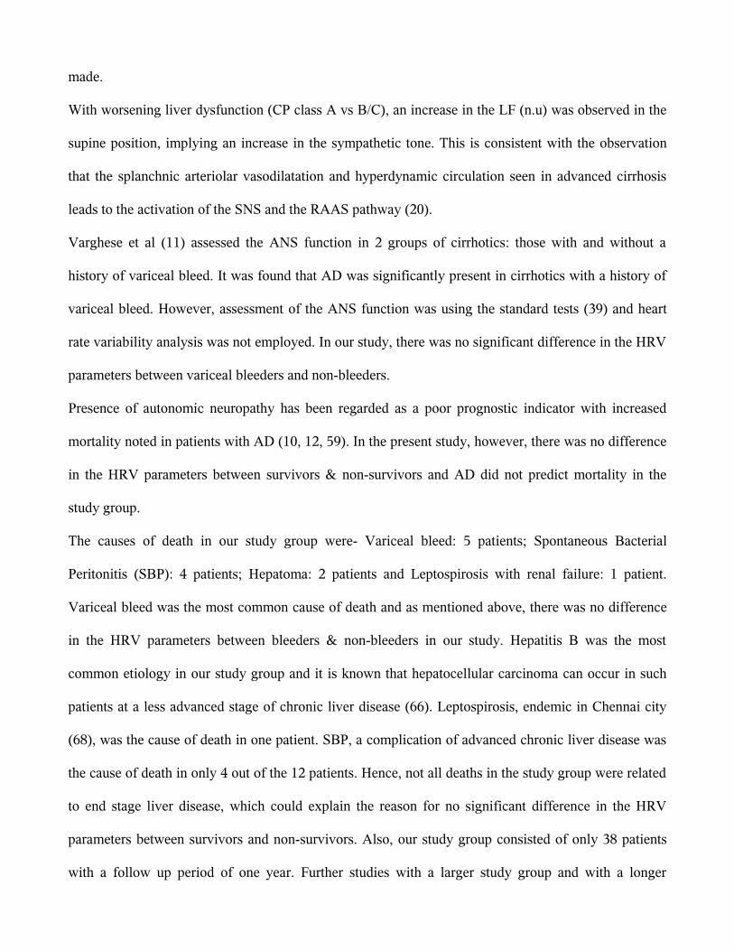
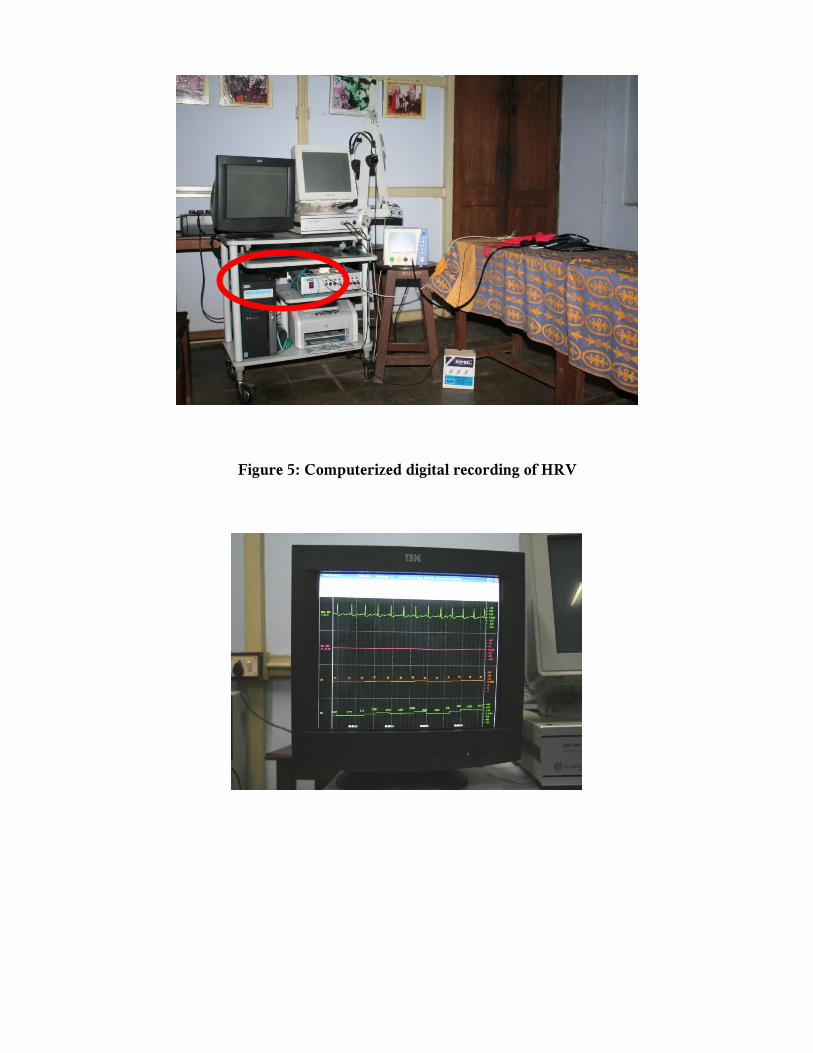
Figure 5 Computerized digital recording of HRV
- CERTIFICATE
- DEAN
-
- Assessment of ANS function
-