health maintenance iii-residents - ncc pediatrics residency maintenance iii... · ncc pediatrics...
TRANSCRIPT

NCC Pediatrics Continuity Clinic Curriculum: Health Maintenance III
Objective: To recognize the most common procedures used in pediatrics for Sensory Screening (hearing and vision), as part of the 2017 AAP Periodicity Schedule.
Pre-Meeting Preparation: Please read the following enclosures, corresponding to the screening procedures: 1) Sensory Screening:
a. Vision: “Vision Screening Essentials” (PIR, 2007)b. Hearing: “Pediatric Hearing Screening”
* Bring a hand-held ophthalmoscope or penlight to clinic, if you have one.
Conference Agenda: • Review “Health-Maintenance III Quiz”• Complete the following group activities for each screening procedure:
1) Vision Screening:a. Snellen Eye-Chartb. Corneal light reflexc. Cover-Uncoverd. SpotVision Conversion Chart (provided as table materials)
2) Hearing Screening:a. Conventional audiometerb. Tympanometry: Test each other with clinic tympanometer!
Extra-Credit: Encouraged for PGY2 & 3 • Vision: Red Reflex Testing; Vision Screening (AAP Clinical Practice Guidelines)
Ø “Pediatric Vision Screening” (ppt by Dr. Erika Beard-Irvine)Ø “Instrument Based Vision Screening: Update and Review (Contemp Peds, 2014)
• Hearing: Hearing Screening Clinical Report; Hearing Screening Policy Statement (AAP)Ø “Pediatric Hearing Screening” (ppt by Candice Ortiz Au.D.; play as slide-show)
© Developed by MAJ Jennifer Hepps. Updated 2016 by LCDR McDonnell, 2018 C. Carr.
Contemporary Pediatrics is a great free journal. Sign up if you don't already have access

Pediatric Vision ScreeningGary L. Rogers, MD,
Catherine Olson Jordan,
MD
Author Disclosure
Drs Rogers and Jordan
have disclosed no
financial relationships
relevant to this article.
This commentary does
not contain
a discussion of an
unapproved/
investigative use of
a commercial product/
device.
Educational Gap
Although early detection of visual disorders can lead to therapy that will prevent per-
manent blindness, compliance with screening guidelines of the American Academy of
Pediatrics is low.
Objectives After completing this article, readers should be able to:
1. Be aware of common vision-threatening conditions that can be detected by using
basic screening examinations.
2. Become familiar with techniques and findings used in vision screening examinations.
3. Understand the role of commercial screening tools.
IntroductionEarly detection of ocular conditions can allow for assessment and treatment of a vision-threatening or life-threatening condition. Amblyopia, or “lazy eye,” can develop if a clearvisual image is not projected onto the retina. Amblyopia can be caused by deprivation, stra-bismus, high refractive error (hyperopia, myopia, or astigmatism), or anisometropia (sig-nificant difference in the refractive error between eyes) and can be unilateral or bilateral.The prevalence of amblyopia is estimated to be 1% to 4%. (1)
Many factors may prevent the achievement of universal vision screening, including lackof education of families; language, financial, and state legislative barriers; and a lack of avail-able providers. (2) Primary care physicians are crucial providers for detecting and referringvision-threatening ocular conditions. From the newborn examination, through subsequenthealth supervision visits, and throughout a child’s life, the pediatrician and family physiciancan perform effective examinations to screen for common and uncommon conditions thatmay be vision-threatening or even potentially life-threatening. (3) However, vision screen-ing in the primary care office should not take the place of a full eye examination. If a patientcannot be screened effectively after two attempts, a referral should be made to an eye careprofessional who is comfortable examining children.
Pediatricians and family physicians should have the ability to perform a thorough ocularexamination. According to the US Preventive Services Task Force, children under age 5years should be screened to detect amblyopia, strabismus, and defects in visual acuity.The American Academy of Pediatrics (AAP) guidelines include screening at all health su-pervision visits, from the newborn period to age 3 years, by using the following compo-nents: ocular history, vision assessment, external examination, ocular motility, pupilexamination, and red reflex examination. For children ages 3 to 5 years, the AAP recom-mends age-appropriate visual acuity measurements and direct ophthalmoscopy. (4) Sensi-tivity of screening examinations increases with age, while specificity remains unchanged. (5)
However, screening is not universal, and compliance with AAP guideline visual acuityscreening is low. (6) In addition, there is controversy over who should provide the screen-ing examinations. In 2000, Kentucky passed legislation mandating diagnostic eye exami-nations by optometrists or ophthalmologists. (7) A survey of primary care physicians foundthat the percentage of pediatricians who expected to perform screening examinations drop-ped from 86% to 71% after the mandate, and the percentage of family physicians who ex-pected to screen dropped from 79% to 50%. The concern with this switch to mandated eyeexaminations is that children may get overlooked by one of the screening programs. In
Department of Ophthalmology, Nationwide Children’s Hospital, Columbus, OH.
Article disorders of the eye
126 Pediatrics in Review Vol.34 No.3 March 2013
by guest on March 31, 2018http://pedsinreview.aappublications.org/Downloaded from
addition, we must consider the increased cost of diagnos-tic eye examinations compared with vision screening pro-grams, given the current health-care crisis.
As discussed, screening from an early age can identifypatients who have poor vision. Whether potentially treat-able or not, low vision or blindness from amblyopia, nys-tagmus, or structural abnormalities certainly can affecta patient’s reading ability and educational progress. Get-ting parents and schools involved early and working withlow-vision specialists and visual aids can help patients ad-just their needs appropriately. Although low-vision servicescan provide essential educational tools, primary preventionand treatment of potentially vision-threatening conditionsis the better goal.
Common Vision ConditionsThe age of the patient is an essential consideration in de-termining a differential diagnosis. From birth to age 1 year,concerning conditions include corneal opacities, cataracts,glaucoma, persistent fetal vasculature, and retinoblas-toma. All of these conditions have the potential to causedeprivational amblyopia if not detected and treated at anearly age (sometimes within the first weeks after birth).Early detection of retinoblastoma couldmean saving a pa-tient’s eye or eyes and possibly some vision, along withtreating a potentially fatal condition. Other conditionsthat may be present from birth, such as congenital ptosisor capillary hemangiomas causing mechanical ptosis orunilateral astigmatism, are also risk factors for amblyopia.
From ages 1 to 3 years, more common eye conditionswith amblyogenic risk factors include strabismus and re-fractive errors such as high hyperopia (farsightedness),high myopia (nearsightedness), astigmatism, and aniso-metropia (significant difference between the refractiveerrors between the eyes). These disorders can be subtle.Preverbal patients are more difficult to examine, but earlydetection will have a substantial impact on a patient’s fu-ture education and life if treatment is initiated promptly.
From ages 3 to 8 years, strabismus and refractive errorscontinue to be significant amblyopic risk factors. As pa-tients age and grow more cooperative, testing for visualacuity becomes more feasible.
StrabismusEarly Detection
Early detection of strabismus or ocular misalignment isessential for the prevention or treatment of amblyopiaand allows the possibility of saving binocular vision. Ifthe misalignment is constant, a child’s developing brainwill ignore the visual input from the misaligned eye to
avoid diplopia. If this situation persists, the eye will be-come “lazy” or amblyopic. If detected and referred earlyenough, treatment of the amblyopia by using penaliza-tion techniques such as patching may improve or resolvethe difference in vision between the two eyes. As childrenget older, treatment is not as effective.
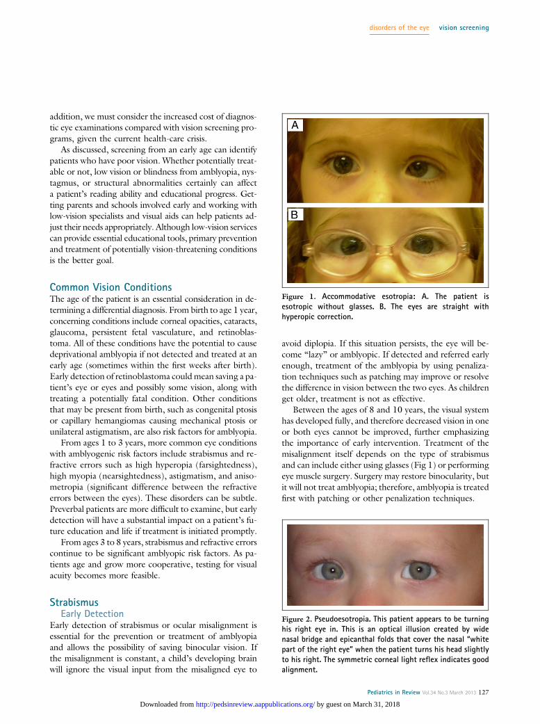
Between the ages of 8 and 10 years, the visual systemhas developed fully, and therefore decreased vision in oneor both eyes cannot be improved, further emphasizingthe importance of early intervention. Treatment of themisalignment itself depends on the type of strabismusand can include either using glasses (Fig 1) or performingeye muscle surgery. Surgery may restore binocularity, butit will not treat amblyopia; therefore, amblyopia is treatedfirst with patching or other penalization techniques.
Figure 1. Accommodative esotropia: A. The patient isesotropic without glasses. B. The eyes are straight withhyperopic correction.
Figure 2. Pseudoesotropia. This patient appears to be turninghis right eye in. This is an optical illusion created by widenasal bridge and epicanthal folds that cover the nasal “whitepart of the right eye” when the patient turns his head slightlyto his right. The symmetric corneal light reflex indicates goodalignment.
disorders of the eye vision screening
Pediatrics in Review Vol.34 No.3 March 2013 127
by guest on March 31, 2018http://pedsinreview.aappublications.org/Downloaded from

Pseudostrabismus Versus True StrabismusPseudostrabismus occurs when one eye appears to turn inbut is straight on cover/uncover testing. This appearanceoccurs most commonly in patients with wide nasal bridgesand epicanthal folds, giving the child the appearance ofesotropia. A true esotropia can be ruled out by using anequal corneal light reflex and normal cover testing (Fig 2).
Examination TechniquesRed Reflex
The red reflex test is the single most important screeningtool for infants and young children. Using the direct oph-thalmoscope to view both eyes simultaneously is the bestway to evaluate the red reflex. The patient’s eyes should beviewed through the direct ophthalmoscope from approx-imately 2 feet away, with a broad beam directed so thatboth eyes are illuminated at the same time. The patientshould be focused on the ophthalmoscope light. Startingwith low illumination and increasing the brightness allowsthe patient to become comfortable with the bright light.
The red reflex will fill the pupil, and the corneal lightreflex will also be centered on the pupil (Brückner reflex)if the patient’s alignment is correct. The red reflex repre-sents the reflection of the light from the retina. There-fore, abnormalities of the red reflex can be caused bya physical blockage of the normal clarity of the visual axis,such as tear film mucus, corneal opacity, cataract, vitreoushemorrhage or retinal detachment, retinoblastoma, orpersistent fetal vasculature. The red reflex also can appeardull in both eyes from a high refractive error (high myo-pia or hyperopia) or unequal due to anisometropia (highrefractive error in only one eye) (Fig 3) or strabismus.
Differences in pigmentation among racial or ethnicgroups also may be responsible for variation in the red
reflex. Darker pigmented patients will appear to have adarker red reflex. The AAP policy states that all neonatesshould have a red reflex examination before dischargefrom the newborn nursery. Urgent referral and directcommunication with the accepting ophthalmologist areessential when abnormalities are detected. High-risk pa-tients who have a family history of retinoblastoma, infan-tile cataracts, glaucoma, or any other ocular disorders thatpresented early in life should be referred but also shouldhave a red reflex examination before leaving the newbornnursery. Any parental concern raised by suspicion ofa white pupil reflex should be referred urgently. (8) Ifthere is ever any concern regarding a child’s red reflex
Figure 3. Asymmetric red reflex. This 7-year-old boy wasfound to be severely hyperopic in the left eye with no need forglasses in the right eye. This is an excellent example ofa patient who might have passed vision screens but shouldhave been referred much earlier for an abnormal red reflex.
Figure 4. Leukocoria.
Figure 5. Partial leukocoria. The patient had a congenital cataractviewed through a surgical microscope just before surgery.
disorders of the eye vision screening
128 Pediatrics in Review Vol.34 No.3 March 2013
by guest on March 31, 2018http://pedsinreview.aappublications.org/Downloaded from

status, the most prudent action is to refer the patient fora complete ocular examination.
LeukocoriaLeukocoria, or a white pupil, occurs when the red reflexappears white rather than the typical red (Fig 4). Themost concerning diagnosis on the differential is retino-blastoma. Toxocariasis, Coats disease, persistent fetal vas-culature, or a chronic retinal detachment will also appearwhite and therefore create a white reflex. Cataracts also cancause leukocoria or just an asymmetric red reflex (Fig 5).Because retinoblastoma is potentially fatal, all cases ofleukocoria require an urgent referral to determine thecause of the condition.
External Examination (Inspection)The external eye examination can be performed witha penlight to look for any external structural abnormalityof the eyelids and adnexa. Paying attention to the eyelidsand the vertical and horizontal fissures can reveal ptosis, cap-illary hemangiomas, and eyelid colobomas, which are impor-tant findings in detecting possible risk factors for obstructiveamblyopia and systemic diseases. The sclera also can be eval-uated easily by using a penlight. Corneal clarity can be eval-uated simply by determining if there is a clear view of the irisand pupil. If there is not a good view of the iris or pupil,there may be a corneal opacity or haze. In newborns andinfants up to age 2 months, asymmetry of the eyes or faceshould be noted. From 3 months on, any face turn or headtilt should be noted, especially if mentioned by the parents.
Visual AcuityVisual acuity improves with age as does the ability to rec-ognize letters or shapes. At age 0 to 2 months, patientsshould manifest a blink response to bright light, equalpupillary response, sporadic fixation, and following thatbecomes more consistent with age. The neonate can haveintermittent strabismus with either an eso- or exodeviationof the eyes (eyes turned in or out), which should resolveby 2 to 4 months, after which the deviation is consideredpathologic. If the infant has a constant strabismus, he orshe should be referred for evaluation.
At age 2 to 6 months, infants should be able to fix andfollow an object, such as a light or mother’s face, and theeye alignment should be straight. From age 6 months to2 years, children should have central fixation, reach fortoys, and demonstrate good alignment.
From age 3 to 5 years, subjective vision can be ob-tained. Children should test to 20/40, or better, on age-appropriate charts with one eye occluded (Allen or LEApictures, tumbling Es, or HOTV letters). There should
be no more than two lines of difference between the eyes.Patients become more cooperative after age 5 years.These children should test to at least 20/30 vision witha regular Snellen chart, with no more than two lines ofdifference between the eyes.
Cover Test and Hirschberg TestThe cover test (Fig 6) and Hirschberg test are used toexamine ocular misalignment. The cover test revealsa manifest deviation (tropia). If a patient is fixating ona target with the right eye and the left eye appears tobe turned in or turned out when the right eye is covered,there will be a shifting movement of the left eye in theopposite direction from its deviated position as the lefteye picks up fixation. This finding is diagnostic of a man-ifest deviation because the misalignment is constant.When the cover is removed from the right eye, the righteye will either continue to be deviated or it will refixate if thereis a right eye preference, indicating a possible amblyopia.
Figure 6. Alternating cover test.
disorders of the eye vision screening
Pediatrics in Review Vol.34 No.3 March 2013 129
by guest on March 31, 2018http://pedsinreview.aappublications.org/Downloaded from

A latent deviation can be induced by using alternatecover testing (Fig 7). This test is performed by movingthe occluder directly from one eye to the other withoutallowing binocular viewing. This maneuver can bringout a phoria (latent deviation) or intermittent tropia(manifest but controlled at times). A manifest deviationis more worrisome due to the higher risk of amblyopiadeveloping. The general pediatrician who is unsureabout the proper performance of these tests shouldask a local ophthalmologist to demonstrate the appro-priate technique.
When there is a constant deviation, the Hirschbergtest can be used to estimate the amount of deviation. Us-ing a penlight directed on both eyes, the light reflex is ex-amined to determine if there is an asymmetry. The lightreflex should be approximately in the center of the pupilin both eyes when the child is fixating on the light, or inthe same spot in both eyes if the patient is fixing on a dif-ferent target. If the light reflex is displaced nasally, thisfinding indicates an exotropia (the eye is turned out)(Fig 8). When the light reflex is displaced temporally, thisfinding indicates an esotropia (the eye is turned in) (Fig 9).This test can be helpful in determining a true deviationversus pseudostrabismus (discussed earlier).
Ocular Motility and NystagmusThe patient’s ocular motility should be evaluated as soonas the child is old enough to fixate and follow an interest-ing target. Eye movements become smoother as infantsget older. Parents may report “funny eye movements,”which could indicate a more complex strabismus suchas congenital fourth nerve palsy, Brown syndrome, orDuane syndrome.
Observing for nystagmus is also important. There aremany types and causes of nystagmus, but all childrenwho manifest symptoms should be referred for evaluationof low vision as a cause. If an obstruction to clear visual de-velopment, such as a congenital cataract, has not been cor-rected at an early age, the presence of nystagmus could bea sign of poor vision. Other types of abnormal eye move-ments, such as opsoclonus, warrant an urgent referral. Op-soclonus can be a presenting sign of neuroblastoma. Thiscondition appears as rapid, involuntary eye movements inall vectors of gaze.
Pupil ExaminationPatients should have equal and reactive pupils from birth.It is more difficult to elicit this response in newborns buthaving the room dark and using a bright penlight oftenare helpful in distinguishing a pupillary response. Theolder a patient gets, the more important it is to have himor her focus at a distant target, dim the lights, and checkthe pupils while standing to the side so the patient doesnot focus on the examiner and induce accommodation.Any evidence of congenital anisocoria, or pupils of differentsizes, also should be referred to evaluate for possible Horner
Figure 7. Alternating exotropia induced by cover testing.
Figure 8. Exotropia. Corneal light reflex in the left eye is nasalcompared with the light reflex on the right eye, which is central.
Figure 9. Esotropia. Corneal light reflex is not as apparent inthis photograph, but the room light reflected off the cornea istemporal in the right eye compared with the central positionin the left eye.
disorders of the eye vision screening
130 Pediatrics in Review Vol.34 No.3 March 2013
by guest on March 31, 2018http://pedsinreview.aappublications.org/Downloaded from

syndrome. The most concerning condition associated withHorner syndrome in children is neuroblastoma.
In addition to the screening examination, takinga complete medical and family history is vital. At birth,it is important to ask about a family history of any con-genital eye conditions or blindness from birth. This in-quiry is imperative in evaluating for possible heritableeye diseases such as retinoblastoma, congenital cataracts,congenital glaucoma, and aniridia.
Commercial Screening ToolsThere has been growing interest in using commercialscreening tools in schools and primary care offices. Stan-dard techniques of visual acuity tests measure visual func-tion directly. Patient cooperation, understanding, age,and attention, as well as the skill and patience of the ex-aminer, play a role in the success of testing visual acuity.
There are a variety of photoscreeners and autorefractorsthat objectively detect amblyopia or amblyogenic risk fac-tors and require little patient cooperation.
The risk factors that should be identified by screen-ing instrumentation include significant anisometropia(>1.50 diopter difference in prescription between thetwo eyes), manifest strabismus, hyperopia greater than3.50 diopters, myopia greater than 3.00 diopters, any vi-sually significant media opacity (>1 mm and in the visualaxis), astigmatism >1.50 diopters in the regular meri-dians or >1.00 in oblique axis, and ptosis. (9)
Not all patients who have these risk factors will de-velop amblyopia. For instance, many infants have a highdegree of clinically significant astigmatism that is eithereliminated or greatly reduced by age 4 years. (10) There-fore, systems with higher sensitivity but lower specificitywill over-refer due to false-positive results. (9)
Table. Vision Screening Recommendations
Age Evaluation Indications for Referral
Newborn (0–1 mo) Examine outer structures of the eye and red reflexbefore the neonate leaves the newborn nursery
Abnormal red reflex requires urgentconsultation
History of retinoblastoma in parent or sibling1 mo–3 y History
Vision assessment; fix and followExternal examinationOcular motilityPupil examinationRed reflex evaluation
Poor tracking by 3 monthsAbnormal red reflexChronic tearing or discharge
3–5 y HistoryVision assessment: LEAand Allen figures, HOTV letters,tumbling Es, Snellen chart
External examinationOcular motilityPupil examinationRed reflex evaluation(photoscreening)Ophthalmoscopy
StrabismusChronic tearing or dischargeFail vision screen (cannot read 20/40 withone or both eyes or two-line differencebetween eyes) or photoscreening
Uncooperative after two attempts
‡5 y HistoryVisual acuityExternal examinationOcular motilityPupil examinationRed reflex evaluation(photoscreening)Ophthalmoscopy
StrabismusCannot read at least 20/30 with one or botheyes or two-line difference between eyes
Fail photoscreeningNot reading at grade level
At-risk childrenof any age
HistoryVisual acuityExternal examinationOcular motilityPupil examinationRed reflexOphthalmoscopy
Retinopathy of prematurityFamily history of retinoblastoma, congenitalglaucoma or congenital cataracts
Systemic diseases with associated retinaldystrophies/degenerations
NystagmusNeurodevelopmental delays
Adapted from American Association for Pediatric Ophthalmology and Strabismus. Vision Screening Recommendations. Accessed online at http://www.aapos.org/terms/conditions/131.
disorders of the eye vision screening
Pediatrics in Review Vol.34 No.3 March 2013 131
by guest on March 31, 2018http://pedsinreview.aappublications.org/Downloaded from

Many studies have compared the different screeningtools. The two most common methods are photoscreening,which involves taking pictures of the red reflex of both eyessimultaneously, and autorefractors, which can estimate thechild’s refractive error. Photoscreening was shown to bemore time-efficient and had a higher positive predictive valuethan traditional screening techniques in 3- to 4-year-olds.(11) A study of vision in preschool-aged children found au-torefractors detected 15% more amblyogenic risk factorsthan photoscreeners. In addition, depending on the criteriaused in the various screening techniques, referral rates can bedifferent. There have been no studies or consensus as towhich method should be standard of care. Ease of referraland compliance of parents to keep the referral appointmentare obstacles regardless of which method is used. (12)
There is no mechanical substitute at present for an ad-equate physical examination conducted by an educatedprimary care physician. Screening programs should be de-signed to easily identify individuals who have amblyopiaor those at risk for developing amblyopia, with the addi-tional concern of keeping the screening inexpensive.
RecommendationsThe table highlights the recommendationsmade by theAmer-ican Association for Pediatric Ophthalmology and Strabismusfor which examinations should be conducted at different agesand also lists guidelines for referral to an ophthalmologist.
References1. Committee on Practice and Ambulatory Medicine and Sectionon Ophthalmology; American Academy of Pediatrics. Use of pho-toscreening for children’s vision screening. Pediatrics. 2002;109(3):524–5252. Castanes MS. Major review: the underutilization of visionscreening (for amblyopia, optical anomalies and strabismus) amongpreschool age children. Binocul Vis Strabismus Q. 2003;18(4):217–2323. Committee on Practice and Ambulatory Medicine Section onOphthalmology; American Association of Certified Orthoptists;American Association for Pediatric Ophthalmology and Strabismus;American Academy of Ophthalmology. Eye examination ininfants, children, and young adults by pediatricians: organizationalprinciples to guide and define the child health care system and/orimprove the health of all children. Ophthalmology. 2003;110(4):860–8654. Grosse S, Biernath K. Vision evidence-statement: screening. In:Campbell KP, Lanza A, Dixon R, Chattopadhyay S, Molinari N,Finch RA, eds. A Purchaser’s Guide to Clinical Preventive Services:Moving Science Into Coverage. Washington, DC: National BusinessGroup on Health; 20065. Schmucker C, Grosselfinger R, Riemsma R, et al. Diagnosticaccuracy of vision screening tests for the detection of amblyopiaand its risk factors: a systematic review. Graefes Arch Clin ExpOphthalmol. 2009;247(11):1441–14546. Clausen MM, Armitage MD, Arnold RW. Overcoming barriersto pediatric visual acuity screening through education plus provisionof materials. J AAPOS. 2009;13(2):151–1547. Kemper AR, Fant KE, Badgett JT. Preschool vision screening inprimary care after a legislative mandate for diagnostic eye exami-nations. South Med J. 2003;96(9):859–8628. American Academy of Pediatrics; Section on Ophthalmology;American Association for Pediatric Ophthalmology and Strabismus;American Academy of Ophthalmology; American Association ofCertified Orthoptists. Red reflex examination in neonates, infants,and children. Pediatrics. 2008;122(6):1401–14049. Donahue SP, Arnold RW, Ruben JB; AAPOS Vision ScreeningCommittee. Preschool vision screening: what should we be detect-ing and how should we report it? Uniform guidelines for reportingresults of preschool vision screening studies. J AAPOS. 2003;7(5):314–31610. Gwiazda J, Scheiman M, Mohindra I, Held R. Astigmatism inchildren: changes in axis and amount from birth to six years. InvestOphthalmol Vis Sci. 1984;25(1):88–9211. Salcido AA, Bradley J, Donahue SP. Predictive value of pho-toscreening and traditional screening of preschool children. J AAPOS.2005;9(2):114–12012. Braverman R. Diagnosis and treatment of refractive errorsin the pediatric population. Curr Opin Ophthalmol. 2007;18(5):379–383
Summary
• Pediatricians and family physicians are essential inassessing the health of the eye and in vision screening.
• Newborns should be evaluated before leaving thenursery and referred urgently for abnormal findings onexternal examination or abnormalities of the red reflex.
• Vision assessment should begin at age 3 years byphysicians, nurses, or technicians trained in visionevaluation. Children should be referred to licensed eyecare professionals for abnormal findings or poorcooperation after two attempts.
• Photoscreeners can be a valuable addition to routinevision screening, especially in preverbal children.
disorders of the eye vision screening
132 Pediatrics in Review Vol.34 No.3 March 2013
by guest on March 31, 2018http://pedsinreview.aappublications.org/Downloaded from

DOI: 10.1542/pir.34-3-1262013;34;126Pediatrics in Review
Gary L. Rogers and Catherine Olson JordanPediatric Vision Screening
ServicesUpdated Information &
http://pedsinreview.aappublications.org/content/34/3/126including high resolution figures, can be found at:
Referenceshttp://pedsinreview.aappublications.org/content/34/3/126#BIBLThis article cites 11 articles, 3 of which you can access for free at:
Subspecialty Collections
mology_subhttp://classic.pedsinreview.aappublications.org/cgi/collection/ophthalOphthalmology_cmehttp://classic.pedsinreview.aappublications.org/cgi/collection/journalJournal CMEfollowing collection(s): This article, along with others on similar topics, appears in the
Permissions & Licensing
.xhtmlhttp://classic.pedsinreview.aappublications.org/site/misc/Permissionsin its entirety can be found online at: Information about reproducing this article in parts (figures, tables) or
Reprints
mlhttp://classic.pedsinreview.aappublications.org/site/misc/reprints.xhtInformation about ordering reprints can be found online:
by guest on March 31, 2018http://pedsinreview.aappublications.org/Downloaded from
Pediatric Hearing Screening (Adapted from http://www.entcolumbia.org/childscrn.html)
I. Newborns and Infants
Hearing screening for newborns before they leave the hospital or maternity center is now becoming a common practice. Without such programs, the average age of hearing loss identification is between 12-25 months. When hearing loss is detected late, language development is already delayed.
Screening Techniques for Newborns and Infants The screening of newborns and infants involves use of non-invasive, objective physiologic measures that include otoacoustic emissions (OAEs) and/or auditory brainstem response (ABR). Both procedures can be done painlessly while the infant is resting quietly.
• Otoacoustic emissions (OAEs) are inaudible sounds from the cochlea when audible sound stimulatesthe cochlea. The outer hair cells of the cochlea vibrate, and the vibration produces an inaudible soundthat echoes back into the middle ear. This sound can be measured with a small probe inserted into theear canal. Persons with normal hearing produce emissions. Those with hearing loss greater than 25-30dB do not. OAEs can detect blockage in the outer ear canal, middle ear fluid, and damage to the outerhair cells in the cochlea.
• Auditory brainstem response (ABRs) is an auditory evoked potential that originates from the auditorynerve. It is often used with babies. Electrodes are placed on the head, and brain wave activity inresponse to sound is recorded. ABR can detect damage to the cochlea, the auditory nerve and theauditory pathways in the stem of the brain.
What happens if an infant does not pass the screening? Infants who do not pass a screening are often given a second screening to confirm findings and then referred for follow-up audiological and medical evaluations that should occur no later than 3 months of age. These evaluations confirm the presence of hearing loss; determine the type, nature, and (whenever possible) the cause of the hearing loss; and help identify options for treatment. Even if the infant passes screening, certain conditions do not produce immediate hearing loss. Rather, the hearing loss occurs later in the child's development.
II. Older Infants and Toddlers
Infants and toddlers (7 months through 2 years) should be screened for hearing loss as needed, requested, mandated, or when conditions place them at risk for hearing disability. Infants not tested as newborns should be screened before three months of age. Other infants should be screened who received neonatal intensive care or special care, or who display other indicators that place them at risk for hearing loss.
Screening Techniques for Infants, Toddlers and Children Two screening methods are suggested as the most appropriate tools for children who are functioning at a development age of 7 months to 3 years, visual reinforcement audiometry (VRA) and conditioned play audiometry (CPA). Both of these methods are behavioral techniques that require involvement and cooperation of the child.
• Visual reinforcement audiometry (VRA) is the method of choice for children between 6 months and 2years of age. The child is trained to look toward (localize) a sound source. When the child gives a

correct response, e.g., looking to a source of sound when it is presented, the child is "rewarded" through a visual reinforcement such as a toy that moves or a flashing light.
• Conditioned play audiometry (CPA) can be used as the child matures. It is widely used between 2 and 3 years of age. The child is trained to perform an activity each time a sound is heard. The activity may be putting a block in a box, placing pegs in a hole, putting a ring on a cone, etc. The child is taught to wait, listen, and respond.
With both of these methods, sounds of different frequencies are presented at a sound level that children with normal hearing can hear. It is ideal if the child will allow earphones to be placed on his or her head so that independent information can be obtained for each ear. If the child refuses earphone placement or earphone placement is otherwise not possible, sounds are presented through speakers inside a sound booth. Since sound field screening does not give ear specific information, a unilateral hearing loss (hearing loss in only one ear) may be missed.
Alternative procedures, such as otoacoustic emissions (OAEs) or auditory brainstem response (ABR) may be used if the child is unable to be conditioned.
What happens if a toddler does not pass the screening? A toddler who does not pass the screening should be rescreened or referred for audiologic evaluation. Confirmation of hearing status should be obtained within 1 month, but no later than 3 months, after the initial screening. III. Hearing Screening in Preschoolers
The goal of screening for hearing loss in preschoolers (ages 3-5 years) is to identify children most likely to have hearing loss that may interfere with communication, development, health, or future school performance. In addition, because hearing loss in this age range is so often associated with middle ear disease, it is also recommended that children in this age group be screened for outer and middle ear disorders (acoustic emmittance screening).
Screening Techniques for Preschoolers • Conditioned play audiometry (CPA) is the most commonly employed procedure. • Acoustic emmittance screening includes tympanometry, acoustic reflex, & static acoustic impedance: o Tympanometry introduces air pressure into the ear canal making the eardrum move back and forth. A
special machine then measures the mobility of the eardrum. Tympanograms, or graphs, are produced which show stiffness, floppiness, or normal eardrum movement. They are classified as type A (normal), type B (flat, clearly abnormal), and type C (indicating a significantly negative pressure in the middle ear, possibly indicative of pathology).
o Acoustic reflex testing measures the response of a tiny ear muscle that contracts when a loud sound occurs. The loudness level at which the acoustic reflex occurs and/or the absence of the acoustic reflex give important diagnostic information.
o Static acoustic impedance testing measures estimate the physical volume of air in the ear canal. This test is useful in identifying a perforated eardrum or whether ear ventilation tubes are still open.
What happens if a preschooler does not pass the screening? • If the child cannot be conditioned to the play audiometry, the child will be screened using infant-
toddler procedures or will be recommended for a more in-depth audiologic assessment.

• If the child did condition and did not pass the screening, then referral for audiological assessmentwill be made. Hearing status of children referred after screening should be confirmed within 1month, but no later than 3 months, after the initial screening.
IV. Hearing Screening for School Age Children and Adolescents (5-18 years)
School-age children should be screened for hearing loss as needed, requested, mandated, or when conditions place them at risk for hearing disability. Screening for hearing loss identifies the school-age children most likely to have hearing impairment that may interfere with development, communication, health, and education. School age children with even minimal hearing loss are at risk for academic and communication difficulties.
School age children should be screened at the following times: on first entry into school; every year from kindergarten through 3rd grade; in 7th & 11th grade; upon entrance into special education; upon grade repetition; upon entering a new school system without evidence of having passed a previous screening.
Screening techniques used for school-age students o Conventional audiometry, in which students are instructed to raise their hand (or point to the
appropriate ear) when they hear a tone, is the commonly used procedure. Conditioned playaudiometry (CPA) is also used.
What happens if a school-age student does not pass the screening? The student should be reinstructed, earphones repositioned, and rescreened in the same session. If the student does not pass the rescreening, he or she should be referred for audiologic assessment. Hearing status of referred students should be confirmed within one month, and no later than 3 months, after initial screening.
V. Risk Factors for Hearing Loss in Children
• Parental, caregiver and/or health care provider concerns regarding hearing, speech, language,and/or developmental delay based on observation and/or standardized developmental screening.
• Family history of permanent childhood hearing loss.• Infections associated with sensorineural hearing loss including bacterial meningitis, mumps.• In utero infections such as cytomegalovirus, herpes, rubella, syphilis, and toxoplasmosis.• Neonatal indicators - specifically hyperbilirubinemia at a serum level requiring exchange
transfusion, persistent pulmonary hypertension of the newborn associated with mechanicalventilation, and conditions requiring the use of extracorporeal membrane oxygenation (ECMO)
• Syndromes associated with progressive hearing loss such as neurofibromatosis, osteopetrosis, andUsher' s syndrome.
• Neurodegenerative disorders, such as Hunter syndrome, or sensory motor neuropathies, such asFriedreich's ataxia and Charcot-Marie-Tooth syndrome.
• Head trauma• Recurrent or persistent otitis media with effusion for at least 3 months.• Anatomic disorders that affect eustachian tube function• Reported exposure to potentially damaging noise levels or to drugs that cause hearing loss.

Health Maintenance III Quiz
1. At what ages does the AAP recommend hearing screening? Do we perform hearing screeningin our clinic? If so, how?
2. At what ages does the AAP recommend vision screening? Do we perform vision screening inthe clinic? If so, how?
3. Please fill in the appropriate sensory screening tests for each of the following age-groups:
Age-Group Hearing Screen Vision Screen Neonate
Toddler
Preschool
School-Age

4. Please define the following terms:a. Myopia
b. Astigmatism
c. Strabismus
d. Esotropia
e. Exophoria
f. Amblyopia
g. 20/40 OU
5. Match the following vision problems in children with the appropriate screening method:
Problem Test(s) Refractive Error ________________________________________
Strabismus ________________________________________
Cataract ________________________________________
6. Which of the following patients is at increased risk for hearing loss?a. 8do former 37+4 week infant, readmitted for bilirubin of 28.5b. 3yo with Trisomy 21c. 18mo female with OME at 12mo, 15mo, and 18mo well-baby visitsd. 14yo with Neurofibromatosis Type II
7. Please list the visual acuity criteria for optometry referral for the following age groups:a. 3 y.o. ________________________________________
b. 5 y.o. ________________________________________
c. > 5 y.o. ________________________________________

Sensory Screening Group Activities (Adapted from http://www.aafp.org/afp/980901ap/broderic.html)
* Remember to add the Snellen chart, audiometer, and tympanometry to your Procedure Log.
Vision Screening
1. Visual acuity (Snellen chart)* Ensure that Snellen chart is 10 or 20 ft away from where the patient stands.* Have the patient cover one eye and read aloud every letter in the chart. If the patient missesonly one letter, have the patient continue reading the next line.* Record the last line the patient reads accurately, and note what the vision is. (Visual acuitymeasures are marked on the Snellen Chart)* Ask the patient to repeat the process with the other eye, and the with both eyes uncovered.* Record the visual acuity for each eye and with both eyes uncovered. Remember— OD =oculus dexter (R eye); OS = oculus sinister (L eye); OU = oculus uterque (both eyes)
2. Corneal light reflex (Hirschberg Test)* Hold a penlight about 3 ft (1m) from both eyes. Note the position of the corneal reflection.* The reflection should fall in the same location in the cornea of each eye, even when the eyesmove. Displacement of the corneal light reflection in one eye suggests strabismus.
How would you interpret these findings? A_______________________________
B_______________________________
C_______________________________
D_______________________________
Other Pediatric Vision screen charts, used for preliterate young children or older children with MR. (L) Allen object recognition; (R) Tumbling E chart.

3. Cover-Uncover test(For demo, see: http://www.youtube.com/watch?v=TxEQWtlXtrI&feature=related)
Example 1: Unilateral Cover-Uncover Test: * Direct the patient to focus on an interesting object about 10ft (3m) away.* For testing of the R eye, cover the L eye and observe the R eye for “fixation” movement.
- If no movement, the patient does NOT have a R eye tropia.- If the R eye moves inward after the left is covered, the patient has a R eye EXOtropia.- If the R eye moves outward after the left is covered, the patient has a R eye ESOtropia
* For testing of the L eye, cover the R eye and observe the L eye for “fixation” movements.* Cover each eye for approximately 3-4 sec, and repeat 3x for each eye.
Example 1: Unilateral Cover-Uncover Test for ‘Tropias’
A. Observe the corneal light reflex at rest,the L eye shows _____________________.
B. Cover the L eye. What happens? _________________________________________.
D. Cover the R eye. What happens to the Leye? _______________________________.
E. Uncover the R eye. What happens to theL eye? _____________________________.
C. Uncover the L eye. What happens? _______________________________________.

Example 2: Alternating Cover-Uncover Test: * Direct the patient to focus on an interesting object about 10ft (3m) away.* For testing of the R eye, cover the R eye for 1-2 sec, then move to cover the L eye for 1-2 sec.Observe the R eye as it is being uncovered to detect “re-fixation” movements.
- If no movement, the patient does NOT have a R eye phoria.- If the R eye moves inward, as it is being uncovered, the patient has a R eye EXOphoria.- If the R eye moves outward, as it is being uncovered, the patient has a R eye ESOphoria.
* For testing of the L eye, cover the L eye, then move to cover the R eye. Observe the L eye asit is being uncovered to detect “re-fixation” movements.
Example 2: Alternating Cover-Uncover Test for ‘Phorias’
And finally, what’s this?
A. Cover and uncover the L eye. Whathappens? ___________________________.This patient has a ____________________.
B. Cover and uncover the L eye. Whathappens? ___________________________.This patient has a ____________________.
C. Cover and uncover the L eye. Whathappens? ___________________________.This patient has a ____________________.

Conversion Chart: Refractive State to “estimated” Visual Acuity[1][2]
[1] Spherical results are based upon minus (-) cylinder convention.
[2] Source: “Composite Chart of Refractive State to V.A.” Derived from Peter’s multiple tables. Peters, H.B. (1961): The Relationship betweenRefractive Error and Visual Acuity at Three Age Levels. A.A.A.O., 38:4.
Not Recommended for conversion of screening results for children screened for amblyopic risk factors
Myopia Hyperopia
Nearsighted Farsighted
Minus (-) Sphere Plus (+) Sphere Plus (+) Sphere Plus (+) Sphere
Ages: All Estimated Visual Acuity Ages: 5y to 15y Ages: 25y to 35y Ages: 45y to 55y Estimated Visual
Acuity
-0.5 20/30-40 +2.00 +1.25 +1.00 20/20
-0.75 20/50 +3.00 +1.75 +1.25 20/25
-1 20/60 +3.25 +2.50 +1.50 20/30
-1.25 20/70 +3.75 +3.00 +1.75 20/40
-1.5 20/100 +4.25 +3.50 +2.00 20/50
-2.5 20/200 +4.75 +4.00 +2.50 20/70

Instructions for: Conversion Chart Refractive State to “estimated” Visual Acuity
>Example 1>DS Result in minus (-), use “myopia” columns>Myopia conversions are for all ages>Conversion for this example indicates an “estimated” visual acuity of20/60 (OD) and 20/60 (OS)
>Example 2>DS Result in plus (+), use “hyperopia” columns>Hyperopia conversions require age selection>Conversion for this example indicates an “estimated” visual acuity of20/40 (OD) and 20/40 (OS) for a 25 to 35 years of age subject
>Example 3>Conversion for this example indicates an “estimated” visual acuity of20/20(OD) and 20/20 (OS) for a 5 to 15 years of age subject>This example indicates a 2.50 diopter variance in refractive power,identified as “anisometropia.”>“Complete Eye Exam Recommended” is properly identified for the subjecteven when the conversion indicates 20/20
1
2
3
Visual Acuity conversions not intended as analternative to the recommendation presented on Spot.

Hearing Screening
1. Conventional Audiometer
Conventional audiometry uses air conduction testing: different sounds go into the ear canal, through the middle ear, to reach the inner ear. An audiogram is a graph that shows the softest sounds a person can hear at different pitches or frequencies. An “O” is used to represent the R ear responses and an “X” for the L ear. The closer the marks are to the TOP of the graph, the softer the sounds that can be heard.
Label the following audiogram examples: Different degrees of hearing loss
Range of pitch and loudness for most of the “speech sounds”
Different degrees of hearing loss
___________ hearing loss ___________ hearing loss ___________ hearing loss
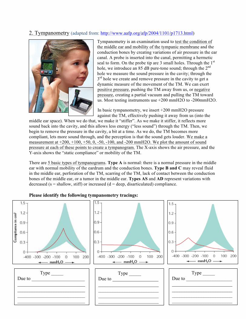
2. Tympanometry (adapted from: http://www.aafp.org/afp/2004/1101/p1713.html)
Tympanometry is an examination used to test the condition of the middle ear and mobility of the tympanic membrane and the conduction bones by creating variations of air pressure in the ear canal. A probe is inserted into the canal, permitting a hermetic seal to form. On the probe tip are 3 small holes. Through the 1st hole, we introduce an 85 dB pure-tone sound; through the 2nd hole we measure the sound pressure in the cavity; through the 3rd hole we create and remove pressure in the cavity to get a dynamic measure of the movement of the TM. We can exert positive pressure, pushing the TM away from us, or negative pressure, creating a partial vacuum and pulling the TM toward us. Most testing instruments use +200 mmH2O to -200mmH2O.
In basic tympanometry, we insert +200 mmH2O pressure against the TM, effectively pushing it away from us (into the
middle ear space). When we do that, we make it “stiffer”. As we make it stiffer, it reflects more sound back into the cavity, and this allows less energy (“less sound”) through the TM. Then, we begin to remove the pressure in the cavity, a bit at a time. As we do, the TM becomes more compliant, lets more sound through, and the perception is that the sound gets louder. We make a measurement at +200, +100, +50, 0, -50, -100, and -200 mmH2O. We plot the amount of sound pressure at each of these points to create a tympanogram. The X-axis shows the air pressure, and the Y-axis shows the “static compliance” or mobility of the TM.
There are 5 basic types of tympanograms. Type A is normal: there is a normal pressure in the middle ear with normal mobility of the eardrum and the conduction bones. Type B and C may reveal fluid in the middle ear, perforation of the TM, scarring of the TM, lack of contact between the conduction bones of the middle ear, or a tumor in the middle ear. Types AS and AD represent variations with decreased (s = shallow, stiff) or increased (d = deep, disarticulated) compliance.
Please identify the following tympanometry tracings:
Type _____ Due to ______________________ ________________________________________________________________________________________________________________
Type _____ Due to ___________________ ____________________________________________________________________________________________________
Type _____ Due to ___________________ ____________________________________________________________________________________________________