head & neck trauma
DESCRIPTION
Head & Neck Trauma. Dr. Mohammad aloulah , MBBS, SBORL(c) Assistant Professor King Saud University. Otolaryngology Consultant. l. Mechanisms of Trauma. • MVA. • Iatrogenic. • Burns and frostbite. • Noise. • Barotrauma. Auricle injuries. • Hematomas. - PowerPoint PPT PresentationTRANSCRIPT
Head & Neck Trauma
Dr. Mohammad aloulah, MBBS, SBORL(c)Assistant Professor King Saud University
Otolaryngology Consultantl
Mechanisms of Trauma
• MVA• Iatrogenic
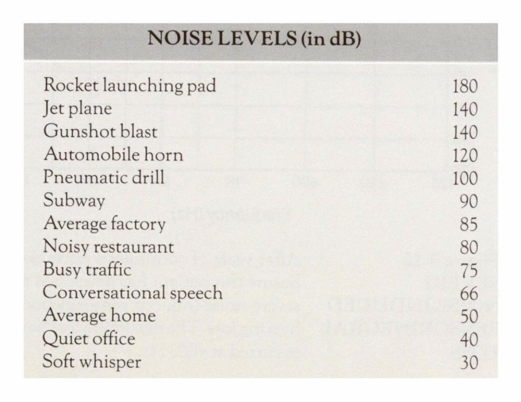
• Burns and frostbite• Noise
• Barotrauma
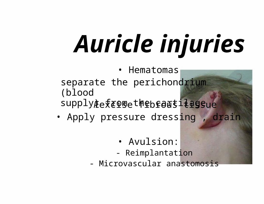
Auricle injuries• Hematomas
separate the perichondrium (bloodsupply) from the cartilageÆexcise fibrous tissue• Apply pressure dressing , drain
• Avulsion:- Reimplantation
- Microvascular anastomosis
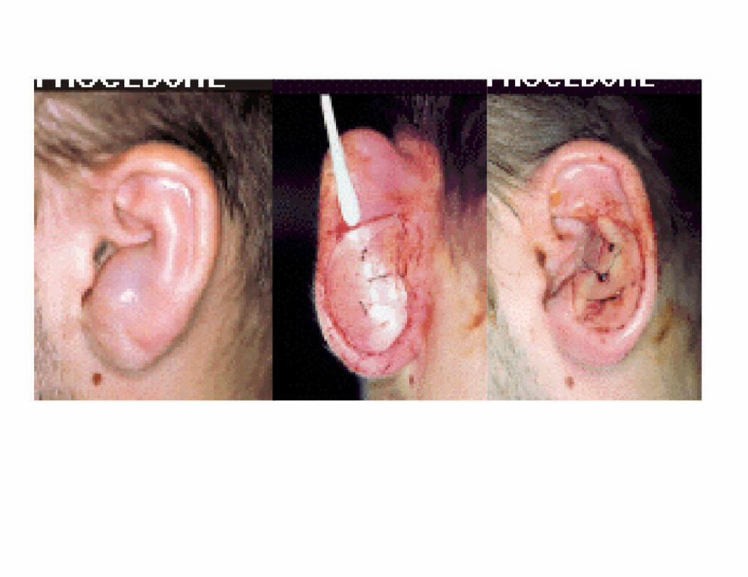
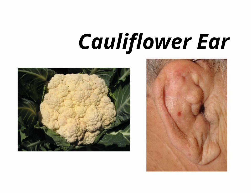
Cauliflower Ear
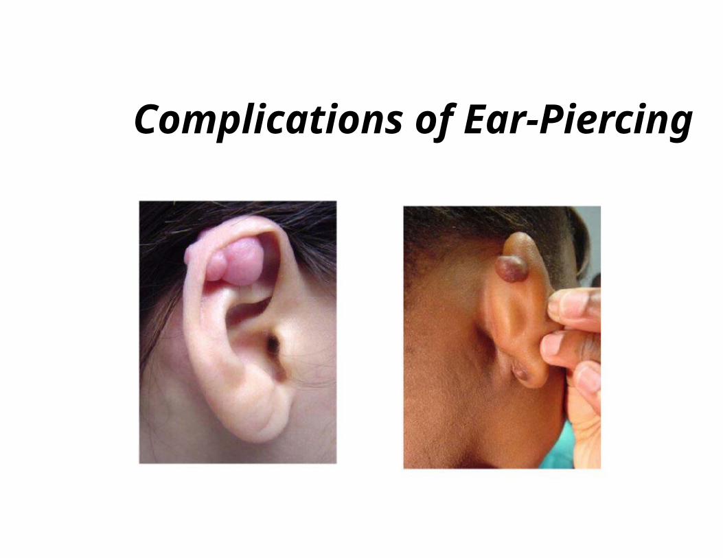
Complications of Ear-Piercing
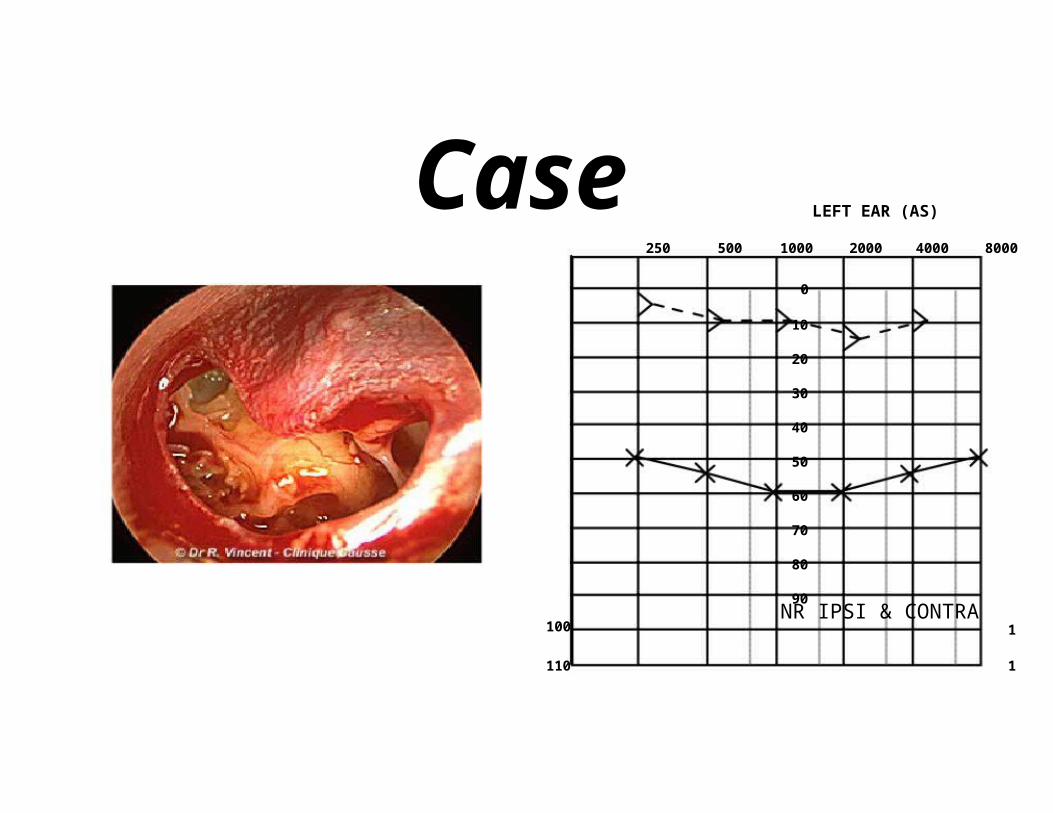
Case LEFT EAR (AS)
250 500 1000 2000 4000 8000
0
10
20
30
40
50
60
70
80
90NR IPSI & CONTRA
100 1
110 1
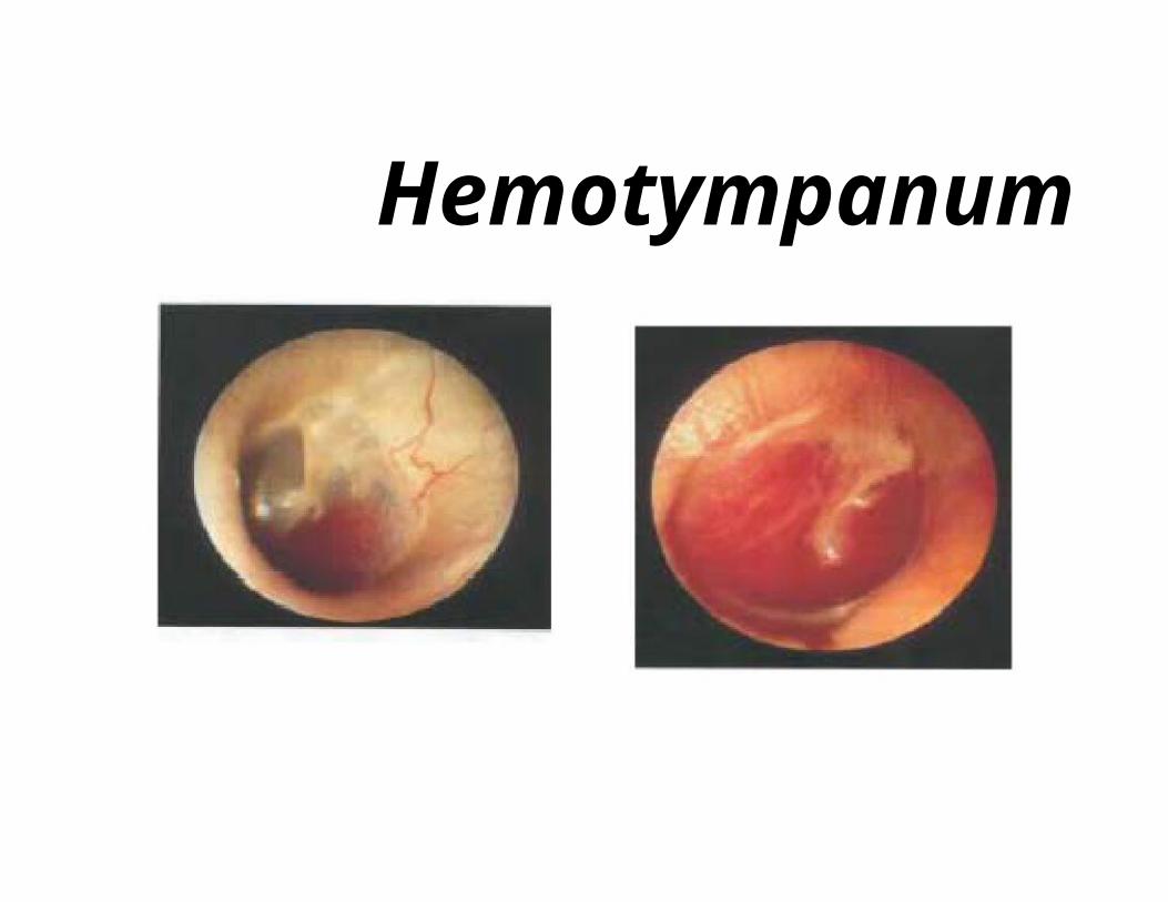
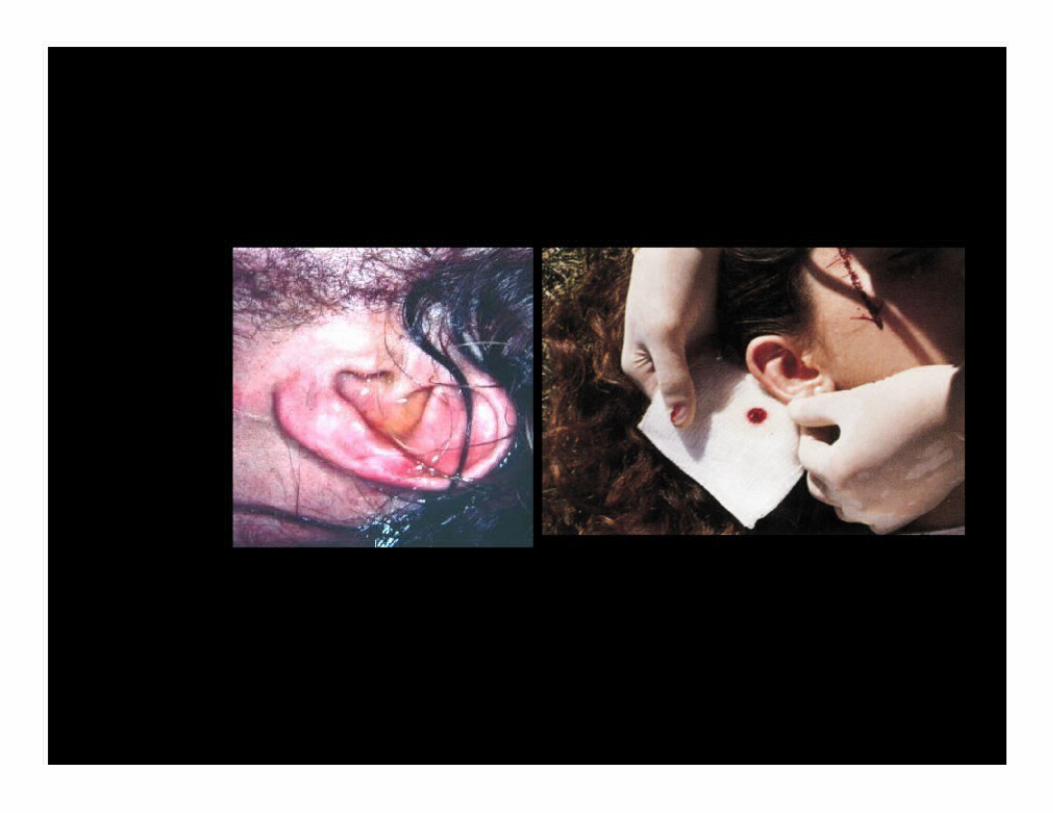
Hemotympanum
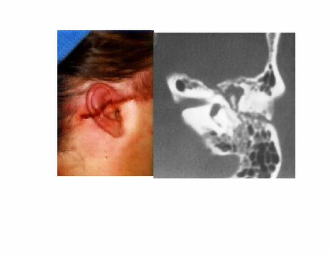
R
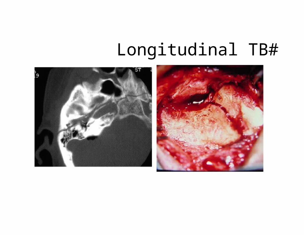
Longitudinal TB#

Complications of TB#• Hearing loss
• Vertigo
• Tinnitus
• Facial paralysis
• CSF leak
• Carotid injury
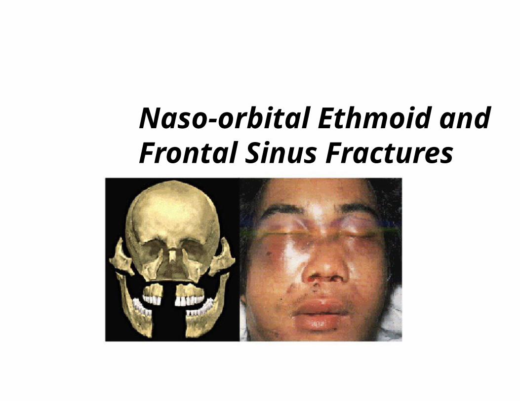
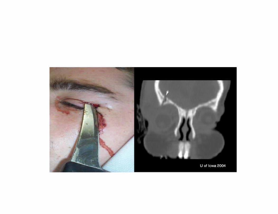
Naso-orbital Ethmoid andFrontal Sinus Fractures
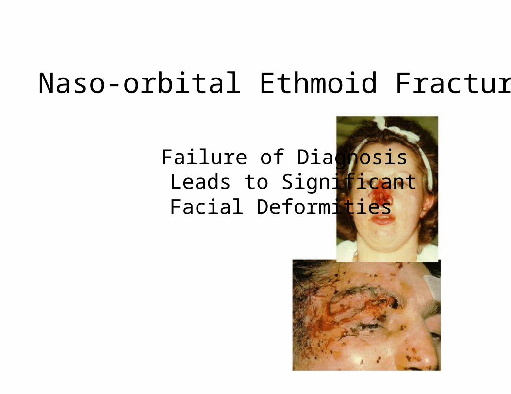
Naso-orbital Ethmoid Fractures
Failure of DiagnosisLeads to SignificantFacial Deformities
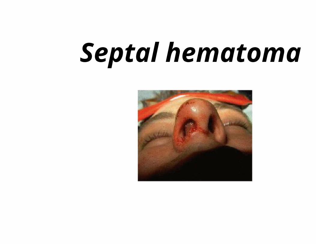
Septal hematoma
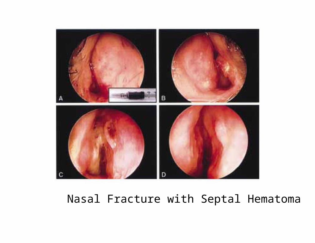
Nasal Fracture with Septal Hematoma
ComplicationNasal Deformity
- Flattened Nasal Dorsum
- Septal Deviation / Dislocation
Intracranial Involvement- Cerebrospinal Fistula
- Pneumocephalus
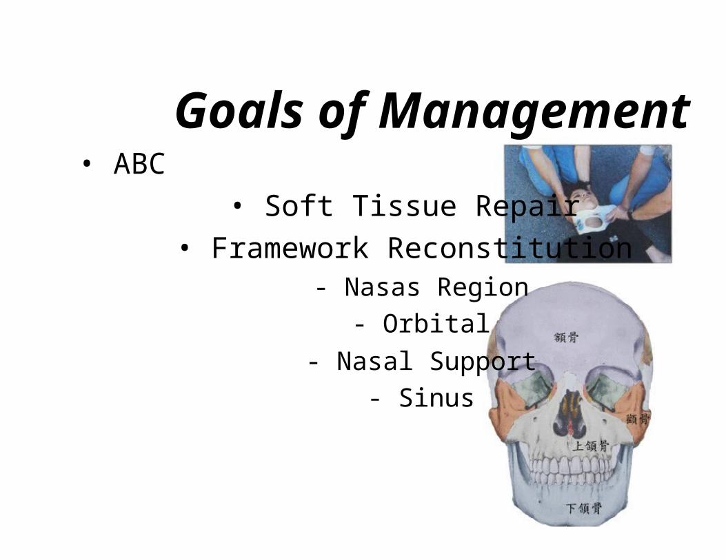
Goals of Management• ABC
• Soft Tissue Repair
• Framework Reconstitution- Nasas Region
- Orbital
- Nasal Support
- Sinus

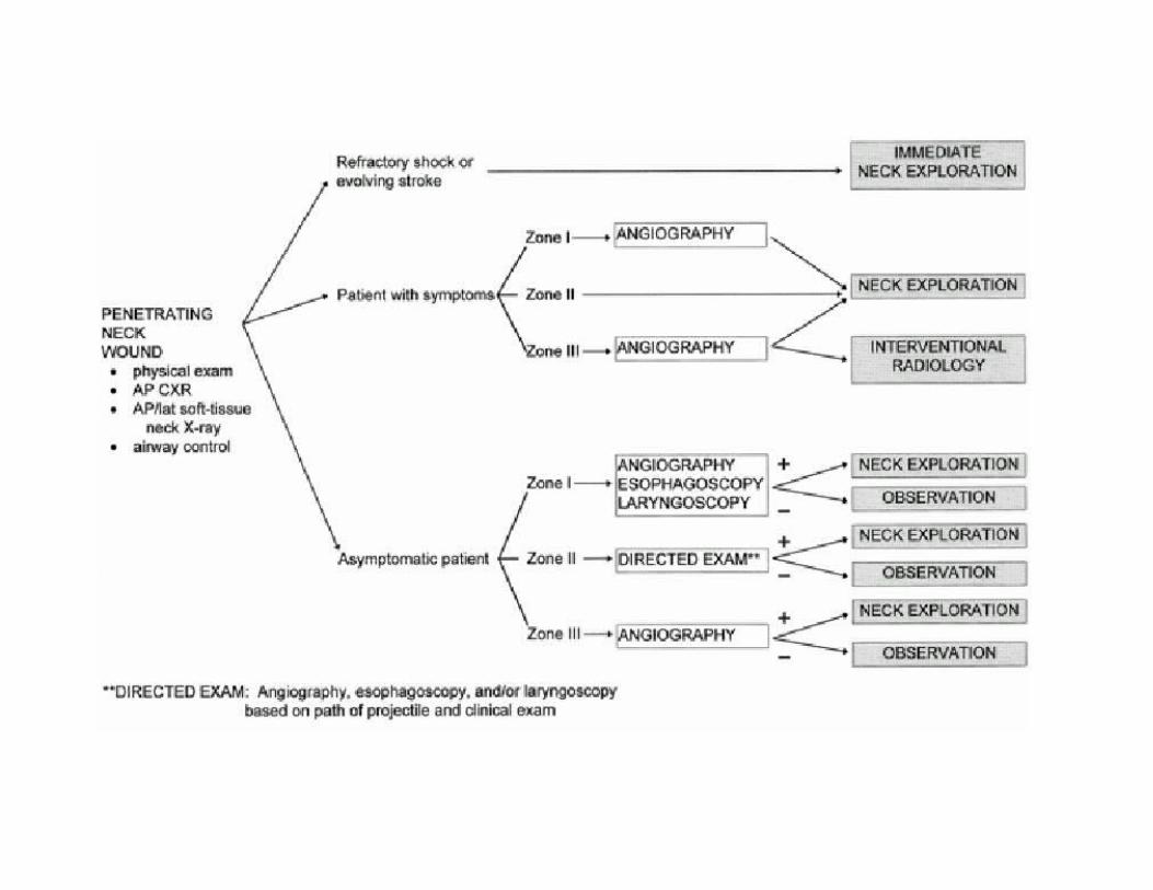
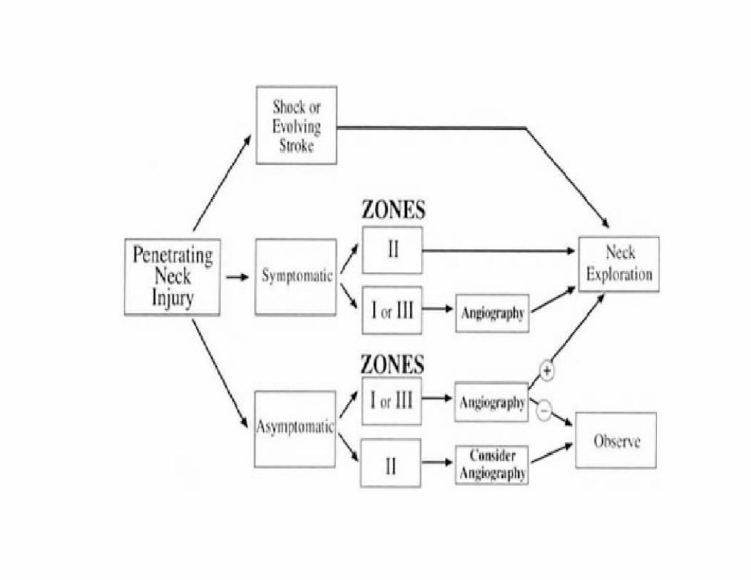
Anatomy/Zone I• Cricoid Æ sternum and clavicles
• Contains the- Subclavian arteries and veins
- Dome of the pleura- Esophagus
- Great vessels of the neck +recurrent nerve- Trachea
• S/S may be hidden from inspection in themediastinum or chest
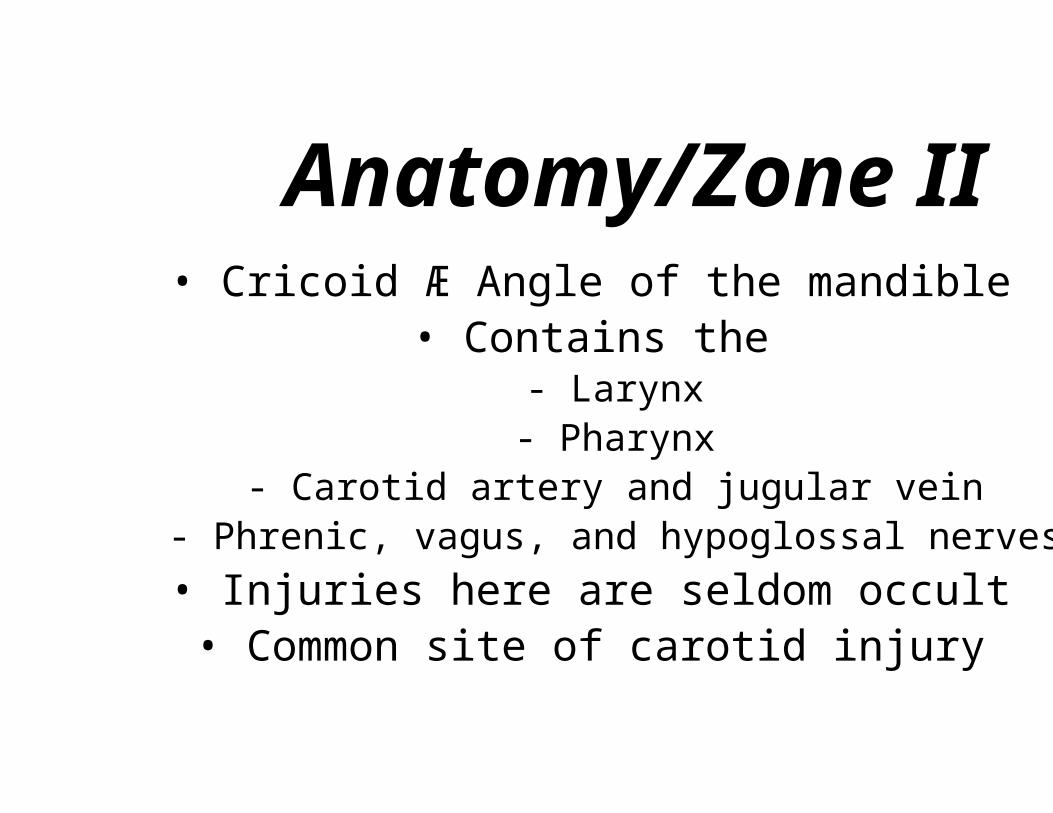
Anatomy/Zone II• Cricoid Æ Angle of the mandible
• Contains the- Larynx- Pharynx
- Carotid artery and jugular vein- Phrenic, vagus, and hypoglossal nerves
• Injuries here are seldom occult• Common site of carotid injury
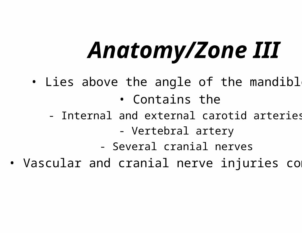
Anatomy/Zone III• Lies above the angle of the mandible
• Contains the- Internal and external carotid arteries
- Vertebral artery
- Several cranial nerves
• Vascular and cranial nerve injuries common
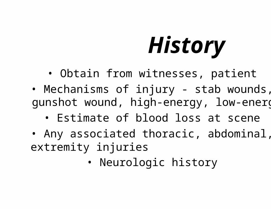
History• Obtain from witnesses, patient
• Mechanisms of injury - stab wounds,gunshot wound, high-energy, low-energy
• Estimate of blood loss at scene
• Any associated thoracic, abdominal,extremity injuries
• Neurologic history
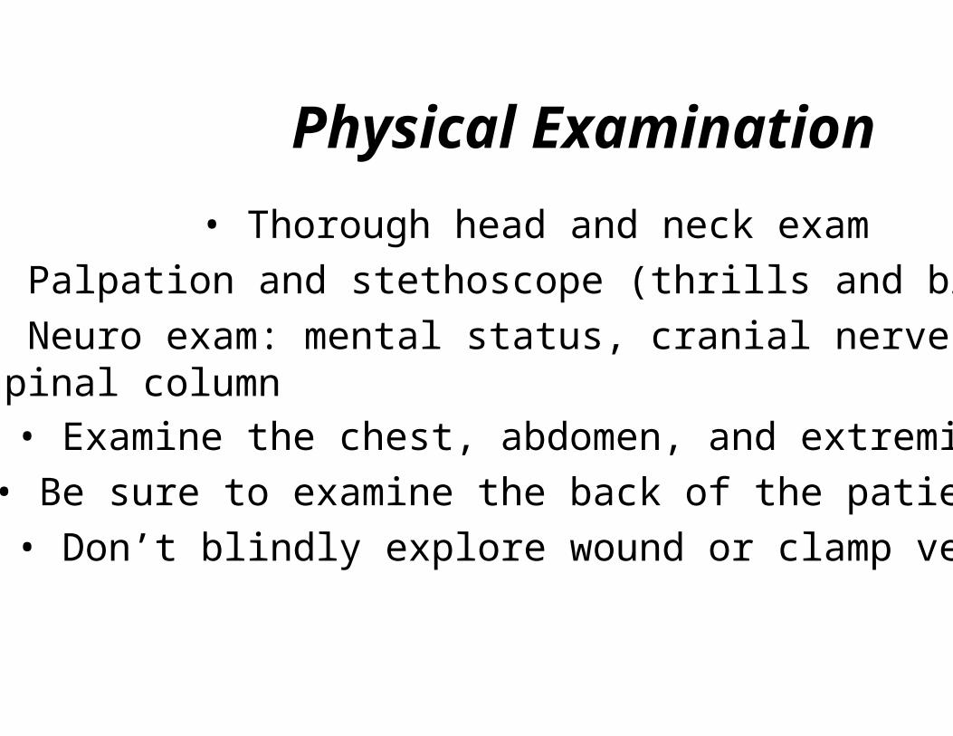
Physical Examination
• Thorough head and neck exam
• Palpation and stethoscope (thrills and bruits)
• Neuro exam: mental status, cranial nerves, andspinal column
• Examine the chest, abdomen, and extremities
• Be sure to examine the back of the patient as
• Don’t blindly explore wound or clamp vessel
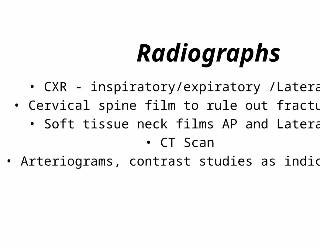
Radiographs• CXR - inspiratory/expiratory /Lateral
• Cervical spine film to rule out fractures
• Soft tissue neck films AP and Lateral
• CT Scan
• Arteriograms, contrast studies as indicated

Intubation: Indications
• Failure to oxygenate
• Failure to remove CO2
• Increased WOB
• Neuromuscular weakness
• CNS failure
• Cardiovascular failure
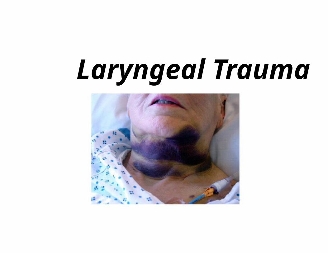
Laryngeal Trauma
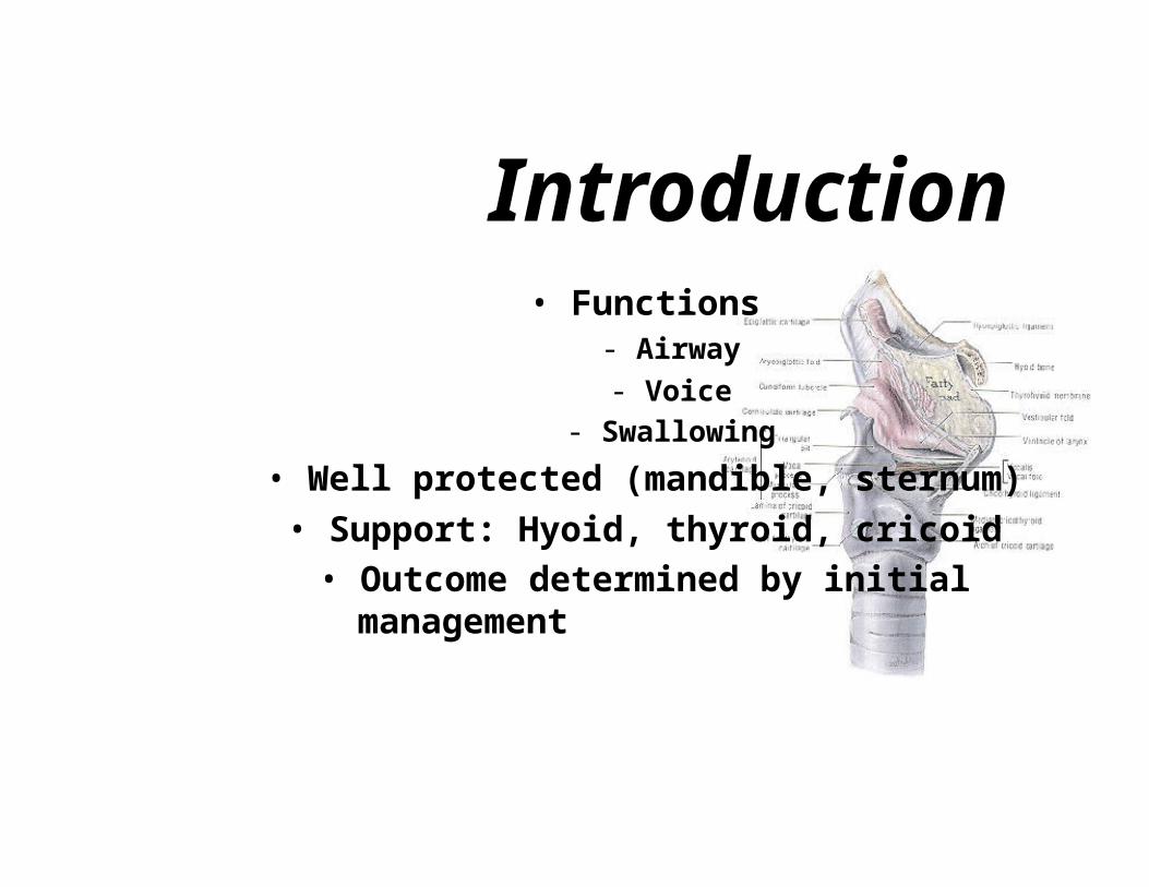
Introduction• Functions
- Airway- Voice
- Swallowing
• Well protected (mandible, sternum)• Support: Hyoid, thyroid, cricoid• Outcome determined by initial
management
Mechanism of Injury
• Blunt- MVA, strangulation, clothesline, sports related
- Significant internal damage, minimal signs
• Penetrating- GSW: damage related to velocity
- Knife: easy to underestimate damage
Initial Evaluation• ABC
• Secure airway - local tracheotomy
• Intubation can worsen airway• Avoid cricothyroidotomy
• Pediatric: tracheotomy over bronchoscope
• Clear C-spine
History• Change in voice - most reliable
• Dysphagia
• Odynophagia
• Difficulty breathing - more severe injury
• Anterior neck pain
• Hemoptysis
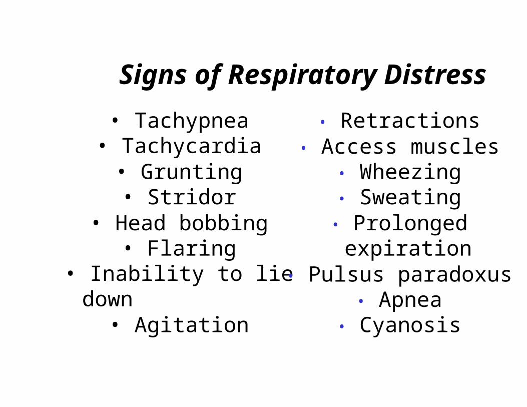
Signs of Respiratory Distress
• Tachypnea• Tachycardia
• Grunting• Stridor
• Head bobbing• Flaring
• Inability to liedown• Agitation
• Retractions• Access muscles
• Wheezing• Sweating
• Prolongedexpiration
• Pulsus paradoxus• Apnea
• Cyanosis
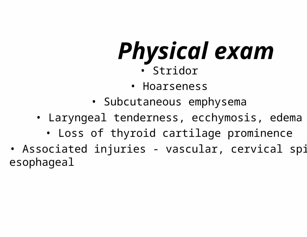
Physical exam• Stridor
• Hoarseness
• Subcutaneous emphysema
• Laryngeal tenderness, ecchymosis, edema
• Loss of thyroid cartilage prominence
• Associated injuries - vascular, cervical spine,esophageal
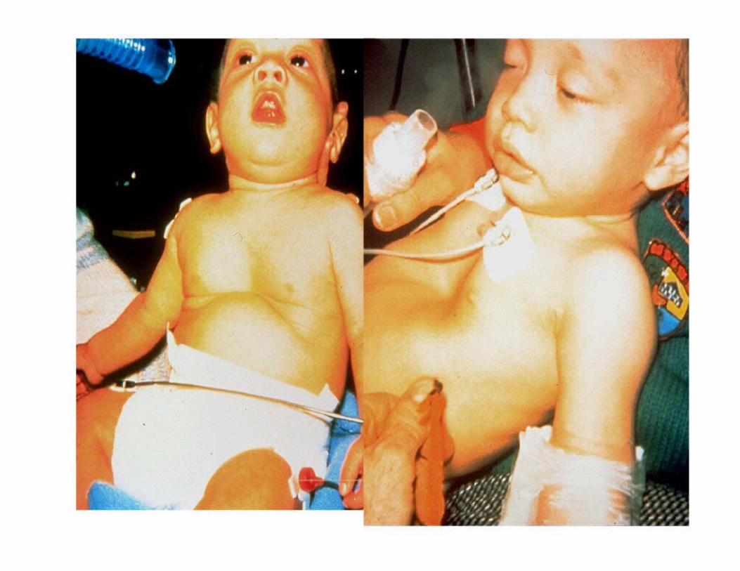

Physical Exam
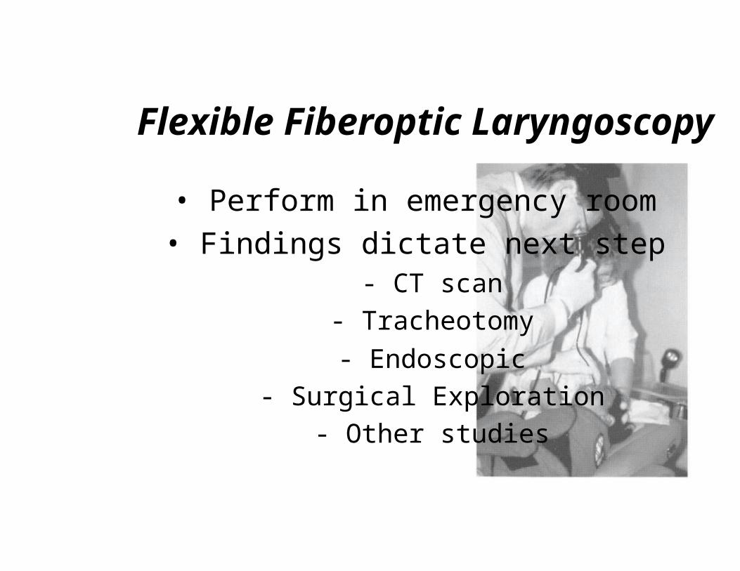
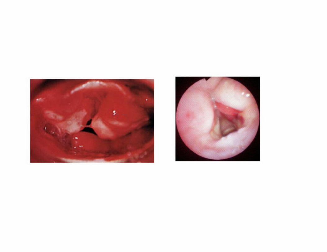
Flexible Fiberoptic Laryngoscopy
• Perform in emergency room
• Findings dictate next step- CT scan
- Tracheotomy
- Endoscopic
- Surgical Exploration
- Other studies
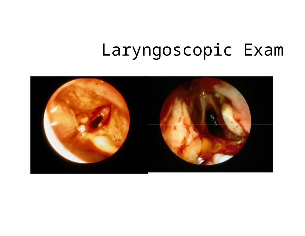
Laryngoscopic Exam
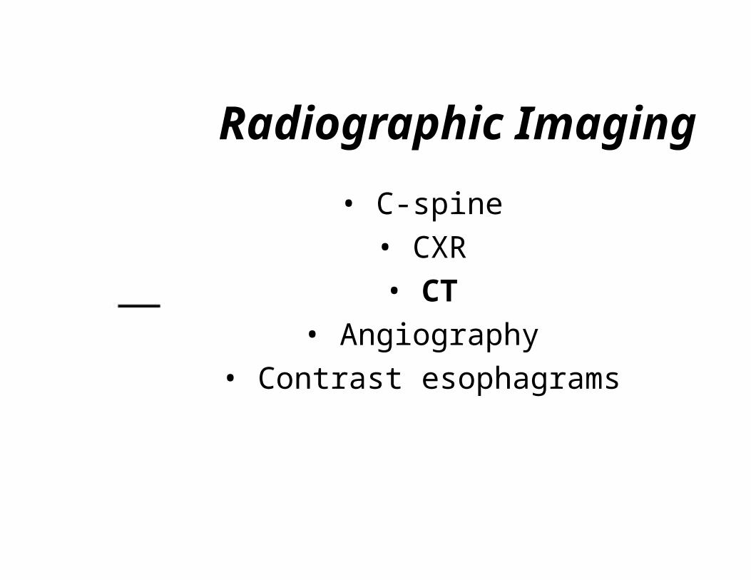
Radiographic Imaging
• C-spine
• CXR
• CT• Angiography
• Contrast esophagrams
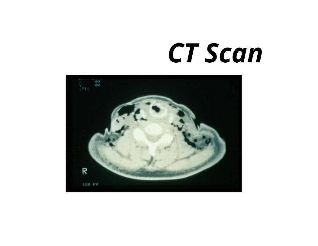
CT Scan
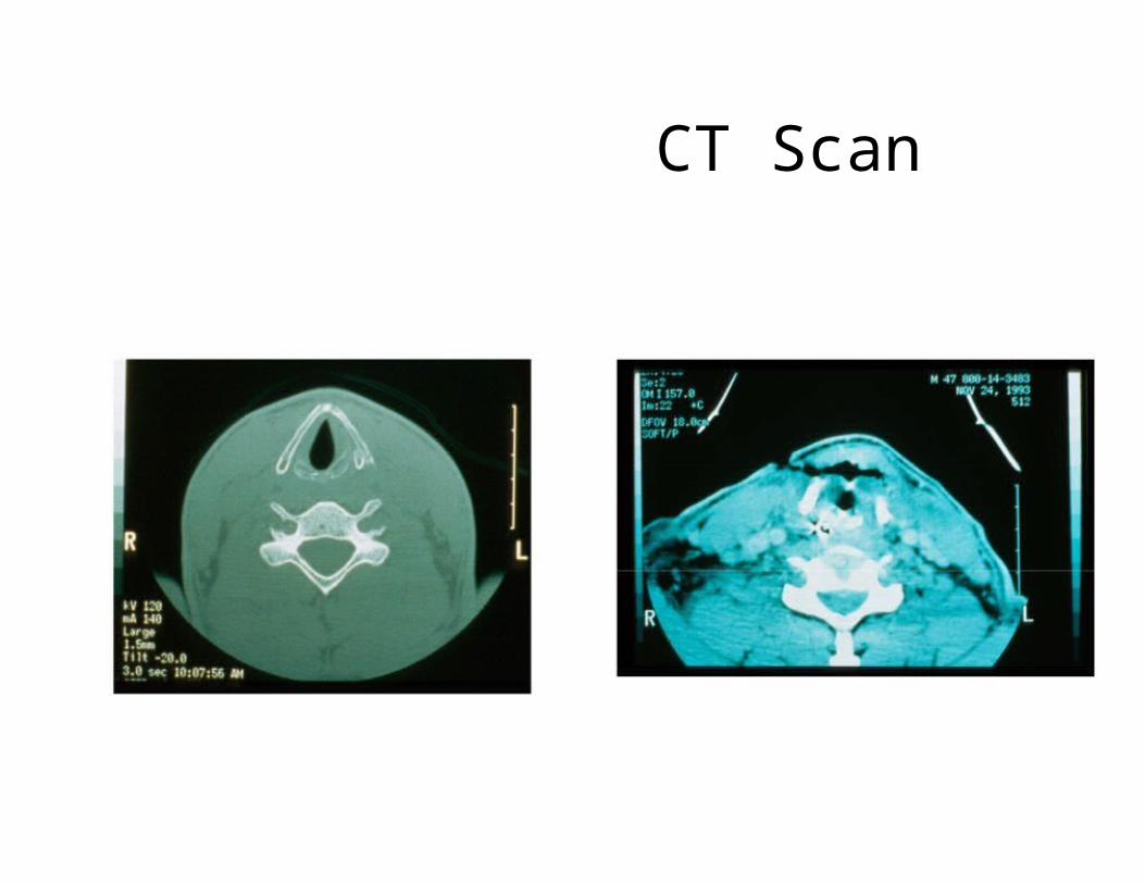
CT Scan
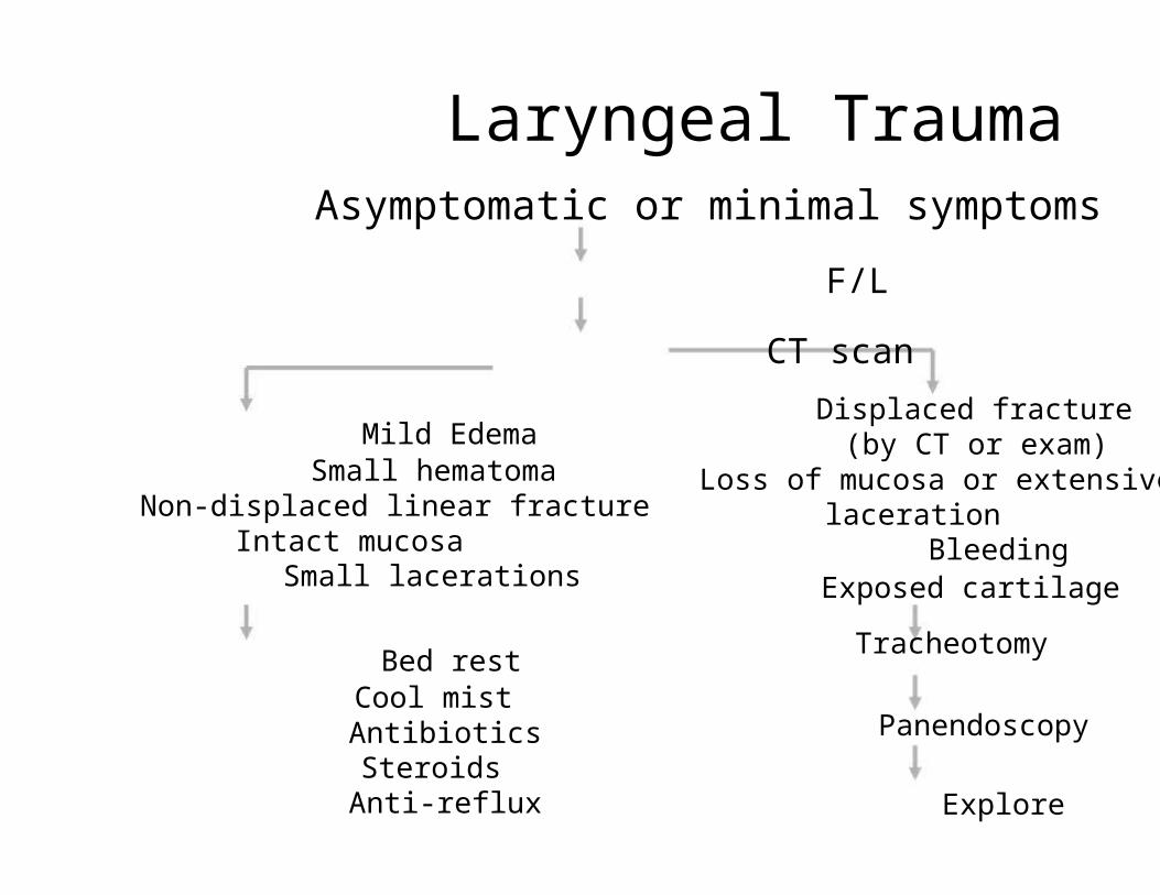
Laryngeal TraumaAsymptomatic or minimal symptoms
F/L
CT scan
Displaced fractureMild Edema
Small hematomaNon-displaced linear fracture
Intact mucosaSmall lacerations
Bed restCool mistAntibioticsSteroids
Anti-reflux
(by CT or exam)Loss of mucosa or extensive
lacerationBleeding
Exposed cartilage
Tracheotomy
Panendoscopy
Explore
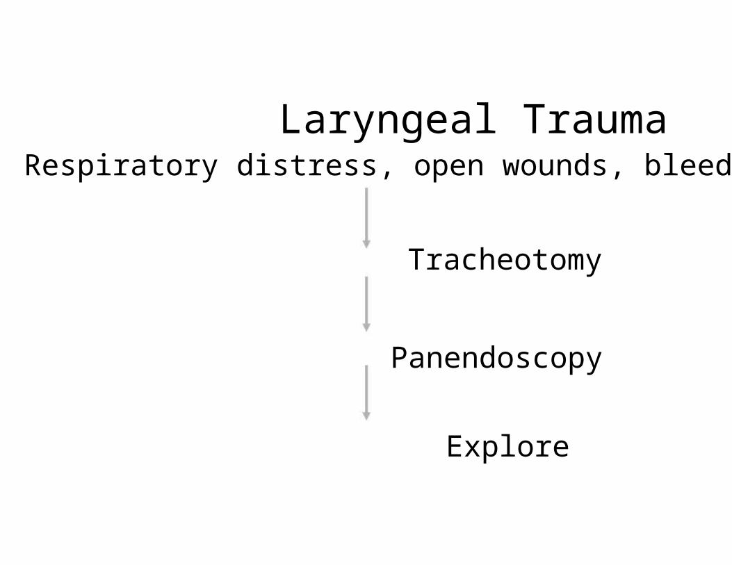
Laryngeal TraumaRespiratory distress, open wounds, bleeding
Tracheotomy
Panendoscopy
Explore
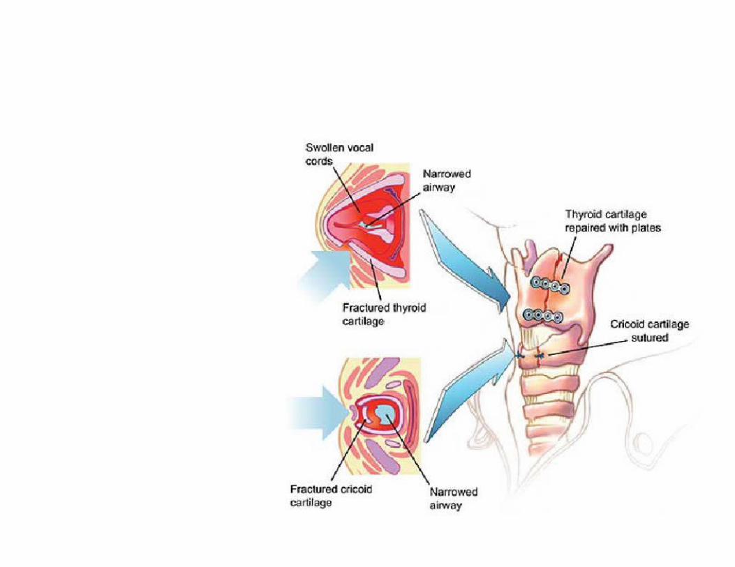
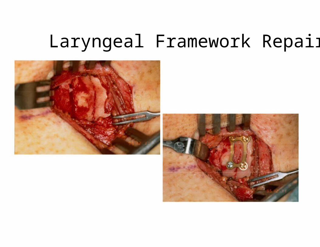
Laryngeal Framework Repair
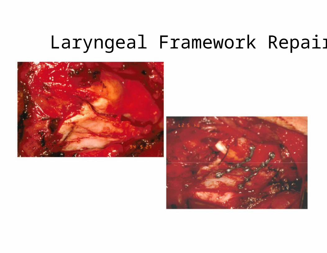
Laryngeal Framework Repair

Treatment Goals• Preservation of airway
• Prevention of aspiration• Restoration of normal voice
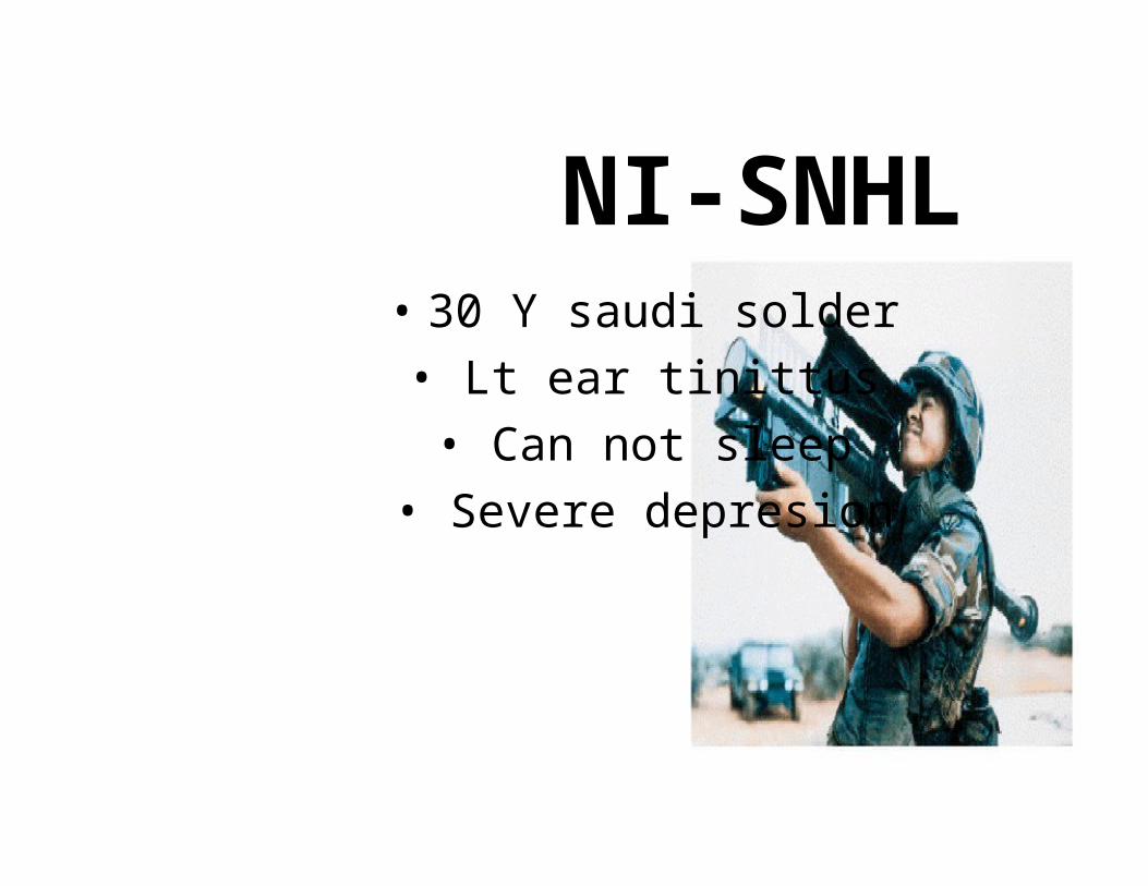
NI-SNHL• 30 Y saudi solder
• Lt ear tinittus
• Can not sleep
• Severe depresion

Trauma & SNHL• NISNHL
• Acoustic trauma• Barotrauma
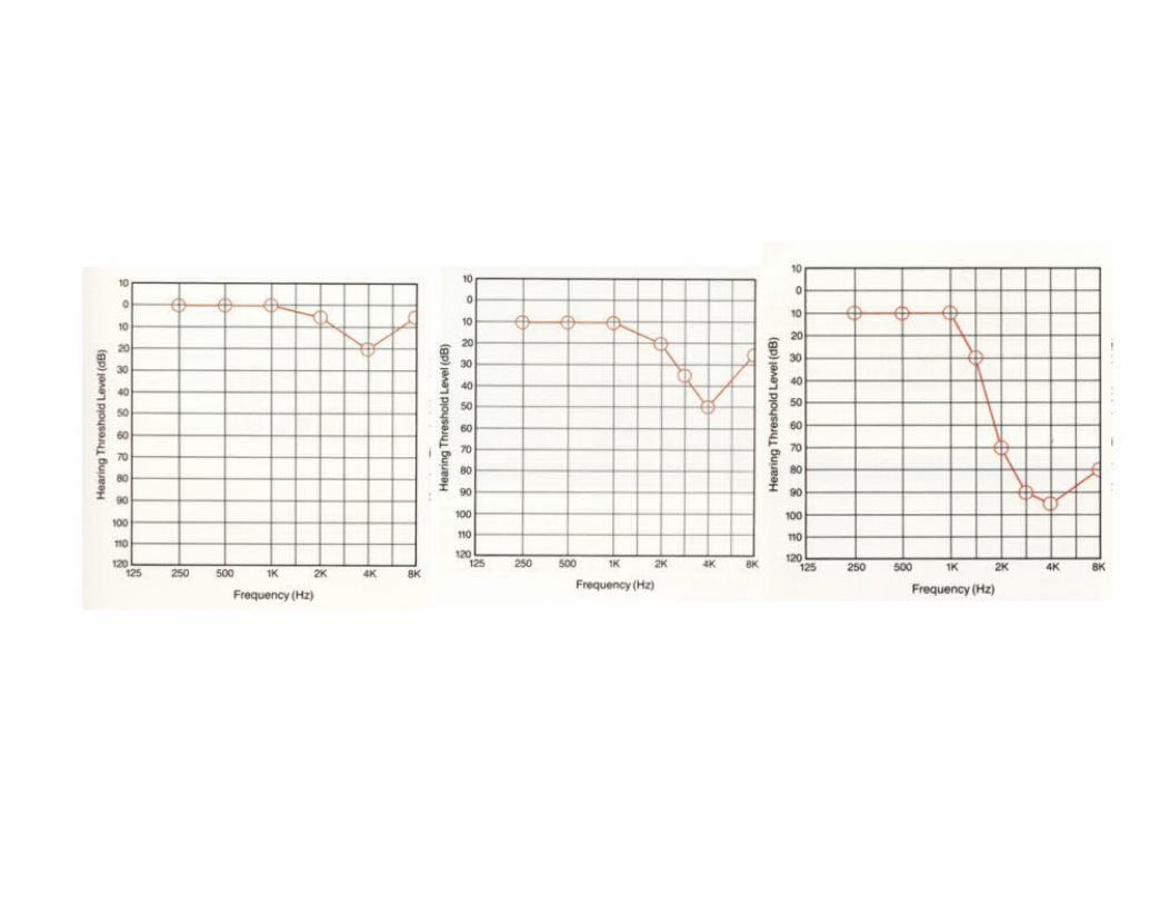
Noise induce SNHL
• one of the most common occupationallyinduced disabilities
• Tinnitus- commonly accompanied NISNHL
- warning sign
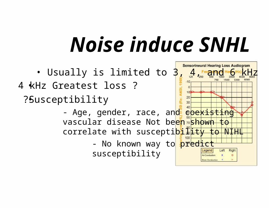
Noise induce SNHL• Usually is limited to 3, 4, and 6 kHz
• 4 kHz Greatest loss ?
• ?Susceptibility- Age, gender, race, and coexistingvascular disease Not been shown tocorrelate with susceptibility to NIHL
- No known way to predictsusceptibility
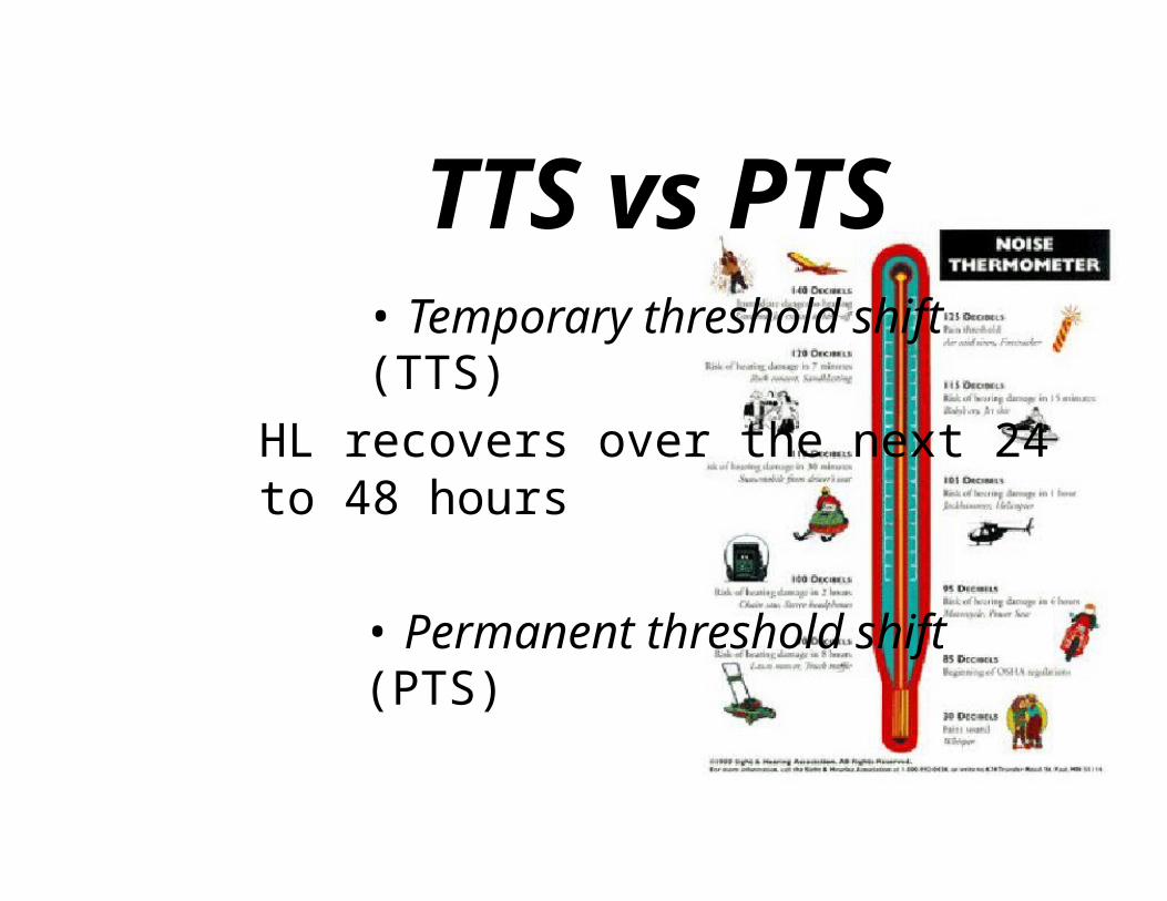
TTS vs PTS• Temporary threshold shift(TTS)
HL recovers over the next 24to 48 hours
• Permanent threshold shift(PTS)
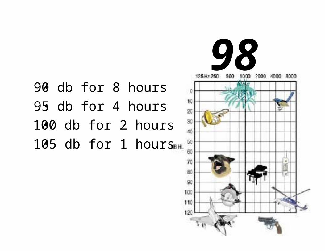
98• 90 db for 8 hours
• 95 db for 4 hours
• 100 db for 2 hours
• 105 db for 1 hours
Primary role of otolaryngologists
• Prevention
• Early identification.
Barotrauma• Injury of the TM and middle ear
• Unequalized pressure differentials betweenthe middle and external ears
• Flying or underwater diving
• ETD may predispose
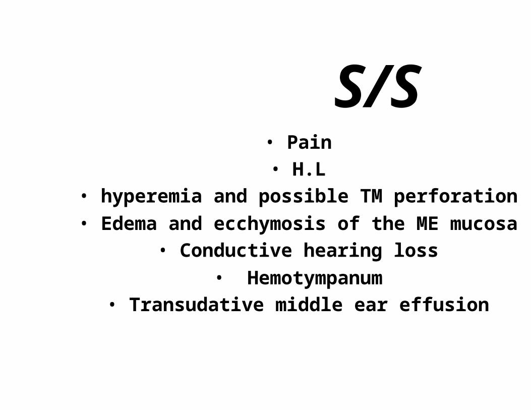
S/S• Pain• H.L
• hyperemia and possible TM perforation
• Edema and ecchymosis of the ME mucosa• Conductive hearing loss
• Hemotympanum• Transudative middle ear effusion
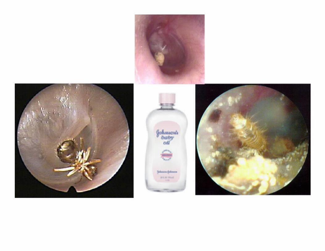
Foreign Bodies of theAerodigestive Tract
Dr. Mohammad Aloulah ,MBBS. SBORLAssistant Professor King Saud University
Otolaryngology Consultant King Abdulaziz Hospital
Foreign Bodies• Foreign body ingestion
• Foreign body aspiration
• Kids- Oral exploration
- Easy distractibility
- Cognitive development
Foreign Body Ingestion• Coins
• Meat
• Vegetable matter
• Less than 24 hours in most
Foreign Body Aspiration• Parental suspicion
• History• Choking• Gagging
• Wheezing• Hoarseness• Dysphonia
• Can mimic asthma, croup, pneumonia

Foreign Body Aspiration• Physical exam
- Larynx/cervical trachea• Inspiratory or biphasic stridor
- Intrathoracic trachea• Prolonged expiratory wheeze
- Bronchi• Unequal breath sounds• Diagnostic triad - <50%
- Unilateral wheeze- Cough
- Ipsilaterally diminished breath sounds
• Fiberoptic laryngoscopy
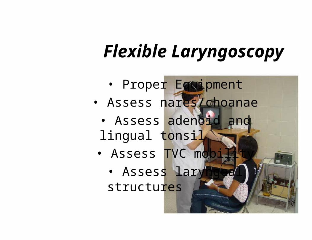
Flexible Laryngoscopy
• Proper Equipment
• Assess nares/choanae
• Assess adenoid andlingual tonsil
• Assess TVC mobility
• Assess laryngealstructures
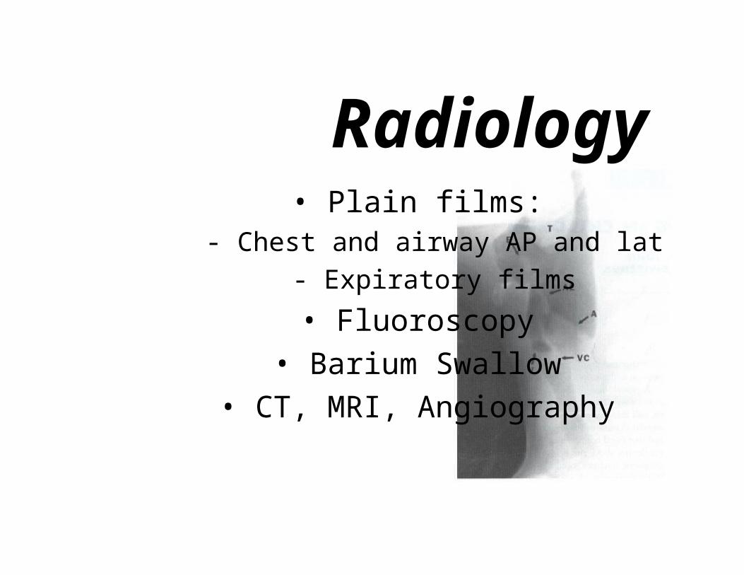
Radiology• Plain films:
- Chest and airway AP and lat
- Expiratory films
• Fluoroscopy
• Barium Swallow
• CT, MRI, Angiography

Direct Laryngoscopy
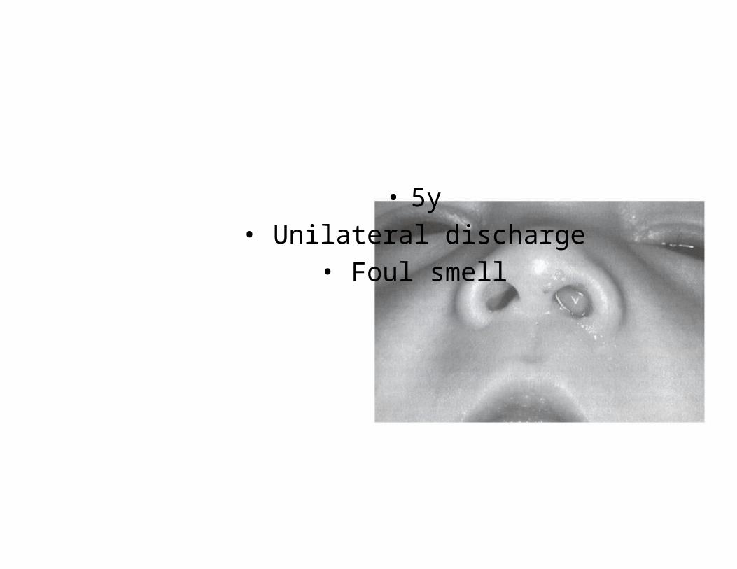
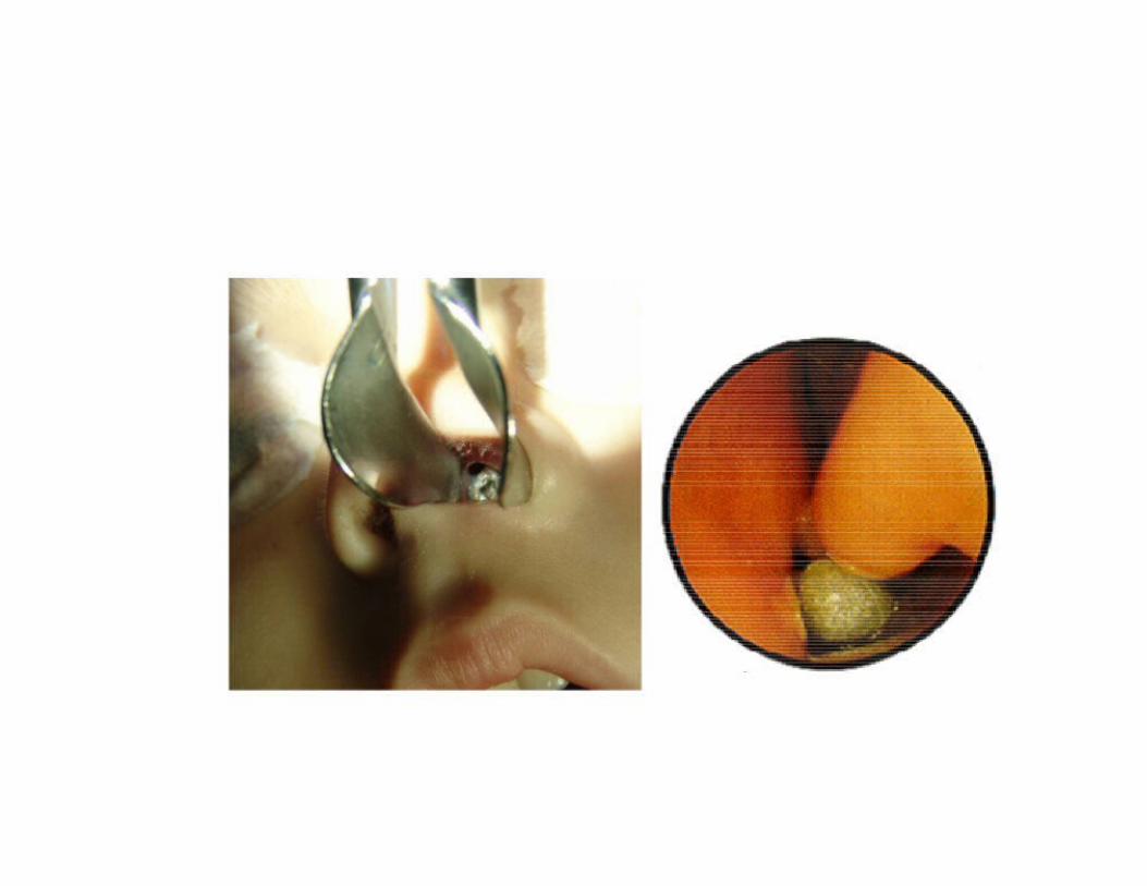
• 5y
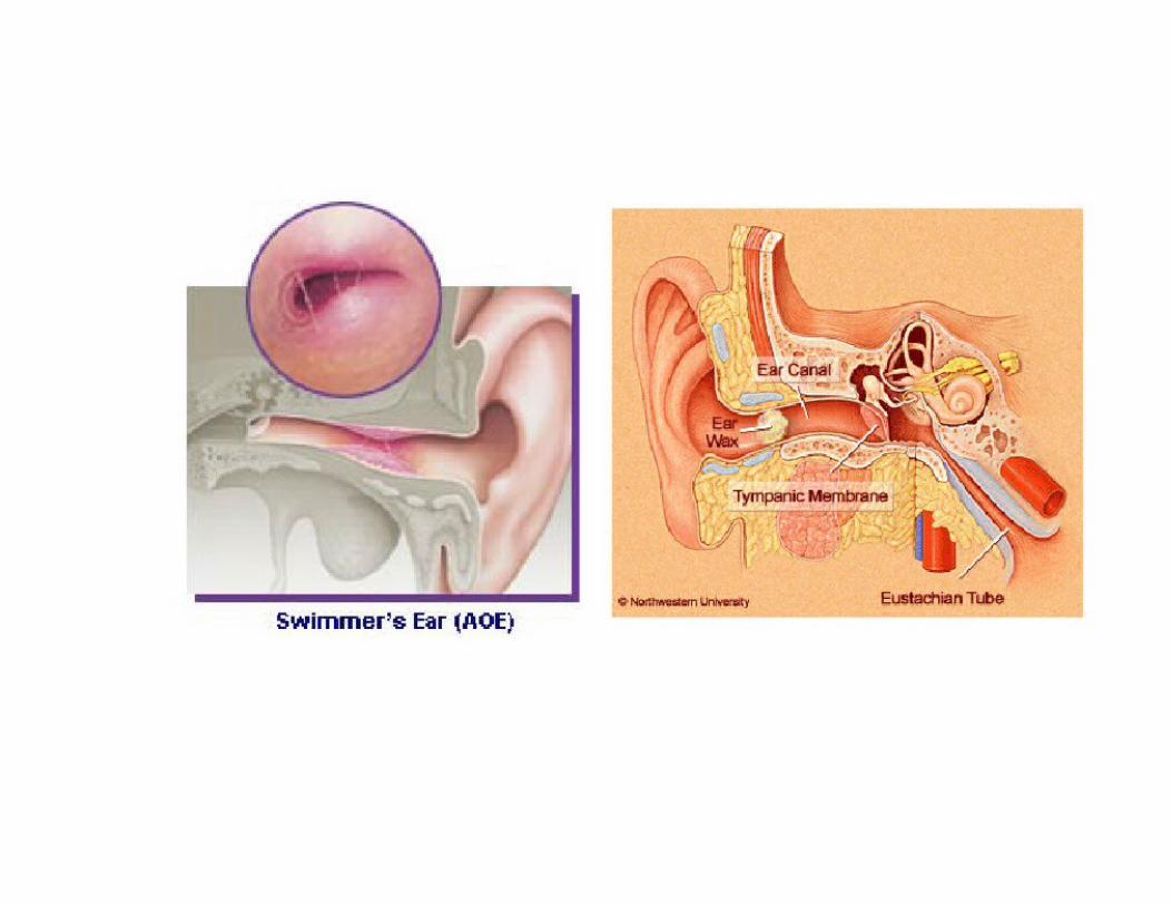
• Unilateral discharge
• Foul smell
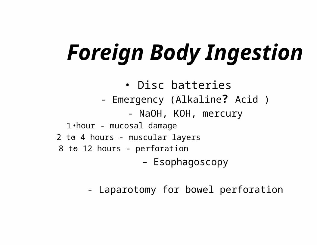
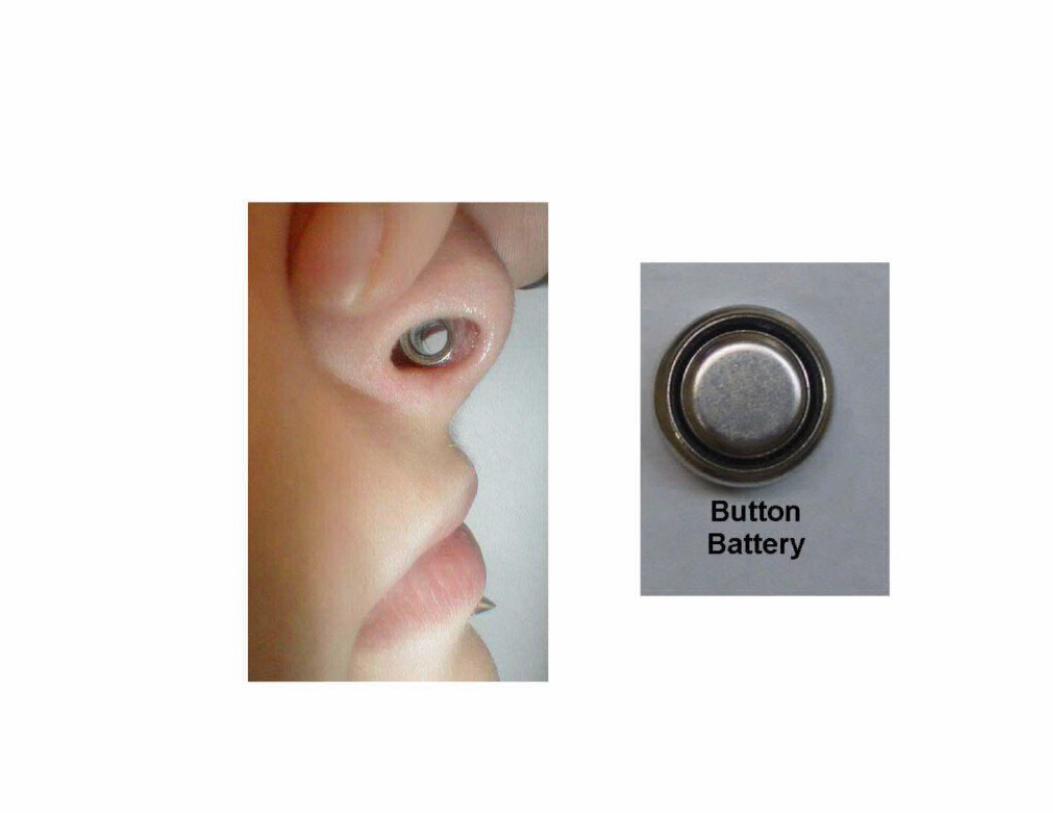
Foreign Body Ingestion• Disc batteries
- Emergency (Alkaline? Acid )
- NaOH, KOH, mercury• 1 hour - mucosal damage
• 2 to 4 hours - muscular layers
• 8 to 12 hours - perforation
– Esophagoscopy
- Laparotomy for bowel perforation
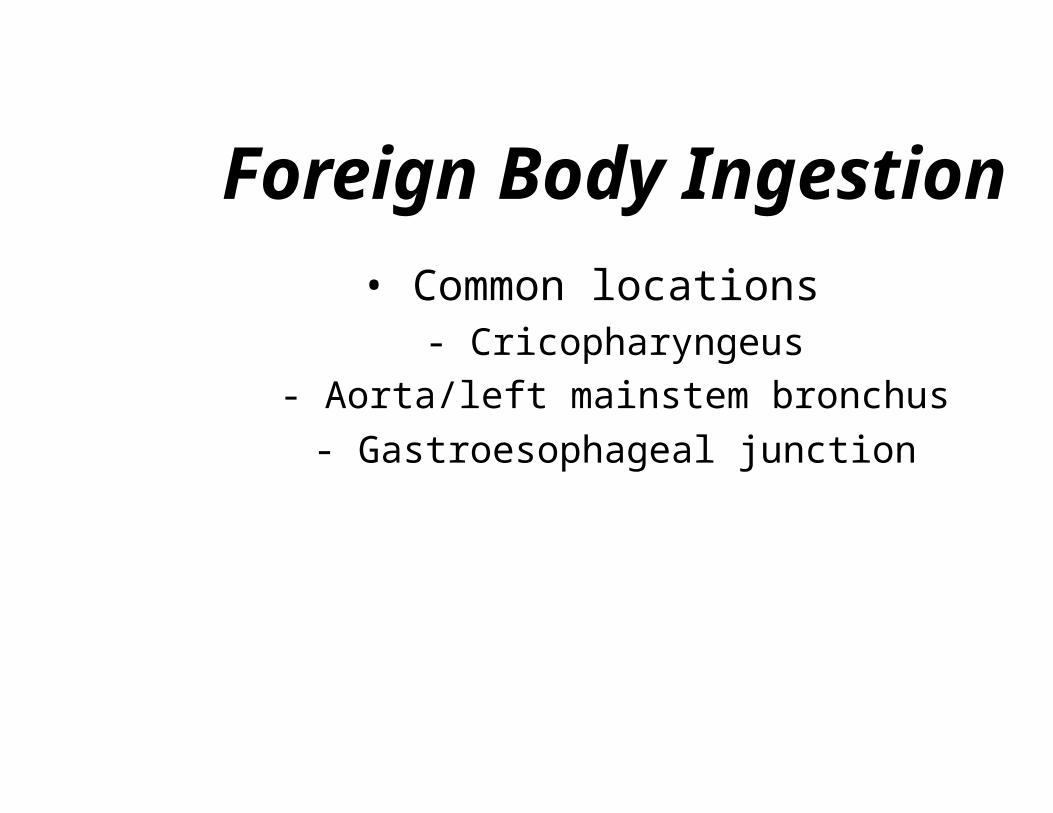
Foreign Body Ingestion• Common locations
- Cricopharyngeus
- Aorta/left mainstem bronchus
- Gastroesophageal junction
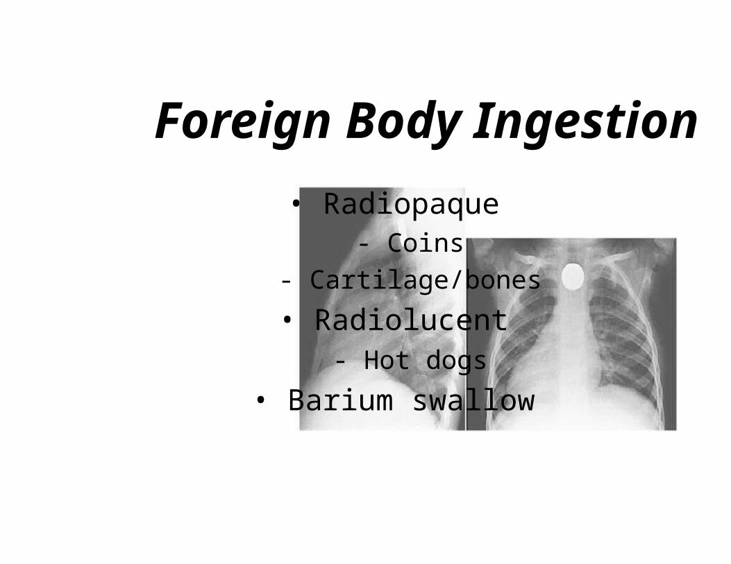
Foreign Body Ingestion
• Radiopaque- Coins
- Cartilage/bones
• Radiolucent- Hot dogs
• Barium swallow
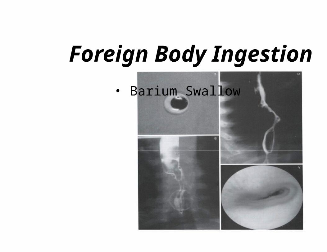
Foreign Body Ingestion
• Barium Swallow
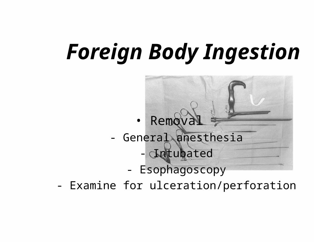
Foreign Body Ingestion
• Removal- General anesthesia
- Intubated
- Esophagoscopy
- Examine for ulceration/perforation

Foreign Body Ingestion• Postoperative management
• NPO for 4-12 hours
• Perforation- Tachycardia
- Tachypnea
- Fever
- Chest pain
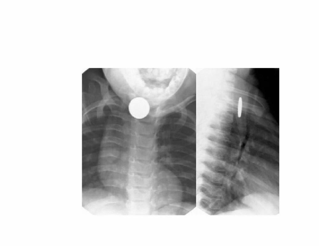
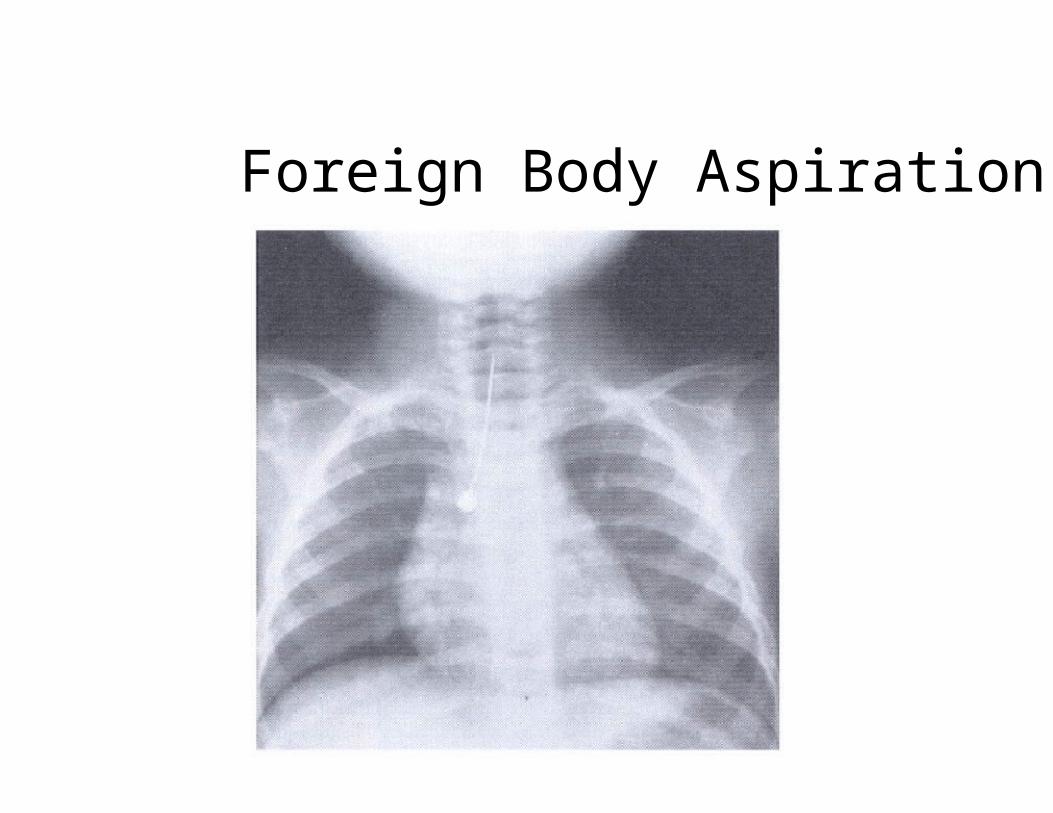
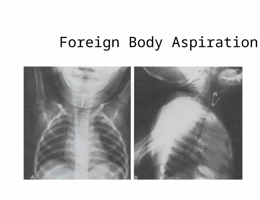
Foreign Body Aspiration
• Radiography- PA & lateral views of chest & neck
- Inspiration & expiration
- Lateral decubitus views
- Airway fluoroscopy
• 25% have normal radiography
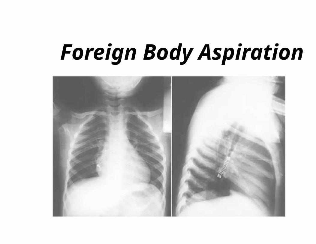
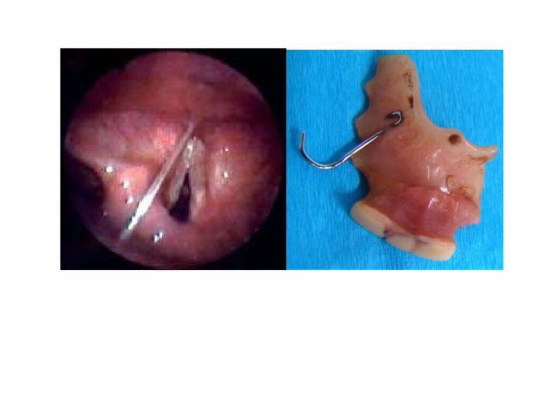
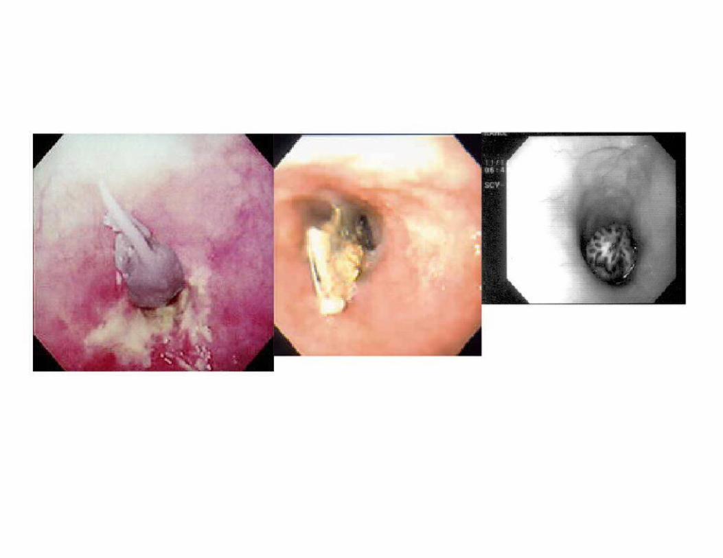
Foreign Body Aspiration
Foreign Body Aspiration
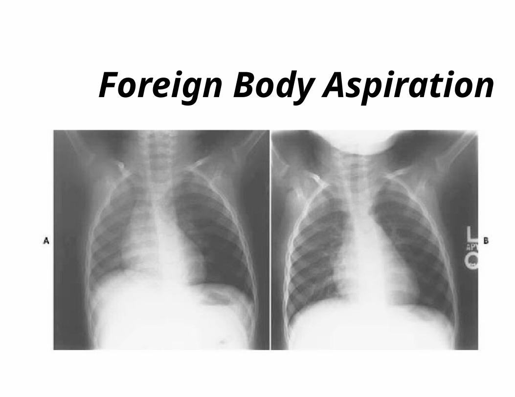
Foreign Body Aspiration
Foreign Body Aspiration
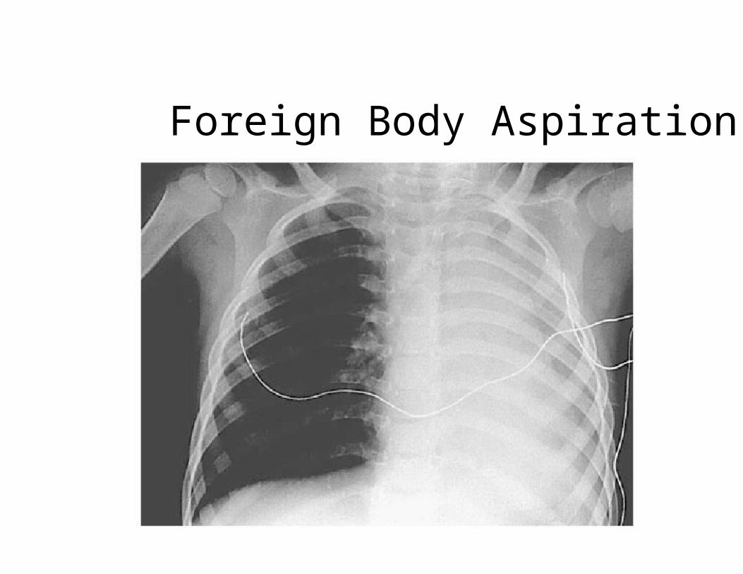
Foreign Body Aspiration

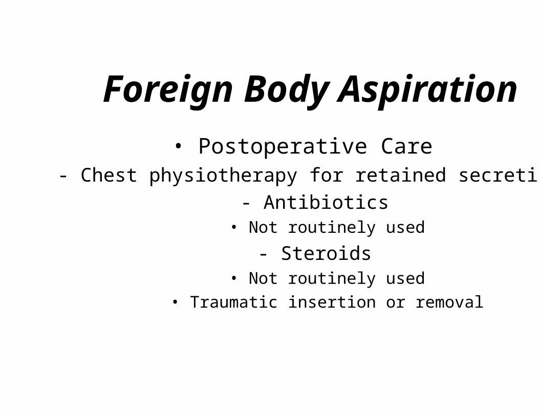
Foreign Body Aspiration• Postoperative Care
- Chest physiotherapy for retained secretions
- Antibiotics• Not routinely used
- Steroids• Not routinely used
• Traumatic insertion or removal
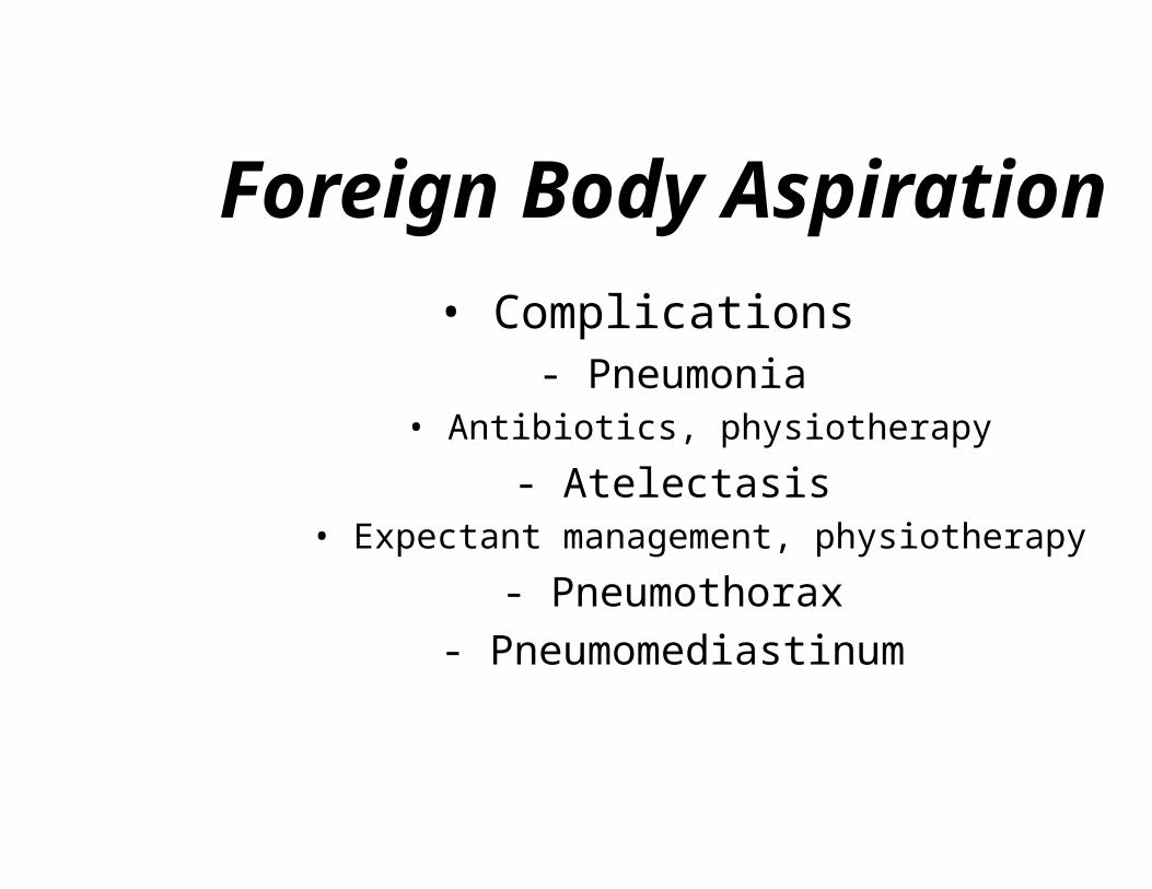
Foreign Body Aspiration• Complications
- Pneumonia• Antibiotics, physiotherapy
- Atelectasis• Expectant management, physiotherapy
- Pneumothorax
- Pneumomediastinum
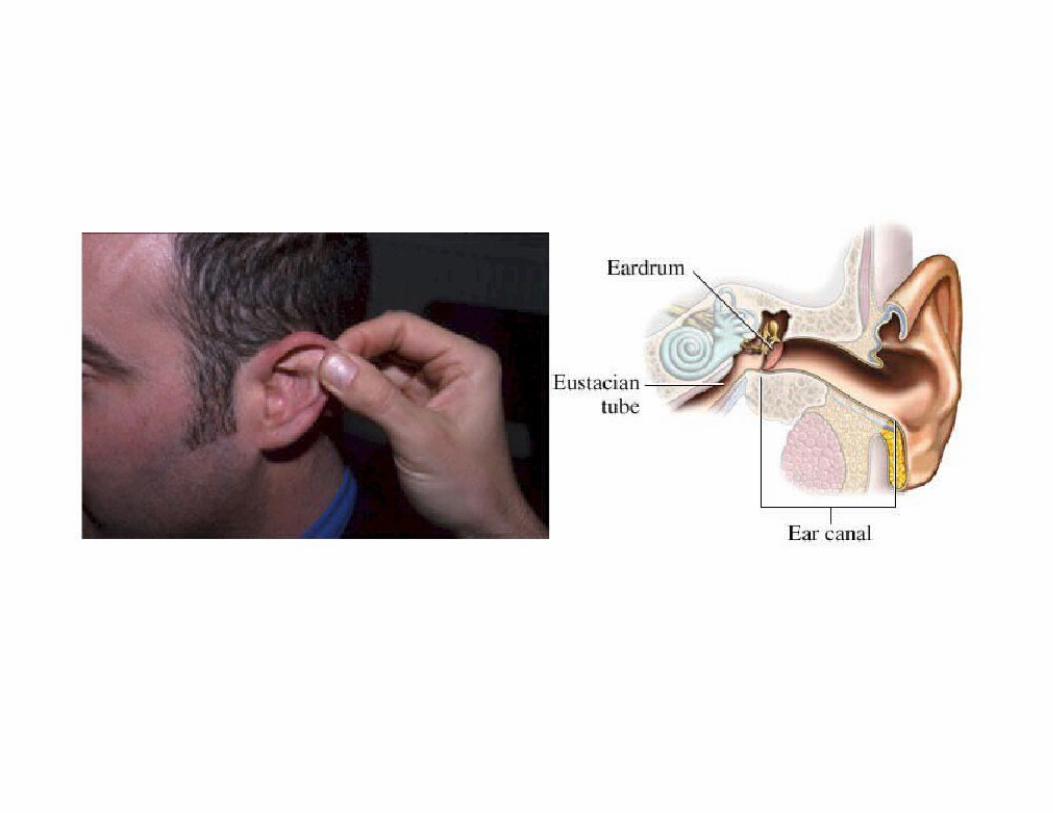
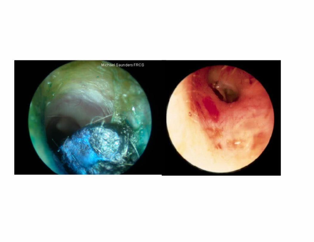
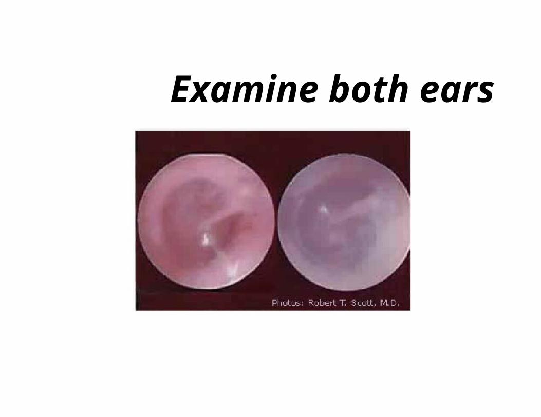
Examine both ears
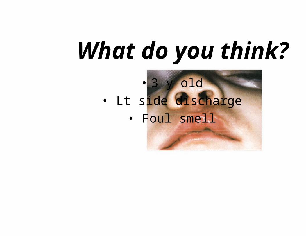
What do you think?• 3 y old
• Lt side discharge
• Foul smell
