head & neck anatomical dissection course 8/14/07
TRANSCRIPT

Head & Neck Anatomical Head & Neck Anatomical Dissection Course 8/14/07Dissection Course 8/14/07
Jason E. Portnof DMD, MDJason E. Portnof DMD, MDChief ResidentChief Resident
Division of Dentistry, Oral & Maxillofacial Division of Dentistry, Oral & Maxillofacial SurgerySurgery
New York Presbyterian HospitalNew York Presbyterian HospitalWeill Cornell Medical CollegeWeill Cornell Medical College

Muscles of Facial ExpressionMuscles of Facial Expression

Muscles of Mouth, Lips, CheekMuscles of Mouth, Lips, CheekCircumoralCircumoral MusclesMuscles
OrbicularisOrbicularis orisorisSphincter of oral aperture.Sphincter of oral aperture.
Dilator MusclesDilator MusclesLevatorLevator labiilabii superiorissuperioris alaquealaque nasinasi
Elevator of upper lip and Elevator of upper lip and alaealae of nose.of nose.MentalisMentalis
Raises skin of chin.Raises skin of chin.BuccinatorBuccinator
Aids mastication by pressing the cheeks against the molar Aids mastication by pressing the cheeks against the molar teeth during chewing.teeth during chewing.

Muscles of Mouth, Lips, CheekMuscles of Mouth, Lips, Cheek
Depressor Depressor angulianguli orisorisLevatorLevator angulianguli orisorisZygomaticusZygomaticus majormajorZygomaticusZygomaticus minorminorLevatorLevator labiilabii superiorissuperiorisDeprssorDeprssor labiilabii inferiorisinferiorisRisoriusRisoriusPlatysmaPlatysma

Lymphatic Drainage of LipsLymphatic Drainage of Lips
Lymph from upper lip and lateral parts of Lymph from upper lip and lateral parts of lower lip drains to lower lip drains to submandibularsubmandibular LNLNLymph from middle part of lower lip drains Lymph from middle part of lower lip drains to to submentalsubmental LNLN


Facial NerveFacial NerveMain trunk emerges from Skull Base at Main trunk emerges from Skull Base at StylomastoidStylomastoidForamen.Foramen.Enters the Parotid Gland.Enters the Parotid Gland.2 trunks emerge from the parotid and radiate 2 trunks emerge from the parotid and radiate anteriorlyanteriorly..
TemporofacialTemporofacialCervicalfacialCervicalfacial
5 Terminal Branches5 Terminal BranchesTemporalTemporalZygomaticZygomaticBuccalBuccalMarginal Marginal MandibularMandibularCervicalCervical

Oral CavityOral Cavity
Boundaries:Boundaries:Extends from lips Extends from lips anteriorlyanteriorly to to palatoglossalpalatoglossal folds folds posteriorlyposteriorly (i.e., anterior to (i.e., anterior to oropharynxoropharynx).).
Structures:Structures:TongueTongueTeethTeethAlveolar BoneAlveolar Bone
The oral vestibule is the space between the The oral vestibule is the space between the cheeks and the teeth/gums.cheeks and the teeth/gums.


PharynxPharynx
NasopharynxNasopharynxOropharynxOropharynxHypopharynxHypopharynx ((LaryngopharynxLaryngopharynx))

Pharyngeal WallPharyngeal Wall
5 Layers5 LayersMucosaMucosaSubmucosaSubmucosaPharyngobasilarPharyngobasilar fasciafasciaMuscular LayerMuscular LayerBuccopharyngealBuccopharyngeal fasciafascia

OropharynxOropharynx
Boundaries of the Boundaries of the oropharynxoropharynx::AnteriorlyAnteriorly: junction of the hard and soft palate : junction of the hard and soft palate and and circumvallatecircumvallate papillae of the tongue base.papillae of the tongue base.Superiorly: imaginary line drawn from the hard Superiorly: imaginary line drawn from the hard palate to the posterior palate to the posterior oropharyngealoropharyngeal wall.wall.Inferiorly: Inferiorly: pharyngoepiglotticpharyngoepiglottic folds. folds. Structures:Structures:
Tonsils, Tonsils, tonsillartonsillar fossafossa, , tonsillartonsillar pillars, tongue pillars, tongue base and a portion of the posterior pharyngeal base and a portion of the posterior pharyngeal wall. wall.
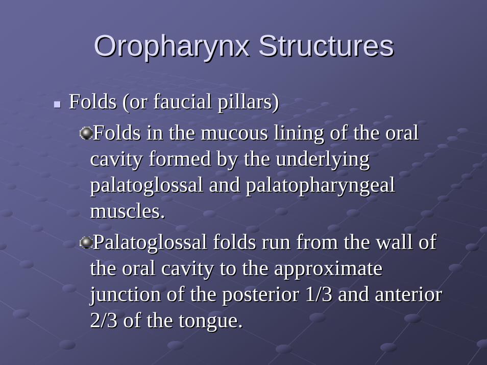
OropharynxOropharynx StructuresStructures
Folds (or Folds (or faucialfaucial pillars)pillars)Folds in the mucous lining of the oral Folds in the mucous lining of the oral cavity formed by the underlying cavity formed by the underlying palatoglossalpalatoglossal and and palatopharyngealpalatopharyngealmuscles.muscles.PalatoglossalPalatoglossal folds run from the wall of folds run from the wall of the oral cavity to the approximate the oral cavity to the approximate junction of the posterior 1/3 and anterior junction of the posterior 1/3 and anterior 2/3 of the tongue.2/3 of the tongue.

OropharynxOropharynx StructuresStructuresTonsilsTonsils
Palatine (Palatine (faucialfaucial) tonsils are located immediately posterior to ) tonsils are located immediately posterior to the the palatoglossalpalatoglossal folds and anterior to the folds and anterior to the palatopharyngealpalatopharyngealfolds; inferior to the soft palate and superior to the tongue; afolds; inferior to the soft palate and superior to the tongue; and nd medial to the superior constrictors, the medial to the superior constrictors, the styloglossusstyloglossus, and the , and the glossopharyngealglossopharyngeal nerve (IX).nerve (IX).Nasopharyngeal (pharyngeal) tonsils are located on the Nasopharyngeal (pharyngeal) tonsils are located on the posterior aspect of the posterior aspect of the nasopharynxnasopharynx..
When enlarged these are referred to as the adenoids.When enlarged these are referred to as the adenoids.Lingual tonsils are lymphoid tissue in the tongue.Lingual tonsils are lymphoid tissue in the tongue.
Continuous with the inferior pole of the palatine tonsil.Continuous with the inferior pole of the palatine tonsil.


NasopharynxNasopharynx
Soft palate separates Soft palate separates nasopharynxnasopharynx from from oropharynxoropharynx..

AdenoidsAdenoids
Pharyngeal tonsils, nasopharyngeal tonsilsPharyngeal tonsils, nasopharyngeal tonsilsMass of Mass of lymphoid tissuelymphoid tissue situated in the roof of situated in the roof of the the nasopharynxnasopharynx

PalatePalate
Vasculature: (Bilateral)Vasculature: (Bilateral)Greater palatine arteryGreater palatine artery
Branch of descending palatine arteryBranch of descending palatine artery
Lesser palatine arteryLesser palatine arteryBranch of descending palatine arteryBranch of descending palatine artery
Ascending palatine arteryAscending palatine arteryBranch of Facial ArteryBranch of Facial Artery
Veins of palateVeins of palateTributaries of Tributaries of pterygoidpterygoid venous plexusvenous plexus

InnervationInnervation of Palateof Palate
Sensory nerves of palate are branches of Sensory nerves of palate are branches of pterygopalatinepterygopalatine ganglionganglionGreater palatine nerve supplies Greater palatine nerve supplies gingivagingiva, , mucous membrane, and glands of hard mucous membrane, and glands of hard palatepalateNasopalatineNasopalatine nerve supplies mucous nerve supplies mucous membrane of anterior part of hard palatemembrane of anterior part of hard palateLesser palatine nerves supply soft palateLesser palatine nerves supply soft palate

Hard PalateHard PalatePalatine processes of Palatine processes of maxilla and horizontal maxilla and horizontal plates of the palatine bones.plates of the palatine bones.PeriosteumPeriosteum of the hard of the hard palate is continuous into the palate is continuous into the soft palate (palatine soft palate (palatine aponeurosisaponeurosis), and is the ), and is the insertion site of the palatal insertion site of the palatal muscles.muscles.LandmarksLandmarks
Incisive ForamenIncisive ForamenGreater Palatine ForamenGreater Palatine ForamenLesser Palatine ForamenLesser Palatine Foramen
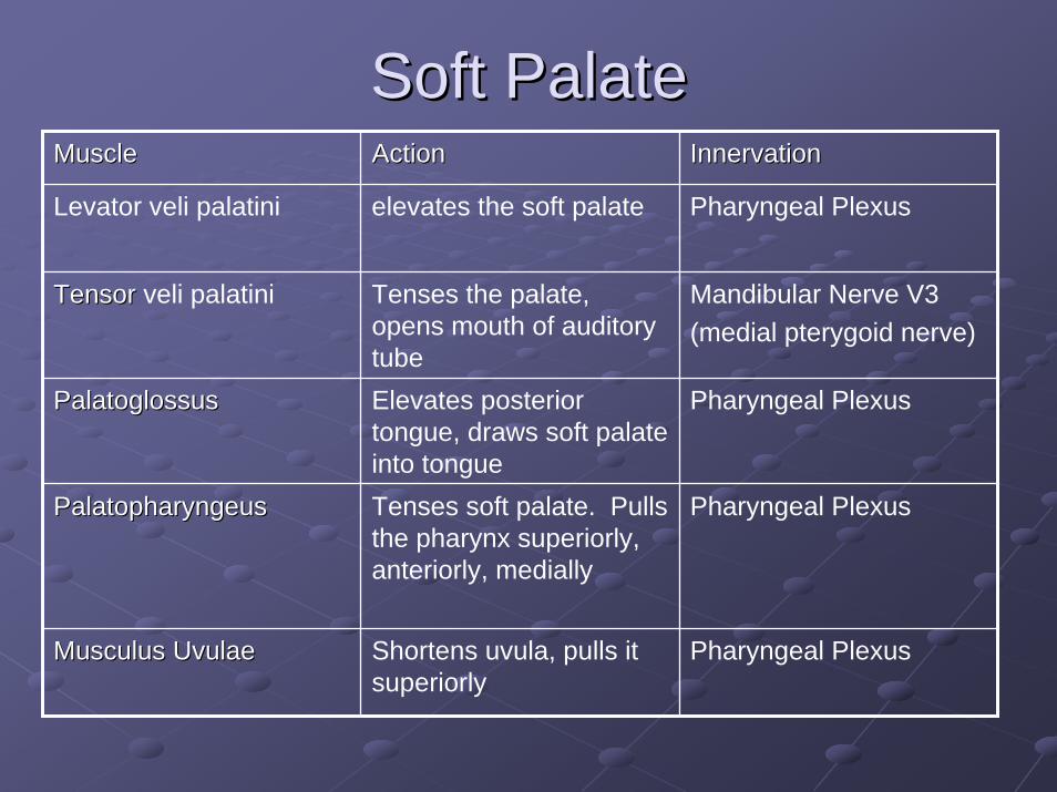
Soft PalateSoft PalateMuscle Muscle ActionAction InnervationInnervation
Levator veli palatini elevates the soft palate Pharyngeal Plexus
Tensor Tensor veli palatini Tenses the palate, opens mouth of auditory tube
Mandibular Nerve V3(medial pterygoid nerve)
PalatoglossusPalatoglossus Elevates posterior tongue, draws soft palate into tongue
Pharyngeal Plexus
PalatopharyngeusPalatopharyngeus Tenses soft palate. Pulls the pharynx superiorly, anteriorly, medially
Pharyngeal Plexus
MusculusMusculus UvulaeUvulae Shortens uvula, pulls it superiorly
Pharyngeal Plexus

Trigeminal NerveTrigeminal Nerve
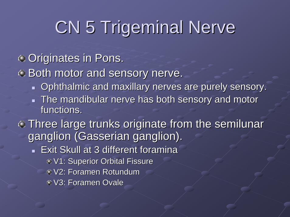
CN 5 Trigeminal NerveCN 5 Trigeminal NerveOriginates in Pons.Originates in Pons.Both motor and sensory nerve.Both motor and sensory nerve.
Ophthalmic and maxillary nerves are purely sensory.Ophthalmic and maxillary nerves are purely sensory.The The mandibularmandibular nerve has both sensory and motor nerve has both sensory and motor functions.functions.
Three large trunks originate from the Three large trunks originate from the semilunarsemilunarganglion (ganglion (GasserianGasserian ganglion).ganglion).
Exit Skull at 3 different foraminaExit Skull at 3 different foraminaV1: Superior Orbital FissureV1: Superior Orbital FissureV2: Foramen V2: Foramen RotundumRotundumV3: Foramen V3: Foramen OvaleOvale

Trigeminal Nerve BranchesTrigeminal Nerve BranchesOpthalmicOpthalmic Nerve Nerve (V1) (V1)
Maxillary Nerve (V2) Maxillary Nerve (V2) MandibularMandibular Nerve Nerve (V3)(V3)
NasociliaryNasociliary nervenerveSupraorbitalSupraorbital nervenerveLacrimalLacrimal nervenerveFrontal nerveFrontal nerveSupratrochlearSupratrochlear nervenerveInfratrochlearInfratrochlear nerve nerve
ZygomaticZygomatic nervenervePosterior superior Posterior superior alveolar nervealveolar nerveMiddle superior Middle superior alveolar nervealveolar nerveAnterior superior Anterior superior alveolar nervealveolar nerveInfraorbitalInfraorbital nervenerveGreater Palatine Greater Palatine nervenerveNasopalatineNasopalatine nerve nerve
AuriculotemporalAuriculotemporalnervenerveLingual nerveLingual nerveBuccalBuccal nervenerveInferior alveolar Inferior alveolar nervenerve(Mental nerve) (Mental nerve)


CN V Dermatome DistributionCN V Dermatome Distribution


MaxillaMaxilla
Each half of the fused Each half of the fused maxilla consists of:maxilla consists of:
The The body of body of maxillamaxillaFour processes Four processes
ZZygomaticygomatic processprocessFFrontal processrontal processAAlveolar processlveolar processPPalatine processalatine process
InfraorbitalInfraorbital foramenforamen


MandibleMandible
CondyleCondyleSigmoid NotchSigmoid NotchRamusRamusAngleAngleExternal Oblique External Oblique RidgeRidgeSymphysisSymphysisMental ForamenMental Foramen

MandibleMandible
SphenomandibularSphenomandibularLigamentLigamentStylomandibularStylomandibularLigamentLigament
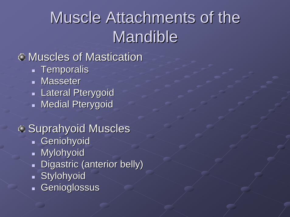
Muscle Attachments of the Muscle Attachments of the MandibleMandible
Muscles of MasticationMuscles of MasticationTemporalisTemporalisMasseterMasseterLateral Lateral PterygoidPterygoidMedial Medial PterygoidPterygoid
SuprahyoidSuprahyoid MusclesMusclesGeniohyoidGeniohyoidMylohyoidMylohyoidDigastricDigastric (anterior belly)(anterior belly)StylohyoidStylohyoidGenioglossusGenioglossus
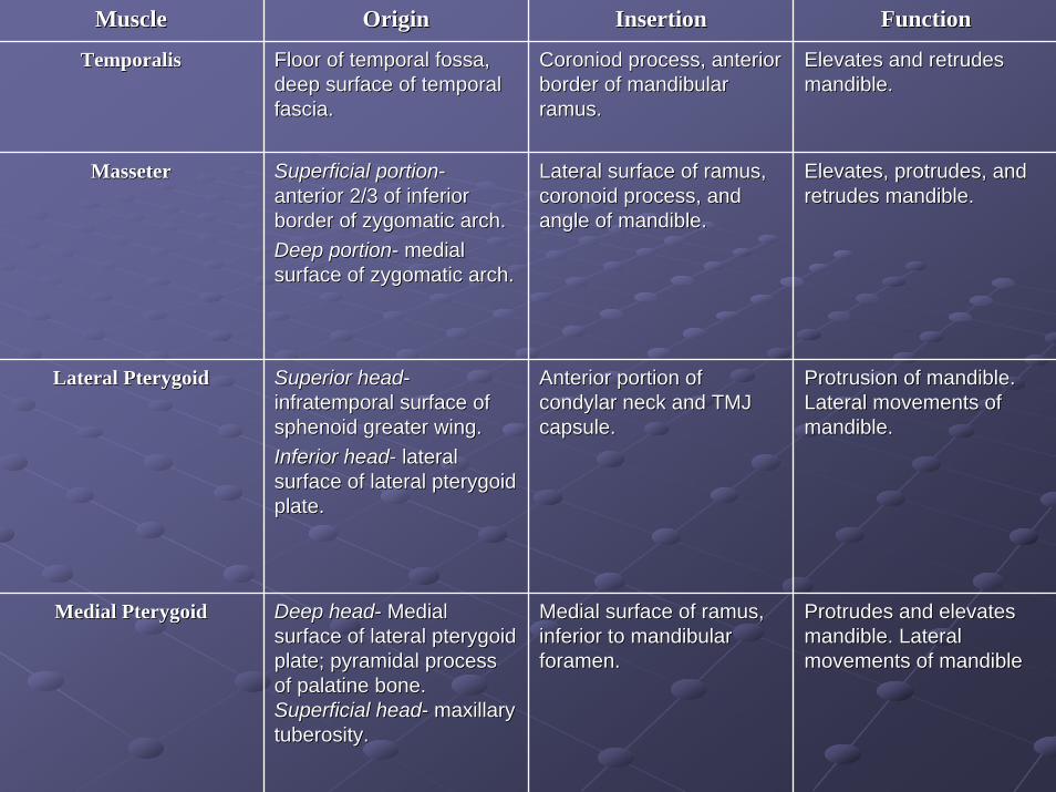
MuscleMuscle OriginOrigin InsertionInsertion FunctionFunction
TemporalisTemporalis Floor of temporal Floor of temporal fossafossa, , deep surface of temporal deep surface of temporal fascia. fascia.
CoroniodCoroniod process, anterior process, anterior border of border of mandibularmandibularramusramus. .
Elevates and Elevates and retrudesretrudesmandible. mandible.
MasseterMasseter Superficial portionSuperficial portion--anterior 2/3 of inferior anterior 2/3 of inferior border of border of zygomaticzygomatic arch.arch.Deep portionDeep portion-- medial medial surface of surface of zygomaticzygomatic arch. arch.
Lateral surface of Lateral surface of ramusramus, , coronoidcoronoid process, and process, and angle of mandible. angle of mandible.
Elevates, protrudes, and Elevates, protrudes, and retrudesretrudes mandible. mandible.
Lateral Lateral PterygoidPterygoid Superior headSuperior head--infratemporalinfratemporal surface of surface of sphenoid greater wing.sphenoid greater wing.Inferior headInferior head-- lateral lateral surface of lateral surface of lateral pterygoidpterygoidplate. plate.
Anterior portion of Anterior portion of condylarcondylar neck and TMJ neck and TMJ capsule. capsule.
Protrusion of mandible. Protrusion of mandible. Lateral movements of Lateral movements of mandible. mandible.
Medial Medial PterygoidPterygoid Deep headDeep head-- Medial Medial surface of lateral surface of lateral pterygoidpterygoidplate; pyramidal process plate; pyramidal process of palatine bone. of palatine bone. Superficial headSuperficial head-- maxillary maxillary tuberositytuberosity..
Medial surface of Medial surface of ramusramus, , inferior to inferior to mandibularmandibularforamen.foramen.
Protrudes and elevates Protrudes and elevates mandible. Lateral mandible. Lateral movements of mandiblemovements of mandible

MuscleMuscle OriginOrigin InsertionInsertion FunctionFunction
GeniohyoidGeniohyoid Inferior genial Inferior genial tubercle on inner tubercle on inner surface of surface of mandibularmandibularsymphysissymphysis. .
Body hyoid bone.Body hyoid bone. Elevates tongue, Elevates tongue, FOM, and hyoid.FOM, and hyoid.
MylohyoidMylohyoid Line from last molar Line from last molar to to mandibularmandibularsymphysissymphysis..((mylohyoidmylohyoid line)line)
RapheRaphe and body of and body of hyoid bone.hyoid bone.
Elevates base of Elevates base of hyoid bone. Raises hyoid bone. Raises floor of mouth and floor of mouth and tongue. tongue.
DigastricDigastric Posterior bellyPosterior belly--mastoid notch mastoid notch (temporal bone).(temporal bone).Anterior bellyAnterior belly--digastricdigastric fossafossa(mandible) .(mandible) .
Intermediate tendon Intermediate tendon attached to hyoid attached to hyoid bone by fibrous bone by fibrous loop. loop.
Depresses Depresses mandible, Elevates mandible, Elevates the hyoid bone. the hyoid bone.
StylohyoidStylohyoid Posterior border of Posterior border of the the styloidstyloid process.process.
Body of hyoid bone.Body of hyoid bone. Elevates base of Elevates base of tongue and hyoid tongue and hyoid bone.bone.

Accessory Muscles to the Muscles Accessory Muscles to the Muscles of Masticationof Mastication
PlatysmaPlatysmaBuccinatorBuccinatorPosterior neck musculaturePosterior neck musculature
SternocleidomastoidSternocleidomastoidTrapeziusTrapeziusIntrinsic neck musclesIntrinsic neck muscles

TemporomandibularTemporomandibular Joint (TMJ)Joint (TMJ)
TMJTMJ
Classification:Classification:GinglymoarthrodialGinglymoarthrodial JointJoint
Translational (gliding) movementTranslational (gliding) movementRotational (hinging) movementRotational (hinging) movement
SynovialSynovial JointJoint

TMJ AnatomyTMJ AnatomyArticulation between the Articulation between the condylecondyle of the mandible and the of the mandible and the squamoussquamous portion of the temporal bone (TMJ portion of the temporal bone (TMJ fossafossa).).ArticularArticular disc lies between disc lies between condylecondyle and and fossafossa..CondylesCondyles
Elliptically shaped. Elliptically shaped. Long axis oriented Long axis oriented mediolaterallymediolaterally..
ArticularArticular Surface of Temporal Bone Surface of Temporal Bone Functional aspect of TMJ.Functional aspect of TMJ.Dense fibrous connective tissue.Dense fibrous connective tissue.ConcaveConcave : : ArticularArticular fossafossa ((GlenoidGlenoid FossaFossa, , MandibularMandibular FossaFossa))ConvexConvex : : ArticularArticular eminenceeminence ((tubercletubercle))

ArticularArticular DiscDiscDense Dense fibrocartilagenousfibrocartilagenous connective tissue.connective tissue.AvascularAvascular and and aneuralaneural..Nutrition of Nutrition of chondrocyteschondrocytes with the movement of with the movement of synovialsynovial fluid is essential for fluid is essential for maintenance of structure and function.maintenance of structure and function.Biconcave structure is a three dimensional space filler between Biconcave structure is a three dimensional space filler between the two the two convex surfaces of the convex surfaces of the condylecondyle and and articulararticular eminence.eminence.Separates joint into inferior and superior joint spaces.Separates joint into inferior and superior joint spaces.Varies in Varies in thicknessthickness..
Intermediate zoneIntermediate zone-- thin (center of disc)thin (center of disc)Anterior and Posterior BandsAnterior and Posterior Bands-- thickthick
Posterior band is thicker and is attached to Posterior band is thicker and is attached to retrodiscalretrodiscal tissues (tissues (bilaminarbilaminar zone, posterior zone, posterior attachment).attachment).Anterior band is attached to the capsular ligament, the lateral Anterior band is attached to the capsular ligament, the lateral pterygoidpterygoid muscle, and the muscle, and the condylecondyle..
RetrodiscalRetrodiscal TissuesTissuesLoose connective tissues.Loose connective tissues.Vascular and Innervated.Vascular and Innervated.

TMJ DisordersTMJ Disorders

TMJ ReconstructionTMJ Reconstruction
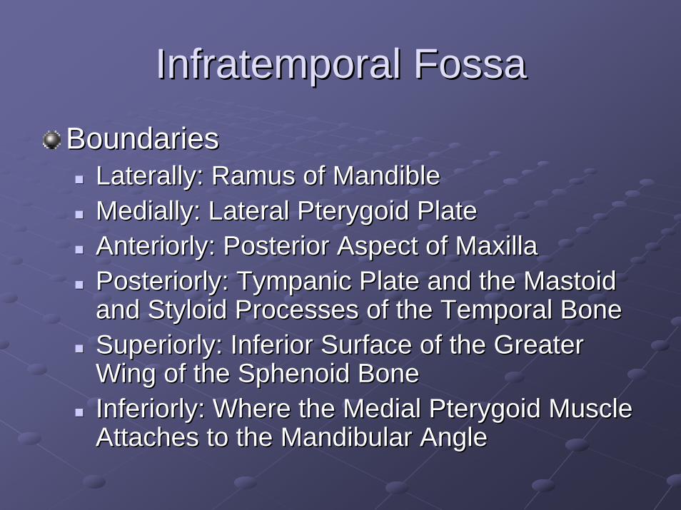
InfratemporalInfratemporal FossaFossa
BoundariesBoundariesLaterally: Laterally: RamusRamus of Mandibleof MandibleMedially: Lateral Medially: Lateral PterygoidPterygoid PlatePlateAnteriorlyAnteriorly: Posterior Aspect of Maxilla: Posterior Aspect of MaxillaPosteriorlyPosteriorly: Tympanic Plate and the Mastoid : Tympanic Plate and the Mastoid and and StyloidStyloid Processes of the Temporal BoneProcesses of the Temporal BoneSuperiorly: Inferior Surface of the Greater Superiorly: Inferior Surface of the Greater Wing of the Sphenoid BoneWing of the Sphenoid BoneInferiorly: Where the Medial Inferiorly: Where the Medial PterygoidPterygoid Muscle Muscle Attaches to the Attaches to the MandibularMandibular AngleAngle

InfratemporalInfratemporal FossaFossa
Contents:Contents:Inferior part of the temporal muscleInferior part of the temporal muscleLateral and medial Lateral and medial pterygoidpterygoid musclesmusclesMaxillary arteryMaxillary arteryPterygoidPterygoid venous plexusvenous plexusMandibularMandibular, Inferior alveolar, lingual, , Inferior alveolar, lingual, buccalbuccal, , and and chordachorda tympani nerves, and tympani nerves, and oticotic ganglionganglion

Arteries of FaceArteries of FaceArteryArtery OriginOrigin CourseCourse DistributionDistributionFacialFacial External Carotid External Carotid
ArteryArteryAscends deep to Ascends deep to submandibularsubmandibulargland, rounds gland, rounds around inferior around inferior border of border of mandible and mandible and enters faceenters face
Muscles of facial Muscles of facial expression and faceexpression and face
Inferior LabialInferior Labial Facial artery near Facial artery near angle of mouthangle of mouth
Runs medially in Runs medially in lower liplower lip
Lower lip and chinLower lip and chin
Superior LabialSuperior Labial Facial artery near Facial artery near angle of mouthangle of mouth
Runs medially in Runs medially in upper lipupper lip
Upper lip and ala Upper lip and ala and septum of noseand septum of nose
Lateral nasalLateral nasal Facial artery as it Facial artery as it ascends ascends alongside nosealongside nose
Passes to ala of Passes to ala of nosenose
Skin on ala and Skin on ala and dorsum of nosedorsum of nose
AngularAngular Terminal branch Terminal branch of facial arteryof facial artery
Passes to medial Passes to medial angle (angle (canthuscanthus) of ) of eyeeye
Superior part of Superior part of cheek and lower cheek and lower eyelideyelid

Arteries of FaceArteries of Face
ArteryArtery OriginOrigin CourseCourse DistributionDistribution
Superficial Superficial temporaltemporal
Smaller terminal Smaller terminal branch of external branch of external carotid arterycarotid artery
Ascends anterior Ascends anterior to ear to temporal to ear to temporal region and ends region and ends in scalpin scalp
Facial muscles Facial muscles and skin of frontal and skin of frontal and temporal and temporal regionsregions
Transverse facialTransverse facial Superficial Superficial temporal artery temporal artery within parotid within parotid glandgland
Crosses face Crosses face superficial to superficial to massetermasseter and and inferior to inferior to zygomaticzygomatic archarch
Parotid gland and Parotid gland and duct, duct, mausclesmausclesand skin of faceand skin of face
MentalMental Terminal branch Terminal branch of Inferior alveolar of Inferior alveolar arteryartery
Emerges from Emerges from mental foramen mental foramen and passes to and passes to chinchin
Facial muscles Facial muscles and skin of chinand skin of chin

Maxillary ArteryMaxillary Artery
Arises from External Carotid ArteryArises from External Carotid ArteryArises posterior to neck of mandibleArises posterior to neck of mandiblePasses Passes anteriorlyanteriorly (deep to neck of (deep to neck of condylecondyle))-- 11stst partpartPasses superficial or deep to the lateral Passes superficial or deep to the lateral pterygoidpterygoid musclemuscle-- 22ndnd partpartPasses through Passes through pterygomaxillarypterygomaxillary fissure to fissure to enter enter infratemporalinfratemporal fossafossa-- 33rdrd partpart

11stst ((MandibularMandibular) Part) PartDeep auricular arteryDeep auricular artery to external acoustic to external acoustic meatusmeatusAnterior tympanic arteryAnterior tympanic artery to tympanic to tympanic membranemembraneMiddle Middle meningealmeningeal arteryartery to to duradura mater and mater and calvariacalvariaAccessory Accessory meningealmeningeal arteriesarteries to the cranial to the cranial cavitycavityInferior alveolar arteryInferior alveolar artery to mandible, to mandible, gingivagingiva, , teethteeth

22ndnd ((PterygoidPterygoid Part)Part)
Deep temporal arteriesDeep temporal arteries, anterior and , anterior and posterior (supply temporal muscle)posterior (supply temporal muscle)PterygoidPterygoid arteriesarteries (supply (supply pterygoidpterygoidmuscles)muscles)MassetericMasseteric arteryartery (supplies deep surface (supplies deep surface of of massetermasseter muscle)muscle)BuccalBuccal arteryartery (supplies (supplies buccinatorbuccinatormuscle)muscle)

33rdrd ((PterygopalatinePterygopalatine) Part) PartPosterior superior alveolar arteryPosterior superior alveolar artery
supplies maxillary molar and premolar teeth, lining of maxillarysupplies maxillary molar and premolar teeth, lining of maxillary sinus, sinus, gingivagingiva
InfraorbitalInfraorbital arteryarterysupplies inferior eyelid, supplies inferior eyelid, lacrimallacrimal sac, side of nose, superior lipsac, side of nose, superior lip
Descending palatine arteryDescending palatine arterysupplies maxillary supplies maxillary gingivagingiva, palatine glands, mucous membrane of roof of , palatine glands, mucous membrane of roof of mouthmouth
Artery of Artery of pterygoidpterygoid canalcanalsupplies superior part of pharynx, supplies superior part of pharynx, pharyngotympanicpharyngotympanic tube, tympanic tube, tympanic cavitycavity
Pharyngeal arteryPharyngeal arterysupplies roof of pharynx, supplies roof of pharynx, sphenoidalsphenoidal sinus, inferior part of sinus, inferior part of pharyngotympanicpharyngotympanic tubetube
SphenopalatineSphenopalatine arteryarterysupplies lateral nasal wall, nasal septum, supplies lateral nasal wall, nasal septum, paranasalparanasal sinusessinuses

Veins of FaceVeins of Face
VeinVein OriginOrigin TerminationTermination Area DrainedArea DrainedFacialFacial Continuation Continuation
of angular vein of angular vein past inferior past inferior margin of orbitmargin of orbit
Internal jugular vein Internal jugular vein opposite or inferior to opposite or inferior to hyoid bonehyoid bone
Anterior scalp and Anterior scalp and forehead, eyelids, external forehead, eyelids, external nose, anterior cheek, lips, nose, anterior cheek, lips, chin, chin, submandibularsubmandibular glandgland
Deep facial Deep facial PterygoidPterygoidvenous plexusvenous plexus
Enters posterior Enters posterior aspect of facial veinaspect of facial vein
InfratemporalInfratemporal fossafossa (most (most areas supplied by areas supplied by maxilarymaxilaryartery)artery)

Veins of FaceVeins of FaceSuperficial Superficial temporaltemporal
Begins from Begins from a widespread a widespread plexus of plexus of veins on side veins on side of scalp and of scalp and along the along the zygomaticzygomaticarcharch
Joins the maxillary Joins the maxillary vein posterior to the vein posterior to the neck of the neck of the mandible to form mandible to form the the retromandibularretromandibularveinvein
Side of scalp, superficial Side of scalp, superficial aspect of temporal aspect of temporal muscle, external earmuscle, external ear
RetromandibularRetromandibular Formed Formed anterior to anterior to the ear by the ear by union of union of superficial superficial temporal and temporal and maxillary maxillary veinsveins
Unites with Unites with posterior auricular posterior auricular vein to form vein to form external jugular external jugular veinvein
Parotid gland and Parotid gland and massetermasseter musclemuscle

Salivary GlandsSalivary Glands
Parotid GlandParotid GlandSublingual GlandSublingual GlandSubmandibularSubmandibular GlandGland

Parotid GlandParotid Gland
Stensen’sStensen’s DuctDuctPierces the Pierces the buccalbuccal fat, fat, buccopharyngealbuccopharyngealfascia and fascia and buccinatorbuccinatormusclemuscle..Opens into the Opens into the vestibule of the mouth vestibule of the mouth opposite the maxillary opposite the maxillary 2nd 2nd molar toothmolar tooth..

Floor of MouthFloor of MouthSublingual Salivary GlandsSublingual Salivary Glands
Sublingual folds (Sublingual folds (plicaplicasublingualissublingualis).).
Run Run anteroposterioranteroposterioralongside the alongside the frenulumfrenulum, one , one per side, converging just per side, converging just anterior to the root of the anterior to the root of the frenulumfrenulum..Overly the sublingual Overly the sublingual salivary glands from which salivary glands from which numerous ducts travel and numerous ducts travel and open onto the top of each open onto the top of each fold.fold.Each gland does not have a Each gland does not have a single duct, but many which single duct, but many which open directly into the oral open directly into the oral cavity.cavity.
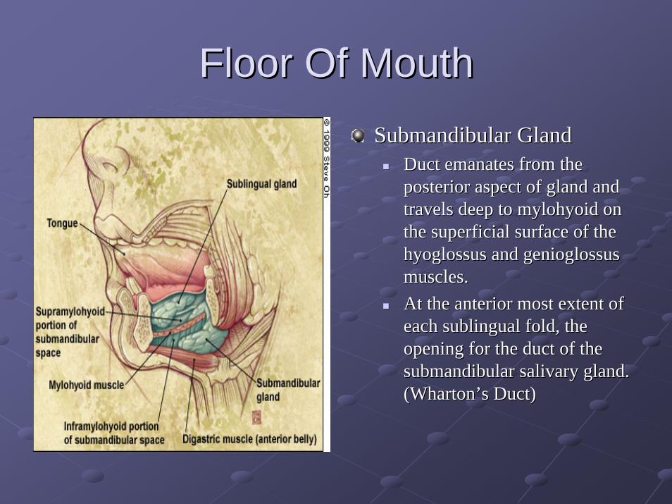
Floor Of MouthFloor Of MouthSubmandibularSubmandibular GlandGland
Duct emanates from the Duct emanates from the posterior aspect of gland and posterior aspect of gland and travels deep to travels deep to mylohyoidmylohyoid on on the superficial surface of the the superficial surface of the hyoglossushyoglossus and and genioglossusgenioglossusmuscles.muscles.At the anterior most extent of At the anterior most extent of each sublingual fold, the each sublingual fold, the opening for the duct of the opening for the duct of the submandibularsubmandibular salivary gland. salivary gland. (Wharton(Wharton’’s Duct)s Duct)

BuccalBuccal Fat PadFat Pad
EncapsuledEncapsuled massmass of of fatfat in the in the cheekcheek on on the the outerouter sideside of the of the buccinatorbuccinator musclemuscleFound in the space between the Found in the space between the massetermassetermusclemuscle and the external surface of the and the external surface of the buccinatorbuccinator

PterygomandibularPterygomandibular RapheRapheTendinousTendinous band of the band of the buccopharyngealbuccopharyngeal fasciafasciaAttached to:Attached to:
HamulusHamulus of the of the medial medial pterygoidpterygoid plateplate..Posterior end of the Posterior end of the mylohyoidmylohyoid lineline of the of the mandiblemandible..
Boundaries:Boundaries:medial surfacemedial surface is covered by the mucous membrane is covered by the mucous membrane of the of the mouthmouth. . lateral surfacelateral surface is separated from the is separated from the ramusramus of the of the mandiblemandible by by adipose tissueadipose tissue. . posterior border posterior border attachesattaches to the to the superior pharyngeal superior pharyngeal constrictor muscleconstrictor muscle..anterior borderanterior border attaches to attaches to buccinatorbuccinator. .


TongueTongue

Surface Anatomy TongueSurface Anatomy TongueSulcusSulcus terminalisterminalis
VV--shaped line (with the point shaped line (with the point facing facing posteriorlyposteriorly) that ) that separates the anterior 2/3 and separates the anterior 2/3 and posterior 1/3 of the tongue.posterior 1/3 of the tongue.At the point, a shallow pit At the point, a shallow pit named the foramen named the foramen cecumcecum is is present (original location of present (original location of thyroid thyroid diverticulumdiverticulum).).Located posterior to a row of Located posterior to a row of vallatevallate papillae.papillae.
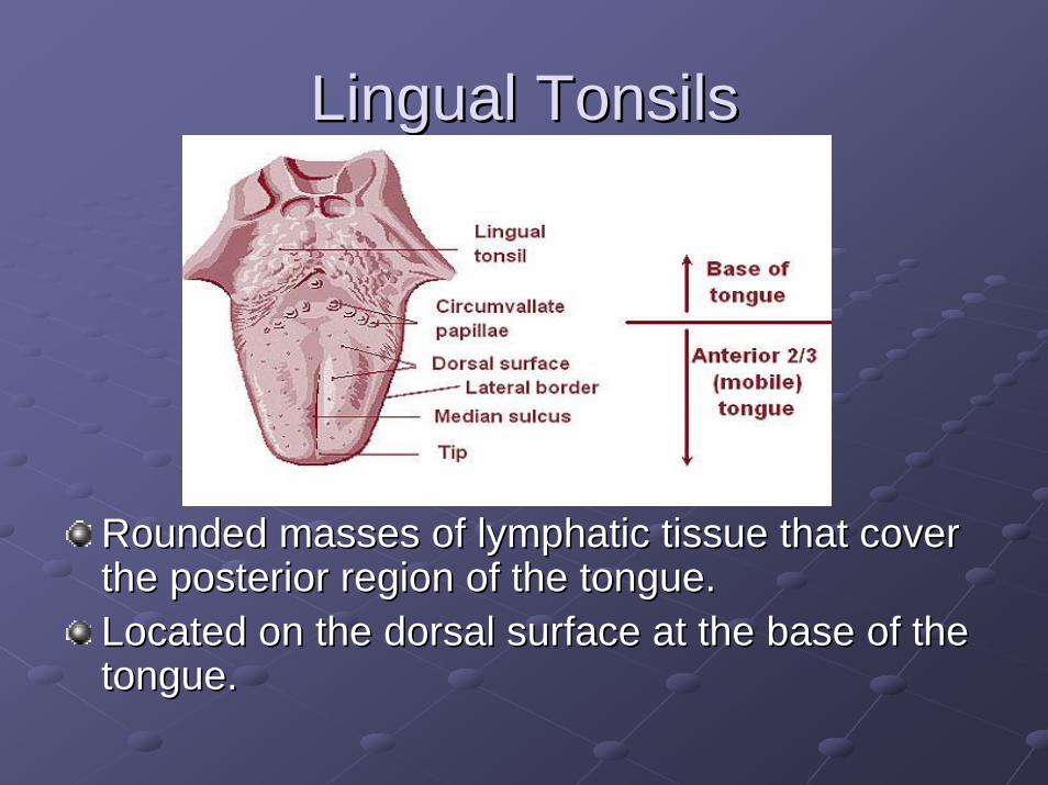
Lingual TonsilsLingual Tonsils
Rounded masses of Rounded masses of lymphatic tissuelymphatic tissue that cover that cover the posterior region of the the posterior region of the tonguetongue..Located on the dorsal surface at the Located on the dorsal surface at the base of the base of the tonguetongue..

InnervationInnervation TongueTongue
Anterior 2/3 Anterior 2/3 Lingual Nerve (V3)Lingual Nerve (V3)-- general sensationgeneral sensationChordaChorda typmanitypmani (CN 7)(CN 7)-- tastetaste
Posterior 1/3Posterior 1/3Lingual branch Lingual branch glossopharyngealglossopharyngeal nerve (CN nerve (CN IX)IX)-- general sensation, tastegeneral sensation, tasteLingual branch facial nerveLingual branch facial nerve-- tastetasteInternal laryngeal branch Internal laryngeal branch VagusVagus-- general general sensation, tastesensation, taste

Motor Motor InnervationInnervation TongueTongue
PalatoglossusPalatoglossus-- pharyngeal plexus (CN XI, pharyngeal plexus (CN XI, CNX)CNX)All other musclesAll other muscles-- CN XII (hypoglossal)CN XII (hypoglossal)

Extrinsic Muscles of TongueExtrinsic Muscles of Tongue
GenioglossusGenioglossusHyoglossusHyoglossusStyloglossusStyloglossusPalatoglossusPalatoglossus

GenioglossusGenioglossus MuscleMuscle
Origin: Superior part mental spine of Origin: Superior part mental spine of mandible.mandible.Insertion: Dorsum of tongue, body of hyoid Insertion: Dorsum of tongue, body of hyoid bone.bone.InnervationInnervation: CN 12 (Hypoglossal Nerve).: CN 12 (Hypoglossal Nerve).Depresses tongue, posterior part pulls tongue Depresses tongue, posterior part pulls tongue anteriorlyanteriorly for protrusionfor protrusion

HyoglossusHyoglossus MuscleMuscle
Origin: Body and Origin: Body and greater horn of hyoidgreater horn of hyoidInsertion: Side and Insertion: Side and inferior aspect of inferior aspect of tonguetongueInnervationInnervation: CN XII: CN XIIDepresses and Depresses and retracts tongueretracts tongue

StyloglossusStyloglossus MuscleMuscle
Origin: Origin: StyloidStyloid process and process and stylohyoidstylohyoidligamentligamentInsertion: Side and inferior aspect of Insertion: Side and inferior aspect of tonguetongueInnervationInnervation: CN 12: CN 12Retracts tongue and draws it up to create Retracts tongue and draws it up to create trough for swallowingtrough for swallowing

PalatoglossusPalatoglossus MuscleMuscle
Origin: Palatine Origin: Palatine aponeurosisaponeurosis of soft palateof soft palateInsertion: Side of tongueInsertion: Side of tongueInnervationInnervation: Cranial root of CN XI via : Cranial root of CN XI via pharyngeal branch of CN X and pharyngeal branch of CN X and pharyngeal plexuspharyngeal plexusElevates posterior part of tongueElevates posterior part of tongue

Intrinsic Muscles TongueIntrinsic Muscles Tongue
Superior LongitudinalSuperior LongitudinalInferior LongitudinalInferior LongitudinalTransverseTransverseVerticalVertical

Tongue VasculatureTongue Vasculature
Lingual artery (from external carotid artery)Lingual artery (from external carotid artery)Dorsal lingual arteriesDorsal lingual arteriesDeep lingual arteryDeep lingual arterySublingual arterySublingual artery
All veins terminate in the Internal Jugular All veins terminate in the Internal Jugular VeinVein
Dorsal lingual veinsDorsal lingual veinsDeep lingual veins (Deep lingual veins (ranineranine veins)veins)Sublingual veinSublingual vein

Lymph Drainage from TongueLymph Drainage from Tongue
Lymph from posterior 1/3Lymph from posterior 1/3 Superior deep Superior deep cervical LNcervical LNLymph from medial part of anterior 2/3 Lymph from medial part of anterior 2/3 Inferior deep cervical LNInferior deep cervical LNLymph from lateral anterior 2/3 Lymph from lateral anterior 2/3 submandibularsubmandibular LNLNApexApex submentalsubmental LNLNPosterior 1/3 & area near midline Posterior 1/3 & area near midline drain drain bilaterallybilaterally

Periodontal AnatomyPeriodontal Anatomy
PeriodontiumPeriodontiumGingivaGingivaPeriodontal Ligament (PDL)Periodontal Ligament (PDL)CementumCementumAlveolar and supporting boneAlveolar and supporting bone

Healthy Healthy GingivaGingiva
Evaluate Color, Contour, Tone and Consistency Evaluate Color, Contour, Tone and Consistency of Gingival Tissueof Gingival Tissue

GingivaGingivaMasticatoryMasticatory mucosa which cover the alveolar process and surround mucosa which cover the alveolar process and surround the cervical portion of teeth. the cervical portion of teeth. Composed of connective tissue and epithelium.Composed of connective tissue and epithelium.Epithelium can be divided into three histological distinct areasEpithelium can be divided into three histological distinct areas::Oral epitheliumOral epithelium
Continuous with epithelial lining of the attached Continuous with epithelial lining of the attached gingivagingiva..Composed of keratinized stratified Composed of keratinized stratified squamoussquamous epithelium.epithelium.
SulcularSulcular epithelium epithelium NonNon--keratinizedkeratinized
JunctionalJunctional epitheliumepitheliumAttached to the tooth by Attached to the tooth by hemidesmosomeshemidesmosomes. . NonNon--keratinized. keratinized. Larger cells with increased intercellular spaces.Larger cells with increased intercellular spaces.

Gingival FibersGingival FibersComposed of type I collagen. Composed of type I collagen. Support the Support the gingivagingiva and attach it to the tooth and and attach it to the tooth and alveolar bone. alveolar bone. Gingival fibers are continuous with the Gingival fibers are continuous with the periodontal ligament.periodontal ligament.Designated by their orientation. Designated by their orientation.
DentogingivalDentogingival fibersfibersDentoperiostealDentoperiosteal fibersfibersCircular fibersCircular fibersAlveologingivalAlveologingival fibers fibers TransseptalTransseptal fibers fibers

Principal Fibers of the Periodontal Principal Fibers of the Periodontal Ligament (PDL)Ligament (PDL)
Bundles of collagen fibers grouped Bundles of collagen fibers grouped according to the direction they extend from according to the direction they extend from the the cementumcementum of the root to the alveolar of the root to the alveolar bone.bone.
Horizontal FibersHorizontal FibersAlveolar Crest FibersAlveolar Crest FibersOblique FibersOblique FibersApical FibersApical FibersInterradicularInterradicular FibersFibers

Tooth AnatomyTooth Anatomy

Universal/National System for Universal/National System for Permanent (Adult) Dentition (1Permanent (Adult) Dentition (1--32)32)
Maxillary Arch (1Maxillary Arch (1--16)16)MandibularMandibular Arch (17Arch (17--32)32)3 Molars, 2 Premolars, 1 3 Molars, 2 Premolars, 1 Canine, 1 Lateral Incisor, Canine, 1 Lateral Incisor, 1 Central Incisor in each 1 Central Incisor in each arch quadrantarch quadrant

The Universal/National System for The Universal/National System for the Primary (Baby) Dentitionthe Primary (Baby) Dentition
Upper case letters A through TUpper case letters A through TNo Premolars in the Primary Dentition No Premolars in the Primary Dentition
A
T
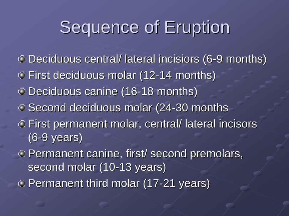
Sequence of EruptionSequence of Eruption
Deciduous central/ lateral Deciduous central/ lateral incisiorsincisiors (6(6--9 months)9 months)First deciduous molar (12First deciduous molar (12--14 months)14 months)Deciduous canine (16Deciduous canine (16--18 months)18 months)Second deciduous molar (24Second deciduous molar (24--30 months30 monthsFirst permanent molar, central/ lateral incisors First permanent molar, central/ lateral incisors (6(6--9 years)9 years)Permanent canine, first/ second premolars, Permanent canine, first/ second premolars, second molar (10second molar (10--13 years)13 years)Permanent third molar (17Permanent third molar (17--21 years)21 years)

Curve of Curve of SpeeSpeeCCurvatureurvature of the of the mandibularmandibular occlusalocclusalplaneplane beginning at the beginning at the tip of the lower tip of the lower cuspidcuspidand following the and following the buccalbuccal cuspscusps of the of the posterior posterior teethteeth, , continuing to the continuing to the terminal terminal molarmolar. . AnteriorAnterior--posterior posterior curvecurve

Curve of WilsonCurve of Wilson
Lateral curveLateral curve

OcclusionOcclusion
Combination of Curve of Combination of Curve of SpeeSpee and Curve and Curve of Wilson create the of Wilson create the OcclusalOcclusal PlanePlaneCentric Relation PositionCentric Relation Position
Anatomic relationship of the TMJ joint. Anatomic relationship of the TMJ joint. Muscles of Mastication are at rest.Muscles of Mastication are at rest.

OcclusionOcclusion

Angle Classification of OcclusionAngle Classification of Occlusion
Class IClass I——patient’s profile patient’s profile is characterized as is characterized as normal. normal. Class IIClass II——patient’s profile patient’s profile is deficient in chin length is deficient in chin length and characterized as a and characterized as a retrudedretruded ((retrognathicretrognathic) ) profile. profile. Class IIIClass III——patient’s patient’s profile is excessive in profile is excessive in chin length and chin length and characterized as characterized as protruded (protruded (prognathicprognathic) ) profile. profile.

OverjetOverjet / Overbite: Horizontal and / Overbite: Horizontal and Vertical OverlapVertical Overlap

OcclusalOcclusal Surface of TeethSurface of Teeth
Canine= Canine= CuspidCuspidPremolar= BicuspidPremolar= Bicuspid
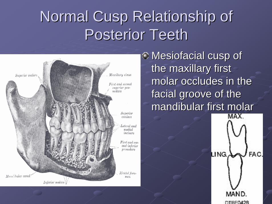
Normal Cusp Relationship of Normal Cusp Relationship of Posterior TeethPosterior Teeth
MesiofacialMesiofacial cusp of cusp of the maxillary first the maxillary first molar occludes in the molar occludes in the facial groove of the facial groove of the mandibularmandibular first molar first molar

OrthognathicOrthognathic SurgerySurgery
Jaw Realignment and Jaw Realignment and Correction of Facial Correction of Facial ProfileProfileMaxillary (oneMaxillary (onemultiplemultiple piece piece osteotomiesosteotomies))MandibularMandibularosteotomiesosteotomies

OrthognathicOrthognathic SurgerySurgery

CephalometricCephalometric AnalysisAnalysis

CepahalometricCepahalometric AnalysisAnalysis

LefortLefort 1 1 OsteotomyOsteotomy

BSRBSR
MandibularMandibular ramusramus sagittalsagittal split split osteotomyosteotomy is the is the most common technique used for most common technique used for mandibularmandibularadvancement advancement
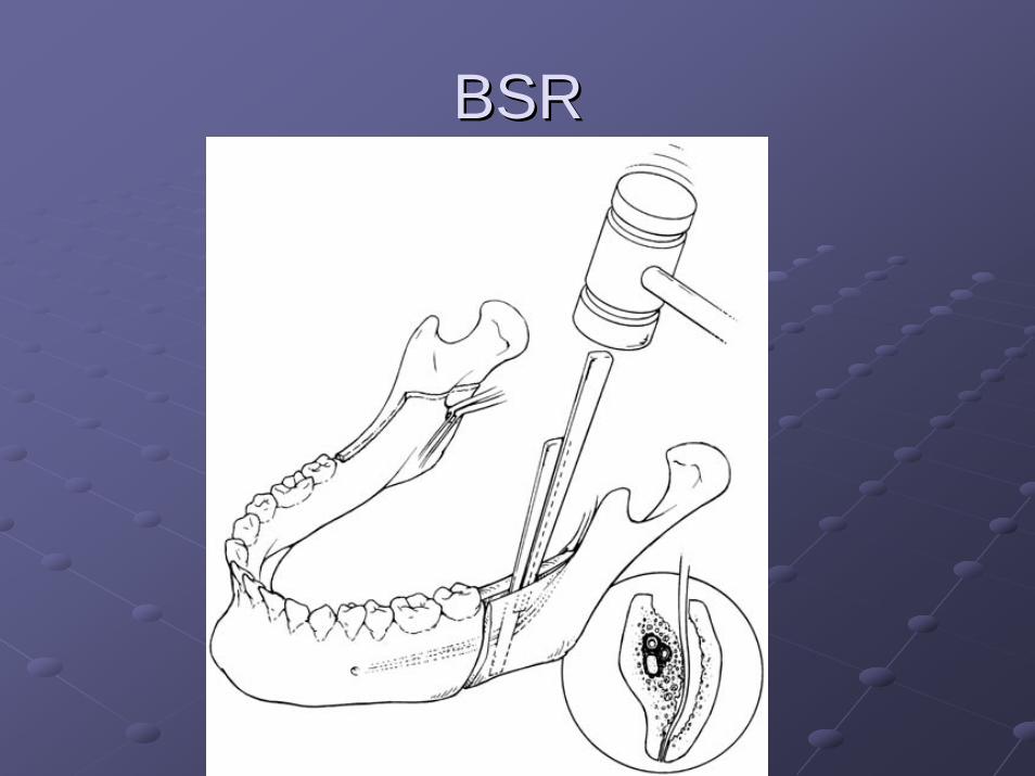
BSRBSR
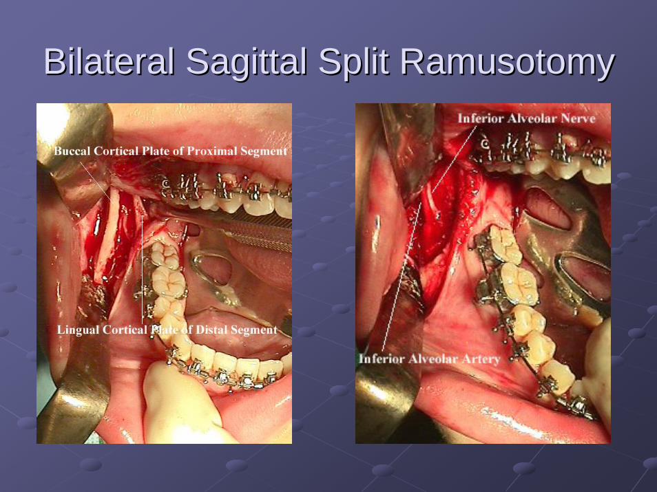
Bilateral Bilateral SagittalSagittal Split Split RamusotomyRamusotomy

Vertical Vertical RamusRamus OsteotomyOsteotomy
Vertical Vertical ramusramus osteotomyosteotomy can be used to set the can be used to set the mandible mandible posteriorlyposteriorly. .

GenioplastyGenioplasty
Anterior Anterior MandibularMandibular Horizontal Horizontal OsteotomyOsteotomy

Facial TraumaFacial Trauma

Facial TraumaFacial Trauma

R R ZygomaticZygomatic Arch FractureArch Fracture

Distribution of Mandible FracturesDistribution of Mandible Fractures

Mandible FractureMandible Fracture

TxTx Mandible Mandible FxFx

ORIF Mandible ORIF Mandible FxFx

LeFortLeFort FracturesFractures

Wisdom TeethWisdom Teeth

Wisdom Teeth (3Wisdom Teeth (3rdrd Molars)Molars)

Extraction of Extraction of MandibularMandibular 33rdrd Molar Molar ToothTooth

Extraction of Extraction of MandibularMandibular 33rdrd Molar Molar ToothTooth

Thank You !Thank You !