hcn4 ion channel function is required for early events...
TRANSCRIPT

RESEARCH ARTICLE
HCN4 ion channel function is required for early events thatregulate anatomical left-right patterning in a nodal and leftyasymmetric gene expression-independent mannerVaibhav P. Pai1, Valerie Willocq1, Emily J. Pitcairn1, Joan M. Lemire1, Jean-François Pare1, Nian-Qing Shi2,Kelly A. McLaughlin1 and Michael Levin1,*
ABSTRACTLaterality is a basic characteristic of all life forms, from single cellorganisms to complex plants and animals. For many metazoans,consistent left-right asymmetric patterning is essential for the correctanatomyof internal organs, suchas the heart, gut, and brain; disruption ofleft-right asymmetry patterning leads to an important class of birth defectsin human patients. Laterality functions across multiple scales, whereearly embryonic, subcellular and chiral cytoskeletal events are coupledwith asymmetric amplification mechanisms and gene regulatorynetworks leading to asymmetric physical forces that ultimately result indistinct left and right anatomical organ patterning. Recent studies havesuggested the existence of multiple parallel pathways regulating organasymmetry. Here, we show that an isoform of the hyperpolarization-activated cyclic nucleotide-gated (HCN) family of ion channels(hyperpolarization-activated cyclic nucleotide-gated channel 4, HCN4)is important for correct left-right patterning. HCN4 channels are presentveryearly inXenopus embryos. BlockingHCNchannels (Ih currents)withpharmacological inhibitors leads to errors in organ situs. This effect isonly seenwhenHCN4channels are blocked early (pre-stage 10) andnotby a later block (post-stage 10). Injections of HCN4-DN (dominant-negative) mRNA induce left-right defects only when injected in bothblastomeres no later than the 2-cell stage. Analysis of key asymmetricgenes’ expression showed that the sidedness ofNodal, Lefty, and Pitx2expression is largely unchanged by HCN4 blockade, despite therandomization of subsequent organ situs, although the area of Pitx2expression was significantly reduced. Together these data identify anovel, developmental role for HCN4 channels and reveal a new Nodal-Lefty-Pitx2 asymmetric gene expression-independent mechanismupstream of organ positioning during embryonic left-right patterning.
KEY WORDS: HCN4, Bioelectricity, Ion channels, Laterality,Xenopus
INTRODUCTIONInvariant left-right asymmetry is a fundamental aspect of all life,from single cell organisms to plants and animals with complex body
plans like humans (Chen et al., 2012; Coutelis et al., 2008; Davisonet al., 2016; Dimonte et al., 2016; Gros et al., 2009; Hashimoto,2002; Kuroda et al., 2009; Naganathan et al., 2014; Petzoldt et al.,2012; Pohl, 2011; Spéder et al., 2007; Thitamadee et al., 2002;Wan et al., 2011; Xu et al., 2007; Yost, 1990, 1991). Consistentorientation of the left-right (LR) axis is a difficult problem for anembryo to solve in a universe that does not macroscopicallydistinguish left from right, and must be done reliably and accuratelyto achieve correct organization of internal organ structures. Errors inleft-right asymmetry form a large and important class of humanbirth defects, affecting almost all major visceral organs, includingthe heart and the brain (Burn, 1991; Cohen et al., 2007; Hoffmanand Kaplan, 2002; Peeters and Devriendt, 2006; Ramsdell, 2005).Hence, understanding the establishment of consistent lateralityin the organization of body plans is a fundamental questionin evolutionary and developmental biology, with importantimplications for addressing birth defects via regenerative medicine.
It is becoming clear that the origins of left-right asymmetry lie inphysical aspects of cytoskeletal chirality (Naganathan et al., 2016;Suzuki et al., 2017; Tee et al., 2015; Wan et al., 2011), amplifiedimmediately post-fertilization in the early embryos of many species(McDowell et al., 2016a,b; Okumura et al., 2008; Spéder et al.,2007; Vandenberg et al., 2013a; Vandenberg and Levin, 2013). Thecytoskeletal chirality-mediated initiation of laterality is highlyconserved across phyla, and even kingdoms (Levin and Nascone,1997; Levin and Palmer, 2007; Lobikin et al., 2012; Okumuraet al., 2008; Spéder et al., 2007). Multiple amplification andreinforcement mechanisms transmit this early left-right asymmetryacross the entire developing embryo. These include asymmetrictransport of ion translocators (Adams et al., 2006; Aw et al., 2008;Bessodes et al., 2012; Levin et al., 2002), charged moleculesthrough gap junctions (Fukumoto et al., 2005a,b; Garic-Stankovicet al., 2008; Oviedo and Levin, 2007; Vandenberg et al., 2013b),and ciliary flow (Basu and Brueckner, 2008; Schweickert et al.,2007). Collectively, these signals trigger an embryo-wideasymmetric gene regulatory network, with a left-side expressionof Nodal-Lefty-Pitx2 as the primary node (Levin, 1998; Mercolaand Levin, 2001; Nakamura and Hamada, 2012; Ramsdell andYost, 1998; Raya and Izpisua Belmonte, 2004a,b). Finally,asymmetric generation of mechanical forces results in asymmetricorgan structure and placement (Granados-Riveron and Brook, 2012;Voronov et al., 2004; Welsh et al., 2013).
Previous work has shown an important role of ion fluxes-mediated regulation of membrane voltage in determination ofleft-right laterality (Adams et al., 2006; Aw et al., 2008, 2010;Garic-Stankovic et al., 2008; Hibino et al., 2006; Levin et al., 2006,2002; Morokuma et al., 2008; Oviedo and Levin, 2007; Shimeldand Levin, 2006). Like other mechanisms involved in left-rightReceived 6 April 2017; Accepted 12 August 2017
1Allen Discovery Center at Tufts University, 200 Boston Ave, Suite 4600, Medford,MA 02155, USA. 2Department of Medicine at University of Wisconsin-Madison,Madison, WI 53792, USA.
*Author for correspondence ([email protected])
M.L., 0000-0001-7292-8084
This is an Open Access article distributed under the terms of the Creative Commons AttributionLicense (http://creativecommons.org/licenses/by/3.0), which permits unrestricted use,distribution and reproduction in any medium provided that the original work is properly attributed.
1445
© 2017. Published by The Company of Biologists Ltd | Biology Open (2017) 6, 1445-1457 doi:10.1242/bio.025957
BiologyOpen
by guest on July 9, 2018http://bio.biologists.org/Downloaded from

laterality determination, these also largely feed into theNodal-Lefty-Pitx2 node of gene regulatory networks. Recent evidence hasuncovered a non-linearity between the cytoskeletal initiation ofleft-right asymmetry and establishment of organ situs, where errorsin key aspects of the gene regulatory network, like the sidedness ofNodal expression, are bypassed to establish correct left-rightorgan situs despite randomized expression of upstream lateralitydeterminant genes (Cota et al., 2006; McDowell et al., 2016a,b).These alternate pathways provide redundancy and robustness tothe establishment of proper left-right organ situs, but are poorlyunderstood. Here we report a new element of the endogenousbioelectric toolbox of left-right patterning, the hyperpolarization-activated cyclic nucleotide-gated channel 4 (HCN4), and presentdata suggesting that it mediates alternative pathways (bypassingthe Nodal-Lefty-Pitx2 cassette) involved in the establishment ofleft-right asymmetry in Xenopus embryos.Hyperpolarization-activated cyclic nucleotide-gated (HCN)
channels are a unique group of voltage-gated channels where thethreshold voltage for opening of the channel is modulated by themetabolic state of the cell (levels of cyclic nucleotides like cAMP)(Biel et al., 2009; Wahl-Schott and Biel, 2009). They open athyperpolarized membrane voltages (negative), giving rise tocurrents which are a mix of sodium and potassium ion fluxes.Among the four HCN isoforms (HCN1-4), HCN4 channels havebeen primarily studied in adult hearts as pacemaker channels(Scicchitano et al., 2012; Verkerk and Wilders, 2015), but recentevidence has demonstrated their presence in human and mouseembryonic cells (Cerbai et al., 1999; Qu et al., 2008; Robinson et al.,1997; Später et al., 2013; Vicente-Steijn et al., 2011; Yasui et al.,2001), and implicated them in cardiac patterning (Pitcairn et al.,2017). Because these channels have not been studied in the contextof developmental bioelectricity (Adams and Levin, 2013; Levin,2013, 2014a,b) or control of embryonic axial patterning, wecharacterized the role of HCN4 channels in embryonic left-rightasymmetry establishment in Xenopus embryos.Here we extend our recent work on HCN4 channels in
cardiogenesis (Pitcairn et al., 2017) using a different set ofmisexpression conditions to target earlier events. We show thatHCN4 channels are present inXenopus embryos beginning at the firstcleavage event (2-cell stage). Pharmacological inhibition of HCNchannels (Ih currents) by ZD7288 causes heterotaxia (randomizationof organ situs) in Xenopus tadpoles, but only if embryos are exposedearly (pre-stage 10, prior to the onset of gastrulation). Similarly,injections of mRNA encoding a dominant-negative protein (whichblocks HCN4 channel function) randomize organ asymmetry ifinjected into both blastomeres at the 2-cell stage. Remarkably, despitethe early period of action (prior to asymmetric gene expression),HCN4 channel disruption does not affect the laterality of subsequentNodal, Lefty, or Pitx2 expression, exhibiting significant latency in itseffects on later organ situs. Together, these results are the first, to ourknowledge, to show an early embryonic axial patterning role of thisimportant ion channel, and implicate it in a pathway bypassing theasymmetric gene expression of Nodal, Lefty, and Pitx2 in regulatingleft-right asymmetry.
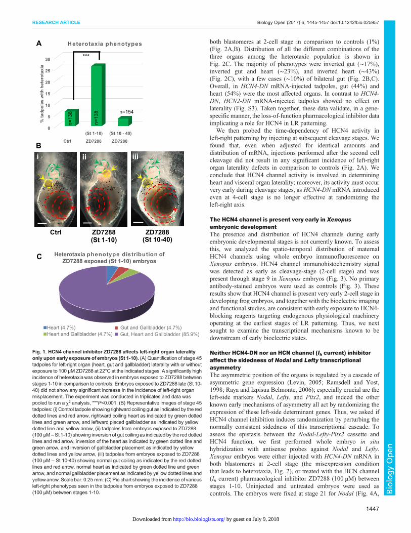
RESULTSEarly exposure to pharmacological inhibitor ZD7288 inducesheterotaxiaOur prior work demonstrated that interference with four differention translocators (two channels and two pumps) inducesheterotaxia, the independent randomization of organ positioningalong the LR axis (Adams et al., 2006; Aw et al., 2008, 2010; Levin
et al., 2002; Morokuma et al., 2008). To test whether HCN4channels also play a role in embryonic left-right lateralitydetermination, we first used the pharmacological inhibitorZD7288 (100 µM), which blocks HCN channels (Ih currents) viaa trapping mechanism (Shin et al., 2001). To probe the timing of theendogenous role of HCN4 channels in development, we comparedthe results of exposures beginning immediately after fertilization tothose that began during gastrulation. Xenopus embryos wereexposed to ZD7288 (100 µM) from stage 1 through stage 10 orstarting at stage 10 until stage 40, followed by anatomical lateralityanalysis at stage 45 (swimming tadpoles). Any instance of mirrorimage reversal of organ position, in the context of normal organpatterning and normal dorso-anterior index was counted as aninstance of heterotaxia. Untreated embryos served as controls.
ZD7288 exposure during stages 1-10 induced a significantly highincidence (∼26%, P<0.001, χ2) of heterotaxia in comparison tocontrols (∼6%) (Fig. 1A,B). In sharp contrast, ZD7288 exposureduring stages 10-40 did not induce heterotaxia (∼6%) in comparisonto controls (∼6%) (Fig. 1A,B). Similar results were observed withanother, more-specific HCN4 channel inhibitor ivabradine (Fig. S1).Distribution of heterotaxia outcomes from the early ZD7288exposure showed various combinations of asymmetric placement ofgut, heart and gallbladder (Fig. 1C). However, the majority (∼86%)of left-right asymmetric tadpoles showed situs inversus – fullmirroring of all three organs (gut, heart and gallbladder) (Fig. 1B,C).Overall, percentage of ZD7288-treated tadpoles showing incorrectplacement of each of the three organswere approximately equal (heart∼34%, gut ∼32%, gallbladder ∼34%). Together, these resultssuggest that HCN4 function is required prior to embryonic stage 10in establishing left-right asymmetry.
These studies took advantage of the temporal control afforded bypharmacological experiments beginning at different timepoints.Treatment with ZD7288, which inhibits HCN (Ih currents), resultedin high incidence of situs inversus. These data implicate the HCN4channel in early (prior to stage 10) processes involved indetermination of left-right patterning in the Xenopus embryo.
HCN4-DN (dominant-negative) causes heterotaxiaTo molecularly validate whether HCN4 channels are specificallyinvolved in organizing left-right organ laterality, we used anHCN4-DN (hyperpolarization-activated cyclic nucleotide-gated channel4-dominant negative) mRNA construct that has been previouslymolecularly characterized and shown to inhibit the HCN4 channelcurrent in mammalian cell culture by direct electrophysiology(Pitcairn et al., 2017). We confirmed that expression of HCN4-DNprotein blocks HCN4 channel function and depolarizes themembrane voltage in Xenopus embryos (Fig. S2). We used adominant-negative construct because many channels are present inXenopus as maternal proteins (Adams et al., 2006; Aw et al., 2008,2010; Levin et al., 2002; Morokuma et al., 2008; Qiu et al., 2005),and thus cannot be targeted by morpholinos or RNAi. Xenopusembryos were injected with HCN4-DN mRNA at 2- and 4-cellstage, in various blastomeres as indicated in Fig. 2A (red arrows),followed by anatomical laterality analysis at stage 45 (swimmingtadpoles). As with the pharmacological inhibitor experiments, anydeviation from the normal laterality of the three organs, in thecontext of otherwise normal patterning, was counted as an instanceof heterotaxia. Uninjected embryos served as controls [as neitherwater nor non-specific mRNA, e.g. β–galactosidase, injectionsaffect left-right asymmetry endpoints (McDowell et al., 2016b)].HCN4-DN mRNA-injected tadpoles showed a significantly highincidence (25%, P<0.001, χ2) of heterotaxia only when injected in
1446
RESEARCH ARTICLE Biology Open (2017) 6, 1445-1457 doi:10.1242/bio.025957
BiologyOpen
by guest on July 9, 2018http://bio.biologists.org/Downloaded from
both blastomeres at 2-cell stage in comparison to controls (1%)(Fig. 2A,B). Distribution of all the different combinations of thethree organs among the heterotaxic population is shown inFig. 2C. The majority of phenotypes were inverted gut (∼17%),inverted gut and heart (∼23%), and inverted heart (∼43%)(Fig. 2C), with a few cases (∼10%) of bilateral gut (Fig. 2B,C).Overall, in HCN4-DN mRNA-injected tadpoles, gut (44%) andheart (54%) were the most affected organs. In contrast to HCN4-DN, HCN2-DN mRNA-injected tadpoles showed no effect onlaterality (Fig. S3). Taken together, these data validate, in a gene-specific manner, the loss-of-function pharmacological inhibitor dataimplicating a role for HCN4 in LR patterning.
We then probed the time-dependency of HCN4 activity inleft-right patterning by injecting at subsequent cleavage stages. Wefound that, even when adjusted for identical amounts anddistribution of mRNA, injections performed after the second cellcleavage did not result in any significant incidence of left-rightorgan laterality defects in comparison to controls (Fig. 2A). Weconclude that HCN4 channel activity is involved in determiningheart and visceral organ laterality; moreover, its activity must occurvery early during cleavage stages, as HCN4-DN mRNA introducedeven at 4-cell stage is no longer effective at randomizing theleft-right axis.
The HCN4 channel is present very early in Xenopusembryonic developmentThe presence and distribution of HCN4 channels during earlyembryonic developmental stages is not currently known. To assessthis, we analyzed the spatio-temporal distribution of maternalHCN4 channels using whole embryo immunofluorescence onXenopus embryos. HCN4 channel immunohistochemistry signalwas detected as early as cleavage-stage (2-cell stage) and waspresent through stage 9 in Xenopus embryos (Fig. 3). No primaryantibody-stained embryos were used as controls (Fig. 3). Theseresults show that HCN4 channel is present very early 2-cell stage indeveloping frog embryos, and together with the bioelectric imagingand functional studies, are consistent with early exposure to HCN4-blocking reagents targeting endogenous physiological machineryoperating at the earliest stages of LR patterning. Thus, we nextsought to examine the transcriptional mechanisms known to bedownstream of early bioelectric states.
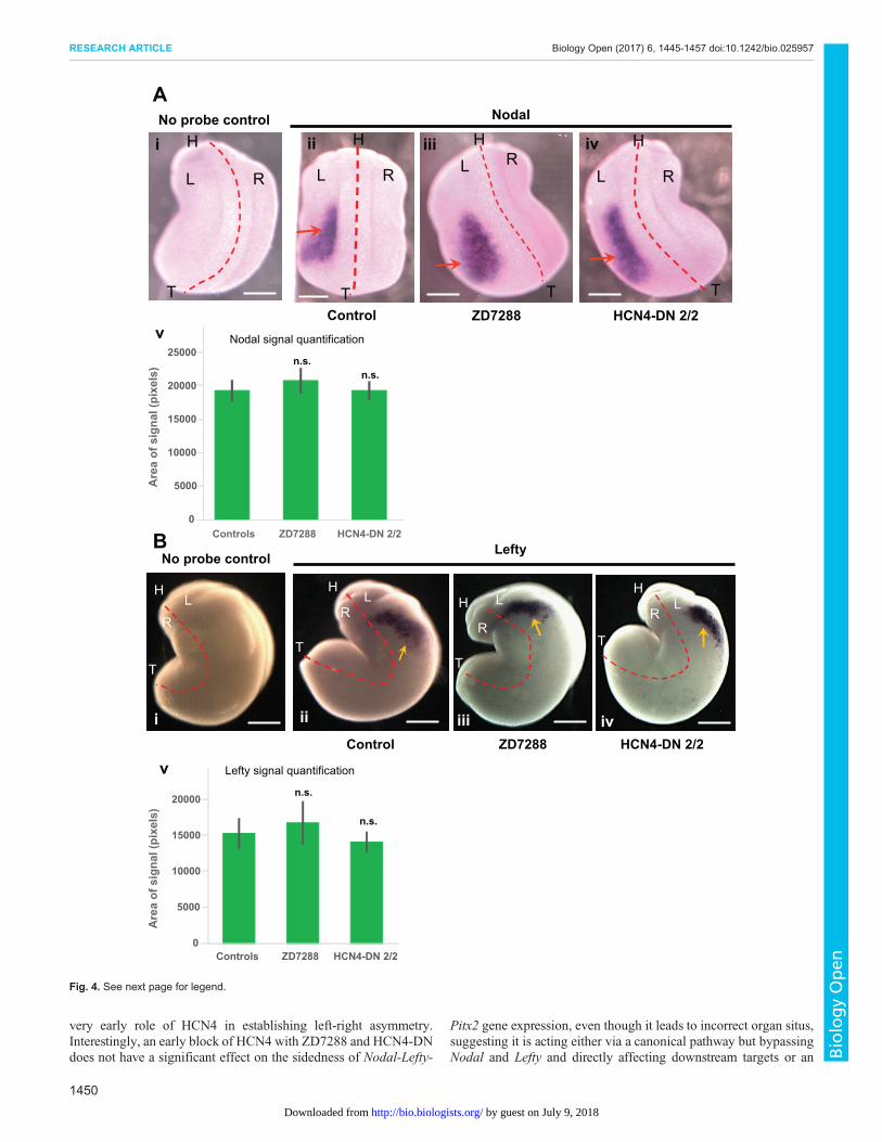
Neither HCN4-DN nor an HCN channel (Ih current) inhibitoraffect the sidedness of Nodal and Lefty transcriptionalasymmetryThe asymmetric position of the organs is regulated by a cascade ofasymmetric gene expression (Levin, 2005; Ramsdell and Yost,1998; Raya and Izpisua Belmonte, 2006); especially crucial are theleft-side markers Nodal, Lefty, and Pitx2, and indeed the otherknown early mechanisms of asymmetry all act by randomizing theexpression of these left-side determinant genes. Thus, we asked ifHCN4 channel inhibition induces randomization by perturbing thenormally consistent sidedness of this transcriptional cascade. Toassess the epistasis between the Nodal-Lefty-Pitx2 cassette andHCN4 function, we first performed whole embryo in situhybridization with antisense probes against Nodal and Lefty.Xenopus embryos were either injected with HCN4-DN mRNA inboth blastomeres at 2-cell stage (the misexpression conditionthat leads to heterotaxia, Fig. 2), or treated with the HCN channel(Ih current) pharmacological inhibitor ZD7288 (100 µM) betweenstages 1-10. Uninjected and untreated embryos were used ascontrols. The embryos were fixed at stage 21 for Nodal (Fig. 4A,
0
5
10
15
20
25
30
(St 1-10) (St 10 - 40)
Ctrl ZD7288 ZD7288
% ta
dpol
es w
ith h
eter
otax
iaHeterotaxia phenotypes
n=13
8
n=13
8 n=154
***
A
B
Heterotaxia phenotype distribution ofZD7288 exposed (St 1-10) embryos
Heart (4.7%) Gut and Gallbladder (4.7%)Heart and Gallbladder (4.7%) Gut, Heart and Gallbladder (85.9%)
C
Ctrl
i
(St 1-10)ZD7288
ii
(St 10-40)ZD7288
iii
Fig. 1. HCN4 channel inhibitor ZD7288 affects left-right organ lateralityonly upon early exposure of embryos (St 1-10). (A) Quantification of stage 45tadpoles for left-right organ (heart, gut and gallbladder) laterality with or withoutexposure to 100 µM ZD7288 at 22°C at the indicated stages. A significantly highincidence of heterotaxiawas observed in embryos exposed to ZD7288 betweenstages 1-10 in comparison to controls. Embryos exposed to ZD7288 late (St 10-40) did not show any significant increase in the incidence of left-right organmisplacement. The experiment was conducted in triplicates and data waspooled to run a χ2 analysis, ***P<0.001. (B) Representative images of stage 45tadpoles: (i) Control tadpole showing rightward coiling gut as indicated by the reddotted lines and red arrow, rightward coiling heart as indicated by green dottedlines and green arrow, and leftward placed gallbladder as indicated by yellowdotted line and yellow arrow, (ii) tadpoles from embryos exposed to ZD7288(100 µM –St 1-10) showing inversion of gut coiling as indicated by the red dottedlines and red arrow, inversion of the heart as indicated by green dotted line andgreen arrow, and inversion of gallbladder placement as indicated by yellowdotted lines and yellow arrow, (iii) tadpoles from embryos exposed to ZD7288(100 µM – St 10-40) showing normal gut coiling as indicated by the red dottedlines and red arrow, normal heart as indicated by green dotted line and greenarrow, and normal gallbladder placement as indicated by yellow dotted lines andyellowarrow. Scale bar: 0.25 mm. (C) Pie chart showing the incidence of variousleft-right phenotypes seen in the tadpoles from embryos exposed to ZD7288(100 µM) between stages 1-10.
1447
RESEARCH ARTICLE Biology Open (2017) 6, 1445-1457 doi:10.1242/bio.025957
BiologyOpen
by guest on July 9, 2018http://bio.biologists.org/Downloaded from

Table 1) and at stage 23 for Lefty (Fig. 4B, Table 2) in situhybridization analysis.In controls, as expected, Nodal was expressed on the left side
of the embryo in the majority (∼84%) of the embryos (Fig. 4A,Table 1). HCN4-DN mRNA-injected and ZD7288-treated embryos
showed no significant change in the laterality of Nodal signal, withthe majority of embryos (∼95% and∼81%, respectively) expressingNodal on the left side (Table 1). Quantification of area of Nodalexpression domain showed no significant difference betweenthe HCN4-DN mRNA-injected and ZD7288-treated embryos, and
HCN4-DN
0
10
20
30
Ctrl HCN4-DN HCN4-DN HCN4-DN HCN4-DN HCN4-DN HCN4-DN HCN4-DN HCN4-DN2/2 cells 1/2 cells 2/4 dorsal 2/4 ventral 1/4 right
ventral1/4 leftventral
1/4 rightdorsal
1/4 leftdorsal
% ta
dpol
es w
ith h
eter
otax
ia
Heterotaxia Phenotypes
n=107
n=12
0
n=122 n=105 n=98 n=125 n=161 n=113 n=126
***
A
B
Heterotaxia phenotype distributionof HCN4-DN injected embryos
Heart (43%) Gut (17%)
Heart and Gut (23%) Gut and Gallbladder (3%)
Heart and Bilateral Gut (3%) Bilateral Gut (11%)
C
Controls
i ii iii
Fig. 2. Early injection of HCN4-DN affects left-right organ laterality in Xenopus laevis. (A) Quantification of stage 45 tadpoles for left-right organ (heart, gutand gallbladder) laterality with or without microinjecting HCN4-DN mRNA apically (∼0.5-1 ng/injection/blastomere) in both blastomeres at 2-cell stage asindicated in the illustrations. A significantly high incidence of heterotaxia was observed in only when HCN4-DNmRNAwas injected in both blastomeres at 2-cellstage, in comparison to controls. The experiment was conducted in triplicate and data was pooled to run a χ2 analysis, ***P<0.001. (B) Representative ventralimages of stage 45 tadpoles: (i) Control tadpole showing rightward coiling gut as indicated by the red dotted lines and red arrow, rightward coiling heart asindicated by green dotted lines and green arrow, and leftward placed gallbladder as indicated by yellow dotted line and yellow arrow, (ii)HCN4-DNmRNA injected(both blastomeres at 2-cell stage) tadpoles showing inversion of gut coiling as indicated by the red dotted lines and red arrow, inversion of the heart asindicated by green dotted line and green arrow, and inversion of gallbladder placement as indicated by yellow dotted lines and yellow arrow, (iii)HCN4-DNmRNAinjected (both blastomeres at 2-cell stage) tadpoles showing bilateral gut coiling as indicated by the red dotted lines and red arrow, with normal heart (green dottedlines and green arrow) and gallbladder (yellow dotted line and yellow arrow). Scale bar: 0.25 mm. (C) Pie chart showing the incidence of various left-rightphenotypes seen in the HCN4-DN mRNA injected (in both blastomeres at 2-cell stage) tadpoles.
1448
RESEARCH ARTICLE Biology Open (2017) 6, 1445-1457 doi:10.1242/bio.025957
BiologyOpen
by guest on July 9, 2018http://bio.biologists.org/Downloaded from

control embryos (Fig. 4Av, ANOVA). A subset of control andtreated embryos from the same batch used for in situ analysis wereraised to stage 45 and scored for left-right organ placement,confirming thatHCN4-DNmRNA and ZD7288 were still effectivelyinducing heterotaxia in this cohort of animals, just as seen in Figs 1and 2 (data not shown). We conclude that very early disruption ofHCN4 function randomizes organ situs, bypassing the normalsequence of asymmetric Nodal expression.In control embryos, Lefty was expressed on the left side in the
majority (∼88%) of the embryos (Fig. 4B, Table 2). Strikingly,HCN4-DN mRNA-injected and ZD7288-treated embryos showedno significant change from wild-type embryos in the laterality ofLefty signal with majority of embryos (∼86% and ∼78%,respectively), showing Lefty on left side as in the set of controlembryos (Table 2). Quantification of area of Lefty expressiondomain showed no significant difference between the HCN4-DNmRNA-injected and ZD7288-treated embryos, and control embryos(Fig. 4Bv, ANOVA). A subset of control and treated embryos fromthe same batch used for in situ analysis were raised until stage 45and scored for left-right organ placement to verify that HCN4-DN
mRNA and ZD7288 were still inducing heterotaxia in this cohort, asseen in Figs 1 and 2 (data not shown).
Thus, HCN4-DN mRNA and ZD7288 induce left-right organrandomization without altering the normal left-sided expression ofNodal and Lefty. These results reveal that HCN4 channel functionaffects organ placement, but bypasses the major asymmetry-regulating gene cassette, Nodal-Lefty.
HCN4-DN and HCN channel (Ih current) inhibition affectPitx2 expressionTo assess if HCN4 channel inhibition affects the normally left-sidedexpression of the late marker Pitx2, we performed whole embryoin situ hybridization against Pitx2. Xenopus embryos were eitherinjected with HCN4-DN mRNA in both blastomeres at 2-cell stageor treated with a pharmacological inhibitor of HCN channels(Ih currents), ZD7288 (100 µM), between stages 1-10. Uninjectedand untreated embryos were used as controls [as these are known tobe equivalent to water- or nonspecific mRNA-injected embryos(McDowell et al., 2016b)]. The embryos were fixed at stage 28 forPitx2 (Fig. 5, Table 3) in situ hybridization analysis. In controls, asexpected, Pitx2 is present on the left side of the embryo in majority(∼87%) of the embryos (Fig. 5A, Table 3). HCN4-DN mRNA-injected and ZD7288-treated embryos caused no significant changein the laterality of Pitx2 signal, with a majority of embryos (∼81%and ∼87%, respectively) showing Pitx2 on the left side of theembryos similar to controls (Table 3). A subsection of control andtreated embryos from the same batch used for in situ analysis wereraised to stage 45 and scored for left-right organ placement,confirming that HCN4-DN mRNA and ZD7288 were still inducingheterotaxia as seen in Figs 1 and 2 (data not shown). We concludethat the randomizing effects of HCN4 inhibition can bypassasymmetric Pitx2 gene expression.
Examining closely the results of treatments that randomizedorgans but not Pitx2 situs, we observed that in contrast to the normalsidedness of expression, there was a significant difference in thespatial extent (pattern) of the Pitx2 transcriptional domain in thetreated embryos in comparison to controls. To analyze this, wequantified the anterior to posterior spread distance and area of thePitx2 signal in embryos (Fig. 5A-C). The anterior-posterior spreadof Pitx2 signal was significantly reduced (***P<0.001, ANOVA) inHCN4-DNmRNA-injected and ZD7288-treated embryos (Table 3).Similarly, the total area of Pitx2 signal was significantly reduced(***P<0.001, **P<0.01, ANOVA) in HCN4-DN mRNA-injectedand ZD7288-treated embryos (Fig. 5C). A similar analysis of theNodal and Lefty expression domains revealed no significantchanges in the signal pattern (Fig. 4). These results show thatwhile HCN4-DN mRNA and ZD7288 treatments do not affect thelaterality of Pitx2 gene expression, both treatments significantlyreduce the anterior to posterior spread distance and the area of Pitx2expression, while inducing left-right organ patterning defects intadpoles.
DISCUSSIONHere, we show that HCN4 channels are present in Xenopus embryosfrom the earliest stages of development, and play an important rolein establishing left-right organ situs in Xenopus tadpoles. BlockingHCN4 channels results in heterotaxia and incorrect organ situs intadpoles. This is effective only during early embryogenesis (pre-stage 10) as post stage 10, HCN4 channel blocking has no effect onorgan situs. Even within the first 10 stages of embryogenesis, theHCN4-DN construct induces heterotaxia only when introduced inall blastomeres during early cleavage stages, further suggesting a
No
prim
ary
Ab
Con
trol
sH
CN
-4
2-CellStage
4-CellStage Stage-9
i
ii
iii
iv
v
vi
vii
viii
ix
x
xi
xiiFig. 3. Xenopus laevis embryos express endogenous HCN4 channelduring early development. Immunofluorescence analysis of whole Xenopusembryos for HCN4 channel protein at indicated stages of development.(i-vi) No primary antibody controls, (vii – xii) HCN4 immunofluorescence,(i, iii, v, vii, ix, xi) bright field images of immunofluorescent embryos,(ii, iv, vi, viii, x, xii) fluorescence images of immunofluorescence embryos.Xenopus embryos at the indicated stage of development showed a prominentHCN4 channel protein (n=15).
1449
RESEARCH ARTICLE Biology Open (2017) 6, 1445-1457 doi:10.1242/bio.025957
BiologyOpen
by guest on July 9, 2018http://bio.biologists.org/Downloaded from
very early role of HCN4 in establishing left-right asymmetry.Interestingly, an early block of HCN4 with ZD7288 and HCN4-DNdoes not have a significant effect on the sidedness of Nodal-Lefty-
Pitx2 gene expression, even though it leads to incorrect organ situs,suggesting it is acting either via a canonical pathway but bypassingNodal and Lefty and directly affecting downstream targets or an
B
NodalANo probe control
Lefty
Control
L R
H
T
ii
ZD7288
L RH
T
iii
HCN4-DN 2/2
L R
H
T
iv
L R
H
T
i
ZD7288iii
T
H
R
L
Control
H
T
LR
ii
No probe control
H
T
L
R
iHCN4-DN 2/2
H
T
LR
iv
0
5000
10000
15000
20000
25000
Controls HCN4-DN 2/2ZD7288
n.s.n.s.
Nodal signal quantification
Are
a of
sig
nal (
pixe
ls)
v
0
5000
10000
15000
20000
Controls HCN4-DN 2/2ZD7288
Lefty signal quantification
Are
a of
sig
nal (
pixe
ls)
n.s.
n.s.
v
Fig. 4. See next page for legend.
1450
RESEARCH ARTICLE Biology Open (2017) 6, 1445-1457 doi:10.1242/bio.025957
BiologyOpen
by guest on July 9, 2018http://bio.biologists.org/Downloaded from

alternative pathway of left-right asymmetry that is able to bypass theNodal-Lefty-Pitx2 cassette completely. Blocking of HCN4 channels(both pharmacologically and physiologically) induces a significantdecrease in Pitx2 signal in its anterior-posterior spread and area ofthe signal.
Pleotropic actions of HCN4 in Xenopus embryonicdevelopmentOur recent study explored HCN4 function in embryonic cardiactissue development (Pitcairn et al., 2017). That study and theexperiments presented here used drastically different injection andculture conditions to target two different lineages/tissues: the animalcap versus the mesodermal heart lineage. The major differences wereas follows. (1) In Pitcairn et al. (2017), where the role of HCN4during heart development was the focus of study, the HCN4-DN wasinjected medially (along the equitorial plane of the embryo) whichtargets mostly mesodermal cardiac and kidney tissues. Here ourinjections were apical, to achieve a more global expression in theembryo, especially targeting the animal cap ectoderm and derivedstructures. (2) In Pitcairn et al. (2017), embryos were injected(equatorially) in one blastomere at the two-cell stage and kept inrelatively high salt (1× MMR) until stage 9 (gastrulation). Underthese high salt conditions, the ion gradients (and hence ion flux) forK+ and Na+ are reversed, affecting all K+ and Na+ ion channel fluxes:blocking HCN4 channel using HCN4-DN is a hyperpolarizingtreatment and leads to subsequent incorrect Nodal-Lefty-Pitx2expression and gross morphology defects of heart. In contrast, theexperiments reported here made use of injections (apical) into bothblastomeres at the 2-cell stage, and the embryos were kept in low salt0.1× MMR (mimicking its natural environment) throughout
development. Under these conditions, an HCN4-DN-mediatedblock of HCN4 ion flux leads to depolarization (Fig. S2) withnormal sidedness and pattern of Nodal-Lefty-Pitx2 expression(Figs 4 and 5, Tables 1-3), but randomized organ situs (withnormal organ morphology) (Figs 1 and 2).
The two different conditions help tease apart two differentfunctions of HCN4: an early activity involved in establishinglaterality of organs and another in developmental morphology ofheart. Previously, it has been shown that moving the membranevoltage in either direction away from wild type leads to defects(Pai et al., 2012, 2015). Since early embryos already havequite depolarized membrane potentials, the HCN4-DN-mediateddepolarization perturbation reported here may be too subtle to affectthe asymmetry of the Nodal-Lefty-Pitx2 expression, but is sufficientto affect a parallel pathway that feeds into organ situs. However,injections targeted to the developing heart in high salt conditions,combined with HCN4 blockade, induce a strong hyperpolarizationperturbation (Pitcairn et al., 2017), which is a more drastic changethat affects several gene regulatory networks involved in organmorphology, including feedback loops between Pitx2 and HCN4during cardiac morphgenesis (Christoffels et al., 2010; Clauss andKaab, 2011; Wang et al., 2010). Work is currently ongoing in ourlaboratories to construct a detailed, testable, physiologically-realistic, and spatialized model of the early frog embryo’sbioelectric circuits, chemical gradients, and relevant gene-regulatory circuitry (Pietak and Levin, 2016).
HCN4 channels can act in a Nodal-Lefty asymmetric geneexpression-independent mannerA recent series of studies and meta-analyses looking at the relationbetween causes of left-right abnormality, asymmetric gene expressionofNodal-Lefty-Pitx2, and organ situs, strongly suggested non-linearityof the laterality pathway (McDowell et al., 2016a,b; Vandenberg,2012). Methods leading to disruption of asymmetricNodal expressioncan still show correct organ situs, suggesting the presence of flexiblegene regulatory networks and the presence of alternative pathwaysconferring redundancy and robustness to laterality and particularlyorgan situs. Some laterality defects were corrected even past Pitx2expression. Perturbation of early cytoskeleton, motor proteins, gap-junctions, and serotonin signaling, all showed significant repair [highincidence of mispatterned Nodal, but a lower incidence of organ situsdefects (McDowell et al., 2016a)] (Fig. 3). However, a particularlyinteresting observation was that defects induced by perturbinglaterality-relevant ion fluxes did not correct over time (incidence oforgan situs defects were higher than incidence of incorrect sidedness ofNodal), strongly suggesting possible bioelectrically mediated Nodal-independent pathway that could correct for errors in the normallaterality-establishing transcriptional pathway.
Here we observed the opposite phenomenon: correct expressionof asymmetric genes Nodal and Lefty but incorrect organ situs afterHCN4 inhibition (Figs 1, 2, 4 and 5). In case of Pitx2 the sidednessof expression was correct (left side) but the area of expression wassignificantly reduced. Although Nodal and Lefty expression was
Fig. 4. Localization of the asymmetric gene Xnr-1 (nodal) is not affectedby HCN4-DN and ZD7288. (A) Representative images of approximately stage21 embryos assayed for Xnr-1 (nodal) expression by in situ hybridization andquantification of area of nodal expression. Red dotted line is midline and Lrepresenting left-side, R representing right-side, H representing head and Trepresenting tail of embryos. Scale bar: 0.25 mm. (i) No probe (negative)untreated control, (ii) control embryos with Xnr-1 signal – red arrow,(iii) ZD7288-treated (from stage 1-10) embryos with Xnr-1 signal – red arrow,(iv) HCN4-DN mRNA injected (in both blastomeres at 2-cell stage) embryoswith Xnr-1 signal – red arrows, and (v) quantification of area of Nodalexpression in embryos showed no significant change in the area of Nodalexpression in HCN4-DN mRNA-injected and ZD7288-treated embryos. N>10;datawas analyzed by one-way ANOVA; n.s., non-significant. (B) Representativeimages of approximately stage 23 embryos assayed for Lefty expression by insitu hybridization and quantification of area of lefty expression. Red dotted line ismidline and L representing left-side, R representing right-side, H representinghead and T representing tail of embryos. Scale bar: 0.25 mm. (i) No probe(negative) untreated control, (ii) control embryos with Lefty signal – yellow arrow,(iii) ZD7288-treated (from stage 1-10) embryos with Lefty signal – yellow arrow,(iv)HCN4-DNmRNA-injected (in both blastomeres at 2-cell stage) embryos withLefty signal – yellow arrows, and (v) quantification of area of Lefty expression inembryos showed no significant change in the area of Lefty expression inHCN4-DNmRNA-injected and ZD7288-treated embryos. N=10; data was analyzed byone-way ANOVA; n.s., non-significant.
Table 1. Number of embryos with in situ hybridization showing Xnr-1(Nodal) laterality in embryos. N=3
Location of expression
Treatment group Left Right Bilateral None
Controls 42 0 0 8HCN4-DN 2/2 20 0 0 1ZD7288 (St 1-10) 22 0 0 5
Table 2. Number of embryos with in situ hybridization showing Leftylaterality in embryos. N=3
Location of expression
Treatment group Left Right Bilateral None
Controls 76 0 0 10HCN4-DN 2/2 54 0 0 9ZD7288 St 1-10 21 2 0 4
1451
RESEARCH ARTICLE Biology Open (2017) 6, 1445-1457 doi:10.1242/bio.025957
BiologyOpen
by guest on July 9, 2018http://bio.biologists.org/Downloaded from

unaltered, it is possible that HCN4 inhibition interferes with thefunction of Nodal and Lefty proteins, resulting in decreased Pitx2expression and/or affecting other downstream pathways involved inleft-right determination, thus acting through the pathway butbypassing the asymmetric gene expression of nodal and leftygenes. Alternatively, the decreased Pitx2 gene expression may bedue to interference in its intronic enhancer binding function.Although Pitx2 is induced by Nodal (which is transientlyexpressed), its expression is maintained long after Nodal bybinding of its intronic enhancer ASE with enhancers, including
Nkx2 and Foxh1 (Shiratori et al., 2001, 2006). It is possible thatHCN4 inhibition may interfere with this enhancer binding, leadingto decreased Pitx2 gene expression.
Previous studies have shown that Pitx2 plays two importantroles during organogenesis; the left-sided expression in the lateralplate mesoderm is important for overall embryonic left-rightdetermination, while the asymmetric (left side) expression withinindividual organ’s tissues (particularly gut and heart) guidesasymmetric morphogenesis of these organs (Campione et al.,1999; Davis et al., 2017). Studies (both in mouse and Xenopus)
SpreadArea
Pitx2A
Control
ii
Left
HCN4-DN 2/2
iii
Left
ZD7288
Left
ivNo probe controli
Left
B
*** ***
020406080
100120140160180200
Controls HCN4-DN 2/2 ZD7288
Spre
ad o
f sig
nal (
Pixe
ls)
P itx2 s igna l quantifica tion C
*** **
0
2000
4000
6000
8000
10000
12000
14000
Controls HCN4-DN 2/2 ZD7288
Area
of s
igna
l (Pi
xels
)
P itx2 s igna l quantifica tion
Fig. 5. Pitx2 expression is affected by HCN4-DN and ZD7288. (A) Representative images of approximately stage 28 embryos assayed for Pitx2 expressionby in situ hybridization. Left orientation of the embryo is indicated at the bottom of the image. Red line indicates the anterior-posterior spread of the Pitx2 expressionand yellow dotted line indicates the area of the Pitx2 expression. (i) No probe (negative) untreated control, (ii) control embryos with Pitx2 signal – yellow arrow,(iii) embryos injected with HCN4-DN mRNA in both blastomeres at 2-cell stage with Pitx2 expression - yellow arrow, (iv) ZD7288-treated (100 µM stage1-10)embryo withPitx2 expression - yellow arrow. Scale bar: 0.25 mm. (B) Quantification of anterior-posterior spread ofPitx2 expression (as indicated by red lines in A) inembryos showed a significant reduction in the spread of Pitx2 expression in HCN4-DNmRNA-injected and ZD7288-treated embryos. N=20; data was analyzed byone-way ANOVA; ***P<0.001. (C) Quantification of area of Pitx2 expression (as indicated by yellow dotted lines in A) in embryos showed a significant reductionin the area of Pitx2 expression in HCN4-DN mRNA-injected and ZD7288-treated embryos. N>25; data was analyzed by one-way ANOVA; ***P<0.001, **P<0.01.
1452
RESEARCH ARTICLE Biology Open (2017) 6, 1445-1457 doi:10.1242/bio.025957
BiologyOpen
by guest on July 9, 2018http://bio.biologists.org/Downloaded from

(Clauss and Kaab, 2011; Davis et al., 2017; Poelmann et al., 2008)have shown that eliminating asymmetric Pitx2 expression withinorgans like heart and gut disrupts asymmetric organ morphogenesisand also leads to disrupted organ morphologies. In our studies, thereduced (but correctly-sided) Pitx2 signal in the lateral platemesodermmay be affecting the Pitx2 expression in the heart and gutorgans. However, unlike the heart and gut Pitx2 expression studies,we do not see morphological abnormalities in organ patterning. Thegut and heart are placed in (left-right orientation) either normal orrevered orientation but with fully formed morphologies.Taken together, these data suggest that HCN4 channel-mediated ion
flux as part of the pathways that bypass Nodal-Lefty asymmetric gene
expression (Fig. 6). To our knowledge, there is only one other report ofsuch a reagent: the Mahogunin (ubiquitin ligase) mutant (Mgrn1-C314D) causes left-right perturbation that bypasses asymmetricNodalgene expression in mouse (Cota et al., 2006) and bypasses Nodal-Lefty-Pitx2 axis asymmetric gene expression in Xenopus (McDowellet al., 2016b). It is not yet known whether Mahogunin and HCN4 arepart of the same or different alternative pathways conferring robustnessto laterality. It is also not yet knownwhether HCN4-mediated lateralitypathways are present in other animals.
HCN4 channel: downstream consequences of its inhibitionSitus inversus is the most frequent phenotype observed uponpharmacologically blocking HCN4 during early embryogenesis(Fig. 1C). In contrast, HCN4-DNmRNA injection-mediated HCN4block (Fig. 2C) resulted in a wider spectrum of defects. Thisdifference is most likely because injected HCN4-DN mRNApersists long beyond stage 9 and this effect on organ morphologymay be due to HCN4-DN action in laterality overlapping with laterorgan morphogenesis steps (Pitcairn et al., 2017).
Left-right asymmetry establishment can be broadly categorizedinto three steps: (1) symmetry breaking, (2) orientation of the axes,
Table 3. Number of embryos with in situ hybridization showing Pitx2laterality in embryos. N=3
Location of expression
Treatment group Left Right Bilateral None
Controls 36 0 0 5HCN4-DN 2/2 21 0 0 5ZD7288 (St 1-10) 26 0 0 4
Fig. 6. Model for HCN4 function inestablishing laterality. The developmentaltimeline along the left illustrates earlycleavage stages to post-gastrulationasymmetric gene expression of the Nodal-Lefty-Pitx2 cascade leading to final organsitus. Previously established importantlaterality events are outlined adjacent to thedevelopmental time line and portray veryearly laterality events of cytoskeletalrearrangement and physiologicalamplification, as well as later events such asgastrulation-stage ciliary flows, all funnel intothe canonical Nodal-Lefty-Pitx2 generegulatory network to bring about invariantasymmetric organ situs. HCN4 action isrequired during early cleavage stages andcan largely bypass the Nodal-Leftyasymmetric gene expression cascade toaffect organ situs. HCN4-mediated Nodal-Lefty asymmetric gene expression-independent effect could be due to directlyacting on downstream factors of the Nodal-Lefty pathway or through a non-canonicalpathway. Players in this HCN4-mediatedlaterality patterning remain to be discovered.
1453
RESEARCH ARTICLE Biology Open (2017) 6, 1445-1457 doi:10.1242/bio.025957
BiologyOpen
by guest on July 9, 2018http://bio.biologists.org/Downloaded from

and (3) amplification steps (Levin, 2005, 2006; Vandenberg et al.,2011). Disrupting each of these three steps is specifically predictedto have three different outcomes on organ situs. Disrupting the firststep of symmetry breaking (cytoskeletal chirality, the directionalaction of motor proteins and transport) will cause the right andleft halves of the embryos to be mirror-images of each other(isomerism), often observed in mice but rarely observed in Xenopus(Levin, 2005, 2006; Vandenberg et al., 2011). Disrupting thesecond step of the orientation of left-right axis with respect toanterior-posterior and dorso-ventral axis (which include asymmetricion translocators and ion fluxes) will lead to a majority ofindividuals with complete reversal of asymmetric organs (situsinversus), as the left-right axis is randomly oriented with respect tothe anterior-posterior and dorso-ventral axis (Levin, 2005, 2006;Vandenberg et al., 2011). Lastly, disruption of the thirdamplification step (which includes gap-junction communication,serotonin transport, ciliary flow, andNodal-lefty-Pitx2 cascade) willresult in each organ making an independent decision leading topredominantly heterotaxic individuals.Interestingly, blocking the HCN4 channel early leads to a major
incidence of situs inversus and a small incidence of the bilateral gut(isomerism) (Figs 1 and 2); it should be noted that gut isomerism isextremely rare in the extensive Xenopus literature on left-right-randomizing treatments. This is consistent with HCN4 acting at thelevel of the second step of orientation of left-right axis in relation tothe other two (anterior-posterior and dorso-ventral axes). This isfurther supported by the observation that HCN4 channels arepresent from early cleavage stages, and their role in laterality seemsto be executed during early cleavage stages. Similar to HCN4,disrupting cytoskeletal dynamics causes a major incidence of situsinversus (Vandenberg et al., 2011). It is not yet clear whethercytoskeletal dynamics are upstream of HCN4 or if they are part oftwo independent mechanisms. The later is likely the case as we donot see consistent asymmetry in the distribution of HCN4 channelwithin the early embryo.What then could be upstream of HCN4 thatis causing them to act physiologically in a different manner? Arecent landmark set of studies showed that, as early as 8-cell stage,the left and right blastomeres are metabolically very different(Onjiko et al., 2016). It is possible that metabolic differences amongblastomeres at the 2-cell stage lead to different physiology of HCN4channel function.
The timing and action of HCN4 channels in development: avery early roleHere we have identified a novel role of HCN4 channels duringembryonic left-right patterning. Many previous reports indicate acritical role of ion translocators and ion fluxes in determiningembryonic left-right asymmetry (Adams et al., 2006; Aw et al.,2008, 2010; Levin et al., 2002; Morokuma et al., 2008). Inparticular, asymmetric functions of H+/K+-ATPase and V-ATPase(Hibino et al., 2006; Kawakami et al., 2005; Levin et al., 2002;Shimeld and Levin, 2006), as well as two other potassium channels(KCNQ1 and KATP) have been implicated. For the majority of theseion translocators, their asymmetric localization and action is post4-cell stage, but before gastrulation. Crucially, HCN4 inhibitionaffects asymmetry during early embryogenesis – an observation thatis incompatible with potential hypotheses about roles in regulatingmuch later events like ciliary motion at gastrulation (Basu andBrueckner, 2008) since all of those events would be targeted byinhibitor exposure starting at stage 10. The same is true of manyother highly-conserved elements of the left-right symmetrybreaking machinery, such as cytoskeletal proteins (Davison et al.,
2016; Lobikin et al., 2012), and reinforces the importance offocusing on intracellular, biophysical events as the earliestcomponents of left-right pattering.
We found that HCN4 channels are already present in 2-cellembryos (most likely maternally loaded) and are uniformly expressedthroughout the embryo all the way through gastrulation (Fig. 3).Hence, the action of HCN4 in establishing laterality is most likely atthe physiological (post-translational gating) level of its function. Thisis not unprecedented, as in zebrafish the H+/K+-ATPase is uniformlyexpressed throughout the embryo and still is involved in lateralitydetermination by its actions at the physiological level (Kawakamiet al., 2005). Moreover, HCN4 is gated by a number of ligands (e.g.cAMP) which can be differentially localized to result in differentialbioelectrical activity even if HCN4 protein is ubiquitous. Futurestudies using fluorescent reporters of cAMP and individual ionconcentrations (being developed by a number of groups but not yetavailable in Xenopus), as well as transgenic Xenopus in which nativeHCN4 is labeled with a fluorescent tag, will dissect the very earlysteps of HCN4 activity. New techniques for introducing material intoXenopus eggs prior to fertilizationmay also be useful in manipulatingthis process, since injections even at 1-cell stage may be attenuated intheir effects by the amount of time needed to make protein from themRNA introduced then.
ConclusionEstablishing invariant laterality is a fundamental aspect of most lifeforms across the tree of life. It is becoming clear that themechanisms of establishing and executing laterality are redundantand highly robust to ensure correct organ situs even in presence ofcertain errors in the pathway. Many fascinating questions remainabout the physiological processes that transmit and amplify physicalchirality of intracellular cytoskeletal structures into embryo-wideprograms of gene expression, and ultimately to the consistentasymmetry of organogenesis. The characterization of a novel player,the HCN4 channel, provides a new entry point into pathways whichact very early during embryogenesis and then bypass the canonicalasymmetric gene expression cascade of nodal-lefty-pitx2 to exerttheir effects much later. Moreover, the discovery of new ionchannels that underlie the endogenous bioelectric signaling that isincreasingly seen to be an important component of developmental(Bates, 2015; Levin, 2012; Levin and Stevenson, 2012) andregenerative (Chifflet et al., 2005; Levin, 2014b; Wang and Zhao,2010) patterning, adds to the toolbox of available targets forunderstanding and control of growth and form. The investigation ofthe dynamic interplay between early bioelectrics, subsequenttranscriptional regulation, and resultant anatomical patterningpresents exciting opportunities for understanding developmentaland evolutionary dynamics. It is also possible that the study ofcompensatory redundant pathways will reveal new approaches forharnessing the robustness of developmental mechanisms forregenerative medicine.
MATERIALS AND METHODSAnimal husbandryXenopus laevis embryos were fertilized in vitro according to standardprotocols (Sive et al., 2000) in 0.1× Marc’s Modified Ringer’s (MMR;10 mM Na+, 0.2 mM K+, 10.5 mM Cl−, 0.2 mM Ca2+, pH 7.8). Xenopusembryos were housed at 14-18°C (14°C overnight after injection andsubsequently at 18°C), except during drug exposure which was at 22°C, andstaged according to Nieuwkoop and Faber (1967). All experiments wereapproved by the Tufts University Animal Research Committee (M2014-79)in accordance with the guide for care and use of laboratory animals.
1454
RESEARCH ARTICLE Biology Open (2017) 6, 1445-1457 doi:10.1242/bio.025957
BiologyOpen
by guest on July 9, 2018http://bio.biologists.org/Downloaded from

MicroinjectionsCapped synthetic mRNAs generated using mMessage mMachine kit(Ambion) were dissolved in nuclease free water and injected into embryosimmersed in 3% Ficoll using standard methods (Sive et al., 2000). Eachinjection delivered between∼0.5-1 nl (0.5-1 ng) of mRNA (per blastomere)into the embryos, at the indicated stages into the middle of the cell in theanimal pole. HCN4-DN was a mammalian (mouse) HCN4, modified as perPitcairn et al. (2017). Briefly, a standard approach was used for generatingdominant-negative channel subunit (Kuzhikandathil and Oxford, 2000;Preisig-Muller et al., 2002; Xue et al., 2002). The HCN4 channel functionwas abolished by altering the highly conserved cation selective sequence inthe pore domain (changed from GYG349-351 to AAA349-351) to generateHCN4-(AAA)-DN mutant from HCN4-WT using primers: 5′CACATGC-TGTGCATTGAGGACGAACGTCAGGCA-3′ (forward) and 5’-TGCCT-GACGTTCGTCCTCAATGCACAGCATGTG-3′ (reverse). The constructwas subcloned into a pCS2 vector to transcribe into mRNAs for Xenopusmicroinjections.
Anatomical laterality assaysXenopus embryos were analyzed as in Levin and Mercola (1998) for position(situs) of three organs: heart, gut and gallbladder at stage 45 (Nieuwkoop andFaber, 1967) using fiber light illumination from the ventral side. Heterotaxicembryos were defined as ones having a reversal in one or more organs.Treatments were titered to levels that gave rise to >90% embryos with normaldorso-anterior development and correctly-formed organs. All reported left-right inversions are embryos with clear (unambiguous) left-right organ situs.It is important to note that our analysis is extremely stringent – itunderestimates the overall effect of any given treatment. Even if 100% ofembryos are affected by some manipulation, the maximum observable effectwill still be capped at 87.5% as some percentage of embryoswill have all threeorgans randomly land in correct orientation making them indistinguishablefrom the wild type and hence will be scored as normal.
Drug exposureXenopus embryos were incubated in pharmacological blocker of HCN4channel ZD7288 (Tocris biosciences) (100 mM stock solution in water)dissolved in 0.1× MMR (final concentration 100 µM) during the stagesindicated in respective experiments followed by several washes with0.1× MMR. Note that these experiments were performed at 22°C sinceZD7288 effect on embryonic left-right asymmetry was found stronger at22°C and not at 14°C and 18°C (data not shown) (Yanagida et al., 2000).Untreated embryos reared at 22°C served as controls.
Imaging Vmem using CC2-DMPE: DiBAC4(3)CC2-DMPE and DiBAC4(3) voltage reporter dyes were obtained fromInvitrogen and used as per the standard protocol, including dark-field andflat-field correction (Adams and Levin, 2012). Briefly, the use of two dyeswith opposite emission profiles simultaneously provides an internal controland allows ratiometric normalization. CC2-DMPE stock (5 mM) wasdissolved 1:1000 in 0.1× MMR and the embryos were incubated in dark inthis solution for at least 1 h followed by washes with 0.1× MMR.DiBAC4(3) stock (1.9 mM) was dissolved 1:4000 in 0.1× MMR and theCC2-DMPE-stained embryos were then incubated in dark in this solutionfor at least 30 min followed by visualization under the microscope. AnOlympus BX-61 microscope equipped with a Hamamatsu ORCA AG CCDcamera, and controlled by Metamorph software (Molecular Devices), wasused to collect signal. NIH Image J software was used to quantify thefluorescence intensities of the CC2-DMPE:DiBAC signal.
Intracellular recordings from embryo cellsMembrane potentials were measured using an oocyte clamp OC-725Camplifier (Warner Instruments, Hamden, CT, USA) with a single voltageelectrode. Microelectrodes were made from thin-walled borosilicate glasspulled with a flaming/brown micropipette puller (p-97, Sutter Instruments,Novato, CA, USA) and back filled with electrode solution (2 M potassiumacetate, 10 mM KCl, 5 mM HEPES pH 7.5). Tip resistances were80-100 MΩ. Electrode penetration of ectodermal cells was by visual
guidance on a fixed-stage microscope (Zeiss) using a three-axismicromanipulator.
ImmunofluorescenceSpatial distribution of HCN4 channel in embryos was detected byimmunofluorescence for the HCN4 channel on whole embryos. Briefly,embryos were fixed overnight in MEMFA at 4°C (Sive et al., 2000). Theembryos were permeabilized in PBS 0.1%Triton-X-100, blocked with 10%goat serum in PBST for 1 h at room temperature, and incubated at 4°Covernightwith primary antibody (Anti-HCN4 – rabbit polyclonal; Abcam ab66501) forHCN4 at 1:500 dilution in PBST+10% goat serum (blocking buffer). Embryoswere washed six times in PBST and incubated with Alexa Fluor-conjugatedfluorescent secondary antibody (Invitrogen) at 1:500 dilution in PBST+10%goat serum overnight at 4°C. Embryos were washed six times in PBST andphotographed using Nikon SMZ-1500 scope with Q-capture software.
In situ hybridizationXenopus embryos were collected and fixed in MEMFA (Sive et al., 2000)and in situ hybridization was performed as previously described (Harland,1991; Sive et al., 2000). The embryos were washed with phosphate bufferedsaline 0.1% Tween-20 (PBST) and transferred through series of methanolwashes (25%, 50%, 75%, 100%). In situ anti-sense probes were generatedin vitro from linearized templates using a DIG labeling mix (Roche).Chromogenic reaction times were optimized for signal to background ratio.Probes used were: Xnodal (Sampath et al., 1997), Xlefty (Meno et al., 1997),and Xpitx2 (Campione et al., 1999). NIH Image J software was used toquantify the in situ signal.
StatisticsAll statistical analysis was performed using Microsoft Excel. As appropriatefor each case, datawere either pooled frommultiple repeat experiments, withχ2 analysis performed on them, or data from various iterations was analyzedby t-test (for 2 groups) or ANOVA (for more than two groups), as indicatedwith each experiment.
AcknowledgementsThis work is dedicated to the memory of Amar Klar. We thank Erin Switzer, andRakela Colon for Xenopus husbandry and general lab assistance, Dany Adams forhelp with microscopy, Chris Wright for Nodal and Lefty antisense probe, andH. Joseph Yost for Pitx2 antisense probes.
Competing interestsThe authors declare no competing or financial interests.
Author contributionsConceptualization: M.L., V.P.P., K.A.M.; Methodology: V.P.P., E.J.P., J.M.L., J.-F.P.;Investigation: V.P.P., V.W., E.J.P., J.M.L., J.-F.P., N.-Q.S.; Writing - original draft:V.P.P.; Writing - review & editing: M.L., V.P.P., E.J.P., J.M.L., J.-F.P., K.A.M.;Supervision:M.L., K.A.M.; Project administration:M.L., K.A.M.; Funding acquisition:M.L.
FundingWe gratefully acknowledge support of the John Templeton Foundation (TWCF0089/AB55), and an Allen Discovery Center award from The Paul G. Allen FamilyFoundation (12171).
Supplementary informationSupplementary information available online athttp://bio.biologists.org/lookup/doi/10.1242/bio.025957.supplemental
This article has an associated First Person interview with the first author(s) of thepaper available online at http://bio.biologists.org/lookup/doi/10.1242/bio.025957.supplemental.
ReferencesAdams, D. S. and Levin, M. (2012). Measuring resting membrane potential using
the fluorescent voltage reporters DiBAC4(3) and CC2-DMPE. Cold Spring Harb.Protoc. 2012, 459-464.
Adams, D. S. and Levin, M. (2013). Endogenous voltage gradients as mediators ofcell-cell communication: strategies for investigating bioelectrical signals duringpattern formation. Cell Tissue Res. 352, 95-122.
Adams, D. S., Robinson, K. R., Fukumoto, T., Yuan, S., Albertson, R. C., Yelick,P., Kuo, L., McSweeney, M. and Levin, M. (2006). Early, H+-V-ATPase-
1455
RESEARCH ARTICLE Biology Open (2017) 6, 1445-1457 doi:10.1242/bio.025957
BiologyOpen
by guest on July 9, 2018http://bio.biologists.org/Downloaded from
dependent proton flux is necessary for consistent left-right patterning of non-mammalian vertebrates. Development 133, 1657-1671.
Aw, S., Adams, D. S., Qiu, D. and Levin, M. (2008). H,K-ATPase proteinlocalization and Kir4.1 function reveal concordance of three axes during earlydetermination of left-right asymmetry. Mech. Dev. 125, 353-372.
Aw, S., Koster, J. C., Pearson, W., Nichols, C. G., Shi, N.-Q., Carneiro, K. andLevin, M. (2010). The ATP-sensitive K(+)-channel (K(ATP)) controls early left-right patterning in Xenopus and chick embryos. Dev. Biol. 346, 39-53.
Basu, B. and Brueckner, M. (2008). Cilia multifunctional organelles at the center ofvertebrate left-right asymmetry. Curr. Top. Dev. Biol. 85, 151-174.
Bates, E. (2015). Ion channels in development and cancer. Annu. Rev. Cell Dev.Biol. 31, 231-247.
Bessodes, N., Haillot, E., Duboc, V., Rottinger, E., Lahaye, F. and Lepage, T.(2012). Reciprocal signaling between the ectoderm and a mesendodermal left-right organizer directs left-right determination in the sea urchin embryo. PLoSGenet. 8, e1003121.
Biel, M., Wahl-Schott, C., Michalakis, S. and Zong, X. (2009). Hyperpolarization-activated cation channels: from genes to function. Physiol. Rev. 89, 847-885.
Burn, J. (1991). Disturbance of morphological laterality in humans. Ciba FoundSymp. 162, 282-296; discussion 296-289.
Campione, M., Steinbeisser, H., Schweickert, A., Deissler, K., van Bebber, F.,Lowe, L. A., Nowotschin, S., Viebahn, C., Haffter, P., Kuehn, M. R. et al.(1999). The homeobox gene Pitx2: mediator of asymmetric left-right signaling invertebrate heart and gut looping. Development 126, 1225-1234.
Cerbai, E., Pino, R., Sartiani, L. and Mugelli, A. (1999). Influence of postnatal-development on If occurrence and properties in neonatal rat ventricular myocytes.Cardiovasc. Res. 42, 416-423.
Chen, T.-H., Hsu, J. J., Zhao, X., Guo, C., Wong, M. N., Huang, Y., Li, Z.,Garfinkel, A., Ho, C.-M., Tintut, Y. et al. (2012). Left-right symmetry breaking intissue morphogenesis via cytoskeletal mechanics. Circ. Res. 110, 551-559.
Chifflet, S., Hernandez, J. A. and Grasso, S. (2005). A possible role for membranedepolarization in epithelial wound healing. Am. J. Physiol. Cell Physiol. 288,C1420-C1430.
Christoffels, V. M., Smits, G. J., Kispert, A. and Moorman, A. F. M. (2010).Development of the pacemaker tissues of the heart. Circ. Res. 106, 240-254.
Clauss, S. and Kaab, S. (2011). Is Pitx2 growing up? Circ. Cardiovasc. Genet. 4,105-107.
Cohen, M. S., Anderson, R. H., Cohen, M. I., Atz, A. M., Fogel, M., Gruber, P. J.,Lopez, L., Rome, J. J. and Weinberg, P. M. (2007). Controversies, genetics,diagnostic assessment, and outcomes relating to the heterotaxy syndrome.Cardiol. Young 17 Suppl. 2, 29-43.
Cota, C. D., Bagher, P., Pelc, P., Smith, C. O., Bodner, C. R. and Gunn, T. M.(2006). Mice with mutations in Mahogunin ring finger-1 (Mgrn1) exhibit abnormalpatterning of the left-right axis. Dev. Dyn. 235, 3438-3447.
Coutelis, J. B., Petzoldt, A. G., Speder, P., Suzanne, M. and Noselli, S. (2008).Left-right asymmetry in Drosophila. Semin. Cell Dev. Biol. 19, 252-262.
Davis, A., Amin, N. M., Johnson, C., Bagley, K., Ghashghaei, H. T. andNascone-Yoder, N. (2017). Stomach curvature is generated by left-rightasymmetric gut morphogenesis. Development 144, 1477-1483.
Davison, A., McDowell, G. S., Holden, J. M., Johnson, H. F., Koutsovoulos,G. D., Liu, M. M., Hulpiau, P., Van Roy, F., Wade, C. M., Banerjee, R. et al.(2016). Formin is associated with left-right asymmetry in the pond snail and thefrog. Curr. Biol. 26, 654-660.
Dimonte, A., Adamatzky, A., Erokhin, V. and Levin, M. (2016). On chirality ofslime mould. Biosystems 140, 23-27.
Fukumoto, T., Blakely, R. and Levin, M. (2005a). Serotonin transporter function isan early step in left-right patterning in chick and frog embryos. Dev. Neurosci. 27,349-363.
Fukumoto, T., Kema, I. P. and Levin, M. (2005b). Serotonin signaling is a very earlystep in patterning of the left-right axis in chick and frog embryos. Curr. Biol. 15,794-803.
Garic-Stankovic, A., Hernandez, M., Flentke, G. R., Zile, M. H. and Smith, S. M.(2008). A ryanodine receptor-dependent Ca(i)(2+) asymmetry at Hensen’s nodemediates avian lateral identity. Development 135, 3271-3280.
Granados-Riveron, J. T. and Brook, J. D. (2012). The impact of mechanical forcesin heart morphogenesis. Circ. Cardiovasc. Genet. 5, 132-142.
Gros, J., Feistel, K., Viebahn, C., Blum, M. and Tabin, C. J. (2009). Cellmovements at Hensen’s node establish left/right asymmetric gene expression inthe chick. Science 324, 941-944.
Harland, R. M. (1991). In situ hybridization: an improved whole mount method forXenopus embryos. In Xenopus laevis: Practical Uses in Cell and MolecularBiology (ed. B. K. Kay and H. B. Peng), pp. 685-695. San Diego: Academic Press.
Hashimoto, T. (2002). Molecular genetic analysis of left-right handedness in plants.Philos. Trans. R. Soc. Lond. B Biol. Sci. 357, 799-808.
Hibino, T., Ishii, Y., Levin, M. and Nishino, A. (2006). Ion flow regulates left-rightasymmetry in sea urchin development. Dev. Genes Evol. 216, 265-276.
Hoffman, J. I. E. and Kaplan, S. (2002). The incidence of congenital heart disease.J. Am. Coll. Cardiol. 39, 1890-1900.
Kawakami, Y., Raya, A., Raya, R. M., Rodrıguez-Esteban, C. and IzpisuaBelmonte, J. C. (2005). Retinoic acid signalling links left-right asymmetric
patterning and bilaterally symmetric somitogenesis in the zebrafish embryo.Nature 435, 165-171.
Kuroda, R., Endo, B., Abe, M. and Shimizu, M. (2009). Chiral blastomerearrangement dictates zygotic left-right asymmetry pathway in snails. Nature 462,790-794.
Kuzhikandathil, E. V. and Oxford, G. S. (2000). Dominant-negative mutantsidentify a role for GIRK channels in D3 dopamine receptor-mediated regulation ofspontaneous secretory activity. J. Gen. Physiol. 115, 697-706.
Levin, M. (1998). Left-right asymmetry and the chick embryo. Semin. Cell Dev. Biol.9, 67-76.
Levin, M. (2005). Left-right asymmetry in embryonic development: a comprehensivereview. Mech. Dev. 122, 3-25.
Levin, M. (2006). Is the early left-right axis like a plant, a kidney, or a neuron? Theintegration of physiological signals in embryonic asymmetry. Birth Defects Res. CEmbryo Today 78, 191-223.
Levin, M. (2012). Molecular bioelectricity in developmental biology: new tools andrecent discoveries: control of cell behavior and pattern formation bytransmembrane potential gradients. BioEssays 34, 205-217.
Levin, M. (2013). Reprogramming cells and tissue patterning via bioelectricalpathways: molecular mechanisms and biomedical opportunities. WileyInterdiscipl. Rev. Syst. Biol. Med. 5, 657-676.
Levin, M. (2014a). Endogenous bioelectrical networks store non-genetic patterninginformation during development and regeneration. J. Physiol. 592, 2295-2305.
Levin, M. (2014b). Molecular bioelectricity: how endogenous voltage potentials controlcell behavior and instruct pattern regulation in vivo. Mol. Biol. Cell 25, 3835-3850.
Levin, M. and Mercola, M. (1998). Gap junctions are involved in the earlygeneration of left-right asymmetry. Dev. Biol. 203, 90-105.
Levin, M. and Nascone, N. (1997). Two molecular models of initial left-rightasymmetry generation. Med. Hypotheses 49, 429-435.
Levin, M. and Palmer, A. R. (2007). Left-right patterning from the inside out:widespread evidence for intracellular control. BioEssays 29, 271-287.
Levin, M. and Stevenson, C. G. (2012). Regulation of cell behavior and tissuepatterning by bioelectrical signals: challenges and opportunities for biomedicalengineering. Annu. Rev. Biomed. Eng. 14, 295-323.
Levin, M., Thorlin, T., Robinson, K. R., Nogi, T. and Mercola, M. (2002).Asymmetries in H+/K+-ATPase and cell membrane potentials comprise a veryearly step in left-right patterning. Cell 111, 77-89.
Levin, M., Buznikov, G. A. and Lauder, J. M. (2006). Of minds and embryos: left-right asymmetry and the serotonergic controls of pre-neural morphogenesis. Dev.Neurosci. 28, 171-185.
Lobikin,M.,Wang,G., Xu, J., Hsieh, Y.-W., Chuang,C.-F., Lemire, J.M. andLevin,M. (2012). Early, nonciliary role for microtubule proteins in left-right patterning isconserved across kingdoms. Proc. Natl. Acad. Sci. USA 109, 12586-12591.
McDowell, G., Rajadurai, S. and Levin, M. (2016a). From cytoskeletal dynamics toorgan asymmetry: a nonlinear, regulative pathway underlies left-right patterning.Philos. Trans. R. Soc. Lond. B Biol. Sci. 371, 20150409.
McDowell, G. S., Lemire, J. M., Pare, J.-F., Cammarata, G., Lowery, L. A. andLevin, M. (2016b). Conserved roles for cytoskeletal components in determininglaterality. Integr. Biol. 8, 267-286.
Meno, C., Ito, Y., Saijoh, Y., Matsuda, Y., Tashiro, K., Kuhara, S. and Hamada, H.(1997). Two closely-related left-right asymmetrically expressed genes, lefty-1 andlefty-2: their distinct expression domains, chromosomal linkage and directneuralizing activity in Xenopus embryos. Genes Cells 2, 513-524.
Mercola, M. and Levin, M. (2001). Left-right asymmetry determination invertebrates. Annu. Rev. Cell Dev. Biol. 17, 779-805.
Morokuma, J., Blackiston, D. and Levin, M. (2008). KCNQ1 and KCNE1 K+channel components are involved in early left-right patterning in Xenopus laevisembryos. Cell. Physiol. Biochem. 21, 357-372.
Naganathan, S. R., Furthauer, S., Nishikawa, M., Julicher, F. and Grill, S. W.(2014). Active torque generation by the actomyosin cell cortex drives left-rightsymmetry breaking. Elife 3, e04165.
Naganathan, S. R., Middelkoop, T. C., Furthauer, S. and Grill, S. W. (2016).Actomyosin-driven left-right asymmetry: from molecular torques to chiral selforganization. Curr. Opin. Cell Biol. 38, 24-30.
Nakamura, T. and Hamada, H. (2012). Left-right patterning: conserved anddivergent mechanisms. Development 139, 3257-3262.
Nieuwkoop, P. D. and Faber, J. (1967). Normal Table of Xenopus laevis (Daudin)2nd edn. Amsterdam: North-Holland Publishing Company.
Okumura, T., Utsuno, H., Kuroda, J., Gittenberger, E., Asami, T. and Matsuno,K. (2008). The development and evolution of left-right asymmetry in invertebrates:lessons from Drosophila and snails. Dev. Dyn. 237, 3497-3515.
Onjiko, R. M., Morris, S. E., Moody, S. A. and Nemes, P. (2016). Single-cell massspectrometry with multi-solvent extraction identifies metabolic differencesbetween left and right blastomeres in the 8-cell frog (Xenopus) embryo. Analyst141, 3648-3656.
Oviedo, N. J. and Levin, M. (2007). Gap junctions provide new links in left-rightpatterning. Cell 129, 645-647.
Pai, V. P., Aw, S., Shomrat, T., Lemire, J. M. and Levin, M. (2012).Transmembrane voltage potential controls embryonic eye patterning inXenopus laevis. Development 139, 313-323.
1456
RESEARCH ARTICLE Biology Open (2017) 6, 1445-1457 doi:10.1242/bio.025957
BiologyOpen
by guest on July 9, 2018http://bio.biologists.org/Downloaded from

Pai, V. P., Lemire, J. M., Pare, J.-F., Lin, G., Chen, Y. and Levin, M. (2015).Endogenous gradients of resting potential instructively pattern embryonic neuraltissue via notch signaling and regulation of proliferation. J. Neurosci. 35,4366-4385.
Peeters, H. and Devriendt, K. (2006). Human laterality disorders. Eur. J. Med.Genet. 49, 349-362.
Petzoldt, A. G., Coutelis, J.-B., Geminard, C., Speder, P., Suzanne, M., Cerezo,D. and Noselli, S. (2012). DE-Cadherin regulates unconventional Myosin ID andMyosin IC in Drosophila left-right asymmetry establishment. Development 139,1874-1884.
Pietak, A. and Levin, M. (2016). Exploring instructive physiological signaling withthe Bioelectric Tissue Simulation Engine (BETSE). Front. Bioeng. Biotechnol. 4,55.
Pitcairn, E., Harris, H., Epiney, J., Pai, V. P., Lemire, J. M., Ye, B., Shi, N.-Q.,Levin, M. and McLaughlin, K. A. (2017). Coordinating heart morphogenesis: anovel role for hyperpolarization-activated cyclic nucleotide-gated (HCN) channelsduring cardiogenesis in Xenopus laevis. Commun. Integr. Biol. 10, e1309488.
Poelmann, R. E., Jongbloed, M. R. M. and Gittenberger-de Groot, A. C. (2008).Pitx2: a challenging teenager. Circ. Res. 102, 749-751.
Pohl, C. (2011). Left-right patterning in the C. elegans embryo: unique mechanismsand common principles. Commun. Integr. Biol. 4, 34-40.
Preisig-Muller, R., Schlichthorl, G., Goerge, T., Heinen, S., Bruggemann, A.,Rajan, S., Derst, C., Veh, R. W. and Daut, J. (2002). Heteromerization of Kir2.xpotassium channels contributes to the phenotype of Andersen’s syndrome. Proc.Natl. Acad. Sci. USA 99, 7774-7779.
Qiu, D., Cheng, S.-M., Wozniak, L., McSweeney, M., Perrone, E. and Levin, M.(2005). Localization and loss-of-function implicates ciliary proteins in early,cytoplasmic roles in left-right asymmetry. Dev. Dyn. 234, 176-189.
Qu, Y., Whitaker, G. M., Hove-Madsen, L., Tibbits, G. F. and Accili, E. A. (2008).Hyperpolarization-activated cyclic nucleotide-modulated ‘HCN’ channels conferregular and faster rhythmicity to beating mouse embryonic stem cells. J. Physiol.586, 701-716.
Ramsdell, A. F. (2005). Left-right asymmetry and congenital cardiac defects: gettingto the heart of the matter in vertebrate left-right axis determination. Dev. Biol. 288,1-20.
Ramsdell, A. F. and Yost, H. J. (1998). Molecular mechanisms of vertebrate left-right development. Trends Genet. 14, 459-465.
Raya, A. and Izpisua Belmonte, J. C. (2004a). Sequential transfer of left-rightinformation during vertebrate embryo development. Curr. Opin. Genet. Dev. 14,575-581.
Raya, A. and Izpisua Belmonte, J. C. (2004b). Unveiling the establishment of left-right asymmetry in the chick embryo. Mech. Dev. 121, 1043-1054.
Raya, A. and IzpisuaBelmonte, J. C. (2006). Left-right asymmetry in the vertebrateembryo: from early information to higher-level integration. Nat. Rev. Genet. 7,283-293.
Robinson, R. B., Yu, H., Chang, F. andCohen, I. S. (1997). Developmental changein the voltage-dependence of the pacemaker current, if, in rat ventricle cells.Pflugers Archiv. 433, 533-535.
Sampath, K., Cheng, A. M., Frisch, A. and Wright, C. V. (1997). Functionaldifferences among Xenopus nodal-related genes in left-right axis determination.Development 124, 3293-3302.
Schweickert, A., Weber, T., Beyer, T., Vick, P., Bogusch, S., Feistel, K. andBlum, M. (2007). Cilia-driven leftward flow determines laterality in Xenopus. Curr.Biol. 17, 60-66.
Scicchitano, P., Carbonara, S., Ricci, G., Mandurino, C., Locorotondo, M.,Bulzis, G., Gesualdo, M., Zito, A., Carbonara, R., Dentamaro, I. et al. (2012).HCN channels and heart rate. Molecules 17, 4225-4235.
Shimeld, S. M. and Levin, M. (2006). Evidence for the regulation of left-rightasymmetry in Ciona intestinalis by ion flux. Dev. Dyn. 235, 1543-1553.
Shin, K. S., Rothberg, B. S. and Yellen, G. (2001). Blocker state dependence andtrapping in hyperpolarization-activated cation channels: evidence for anintracellular activation gate. J. Gen. Physiol. 117, 91-101.
Shiratori, H., Sakuma, R., Watanabe, M., Hashiguchi, H., Mochida, K., Sakai, Y.,Nishino, J., Saijoh, Y., Whitman, M. and Hamada, H. (2001). Two-stepregulation of left-right asymmetric expression of Pitx2: initiation by nodal signalingand maintenance by Nkx2. Mol. Cell 7, 137-149.
Shiratori, H., Yashiro, K., Shen, M. M. and Hamada, H. (2006). Conservedregulation and role of Pitx2 in situs-specific morphogenesis of visceral organs.Development 133, 3015-3025.
Sive, H. L., Grainger, R. M. and Harland, R. M. (2000). Early Development ofXenopus Laevis. New York: Cold Spring Harbor Laboratory Press.
Spater, D., Abramczuk, M. K., Buac, K., Zangi, L., Stachel, M. W., Clarke, J.,Sahara, M., Ludwig, A. and Chien, K. R. (2013). A HCN4+ cardiomyogenicprogenitor derived from the first heart field and human pluripotent stem cells. Nat.Cell Biol. 15, 1098-1106.
Speder, P., Petzoldt, A., Suzanne, M. and Noselli, S. (2007). Strategies toestablish left/right asymmetry in vertebrates and invertebrates. Curr. Opin. Genet.Dev. 17, 351-358.
Suzuki, K., Miyazaki, M., Takagi, J., Itabashi, T. and Ishiwata, S. (2017). Spatialconfinement of active microtubule networks induces large-scale rotationalcytoplasmic flow. Proc. Natl. Acad. Sci. USA 114, 2922-2927.
Tee, Y. H., Shemesh, T., Thiagarajan, V., Hariadi, R. F., Anderson, K. L., Page,C., Volkmann, N., Hanein, D., Sivaramakrishnan, S., Kozlov, M. M. et al.(2015). Cellular chirality arising from the self-organization of the actincytoskeleton. Nat. Cell Biol. 17, 445-457.
Thitamadee, S., Tuchihara, K. and Hashimoto, T. (2002). Microtubule basis forleft-handed helical growth in Arabidopsis. Nature 417, 193-196.
Vandenberg, L. N. (2012). Laterality defects are influenced by timing of treatmentsand animal model. Differentiation 83, 26-37.
Vandenberg, L. N. and Levin, M. (2013). A unified model for left-right asymmetry?Comparison and synthesis of molecular models of embryonic laterality. Dev. Biol.379, 1-15.
Vandenberg, L. N., Pennarola, B. W. and Levin, M. (2011). Low frequencyvibrations disrupt left-right patterning in the Xenopus embryo. PLoS ONE 6,e23306.
Vandenberg, L. N., Lemire, J. M. and Levin, M. (2013a). It’s never too early to get itRight: a conserved role for the cytoskeleton in left-right asymmetry. Commun.Integr. Biol. 6, e27155.
Vandenberg, L. N., Lemire, J. M. and Levin, M. (2013b). Serotonin has early, cilia-independent roles in Xenopus left-right patterning. Dis. Model. Mech. 6, 261-268.
Verkerk, A. O. and Wilders, R. (2015). Pacemaker activity of the human sinoatrialnode: an update on the effects of mutations in HCN4 on the hyperpolarization-activated current. Int. J. Mol. Sci. 16, 3071-3094.
Vicente-Steijn, R., Passier, R., Wisse, L. J., Schalij, M. J., Poelmann, R. E.,Gittenberger-de Groot, A. C. and Jongbloed, M. R. M. (2011). Funny currentchannel HCN4 delineates the developing cardiac conduction system in chickenheart. Heart Rhythm 8, 1254-1263.
Voronov, D. A., Alford, P. W., Xu, G. and Taber, L. A. (2004). The role ofmechanical forces in dextral rotation during cardiac looping in the chick embryo.Dev. Biol. 272, 339-350.
Wahl-Schott, C. and Biel, M. (2009). HCN channels: structure, cellular regulationand physiological function. Cell. Mol. Life Sci. 66, 470-494.
Wan, L. Q., Ronaldson, K., Park, M., Taylor, G., Zhang, Y., Gimble, J. M. andVunjak-Novakovic, G. (2011). Micropatterned mammalian cells exhibitphenotype-specific left-right asymmetry. Proc. Natl. Acad. Sci. USA 108,12295-12300.
Wang, E. T. and Zhao, M. (2010). Regulation of tissue repair and regeneration byelectric fields. Chin. J. Traumatol. 13, 55-61.
Wang, J., Klysik, E., Sood, S., Johnson, R. L., Wehrens, X. H. T. andMartin, J. F.(2010). Pitx2 prevents susceptibility to atrial arrhythmias by inhibiting left-sidedpacemaker specification. Proc. Natl. Acad. Sci. USA 107, 9753-9758.
Welsh, I. C., Thomsen, M., Gludish, D.W., Alfonso-Parra, C., Bai, Y., Martin, J. F.and Kurpios, N. A. (2013). Integration of left-right Pitx2 transcription and Wntsignaling drives asymmetric gut morphogenesis via Daam2. Dev. Cell 26,629-644.
Xu, J., Van Keymeulen, A., Wakida, N. M., Carlton, P., Berns, M. W. and Bourne,H. R. (2007). Polarity reveals intrinsic cell chirality.Proc. Natl. Acad. Sci. USA 104,9296-9300.
Xue, T., Marban, E. and Li, R. A. (2002). Dominant-negative suppression of HCN1-and HCN2-encoded pacemaker currents by an engineered HCN1 construct:insights into structure-function relationships and multimerization. Circ. Res. 90,1267-1273.
Yanagida, H., Inoue, R., Tanaka, M. and Ito, Y. (2000). Temperature-sensitivegating of cation current in guinea pig ileal muscle activated by hyperpolarization.Am. J. Physiol. Cell Physiol. 278, C40-C48.
Yasui, K., Liu, W., Opthof, T., Kada, K., Lee, J.-K., Kamiya, K. and Kodama, I.(2001). If current and spontaneous activity in mouse embryonic ventricularmyocytes. Circ. Res. 88, 536-542.
Yost, H. J. (1990). Inhibition of proteoglycan synthesis eliminates left-rightasymmetry in Xenopus laevis cardiac looping. Development 110, 865-874.
Yost, H. J. (1991). Development of the left-right axis in amphibians. Ciba FoundSymp. 162, 165-176; discussion 176-181.
1457
RESEARCH ARTICLE Biology Open (2017) 6, 1445-1457 doi:10.1242/bio.025957
BiologyOpen
by guest on July 9, 2018http://bio.biologists.org/Downloaded from