harris et al., eds., 2006, the triassic-jurassic
TRANSCRIPT

543Harris et al., eds., 2006, The Triassic-Jurassic Terrestrial Transition. New Mexico Museum of Natural History and Science Bulletin 37.
REINTERPRETATION OF THE HOLOTYPE OF MALERISAURUS LANGSTONI, A DIAPSIDREPTILE FROM THE UPPER TRIASSIC CHINLE GROUP OF WEST TEXAS
JUSTIN A. SPIELMANN1, SPENCER G. LUCAS1, ADRIAN P. HUNT1 AND ANDREW B. HECKERT2
1New Mexico Museum of Natural History and Science, 1801 Mountain Rd., Albuquerque, NM 87104-1375;2Department of Geology, Appalachian State University, ASU Box 32067, Boone, NC 28608-2067
Abstract—The holotype of Malerisaurus langstoni from the Late Triassic (Otischalkian) Trilophosaurus quarry2 of West Texas is a chimera. The holotype represents at least 6-7 individuals of four reptilian groups:Trilophosauridae, Rhynchosauridae, Parasuchidae and Aetosauria. The majority of the material, including all of thecranial fragments, are re-identified as Trilophosaurus buettneri. Based on the chimeric nature of the specimen, theholotype of M. langstoni is restricted to the skull fragments and considered a junior subjective synonym of T.buetterni. This reassessment of M. langstoni calls into question the validity of M. robinsonae from the MaleriFormation in India and suggests that at least some of the elements referred to this taxon may, in fact, be a record ofTrilophosaurus. This would extend the paleogeographic range of Trilophosaurus from a taxon endemic to theAmerican Southwest to a nearly Pangean distribution during the Late Triassic.
INTRODUCTION
Malerisaurus langstoni is a Late Triassic (Otischalkian) diapsidreptile known only from a single partial skeleton, the holotype, collectedfrom Trilophosaurus quarry 2. Located in Howard County, West Texas,25 km southeast of Big Spring, the Trilophosaurus quarries in the Colo-rado City Formation of the Chinle Group (Fig. 1; Lucas et al., 1993;Lucas and Anderson, 1993) have been among the most important LateTriassic bonebeds in the American Southwest since their initial excava-tion and collection between 1939 and 1941 by the Work Projects Admin-istration (WPA). Over the last 60-plus years, studies of the materialfrom the various Trilophosaurus quarries have focused on osteology(Gregory, 1945; Parks, 1969; Demar and Bolt, 1981), taphonomy of thequarry and paleoecology (Elder 1978, 1987), and the quarries’ faunalcomposition with regard to its biostratigraphic utility (Hunt and Lucas,1993; Lucas et al., 1993; Long and Murry, 1995). Indeed, the vertebratefauna of the Trilophosaurus quarries is the “type” fauna of theOtischalkian land-vertebrate faunachron (lvf) of Hunt and Lucas (1993).
Malerisaurus langstoni, a member of this fauna, has only beenexamined in detail once, in its initial description by Chatterjee (1986). Inhis description, Chatterjee compared the holotype of M. langstoni to theholotype and paratype of Malerisaurus robinsonae, a diapsid reptilefrom the Maleri Formation of India also initially described by Chatterjee(1980). Nevertheless, our examination of the holotype of M. langstoniidentifies it as a chimera, consisting of the skull, axial skeleton and femoraof the archosauromorph Trilophosaurus buettneri, which are locally abun-dant in quarry 2, the humeri of the rhynchosaur Otischalkia elderae, aprobable aetosaur braincase and portions of a juvenile phytosaur. Here,we also photographically illustrate key elements of the holotype of M.langstoni for the first time, including the skull, braincase, axial skeleton,humerus and femur. In this paper, TMM = Texas Memorial Museum,Austin.
THE HOLOTYPE OF MALERISAURUS LANGSTONI
Chatterjee (1986) described the holotype of Malerisauruslangstoni, TMM 31099-11, as a nearly complete skeleton of a singleindividual consisting of: cranial elements; a braincase; vertebrae 2 through8 (cervical), 10 through 25 (dorsal), 26 and 27 (sacral), 30 (proximalcaudal) and 37 (distal caudal); interclavicle; shoulder girdle; humerus;radius; pelvis; femur; and tibia. We focus our discussion on the skullelements, braincase, axial skeleton, humerus and femur, all of which havediagnostic value and are generally distinct between Late Triassic reptiletaxa.
Skull
The skull of M. langstoni (Fig. 2) is incomplete. Chatterjee (1986,p. 298) interpreted the elements present as “the posterior half of theskull roof, quadrate, left jugal, and right mandible…held together in ma-trix.” In Chatterjee’s (1986, fig. 2) diagrams of the skull there are threedistinct groups of elements: the parietal/quadrate, the postorbital and thelower jaw. The initial identifications of Chatterjee (1986) will hereafterbe placed in quotation marks, whereas our current interpretation will bewithout quotes.
FIGURE 1. Index map and stratigraphic column showing the location ofTrilophosaurus quarry 2 within the Late Triassic stratigraphy of West Texas.

544
The “parietal/quadrate” (Fig. 2A-D; Chatterjee, 1986, fig. 2) isactually a conglomeration of bone and matrix, although this is difficult todiscern due to the thick preservative used on the specimen. What wasinitially interpreted as a “parietal” is actually a partial right maxilla of T.buettneri seen in medial view (Fig. 2A-B, F). This maxilla, as illustrated,has been rotated approximately 135° counterclockwise from its originallife position. The gentle curve of the dorsal edge of the maxilla, located atthe lower left in Figure 2B, forms the ventral margin of the right orbit.The gentle, wave-like sculpturing of the ventral surface of the maxilla,located at the upper right in the same illustration (Fig. 2B), are the edgesof multiple tooth sockets. The “parietal suture” is the margin betweenthe mediolaterally-thicker dorsal maxilla and the thinner, more laterally-placed tooth row. The “parietal foramen,” which Chatterjee (1986) citedin his diagnosis of the species, is damage to the specimen—simply adivot in the maxilla—and does not represent a genuine morphologicfeature.
We reinterpret the “postorbital” as the right anterior portion ofthe skull of T. buettneri in medial view, consisting of a partial nasal and anincomplete premaxilla (Fig. 2A-B, E). As illustrated, the anterior portionof the skull, like the incomplete right maxilla, has been rotated 135°
counterclockwise from its original life position. Reidentification of thiselement is based on its overall beak-like-shape and its hollow medialconvexity. The curved posterior margin of the premaxilla may representthe anterodorsal margin of the external nares. The “postorbital suture” isactually a glue joint, not a morphologic feature. However, a suture be-tween the premaxilla and nasal is present and is oriented at a 45° anglefrom the anterior tip of the two elements (Fig. 2B).
The “right mandible” is actually an incomplete skull roof consist-ing of a pair of prefrontals? and frontals? separated by the midline suture(Fig. 2A-D). A second suture angled towards the midline divides theprefrontal? and frontal? Only the posterior portion of the skull can bediscerned, although it is likely that more anterior portions of the skullroof may be present.
Based on our reinterpretation, all of the skull fragments of theholotype of Malerisaurus langstoni pertain to Trilophosaurus buettneri.Our comparisons are based on first-hand examinations of numerous speci-mens in the TMM as well as osteologies of Trilophosaurus skulls byGregory (1945), Parks (1969) and Heckert et al. (2006).
Braincase
The braincase of “M. langstoni” is incomplete; the only preservedportions are the occipital condyle, basisphenoid, basal tuber, opisthotic?and cultriform process (Fig. 2G-I). The occipital condyle is oval in pos-terior view with a flattened dorsal margin. The basisphenoid has an oval-shaped depression between the basipterygoid processes and the basaltubera. The cultriform process is relatively short and constrictedmediolaterally. A flange of bone that we interpret as a possible opisthoticextends laterally from the basisphenoid; this flange may represent aportion of the paraoccipital process.
Among Late Triassic reptiles, a basisphenoid with a hemispheri-cal depression is only present in the aetosaurs Coahomasuchus,Stagonolepis, Longosuchus, Desmatosuchus, Typothorax andParatypothorax and the rauisuchians Sarcosuchus and Riojasuchus(Desojo and Heckert, 2004, and references cited therein). Based on therelative rarity of rauisuchians in Otischalkian deposits and the presenceof aetosaurs within these same deposits, we believe this braincase mostlikely is that of an aetosaur. While the size of the braincase is consistentwith a small aetosaur, such as Coahomasuchus, the relatively shortcultriform process of the “M. langstoni” braincase is not consistent withthe elongate cultriform process of Coahomasuchus (Desojo and Heckert,2004), so the braincase of “M. langstoni” cannot be assigned to thattaxon.
Axial Skeleton
The holotype of M. langstoni preserves a complete cervical se-ries, more than two-thirds of the dorsal series, two of three sacral verte-brae and two caudals, one proximal and one distal. However, a number ofanomalies and omissions are present within the axial skeleton. The cervi-cal vertebrae of the holotype (Fig. 3A-G) are considerably larger than thedorsal vertebrae (Fig. 3H-I), especially the anterior dorsals (Fig. 3H),which are approximately half the length of the cervical vertebrae. Thisdifference in size suggests that the axial skeleton of the holotype isderived from more than one individual. The cervical vertebrae, “vertebrae2-7” of Chatterjee (1986, fig. 5), have a consistent overall size and areprobably from a single individual. The two dorsal vertebral series (theanterior series is vertebrae 10-13 and the posterior series is vertebrae 19-25 of Chatterjee, 1986, fig. 5) show slight differences in size, which mayrepresent either variation within the dorsal series or indicate that they arefrom two different individuals. The sacral and caudal vertebrae appearconsistent in size with the posterior dorsal series and likely all originatedfrom the same individual. The anterior series of dorsal vertebrae (Fig.3H) is illustrated by Chatterjee (1986, fig. 5) as consisting of four com-plete vertebrae, but when we examined the holotype this series of verte-brae consisted of three nearly complete vertebrae all with portions of
FIGURE 2. Holotype of “Malerisaurus langstoni,” TMM 31099-11, A-F,skull fragments in A, medial view, B, interpretative line drawing of elementsin A, C, lateral view, D, interpretative line drawing of elements in C, E,closeup of right maxillary fragment in medial view, F, closeup of anteriorskull fragments in medial view, G-I, braincase in G, ventral view, Hinterpretative line drawing of elements in G, and H, left lateral view. Notethat E-F have been rotated from their life positions. Note that in the linedrawings, white areas represent exposed bone, gray areas represent matrix,black areas represent perforations in the specimen and hatched areas representbroken bone surfaces. Abbreviations: bd – basisphenoid depression; bt –basal tuber; cv – cultriform process; fr? – frontal?; mx – maxilla; na – nasal;o? – opisthotic?; oc – occipital condyle; prf? – prefrontal?; pmx – premaxilla;ts – tooth sockets.

545
their neural spines missing and a partial centrum attached to the poste-rior end of the series. Chatterjee (1986) illustrated the posterior series ofdorsal vertebrae as unobstructed in left lateral view (Chatterjee, 1986,fig. 5), however, a number of ribs have been crushed dorsally into thesides of the posterior four vertebrae of this series, totally obscuring themin left lateral view (Fig. 3I).
All of the vertebrae of Malerisaurus langstoni can be assigned toT. buettneri. The cervical series of M. langstoni shares the followingfeatures with the cervical vertebrae of T. buettneri: tall semicircular neuralspine of vertebra 2; anteroventrally-facing, laterally-projecting diapo-physes of the vertebrae; and elongate centra that are arched in lateralview. The dorsal series shares the following similarities with T. buettneri:neural spines displaced posteriorly and postzygapophyses that extendpast the posterior margin of the centra in the posterior vertebrae (Gre-gory, 1945). The caudal vertebrae are similar to T. buettneri in that theyboth possess extensive transverse processes that have concave dorsalsurfaces. The caudal series is assigned to T. buettneri based on: elongatecylindrical centra of the vertebrae with no ventral keel; and in the distalcaudals, the prezygapophyses extending directly from the centrum withno discernable neural arch. Also, Chatterjee (1986, fig. 5) illustrated whathe considered the 37th vertebra, with the anterior end facing towards theright side of the page; this is not corrected in Figure 3M in order to showthe exact orientation of the specimen as illustrated by Chatterjee (1986).
Pectoral Girdle
The pectoral girdle of “Malerisaurus langstoni” consists of a
narrow, high scapula, a coracoid with a post-glenoid projection and aglenoid with tubercle above it for the origin of the triceps muscle(Chatterjee, 1986, fig. 6B). However, these features are all present in thepectoral girdle of Trilophosaurus buettneri, so the pectoral girdle of “M.langstoni” possesses no features that distinguish it from T. buettneri(Gregory, 1945, fig. 7). Therefore we reidentify the pectoral girdle of “M.langstoni” as a small individual of T. buettneri.
Interclavicle
The interclavicle of “M. langstoni” (Chatterjee, 1986, fig. 6) has aT-shaped anterior end, that with its slightly triangular anterior end, ap-pears very similar to a phytosaur interclavicle (Camp, 1930, fig. 14B).Based on the relatively small size of the specimen we tentatively iden-tify this element as a juvenile phytosaur interclavicle (Parasuchidae inde-terminate).
Humerus
The humerus of M. langstoni is that of a rhynchosaur. We directlycompared it with the humeri of the holotype of Otischalkia elderae (Fig.4A-H; Hunt and Lucas, 1991), and a number of similarities are apparent,including: expanded proximal and distal ends; a shaft that is orientedsymmetrical to the proximal end; and a proximal end that is tetralobate.These characteristics are also shared with Trilophosaurus buettneri. In-deed, the similarity of T. buettneri and O. elderae humeri has made itdifficult to distinguish these taxa based solely on isolated humeri (Longand Murry, 1995). However, both the humeri of “M. langstoni” and O.elderae have distal ends that are bilobate in posterior view with ulnar andradial condyles that are separated by a shallow sulcus, features notpossessed by T. buettneri, which has appressed condyles and no sulcus(Spielmann et al., 2005, fig. 3c-d). Nevertheless, the humeri of “M.langstoni” cannot be confidently assigned to O. elderae due to their lackof a hook-like process on the supinator crest. Furthermore, the “M.langstoni” humeri do not have prominent supinator crests and lack thetripodal distal ends that are common among large Late Triassicrhynchosaurs. However, a similar lack of a prominent supinator crestand a bilobate distal end is seen in the primitive, gracile rhynchosaurMesosuchus browni from the Early Triassic of South Africa (Dilkes,1998). Thus, the gracile nature of the humeri of “M. langstoni” mayindicate that they represent either a small, adult rhynchosaur, heretoforeundescribed from the Otischalkian fauna, or, more likely, a juvenilerhynchosaur (Rhynchosauria indet.).
Radius
The radius of “Malerisaurus langstoni” (Chatterjee, 1986, fig. 7)is a typical Late Triassic reptile radius, without any features that distin-guish it from many of the other taxa from the Trilophosaurus quarries.Based on the predominance of T. buettneri from the quarry we tenta-tively assign the radius of “M. langstoni” to cf. Trilophosaurus buettneri.
Pelvic girdle
The pelvic girdle of “Malerisaurus langstoni” consists of an iliumthat bears a long, low triangular iliac blade with an anterior process, a gapbetween the pubis and ischium, a closed acetabulum and a prominentobturator foramen. All these features are shared with the pelvic girdles ofphytosaurs (compare Chatterjee, 1986, fig. 7a with Camp, 1930, fig. 16),so based on size we assign the pelvis of “M. langstoni” to a juvenilephytosaur (Parasuchidae indeterminate).
Femur
The femora of “Malerisaurus langstoni” (Fig. 4I-T) are not asarched distally as illustrated by Chatterjee (1986, fig. 7). However, theyare nearly identical to femora of T. buettneri from TMM quarry 2. Both“M. langstoni” and T. buettneri have oval proximal ends with slight
FIGURE 3. Holotype of “Malerisaurus langstoni,” TMM 31099-11. A-G,Cervical vertebrae in left lateral view. H-I, Dorsal vertebrae in left lateralview. J-K, Sacral vertebrae in anterior view. L, Anterior caudal vertebrae inleft lateral view. M, Posterior caudal vertebrae in right lateral view. Layoutof figure modified from Chatterjee (1986, fig. 5).
546
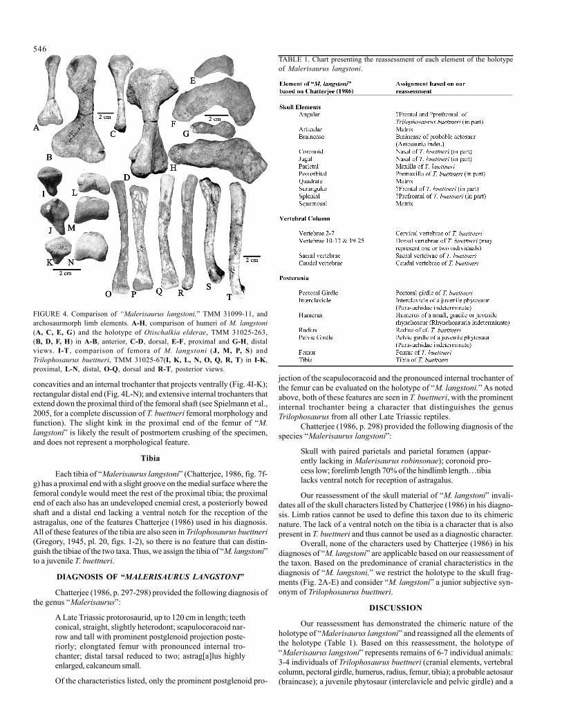
concavities and an internal trochanter that projects ventrally (Fig. 4I-K);rectangular distal end (Fig. 4L-N); and extensive internal trochanters thatextend down the proximal third of the femoral shaft (see Spielmann et al.,2005, for a complete discussion of T. buettneri femoral morphology andfunction). The slight kink in the proximal end of the femur of “M.langstoni” is likely the result of postmortem crushing of the specimen,and does not represent a morphological feature.
Tibia
Each tibia of “Malerisaurus langstoni” (Chatterjee, 1986, fig. 7f-g) has a proximal end with a slight groove on the medial surface where thefemoral condyle would meet the rest of the proximal tibia; the proximalend of each also has an undeveloped cnemial crest, a posteriorly bowedshaft and a distal end lacking a ventral notch for the reception of theastragalus, one of the features Chatterjee (1986) used in his diagnosis.All of these features of the tibia are also seen in Trilophosaurus buettneri(Gregory, 1945, pl. 20, figs. 1-2), so there is no feature that can distin-guish the tibiae of the two taxa. Thus, we assign the tibia of “M. langstoni”to a juvenile T. buettneri.
DIAGNOSIS OF “MALERISAURUS LANGSTONI”
Chatterjee (1986, p. 297-298) provided the following diagnosis ofthe genus “Malerisaurus”:
A Late Triassic protorosaurid, up to 120 cm in length; teethconical, straight, slightly heterodont; scapulocoracoid nar-row and tall with prominent postglenoid projection poste-riorly; elongtated femur with pronounced internal tro-chanter; distal tarsal reduced to two; astrag[a]lus highlyenlarged, calcaneum small.
Of the characteristics listed, only the prominent postglenoid pro-
jection of the scapulocoracoid and the pronounced internal trochanter ofthe femur can be evaluated on the holotype of “M. langstoni.” As notedabove, both of these features are seen in T. buettneri, with the prominentinternal trochanter being a character that distinguishes the genusTrilophosaurus from all other Late Triassic reptiles.
Chatterjee (1986, p. 298) provided the following diagnosis of thespecies “Malerisaurus langstoni”:
Skull with paired parietals and parietal foramen (appar-ently lacking in Malerisaurus robinsonae); coronoid pro-cess low; forelimb length 70% of the hindlimb length…tibialacks ventral notch for reception of astragalus.
Our reassessment of the skull material of “M. langstoni” invali-dates all of the skull characters listed by Chatterjee (1986) in his diagno-sis. Limb ratios cannot be used to define this taxon due to its chimericnature. The lack of a ventral notch on the tibia is a character that is alsopresent in T. buettneri and thus cannot be used as a diagnostic character.
Overall, none of the characters used by Chatterjee (1986) in hisdiagnoses of “M. langstoni” are applicable based on our reassessment ofthe taxon. Based on the predominance of cranial characteristics in thediagnosis of “M. langstoni,” we restrict the holotype to the skull frag-ments (Fig. 2A-E) and consider “M. langstoni” a junior subjective syn-onym of Trilophosaurus buettneri.
DISCUSSION
Our reassessment has demonstrated the chimeric nature of theholotype of “Malerisaurus langstoni” and reassigned all the elements ofthe holotype (Table 1). Based on this reassessment, the holotype of“Malerisaurus langstoni” represents remains of 6-7 individual animals:3-4 individuals of Trilophosaurus buettneri (cranial elements, vertebralcolumn, pectoral girdle, humerus, radius, femur, tibia); a probable aetosaur(braincase); a juvenile phytosaur (interclavicle and pelvic girdle) and a
FIGURE 4. Comparison of “Malerisaurus langstoni,” TMM 31099-11, andarchosaurmorph limb elements. A-H, comparison of humeri of M. langstoni(A, C, E, G) and the holotype of Otischalkia elderae, TMM 31025-263,(B, D, F, H) in A-B, anterior, C-D, dorsal, E-F, proximal and G-H, distalviews. I-T , comparison of femora of M. langstoni (J, M, P, S) andTrilophosaurus buettneri, TMM 31025-67(I, K, L, N, O, Q, R, T) in I-K,proximal, L-N, distal, O-Q, dorsal and R-T, posterior views.
TABLE 1. Chart presenting the reassessment of each element of the holotypeof Malerisaurus langstoni.

547
small, gracile and/or juvenile rhynchosaur (humerus). Thus, the holo-type represents elements of at least four disparate taxa of Late Triassicreptiles: Trilophosauridae, Parasuchidae, Rhynchosauridae andAetosauria. This is consistent with the known macrovertebrate faunafrom the Trilophosaurus quarries, which are dominated by specimens of
FIGURE 5. Paleogeographic map of the Late Triassic world withTrilophosaurus and Malerisaurus robinsonae localities highlighted.
T. buettneri with occasional remains of phytosaurs rhynchosaurs andaetosaurs (Gregory, 1945; Long and Murry, 1995; Heckert, 2004).
“Malerisaurus langstoni” thus joins a list of several taxa from theLate Triassic of West Texas that have, upon reexamination, been revealedas chimerae of elements of multiple taxa brought into association bytaphonomic agents. These taxa include Postosuchus (Chatterjee, 1985),Shuvosaurus (Chatterjee, 1993) and, most prominently, Protoavis(Chatterjee, 1991) (see Long and Murry, 1995; Witmer, 2001; and othersfor relevant discussions).
This reassessement of “M. langstoni” also raises questions aboutthe validity of Malerisaurus robinsonae from the Maleri Formation ofIndia (Chatterjee, 1980) and whether portions of it also pertain toTrilophosaurus. Based on photographs of the holotype of M. robinsonae,the femora of the specimen have a prominent internal trochanter thatextends down the proximal third of the femoral shaft. This feature isquite rare among Permo-Triassic tetrapods and is only possessed byTrilophosaurus and Araeoscelis. If the holotype of M. robinsonae doesindeed represent a partial skeleton of Trilophosaurus, then the paleogeo-graphic distribution of Trilophosaurus would change from a fairly en-demic distribution in the American Southwest (Fig. 5; Heckert et al.,2006) to a nearly Pangean distribution. Also, this would provide thepotential for a stronger correlation between the Maleri Formation ofIndia and the Chinle Group of the American Southwest (Fig. 5), espe-cially since it has been determined that isolated Trilophosaurus teeth canbe identified to the species level (Heckert et al., 2006).
ACKNOWLEDGMENTS
We thank Timothy Rowe and the staff of the Texas MemorialMuseum for access to their collection. Jerry Harris and Robert Sullivanprovided helpful reviews of the manuscript.
REFERENCES
Camp, C.L., 1930, A study of the phytosaurs with description of new mate-rial from western North America: Memoirs of the University of Califor-nia, v. 10, 174 pp.
Chatterjee, S., 1980, Malerisaurus, a new eosuchian reptile from the lateTriassic of India: Philosophical Transactions of the Royal Society ofLondon, Series B, v. 291, p. 163-200.
Chatterjee, S., 1985, Postosuchus, a new thecodontian reptile from theTriassic of Texas and the origin of tyrannosaurs: Philosophical Trans-actions of the Royal Society of London B, v. 309, p. 395-460.
Chatterjee, S., 1986, Malerisaurus langstoni, a new diapsid reptile from theTriassic of Texas: Journal of Vertebrate Paleontology, v. 6, p. 297-312.
Chatterjee, S., 1991, Cranial anatomy and relationships of a new Triassicbird from Texas: Philosophical Transactions of the Royal Society ofLondon B, v. 332, p. 277-342.
Chatterjee, S., 1993, Shuvosaurus, a new theropod: National GeographicResearch and Exploration, v. 9, p. 274-285.
Demar R. and Bolt, J.R., 1981, Dentitional organization and function in aTriassic reptile: Journal of Paleontology, v. 55, p. 967-984.
Desojo, J.B. and Heckert, A.B., 2004, New information on the braincaseand mandible of Coahomasuchus (Archosauria: Aetosauria) from theOtischalkian (Carnian) of Texas: Neues Jahrbuch für Geologie undPaläontologie Monatshefte, v. 2004, p. 605-616.
Dilkes, D.W., 1998, The Early Triassic rhynchosaur Mesosuchus browniand the interrelationships of basal archosauromorph reptiles: Philo-sophical Transactions of the Royal Society of London B, v. 353, p. 501-541.
Elder, R.L., 1978, Paleontology and paleoecology of the Dockum Group,Upper Triassic, Howard County, Texas [M.S. thesis]: Austin, Universityof Texas, 205 p.
Elder, R.L., 1987, Taphonomy and paleoecology of the Dockum Group,Howard County, Texas: Journal of the Arizon-Nevada Academy of Sci-ences, v. 22, p.85-94.
Gregory, J.T., 1945, Osteology and relationships of Trilophosaurus: Uni-versity of Texas, Publication 4401, p. 273-359.
Heckert, A.B., Lucas, S.G., Rinehart, L.R., Hunt, A.P. and Kahle, R., 2006,Revision of the archosauromorph reptile Trilophosaurus, with a de-scription of the first skull of Trilophosaurus jacobsi, from the UpperTriassic Chilne Group, West Texas, USA: Palaeontology, v. 49, p. 1-20.
Hunt, A.P. and Lucas, S.G., 1991, A new rhynchosaur from the UpperTriassic of West Texas, and the biochronology of Late Triassicrhynchosaurs: Palaeontology, v. 34, p. 927-938.
Hunt, A.P. and Lucas, S.G., 1993, Triassic vertebrate paleontology andbiochronology of New Mexico: New Mexico Museum of Natural His-tory and Science, Bulletin 2, p. 49-60.
Long, R.L. and Murry, P.A., 1995, Late Triassic (Carnian and Norian)tetrapods from the southwest United States: New Mexico Museum ofNatural History, Bulletin 4, 254 pp.
Lucas, S.G., Hunt, A.P. and Kahle, R., 1993, Late Triassic vertebrates fromthe Dockum Formation near Otis Chalk, Howard County, Texas: NewMexico Geological Society, Guidebook 44, p. 237-244.
Lucas, S.G. and Anderson, O.J., 1993, Triassic stratigraphy in southeasternNew Mexico and southwestern Texas: New Mexico Geological Society,Guidebook 44, p. 231-235.
Parks, P., 1969, Cranial anatomy and mastication of the Triassic reptileTrilophosaurus [M.S. thesis]: University of Texas, 100 p.
Spielmann, J.A., Heckert, A.B. and Lucas S.G., 2005, The Late Triassicarchosauromoph Trilophosaurus as an arboreal climber: Rivista Italianadi Paleontologia e Stratigrafia, v. 111, p. 395-412.
Witmer, L.M., 2001, The role of Protoavis in the debate on avian origins;in Gauthier, J. and Gall, J.F. eds., New Perspectives on the origin andearly evolution of Birds, Proceedings of the International Symposium inHonor of John H. Ostrom, New Haven, Yale Peabody Museum, p. 537-548.