harnessing neuroplasticity with brain stimulation and
TRANSCRIPT
Neuromodulation:
Harnessing Neuroplasticity with Brain
Stimulation and Rehabilitation
Cecília N. Prudente, PT, MS, PhD1
Bernadette T. Gillick, PT, MS, PhD1
Colum MacKinnon, PhD2
Teresa J.Kimberley, PT, PhD1
1Dept. of Rehabilitation Medicine 2Dept. of Neurology
Presenters:

Conflicts of interest
TJK: consulting income from MicroTransponder
Others: Nothing to declare
Learning objectives
1. Be familiar with forms of brain stimulation
2. Be able to identify safety and feasibility of each
technique
3. Understand the purposes of using the
parameters of brain stimulation
4. Translate brain stimulation research into clinical
implications

Harnessing neuroplasticity to improve
motor function
1. Neuromodulation tools
2. Down-regulation
3. Up-regulation
4. Hijacking neural firing patterns
5. Where are we now, where are we going, and
how do we get there?
6. Discussion
Harnessing neuroplasticity to improve
motor function
1. Neuromodulation tools
2. Down-regulation
3. Up-regulation
4. Hijacking neural firing patterns
5. Where are we now, where are we going, and
how do we get there?
6. Discussion

What is neuromodulation?
http://blog.cambridgeconsultants.com/medical-technology/wp-
content/uploads/2014/05/Neuromodulation.jpg
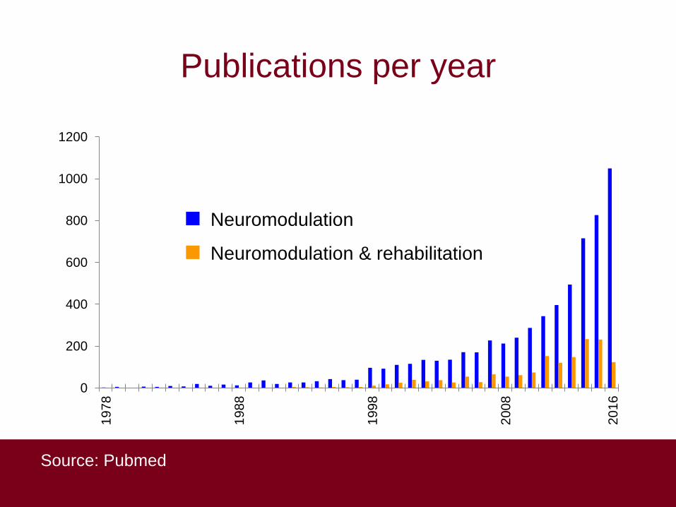
0
200
400
600
800
1000
1200
19
78
19
88
19
98
20
08
Publications per year
Source: Pubmed
Neuromodulation
Neuromodulation & rehabilitation
20
16

How to neuromodulate?
Healthy state
Injury
Neuromodulation Medications
Rehabilitation
Neuromodulation tools
Neuroplasticity
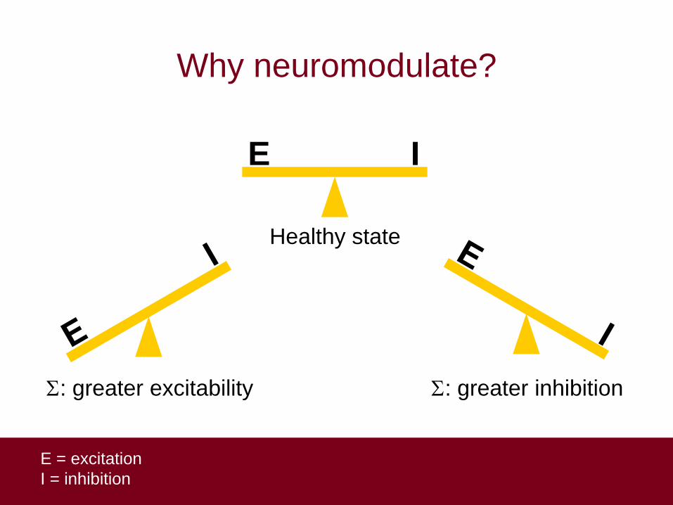
Why neuromodulate?
E I
Healthy state
E = excitation
I = inhibition
: greater excitability : greater inhibition
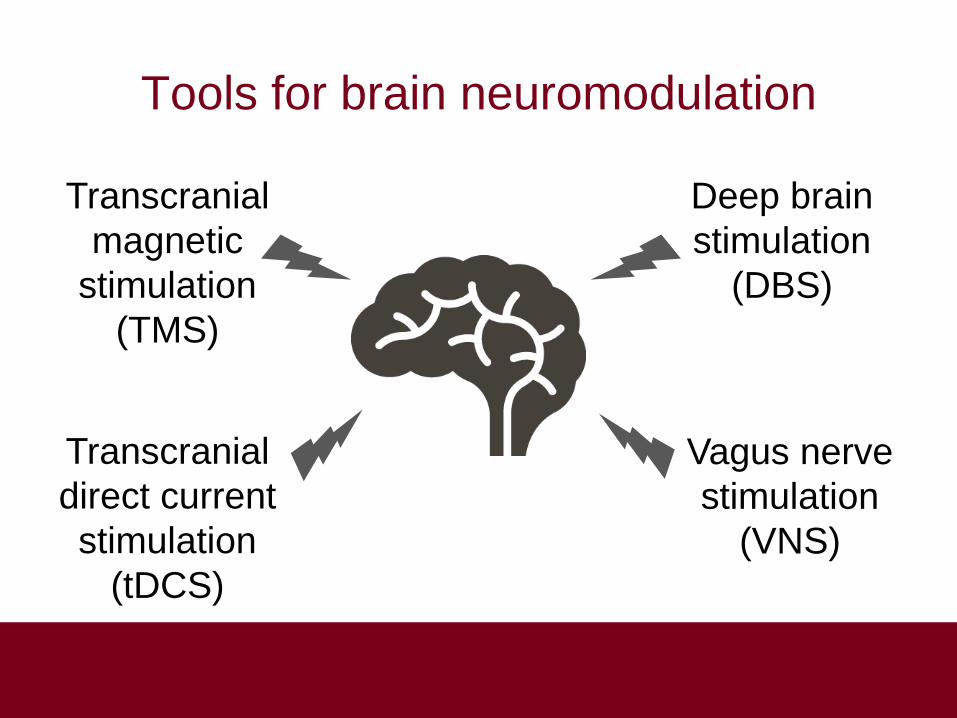
Tools for brain neuromodulation
Transcranial
magnetic
stimulation
(TMS)
Vagus nerve
stimulation
(VNS)
Deep brain
stimulation
(DBS)
Transcranial
direct current
stimulation
(tDCS)

Transcranial magnetic stimulation
(TMS)
• Pulsating magnetic fields on the scalp to induce
an electrical current within the brain
Wagner et al., 2007
George & Aston-Jones, 2010
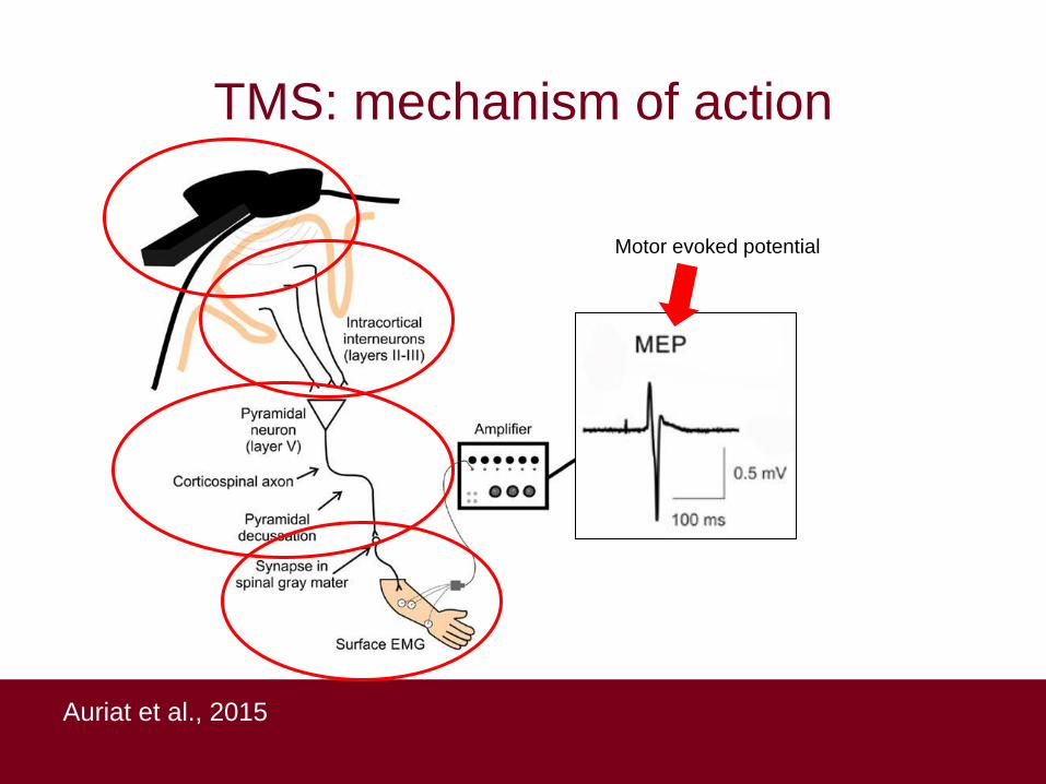
TMS: mechanism of action
Auriat et al., 2015
Motor evoked potential
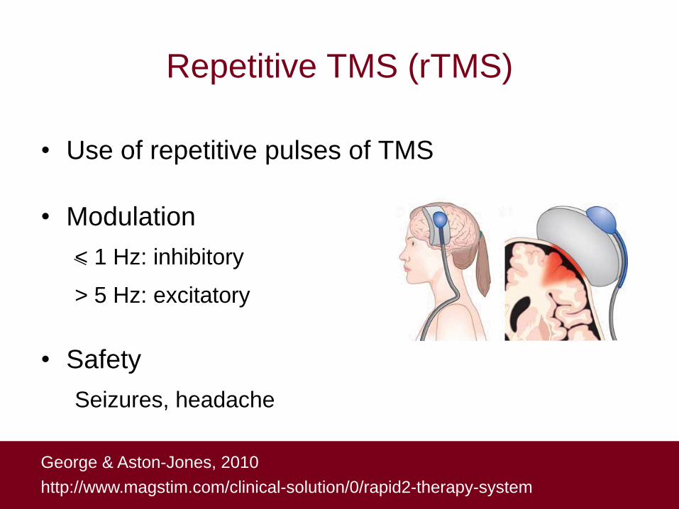
Repetitive TMS (rTMS)
• Use of repetitive pulses of TMS
• Modulation
< 1 Hz: inhibitory
> 5 Hz: excitatory
• Safety
Seizures, headache
George & Aston-Jones, 2010
http://www.magstim.com/clinical-solution/0/rapid2-therapy-system
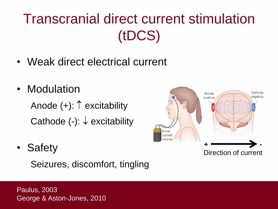
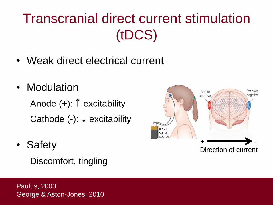
• Weak direct electrical current
• Modulation
Anode (+): excitability
Cathode (-): excitability
• Safety
Seizures, discomfort, tingling
Transcranial direct current stimulation
(tDCS)
Paulus, 2003
George & Aston-Jones, 2010
+ -
Direction of current
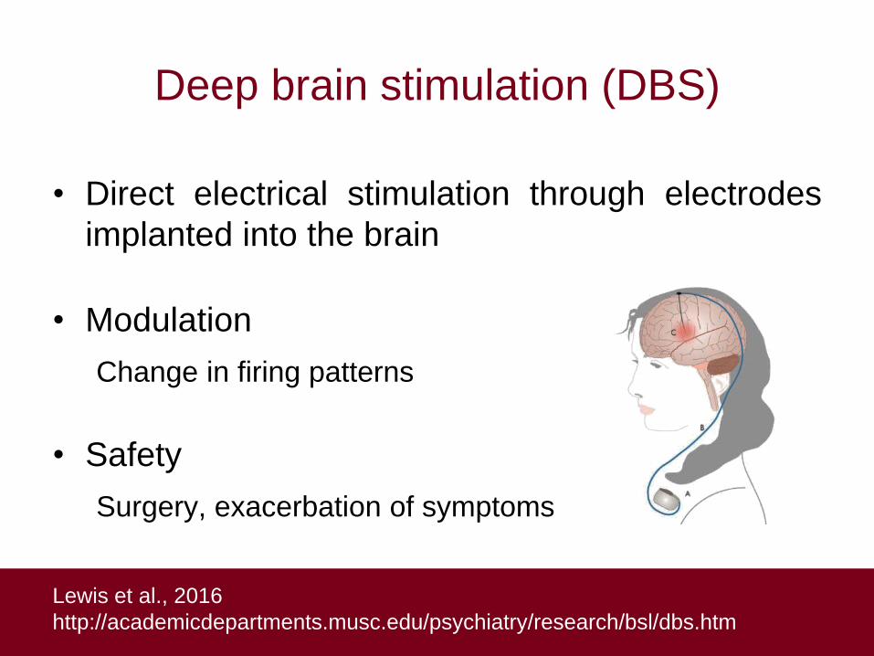
Deep brain stimulation (DBS)
• Direct electrical stimulation through electrodes
implanted into the brain
• Modulation
Change in firing patterns
• Safety
Surgery, exacerbation of symptoms
Lewis et al., 2016
http://academicdepartments.musc.edu/psychiatry/research/bsl/dbs.htm

Vagus nerve stimulation (VNS)
• Electrical stimulation of vagus nerve through
implantable pulse generator
• Modulation
Norepinephrine and acetylcholine
• Safety
Surgery, cough, hoarseness
George & Aston-Jones, 2010
http://academicdepartments.musc.edu/psychiatry/research/bsl/vns.htm
Harnessing neuroplasticity to improve
motor function
1. Neuromodulation tools
2. Down-regulation
3. Up-regulation
4. Hijacking neural firing patterns
5. Where are we now, where are we going, and
how do we get there?
6. Discussion

Why down-regulate?
E = excitation
I = inhibition
: greater excitability

Dystonia and impaired inhibition
• Excessive and involuntary contractions
Quartarone & Hallett, 2013
Writer’s cramp

Dystonia and impaired inhibition
Quartarone & Hallett, 2013
Prudente et al., 2016
Focal hand dystonia Cervical dystonia
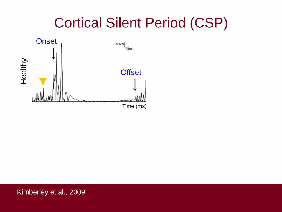
Cortical Silent Period (CSP)
Kimberley et al., 2009
Onset
Offset
Healthy
Time (ms)

Cortical Silent Period (CSP)
Kimberley et al., 2009
Onset
Onset
Offset
Offset
Shorter CSP
=
decreased inhibition Dysto
nia
H
ealthy
Time (ms)
Time (ms)

rTMS in focal hand dystonia
Murase et al., 2005
• Groups:
Experimental: writer’s cramp (n=9)
Control: healthy adults (n=7)
• rTMS: 0.2 Hz, 1 session
• Targets: primary motor cortex, premotor cortex,
supplementary motor area; real vs. sham
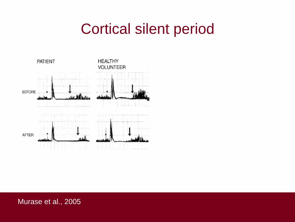
Cortical silent period
Murase et al., 2005

Cortical silent period
Murase et al., 2005
B: before rTMS; A: after rTMS
PMC: premotor cortex; MC: motor cortex; SMA: supplementary motor area

rTMS combined with rehabilitation
Kimberley et al., 2015
• Groups (n=8):
Randomized single subject design with crossover
Experimental: rTMS + sensorimotor training
Control: rTMS + control therapy
• rTMS: 1 Hz, 5 sessions
• Target: premotor cortex

Kimberley et al., 2015
Byl et al., 2002
Sensorimotor training
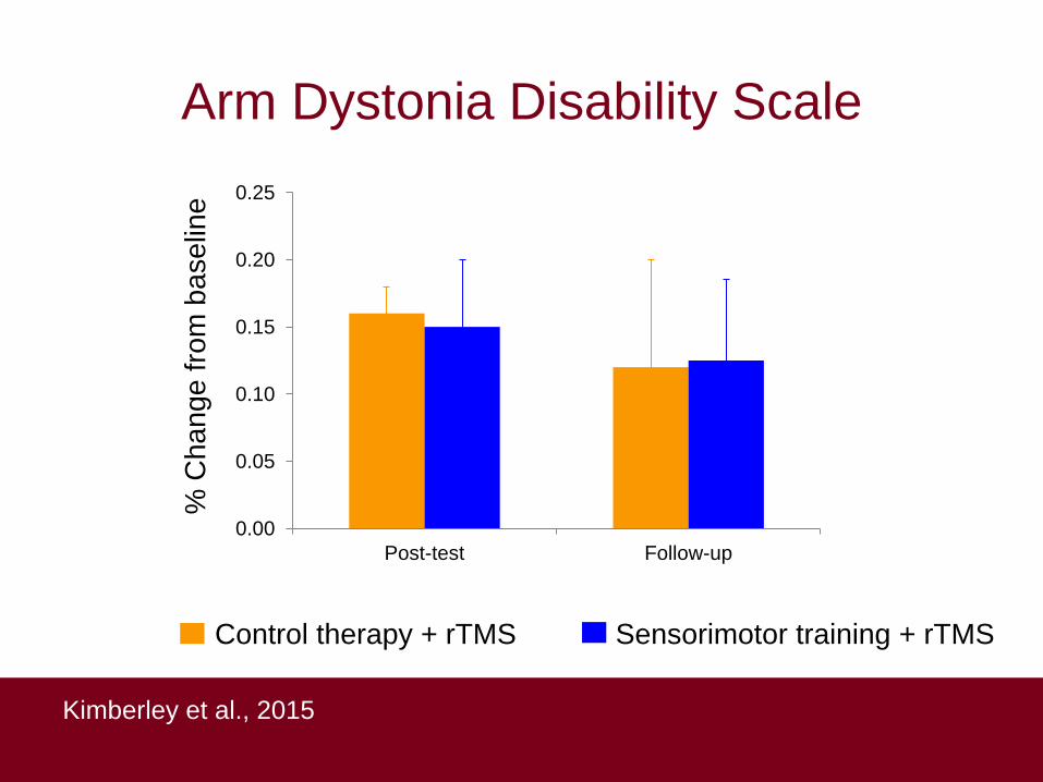
Arm Dystonia Disability Scale
Kimberley et al., 2015
% C
hang
e f
rom
baselin
e
0.00
0.05
0.10
0.15
0.20
0.25
Post-test Follow-up
Control therapy + rTMS Sensorimotor training + rTMS
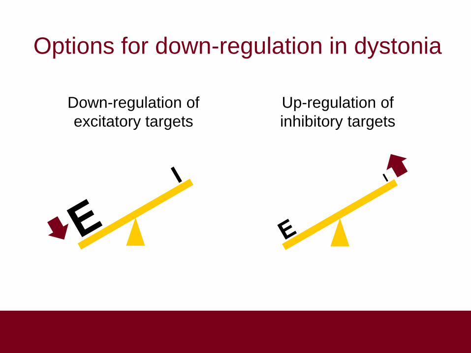
Options for down-regulation in dystonia
Down-regulation of
excitatory targets
Up-regulation of
inhibitory targets
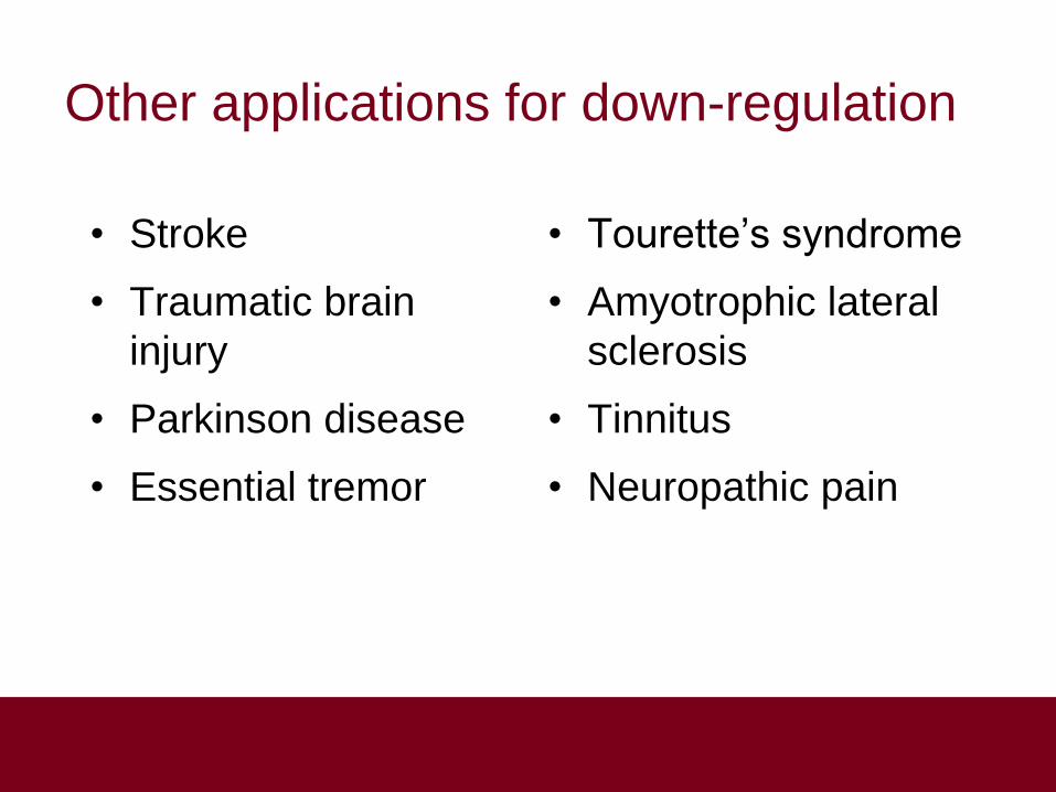
Other applications for down-regulation
• Stroke
• Traumatic brain
injury
• Parkinson disease
• Essential tremor
• Tourette’s syndrome
• Amyotrophic lateral
sclerosis
• Tinnitus
• Neuropathic pain

Harnessing neuroplasticity to improve
motor function
1. Neuromodulation tools
2. Down-regulation
3. Up-regulation
4. Hijacking neural firing patterns
5. Where are we now, where are we going, and
how do we get there?
6. Discussion

Why up-regulate?
E = excitation
I = inhibition
= greater
inhibition

Stroke
Doubly-Disabled
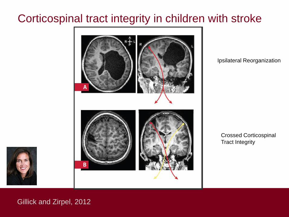
Corticospinal tract integrity in children with stroke
Gillick and Zirpel, 2012
Ipsilateral Reorganization
Crossed Corticospinal
Tract Integrity
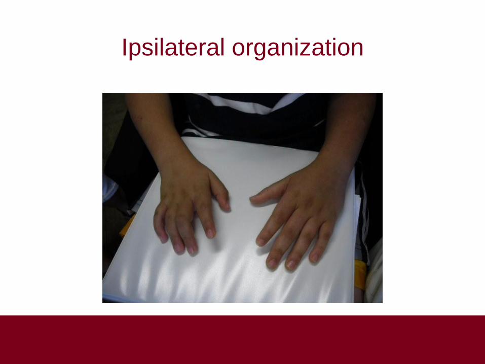
Ipsilateral organization

Tricky Triad in Tots
• “Doubly-Disabled”
• “Developmental Disuse”

Modulation options for goal of up
regulating lesioned hemisphere

Repetitive TMS (rTMS)
• Use of repetitive pulses of TMS
• Modulation
< 1 Hz: inhibitory
> 5 Hz: excitatory
• Safety
Seizures, headache
George & Aston-Jones, 2010
http://www.magstim.com/clinical-solution/0/rapid2-therapy-system
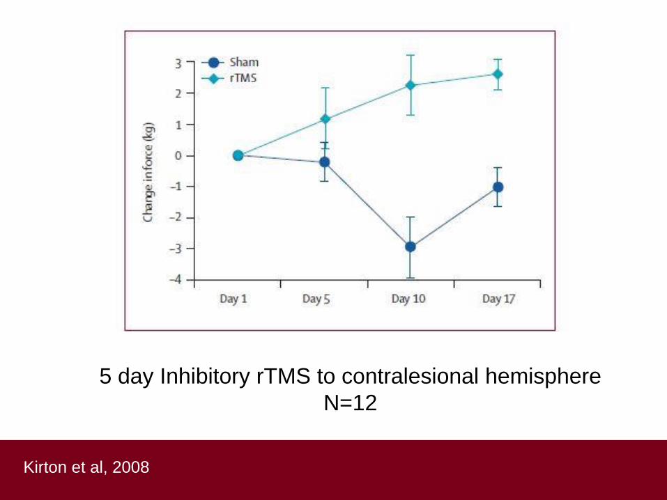
Kirton et al, 2008
5 day Inhibitory rTMS to contralesional hemisphere
N=12

Kirton et al, 2008

rTMS and Constraint-Induced Movement
Therapy (CIMT) in Pediatric Hemiparesis
Gillick et al, 2013
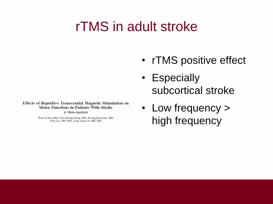
rTMS in adult stroke
• rTMS positive effect
• Especially
subcortical stroke
• Low frequency >
high frequency
• Weak direct electrical current
• Modulation
Anode (+): excitability
Cathode (-): excitability
• Safety
Discomfort, tingling
Transcranial direct current stimulation
(tDCS)
Paulus, 2003
George & Aston-Jones, 2010
+ -
Direction of current

Gillick et al , 2015
Single Session Bihemispheric tDCS:
Safe and feasible
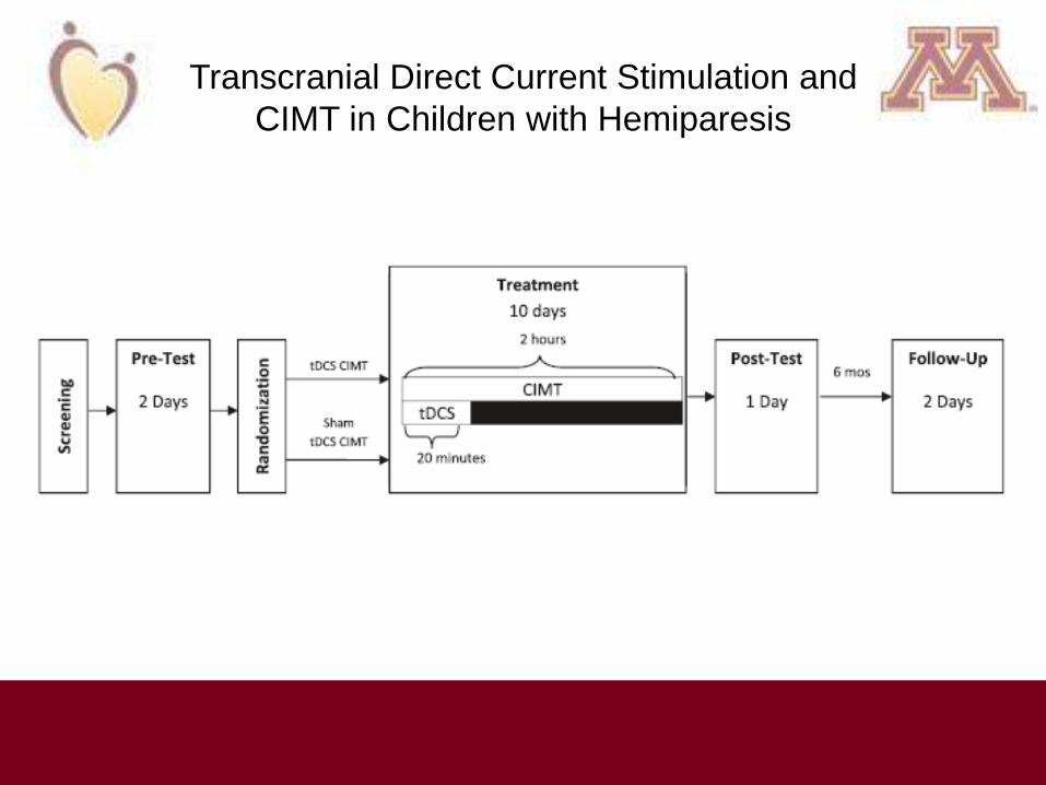
Transcranial Direct Current Stimulation and
CIMT in Children with Hemiparesis
Gillick et al, 2015

Harnessing neuroplasticity to improve
motor function
1. Neuromodulation tools
2. Down-regulation
3. Up-regulation
4. Hijacking neural firing patterns
5. Where are we now, where are we going, and
how do we get there?
6. Discussion

Neuromodulation: Harnessing Neuroplasticity with Brain
Stimulation and Rehabilitation
Invasive Neuromodulation: Hijacking neural firing patterns and harnessing
neuroplasticity to improve motor function
Colum D. MacKinnon PhD Department of Neurology
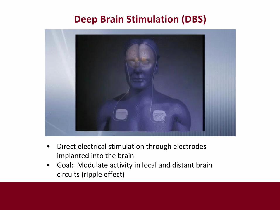
Deep Brain Stimulation (DBS)
• Direct electrical stimulation through electrodes implanted into the brain
• Goal: Modulate activity in local and distant brain circuits (ripple effect)
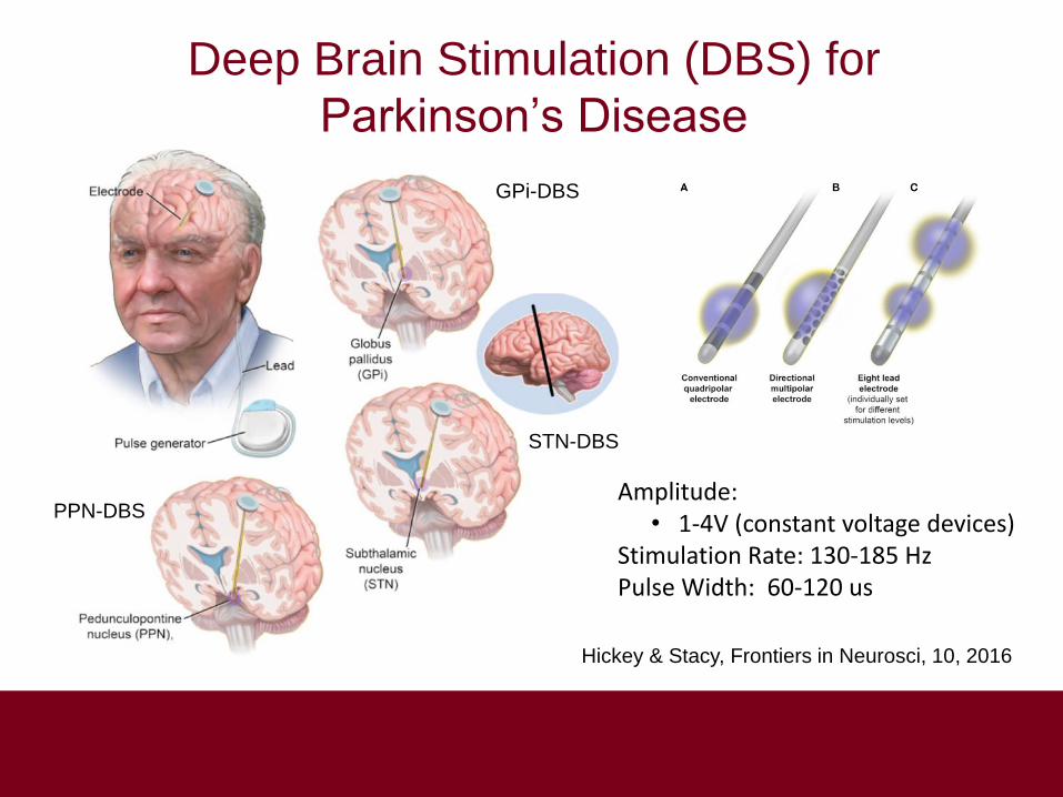
Deep Brain Stimulation (DBS) for
Parkinson’s Disease
Hickey & Stacy, Frontiers in Neurosci, 10, 2016
GPi-DBS
STN-DBS
PPN-DBS Amplitude:
• 1-4V (constant voltage devices) Stimulation Rate: 130-185 Hz Pulse Width: 60-120 us
Lozano and Lipsman, 77, Neuron, 2013
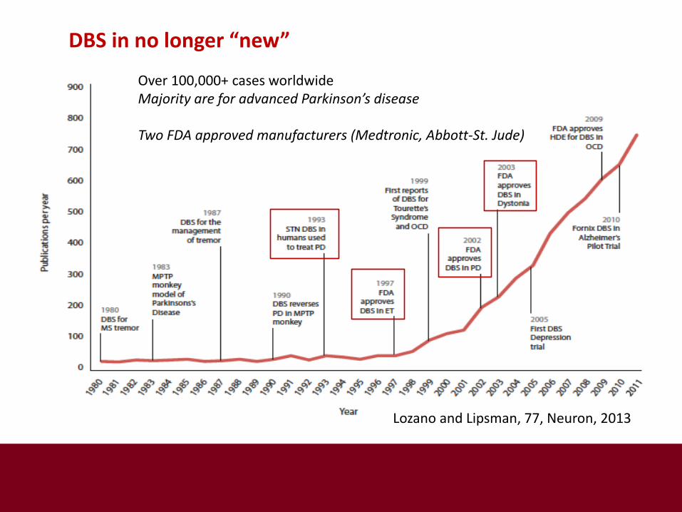
DBS in no longer “new”
Over 100,000+ cases worldwide Majority are for advanced Parkinson’s disease Two FDA approved manufacturers (Medtronic, Abbott-St. Jude)

Clinical Indications: •Parkinson’s disease •Essential tremor •Dystonia •Obsessive-Compulsive Disorder Future Indications •Tourette’s •Depression •Pain •Obesity •Addiction •Alzheimer’s disease •Stroke? (deep cerebellar stimulation)
Deep Brain Stimulation (DBS)

Why use DBS?
• Effect size and efficacy is large
• Consistent increase in quality of life
• Broad network effects (not symptom- or segment-specific)
but…
• Higher level of risk
• Higher cost
• High variability across individuals
• Some motor and non-motor symptoms can worsen
• Some symptoms do not respond to DBS
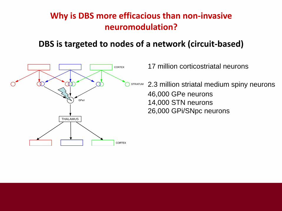
Why is DBS more efficacious than non-invasive neuromodulation?
DBS is targeted to nodes of a network (circuit-based)
THALAMUS
17 million corticostriatal neurons
2.3 million striatal medium spiny neurons
46,000 GPe neurons
14,000 STN neurons
26,000 GPi/SNpc neurons

7T MRI, SWI N. Harel, CMRR, U Minnesota
Non-invasive approaches to treating Parkinson’s disease
J
• rTMS sensorimotor cortex
• anodal tDCS sensorimotor cortex
• rTMS supplementary motor area
• anodal tDCS of the
supplementary motor area

DBS acts on nodes of the basal ganglia-thalamocortical network

DBS is targeted to nodes of a network (circuit-based)
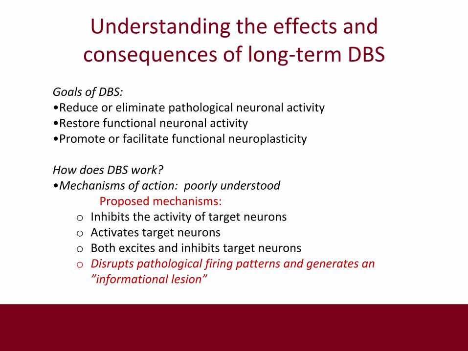
Understanding the effects and consequences of long-term DBS
Goals of DBS: •Reduce or eliminate pathological neuronal activity •Restore functional neuronal activity •Promote or facilitate functional neuroplasticity How does DBS work? •Mechanisms of action: poorly understood Proposed mechanisms:
o Inhibits the activity of target neurons o Activates target neurons o Both excites and inhibits target neurons o Disrupts pathological firing patterns and generates an
”informational lesion”
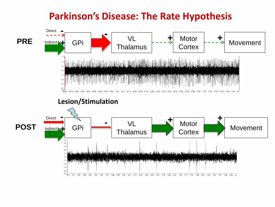
GPi VL
Thalamus
Motor
Cortex
+ - Movement
+ PRE -
Direct
Indirect +
-
+ + POST VL
Thalamus
Motor
Cortex
- Movement GPi
Direct
Indirect +
-
Lesion/Stimulation
Parkinson’s Disease: The Rate Hypothesis

Abnormal patterns at rest: • Increased bursting • Rhythmic activity (particularly low frequency in the theta,
alpha and beta bands) • Correlated firing both within and between nuclei
Wichmann and Delong, 2003, Ann NY Acad, Sci, 991: 199-213.
THE PATTERN HYPOTHESIS OF PARKINSON’S DISEASE
Mean = 81 Hz Mean = 78 Hz
GPe GPi
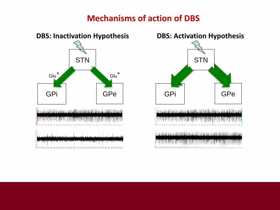
Mechanisms of action of DBS
STN
DBS: Inactivation Hypothesis DBS: Activation Hypothesis
GPi GPe
STN
Glu Glu + +

GPe GPi
DBS hijacks the abnormal firing pattern and effectively produces an informational lesion
STN DBS ON
Hashimoto, J Neurosci, 2008
STN
DBS: Inactivation Hypothesis DBS: Activation Hypothesis
GPi GPe
STN
STN DBS ON

Wichmann and Delong, 2003, Ann NY Acad, Sci, 991: 199-213.
DBS hijacks the abnormal firing pattern and effectively produces an informational lesion
DBS- induced neuronal activity
at 130 Hz

• Increased velocity (decreased bradykinesia)
• Increased movement amplitude (decreased hypokinesia)
• Improved muscle activation (increased force output)
• Marked suppression of tremor
• Marked suppression of rigidity
• Marked reduction of levodopa-induced dyskinesias
• **Improved quality of life
Beneficial motor effects of the informational
lesion caused by STN-DBS or GPi-DBS

Effects of STN-DBS on muscle activation
Vaillancourt et al., Brain, 127, 2004

• Postural stability • Anticipatory postural adjustments • Temporal and balance components of gait • Speech (particularly with bilateral stimulation) • Eye movements (saccades)
Also…. • Cognition (exacerbation of dual-task deficits)
Motor features that can be worsened by the
informational lesion induced by STN-DBS or GPi-DBS

Rocchi et al., J Neurosurg, 2014
Some motor symptoms are worsened by
STN-DBS or GPi-DBS: an opportunity for PT intervention

• High-rate repetitive or sequential movements • Freezing of gait (initially effective in individuals with a good response to levodopa preoperatively)
Motor features that are resistant to the effects
of STN-DBS or GPi-DBS
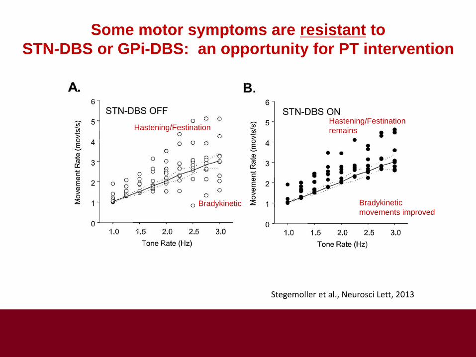
Stegemoller et al., Neurosci Lett, 2013
Hastening/Festination
Bradykinetic
Hastening/Festination
remains
Bradykinetic
movements improved
Some motor symptoms are resistant to
STN-DBS or GPi-DBS: an opportunity for PT intervention
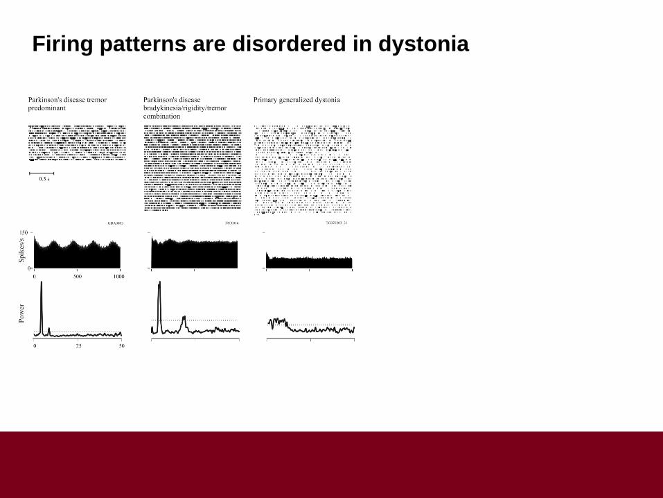
Firing patterns are disordered in dystonia

Ruge et al., Mov Disord, 26, 2011
DBS-evoked changes in motor function in dystonia can
take weeks to months to reach maximal efficacy
Clin
ica
l R
ati
ng
of
Se
ve
rity
(B
MF
)
Critical window during neuroplastic changes for therapeutic intervention to improve function
OPPORTUNITY
Time Course of Clinical Improvement with GPi-DBS in Primary Dystonia
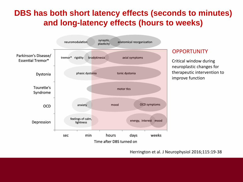
Herrington et al. J Neurophysiol 2016;115:19-38
DBS has both short latency effects (seconds to minutes)
and long-latency effects (hours to weeks)
Critical window during neuroplastic changes for therapeutic intervention to improve function
OPPORTUNITY
Todd M. Herrington et al. J Neurophysiol 2016;115:19-38
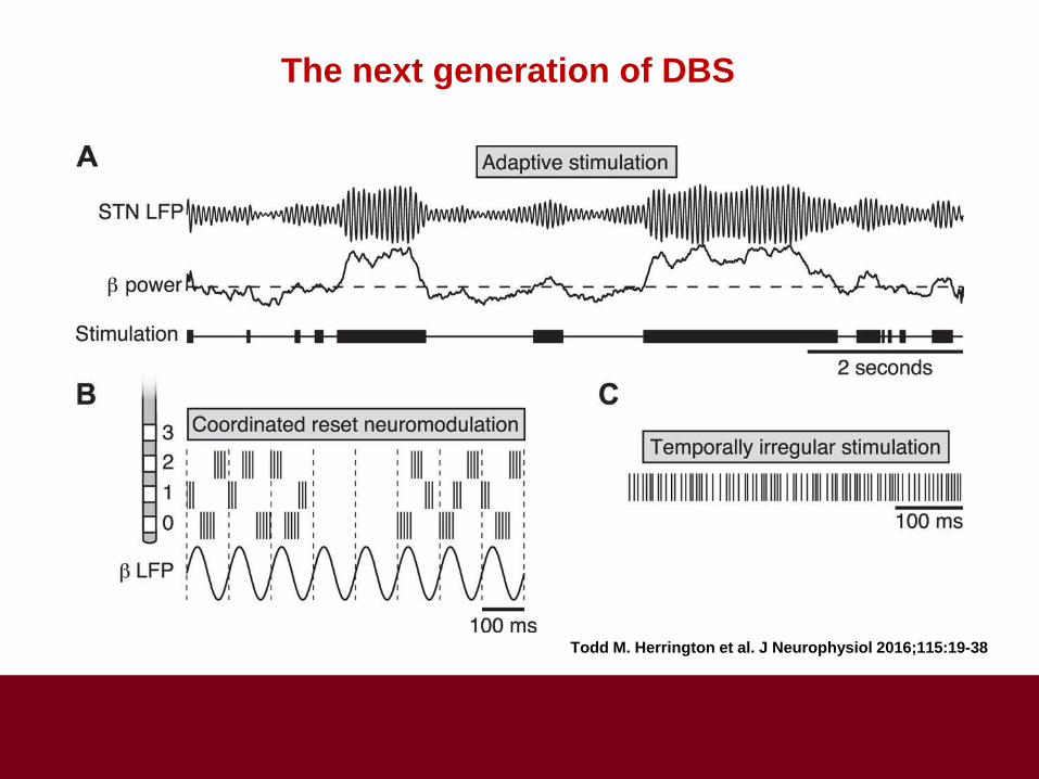
The next generation of DBS

The Next Generation of DBS
Harness Neuroplasticity
(e.g. Coordinated Reset or CR-DBS)
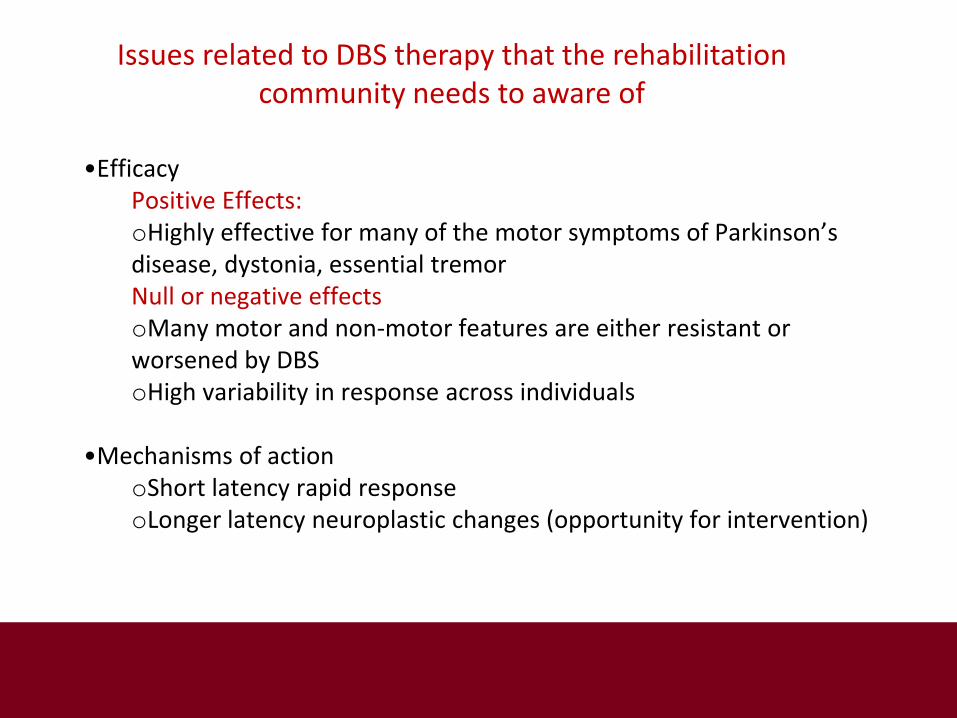
•Efficacy Positive Effects: oHighly effective for many of the motor symptoms of Parkinson’s disease, dystonia, essential tremor Null or negative effects oMany motor and non-motor features are either resistant or worsened by DBS oHigh variability in response across individuals
•Mechanisms of action
oShort latency rapid response oLonger latency neuroplastic changes (opportunity for intervention)
Issues related to DBS therapy that the rehabilitation community needs to aware of
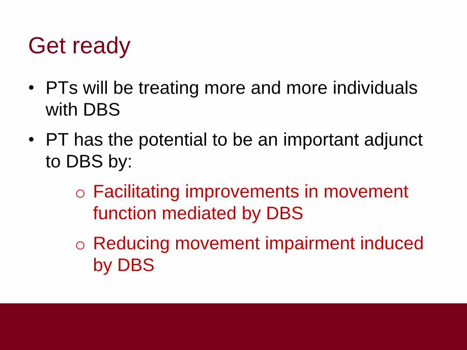
Get ready
• PTs will be treating more and more individuals
with DBS
• PT has the potential to be an important adjunct
to DBS by:
o Facilitating improvements in movement
function mediated by DBS
o Reducing movement impairment induced
by DBS

Vagus nerve stimulation for poststroke
upper extremity hemiparesis
Prime rehabilitation effects
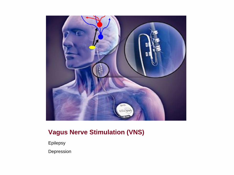
Vagus Nerve Stimulation (VNS)
Epilepsy
Depression

Locus
Coeruleus
Nucleus
Basalis Vagus
Nerve
Acetylcholine + Norepinephrine
Left Vagal Nerve Stimulation
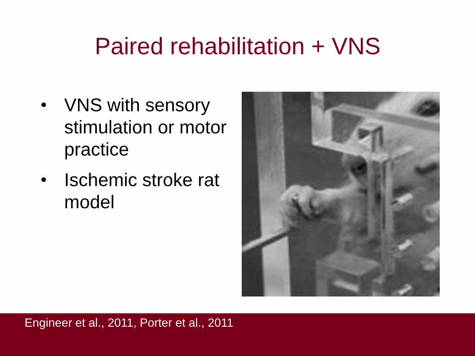
Paired rehabilitation + VNS
• VNS with sensory
stimulation or motor
practice
• Ischemic stroke rat
model
Engineer et al., 2011, Porter et al., 2011

Ischemic stroke rat model
Khodaparast et al, 2013
Paired VNS improves recovery of hit rate performance and force on
compared with Rehab alone and unpaired VNS
Paired
Un-Paired

Upper extremity therapy
• Graded, progressive
task practice
• ~300 repetitions
• Average 72 minutes
• 18 sessions
(3x/week for 6
weeks)
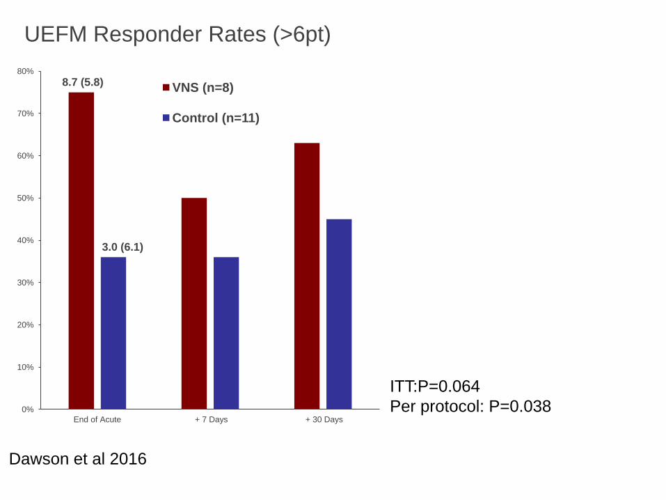
UEFM Responder Rates (>6pt)
8.7 (5.8)
3.0 (6.1)
0%
10%
20%
30%
40%
50%
60%
70%
80%
End of Acute + 7 Days + 30 Days
VNS (n=8)
Control (n=11)
Dawson et al 2016
ITT:P=0.064
Per protocol: P=0.038

Blinded, Randomized Preliminary Clinical Trial
• VNS during rehabilitation for improved upper
limb motor function after stroke
• Purpose: establish safety and effect size for
definitive FDA trial
• 17 people (4 sites)
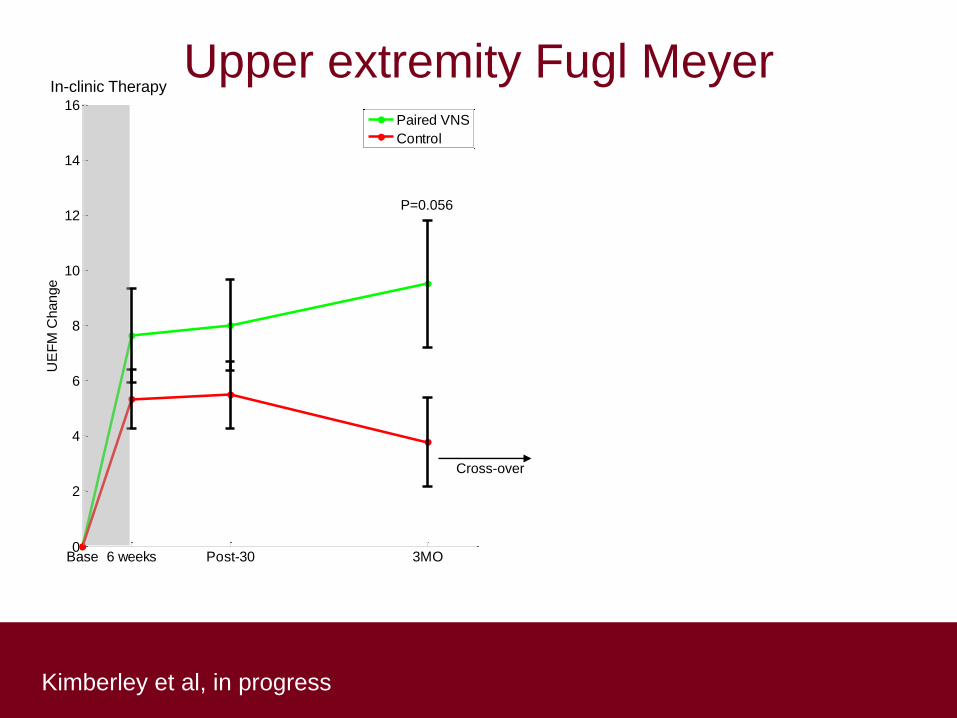
Kimberley et al, in progress
Base 6 weeks Post-30 3MO0
2
4
6
8
10
12
14
16
UE
FM
Change
Paired VNS
Control
P=0.056
In-clinic Therapy
Cross-over
Upper extremity Fugl Meyer

Kimberley et al, in progress
Upper extremity Fugl Meyer
Rebaseline 6 weeks0
2
4
6
8
10
12
14
16
UE
FM
Change
Base 6 weeks Post-30 3MO0
2
4
6
8
10
12
14
16
UE
FM
Change
Paired VNS
Control
P=0.056
In-clinic Therapy In-clinic Therapy
n=4
Cross-over Paired VNS
Kimberley et al, in progress
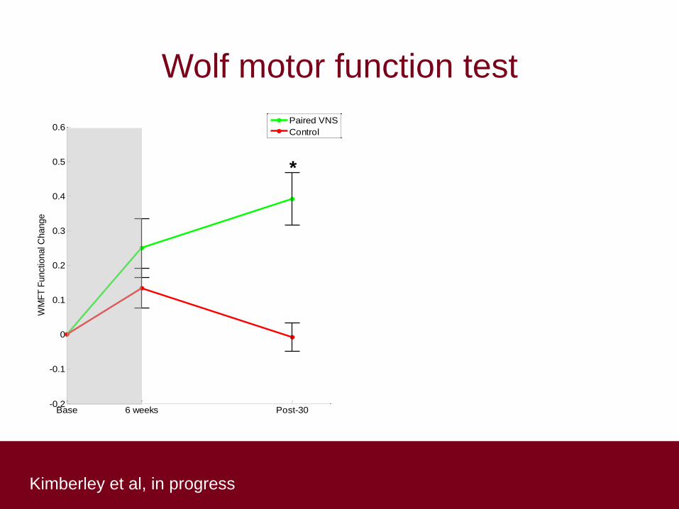
Wolf motor function test
*
Base 6 weeks Post-30-0.2
-0.1
0
0.1
0.2
0.3
0.4
0.5
0.6
WM
FT
Functional C
hange
Paired VNS
Control
*
Kimberley et al, in progress
Wolf motor function test
*
Base 6 weeks Post-30-0.2
-0.1
0
0.1
0.2
0.3
0.4
0.5
0.6
WM
FT
Functional C
hange
Paired VNS
Control
*
Base 6 weeks Post-30
-0.5
-0.4
-0.3
-0.2
-0.1
0
WM
FT
Tim
e C
hange (
log tra
nsfo
rm)
Paired VNS
Control
p=0.13
Harnessing neuroplasticity to improve
motor function
1. Neuromodulation tools
2. Down-regulation
3. Up-regulation
4. Hijacking neural firing patterns
5. Where are we now, where are we going, and
how do we get there?
6. Discussion

Where are we now?

FDA approved indications
George & Aston-Jones, 2010
Device Disease FDA status
TMS Treatment-resistant depression General approval
tDCS No indication
DBS Parkinson disease General approval
Dystonia Humanitarian device
Exemption approval
Obsessive-compulsive disorder Humanitarian device
Exemption approval
VNS Epilepsy General approval
Treatment-resistant depression General approval

Where are we going?

Another tool: get ready
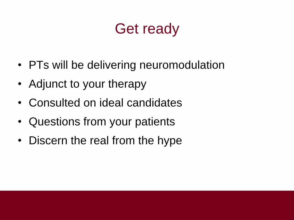
Get ready
• PTs will be delivering neuromodulation
• Adjunct to your therapy
• Consulted on ideal candidates
• Questions from your patients
• Discern the real from the hype

Caution

How do we get there?
Need to understand these tools better
Why non-invasive brain stimulation?
(tDCS, rTMS)
• Evidence that patients have more capacity
• Clinical use
• Ease of use
• Low cost
• Targeted brain area
• Low risk

Why invasive brain stimulation?
(DBS, VNS)
• Broad network effects
• Effect size and efficacy
• Higher level of risk
• Higher cost

Does it work?
• Case series
• Small n studies
• Large scale RCT have not yet been done
• Can they? Should they?
Different models
• Allow failure
• Pragmatic design and report
– Allow clinicians to evaluate how and with whom
• Acute testing
• Models of patient selection
– E.g. PREP algorithm (Stinear et al, 2010, 2012)
– TMS + neuroimaging +genetics + clinical assessment

In clinic private pay
• Why?
– Some efficacy
– If we wait, it may
never happen
– Data
• Off label rTMS
• Post ischemic stroke
• 10 session ($2115)
• (depression: $8000-
$14,000)
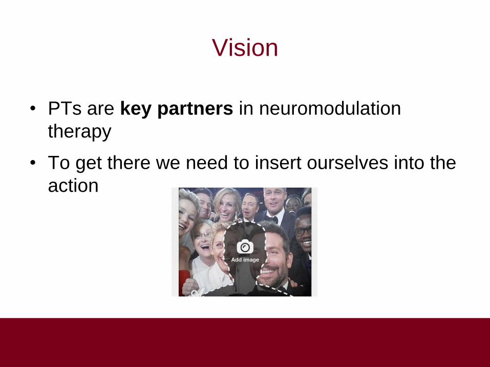
Vision
• PTs are key partners in neuromodulation
therapy
• To get there we need to insert ourselves into the
action
Vision: we need neuromod and
neuromod needs us
• Understand the brain target and effect on
circuitry
• Model of patient selection
• Dose and duration of effect
• Ideal timing/type of rehabilitation
• Multicenter studies: info from all sources

Work together

Work together

Acknowledgements
Brain Plasticity
Laboratory