handbook of hospital medicine || cardiology
TRANSCRIPT
1
CARDIOLOGY
Management of Cardiac Arrest
Usually occurs in the context of acute myocardial infarction, but also with hypoxia, anaesthesia, surgery, shock, electrocution, drowning, hypothermia, biochemical abnormalities, drugs (digoxin, adrenaline), anaphylaxis, etc.
1. Establish the diagnosis - absent pulses, apnoea, unconsciousness.
2. Note the time.
3. A blow to the praecordium is occasionally successful in restoring normal rhythm.
4. Give an immediate DC shock with 400 J if a defibrillator is available. Do not delay defibrillation for lack of an ECG monitor as ventricular fibrillation is the commonest arrhythmia. Newer defibrillators may record the ECG via their paddles.
5. Begin external cardiac massage at about 60 compressions/min. Check efficiency at the femoral pulses. The patient must be on a hard surface, the floor if necessary.
6. Artificial respiration. First remove dentures and clear the airway. Use mouth to mouth ventilation if there is no equipment to hand, but use Brook airway/Ambu bag/endotracheal tube etc. if available. Ventilate with 100% oxygen. There is no reason for cardiac massage and ventilation to alternate; indeed, they are more efficient if simultaneous.
7. Establish an intravenous line. Give 8.4% bicarbonate, l ml/kg body weight; repeat every 10 min.
1
P. J. Mitchell et al., Handbook of Hospital Medicine© MTP Press Limited 1983
2 Handbook of Hospital Medicine
8. ECG monitor lead. Check the rhythm. (a) If VT or VF - 400 J DC shock. Repeat if necessary. If
unsuccessful, give lignocaine 100 mg i. v. and repeat DC shock. If still unsuccessful with VF, give adrenaline 10 ml of 1 in 10,000 or 1 ml of 1 in 1000 i.v. and calcium chloride or gluconate 10 ml of 10% solution i.v. Repeat DC shock after further cardiac massage.
(b) If asystole - adrenaline 1 ml of 1 in 1000 solution i.v. - calcium chloride or gluconate 10 ml of 10% solution i. v. - continue cardiac massage and ventilation. - repeat adrenaline and calcium after 5-10 min if required.
(c) If fairly "normal" rhythm and no cardiac output, then a mechanical catastrophe is likely, e.g. ruptured ventricle, massive pulmonary embolism, tamponade. Try adrenaline, calcium and further massage.
9. Once stable (a) If the cause was VT or VF give lignocaine 100 mg i. v. over 2
min and then put up a lignocaine infusion at 4 mg/min for 30 min, then 3 mg/min for 30 min, then 2 mg/min.
(b) Check arterial gases and serum potassium. ( c) If the rhythm and output are restored but the patient does
not breathe and the cause of the arrest is not clear, give naloxone 400-800 ~g i.v. Repeat after 3-4 min if required.
(d) If massive pulmonary embolism seems likely give heparin 15,000 units i.v.
(e) Continue oxygen by mask. (f) Arrange a chest X-ray to look for pneumothorax, fractured
ribs etc.
NOTE
1. In asystole or profound bradycardia, consider emergency pacing.
2. If tamponade is likely, immediate thoracotomy should be performed. Attempts at needle aspiration are pointless.
3. If resuscitation is successful but delayed and acute brain injury seems likely, consider (a) artificial ventilation (b) i.v. dexamethasone 12 mg stat then 4 mg 6-hourly (c) 20% mannitol 2.5 ml/kg stat then 0.5 ml/kglhour (d) frusemide 40-80 mg i.v.
Cardiology 3
4. If there is significant hypokalaemia begin correction with 10-20 mmol potassium chloride i.v. over 5-10 min.
5. Do not give calcium into i.v. line containing bicarbonate as this will cause precipitation.
6. Pupillary dilatation is sometimes an unreliable guide to progress or lack of it.
7. It is more difficult to hit the heart with an intracardiac injection than might be expected and the heart may be damaged. The needle usually enters the right ventricle and thus is not necessarily more effective than an i.v. injection.
8. The various steps are listed in order of their urgency but, obviously, if plenty of help is available then several steps can be performed simultaneously.
D.C. Cardioversion
This section deals with synchronised DC cardioversion - for emergency cardioversion see "Cardiac Arrest".
INDICATIONS.
Most effective against - ventricular tachycardia - paroxysmal SVT - atrial fibrillation - atrial flutter
especially if associated with haemodynamic embarrassment. In AF consider cardioversion if
- of recent onset - there is no identifiable underlying cause - post-operative - underlying thyrotoxicosis has been treated.
Usually ineffective if AF - is secondary to mitral valve disease - is associated with poor left ventricular function - is longstanding (over 6 months) - has relapsed after previous cardioversion.
A void cardioversion in - sick sinus syndrome - digoxin toxicity - AF with complete heart block.
4 Handbook of Hospital Medicine
PRECAUTIONS
Usually performed under brief general anaesthetic (eg. i.v. methohexitone) with an anaesthetist and full resusitation facilities.
If urgent or no anaesthetist available it may be performed with i. v. diazepam. In elective cardioversion for AF it is best to stop digoxin for a few days beforehand. Anticoagulation: if cardioversion is an elective procedure for AF or atrial flutter, arrange oral anticoagulation for 2-4 weeks before and 2-4 weeks afterwards.
METHOD
1. Monitor the ECG on an oscilloscope.
2. Use a defibrillator which can be "synchronised" with the QRS complex on the patient's ECG. See precise instructions with machine.
3. Use jelly pads to avoid burning the skin (rather than electrode jelly).
4. In AF, especially if on digoxin, begin with a low energy level such as 20 J.
5. In VT start with 50--100 J.
6. If unsuccessful, try a higher energy shock.
7. Position one paddle below the right clavicle, the other on the lateral left chest wall (if a posterior paddle is used, it should be placed just below the left scapula).
8. Watch the ECG immediately after the shock - transient nodal rhythm is not uncommon - transient complete heart block occasionally occurs - if VF occurs give an immediate de synchronised shock of
200--400 J - if there is a bradycardia, consider i. v. atropine - ventricular extrasystoles are not uncommon -if asystole is produced, procede to immediate resuscitation
(with isoprenaline and emergency pacing if necessary). Consider maintainance treatment such as oral disopyramide to hold sinus rhythm.
Cardiology 5
Acute Myocardial Infarction
PRESENTATION
Usually with pain (but may be "silent"); also with pulmonary oedema, shock, syncope, and embolic complications.
EXAMINATION
Take particular note of general condition, pulse rate and rhythm, blood pressure, signs of cardiac failure, presence of pulses and murmurs.
INITIAL MANAGEMENT
1. Admit to a coronary care unit if possible.
2. Monitor the ECG as serious arrhythmias are common in the early stages.
3. i.v. cannula (for drugs and use in cardiac arrest). Avoid i.m. injections as they may affect serum enzymes.
4. Analgesia: i.v. diamorphine 2.5 mg repeated as necessary. Give with an antiemetic (cyclizine 50 mg or prochlorperazine 12.5 mg i.v.)
5. Give oxygen (35 % or more) and frusemide 40-80 mg i. v. if there are signs of failure.
INVESTIGATIONS
ECG - note the date, time and whether the patient was in pain on all recordings.
- normal progression (a) ST elevation is the earliest sign (b) Q waves (may not appear) (c) T wave inversion is usually later.
- may show ST depression, smaller R waves, bundle branch block or no change.
- record at least daily for 3-4 days. Chest X-ray - look for signs of failure (distended upper lobe veins,
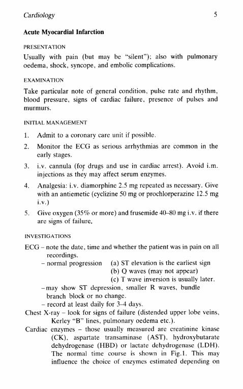
Kerley "B" lines, pulmonary oedema etc.). Cardiac enzymes - those usually measured are creatinine kinase
(CK), aspartate transaminase (AST), hydroxybutarate dehydrogenase (HBD) or lactate dehydrogenase (LDH). The normal time course is shown in Fig.1. This may influence the choice of enzymes estimated depending on
6 Handbook of Hospital Medicine
how soon the patient is seen after the onset of symptoms. Also consider other causes of abnormal enzyme results (liver disease, haemolysis, i.m. injections, etc.). Measure daily for the first 3 or 4 days and later if there is any suggestion of re-infarction
8 Enzyme Level (x normal)
4
o 1 2 3
Figure 1 Pattern of serum enzyme elevation following acute myocardial infarction.
LDH
4 5 6
Time (days)
Also check - WBC (often raised), haemoglobin, urea and electrolytes (potassium may need correction), blood glucose. Monitor the temperature chart as transient pyrexia is common.
Mobilisation - depends on the patient's age, size of infarction, complications and other diseases. The average is about 2 days in bed and 10 days in hospital; slower if heart failure, angina or re-infarction occur.
Rehabilitation - early exercise testing, before discharge with a modified protocol, is becoming widely used and has important prognostic significance. Most patients will be off work until the first review appointment at 4-6 weeks and the course thereafter depends on age, infarct size, occupation, motivation etc.
Cardiology 7
Reassurance - explain and reassure at every stage. Many hospitals distribute explanatory leaflets for patients to read.
Anticoagulation - in the acute phase, prevents DVT and pulmonary embolism but has not been shown to influence the course of infarction. A common compromise is sub-cutaneous heparin 5000 units b.d.
Risk factors - ban smoking, treat hypertension, and encourage weight loss. In the acute phase serum lipids are artificially low and difficult to interpret. Check the lipid profile at outpatient review in younger patients.
At the follow up clinic check - does the patient have angina? - are there any signs of failure? - measure the blood pressure - consider fasting lipid profile.
COMPLICATIONS
Arrhythmias
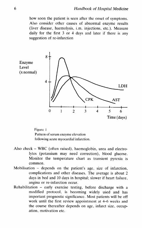
1. Sinus bradycardia (see Fig. 2a) -common, especially in inferior infarction. -treat with atropine 0.6 mg i.v. if rate below 4O/min or
hypotensive. Repeat as necessary.
2. Sinus tachycardia (see Fig. 2b) -usually reflects pain, anxiety, heart failure or shock. Treat
the underlying problem.
3. Nodal or supra-ventricular tachycardia (see Fig. 2c) -try carotid sinus massage first. Use cardioversion if
shocked, otherwise practolol 10--20 mg i.v. slowly, repeated after 15 min if necessary or verapamil 1 mg/min i.v. up to 10 mg, but not both drugs, because there is a risk of asystole.
4. Atrial flutter and fibrillation (see Figs. 2d & 2e) -often transient. Both arrhythmias respond well to DC
cardioversion if haemodynamic disturbance is present. Otherwise give verapamil as above or deslanoside 800--1200 ILg i. v. (cardiac glycosides are better avoided in acute infarction if possible, but controlling the ventricular rate may be more important).
8 Handbook of Hospital Medicine
Figure 2 (a) Sinus bradycardia - sinus rhythm with rate below 6O/min; (b) Sinus tachycardia - sinus rhythm with rate above 100/min; (c) Supra-ventricular tachycardia - QRS complexes usually narrow; (d) Atrial flutter-conduction is usually2:1 or4:1 (e )Atrial fibrillation - irregular QRS complexes with no Pwaves; (f) Ventricular ectopics - broad premature complexes, usually with a compensatory pause (g)Ventricular tachycardia - broad complexes,usually 150-250/min and more or less regular: look for independent P waves
(a)
(b)
(c)
(d)
(e)
(f)
(g)
Cardiology 9
5. Atrial ectopics -common, and do not require treatment. May precede
atrial fibrillation.
6. Ventricular ectopics (see Fig. 2f) -very common. Evidence that their suppression is
beneficial is now less convincing. "R on T" ectopics are the most ominous. If the sinus rate is below 60, give atropine, if above 110 give practolol. Otherwise use lignocaine as below.
7. Ventricular tachycardia (see Fig. 2g) -if shock or syncope use immediate DC cardioversion.
Otherwise give lignocaine 50-150 mg i.v. followed by an infusion of 4 mg/min, reducing to 2 mg/min. If ineffective, use disopyramide 2 mg/kg i.v. over 5 min to a maximum of 150 mg and up to 300 mg in the first hour; or mexiletine 150-250 mg i.v. over 5 min, and, failing that, or with a recurrence, a combination of drugs may be needed. Overdrive pacing may also help if drugs fail (see "Temporary Pacing").
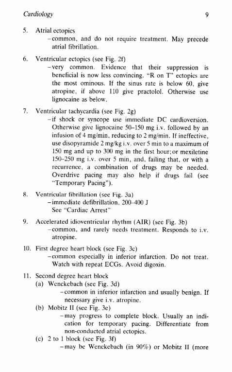
8. Ventricular fibrillation (see Fig. 3a) - immediate defibrillation, 200-400 J
See "Cardiac Arrest"
9. Accelerated idioventricular rhythm (AIR) (see Fig. 3b) -common, and rarely needs treatment. Responds to i.v.
atropine.
10. First degree heart block (see Fig. 3c) -common especially in inferior infarction. Do not treat.
Watch with repeat ECGs. Avoid digoxin.
11. Second degree heart block (a) Wenckebach (see Fig. 3d)
-common in inferior infarction and usually benign. If necessary give i.v. atropine.
(b) Mobitz II (see Fig. 3e) -may progress to complete block. Usually an indi
cation for temporary pacing. Differentiate from non-conducted atrial ectopics.
(c) 2 to 1 block (see Fig. 3f) -may be Wenckebach (in 90%) or Mobitz II (more
10 Handbook of Hospital Medicine
Figure 3 (a) Ventricular fibrillation-unco-ordinated electrical activity. (b) Accelerated idio-ventricular rhythm-broad QRS complexes between 60
and lOO/min and faster than the sinus rate; (e) First degree heart block-PR interval greater than 0.22 second; (d) Wenekebaeh second degree block-increasing PR interval with dropped
QRS. (e) Mobitz II second degree bloek-constant PR interval with dropped QRS; (f) 2:1 second degree block-alternate P waves are conducted; (g) Complete heart block-unrelated P waves and QRS complexes. The
QRS is narrow in this example
(a)
(b)
(c)
(d)
(e)
(g)
(f)
Cardiology 11
likely if BBB is present). Try atropine 0.6 mg i.v. or temporary pacing if the bradycardia produces hypotension.
12. Complete heart block (CHB)(see Fig. 3g) CHB with a narrow QRS in inferior MI is common. Treat with atropine if there is a significant bradycardia. Isoprenaline infusion may be used until temporary pacing can be arranged if there is haemodynamic embarassment. CHB with broad QRS in anterior MI - ominous, often implying extensive infarction. Insert a temporary pacemaker. May take up to 3 weeks to resolve and occasionally a permanent pacemaker is needed.
13. Indications for temporary pacing in acute infarction. (a) almost always in
. - CHB with anterior infarction - Mobitz II second degree block - intermittent ventricular standstill.
(b) often advised in - new RBBB and left posterior hemiblock -new RBBB and left anterior hemiblock (LAHB) with
long PR interval. (c) occasionally in
- CHB with inferior infarction - new LBBB - new RBBB and LAHB - any symptomatic bradycardia - resistant VT (overdrive pacing).
Failure -present if there are basal crepitations, third heart sound,
congestion on chest X-ray. - treat with frusemide 40 mg , repeat if necessary. - maintenance treatment is not always needed and a thiazide
may be sufficient. If not, use frusemide and then as the next step add isosorbide. Potassium replacement is necessary.
- see also "Pulmonary Oedema"
Shock - see under "Cardiogenic Shock"
12 Handbook of Hospital Medicine
Pericarditis -common; the friction rub is often transient and may be absent.
Usually responds to indomethacin 25 mg t.d.s.
Onset of Murmur -a quiet systolic murmur is common. No treatment is necessary
if the patient is haemodynamically stable. -a loud pansystolic murmur with a third heart sound, a thrill,
clinical deterioration and often shock or pulmonary oedema: the differential diagnosis is between a ruptured ventricular septum and acute mitral regurgitation. Refer for investigation.
LATER PROBLEMS
1. Dressler's syndrome -uncommon. Presents at 2-6 weeks with pain, failure,
pleural and pericardial effusions, fatigue, fever, pericardial rub.
-check temperature, ESR, ECG, cardiac enzymes, anticardiac antibodies.
- treat with an analgesic and consider a course of prednisolone if severe.
2. Left ventricular aneurysm -suggested by persisting failure, arrhythmias, emboli,
paradoxical apex beat. -chest X-ray may show a "boot-shaped" heart. - ECG may show persisting ST elevation. -consider referral for investigation (the diagnosis can often
be confirmed non-invasively with gated blood pool scanning or a 2D echocardiogram).
3. Emboli - from mural thrombus, see under "Systemic Emboli".
Cardiac Arrhythmias
Cardiac arrhythmias are considered in detail under "Acute Myocardial Infarction" but also occur in circumstances where the indications and recommendations for treatment may be different.
1. Paroxysmal supraventricular tachycardia (see Fig. 2c) - regular, usually above 150/min.
Cardiology
- distinguish from sinus tachycardia, AF, atrial flutter, VT. -causes include ischaemia, Wolff-Parkinson-White and
Lown-Ganong-Levine syndromes, and digoxin toxicity, but often none is found.
(a) if the patient is well and the attacks are self limiting, it may be sufficient to observe for a while.
(b) try carotid sinus massage. (c) use DC cardioversion if there is haemodynamic embarass
ment. (d) drugs - verapamil 5-10 mg i. v. slowly
OR - practolol 5-20 mg i.v. slowly OR - disopyramide 2 mg/kg OR - digoxin (see "Digoxin")
If one drug fails another may be given later, but not verapamil AND practolol, and take care with disopyramide and verapamil.
(e) for long term prevention a beta-blocker or disopyramide is the drug of choice.
2. Atrial fibrillation (see Fig. 2e) - irregularly irregular with absent P waves.
13
- seen in ischaemic, valvular, thyrotoxic and alcoholic heart disease, cardiomyopathy, ASD, pericarditis etc.
- usually treated with digoxin (for doses see "Digoxin"). - if not fully controlled add a beta-blocker or verapamil. - to prevent recurrence of paroxysmal AF try disopyramide
or amiodarone or procainamide.
3. Atrial flutter (see Fig. 2d) -the ventricular rate is often almost exactly l50/min (i.e.
2:1 conduction) when untreated. - seen in ischaemia, cardiomyopathy, after cardiac surgery,
etc. -responds well to DC shock or overdrive pacing. -treat with digoxin or beta-blocker or verapamil as above. - beware of disopyramide which may slow the flutter rate
enough to allow 1:1 conduction and thereby increase the ventricular rate.
4. Sinus bradycardia (see Fig. 2a) - sinus rhythm below 6O/min. -seen in normal people, athletes, sino-atrial disease, drug
treatment (digoxin, beta blockade), myxoedema, raised
14 Handbook of Hospital Medicine
intracranial pressure etc. - rarely needs acute treatment but, if prolonged and
symptomatic, permanent pacing is sometimes required.
5. Ventricular ectopics (see Fig. 2f) - very common but rarely require treatment. -apart from infarction they may be important in hypertro-
phic cardiomyopathy and in patients taking digoxin.
6. Ventricular tachycardia (see Fig. 2g) - by definition is a run of three or more ventricular ectopics. -with a broad-complex tachycardia it is sometimes difficult
to distinguish between VT and SVT with aberration. The following may help:
(a) look for independent P waves in VT and related P waves in SVT.
(b) capture beats and fusion beats are seen in VT. (c) if there is a classical LBBB or RBBB pattern, SVT
with aberration is more likely. (d) in lead V1 if R is taller than R', VT is likely. (e) if the QRS is predominantly upright in all chest leads,
VT is more likely. - the main causes are ischaemic heart disease and digoxin
toxicity. VT may be a cause of Stokes-Adams attacks in complete heart block.
-in the acute situation, treatment is as detailed under "Acute Myocardial Infarction". If not related to an acute problem, oral suppressive treatment will be necessary:
oral disopyramide 100--150 mg 6 or 8 hourly oral mexiletine 200--300 mg 8 hourly oral amiodarone 400 mg daily
Management of Acute Pulmonary Oedema
CLINICAL FEATURES
Increasing dyspnoea, orthopnoea, paroxysmal nocturnal dyspnoea, sweating cough and pink sputum and cardiac pain.
DIAGNOSIS
On history, chest auscultation and chest X-ray.
Cardiology 15
CAUSES
Myocardial infarction, hypertension, beta-blockers, valve disease (especially with the onset of uncontrolled AF), overtransfusion.
DIFFERENTIAL DIAGNOSIS
Asthma, acute - on - chronic bronchitis, pulmonary embolism, severe pneumonia, etc.
MANAGEMENT
1. Sit the patient up.
2. Give 40% oxygen by mask or nasal catheter (if the patient also has chronic chest disease, monitor blood gases).
3. Drugs -diamorphine 2.5 mg i.v. repeated as necessary (have
naloxone available). Avoid in chronic lung disease or if the diagnosis is in doubt. It may be safer to give 1 mg at a time.
-anti-emetic; cyclizine 50 mg or prochlorperazine 12.5 mg i.v. with the diamorphine.
- frusemide 40--80 mg i.v. - aminophylline 250--500 mg i. v. slowly. - digoxin, if the patient is not on digoxin and there is no
evidence of acute infarction (see "Digoxin"). -vasodilators: glyceryl trinitrate, nitroprusside, isosorbide.
Nitroprusside is first choice in hypertensive heart failure, although a mild elevation of blood pressure is common in pulmonary oedema.
4. Specific treatment of dysrhythmia. VT and uncontrolled AF may be best treated by cardioversion.
5. Venesection is best for overtransfusion.
6. Consider mechanical ventilation if no improvement.
7. A Swan-Ganz catheter may help in management, especially if vasodilators or ventilation are used.
8. Once the situation is stable, identify the cause. If none is obvious, consider rarer causes ego myxoma, inhaled gases, etc.
9. In valve disease, if there is no improvement, consider referral for urgent investigation and operation.
16 Handbook of Hospital Medicine
10. Check Hb, albumin, urea and electrolytes, and cardiac enzymes.
11. Monitor improvement clinically and with fluid balance, daily weight and repeat chest X-ray.
12. Drain pleural effusion if large enough to be contributing to dyspnoea.
Management of Cardiogenic Shock
DEFINITION
A syndrome produced by pump failure and characterised by hypotension (systolic <80 mmHg) , oliguria and peripheral vasoconstriction, often accompanied by pulmonary oedema.
CAUSES
- acute myocardial infarction - acute mitral or aortic regurgitation - acute ventricular septal defect - cardiac tamponade
DIFFERENTIAL DIAGNOSIS
Includes haemorrhage, septicaemia, massive pulmonary embolism.
MANAGEMENT
1. Identify any treatable causes (e.g. acute VSD in infarction).
2. Before treatment, THINK - in view of the poor prognosis is aggressive treatment justified in this patient? Does age or other disease suggest it would be best to avoid invasive procedures with little hope of success?
3. Give oxygen by face mask or nasal cannulae.
4. Treat pain with diamorphine 2.5 mg i.v. as required.
5. Haemodynamic monitoring Swan-Ganz catheter via subclavian vein to monitor pulmonary wedge pressure.
- peripheral arterial line for BP and blood gases. - urinary catheter to measure hourly output.
Cardiology 17
6. If the wedge pressure is below 18-20 mmHg, raise to this level by infusion of normal saline (to provide preload for the left ventricle) .
7. Commence low dose infusion of nitroprusside if BP allows, but watch BP carefully.
8. Begin dopamine infusion -at 2.5-5.0 p,g/kg/min renal blood flow, cardiac output and
contractility increase. - above 5-10 p,g/kg/min, peripheral resistance and heart
rate increase, thus increasing cardiac work. - watch for dysrhythmias.
9. Dobutamine infusion up to 10 p,g/kg/min produces an increase in cardiac output while reducing LV filling pressure. There is less effect on rate and less irritability than with dopamine. It can be used in combination with dopamine and nitroprusside.
10. Intra-aortic balloon counterpulsation, if available, can improve and stabilise the situation, allowing time for investigation.
11. If there is no obvious cause, an echo cardiogram must be done to exclude tamponade or left atrial myxoma.
Pericardial Tamponade
CAUSES
Trauma (may be non-penetrating), malignancy, uraemia, infection, aortic dissection, connective tissue diseases.
SYMPTOMS
Dyspnoea, fatigue, faintness, pain, collapse.
PHYSICAL SIGNS
Tachycardia, raised JVP, hypotension, pulsus paradoxus (more than 10 mmHg fall in systolic BP on inspiration), friction rub.
INVESTIGATIONS
Echocardiography - the investigation of choice if available. Chest X-ray - may show cardiomegaly.
18 Handbook of Hospital Medicine
ECG - low voltage and T wave inversion (non specific). Screening with a catheter in the right atrium or a right atrial angiogram are now rarely indicated but may help with the diagnosis if no other facilities are available.
PERICARDIAL ASPIRATION
Indications -hypotension, marked signs, deterioration, and to provide fluid
for investigations.
Technique - the xiphisternal route is preferable. - use a lumbar puncture needle under local anaesthetic. - advance the needle to the left of the xiphisternum, cephalad at
45 degrees and aimed slightly to the left. - ECG monitoring via the needle is often advocated but may be
difficult to use because of respiratory swing on the trace. - send fluid for Hb and pev, protein, sugar, cytology, culture
and Gram stain. - if a large volume is present a cannula may be used to facilitate
aspiration and reduce the risk of damage to the heart.
Complications -include RV puncture, coronary artery or vein laceration and
pneumothorax. Refer for surgical drainage if
- haemopericardium - malignant effusion - rapid recurrence.
Pericarditis and Pericardial Effusion
The causes of each are similar and the list of aetiologies suggests the necessary investigations.
CAUSES
- viral, bacterial, tuberculous infection - myocardial infarction - malignant infiltration - congestive heart failure, uraemia, hypoalbuminaemia
Cardiology 19
- trauma, post-cardiotomy syndrome, post radiotherapy - Dressler's syndrome, myxoedema, SLE.
INVESTIGATIONS
Chest X-ray - often normal. At least 250 ml of fluid are needed to produce enlargement of the cardiac shadow.
ECG - widespread ST elevation in awk pericarditis, classically concave upwards. T wave changes, low voltage, and electrical alternans are seen in pericardial effusion.
Echocardiogram - the best way to demonstrate an effusion. Other investigations as suggested by the list of causes. Pericardiocentesis is useful to establish the diagnosis in bacterial, tuberculous and malignant pericarditis.
MANAGEMENT
1. Analgesia.
2. Watch for signs of tamponade (see above).
3. Establish the cause as above.
4. Specific treatment depending on the cause.
Management of Chronic Stable Angina
HISTORY
Establish the diagnosis and its severity from the history. Distinguish from other causes of chest pain as far as possible. Also ask about past hypertension, myocardial infarction, risk factors (smoking, obesity, etc.) and diseases affecting the choice of treatment (asthma, peripheral vascular disease, etc.).
EXAMINATION
Measure BP, look for signs of heart failure and peripheral vascular disease. Factors other than ischaemic heart disease may contribute to or cause angina (eg anaemia, aortic stenosis).

20 Handbook of Hospital Medicine
ECG - will be normal in over 50%. Chest X-ray - normal unless heart failure or hypertension etc. are
present. FBC - if anaemia is present, its correction will often improve
angina. Lipids - check fasting profile in younger patients. Exercise testing - very helpful but if negative does not exclude the
diagnosis. Useful in assessing severity and response to treatment and in identifying those patients with severe disease.
Coronary arteriography - indicated in patients in whom medical treatment has failed, in those who may have severe disease, and when a job may be at risk (e.g. pilots, drivers).
MANAGEMENT
1. Explain the diagnosis and reassure the patient. Stress the good prognosis.
2. Correct any risk factors - hypertension, smoking, obesity etc.
3. Start glyceryl trinitrate. Explain its use, particularly as prophylaxis before exertion. It should be taken as often as needed.
4. The next stage is a beta-blocker if not contra-indicated (by asthma, cardiac failure, vascular disease). Use propranolol unless a cardio selective drug is indicated. Begin with 40 mg t.d.s. and increase as necessary. Change to another beta-blocker if minor side-effects are troublesome.
5. Add a long-acting nitrate, such as isosorbide dinitrate 10 mg t.d.s. Increase the dose if well tolerated.
6. Consider adding nifedepine, perhexiline, lidoflazine, or verapamil, especially in those in whom investigation and/or surgery is ruled out by age, past infarction, or other illnesses.
7. Consider coronary arteriography with a view to coronary bypass grafting. Except in the case of left main stem stenosis and triple vessel disease the decision is made on the basis of symptoms so there must be no doubt about the diagnosis and its severity before investigation. Arteriography is rarely indicated to confirm or exclude the diagnosis.
Cardiology
Management of Unstable Angina
DEFINITION
21
Includes patients with progressive angina of recent onset, those with rapidly worsening angina, and pain at rest.
ECG
Shows variable ST/T changes or may be normal if pain-free. ST depression and T wave inversion are not uncommon. Prinzmetal angina produces ST segment elevation during pain.
MANAGEMENT
1. Bed rest.
2. Glyceryl trinitrate tablets as required. Topical GTN paste is often useful.
3. Diamorphine if no relief or for prolonged pain.
4. A beta-blocker, increasing the dose as necessary to produce a resting bradycardia.
5. Add isosorbide 10-20 mg q.d.s., increasing rapidly if pain persists. Can also be given by infusion.
6. Verapamil 40-S0 mg or more t.d.s. and nifedepine 10-20 mg t.d.s. are further possibilities.
7. Heparin is sometimes advocated. So far there is no proof that it influences the incidence of infarction, but it will help to prevent DVT or pulmonary embolism.
S. Refer for coronary arteriography if the pain does not settle rapidly. The patient can often be stabilised on intra-aortic balloon pumping and this may be necessary before investigation.
Management of Hypertension
HISTORY
Most patients are asymJ'1omatic. Ask about
-complications such as angina, claudication, previous myocar-dial infarction or stroke
-contributory factors: family history, obesity, salt intake - others risk factors: smoking, diabetes, diet - medication.
22 Handbook of Hospital Medicine
EXAMINATION
Measure BP at rest, with the patient relaxed. Assess target organ damage: fundi, LV hypertrophy, peripheral pulses. Look for an underlying cause: femoral pulses, Cushingoid facies, renal masses, renal bruits.
INVESTIGATIONS
Chest X-ray - looking for cardiomegaly, rib notching, pulmonary congestion. ECG - may show left ventricular hypertrophy. Urine - stick testing and microscopy. Serum urea, creatinine and electrolytes. Investigation rarely reveals an underlying cause but may be indicated in younger patients or where the history, examination or simple tests suggest an underlying disease. Renal artery stenosis - often a bruit present. IVP is suggestive in
70% of cases. Confirm with renal vein renin samples and renal arteriography.
Conn's syndrome - suggested by t K, t Na, and t bicarbonate on biochemical screen if not on a diuretic. Confirm with plasma renin (low) and aldosterone (high).
Cushing's syndrome - typical clinical features, glycosuria, hypokalaemia. Further investigations include diurnal plasma cortisol levels, 24 hour urinary cortisol and low dose dexamethasone suppression test.
Phaeochromocytoma - history of anxiety, sweating, tremor and palpitation. The hypertension is often sustained. Check 24 hour urinary VMA excretion on several samples. Confirm with plasma catecholamines.
Coarctation of the aorta - suggested by delayed or absent femoral pulses, a systolic murmur, evidence of collateral vessels (especially around the scapulae) and by abnormal aortic knuckle and rib-notching on chest X-ray.
TREATMENT
1. Correct other risk factors.
2. Encourage weight loss and reduce salt intake.
3. Drugs (a) begin with a beta-blocker (eg. propranolol 40 mg b.d. or
Cardiology
atenolol 100 mg daily) in younger patients. (b) use a thiazide diuretic (eg. bendrofluazide 5 mg daily) in
older patients. (c) combine beta-blocker and thiazide if control is not achieved
after a few weeks. (d) a vasodilator ego prazosin or hydralazine is usually the next
step in resistant cases. (e) consider methyl dopa, labetalol, or debrisoquine if the
above regime fails. (f) newer drugs for resistant hypertension include minoxidil
and captopril. (g) remember that non-compliance is a common cause of
treatment "failure".
23
The level of hypertension to treat and the degree of control to accept remain controversial. Treat, and aim for, lower pressures in younger patients. Use the above format as a guide only.
Accelerated Hypertension
Hypertension should be regarded as an emergency if any of the following is present:
1. diastolic pressure greater than 140 mmHg
2. pulmonary oedema
3. encephalopathy
4. renal failure
5. aortic dissection. In the first instance do not reduce the blood pressure too quickly, or aim to reduce it below 150/100. Aortic dissection is an exception, where a systolic of 100--110 mmHg is preferable.
TREATMENT
1. Labetalol - mix 200 mg in 200 ml of dextrose-saline. - infuse 2 mg/min until a satisfactory response is obtained. - the usual dose needed is in the range 50--200 mg. - once controlled, transfer to oral treatment.
2. Nitroprusside - begin with 0.5-1.5 Iotg/kg/min.

24 Handbook of Hospital Medicine
- increase to 0.5-8 ILg/kg/min depending on response. - effective dose is usually in the range 50-400 ILg/min. - the maximum dose is 800 ILg/min. - treatment of choice in LVF or aortic dissection. - avoid in patients with renal or hepatic failure. - protect from light and avoid extravasation. - monitor BP carefully. - do not stop infusion suddenly.
3. Hydralazine - 20-40 mg i.v. slowly or by infusion. - may cause tachycardia and worsening of angina.
4. Propranolol and hydralazine orally will often produce a satisfactory response, even when hypertension is severe.
Dissecting Aneurysm of the Aorta
PRESENTATION
Pain (sudden, severe, tearing pain in the back, chest and abdomen), collapse, shock and stroke.
EXAMINATION
Measure the BP (often raised although the patient looks shocked). Check all peripheral pulses. Look for signs of peripheral ischaemia (stroke, paraplegia, silent abdomen, cold leg, etc). Listen carefully for an aortic diastolic murmur.
INVESTIGATIONS
Chest X-ray -widening of the mediastinum is common but may take time to
develop. - a small left pleural effusion may be present.
ECG - changes of left ventricular hypertrophy due to hypertension are
often seen. - acute infarction changes may be present.
MANAGEMENT
1. Admit to intensive care unit if possible.
Cardiology 25
2. Relieve pain with i.v. diamorphine.
3. Establish venous and arterial lines.
4. If hypertensive, begin nitroprusside infusion (see under "Accelerated Hypertension").
5. Refer for emergency aortography. In general, surgical treatment is better for type I and II dissections (involving the ascending aorta) and medical, i.e. hypotensive, treatment for type III which are more distal. In time CAT scanning may be a rapid non-invasive technique to distinguish between them.
Note Decisions about management and surgery will obviously be affected by the presence of stroke or other complications and the age and previous health of the patient.
Management of Congestive Cardiac Failure
DEFINITION
Usually taken to mean combined left and right heart failure. Most of the comments below also apply to pure right heart failure.
PHYSICAL SIGNS
The most important sign of right heart failure is elevation of the jugular venous pressure. Others such as hepatomegaly, peripheral oedema and cachexia occur in a variety of conditions.
CAUSES OF RIGHT HEART FAILURE
- secondary to left heart failure - cor pulmonale (secondary to chronic lung disease) - rarer: cardiomyopathy, thyrotoxicosis, atrial septal defect,
pericardial constriction, etc.
INVESTIGATIONS
Will be suggested by possible causes but must include: Chest X-ray -note the heart size and look for signs of lung disease,
pleural effusions, etc. ECG U+E - especially the potassium.
26 Handbook of Hospital Medicine
LFTs - especially albumin. Abnormal LFTs are common and improve with treatment.
Also consider FBC, thyroid function tests, echocardiography, and pulmonary function tests depending on the individual case.
MANAGEMENT
1. Rest, either in bed or in a chair.
2. Correct underlying abnormalities such as anaemia, hyperthyroidism, tachycardia and bradycardia. Stop cardio-depressant or fluid-retaining drugs. (beta blockers, NSAID etc)
3. Salt restriction - "no added salt diet".
4. Consider anticoagulation to prevent DVT or pulmonary embolism.
5. Aspiration of pleural effusions if large.
6. Diuretics: if failure is moderate or severe begin with frusemide 40-80 mg daily, increasing the dose as necessary, together with potassium replacements or a potassium retaining diuretic eg amiloride 5 mg daily. In mild failure begin with a thiazide diuretic ego bendrofluazide K.
7. Digoxin - often beneficial even in sinus rhythm (see "Digoxin").
8. Vasodilators -isosorbide dinitrate 10 mg Ld.s. up to 40 mg g.d.s.
especially if the patient also has angina. -prazosin 0.5 mg first dose in the evening, then 0.5 mg
t.d.s. increasing as necessary. -hydralazine 50-75 mg g.d.s. or sometimes more up to a
maximum of 800 mg/day. With the higher doses there is an increased risk of developing a lupus-like syndrome.
If the patient is on the above treatment and signs of failure persist, consider the following steps:
9. Change from potassium supplements to a potassium-sparing diuretic.
10. Increase the dose of diuretic.
11. Add metolazone 5 mg to the frusemide. This often produces a brisk diuresis but may cause marked hypokalaemia. Anticipate by increasing potassium supplements.
Cardiology 27
11. Dopamine or dobutamine infusion. For details see "Cardiogenic Shock". The improvement is not long-lasting but it may be worth considering if the patient is not in end-stage cardiac failure.
Look for correctable lesions and refer for invasive investigation if necessary.
DURING TREATMENT MONITOR
1. Daily weight - more accurate than fluid balance.
2. Blood urea - often rises, especially in the elderly.
3. Electrolytes - low sodium when failure persists is an ominous sign. Watch potassium closely.
4. Chest X-ray - looking for reduction in heart size and to check the progress of pleural effusions.
Digoxin
Starting treatment: Routine - 500 f.Lg b.d. for 2 days.
or - 250 f.Lg b.d. for 7 days. Rapid - SOO f.Lg i.v. by infusion over IS minutes.
repeat after 1 hour, then 2S0 f.Lg 4 hourly. or - oubain 2SG-SOO f.Lg i. v. slowly, then 100 f.Lg hourly or oral
digoxin. or - lanatoside 80G-1200 f.Lg i. v. then 200 f.Lg 2-hourly to total
dose of 1.S mg in 24hours. Maintenance treatment:
- in the range 125-S00 f.Lglday. - reduced in renal impairment, old age, hypokalaemia. - serum digoxin concentration. - an inappropriately high blood concentration helps to
confirm toxicity. - measure at 6--8 hours post dose. _ normal therapeutic range 1-2 ng/ml (1.3-2.6 nmol/l). - check serum potassium at the same time.
DIGOXIN TOXICITY
-more likely in elderly, renal failure, hypokalaemia, after diarrhoea and vomiting, after starting amiodarone.
-produces nausea, anorexia, vomiting, xanthopsia,etc.
28 Handbook of Hospital Medicine
-ECG may show ventricular bigeminy, VT, multiform ventricular ectopics, atrial tachycardia with block, A-V block etc.
Treatment 1. Stop digoxin.
2. Increase potassium supplements (an infusion of 40 mmol in 1 hour can be given safely). Monitor serum potassium.
3. Give practolol 5-20 mg Lv. slowly for supraventricular tachycardias.
4. Use lignocaine for VT (see under "Arrhythmias").
5. Cardioversion can be used to treat VT when urgent restoration of normal rhythm is necessary. Start with low energy, such as 20 J, increasing if ineffective.
6. Give atropine 0.6 mg i.v. for A-V block or bradycardia. A temporary pacing wire may be needed.
7. Overdrive pacing of the atrium for SVT or ventricle for VT is often successful and can be repeated if the dysrhythmia recurs (see under "Temporary Pacing").
Antibiotic Prophylaxis Against Bacterial Endocarditis
Higher risk in patients with aortic valve disease, VSD, mitral regurgitation and prosthetic valves. Lower risk in those with mitral stenosis, secundum ASD, "innocent" murmur . Many advocate no cover for normal delivery in pregnant women.
RECOMMENDED TREATMENT
1. Dental extraction, scaling etc. - 1 vial of Triplopen i.m. 30 mins before the procedure.
or - amoxycillin 3 g orally 60 mins before procedure. or -in patients allergic to penicillin use erythromycin 500 mg
orally 60 mins before and then 6 hourly for 24 hours after.
2. Genito-urinary procedures (sigmoidoscopy, cystoscopy, D+C) and abdominal surgery.
-ampicillin 1 g plus gentamicin 80 mg i.m. 30 mins before
Cardiology
procedure or Lv. with the anaesthetic if given. Same dose of both drugs at 8 and 16 hours.
or -for patients allergic to penicillin give vancomycin 1 g i.v. (plus gentamicin 80 mg Lv.) by infusion over 30 mins about 30 mins before the procedure.
3. Patients with prosthetic valves - can be treated as above after 1 year. - earlier than that, consult the Bacteriologist.
4. Patients already on antibiotic treatment. - consult the Bacteriologist.
Management of Infective Endocarditis
INVESTIGATIONS I
29
- always be suspicious in patients with murmurs or undiagnosed fever
-3 blood cultures are usually sufficient (more if the patient has recently received antibiotics or following cardiac surgery)
- 4 hourly temperature chart -FBC, ESR - immunoglobulins, complement levels - urine microscopy (for red cells) -echocardiogram, looking for vegetations and for comparison
later in the course of the illness. If cultures are positive, the laboratory will measure antibiotic sensitivity and minimum inhibitory concentration (MIC). If cultures are negative, repeat and also consider rare causes such as Q fever, psittacosis (diagnose on CFT) or fungi (check precipitins)
PRINCIPLES OF TREATMENT
Antibiotics and doses should be chosen in consultation with the Bacteriologist. High doses of bactericidal antibiotics are given intravenously and the course of treatment usually lasts six weeks, although in the later stages the drugs may be given orally. Great care must be taken to avoid infection at the site of the infusion. Subclavian lines are convenient, reduce the risk of thrombophlebitis and allow the patient more mobility. Probenecid is often advocated to increase blood levels of penicillin but makes drug rashes more
30 Handbook of Hospital Medicine
likely and is probably best avoided. Common organisms - Strep. viridans, Strep. faecalis, Staph. aureus
(in acute endocarditis). Others are less common. Commonly used antibiotics - benzylpenicillin, gentamlcm,
ampicillin, vancomycin (in patients allergic to penicillin), flucloxacillin.
During treatment monitor:
1. temperature 4 hourly
2. physical signs (murmurs, diastolic BP in aortic regurgitation,)
3. urine output
4. back titrations (in conjunction with the Bacteriologist)
5. renal function
6. Hb, WBC, ESR (anaemia and raised ESR may lag behind clinical improvement)
7. ECG (development of conduction defects is ominous)
8. chest X-ray (looking for any increase in heart size or sign of failure).
Persisting fever or recurrence of fever may be due to:
1. inadequate treatment
2. infection at the infusion site (remove cannula and send the tip for culture, including fungi)
3. drug fever (check for eosinophilia)
4. unrelated causes (urinary tract infection, DVT, pneumonia, etc.).
Systemic Embolism
CAUSES
- mural thrombus in myocardial infarction, LV aneurysm -left atrial thrombus in mitral valve disease, AF, cardio-
myopathy (note that emboli can occur with mitral stenosis in sinus rhythm)
- aortic atheroma

Cardiology
- thrombus from prosthetic valve - vegetations in infective endocarditis - tumour in left atrial myxoma.
EFFECTS
- cerebral: TIA, stroke, amaurosis fugax - mesenteric: pain, vomiting, rectal bleeding, peritonitis - renal: loin pain, haematuria -limb: pallor, pain, paraesthesiae, loss of pulse, weakness.
DIFFERENTIAL DIAGNOSIS
- arterial thrombosis - arterial injury - DVT with peripheral vascular disease.
INVESTIGATIONS
- blood cultures -EeG - cardiac enzymes
31
- echocardiography looking for evidence of mitral valve disease, left ventricular disease, vegetations, myxoma
-culture and histology of embolus removed at embolectomy.
MANAGEMENT
1. anticoagulate unless endocarditis or other contra-indication. May need to be delayed in stroke.
2. consider dextran 40 infusion.
3. angiography and streptokinase infusion may be indicated in renal emboli.
4. laparotomy may be necessary with mesenteric emboli.
5. in limb ischaemia, give analgesia and consider embolectomy.
Electrocardiography
1. Calibration. Normal paper speed is 25 mm/sec. 5 large squares = 1 sec 1 large square = 0.2 sec
32 Handbook of Hospital Medicine
1 small square = 0.04 sec 1 m V = 10 mm. Check this on each recording.
2. Standard I2-lead recording. The two commonest faults are transposed arm leads (giving inverted P waves in lead I) and chest leads all recorded on aVF. Chest leads are positioned as follows:
VI 4th intercostal space right sternal edge
V2 4th intercostal space left sternal edge
V3 over the 5th rib between V2 and V 4
V4 5th intercostal space mid-clavicular line
V5 on horizontal line through V4 anterior axillary line
V6 on horizontal line through V4 mid-axillary line
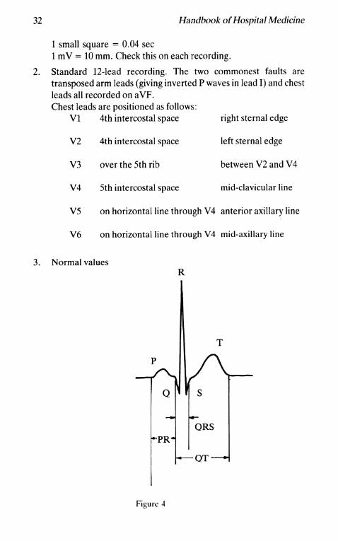
3. Normal values R
T
Figure 4
Cardiology
P wave <2.5 mm tall, <0.12 sec duration PR interval 0.12-0.22 sec QRS duration <0.10 sec QT interval depends on heart rate
<0.43 at 60/min <0.38 at 80/min <0.35 at 100/min
4. Rate. If regular count large squares between complexes 2 squares = ISO/min 3 squares = 100/min 4 squares = 75/min 5 squares = 60/min, etc.
33
H irregular, count the number of complexes in 30 large squares (6 seconds) and multiply by 10 for rate per min.
5. Axis. Normal range is -300 to +900 (or more in children). Left axis deviation is usually due to inferior infarction or left anterior hemiblock. Right axis deviation is usually due to anterolateral infarction or right ventricular hypertrophy or dominance.
6. P waves - look especially at leads II and VI. - bifid in left atrial enlargement - peaked in right atrial enlargement (congenital heart
disease or chronic chest disease).
7. PR interval -short in Wolff-Parkinson-White syndrome and some nodal
rhythms. -long in first degree block (ischaemia, digoxin, etc.).
8. Q waves - small 'q' is normal in leads I, aVL, and lateral chest leads with a liorizontal heart and in II, III, and aVF with a vertical heart and is due to septal depolarisation. Deeper, wider Q or QS may be seen in a VR and VI in normals.
- abnormal if 0.04 sec or more in duration 2 mm or deeper >25% of R wave in the same lead.
-seen in infarction, pulmonary embolism, LBBB (in right precordial leads).
34 Handbook of Hospital Medicine
9. QRS - increased voltage in (a) thin people
(b) ventricular hypertrophy - reduced voltage in (a) obesity
(b) chronic chest disease (c) myocardial infarction (d) myxoedema (e) pericardial effusion
- increased duration in (a) ventricular hypertrophy 0.09-0.12 sec
(b) bundle branch block >0.12 sec.
10. Left ventricular hypertrophy. It is difficult to make a definite diagnosis on EeG unless the changes are very marked. Shows as(a) increased amplitude and duration
(b) "strain" pattern in anterolateral ST segments and T waves
(c) left atrial hypertrophy. "Voltage criteria" - QRS >30 mm in any lead
S in V1 + R in V5 >40 mm S in VI + R in V6 >37 mm
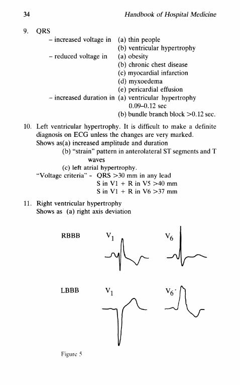
11. Right ventricular hypertrophy Shows as (a) right axis deviation
RBBB
LBBB
Figure :'1
Cardiology
(b) clockwise rotation (c) R > S in Vl, R in Vl > 7 mm, Sin V5 >7 mm Other causes of tall R wave in Vl are: incomplete RBBB, posterior infarction, W-P-W syndrome.
12. Left bundle branch block (see Fig. 5). Always pathological.
35
It is usually impossible to diagnose ischaemia or infarction as well. Causes include ischaemia, hypertension, aortic valve disease, cardiomyopathy.
13. Right bundle branch block (see Fig. 5). Allows diagnosis of underlying infarction. Seen in normals, pulmonary embolism, ischaemia, ASD, cpnducting tissue disease. May be "incomplete" i.e. <0.12 sec.
14. ST segment. Depression in ischaemia, infarction, ventricular hypertrophy, bundle branch block, digoxin, hypokalaemia. Elevation in infarction, pericarditis, ventricular aneurysm. May be raised in normals or in LBBB in right chest leads.
15. T waves. May normally be inverted in III, aVR, aVL, VI, V2. Abnormalities are very non-specific and may be seen in many forms of heart disease and other diseases as well as being related to a variety of physiological activities (eg. hyperventilation, anxiety, change in body position, pre and post prandial.).
16. U waves. Best seen in V2-4. Inverted in anterolateral leads in ischaemia, and hypertensive heart disease. Prominent in V3-5 in hypokalaemia.
17. QT interval. Shortened by digoxin treatment and hypercalcaemia. Prolonged by ischaemia, infarction, hypocalcaemia, stroke, head injury, hypothermia, beta-blockers, amiodarone.
18. Electrolyte effects. Hypokalaemia - prominent U waves, flat or inverted T waves,
ST depression, rarely first degree heart block. Hyperkalaemia - tall peaked T waves, smaller broader QRS
complexes, small or absent P waves.
36 Handbook of Hospital Medicine
Monitoring Central Venous Pressure
INDICATIONS
1. Differential diagnosis and management of shock
2. Fluid replacement where the circulatory balance is precarious (heart disease, elderly, etc.)
3. Fluid balance in general anaesthesia and intensive care.
REFERENCE POINT
This must be defined and kept constant. It is usually either the sternal angle or the mid-thorax at the level of the sternal angle. Normal range depends on the point chosen. Recheck the zero if the patient's position is changed.
TECHNIQUE
1. Percutaneous puncture of the subclavian (either supra- or infraclavicular route), external or internal jugular or antecubital vein depending on your experience.
2. Insert catheter and advance until tip is in the lower SVC (but not in the right atrium). Check that blood can be aspirated easily.
3. Secure to the skin with a suture.
4. Connect to a saline or dextrose manometer.
5. Check that the column of fluid swings with respiration.
6. Zero the manometer with the chosen anatomical reference point.
7. Measure and record the CVP.
8. Arrange a chest X-ray to check the catheter position.
COMPLICATIONS
Mainly those associated with venous cannulation ego thrombosis, septicaemia etc.
Cardiology 37
INTERPRETATION
1. high CVP
2.
- heart failure - overtransfusion - pulmonary embolism - superior vena cava obstruction -tamponade
10wCVP - haemorrhage } - fluid depletion - dehydration
peripheral shutdown
- septicaemia
} - drug overdose - anaphylaxis
peripheral vasodilation
NOTE
The CVP is a poor guide to pulmonary wedge pressure in the presence of
heart or lung disease or drug treatment.
A CVP line can also be used for drug infusion. i.v. feeding and blood
sampling. The trend of readings is much more use than an isolated recording.
Swan-Ganz Catheterisation
A Swan-Ganz catheter is a flow-directed bailon-tipped catheter which can be passed to the pulmonary artery without X-ray screening to measure pulmonary wedge pressure (indirect left atrial pressure and hence left ventricular filling pressure).
INDICATIONS
When monitoring of left ventricular end diastolic pressure (L VEDP) is needed, ego in shock or pulmonary oedema when vasodilators are used. Where repeated thermodilution measurements of cardiac output are needed. To differentiate between VSD and acute mitral valve regurgitation following infarction (by multiple oxygen saturation measurements).
TECHNIQUE
1. Standard subclavian vein puncture as in "Temporary Pacing" leaving a plastic cannula in place.
38 Handbook of Hospital Medicine
2. Note the balloon capacity and prime the catheter with saline.
3. Inflate the balloon to check it is not punctured, and then deflate.
4. Advance the catheter 10-15 cm to right atrium (RA).
5. Inflate balloon. If X-ray screening is available it makes advancement to the pulmonary artery much easier but, if not, connect the catheter to a pressure transducer with an oscilloscope display and a continuous flush system of heparinised saline.
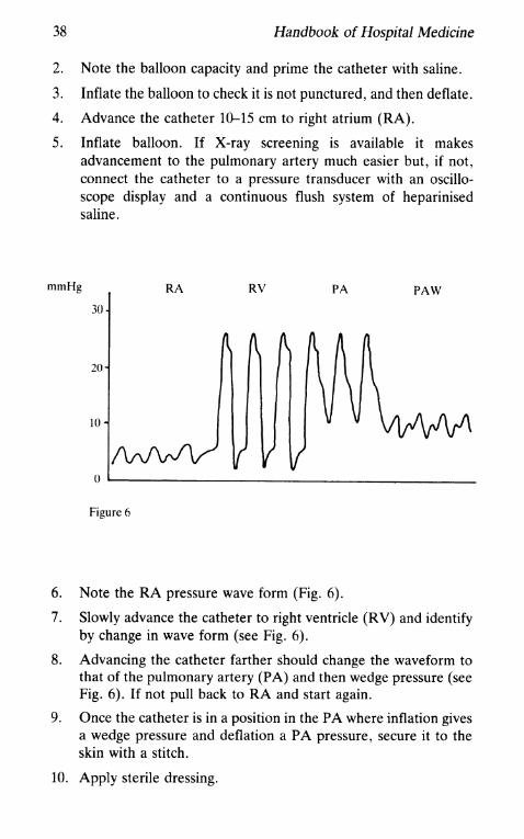
mmHg RA RV PA PAW
30
20
10
o ~----------------________________________ __
Figure 6
6. Note the RA pressure wave form (Fig. 6).
7. Slowly advance the catheter to right ventricle (RV) and identify by change in wave form (see Fig. 6).
8. Advancing the catheter farther should change the waveform to that of the pulmonary artery (PA) and then wedge pressure (see Fig. 6). If not pull back to RA and start again.
9. Once the catheter is in a position in the PA where inflation gives a wedge pressure and deflation a PA pressure, secure it to the skin with a stitch.
10. Apply sterile dressing.
Cardiology 39
11. Check catheter position with chest X-ray.
COMPLICATIONS
- those associated with subclavian vein cannulation - balloon bursting: avoid overinflation - catheter clotting: avoid by slow infusion - pulmonary infarction: do not leave the ballon inflated in the
PA -failure to pass to the PA. Unusual, but may occur if the RA or
RV are dilated, in which case X-ray screening is especially useful.
The catheter can probably be left in position for several days without problems but the latex does degenerate and the manufacturer will provide recommendations.
Temporary Pacing
INDICATIONS
-Stokes-Adams attacks before implantation of permanent pacemaker
- symptomatic bradycardia in acute infarction -complete heart block or Mobitz II block in anterior infarction - prophylactic in bifascicular block in acute infarction - for overdrive pacing of SVT or VT.
PROCEDURE
1. Seldinger technique to cannulate the right subclavian vein (either supra- or infra-clavicularly). If not possible, try the left side. Alternatives are the medial antecubital or femoral veins but these are less convenient.
2. Introduce a 6F bipolar pacing electrode.
3. Using X-ray screening, position the tip of the electrode in the apex of the right ventricle.
4. Connect to the pacing box.
5. Pace on demand at 70/min (or faster if patient's own heart rate is over 70/min).
6. Slowly reduce the voltage to measure the threshold. Less than
40 Handbook of Hospital Medicine
1.0 V is acceptable, less than 0.5 V ideal. If higher than 1.0 V, reposition the electrode.
7. Check stability of electrode position by asking the patient to cough and breathe deeply.
8. Secure the electrode to the skin with a suture.
9. Apply a sterile dressing.
10. Leave the box set at 70 beats/min at 2-3 times the threshold (sometimes should be set slower or faster depending on the indication for pacing).
11. For overdrive pacing, the rate is set 10--20 beats per minute faster than the tachycardia rate to "capture" and is then slowly reduced until the rhythm returns to normal. Pace the right atrium for SVT and the right ventricle for VT. Ensure that the pacing box is capable of rapid pacing. Avoid sustained ventricular pacing at rates near or above 2001 min because of the risk of VF.
COMPLICA nONS
- those of subclavian vein puncture (bleeding, pneumothorax etc. ).
-myocardial perforation is not uncommon. Normally does not cause serious problems. Recognise by pacing failure, friction rub, wire position on chest X-ray.
- pacing failure- ie. failure to capture or sense correctly. Check all connections. Review electrode position by X-ray screening. Reposition electrode if necessary.
-rising threshold. The threshold should be checked once or twice daily and will normally rise 2 or 3 fold over the first 7-10 days.
Conducting an Exercise Test
Despite the very small risk, all exercise tests should be supervised by a doctor. The commonest protocol is the Bruce test, which is
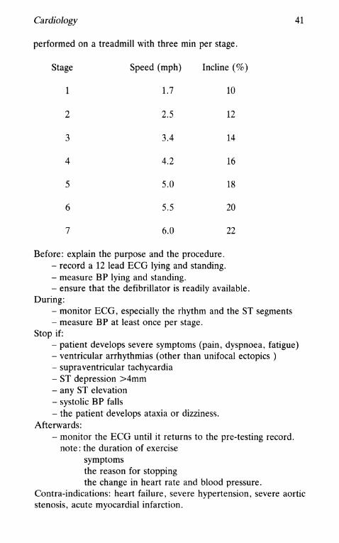
Cardiology
performed on a treadmill with three min per stage.
Stage Speed (mph) Incline (%)
1 1.7
2 2.5
3 3.4
4 4.2
5 5.0
6 5.5
7 6.0
Before: explain the purpose and the procedure. - record a 12 lead ECG lying and standing. - measure BP lying and standing.
10
12
14
16
18
20
22
- ensure that the defibrillator is readily available. During:
- monitor ECG, especially the rhythm and the ST segments - measure BP at least once per stage.
Stop if:
41
- patient develops severe symptoms (pain, dyspnoea, fatigue) - ventricular arrhythmias (other than unifocal ectopics ) - supraventricular tachycardia - ST depression >4mm - any ST elevation - systolic BP falls - the patient develops ataxia or dizziness.
Afterwards: - monitor the ECG until it returns to the pre-testing record.
note: the duration of exercise symptoms the reason for stopping the change in heart rate and blood pressure.
Contra-indications: heart failure, severe hypertension, severe aortic stenosis, acute myocardial infarction.
42 Handbook of Hospital Medicine
NOTE
- if VF occurs, it is usually in the first few minutes after exercise. - be prepared to admit the patient for observation if necessary (ie. prolonged pain or ECG changes, serious arrhythmias etc.). - early post-myocardial infarction testing is becoming more popular as an indicator of prognosis. A much lower work load and a shorter protocol are used.
SUGGESTED READING
1. L.H.Opie Drugs and the Heart. 1980 The Lancet. London.
2. L. Schamroth An Introduction to Electrocardiography 1976. Blackwell Scientific Publications.