guidelines for resuscitation training for ministry …hqe.moh.gov.my/web/ict/resuscitation training...
TRANSCRIPT
GUIDELINES FOR RESUSCITATION TRAINING FOR MINISTRY OF HEALTH
GUIDELINES FOR RESUSCITATION TRAINING FOR MINISTRY OF HEALTH
First published in Malaysia in March 2016
GUIDELINES FOR RESUSCITATION TRAINING FOR MINISTRY OF HEALTH
Procedures were coordinated by:
Surgical and Emergency Services Unit,
Medical Services Development Section,
Medical Development Division,
Ministry of Health Malaysia.
©The Ministry of Health Malaysia 2015 www.moh.gov.my
Institute for Medical Research Cataloguing in Publication Data
A catalogue record for this book is available from the
Institute for Medical Research, Ministry of Health Malaysia
National Library of Malaysia Cataloguing in Publication Data
A catalogue record for this book is available from the
National Library of Malaysia
All rights reserved: no part of this publication may be
reproduced, stored in a retrieval system, or transmitted
in any form or by any means, electronic, mechanical,
photocopying, recording, or otherwise without the prior
permission of the Ministry Of Health Malaysia
GUIDELINES FOR RESUSCITATION TRAINING FOR MINISTRY OF HEALTH
LIST OF ABBREVIATIONS v
FOREWORD by DIRECTOR GENERAL of HEALTH MALAYSIA vi
INTRODUCTION by CHAIRMAN OF NATIONAL COMMITTEE ON RESUSCITATION TRAINING (NCORT) vii
1.0 BASIC LIFE SUPPORT 1
1.1 Adult Basic Life Support 1
1.2 Paediatric Basic Life Support 5
1.3 Choking 8
1.4 Automated External Defibrillator 9
2.0 ADVANCED LIFE SUPPORT 12
2.1 Adult Cardiac Arrest (Pulseless Arrest) 12
2.2 Defibrillation 14
2.3 Airway Management and Ventilation 16
2.4 Drugs 17
2.5 Intravenous and Intraosseous Route 17
2.6 Monitoring During Adult Advanced Life Support 18
2.7 Cardiac Arrest in Special Circumstances 19
2.8 Post Resuscitation Care 19
3.0 ADVANCED PAEDIATRIC LIFE SUPPORT 22
3.1 Prevention of cardiac arrest 22
3.2 Advanced life support during cardiac arrest 24
3.3 Post Resuscitation Care 28
4.0 NEONATAL RESUSCITATION PROGRAMME (NRP) 31
5.0 CARDIAC ARREST IN SPECIAL CIRCUMSTANCES 33
5.1 Drowning - Cardiac arrest caused by drowning 33
5.2 Trauma - Traumatic cardiac arrest (TCA) 33
5.3 Dispatcher Assisted CPR (DA-CPR) 33
CONTENT
6.0 EDUCATION, IMPLEMENTATION, SAFETY AND ETHICS 35
6.1 Training 35
6.2 Implementation 35
6.3 Safety 38
6.4 Ethics of Resuscitation and End of Life Issues 39
7.0 APPENDICES 40
Appendix 1 : In Hospital Resuscitation 41
Appendix 2 : Adult BLS Algorithm for Healthcare Providers in MOH Hospitals & Healthcare Facilities 42
Appendix 3 : Paediatric BLS Algorithm for Healthcare Providers in MOH Hospitals & Healthcare Facilities 43
Appendix 4 : Paediatric Foreign Body Airway Obstruction Algorithm 44
Appendix 5 : Adult Cardiac Arrest Algorithm – 2015 Update 45
Appendix 6 : Tachycardia Algorithm (With Pulse) 46
Appendix 7 : Bradycardia Algorithm 47
Appendix 8 : Neonatal Resuscitation Algorithm 48
Appendix 9 : Drowning Treatment Algorithm 49
Appendix 10 : Traumatic Cardiac Arrest Algorithm 50
8.0 TECHNICAL COMMITTEE 51
v
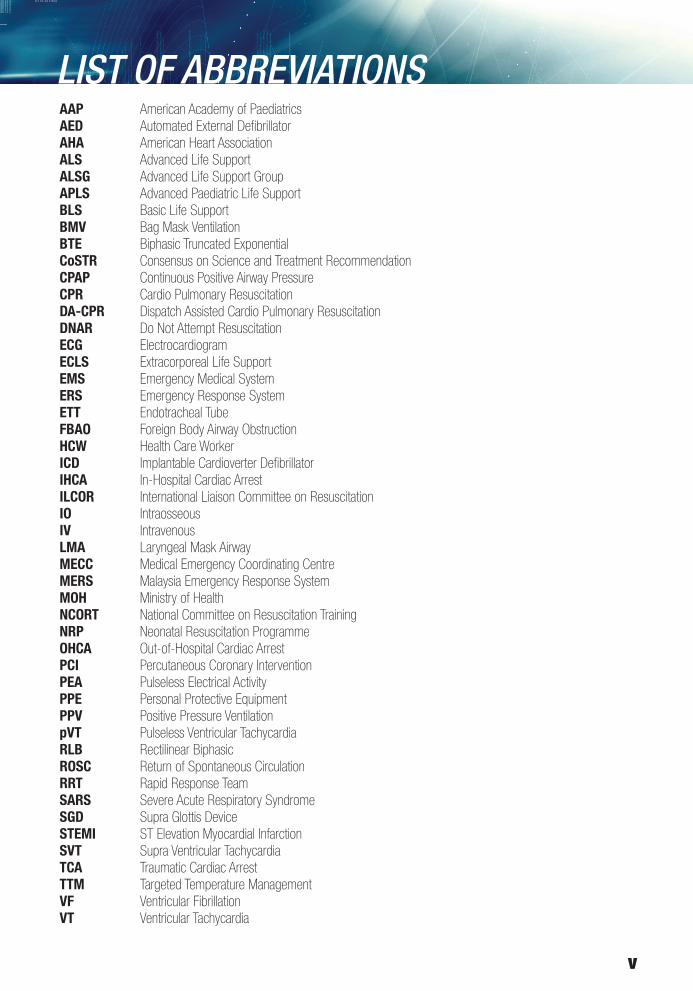
AAP American Academy of PaediatricsAED Automated External DefibrillatorAHA American Heart AssociationALS Advanced Life SupportALSG Advanced Life Support GroupAPLS Advanced Paediatric Life SupportBLS Basic Life SupportBMV Bag Mask VentilationBTE Biphasic Truncated ExponentialCoSTR Consensus on Science and Treatment RecommendationCPAP Continuous Positive Airway PressureCPR Cardio Pulmonary ResuscitationDA-CPR Dispatch Assisted Cardio Pulmonary ResuscitationDNAR Do Not Attempt ResuscitationECG ElectrocardiogramECLS Extracorporeal Life SupportEMS Emergency Medical SystemERS Emergency Response SystemETT Endotracheal TubeFBAO Foreign Body Airway ObstructionHCW Health Care WorkerICD Implantable Cardioverter DefibrillatorIHCA In-Hospital Cardiac ArrestILCOR International Liaison Committee on ResuscitationIO IntraosseousIV IntravenousLMA Laryngeal Mask AirwayMECC Medical Emergency Coordinating CentreMERS Malaysia Emergency Response SystemMOH Ministry of HealthNCORT National Committee on Resuscitation TrainingNRP Neonatal Resuscitation ProgrammeOHCA Out-of-Hospital Cardiac ArrestPCI Percutaneous Coronary InterventionPEA Pulseless Electrical ActivityPPE Personal Protective EquipmentPPV Positive Pressure VentilationpVT Pulseless Ventricular TachycardiaRLB Rectilinear BiphasicROSC Return of Spontaneous CirculationRRT Rapid Response TeamSARS Severe Acute Respiratory SyndromeSGD Supra Glottis DeviceSTEMI ST Elevation Myocardial InfarctionSVT Supra Ventricular TachycardiaTCA Traumatic Cardiac ArrestTTM Targeted Temperature ManagementVF Ventricular FibrillationVT Ventricular Tachycardia
LIST OF ABBREVIATIONS

vi
First and foremost, I would like to thank and congratulate the National Committee on Resuscitation Training (NCORT) and the subcommittees for producing the Guidelines for Resuscitation to be used in all MOH facilities.
Resuscitation training in Ministry of Health Malaysia (MOH) is one of the important areas that needs to be focused upon in order to ensure all healthcare workers in MOH are equipped with life support training that is based on current recommendations practiced worldwide.
The National Committee on Resuscitation Training (NCORT) was formed in 2006 and was given the task to oversee the whole aspect of resuscitation training in MOH with the main aim to streamline and provide direction for resuscitation training in MOH.
To the public and our patients, saving lives is their number one expectation from us and as healthcare workers, we are expected to know how to perform cardiopulmonary resuscitation (CPR). Over the years, MOH Malaysia has provided resuscitation training to all health workers at various levels, from basic to advanced training. The National Committee on Resuscitation Training has produced the resuscitation training guidelines based on the consensus document from International Liaison Committee on Resuscitation (ILCOR) to suit our local culture, and the economic and systemic differences in practice and resources.
With the development of this guideline, it is hoped that it will provide the trainers of life support training a guide to conduct life support courses based on the latest updates and for the Ministry; a standardised and quality training that can be achieved.
FOREWORD
Datuk Dr. Noor Hisham Bin Abdullah
Director General Ministry of Health Malaysia
vii
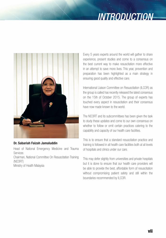
Every 5 years experts around the world will gather to share experience, present studies and come to a consensus on the best current way to make resuscitation more effective in an attempt to save more lives. This year, prevention and preparation has been highlighted as a main strategy in ensuring good quality and effective care.
International Liaison Committee on Resuscitation (ILCOR) as the group is called has recently released the latest consensus on the 15th of October 2015. The group of experts has touched every aspect in resuscitation and their consensus have now made known to the world.
The NCORT and its subcommittees has been given the task to study these updates and come to our own consensus on whether to follow or omit certain practices catering to the capability and capacity of our health care facilities.
This is to ensure that a standard resuscitation practice and training is followed in all health care facilities both at all levels of hospitals and clinics under our care.
This may defer slightly from universities and private hospitals but it is done to ensure that our health care providers will be able to provide the best, affordable form of resuscitation without compromising patient safety and still within the boundaries recommended by ILCOR.
INTRODUCTION
Dr. Sabariah Faizah Jamaluddin
Head of National Emergency Medicine and Trauma ServicesChairman, National Committee On Resuscitation Training (NCORT)Ministry of Health Malaysia

1
GUIDELINES FOR RESUSCITATION TRAINING FOR MINISTRY OF HEALTH
CHAPTER 1- 6
2
GUIDELINES FOR RESUSCITATION TRAINING FOR MINISTRY OF HEALTH
1.0 BASIC LIFE SUPPORT
1.1 Adult Basic Life Support
Highlights in 2015
1. Guidelines 2015 highlights the critical importance of the interactions between the emergency medical dispatcher, the bystander who perform CPR and the use of AED.
2. Emphasis on importance of early recognition of cardiac arrest (telephone CPR - dial 999) via MERS (Malaysian Emergency Response System).
3. Emphasis on high quality CPR.
3.1 Compression rate 100-120 compression per minute
3.2 The depth is 5cm but not more than 6cm
3.3 Minimal interruption in chest compression < 10 seconds
3.4 Allow spontaneous recoil of the chest wall in between compression.
4. Be aware that seizures can be a sign of cardiac arrest.
5. Real time CPR feedback should be used to ensure high quality CPR if available. The use of real time CPR feedback in clinical practice should be considered as part of a comprehensive system for care for cardiac arrest.
6. Implementation of public-access defibrillator programme.
Issues addressed:
1.1.1 Assessing Danger and Safety to Health Care Worker Prior to Resuscitation
HCW (Health Care Worker) shall be taught to protect themselves from danger while providing CPR, these includes:
1.1.1.1 Wear PPE (gloves, apron, mask) if available.
1.1.1.2 Look out for blood spills, sharps, electric wires.
1.1.1.3 Unsteady beds, trolley
1.1.2 Responsiveness
1.1.2.1 HCW shall be taught to check for the victim’s responsiveness by gently tapping the shoulder and ask loudly: ‘Are you ok?’
1.1.3 Shout for help
1.1.3.1 HCW shall be taught to activate the Emergency Response System (ERS) or to shout the following words after suspecting a cardiac arrest. ‘Emergency! Emergency! Bring the resuscitation trolley and defibrillator!’
CHAPTER 1
3
Chapter 1 : Basic Life Support
1.1.4 Positioning
1.1.4.1 Victims found on the floor should be initially managed on the floor.
1.1.4.2 Face down victims shall be rolled over to the supine position.
1.1.4.3 Air filled mattresses should be deflated during CPR.
1.1.4.4 Backboard use is not recommended because of delays in initiation or interruptions of compressions and the potential of dislodging tubes and catheters during backboard placement.
1.1.5 Airway
1.1.5.1 The airway should be opened by using head tilt-chin lift or jaw thrust if suspected cervical injury.
1.1.6 Breathing
1.1.6.1 Absent or abnormal breathing shall be determined simultaneously while opening the airway by looking at the chest, neck and face for not more than 10s.
1.1.6.2 HCW shall be taught to recognize absence of breath and presence of abnormal breathing treat as a sign of cardiac arrest.
1.1.6.3 Chest compression shall begin with absence of normal breathing.
1.1.6.4 HCW shall be taught that in cases if in doubt whether breathing is normal, act as if it is they are not breathing normally and prepare to start CPR.
1.1.7 Chest compression
Emphasize on high quality chest compression during BLS training.
The components include:
1.1.7.1 Location:
The lower half of the chest shall be the site for hand placement. This is taught as ‘place the heel of your hand in the centre of the chest with the other hand on top’. This instruction shall be accompanied by the demonstration of placing the hands on the lower half of the sternum.
1.1.7.2 Rate:
100 to 120 compressions per minute.
1.1.7.3 Depth:
5 cm but not greater than 6cm.
1.1.7.4 Recoil:
Complete recoil of the chest must be allowed after each compression.
1.1.7.5 Minimize interruption on chest compression for:
Delivery of rescue breaths, shocks, ventilations and rhythm analysis.
Pre- and post-shock pauses of less than 10 s.

4
GUIDELINES FOR RESUSCITATION TRAINING FOR MINISTRY OF HEALTH
1.1.8 Ratio of chest compression to ventilation
The compression-ventilation ratio shall be 30:2 if the airway is not secured.
1.1.9 Ventilation
1.1.9.1 In the MOH hospital scenario, bag mask devices are usually available. Performing mouth to mouth ventilations is hardly done as there is fear of disease transmission from hospitalized patients. It however may need to be done in areas without a bag-mask device. Protective devices like pocket mask and face shields are available to reduce the uneasiness of mouth to mouth ventilation.
1.1.9.2 Use of bag-mask device or bag-valve mask shall be taught to all HCW.
1.1.9.3 Each breath shall be given within 1 second inspiratory time until a chest rise is observed.
1.1.9.4 In areas without bag-mask device, HCW shall be taught to perform chest compression only CPR.
1.1.10 Defibrillation
Early defibrillation is an essential step in the chain of survival for victims of cardiac arrest.
1.1.10.1 HCW shall be taught on defibrillation during BLS.
1.1.10.2 HCW shall be taught to deliver defibrillation as soon as it is available in shockable rhythm and resume chest compression immediately after defibrillation.
1.1.10.3 Minimize interruption of chest compression during attachment of defibrillator and rhythm analysis.
1.1.10.4 Encourage the use AED function in manual defibrillator if the provider is untrained to use manual defibrillation for IHCA.
1.1.11 Reassessment of during CPR
1.1.11.1 After every 5 cycles or 2 minutes of CPR, HCW shall check for normal breathing.
1.1.12 Teaching of pulse check
1.1.12.1 If a cardiac monitor is available, the carotid shall be checked when an organized rhythm is seen.
1.1.13 Stopping CPR
1.1.13.1 CPR can be stopped in following circumstances:
1.1.13.1.1 Victim recovers with normal breathing.
1.1.13.1.2 HCW is exhausted.
1.1.13.1.3 Assistance arrives to take over CPR.
1.1.13.2 HCW shall be taught to switch the role of chest compressions every 5 cycles or 2 minutes to avoid fatigue.
5
Chapter 1 : Basic Life Support
1.1.14 Recovery position
1.1.14.1 Recovery position is applied when victims resume normal breathing but remain unresponsive.
1.1.14.2 HCW shall be taught the recovery position during the BLS course. The technique taught must ensure the following:
1.1.14.2.1 Victim is in the true lateral position.
1.1.14.2.2 Head in the dependant position.
1.1.14.2.3 Position is stable.
1.1.14.2.4 Position is safe and comfortable to the victim.
1.2 Paediatric Basic Life Support
HCW who have been taught adult BLS and have no specific knowledge of paediatric resuscitation may use the adult sequence as outcome is worse if they do nothing.
The following sequence is to be followed in a Paediatric emergency situation.
1.2.1 Ensure The Safety of Rescuer and Child
1.2.1.1 The HCW should ensure the external environment is safe and the child is not in danger.
1.2.2 Check for the Child’s Responsiveness
1.2.2.1 Gently stimulate the child and ask loudly “Are you alright?”.
1.2.2.2 If the child responds by answering or moving, let him in the position in which you find him (provided he is safe), check his condition and seek help.
1.2.2.3 Reassess him regularly.
1.2.2.4 If he does not respond, shout for help.
1.2.3 Shout for Help
1.2.3.1 Shout for help by saying out loudly ‘Emergency! Emergency! Bring the resuscitation trolley and defibrillator!’.
1.2.4 Positioning of the Child
1.2.4.1 Victims found on the floor should be initially managed on the floor.
1.2.4.2 Face down victims shall be rolled over to the supine position.
1.2.4.3 Air filled mattresses should be deflated during CPR.
1.2.4.4 Backboard use is not recommended because of delays in initiation or interruptions of compressions and the potential of dislodging tubes and catheters during backboard placement.
1.2.5 Open The Airway
1.2.5.1 Open the airway by the head tilt chin lift manoeuvre.

6
GUIDELINES FOR RESUSCITATION TRAINING FOR MINISTRY OF HEALTH
1.2.6 Breathing
1.2.6.1 Checking for normal breathing should not take more than 10 seconds.
1.2.6.1.1 Keeping the airway open, look, listen and feel for normal breathing.
1.2.6.1.2 Look for chest movements.
1.2.6.1.3 Listen for breath sounds at the child’s nose and mouth.
1.2.6.1.4 Feel for air movements on your cheek.
1.2.6.2 If the child is breathing normally,
1.2.6.2.1 Turn him on his side into the recovery position.
1.2.6.2.2 Send or go for help.
1.2.6.2.3 Check for continuous normal breathing.
1.2.6.3 If breathing is NOT normal or absent,
1.2.6.3.1 Give 5 initial rescue breaths.
1.2.6.3.2 The duration of delivering a breath is about 1 second sufficient to produce a visible chest rise.
1.2.7 Assessing the Child’s Circulation
1.2.7.1 Take not more than 10 seconds to check for signs of life and to palpate the pulse.
1.2.7.2 For an infant feel the brachial pulse.
1.2.7.3 For a child feel the carotid pulse.
1.2.7.4 The femoral pulse may be felt in both infant and child.
1.2.7.4.1 If there is a palpable pulse ≥ 60/minute but there is inadequate breathing, continue rescue breaths at 12 - 20 breaths per minute till the child starts breathing effectively on his own.
1.2.7.4.2 If pulse is < 60/minute and there are signs of poor perfusion or no signs of life, start chest compressions.
1.2.7.5 The signs of life to look for are:
1.2.7.5.1 Any spontaneous movement
1.2.7.5.2 Coughing
1.2.7.5.3 Normal breathing (gasps or infrequent, irregular breaths are abnormal breathing)
1.2.8 Chest Compressions
“Push hard” and “Push fast”, allowing full chest recoil after each compression, minimize interruptions of chest compressions and avoid excessive ventilation.
1.2.8.1 Technique:
1.2.8.1.1 For one rescuer CPR in an infant, the rescuer compresses with the tips of 2 fingers.
7
Chapter 1 : Basic Life Support
1.2.8.1.2 For two rescuers CPR in an infant, the two thumb chest compression technique is used.
1.2.8.1.3 For a child >1year old, use the heel of one hand over the lower sternum.
1.2.8.1.4 For a larger child, use 2 hands with fingers interlocked.
1.2.8.2 Site of Compression:
1.2.8.2.1 For Infant - lower half of the sternum
1.2.8.2.2 For Child - lower half of the sternum
1.2.8.3 Depth of Compression:
1.2.8.3.1 For Infant - at least 1/3 the depth of the chest or 4 cm
1.2.8.3.2 For Child - at least 1/3 the depth of the chest or 5 cm
1.2.8.4 Rate of Compression:
1.2.8.4.1 Push at the rate of at least 100-120/mm
1.2.8.5 Ratio of Compressions to Breaths:
1.2.8.5.1 One Rescuer CPR - 15:2
1.2.8.5.2 Two Rescuers CPR - 15:2
Switch the role of chest compressions every 10 cycles or 2 minutes to avoid fatigue.
When an advance airway is in place, compressions should not be interrupted for ventilations.
1.2.9 Ventilation
1.2.9.1 In the MOH hospital scenario, bag mask devices are usually available. Performing mouth to mouth ventilations is hardly done as there is fear of disease transmission from hospitalized patients. It however may need to be done in areas without a bag-mask device. Protective devices like pocket mask and face shields are available to reduce the uneasiness of mouth to mouth ventilation.
Recommendations
1. Use of bag-mask device shall be taught to all HCW.
2. Mouth to mouth ventilation shall be taught for use outside of the - hospital and in hospital areas without bag mask devices.
3. Use of protective devices shall be taught during BLS courses.
1.2.10 Defibrillation
1.2.10.1 Defibrillation (AED and/or Manual) training will be part of Paediatric BLS course content.
1.2.10.2 HCW will be taught to attach the defibrillator as soon as it is available, with minimal interruptions to chest compression.
1.2.10.3 AED with paediatric pads or programs which attenuate the output to 50-75J is preferred to be used for children between 1 to 8 years. Unmodified adult AED may be used if this is not available.
8
GUIDELINES FOR RESUSCITATION TRAINING FOR MINISTRY OF HEALTH
1.2.11 Stopping CPR
1.2.11.1 CPR can be stopped in following circumstances:
1.2.11.1.1 The child shows signs of life (starts to wake up, move, open eyes and breath normally).
1.2.11.1.2 More HCW arrive to assist or take over.
1.2.11.1.3 HCW is exhausted.
1.2.12 Recovery Position
1.2.12.1 Recovery position is applied when victims resume normal breathing but remain unresponsive.
1.2.12.2 HCW will be taught the recovery position during the BLS course. The technique taught must ensure the following:
1.2.12.2.1 Victim is in the true lateral position.
1.2.12.2.2 Avoid any pressure on the child’s chest that may impair breathing.
1.2.12.2.3 Head in the dependent position.
1.2.12.2.4 Position is stable.
1.2.12.2.5 Position is safe and comfortable to the victim.
1.2.12.2.6 Regularly change side to avoid pressure points (every 30 min).
1.3 Choking
1.3.1 Adult with Foreign Body Airway Obstruction (FBAO)
1.3.1.1 If cough is effective, encourage the victims to cough until the obstruction is relieved.
1.3.1.2 If cough is ineffective, apply 5 back blows, if unable to relieve the obstruction, to apply 5 abdominal thrusts.
1.3.1.3 If the obstruction is still not relieved, continue alternating five back blows with five abdominal thrusts.
1.3.1.4 If patient deteriorates to become unconscious, HCW should begin CPR.
1.3.2 Paediatric Foreign Body Airway Obstruction (FBAO)
1.3.2.1 If the child is coughing effectively, no manoeuvre is necessary.
1.3.2.1.1 Encourage the child to cough.
1.3.2.1.2 Continue monitoring the child’s condition.
1.3.2.2 If the child’s coughing is becoming ineffective,
1.3.2.2.1 Shout for help immediately.
1.3.2.2.2 Determine the child’s conscious level.
1.3.2.2.3 For a child, perform back blows or abdominal trusts until the object is expelled or the child becomes unresponsive.
1.3.2.2.4 For an infant, deliver repeated cycles of 5 back blows (slaps) followed by 5
9
Chapter 1 : Basic Life Support
chest thrusts until the object is expelled or the victim becomes unconscious.
1.3.2.2.5 Abdominal trusts are not recommended in infants as it may cause liver injury.
1.3.2.3 If the victim becomes unresponsive:
1.3.2.3.1 Call for help if it is still not available.
1.3.2.3.2 Open the mouth and look for any obvious object. If one is seen, make an attempt to remove it with a single finger sweep. Do not attempt blind or repeated finger sweeps.
1.3.2.3.3 Open the airway using a head tilt/chin lift.
1.3.2.3.4 Attempt five rescue breaths. If a breath does not make the chest rise, reposition the head before making the next attempt.
1.3.2.3.5 If there is no response (moving, coughing, spontaneous breaths) proceed to chest compressions without further assessment of the circulation.
1.3.2.3.6 After 15 compressions and 2 ventilations, activate the EMS if no one has done so.
1.3.2.3.7 Continue with cycles of 15 chest compressions and 2 ventilations until the object is expelled.
1.4 Automated External Defibrillator
Highlights
1. All BLS providers are trained on use of an AED.
2. VF and pulseless VT are treatable cardiac arrest rhythms with outcomes closely related to the rapidity of recognition and treatment.
3. Survivals in victims of VF/ pulseless VT is highest when bystander deliver CPR and defibrillation is attempted within 3-5 minutes of collapse.
Issues addressed:
1.4.1 CPR before defibrillation
1.4.1.1 For witnessed adult cardiac arrest when an AED is immediately available, it is reasonable that the defibrillator be used as soon as possible.
1.4.1.2 For adults with unmonitored cardiac arrest or for whom an AED is not immediately available, it is reasonable that CPR be initiated while the defibrillator equipment is being retrieved and applied and that defibrillation, if indicated, be attempted as soon as the device is ready for use.
1.4.2 Timing of rhythm check
1.4.2.1 It may be reasonable to immediately resume chest compression after shock delivery for adults in cardiac arrest in any setting.
1.4.2.2 If there is alternative physiologic evidence of ROSC (e.g. arterial waveform or rapid rise in ETCO2), chest compression can be paused briefly for rhythm check.
10
GUIDELINES FOR RESUSCITATION TRAINING FOR MINISTRY OF HEALTH
1.4.3 Use of AED
1.4.3.1 Standard AEDs are suitable for use in children older than 8 years old.
1.4.3.2 Paediatric pads with attenuator or paediatric mode should be used in children between 1 and 8 years old. If these are not available, the AED should be used.
1.4.3.3 The incidence of shockable rhythms in infants is very low except when there is cardiac disease. In these rare cases, if an AED is the only defibrillator available, its use should be considered (preferably with a dose attenuator).
1.4.4 Public-access defibrillation
1.4.4.1 Implementation of public-access defibrillation program for patients with OHCAs.
1.4.4.2 AEDs to be placed in:
1.4.4.2.1 Public places with a high density and movement of people such as airports, railway stations, bus terminals, shopping malls, offices and centres.
1.4.4.2.2 Remote locations where an emergency ambulance response would be likely to be delayed (e.g. aircraft, ferries and off-shore locations).
1.4.4.2.3 Places where cardiac arrest are frequently witnessed e.g. clinics, pharmacy and stadiums.
1.4.4.3 Registration of defibrillations with local ambulance services is highly desirable so that the dispatches can direct CPR providers to the nearest AED.
1.4.4.4 Placement of AEDs in areas where one cardiac arrest per 5 years can be expected is considered cost-effective and comparable to other medical interventions.
1.4.5 Use of defibrillator with AED function
1.4.5.1 It is recommended to use AED function in manual defibrillator if the provider is untrained to use manual defibrillation for IHCA.
11
GUIDELINES FOR RESUSCITATION TRAINING FOR MINISTRY OF HEALTH
2.0 ADVANCED LIFE SUPPORT
Summary of major changes in Adult Advanced Life Support 2015
The 2015 Advanced Life Support (ALS) guidelines have emphasized the changes aimed at improving patient care and outcome during and after cardiopulmonary resuscitation.
The key changes since 2010 are:
1. Continuing emphasis on the approach to prevention of in-hospital cardiac arrest that includes staff education, monitoring of patients, recognition of patient deterioration and implementation of the effective rapid response system during cardiac arrest.
2. Performing minimally interrupted high quality CPR is the cornerstone for cardiac arrest. Interruption in chest compression must be less than 5 seconds when attempting defibrillation or tracheal intubation.
3. Monitoring physiological parameters during CPR may be reasonable.
4. Continuous waveform capnography is recommended as the most reliable method of confirming the correct placement of ETT, monitoring CPR quality, detect the return of spontaneous circulation and predict CPR outcome.
5. There is a broad recommendation to airway management during CPR. Using either bag mask device or advanced airway device is determined mainly by the skill and experience of the provider.
6. Self –adhesive defibrillation pads should always be used in preference when available.
7. In general, there are no major changes in the recommendation for the drug therapy during CPR. But, administration of vasopressin as the sole vasoactive drug during CPR has been removed from the algorithm.
8. There is possibility of bundling treatment of steroid, vasopressin and epinephrine (adrenaline) in the treatment of in-hospital cardiac arrest.
9. Ultrasound may be considered during management of cardiac arrest in cases with potentially reversible causes.
10. The routine use of mechanical chest compression devices is not recommended. However, it may be useful in situation where sustained high quality manual chest compression is impractical or compromise provider safety.
11. Extracorporeal life support technique (ECPR) may be considered for selected cardiac arrest patient with potential reversible cause where standard ALS measures are not recommended.
12. Targeted temperature management for post cardiac arrest patient is 32-360C for at least 24 hours.
CHAPTER 2
12
GUIDELINES FOR RESUSCITATION TRAINING FOR MINISTRY OF HEALTH
Treatment Algorithm in 2015 Guidelines
There are basically no major changes in treatment algorithm in 2015 Guidelines in comparison to 2010. Thus the 2010 Guidelines is still relevant.
2.1 Adult Cardiac Arrest (Pulseless Arrest)
The only major changes are, Vasopressin has been taken out from the algorithm. Vasopressin is no longer given to replace 1st or 2nd dose of adrenaline as in 2010 guidelines.
2.1.1 Shockable Rhythm. (VF/pVT) (Refer to Appendix 5)
2.1.1.1 In shockable rhythm (VF/pVT) early defibrillation is the utmost important and high quality CPR takes precedence over the drugs and airway intervention.
2.1.1.2 Defibrillation is by biphasic defibrillator, at device specific energy by manufacturer. However if unknown, the maximum energy should be used.
2.1.1.3 It is appropriate to consider escalating the shock energy if feasible, after a failed shock and for patients where Refibrillation occurs.
2.1.1.4 Differentiate between Refractory VF (when fibrillation persists after one or more shocks) and Refibrillation (when recurrence of VF during a documented cardiac arrest episode, occurring after initial termination of VF while the patient remains under the care of the same providers). Higher termination of VF if higher energy is used to defibrillate, if the machine is capable of giving higher energy.
2.1.1.5 Avoid pre-shock pause of more than 5 secs. Even a 5-10 secs delay will reduce the chance of survival. Use of self-adhesive pad is emphasized.
2.1.1.6 To continue giving high quality chest compression for 2 minutes after delivery of shock to improve coronary and cerebral perfusion. Only to check the pulse and rhythm after complete 2 minutes cycle of CPR.
2.1.2 Shockable, Witnessed (monitored VF/pVT )
2.1.2.1 Three quick successive (stacked) shocks can be considered if a patient has a monitored and witnessed cardiac arrest in the catheter laboratory, coronary care unit, a critical area or whilst monitored after cardiac surgery and a manual defibrillator is rapidly available.
2.1.2.2 Although there are no data supporting a three-shock strategy in these circumstances, it is unlikely that chest compression will improve the already very high chance of ROSC when defibrillation occurs early in the electrical phase, immediately after the onset of VF/pVT.
2.1.2.3 If this initial three-shock strategy is unsuccessful for a monitored VF/pVT cardiac arrest, the ALS algorithm should be followed and these three-shocks treated as if only the first sig le shock has been given.
2.1.2.4 The use of CO2 capnography may enable ROSC to be detected without pausing for chest compression and may avoid giving adrenaline after ROSC. If ROSC is suspected during CPR, to withhold adrenaline.
2.1.2.5 Give adrenaline if cardiac arrest is confirmed at the next rhythm check. Adrenaline has shown to give poor neurological outcome. Studies on the adrenaline dosages and timing are ongoing.
2.1.2.6 The use of bedside ultrasound to confirm ETT placement can be considered if available.
2.1.2.7 I/V amiodarone (300mg followed by 900mg infusion, 2g/day, max). A repeat amiodarone 150mg can also be given.
13
Chapter 2 : Advanced Life Support
2.1.2.8 In refractory VF/pVT, as an alternative to amiodarone, lignocaine can be used (initial dose of 100mg (1-1.5mg/kg, then additional 50mg if necessary, maximum 3mg/kg during first hour). Only to use if amiodarone is unavailable.
2.1.3 Shockable Rhythm (Ventricular fibrillation (VF), persistent Ventricular Tachycardia (pVT))
2.1.3.1 Consider changing the position of pads/paddles. To review all possible causes of 4Hs and 4Ts and to treat if identified.
2.1.3.2 Persistent VF/pVT may be an indication for percutaneous coronary intervention (PCI)-in these cases, a mechanical chest compression can be used to maintain high quality chest compression for transport and PCI.
2.1.3.3 The use of extracorporeal should also be considered to support the circulation whilst a reversible cause is treated.
2.1.3.4 A single precordial thump has a very low success rate for termination of a shockable rhythm. Its routine used is therefore not recommended. It may be appropriate therapy only when used without delay whilst awaiting the arrival of a defibrillator in a monitored VF/pVT.
2.1.4 Non shockable rhythm (PEA/asystole) (Refer to Appendix 5)
2.1.4.1 It may be reasonable to administer adrenaline as soon as feasible after the onset of cardiac arrest due to an initial non-shockable rhythm.
2.1.4.2 A very large observational study of cardiac arrest with non-shockable rhythm comparing adrenaline given at 1 minutes to 3 minutes intervals with adrenaline given at 3 later time intervals (4 to 6, 7 to 9 and greater than 9 minutes). The study found as association between early administration of adrenaline and increase ROSC, survival to hospital discharge and neurologically intact survival.
2.1.4.3 Whenever a diagnosis of asystole is made, check the ECG carefully for the presence of P waves, because this may respond to cardiac pacing. There is no benefit in attempting a true asystole.
2.1.4.4 If there is a doubt about whether the rhythm asystole or fine VF, do not attempt defibrillation; instead continue chest compression and ventilation.
2.1.4.5 Continuing high quality CPR however may improve the amplitude and frequency of the VF and improve the chance of successful defibrillation to a perfusing rhythm.
2.1.4.6 At all times during cardiac arrest, attempt should be made to find any reversible causes of 4Hs-Hypoxia, Hypovolaemia, Hyperkalemia/Hypokalemia, Hypothermia and 4Ts-Thrombosis, Tension Pneumothorax, Cardiac Tamponade and Toxin.
Use of ultrasound (Focused Cardiac Ultrasound) imaging during advanced life support
1. Echocardiography has the potential to detect reversible causes of cardiac arrest e.g. Cardiac Tamponade, Pulmonary Embolism, hypovolaemia, pneumothorax and pseudo-PEA (when the cardiac contraction is too weak to produce detectable pulse of blood pressure).
2. Specific protocol may be helpful, but require considerable training to minimize interruption to chest compression.
14
GUIDELINES FOR RESUSCITATION TRAINING FOR MINISTRY OF HEALTH
2.1.5 Peri-arrest Arrhythmias
Similar guidelines as 2010.Initial assessment of ABCDE, followed by medications if stable or cardioversion if unstable.
2.1.6 Tachycardia Algorithm (Refer to Appendix 6)
2.1.6.1 Unstable tachycardia (symptomatic when HR> 150beats/min or systolic pressure <90mmHg).
2.1.6.2 If clinically and haemodynamically unstable, synchronized cardioversion must be attempted, if fail to follow with medication (amiodarone 300mg, then 900mg infusion). Then, repeat cardioversion.
2.1.6.3 SVT, Atrial flutter - start with 70-120J, escalate if necessary.
2.1.6.4 VT, Atrial regular fibrillation - to start with 120-150J, escalate if necessary.
1. Stable narrow complex tachycardia (regular) (rate control manoeuvres and drugs e.g. Adenosine.
2. Stable narrow complex tachycardia (irregular) rate control with beta-blocker or diltiazem or amiodarone if any evidence of heart failure. Anticoagulant if duration more than 24 hours.
3. Stable broad complex tachycardia, rate control medication with antiarrhythmic e.g. amiodarone
2.1.7 Bradycardia algorithm (Refer to Appendix 7)
Same as 2010 guidelines. Symptomatic when HR < 60 beats/min. Initial ABCDE, followed with medications. If unresponsive for pacing.
2.2 Defibrillation
2.2.1 The defibrillation strategy for the 2015 Resuscitation Guidelines has seen little change from the former guidelines. Below are some of the updated recommendations:
2.2.1.1 The importance of early, uninterrupted chest compressions remains an emphasis throughout these guidelines, together with minimizing the duration of pre-shock and post-shock pauses.
2.2.1.2 Continued chest compressions is recommended during defibrillator charging, deliver defibrillation with an interruption in chest compressions of no more than 5 seconds and immediately resume chest compressions following defibrillation.
2.2.1.3 Self-adhesive defibrillation pads have a number of advantages over manual paddles and should always be used in preference when they are available.
2.2.1.4 CPR should be continued while a defibrillator or automated external defibrillator (AED) is retrieved and applied but defibrillation should not be delayed longer than needed to establish the need for defibrillation and charging.
2.2.1.5 The use of up to three-stacked shocks may be considered if initial VF/pVT occurs during a witnessed, monitored arrest with a defibrillator immediately available example during cardiac catheterization.
15
Chapter 2 : Advanced Life Support
2.2.1.6 Although it is recognized that some areas continue to use the older monophasic waveforms, when possible, biphasic waveforms should be used in preference to the older monophasic waveform for the treatment of both atrial and ventricular arrhythmias. Defibrillation recommendations in the 2015 guidelines apply only to biphasic waveforms. For monophasic defibrillators, the 2010 guidelines still apply.
2.2.1.7 In an oxygen-enriched atmosphere, sparking from poorly applied defibrillator paddles can cause a fire and significant burns to a patient. The absence of case reports of fires caused by sparking where defibrillation was delivered using self-adhesive defibrillation pads suggests that the latter minimize the risk of electrical arcing and should always be used when possible.
2.2.1.8 More patients are presenting with implantable medical devices (e.g. permanent pacemaker, implantable cardioverter defibrillator (ICD)). Medic alert bracelets are recommended for these patients. These devices may be damaged during defibrillation if current is discharged through electrodes placed directly over the device. Place the electrode away from the device (at least 8cm).
2.2.1.9 When defibrillation is warranted, give a single shock and resume chest compressions immediately following the shock. Do not delay CPR for rhythm reanalysis or a pulse check immediately after a shock. Continue CPR (30 compressions:2 ventilations) for 2 min until rhythm reanalysis is undertaken and another shock given (if indicated). Even if the defibrillation attempt is successful, it takes time until the post shock circulation is established and it is very rare for a pulse to be palpable immediately after defibrillation. Patients can remain pulseless for over 2 min and the duration of asystole before ROSC can be longer than 2 min in as many as 25% of successful shocks.
2.2.2 Energy levels
Defibrillation requires the delivery of sufficient electrical energy to defibrillate a critical mass of myocardium, abolish the wave fronts of VF and enable restoration of spontaneous synchronized electrical activity in the form of an organized rhythm. The optimal energy for defibrillation is that which achieves defibrillation whilst causing the minimum of myocardial damage.
2.2.2.1 First shock
2.2.2.1.1 Defibrillation shock energy levels are unchanged from the 2010 guidelines.
2.2.2.1.2 The initial biphasic shock should be no lower than 120 J for RLB (rectilinear biphasic) waveforms and at least 150 J for BTE (biphasic truncated exponential) waveforms. Ideally, the initial biphasic shock energy should be at least 150 J for all waveforms.
2.2.2.2 Second and subsequent shocks
2.2.2.2.1 The 2010 guidelines recommended either a fixed or escalating energy strategy for defibrillation.
2.2.2.2.2 There remains no evidence to support either a fixed or escalating energy protocol, although an escalating protocol may be associated with a lower incidence of refibrillation. Both strategies are acceptable; however, if the first shock is not successful and the defibrillator is capable of delivering shocks of higher energy it is reasonable to increase the energy for subsequent shocks.
2.2.2.3 Recurrent Ventricular Fibrillation (refibrillation)
2.2.2.3.1 Refibrillation is common and occurs in the majority of patients following initial first-shock termination of VF.
16
GUIDELINES FOR RESUSCITATION TRAINING FOR MINISTRY OF HEALTH
2.2.2.3.2 It is recommended that if a shockable rhythm recurs after successful defibrillation with ROSC, and the defibrillator is capable of delivering shocks of higher energy it is reasonable to increase the energy for subsequent shocks.
2.2.3 Cardioversion
If electrical cardioversion is used to convert atrial or ventriculartachyarrhythmias, the shock must be synchronized to occur with the R wave of the electrocardiogram rather than with the T wave: VF can be induced if a shock is delivered during the relative refractory portion of the cardiac cycle.
2.3 Airway Management and Ventilation
The options of airway management during resuscitation is much depend on patient factors, the phase of resuscitation (during CPR or after ROSC) and the skill of the rescuers. A bag mask ventilation (BMV), supra glottis devices (e.g. LMA, Supreme) and endotracheal tube are often used during resuscitation as a part of stepwise approach to airway management. After ROSC, ultimately an endotracheal placement is needed in order to continue care in the post resuscitation periods.
2.3.1 Oxygen during CPR
2.3.1.1 The ultimate goals in performing CPR are to restore circulation to vital organ (e.g. heart) in order for it to resume work and adequate oxygen delivery is vital to achieve this goal.
2.3.1.2 When supplementary oxygen is available, use maximal feasible inspired oxygen concentration during CPR. The detrimental effect of hypoxia superseded the harmful effect of hyperoxia that may exist in the immediate post cardiac arrest periods.
2.3.1.3 After ROSC, titrate the inspired oxygen concentration to maintain the arterial blood oxygen saturation in the range of 94-98%.
2.3.2 Adjunct for Airway Management and Ventilation
2.3.2.1 When cardiac arrest occurs, rescuers must determine the best way to provide oxygen to victims to prevent detrimental effect of hypoxia. Option includes bag mask ventilation (BMV), supra glottis devices (SGD) and endotracheal tube (ETT).
2.3.2.2 There is inadequate evidence to show a difference in survival or favourable neurologic outcome with the use of bag mask devices (BMV), supra glottis devices (SGD) and endotracheal tube (ETT). Either a bag mask devices or an advance airway may be used for oxygenation and ventilation during CPR. Advance airway devices should be inserted by trained rescuers who are familiar with devices insertion and confirming its placement.
2.3.2.3 A supra glottis devices e.g. laryngeal mask, I-gel, Supreme are relatively easier to insert in comparison to an endotracheal intubation. A rescuer inserting an endotracheal should not interrupt chest compression for more than 5 seconds.
2.3.2.4 Continuous waveform capnogram is recommended in addition to clinical assessment as the most reliable method of conforming and monitoring correct placemat of an endotracheal tube. Other methods includes non-wave CO2 detector, esophageal detection device or ultrasound used by an experience operator are a reasonable alternative.
2.3.2.5 The emerging era of video-laryngoscopy, even though they enable a better view of the larynx and improved successful rate of endotracheal placement needs further study and data before recommendations can be made for wider use during CPR.
2.3.2.6 Once an advanced airway inserted, the ventilation rate should be 10 breaths per minute (1 breath every 6 seconds) while continuous chest compression being performed. If performing ventilation either by using bag mask, supra glottis devices or even
17
Chapter 2 : Advanced Life Support
endotracheal tube seems impossible, delivery of oxygen through a cannula or surgical cricothyrodotomy may be lifesaving. A tracheostomy is not advisable in this situation as it is time consuming.
2.4 Drugs
1. The ILCOR 2015 systemic reviews found insufficient evidence to comment on critical outcomes such as survival to discharge and survival discharge with good neurological outcome with any drug during CPR.
2. There was also insufficient evidence to comment on the best time to give drugs to optimize outcome.
3. Thus, although drugs are still included among ALS interventions, they are secondary importance to high quality uninterrupted chest compressions and early defibrillation.
2.4.1 Vasopressor
No placebo-controlled study had shown the routine use of any vasopressor increases the survival to hospital discharge but documented improvement in short term survival.
However, vasopressor continues to be recommended as a means of increasing cerebral and coronary perfusion during CPR.
2.4.1.1 Adrenaline Vs Vasopressin
2.4.1.1.1 Administration of vasopressin as the sole vasoactive drug during CPR has been removed from the algorithm.
2.4.1.1.2 Adrenaline in-patient with out of hospital cardiac arrest didn’t demonstrate any significant difference in term of survival to hospital discharge or neurological outcome.
2.4.1.2 Vasopressin+ Adrenaline Vs Adrenaline
2.4.1.2.1 Trials showed no added benefit in the vasopressin/ Adrenaline combination when compared to using adrenaline alone in cardiac arrest. Therefore, the current recommendation is vasopressin should not be used in cardiac arrest instead of adrenaline.
2.4.2 Steroid
2.4.2.1 The use of steroid alone in out and in-hospital cardiac arrest is not recommended. However, 2 studies have suggested a bundled regimen of Adrenaline (1mg), vasopressin (20mg) and methylprednisolone (40mg) at first cycle of CPR plus hydrocortisone 300mg in case of post-resuscitation shock improved survival in in-hospital cardiac arrest.
2.4.2.2 The current recommendation is steroid maybe considered but not for routine use.
2.5 Intravenous and intraosseous route
2.5.1 Peripheral versus central venous drug delivery
2.5.1.1 Although drugs given through central venous access can achieve higher peak concentration and shorter circulation time compared to peripheral line, insertion of the catheter will interrupt CPR.
2.5.1.2 Therefore, peripheral venous cannulation is a better choice. Drugs injected peripherally must be followed by a flush of 20mls of fluid and elevation of the extremities for 10-20 seconds.
18
GUIDELINES FOR RESUSCITATION TRAINING FOR MINISTRY OF HEALTH
2.5.2 Intraosseous route
2.5.2.1 If intravenous is difficult or impossible, we can consider the intraosseous route. This route is now established as an effective route in adult and achieves adequate plasma concentration comparable with intravenous route.
2.5.2.2 The insertion sites include the humerus, proximal or distal tibia and sternum. Animal studies showed the sternal route more closely approaches the pharmacokinetic of intravenous route.
2.6 Monitoring during Adult Advanced Life Support
2.6.1 Monitoring both provider performance and patient physiologic parameters during CPR is essential to optimizing CPR quality. Even though there is currently no evidence that titrating resuscitation effort to physiologic parameters during CPR improves patient outcome, it may be reasonable to use physiologic parameters (waveform capnography, arterial relaxation diastolic pressure, arterial pressure monitoring and central venous oxygen saturation) when feasible to monitor and optimize CPR quality, guide vasopressor therapy and detect ROSC.
2.6.2 The following methods can be used to monitor patient during CPR and guide the ALS interventions:
2.6.2.1 Clinical signs such as breathing effort, movements and eye opening during CPR may indicate ROSC.
2.6.2.2 Pulse check when there is an ECG rhythm compatible with an output can be used to identify ROSC.
2.6.2.3 The use of CPR feedback or prompt devices during CPR should only be considered as part of a broader system of care that should include comprehensive CPR quality improvement initiatives rather than an isolated intervention.
2.6.2.4 Monitoring heart rhythm through pads, paddles or ECG electrodes is a standard part of ALS.
2.6.2.5 End tidal carbon dioxide with waveform capnography. The role of waveform capnography during CPR includes:
2.6.2.5.1 As the most reliable method of confirming and monitoring correct placement of ETT.
2.6.2.5.2 Monitoring the quality of chest compression during CPR.
2.6.2.5.3 Identifying ROSC during CPR.
2.6.2.5.4 Prognostication during CPR (End tidal CO2 value should be considered only as part of a multi-modal approach to decision-making for prognostication during CPR).
2.6.2.6 Blood sampling and analysis during CPR can be used to identify potentially reversible causes of cardiac arrest.
2.6.2.7 Analysis of central venous blood may provide a better estimation of tissue PH than arterial blood gas values.
2.6.2.8 Invasive cardiovascular monitoring in critical care settings, e.g. invasive arterial blood pressure may enable the detection of low blood pressure values when ROSC is achieved.
2.6.2.9 Ultrasound may be considered during CPR to identify and treat reversible causes of cardiac arrest, and identify low cardiac output states.
19
Chapter 2 : Advanced Life Support
2.7 Cardiac Arrest in Special Circumstances
2.7.1 Survival after an asphyxia-induced cardiac arrest is rare and survivors often have severe neurological impairment. Thus, during CPR early effective ventilation of supplementary oxygen is essential.
2.7.2 A high degree of clinical suspicion and aggressive treatment can prevent cardiac arrest from electrolytes abnormalities especially from life-threatening hyperkalaemia. Early recognition and prompt treatment must be done immediately and a new algorithm has been introduced for management of hyperkalaemia. Other electrolytes disorders also important to be managed correctly to minimize complications especially cardiac arrest namely hypercalcemia, hypocalcaemia, hypermagnesaemia and hypomagnesaemia.
2.7.3 Hypothermic patient without signs of cardiac instability (systolic blood pressure >90 mm Hg, absence of ventricular arrhythmias or core temperature > 28oC) can be rewarmed externally using minimally invasive techniques (e.g. with warm forced air and warm intravenous fluid). Patients with signs of cardiac instability should be transferred directly to a centre capable of extracorporeal life support (ECLS). The mainstay of therapy for hyperthermia including heat stroke is still supportive and rapidly cooling the victim.
2.7.4 Hypovolaemia is a potentially treatable cause of cardiac arrest that usually results from a reduced intravascular volume (i.e. haemorrhage), but relative hypovolaemia may also occur in patients with severe vasodilation (e.g. anaphylaxis, sepsis). Early recognition and immediate treatment with intramuscular adrenaline remains the mainstay of emergency treatment for anaphylaxis. Intravenous adrenaline should only be used by those experienced in the use and titration of vasopressors in their normal clinical practice (e.g. anaesthetists, emergency physicians, intensive care doctors).
2.7.5 Diagnosis of tension pneumothorax in a patient with cardiac arrest or hemodynamic instability must be based on clinical examination. During CPR, presentation is not always classical, but when it is suspected in the presence of cardiac arrest or severe hypotension, chest decompression should be carried out immediately before radiographic confirmation.
2.7.6 There is limited evidence for recommending the routine transport of patients with continuing CPR after out-of-hospital cardiac arrest (OHCA) of suspected cardiac origin. Transport may be beneficial in selected patients where there is immediate hospital access to the catheterization laboratory and an infrastructure providing pre-hospital and in-hospital teams experienced in mechanical or hemodynamic support and percutaneous coronary intervention (PCI) with ongoing CPR.
2.7.7 Recommendations for administration of fibrinolytics when pulmonary embolism is the suspected cause of cardiac arrest remain unchanged. Routine use of surgical embolectomy or mechanical thrombectomy is however is not recommended. Consider these methods when there is a known diagnosis of pulmonary embolism.
2.7.8 Routine use of gastric lavage for gastrointestinal decontamination in poisoning is no longer recommended. The preferred method of gastrointestinal decontamination in patients with intact or protected airway is activated charcoal especially if been given within 1 hour of the time of ingestion. Reduced emphasis is placed on hyperbaric oxygen therapy in carbon monoxide poisoning. For up-to-date guidance in severe or uncommon poisonings, seek advice from a poison centre.
2.8 Post Resuscitation Care
Return of spontaneous circulation (ROSC) following a successful cardio-respiratory resuscitation is only the first step towards achieving complete and meaningful recovery from a cardiac arrest, regardless of its cause.
Post cardiac arrest syndrome may manifest as a result of not only organ ischemia and hypoxia but also the subsequent organ reperfusion response during CPR and successful ROSC.
The treatment provided during this period of post resuscitation in the forms of multiple organ support influences significantly the overall outcome of the victims, particularly their neurological recovery and eventual quality of life.
20
GUIDELINES FOR RESUSCITATION TRAINING FOR MINISTRY OF HEALTH
Updated guidelines:
2.8.1 Oxygenation
2.8.1.1 Maintain optimal oxygenation during period of immediate post cardiac arrest. Both hypoxemia and hyperoxemia should be avoided.
2.8.1.2 Once a reliable arterial blood sample if available, titrate the inspired fraction of oxygen (Fi02) targeting arterial blood oxygen saturation between 94-98%.
2.8.2 Ventilation
2.8.2.1 Maintain normocarbia by monitoring arterial blood paC02 or end tidal CO2 levels.
2.8.2.2 Avoid hypocapnia as is has been associated with poor neurological recovery.
2.8.2.3 Protective lung ventilation should be applied in mechanically ventilated patients targeting tidal volumes of between 6-8 ml per kg of adjusted body weight with PEEP of between 4-8 cmH20
2.8.3 Hemodynamics management
2.8.3.1 Optimize hemodynamics using adequate fluid volume, vasopressors ± inotropes guided by serial echocardiocardiography and clinical parameters including blood pressure, heart rate, urine output, rate of plasma lactate clearance and central venous oxygen saturation.
2.8.3.2 In hemodynamically unstable patients, continuous arterial line and cardiac output monitoring may be used although there is no effect on outcome.
2.8.3.3 Steroids should not be used routinely in in-hospital cardiac arrest (IHCA) and not recommended in out of hospital cardiac arrest (OHCA).
2.8.4 Early PCI post ROSC
2.8.4.1 Early PCI is recommended in STEMI, if such facility is readily available and if the cause of arrest is likely cardiac in nature.
2.8.5 Glucose control
2.8.5.1 In view of the strong association between hyperglycemia following cardiac arrest and poor neurological outcome, maintain normoglycemia with a target blood glucose level of < 10mmol/l after ROSC using insulin infusion if necessary.
2.8.5.2 Close blood glucose monitoring is essential to prevent hypoglycemia especially in the comatose patients.
2.8.6 Temperature management
2.8.6.1 Maintain a constant temperature of between 32 to 36 degrees Celsius for a period of at least 24h.
2.8.6.2 Temperature control and monitoring should be started early.
2.8.6.3 The term targeted temperature management (TTM) is preferred over the previously used therapeutic hypothermia.
2.8.6.4 It is important to avoid fever during the first 72h of post resuscitation with the use of antipyretics or active cooling.
21
Chapter 2 : Advanced Life Support
2.8.7 Seizure management
2.8.7.1 Early detection and control of seizure to reduce cerebral metabolic oxygen consumption. Intermittent or continuous EEG may be used if non clinical seizure is suspected.
2.8.8 Prognosis
2.8.8.1 Prognostication following cardiac arrest using multimodal strategy is recommended and sufficient time should be given for neurological recovery and to allow clearance of sedatives.
2.8.8.2 A period of at least 72h is recommended before prognostication is made using clinical, diagnostic imaging, EEG and/or biomarkers as tools.
2.8.9 Rehabilitation
2.8.9.1 Rehabilitation of post cardiac arrest victims should include laying the systematic organizational foundations for follow-up care, including screening and support for potential emotional and cognitive dysfunction.
22
GUIDELINES FOR RESUSCITATION TRAINING FOR MINISTRY OF HEALTH
3.0 ADVANCED PAEDIATRIC LIFE SUPPORT
1. Ministry of Health Malaysia (MOH) runs the accredited Advanced Paediatric Life Support (APLS) developed by Advanced Life Support Group (ALSG) United Kingdom (UK). APLS guidelines updated by ALSG group in 2015 are an interpretation of the evidences presented at the European Resuscitation Council Guidelines for Resuscitation 2015. The following are issues addressed in the APLS guidelines and shall be taught during APLS training by MOH institutions in Malaysia using the latest edition of the APLS textbook.
2. Issues addressed in this chapter are:
2.1 Prevention of cardiac arrest.
2.2 Advanced life support during cardiac arrest
2.3 Post resuscitation care
3.1 Prevention of cardiac arrest
3.1.1 Assessment of the seriously ill or injured child-The prevention of cardiopulmonary arrest.
3.1.1.1 In contrast to adult’s cardiac arrest, infants’ and children’s do not usually result from a primary cardiac cause; it is more often the terminal result of progressive respiratory failure or shock. The asphyxial arrest or respiratory arrest are also more common in young adulthood (e.g. trauma, drowning and poisoning). The outcome for children following cardiac arrest is, in general, poor. Early recognition and management of potential respiratory, circulatory or neurological failure will reduce mortality and secondary morbidity.
3.1.1.2 Initial physiological response will be in the circulatory/respiratory system. As things worsen, other systems may become involved as part of the compensatory mechanism. Further deterioration will lead to decompensated circulatory and/or respiratory failure. Further physiological deterioration occurs leading to inevitable cardiopulmonary arrest.
3.1.1.3 The order of assessment and intervention for any seriously ill/injured child follows the ABCDE principles:
A - AirwayB - BreathingC - CirculationD - DisabilityE - Exposure
3.1.1.4 Please be mindful that interventions are made at each step of the assessment as abnormalities are identified.
3.1.1.5 The team leader is to coordinate care and to anticipate problems and each team member must be aware of the ABCDE principles. Should deterioration occur, reassessment based on ABCDE is strongly recommended.
3.1.1.6 The followings are changes which require re-emphasis in Paediatric Advanced Life Support:
CHAPTER 3
23
Chapter 3 : Advanced Paediatric Life Support
3.1.1.6.1 The use of medical emergency teams or rapid response teams (RRT) with early detection of deterioration may reduce the risk of respiratory and cardiac arrests in selected paediatric in-patient settings.
3.1.1.6.2 Teamwork shall continue to be emphasized.
3.1.2 Diagnosing respiratory failure: Assessment of A and B
3.1.2.1 Assessment of a seriously ill child starts with the assessment of airway (A) and breathing (B). Respiratory failure can be defined as the body’s inability to maintain adequate blood levels of oxygen and carbon dioxide.
3.1.2.2 The signs of respiratory failure include:
3.1.2.2.1 Respiratory rate outside the normal range for the children’s age - either too fast or too slow.
3.1.2.2.2 Initially increased work of breathing, which may progress to inadequate work of breathing as the child’s compensatory mechanism fail.
3.1.2.2.3 Additional noises: stridor, wheeze, crackles, grunting or loss of breath sounds.
3.1.2.2.4 Decreased tidal volume marked by shallow breathing, decreased chest expansion or decreased air entry on auscultation.
3.1.2.2.5 Hypoxemia with or without supplemental oxygen.
Please note: In patients with abnormal neurological conditions i.e. coma, intoxication or muscular disorder as well as in the exhausted child, the above signs may not be presence. There may be associated signs in other organ system even though the primary problem is respiratory.
3.1.2.3 Signs in other system:
3.1.2.3.1 Increasing tachycardia (compensatory mechanism to increase tissue oxygen delivery).
3.1.2.3.2 Pallor
3.1.2.3.3 Bradycardia (an ominous indicator of the loss of compensatory mechanism).
3.1.2.3.4 Alteration in the level of consciousness (a sign that compensatory mechanism are failing) owing to poor perfusion to the brain.
3.1.3 Diagnosing circulatory failure: Assessment of C
3.1.3.1 Circulatory failure is characterized by a mismatch between the metabolic demand by the tissues, and the delivery of oxygen and nutrients by the circulation.
3.1.3.2 Signs of circulatory failure might include:
3.1.3.2.1 Increased heart rate (bradycardia is an ominous sign of physiological decompensation).
3.1.3.2.2 Decreased systemic blood pressure.
3.1.3.2.3 Decreased peripheral perfusion (prolonged capillary refill time, decreased skin temperature, pale or mottled skin) - signs of increased vascular resistance.

24
GUIDELINES FOR RESUSCITATION TRAINING FOR MINISTRY OF HEALTH
3.1.3.2.4 Bounding pulses, vasodilatation with widespread erythema may be seen in conditions with decreased vascular resistance.
3.1.3.2.5 Weak or absent peripheral pulses.
3.1.3.2.6 Decreased intravascular volume.
3.1.3.2.7 Decreased urine output.
3.1.3.3 Signs in other system:
3.1.3.3.1 Tachypnea as an attempt to improve oxygen delivery which later becoming slower, this usually accompanied by decompensated circulatory failure.
3.1.3.3.2 Reduce level of consciousness due to poor cerebral perfusion.
3.1.3.3.3 Poor cardiac functioning can lead to other signs, such as pulmonary oedema, enlarged liver, and raised jugular veins.
3.1.3.3.4 Poor tissue perfusion, metabolic acidosis and increased/increasing blood lactate levels.
3.2 Advanced life support during cardiac arrest
3.2.1 Diagnosing cardiopulmonary arrest.
3.2.1.1 Signs of cardiopulmonary arrest.
3.2.1.2 Unresponsiveness to pain.
3.2.1.3 Apnoea or gasping respiratory pattern.
3.2.1.4 Absent circulation.
3.2.1.5 Pallor or deep cyanosis.
3.2.2 Pulse Check versus Check for Signs of Life.
3.2.2.1 Palpation of a pulse (or its absence) is not reliable as the sole determinant of cardiac arrest and need for chest compressions. If the victim is unresponsive, not breathing normally, and there are no signs of life, lay rescuers should begin CPR.
3.2.2.2 Absence signs of life include:
3.2.2.2.1 No breathing or gasping.
3.2.2.2.2 No movement.
3.2.2.2.3 No Coughing/gagging.
3.2.2.3 In infants and children with no signs of life, healthcare providers should begin CPR unless they can definitely palpate a pulse within 10 seconds.
3.2.3 Echocardiography
3.2.3.1 If personnel skilled in echocardiography are available, this investigation may help to detect cardiac activity and potentially treatable causes for arrest. However, echocardiography must not interfere with or delay the performance of chest compression.
25
Chapter 3 : Advanced Paediatric Life Support
3.2.4 Airway and Breathing
3.2.4.1 Opening and maintaining the patency of the airway are fundamental to paediatric CPR often due to asphyxia arrest. If a child is not breathing it may be because the airway is blocked by the tongue falling back thus obstructing the oro-pharynx. An attempt to open the airway should be made using the head tilt/chin lift manoeuvre. The desirable degrees of tilt are neutral in the infant and sniffing in the children.
3.2.4.1.1 Give adequate oxygenation, starts with 100% oxygen and titrate accordingly.
3.2.4.1.2 Establish respiratory monitoring (first line choice is pulse oximetry).
3.2.4.1.3 When airway control is not effective supraglottic airways e.g. LMA, may be helpful if inserted by trained personnel.
3.2.4.1.4 For intubated children, end tidal carbon dioxide levels should be monitored. Routine confirmation of tracheal placement with capnography or capnometry is encouraged where available with the caveat that ETCO2 may be below detectable levels during cardiac arrests. Capnography may provide information on the effectiveness of chest compression and can give an early indication of return of systemic circulation (ROSC).
3.2.5 Circulation
3.2.5.1 Establish cardiac monitoring (first line- pulse oximetry/SpO2, ECG, and non-invasive blood pressure.
3.2.5.2 Intravenous or intra-osseous access.
3.2.5.3 Fluid boluses (20ml/kg) and or drugs (e.g. inotropes, vasopressors, anti-arrhythmic) to treat circulatory failure due to hypovolemia, septic shock or anaphylaxis.
3.2.5.4 Consider carefully the use of fluid bolus in primary cardiac function disorders e.g. myocarditis or cardiomyopathy.
3.2.5.5 Be cautious about giving fluid bolus in severe febrile illness when circulatory failure is absent. Refer Maitland K; FEAST Trial 2011, 2013; Dung NM; Fluid replacement in dengue shock syndrome 1999.
3.2.5.6 Isotonic crystalloids are recommended as initial resuscitation fluid in infants and children with any type of shock, including septic shock.
3.2.5.7 Serial assessment is indicated and measurement of blood gas and lactate is required.
3.2.5.8 During treatment: capnography, invasive monitoring of arterial blood pressure, blood gas analysis, cardiac output monitoring, echocardiography and ScvO2 are useful to guide the treatment.
3.2.6 Tracheal intubation
Tracheal intubation is the most secure and effective way to establish and maintain airway. The oral route is preferable during resuscitation as it is simpler with less complication. Intubation during cardiopulmonary arrest does not require sedation or analgesia to be intubated. An experienced and trained practitioner should undertake intubation of the seriously ill or injured child.
3.2.6.1 Cuffed Versus Un-cuffed Tracheal Tube
3.2.6.1.1 The correctly sized cuffed tracheal tube is as safe as uncuffed tube for infant and children (cuffed tube is not recommended in neonates) and when cuffed tubes are used there is a need to avoid excessive pressure.
26
GUIDELINES FOR RESUSCITATION TRAINING FOR MINISTRY OF HEALTH
3.2.6.1.2 If a cuffed tracheal tube is used in infants 3.5 kg and <1 year of age, it is reasonable to use a tube with an ID of 3.0mm. If a cuffed tracheal tube is used in children between 1 and 2 years of age, it is reasonable to use a tube with an ID of 3.5mm. After the age of 2, it is reasonable to estimate the cuffed tracheal tube size with the formula ID (mm) = (age in years/4) +3.5. If the tracheal tube meets resistance during insertion, a tube with an ID 0.5mm smaller should be used. If there is no leak around the tube with the cuff deflated, reintubation with a tube ID 0.5mm smaller may be beneficial when the patient is stable.
3.2.7 Ventilation
A simple guide to deliver an appropriate tidal volume is to achieve normal chest wall rise. Use a ratio of 15 compressions to 2 ventilations and a compression rate of 100-120 per min. Once the airway is protected by tracheal intubation, rate of ventilation is at 10 per minute. Both hypocarbia and hypercarbia associated with poorer outcome. Child with ROSC should usually be ventilated at the rate of 12-24/min according to their age normal values.
3.2.7.1 Bag-Mask Ventilation
3.2.7.1.1 Studies showed a significantly greater rate of failed intubation and complications in children compared with adults in out of hospitals and emergency department settings.
3.2.7.1.2 BMV is recommended over tracheal intubation in infants and children. BMV remains the preferred technique for emergency ventilation during the initial steps of paediatric resuscitation. In infants and children for whom BMV is unsuccessful, use of the LMA by appropriately trained providers may be considered for either airway rescue or support of ventilation in the out-of-hospital setting when transport time is short.
3.2.7.2 Minute Ventilation
3.2.7.2.1 Following placement of a secure airway, avoid hyperventilation of infants and children during resuscitation from cardiac arrest, whether asphyxia or due to VF. A reduction in minute ventilation to less than baseline for age is reasonable to provide sufficient ventilation to maintain adequate ventilation-to-perfusion ratio during CPR while avoiding the harmful effects of hyperventilation.
3.2.7.3 Cricoid Pressure
3.2.7.3.1 There are no data to show that cricoid pressure prevents aspiration during rapid sequence or emergency tracheal intubation in infants or children. If cricoid pressure is used during emergency intubations in infants and children it should be discontinued if it impedes ventilation or interferes with the speed or ease of intubation.
3.2.8 Chest Compression and Compression Ventilation Ratio.
The concept of chest compression-only CPR is appealing because it is easier to teach than conventional CPR, and immediate chest compressions may be beneficial for resuscitation from sudden cardiac arrest caused by VF or pulseless VT. In a large study of out-of-hospital paediatric cardiac arrest, few children with asphyxial arrest received compression-only CPR and their survival was no better than in children who received no CPR.
Rescuers should provide conventional CPR (rescue breathing and chest compressions) for in-hospital and out-of-hospital paediatric cardiac arrests. Lay rescuers who cannot provide rescue breathing should at least perform chest compressions for infants and children in cardiac arrest.
27
Chapter 3 : Advanced Paediatric Life Support
3.2.8.1 Chest Compression
3.2.8.1.1 Either a one- or two-hand technique can be used for performing chest compressions in children. In infant, chest compression can be more effectively achieved using hand-encircling method but only possible if two rescuers are available. Single rescuer should use two- finger method with compression depth of at least one-third the anterior-posterior chest dimension or approximately 4cm. In children, rescuers should be taught to compress the chest by at least one-third the anterior-posterior dimension or approximately 5cm.
3.2.8.1.2 For ease of teaching and retention, a compression-ventilation ratio of 30:2 is recommended for the lay rescuer performing CPR in infants and children, as is used for adults. For healthcare providers performing two-rescuer CPR in infants and children, a compression-ventilation ratio of 15:2 is recommended. When a tracheal tube is in place, compressions should not be interrupted for ventilations.
3.2.8.1.3 For newborn resuscitation we shall follow guidelines as taught in the Neonatal Resuscitation Programme (NRP)
3.2.9 Vascular Access and Drug Delivery
There are no studies comparing intraosseous (IO) with intravenous (IV) access in children with cardiac arrest. IO cannulation is a rapid, safe and effective route to give drugs, fluids and blood products in infants and children with cardiac arrest. The onset of action and time to achieve adequate plasma drug concentrations are similar to that achieved via the central venous route. It should be considered early in the care of critically ill children whenever venous access is not readily attainable. The preferred routes of drug delivery for infants and children in cardiac arrest are IV and IO. The tracheal route for administration of drugs is no longer recommended.
3.2.10 Defibrillation
3.2.10.1 Paddle Size and Orientation
For ease of placement anterolateral is recommended. When no paediatric paddle is available an anterior-posterior placement is recommended.
3.2.10.2 Number of Shock
3.2.10.2.1 The recommended dose of 4J/kg followed by immediate CPR (beginning with chest (compressions) is recommended for children with out of hospital or in-hospital VF/pulseless VT. Preferred delivery by device is listed in order of preference below. If there is any delay in availability of the preferred device, the available device should be used. The AED algorithm demonstrates high specificity and sensitivity for detecting shock-able rhythms in infants. The order of preference is as follows:
3.2.10.2.1.1 Manual defibrillator.
3.2.10.2.1.2 AED with dose attenuator.
3.2.10.2.1.3 AED without dose attenuator.
3.2.10.2.2 It is reasonable to use synchronized electric cardioversion as the preferred first therapy for paediatric VT with hypotension or evidence of poor perfusion. If drug therapy is used to treat unstable VT, amiodarone may be a reasonable choice, with careful hemodynamic monitoring performed during its slow delivery.
28
GUIDELINES FOR RESUSCITATION TRAINING FOR MINISTRY OF HEALTH
3.2.10.2.3 For infants and children with SVT with a palpable pulse, adenosine should be considered the preferred medication. Verapamil may be considered as alternative therapy in older children but should not be routinely used in infants. Procainamide or amiodarone given by a slow IV infusion with careful hemodynamic monitoring may be considered for refractory SVT. Synchronized cardio version in SVT is recommended to start at 1J/kg then 2J/kg for subsequent doses.
3.3 Post Resuscitation Care
3.3.1 Shock
3.3.1.1 Controlled volume administration is indicated in child with signs of circulatory failure secondary to hypovolaemia and cautious fluid therapy in children with febrile illness that are not showing signs of circulatory failure. Isotonic crystalloids are recommended as the initial resuscitation fluids for infant and children with any type of circulatory failure. In life threatening hypovolaemic shock seen in trauma, limiting the use of crystalloids in favour of a regime of massive blood transfusion may be required. There is growing evidence that balanced crystalloid induce less hyperchloraemic acidosis; Albumin or colloids may be considered in children required repeated boluses of fluid. Consider tracheal intubation and mechanical ventilation if 40ml/kg of fluid are required with signs of ongoing shock.
3.3.1.2 A protocol-driven therapy, which includes titration to a superior vena caval oxygen saturation above 70%, may be beneficial for infants and children (without cyanotic congenital heart disease) with fluid-refractory septic shock. No treatment recommendations can be made to target ScvO2 saturation in the management of fluid-refractory septic shock in paediatric patients with cyanotic congenital heart disease or for other forms of paediatric shock.
3.3.2 Vasoactive Agent in Distributive Shock
There is insufficient evidence to recommend a specific inotrope or vasopressor to improve mortality in paediatric distributive shock. Selection of an inotrope or vasopressor to improve hemodynamics should be tailored to each patient’s physiology and adjusted as clinical status changes.
3.3.3 Vasoactive Agent in Cardiogenic Shock
The catecholamine dose for inotropic support in cardiogenic shock must be individually titrated because there is a wide variability in clinical responses. It is reasonable to use adrenaline, levosimendan, dopamine, or dobutamine for inotropic support in infants and children with cardiogenic shock. Milrinone may be beneficial for the prevention and treatment of low cardiac output following cardiac surgery.
3.3.4 Corticosteroid in Shock
There is insufficient evidence to support or refute the routine use of stress-dose or low-dose hydrocortisone and/or other corticosteroids in infants and children with septic shock. In refractory shock with suspected or definite adrenal deficiency, take blood for cortisol level and give IV hydrocortisone infusion at the dose of 1-2mg/kg.
3.3.5 Medications during Cardiac Arrest
3.3.5.1 In infants and children without- of- hospital or in-hospital cardiac arrest, the appropriate dose of IV adrenaline is 10µg/kg per dose for the first and subsequent dose. The maximum single dose is 1mg. The use of single higher doses of adrenaline above 10µg/kg is not recommended because it does not improve survival or neurological outcome after cardiopulmonary arrest. Routine administration of sodium bicarbonate is not recommended in the management of paediatric cardiac arrest.

29
Chapter 3 : Advanced Paediatric Life Support
3.3.5.2 There is insufficient evidence for or against the administration of vasopressin or its long-acting analogue, terlipressin in paediatric cardiac arrest.
3.3.5.3 Routine use of calcium for infants and children with cardiopulmonary arrest is not recommended in the absence of hypocalcaemia, calcium channel blocker overdose, hypermagnesaemia, or hyperkalaemia but may be required when massive blood transfusion is given.
3.3.5.4 Adrenaline may be used for infants and children with bradycardia and poor perfusion that is unresponsive to ventilation and oxygenation. It is reasonable to administer atropine for bradycardia caused by increased vagal tone or cholinergic drug toxicity. There is insufficient evidence to support or refute the routine use of atropine for paediatric cardiac arrest.
3.3.6 Extracorporeal Cardiac Life Support
ECLS may be beneficial for infants and children with cardiac arrest if they have heart disease amenable to recovery or transplantation and the arrest occurs in a highly supervised environment such as an ICU with existing clinical protocols and available expertise.
3.3.7 Hypothermia
The THAPCA out of hospital study showed both hypothermia (32-34’C) and controlled normothermia (36-37.5’C) could be used in children with no significant difference of neurological outcome.
3.3.8 Vasoactive Drugs
There are no studies evaluating the role of vasoactive medications after ROSC in children. Studies however show evidence of myocardial dysfunction and vascular instability following resuscitation from cardiac arrest. It is reasonable to administer vasoactive medications to infants and children with docu-mented or suspected cardiovascular dysfunction after arrest. These drugs shall be titrated to improve function and reduce adverse effects.
3.3.9 Blood Glucose
It is appropriate to monitor blood glucose levels and avoid hypoglycaemia as well as sustained hyperglycaemia following cardiac arrest. There is insufficient evidence to recommend specific strategies to manage hyperglycaemia in infants and children with ROSC following cardiac arrest. If hyperglycaemia is treated following ROSC in children, blood glucose concentrations should be carefully monitored to reduce episodes of hypoglycaemia.
3.3.10 Prognosis and Decision to Terminate CPR
There is insufficient evidence to allow a reliable prediction of success or failure to achieve ROSC or survival from cardiac arrest in infants and children.
3.3.11 Family Presence during Resuscitation
Family presence during resuscitations has been shown to be beneficial for the grieving process and in general was not found to be disruptive. Thus, family presence is supported if it does not interfere with the resuscitative effort. One person in the team should be assigned to remain with the family to comfort, answer questions and provide support.
3.3.12 Algorithms
Please refer latest APLS Textbook ALSG

30
GUIDELINES FOR RESUSCITATION TRAINING FOR MINISTRY OF HEALTH
References:
1. Ian K Maconochie et al. European resuscitation council guidelines for resuscitation 2015 section 6 paediatric life support. 2015. Resuscitation 95:223-248.
2. APLS Manual 6e 2015. ALSG UK.
31
GUIDELINES FOR RESUSCITATION TRAINING FOR MINISTRY OF HEALTH
4.0 NEONATAL RESUSCITATION PROGRAMME (NRP)
4.1 Ministry of Health institutions in Malaysia adopt the Neonatal Resuscitation Program (NRP) developed by the American Heart Association (AHA) and the American Academy of Paediatrics (AAP). The NRP guidelines updated by the AHA in 2015 are an interpretation of the evidence presented in the 2015 International Consensus on Cardiopulmonary Resuscitation and Emergency Cardiovascular Care Science with Treatment Recommendations.
4.2 The following are the issues addressed in the NRP guidelines and shall be taught during NRP training in Ministry of Health institutions in Malaysia using the latest edition of the Textbook of Neonatal Resuscitation (American Academy of Paediatrics).
4.2.1 Anticipation of Resuscitation Need
Anticipation, adequate preparation, accurate evaluation and prompt initiation of support are critical for successful neonatal resuscitation and shall be emphasized.
4.2.2 Prevention of hypothermia
Prevention of hypothermia is of paramount importance in the outcome and shall be emphasized.
4.2.3 Assessment
The following 3 assessment questions will determine the need for resuscitation:
4.2.3.1 Term gestation?
4.2.3.2 Good tone?
4.2.3.3 Breathing or crying?
4.2.4 Delayed cord clamping
Delayed cord clamping for longer than 30 seconds is reasonable for both term and preterm infants who do not require resuscitation at birth.
4.2.5 Initial Steps
The initial steps are providing warmth by placing the baby under a radiant heat source, positioning the head in a “sniffing position” to open the airway, clearing the airway if necessary, drying the baby, and stimulating breathing. Routine intubation for tracheal suction is no longer recommended in non-vigorous babies with meconium stained liquor.
4.2.6 Re-evaluation
The infant is re-evaluated after completing the initial steps. The decision to progress beyond the initial steps is determined by the simultaneous assessment of respirations and heart rate. The use of a 3-lead ECG may be reasonable.
4.2.7 Administration of Supplemental Oxygen
Resuscitation of preterm newborns of less than 35 weeks gestational age shall be initiated with low oxygen concentration (between 21% to 30%) whereas resuscitation of term newborns shall be initiated with room air and guided by pre-ductal oxygen saturation.
CHAPTER 4
32
GUIDELINES FOR RESUSCITATION TRAINING FOR MINISTRY OF HEALTH
4.2.8 Positive Pressure Ventilation (PPV)
PPV is started if the infant remains apnoeic or gasping, or if the heart rate remains <100 per minute after administering the initial steps. The use of self-inflating bags ± T-piece resuscitator shall be taught.
4.2.9 Continuous Positive Airway Pressure (CPAP)
Spontaneously breathing preterm infants who have respiratory distress may be supported with CPAP initially rather than with intubation and mechanical ventilation
4.2.10 Endotracheal Tube Placement
Endotracheal tube placement and confirmation of correct tube placement shall be taught. A laryngeal mask airway may be considered as an alternative to tracheal intubation in newborns more than 34 weeks gestation when tracheal intubation is unsuccessful or not feasible.
4.2.11 Chest Compressions
The 2 thumb-encircling hands technique with 90 compressions and 30 breaths per minute (compression to ventilation ratio of 3:1) shall be taught.
4.2.12 Medications
The use of epinephrine and volume expanders shall be taught.
4.2.13 Post-resuscitation Care
Post-resuscitation care shall be discussed at the course.
4.2.14 Other Issues
Other issues to be discussed include induced therapeutic hypothermia and guidelines for withholding and discontinuing resuscitation.

33
GUIDELINES FOR RESUSCITATION TRAINING FOR MINISTRY OF HEALTH
5.0 CARDIAC ARREST IN SPECIAL CIRCUMSTANCES
5.1 Drowning - Cardiac arrest caused by drowning
5.1.1 Drowning is a preventable cause of death. The duration and severity of hypoxia sustained as a result of drowning is the single most important determinant of outcome.
5.1.2 Rescuers should provide CPR, particularly rescue breathing (5 rescue breaths), as soon as an unresponsive submersion victim is removed from the water.
5.1.3 Mouth-to-mouth ventilation in the water may be helpful when administered by a trained rescuer.
5.1.4 Chest compression is not recommended to perform in the water as that may not be effective and could potentially cause more harm to both rescuer and victim.
5.1.5 It is reasonable for the lone healthcare provider to give 5 cycles (about 2 minutes) of CPR before leaving the victim to activate ERS.
5.1.6 Algorithm drowning (Refer to Appendix 9)
5.2 Trauma - Traumatic cardiac arrest (TCA).
5.2.1 Traumatic cardiac arrest carries a very high mortality, but in those where ROSC can be achieved, neurological outcome in survivors appears to be much better than in other causes of cardiac arrest.
5.2.2 Immediate resuscitative efforts in TCA focus on simultaneous treatment of reversible causes, which takes priority over chest compressions.
5.2.3 Have a low threshold for suspecting injury to the neck. If you suspect this, try to open the airway using jaw thrust alone. If this is unsuccessful, add head tilt gradually until the airway is open. Establishing an open airway takes priority over concerns about the cervical spine.
5.2.4 Consider withholding resuscitation in TCA in any of the following conditions:
5.2.4.1 No signs of life within the preceding 15 min.
5.2.4.2 Massive trauma incompatible with survival (e.g. decapitation, penetrating heart injury, loss of brain tissue).
5.2.5 We suggest termination of resuscitative efforts should be considered if there is:
5.2.5.1 No ROSC after reversible causes have been addressed;
5.2.5.2 No detectable ultrasonographic cardiac activity.
5.2.6 Algorithm Traumatic Cardiac Arrest (Refer to Appendix 10)
CHAPTER 5
34
GUIDELINES FOR RESUSCITATION TRAINING FOR MINISTRY OF HEALTH
5.3 Dispatcher Assisted CPR (DA-CPR)
5.3.1 In the Medical Coordinating Centre (MECC) and other Ambulance Dispatch Centres, the emergency medical dispatcher plays critical role in the diagnosis of cardiac arrest and the provision of DA-CPR.*
5.3.2 Bystander CPR rates are low in our communities, thus DA-CPR instructions can improve bystander CPR rates, reduce the time to first CPR and increase the number of chest compressions delivered.*
5.3.3 Dispatchers should provide DA-CPR instructions in all cases of suspected cardiac arrest unless a trained provider is already delivering CPR.*
5.3.4 Where instructions are required for an adult victim, dispatchers shall provide chest-compression with or without ventilation instructions to callers of suspected out-of-hospital cardiac arrest (OHCA) victims.*
5.3.5 If the victim is a child, dispatchers should instruct callers to provide both ventilations and chest compressions.
* From “Recommendation of Minimum Standard for Management of Out of Hospital Cardiac Arrest in PHC Services” by the College of Emergency Physicians, AMM
35
GUIDELINES FOR RESUSCITATION TRAINING FOR MINISTRY OF HEALTH
6.0 EDUCATION, IMPLEMENTATION, SAFETY AND ETHICS
The following is a summary of the most important new reviews or changes in recommendations for education, implementation, and teams since the last ILCOR review, in 2010:
6.1 Training
6.1.1 High fidelity training manikins provide greater physical realism and their use is popular with learners. They are, however, more expensive than standard lower fidelity manikins. In centres that have the resources to purchase and maintain high fidelity manikins, their use were recommended. The use of lower fidelity manikins however is appropriate for all levels of training of resuscitation.
6.1.2 Directive CPR feedback devices are useful for improving compression rate, depth, release, and hand position. Tonal devices improve compression rates only and may have a detrimental effect on compression depth while rescuers focus on the rate.
6.1.3 The intervals for retraining will differ according to the characteristics of the participants (e.g. lay or healthcare). It is known that CPR skills deteriorate within months of training and therefore annual retraining strategies may not be frequent enough. Whilst optimal intervals are not known, frequent ‘low dose’ retraining may be beneficial.
6.1.4 Training in non-technical skills (e.g. communication skills, team leadership and team member roles) is an essential adjunct to the training of technical skills. This type of training should be incorporated into life support courses.
6.1.5 Ambulance service dispatchers have an influential role to play in guiding lay rescuers how to deliver CPR. This role needs specific training in order to deliver clear and effective instructions in a stressful situation.
6.2 Implementation
6.2.1 Data-driven performance-focused debriefing has been shown to improve performance of resuscitation teams. It is highly recommend their use for teams managing patients in cardiac arrest.
6.2.2 Regional systems including cardiac arrest centres are to be encouraged, as there is an association with increased survival and improved neurological outcome in victims of out-of-hospital cardiac arrest.
6.2.3 The use of innovative technologies and social media can be beneficial or the deployment of rapid responders to victims of out-of-hospital cardiac arrest. Novel systems are also being developed to alert bystanders to the location of the nearest AED. Any technology that improves the delivery of swift bystander CPR with rapid access to an AED is to be encouraged.
6.2.4 “It takes a system to save a life”. Healthcare systems with a responsibility for the management of patients in cardiac arrest (e.g. EMS organisations, cardiac arrest centres) should evaluate their processes to ensure that they are able to deliver care that ensures the best achievable survival rates.
CHAPTER 6
36
GUIDELINES FOR RESUSCITATION TRAINING FOR MINISTRY OF HEALTH
Recommendation by NCORT on Education, Implementation of the Resuscitation Training in MOH Hospitals
1. Efforts to implement new resuscitation guidelines are likely to be more successful if a carefully planned, multifaceted implementation strategy is being used. Education, while essential, is only one element of a comprehensive implementation strategy.
2. All courses should be evaluated to ensure that they reliably achieve the program objectives. Training should aim to ensure that learners acquire and retain the skills and knowledge that will enable them to act correctly in actual cardiac arrests.
3. Life support knowledge and skills, both basic and advanced, can deteriorate in as little as 3 - 6 months. Frequent assessments and, when needed, refresher training are recommended to maintain knowledge and skills.
4. Short video/computer self-instruction courses with minimal or no instructor coaching, combined with hands-on practice (practice-while-you-watch), can be considered as an effective alternative to a refresher course.
5. AED use should not be restricted to trained personnel. Allowing use of AEDs by individuals without prior formal training can be beneficial and may be lifesaving. Since even brief training improves performance (e.g. speed of use, correct pad placement), it is recommended that training in the use of AEDs be provided.
6. CPR prompt or feedback devices improve CPR skill acquisition and retention and may be considered during CPR training for healthcare professionals. These devices may be considered for clinical use as part of an overall strategy to improve the quality of CPR.
7. Targeted Populations – Recommendation
7.1 All Healthcare Workers (HCW) in MOH Hospitals and healthcare facilities shall be trained in Basic Life Support (BLS).
7.2 All house-officers shall be encouraged be trained in Advanced life support (ALS) during their training period.
7.3 All doctors working in critical areas (e.g. acute admission wards, ED, ICU, HDW and CCU) shall be trained in ALS.
8. Pre Course Preparation
8.1 NCORT subcommittees shall review/adapt/produce training material that may include written self-instruction materials, video-based learning, textbook reading, pre-tests, skills scenarios and testing and written assessments as part of the respective resuscitation courses.
8.2 NCORT subcommittees will consider implementing a validation process to ensure the learning objectives will be achieved.
9. Instructional Methods Using Video/ IT based
9.1 Short video/computer self-instruction in basic life support may be used and considered as alternative to instructor-led courses. Any alternative instructor method in basic life support must go through a validation process to ensure the learning objectives can be achieved.
37
Chapter 6 : Education, Implementation, Safety and Ethics
9.2 NCORT subcommittees shall review and endorse all short video and computer self-instruction modules for use in MOH Hospitals.
10. Use of CPR Prompt/Feedback Devices
10.1 CPR prompt/feedback devices may be used during CPR training for MOH HCW.
10.2 CPR prompt/feedback devices may be used for clinical use as part of an overall strategy to improve the quality of CPR.
10.3 Instructors and rescuers should be made aware that a compressible support surface may cause a feedback device to overestimate depth of compression.
11. Course Duration
There are knowledge gaps on optimal timing and form of assessment to optimise learning, retention and application of resuscitation skills. The recommendation made is based on the current scenario in Ministry of Health hospitals. The economy and logistic of shorter intervals for update and training need to be look into based on local system and settings.
11.1 A minimum of one day duration of instructor-led BLS course.
11.2 Brief reassessment at 6 months may be considered by MOH Hospitals to improve skills and retention of providers.
11.3 New course formats introduced should be assessed and endorsed by NCORT committee to ensure that they achieve their objectives.
12. Ratio Between Instructor and Provider for BLS Training
12.1 We recommend an instructor to participant ratio of 1:6 for BLS Provider courses, with at least one manikin and one AED for each group of 6.
13. Retraining Intervals
13.1 CoSTR states ‘For CPR courses, skills assessment and, if required, a skills refresher should be undertaken more often than the current commonly recommended training interval of 12 - 24 months’.
13.2 CPR training activities currently vary among MOH Hospitals. Some are well established with frequent training programs and will be able to conduct frequent retraining. Some hospitals are just expanding their programs and have difficulty conducting even one course for all healthcare workers.
13.3 All HCW in MOH Hospitals will be reassessed or refreshed in BLS skills based on the resources available in individual training centres.
14. Assessments
14.1 All resuscitation courses in MOH Hospitals shall emphasise a practical skills assessment component.
15. Use of Check Lists during Actual CPR
15.1 Cognitive aids (e.g. checklists) can be used during resuscitation, provided that they do not delay the start of resuscitative efforts.
15.2 Cognitive aids (e.g. checklists) shall be created for use during actual resuscitation in MOH Hospitals.
38
GUIDELINES FOR RESUSCITATION TRAINING FOR MINISTRY OF HEALTH
15.3 Team Briefing and Debriefing
15.4 Briefing and debriefing can be used as part of learning activities during the course and actual resuscitation activities in MOH Hospitals.
16. Implementation Strategies – Recommendation
16.1 In efforts towards implementation of these guidelines in MOH Hospitals, we shall use a comprehensive, multifaceted approach including clinical champions, a consensus-building process, multidisciplinary involvement, written protocols, detailed process description, practical logistic support, multimodality/multilevel education, and rapid cycle improvement methods
6.3 Safety
6.3.1 Physical Effects on Rescuers
6.3.1.1 It shall be emphasised to participants at CPR courses that training and actual performance is safe in most circumstances.
6.3.1.2 Individuals undertaking CPR training shall be advised before the course of the nature and extent of the physical activity required during the training program.
6.3.1.3 Learners who develop significant symptoms (e.g. chest pain, severe shortness of breath) during CPR shall be advised to stop.
6.3.1.4 When performing chest compressions, if feasible, encourage changing rescuers after about 2 minutes to prevent rescuer fatigue.
6.3.2 Disease Transmission
‘There are only a very few cases reported where performing CPR has been implicated in disease transmission. Salmonella, Staphylococcus Aureus, Severe Acute Respiratory Syndrome (SARS) Meningococcal Meningitis, Helicobacter Pylori, Herpes Simplex Virus, Cutaneous Tuberculosis, Stomatitis, Tracheitis, Shigella and Streptococcus Pyogenes have been implicated. One report described herpes simplex virus infection as a result of training in CPR. One systematic review found that in the absence of high-risk activities, such as intravenous cannulation, there were no reports of transmission of Hepatitis B, Hepatitis C, Human Immunodeficiency Virus (HIV), or Cytomegalovirus during either training or actual CPR’.
6.3.2.1 It shall be emphasised during CPR classes that the risks of disease transmission during training and actual CPR performance is very low.
6.3.2.2 The following safety precautions shall be instituted at all CPR classes.
6.3.2.2.1 The use of barrier devices (e.g. face shields) for mouth to mouth breathing.
6.3.2.2.2 Use of gloves during chest compressions.
6.3.2.2.3 Hand hygiene (anti-septic rubs) before and after manikin use as an alternative to gloves.
6.3.2.3 The use of the following person protective equipment shall be encouraged during actual CPR performance
6.3.2.3.1 Gloves
6.3.2.3.2 Masks
6.3.2.3.3 Plastic aprons
39
Chapter 6 : Education, Implementation, Safety and Ethics
6.3.2.4 Instructors/equipment managers shall be responsible for effective cleaning, disinfection and changing of disposable parts (e.g. lungs) of manikins as recommended by the manufacturers.
6.3.2.5 Hospital resuscitation training committees shall be responsible to institute and monitor proper management of all training equipment.
6.4 Ethics of resuscitation and end of life issues
6.4.1 Emphasis on understanding the ethics of resuscitation in medical decision- making
A shift from doctor-centred (beneficence) to patient-centred (autonomy) in resuscitation and end of life decisions. Greater importance has been given to respecting patient’s wishes for CPR as part of medical therapy. Healthcare providers are responsible to equip themselves with sound knowledge in resuscitation and ethical issues pertaining to it in making end of life decisions.
6.4.2 Advanced directives
Advanced medical plan and directives when made, should be clearly documented and visible in patient’s notes for other healthcare personnel to recognize on the spot. However, over time the situation or perspectives of patient might change and DNAR orders should be revised accordingly. Exemption from DNAR should also be clearly specified.
6.4.3 Withdrawal and withholding therapy
Healthcare provider should consider withholding or withdrawing CPR when
6.4.3.1 Safety of provider can no longer be sufficiently assured
6.4.3.2 Obvious mortal injury or irreversible death
6.4.3.3 Advanced medical directive becomes available
6.4.3.4 There is strong evidence that further CPR would be against patient’s values and preferences or considered ’futile’
6.4.3.5 Asystole for more than 20 minutes despite ongoing ALS in absence of reversible cause
6.4.4 Family presence during resuscitation
The concept of family members being present during resuscitation process has become an accepted practice since the 1980s. Therefore, family members should be given the choice to be present during resuscitation attempt after considering the cultural, religious and social background. Observing the resuscitation attempt may allow time for family to accept the reality of death, reduce guilt or disappointment and may help the grieving process. Whenever possible, an experienced staff should facilitate and support the relative during this process. The next-of-kin shall be updated on the progress of the patient during resuscitation.
40
GUIDELINES FOR RESUSCITATION TRAINING FOR MINISTRY OF HEALTH
APPENDICES

41
Appendix
Appendix 1
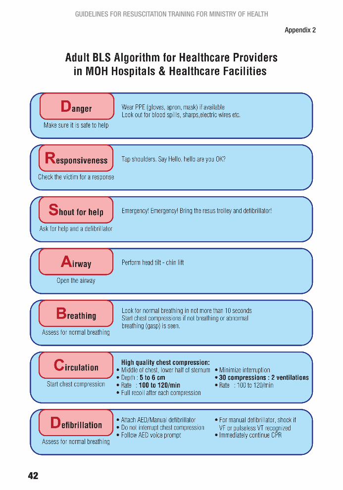
42
GUIDELINES FOR RESUSCITATION TRAINING FOR MINISTRY OF HEALTH
Appendix 2

43
Appendix
Appendix 3
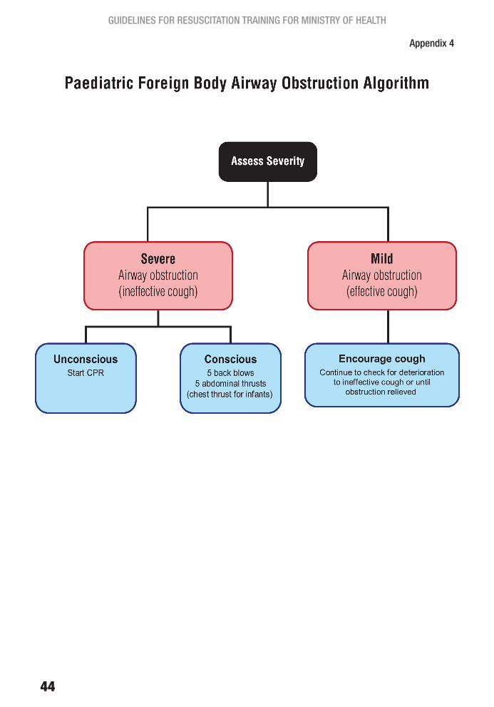
44
GUIDELINES FOR RESUSCITATION TRAINING FOR MINISTRY OF HEALTH
Appendix 4

45
Appendix
Appendix 5
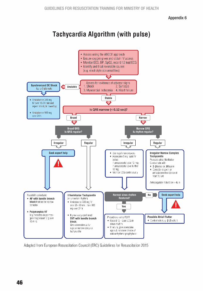
46
GUIDELINES FOR RESUSCITATION TRAINING FOR MINISTRY OF HEALTH
Appendix 6

47
Appendix
Appendix 7
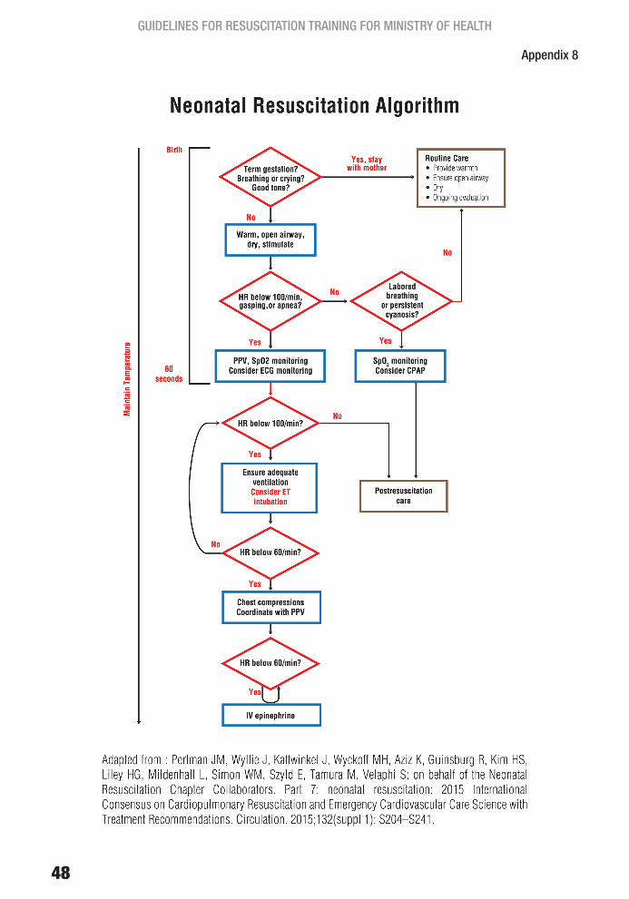
48
GUIDELINES FOR RESUSCITATION TRAINING FOR MINISTRY OF HEALTH
Appendix 8
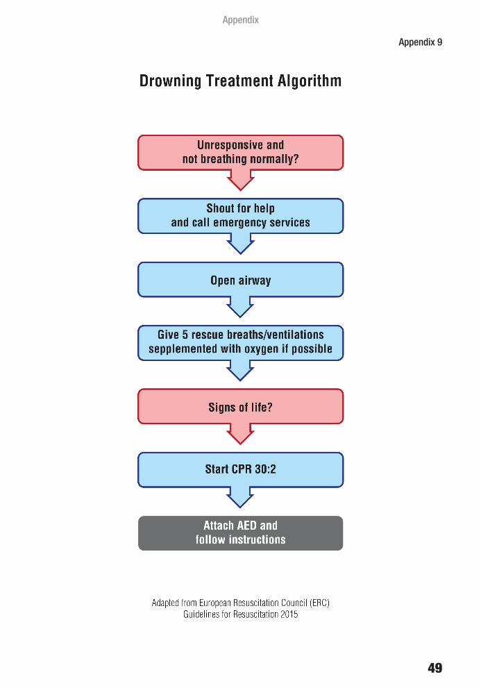
49
Appendix
Appendix 9

50
GUIDELINES FOR RESUSCITATION TRAINING FOR MINISTRY OF HEALTH
Appendix 10
51
TECHNICAL COMMITTEE
NATIONAL COMMITTEE AND RESUSCITATION TRAINING (NCORT)
1. Dr Sabariah Faizah Binti JamaluddinHead of National Emergency Medicine and Trauma ServicesChairman of National Committee On Resuscitation Training (NCORT)Emergency and Trauma DepartmentHospital Sungai BulohSelangor
2. Dr Luah Lean WahConsultant AnaesthesiologistDepartment of Anaesthesiology and Intensive CareHospital Pulau PinangPulau Pinang
3. Dr Thohiroh Binti Abdul RazakConsultant AnaesthesiologistDepartment of Anaesthesiology and Intensive CareHospital Kuala LumpurWilayah Persekutuan Kuala Lumpur
4. Dr Ridzuan Bin Dato’ Mohd IsaConsultant Emergency Physician and HeadEmergency and Trauma DepartmentHospital AmpangSelangor
5. Dr Neoh Siew HongConsultant Paediatrician (Neonatology)Paediatric InstituteHospital Kuala LumpurWilayah Persekutuan Kuala Lumpur
6. Dr Mohd Lotfi Bin HamzahEmergency Physician and HeadEmergency and Trauma DepartmentHospital Sultanah Nur Zahirah, Kuala TerengganuTerengganu
7. Dato’ Dr Teh Keng HwangConsultant PaediatricianPaediatric DepartmentHospital Sultan Abdul Halim, Sungai PetaniKedah
BASIC LIFE SUPPORT SUB-COMMITTEE
1. Dr Mohd Lotfi Bin HamzahChairman of BLS Sub-CommitteeEmergency Physician and HeadEmergency and Trauma DepartmentHospital Sultanah Nur Zahirah, Kuala TerengganuTerengganu
2. Dr Mohd Amin Bin MohidinEmergency PhysicianEmergency and Trauma DepartmentHospital Sultanah Aminah, Johor BahruJohor
3. Dr Shukrudeen Bin SallehEmergency Physician and HeadEmergency and Trauma DepartmentHospital Kuala KraiKelantan
4. Dr Abdul Kursi Bin Abdul LatifEmergency PhysicianEmergency and Trauma DepartmentHospital Raja Permaisuri Bainun, IpohPerak
5. Dr Khor Cheng HoonAnaesthesiologistDepartment of Anaesthesiology and Intensive CareHospital Taiping Perak
6. Dr Cheah Pike KuanAnaesthesiologistDepartment of Anaesthesiology and Intensive CareHospital Raja Permaisuri Bainun, IpohPerak
52
GUIDELINES FOR RESUSCITATION TRAINING FOR MINISTRY OF HEALTH
ADVANCED LIFE SUPPORT SUB-COMMITTEE
1. Dr Luah Lean WahChairman of ALS Sub-CommitteeConsultant AnaesthesiologistDepartment of Anaesthesiology and Intensive CareHospital Pulau Pinang
2. Datin Dr Najah Binti HarussaniConsultant AnaesthesiologistDepartment of Anaesthesiology and Intensive CareHospital Raja Permaisuri Bainun, IpohPerak
3. Dr Adi Bin OsmanConsultant Emergency Physician and HeadEmergency and Trauma DepartmentHospital Raja Permaisuri Bainun, IpohPerak
4. Dr Thohiroh Binti Abdul RazakConsultant AnaesthesiologistDepartment of Anaesthesiology and Intensive CareHospital Kuala LumpurWilayah Persekutuan Kuala Lumpur
5. Dr Ridzuan Bin Dato’ Mohd IsaConsultant Emergency Physician and HeadEmergency and Trauma DepartmentHospital AmpangSelangor
6. Dr Ismail Tan Bin Mohd Ali TanAnaesthesiologistDepartment of Anaesthesiology and Intensive CareHospital Kuala LumpurWilayah Persekutuan Kuala Lumpur
7. Dr Anuwar Ariff Bin Md OmarAnaesthesiologistDepartment of Anaesthesiology and Intensive CareHospital MelakaMelaka
8. Dr Khairuddin Bin Ismail AnaesthesiologistDepartment of Anaesthesiology and Intensive CareHospital Sultanah Nur Zahirah, Kuala TerengganuTerengganu
PAEDIATRIC LIFE SUPPORT/ADVANCED PAEDIATRIC LIFE SUPPORT SUB-COMMITTEE
1. Dato’ Dr Teh Keng HwangChairman of PLS/APLS Sub-CommitteeConsultant PaediatricianPaediatric DepartmentHospital Sultan Abdul Halim, Sungai PetaniKedah
2. Dr Maznisah Binti MahmoodConsultant PaediatricianPaediatric InstituteHospital Kuala LumpurWilayah Persekutuan Kuala Lumpur
3. Dr Zainah Binti HedraConsultant Paediatrician and HeadPaediatric DepartmentHospital Sultanah Nora Ismail, Batu PahatJohor
4. Dr Pon Kah MinConsultant PaediatricianPaediatric DepartmentHospital Pulau PinangPulau Pinang
5. Dr Chew Eng LaiPaediatricianPaediatric DepartmentHospital Raja Permaisuri Bainun, IpohPerak
53
TECHNICAL COMMITTEE
TRAUMA LIFE SUPPORT SUB-COMMITTEE
1. Dr Ridzuan Bin Dato’ Mohd IsaChairman of TLS Sub-CommitteeConsultant Emergency Physician and HeadEmergency and Trauma DepartmentHospital AmpangSelangor
2. Dr Andre DasConsultant General Surgery and HeadDepartment of General SurgeryHospital KajangSelangor
3. Dr Mohd Amin Bin MohidinEmergency PhysicianEmergency and Trauma DepartmentHospital Sultanah Aminah, Johor BahruJohor
4. Dr Shah Jahan Bin Mohd YusopEmergency PhysicianEmergency and Trauma DepartmentHospital Kuala LumpurWilayah Persekutuan Kuala Lumpur
5. Dr Azuddin Bin Mohd KhairyGeneral SurgeonDepartment of General SurgeryHospital Tuanku Ampuan Rahimah, KlangSelangor
NEONATAL RESUSCITATION PROGRAMME
1. Dr Neoh Siew HongChairman of Neonatal Resuscitation ProgrammeConsultant Paediatrician (Neonatology)Paediatric InstituteHospital Kuala LumpurWilayah Persekutuan Kuala Lumpur
2. Dr Kok Juan LoongConsultant PediatricianPaediatric DepartmentHospital Umum Sarawak, KuchingSarawak
3. Dr See Kwee ChingPaediatrician (Neonatology)Paediatric DepartmentHospital Sungai BulohSelangor
4. Dr Wong Ann ChengPaediatricianPaediatric DepartmentHospital Umum Sarawak, KuchingSarawak
5. Dr Pauline Choo Poh LingPaediatricianPaediatric DepartmentHospital Tuanku Ja’afar, SerembanNegeri Sembilan
SECRETARIAT
1. Dr Kasuadi Bin HussinSenior Principle Assistant DirectorEmergency Services UnitMedical Development DivisionMinistry of Health
2. Dr Nor Mashitah Binti Hj JobliPrinciple Assistant DirectorEmergency Services UnitMedical Development DivisionMinistry of Health
3. Mohd Arif Bin Mohd YusoffSenior Assistant Medical OfficerEmergency Services UnitMedical Development DivisionMinistry of Health
4. Mohd Faiz Bin JohariSenior Assistant Medical OfficerEmergency Services UnitMedical Development DivisionMinistry of Health