guidelines for non operative diagnostic procedures and reporting in
TRANSCRIPT
GUIDELINES FOR NON-OPERATIVE DIAGNOSTIC PROCEDURES AND REPORTING
IN BREAST CANCER SCREENING
Non-operative Diagnosis Subgroup of the National Coordinating Group for Breast Screening Pathology
NHSBSP Publication No 50
June 2001
Archive
d on 2
2/03/2
017

First published by:
NHS Cancer Screening Programmes
The Manor House
260 Ecclesall Road South
Sheffi eld S11 9PS
Tel: 0114 271 1060
Fax: 0114 271 1089
Email: nhs.screening@sheffi eld-ha.nhs.uk
Web site: www.cancerscreening.nhs.uk
© NHS Cancer Screening Programmes 2001
The contents of this document may be copied for use by staff working in the public sector but
may not be copied for any other purpose without prior permission from the NHS Cancer Screening
Programmes.
ISBN 1 871997 44 5
Typeset by Prepress Projects Ltd, Perth (www.prepress-projects.co.uk)
Printed by Streamline Offset, Hoddesdon, Herts
Archive
d on 2
2/03/2
017

NHSBSP June 2001 iii
Non-operative diagnostic procedures and reporting
CONTENTS
Page No
1. USE OF NON-OPERATIVE DIAGNOSTIC TECHNIQUES 1
1.1 Role of non-operative diagnostic techniques 1
1.2 Choice of sampling technique – FNAC or core biopsy 2
1.3 False positive cytology 4
1.4 Resource implications 4
2. SAMPLING TECHNIQUES AND PROCEDURES 6
2.1 Sampling techniques 6
2.2 Use of image guidance for breast biopsy 6
2.3 Fine needle aspiration cytology (FNAC) 7
2.4 Core biopsy 13
2.5 Large volume sampling techniques 15
2.6 Complications of FNAC and WBN 16
3. FNAC REPORTING GUIDELINES 18
3.1 Using the cytopathology reporting form 18
3.2 Recording basic information 19
3.3 Reporting categories 20
3.4 Calcifi cation in FNAC 23
3.5 General diagnostic patterns 23
4. DIAGNOSTIC PITFALLS IN INTERPRETATION OF BREAST FNAC 24
4.1 Common conditions resulting in a false positive diagnosis 24
4.2 Uncommon lesions causing a false positive diagnosis 28
4.3 Conditions causing a false negative diagnosis 28
4.4 Other unusual lesions 30
4.5 Prognostic information 31
5. CORE BIOPSY REPORTING GUIDELINES 32
5.1 Core biopsy specimen information and handling 32
5.2 Using the core biopsy (WBN) reporting form 32
5.3 Recording basic information 33
5.4 Reporting categories 35
5.5 Calcifi cation in core biopsy 40
5.6 Problems and pitfalls in diagnosis 40
5.7 Rare lesions 43
5.8 Assessment of prognostic information 44
5.9 Oestrogen receptor (ER) assessment 45Arch
ived o
n 22/0
3/201
7

Non-operative diagnostic procedures and reporting
NHSBSP June 2001 iv
Page No
6. QUALITY ASSURANCE 46
6.1 Defi nitions 46
6.2 How to calculate quality assurance statistics 46
6.3 Cytology performance fi gures 48
6.4 How to interpret the results 48
6.5 Education and training 51
APPENDICES
1: How the national screening system treats cytology data 52
2: Immunohistochemical detection of steroid receptors in breast cancer 54
REFERENCES 57
Archive
d on 2
2/03/2
017

NHSBSP June 2001 v
Non-operative diagnostic procedures and reporting
WRITING GROUP
Dr I O Ellis Reader in Pathology
University of Nottingham
Department of Histopathology
Nottingham City Hospital
Hucknall Road
Nottingham NG5 1PB
Dr S Humphreys Consultant Pathologist
Preston Hall Hospital
Maidstone
ME20 7NH
Dr M Michell Consultant Radiologist
King’s College Hospital
London SE5 9PS
Dr S E Pinder Senior Lecturer
University of Nottingham
Department of Histopathology
Nottingham City Hospital
Hucknall Road
Nottingham NG5 1PB
Dr C A Wells Consultant Pathologist
St Bartholomew’s Hospital
West Smithfi eld
London EC1A 7BE
Dr H D Zakhour Consultant Pathologist
Department of Histopathology
Arrowe Park Hospital
Arrowe Park Road
Upton
Wirral CH49 5PE
Archive
d on 2
2/03/2
017
Non-operative diagnostic procedures and reporting
NHSBSP June 2001 vi
PREFACE
In 1989, a working party of the Royal College of Pathologists produced
two documents giving guidance on breast cancer screening as it
related to histopathology.1,2 Subsequently, the National Coordinating
Committee for Breast Cancer Screening Pathology published guidelines
for cytology procedures and reporting in breast cancer screening.3 These
guidelines have essentially been adopted with minor modifi cations
by the European Union (EU) and form the basis of the European
guidelines.4 In addition, the 1989 guidelines have been updated in
two revised documents.5,6
Non-operative diagnosis has become the norm in breast screening
assessment. Until now, fi ne needle aspiration cytology (FNAC) has
been the sampling method of choice, but the more recent introduction
of automated core biopsy guns, which facilitate sampling, has led to the
implementation of core biopsy in assessment units.
The purpose of this document is to update pathologists on the role
and use of FNAC and core biopsy in breast screening assessment. It
also details the mechanisms used to assure the quality of cytological
and core biopsy diagnosis.
The document constitutes the second edition of guidelines for cytology
procedures and reporting in breast cancer screening. It updates and
replaces the previous guidelines published as NHSBSP Publication
No 22.3
Archive
d on 2
2/03/2
017
NHSBSP June 2001 1
Non-operative diagnostic procedures and reporting
1. USE OF NON-OPERATIVE
DIAGNOSTIC TECHNIQUES
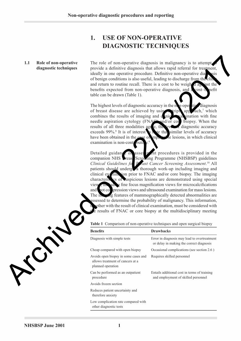
The role of non-operative diagnosis in malignancy is to attempt to
provide a defi nitive diagnosis that allows rapid referral for treatment,
ideally in one operative procedure. Defi nitive non-operative diagnosis
of benign conditions is also useful, leading to discharge from the clinic
and return to routine recall. There is a cost to be weighed against the
benefi ts expected from non-operative diagnosis, and a cost–benefi t
table can be drawn (Table 1).
The highest levels of diagnostic accuracy in the non-operative diagnosis
of breast disease are achieved by using a triple approach,7 which
combines the results of imaging and clinical examination with fi ne
needle aspiration cytology (FNAC) and/or core biopsy. When the
results of all three modalities agree, the level of diagnostic accuracy
exceeds 99%.8 It is of interest to note that similar levels of accuracy
have been obtained in the case of impalpable lesions, in which clinical
examination is non-contributory.9
Detailed guidance on assessment procedures is provided in the
companion NHS Breast Screening Programme (NHSBSP) guidelines
Clinical Guidelines for Breast Cancer Screening Assessment.10 All
patients should undergo a thorough work-up including imaging and
clinical examination prior to FNAC and/or core biopsy. The imaging
characteristics of suspicious lesions are demonstrated using special
views, including fi ne focus magnifi cation views for microcalcifi cations
and spot compression views and ultrasound examination for mass lesions.
The imaging features of mammographically detected abnormalities are
assessed to determine the probability of malignancy. This information,
together with the result of clinical examination, must be considered with
the results of FNAC or core biopsy at the multidisciplinary meeting
1.11.1 Role of non-operative
diagnostic techniques
Table 1 Comparison of non-operative techniques and open surgical biopsy
Benefi ts Drawbacks
Diagnosis with simple tests Error in diagnosis may lead to overtreatment
or delay in making the correct diagnosis
Cheap compared with open biopsy Occasional complications (see section 2.6 )
Avoids open biopsy in some cases and Requires skilled personnel
allows treatment of cancers at a
planned operation
Can be performed as an outpatient Entails additional cost in terms of training
procedure and employment of skilled personnel
Avoids frozen section
Reduces patient uncertainty and
therefore anxiety
Low complication rate compared with
other diagnostic tests
Archive
d on 2
2/03/2
017
Non-operative diagnostic procedures and reporting
NHSBSP June 2001 2
when deciding further management. This combined approach must be
adhered to in the UK National Breast Screening Programme. Under
no circumstances should a cytological opinion of malignancy in the
absence of mammographic and/or clinical evidence of malignancy
be taken as authority for therapeutic surgery. It is recognised that
false positive core biopsy interpretation occasionally occurs, and all
cases should be subject to multidisciplinary review before defi nitive
treatment.
Cytology and core biopsy results from impalpable lesions should not be
interpreted in isolation. Inevitably, inadequate and false negative results
are significantly higher for impalpable lesions. When the imaging
findings are considered to be strongly suspicious of malignancy,
and FNAC or core biopsy is inadequate, normal, or benign, then
management should be based on the imaging fi ndings. The case should
be reviewed at a multidisciplinary meeting and a decision made as to
whether to repeat the sampling procedure or to refer the subject for
open biopsy or localisation biopsy. If the initial sampling procedure
was FNAC then consideration should be given to the use of core biopsy
as the repeat procedure. In cases where there is disagreement between
modalities with a failure to achieve consensus after multidisciplinary
discussion, diagnostic histopathological biopsy is the appropriate
procedure. Frozen section for the diagnosis of screen-detected
lesions is inappropriate.
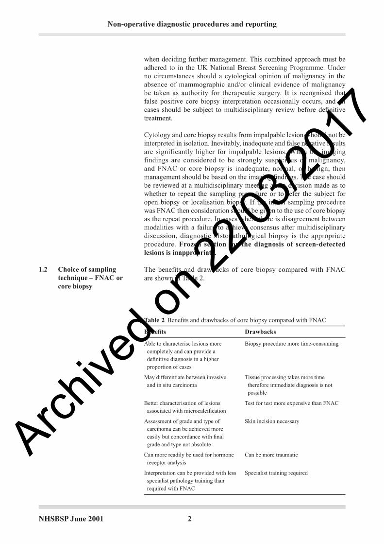
The benefits and drawbacks of core biopsy compared with FNAC
are shown in Table 2.
1.21.2 Choice of sampling
technique – FNAC or
core biopsy
Table 2 Benefi ts and drawbacks of core biopsy compared with FNAC
Benefi ts Drawbacks
Able to characterise lesions more Biopsy procedure more time-consuming
completely and can provide a
defi nitive diagnosis in a higher
proportion of cases
May differentiate between invasive Tissue processing takes more time
and in situ carcinoma therefore immediate diagnosis is not
possible
Better characterisation of lesions Test for test more expensive than FNAC
associated with microcalcifi cation
Assessment of grade and type of Skin incision necessary
carcinoma can be achieved more
easily but concordance with fi nal
grade and type not absolute
Can more readily be used for hormone Can be more traumatic
receptor analysis
Interpretation can be provided with less Specialist training required
specialist pathology training than
required with FNAC
Archive
d on 2
2/03/2
017
NHSBSP June 2001 3
Non-operative diagnostic procedures and reporting
The choice of sampling method in any centre should be determined
by:
• the sensitivity and specifi city of the technique in the centre
• the diagnostic information required for malignant lesions
• patient comfort
• cost
• the availability of staff skilled and experienced in using the
procedures, particularly FNAC sampling and interpretation.
The number of personnel performing FNAC or core biopsy in any
centre should be kept to a minimum and their performance audited
as a matter of routine.
The accuracy of FNAC depends on three main factors:
• a sample that is adequate and representative of the lesion
• suitable processing and staining without artefact
• accurate interpretation of the cytological material with a clear report
conveyed to the rest of the clinical team.
The procedure can fail at any of the stages of preparation (aspiration,
spreading and staining) even before diagnostic interpretation. The
confi dence and experience of the aspirator are vital for obtaining a
satisfactory sample. This part of the procedure, like other parts, should
not be delegated to the novice. Although FNAC has been successfully
used by many centres, its effectiveness in some has been limited by
inadequate and/or equivocal sample rates of 10–30%. Poor cytology
specimens are more likely to be obtained from paucicellular lesions such
as sclerosed fi broadenomas and microcalcifi cation due to fi brocystic
change.
The availability of automated core biopsy guns and the publication of
results from several centres showing very high sensitivity and specifi city
have resulted in the more widespread use of image guided core biopsy.
A recent review of published series of image guided breast core biopsy
shows that the sensitivity and specifi city of core biopsy are high when
compared with FNAC.11 No difference in patient discomfort between
FNAC and core biopsy has been demonstrated. Information regarding
the invasive nature and grade of the tumour can be obtained in most
malignant mass lesions. For malignant microcalcifi cation, if an invasive
tumour is present, 14G core biopsy will detect the invasive element in
approximately 40% of cases.
Recent evidence indicates that for certain types of mammographic
abnormality, such as moderate to low level suspicion microcalcifi cation,
a larger volume of tissue is required for accurate diagnosis.12 In the case
of such lesions, when the use of conventional FNAC or 14G core biopsy
carries a risk of an equivocal result, use of larger volume sampling
techniques may increase the accuracy of biopsy. Recently published
results of vacuum assisted core biopsy have demonstrated a lower
equivocal sample rate and increased accuracy in the detection of small
1.2.1 Accuracy of FNAC
1.2.2 Accuracy of core biopsy
Archive
d on 2
2/03/2
017
Non-operative diagnostic procedures and reporting
NHSBSP June 2001 4
invasive tumours associated with an area of ductal carcinoma in situ
(DCIS). Consideration of the likely underlying histological nature of the
lesion from the imaging features should therefore be taken into account
when deciding on the sampling method to be used.
False positive cytology is recognised in the published literature and
is an important quality assurance measure. UK practice currently
accepts a relatively high false positive rate of up to 1%. In the clinical
management of individual patients, the risk of overtreatment can be
reduced by strict adherence to the principles of the triple diagnostic
approach as emphasised in these guidelines.
All those involved with the diagnosis of breast disease should be aware
that some lesions currently classifi ed as benign histologically exhibit
malignant cytomorphological features. These conditions generally
fall within the category of borderline hyperplastic lesions, especially
atypical hyperplasia. This histological diagnostic does not rely only
on cytomorphology but takes into account the extent and purity of the
changes present. These are not demonstrable in cytological preparations.
These lesions are not clinically regarded or managed as established
malignancy and therefore, at present, these cases should be regarded
biostatistically as false positive cases. For these reasons, all false positive
cases should be subject to internal unit review and should not necessarily
be considered a problem of cytological misdiagnosis/classifi cation.
In the Forrest Committee Report in 1985, neither FNAC nor core
biopsy was costed as an essential technique in breast cancer screening.
Indeed, to our knowledge, no detailed economic analysis has yet been
performed on FNAC or core biopsy in screening in the UK. Time has
demonstrated that a reduction in the number of benign biopsies and
a reduction in second operations on cancer-bearing breasts can be
achieved in centres which have a high level of expertise. This is borne
out by the low benign biopsy fi gures reported by the Epping unit13 as
well as by the fi gures for benign to malignant ratios at open biopsy
presented in the section on sensitivity and specifi city.
Many of the existing data on economics of non-operative diagnosis
relate to FNAC rather than core biopsy; studies in the USA have
discovered a reduction in costs of approximately 90%.14 An evaluation
of the subject showing a similar value in the UK was conducted
by Kocjan.15 The FNA costs for stereotactic aspirations are higher
(estimated approximately double), but marker biopsy costs are also
much higher. It is therefore apparent that the cost of the FNACs, even
if performed on several more patients than would otherwise have been
subjected to open biopsy, is likely to be outweighed by the fi nancial
savings, even without taking into consideration the benefi ts in terms
of reduced morbidity.
An estimate of the number of biopsies saved can be gained from a
study conducted in Guildford which looked specifi cally at stereotactic
aspiration.16 The estimated benign to malignant ratio without stereo-
tactic aspiration in 250 cases of impalpable lesions based on the
1.41.4 Resource implications
1.31.3 False positive cytology
Archive
d on 2
2/03/2
017

NHSBSP June 2001 5
Non-operative diagnostic procedures and reporting
mammographic appearances alone would have been 1:0.94. The
actual benign to malignant ratio achieved with stereotactic aspiration
was 1:2.5. In addition, seven extra cancers were discovered in the
mammographically low risk group that would otherwise have not been
biopsied at the fi rst assessment.
Data on core biopsy use from the US show a cost saving of core biopsy
over specimen biopsies of approximately 300% per case in one unit
($243 vs. $698)17 and $744 for ultrasound biopsy or $519 for stereotactic
biopsy over the cost of surgical biopsy in a second unit.18 FNAC and
core biopsy will be most effective within the framework of an integrated
team approach to assessment and its quality must, like other aspects of
the screening programme, be monitored.
Archive
d on 2
2/03/2
017
Non-operative diagnostic procedures and reporting
NHSBSP June 2001 6
2. SAMPLING TECHNIQUES AND
PROCEDURES
This chapter considers the following techniques:
• fi ne needle aspiration cytology (FNAC)
• core biopsy
• large volume core techniques
– vacuum assisted core biopsy (Mammotome)
– minimally invasive breast biopsy (MIB)
– advanced breast biopsy instrumentation (ABBI)
– Site Select percutaneous breast biopsy (PBB)
All of these procedures are best carried out by experienced clinicians or
radiologists who are specialists in breast imaging or by a multidisciplinary
team with a specialist radiologist present.
A high proportion of mammographically detected lesions are impalpable
and require image guidance for FNAC or core biopsy sampling. In
addition, image guidance, particularly ultrasound, can have advantages
over freehand procedures when sampling some palpable lesions
Most soft tissue lesions in the breast are visible using modern high
frequency apparatus with a frequency range of up to 10–13 MHz.
Ultrasound guided FNAC or core biopsy is the technique of fi rst choice
for sampling impalpable breast lesions as it is easier to perform,
more comfortable for the patient and less time-consuming than the
x-ray guided techniques. It allows real time demonstration of the
needle traversing the lesion.. The radiologist must be certain that the
abnormality seen on ultrasound is the same as the abnormality seen on
mammography. X-ray guided FNAC and core biopsy should be used
where there is any doubt about the ultrasound appearances. Ultrasound
is also increasingly being used to guide needle biopsy of palpable
masses to ensure accurate sampling. Some clusters of microcalcifi cation,
particularly coarser comedo-type calcification, are visible on high
frequency ultrasound and may therefore be sampled by ultrasound
guidance.
X-ray stereotaxis is used for image guided biopsy of most suspicious
microcalcifi cations, areas of parenchymal distortion/stellate lesions
or small soft tissue masses that cannot be adequately visualised by
ultrasound. Stereotactic localisation can be carried out with the patient
in the upright or prone positions. Upright stereotactic units are more
widely available and less expensive than dedicated prone stereotactic
units. Digital imaging is becoming available for use with conventional
upright stereotactic units, and this improves the accuracy of the
technique because of the shorter image acquisition time and because of
the improved quality of the digital images.
The main problems encountered with the use of the upright stereotactic
2.12.1 Sampling techniques
2.22.2 Use of image guidance
for breast biopsy
2.2.1 When to use ultrasound
guidance
2.2.2 When to use stereotactic
guidance
Archive
d on 2
2/03/2
017
NHSBSP June 2001 7
Non-operative diagnostic procedures and reporting
units are vaso-vagal episodes and diffi culty in accurately targeting
lesions that are very posteriorly situated. It is possible with some units
to carry out the stereotactic biopsy procedure with the patient lying on
her side. Dedicated prone breast biopsy systems use a table on which
the patient lies in the prone oblique position and the breast passes
through a rounded aperture in the table. The advantages of the prone
system are the negligible risk of a vaso-vagal episode, a stable position
with minimal patient movement and improved access to lesions situated
in the inferior or posterior parts of the breast. The dedicated prone
breast biopsy systems are supplied with digital imaging, allowing rapid
acquisition of stereotactic images, manipulation of the digital images
including magnifi cation, image reversal and contrast adjustment for
improved visualisation of the target abnormalities. The disadvantages
of the dedicated prone breast biopsy systems are the high capital cost
of the equipment and the need for a dedicated room, which cannot be
otherwise used for diagnostic mammography.
The success of FNAC is directly related to the skill and experience
of the operators. The number of staff involved should be kept to a
minimum. A core team of aspirators who perform all breast needling
procedures should be established in each centre. Trainees must be
closely supervised. An experienced radiographer is essential for x-ray
guided procedures. For both x-ray and ultrasound guided FNAC it
is recommended that an assistant skilled in specimen preparation,
preferably a medical laboratory scientific officer (MLSO) or a
pathologist, is present. The procedure time is signifi cantly shortened if
an assistant deals with the specimens and smears the slides while the
aspirator obtains further samples. If a trained MLSO or pathologist
is available to assess the adequacy of the aspirate immediately using
a rapid staining technique, recall for repeat cytology can be avoided,
therefore reducing delay and distress.
Needles (22 or 23 gauge) of appropriate type and length are ideal
for FNAC. A 10- or 20-ml syringe is used to apply suction. A short
extension tube between the needle and syringe is usually required for
image guided procedures. A syringe holder is desirable but not essential
although it does make manipulation of the syringe with simultaneous
constant suction much easier. Several makes of holder, eg Cameco Ltd,
Nyegaard Aspirator, R H syringe holder, are available in appropriate
sizes. For superfi cial lesions or in small breasts, a 23G (blue) needle
is suffi cient and may produce less bleeding. Needles that have no dead
space in the needle hub are best for FNAC (eg B & D Microlance).
Finer gauge needles, eg 25G (orange), are used in some units with
excellent results.19 In large breasts or in deeper lesions, a 22G (grey)
needle may be necessary because of its extra length. A needle with a
trocar may be preferred for deep lesions as it is more rigid and is less
likely to become blocked or contaminated during insertion.
Local anaesthetic is not usually necessary for freehand FNAC
procedures, and excessive use of anaesthetic can lead to specimen
2.32.3 Fine needle aspiration
cytology (FNAC)
2.3.1 Personnel
2.3.2 Equipment
2.3.3 Local anaesthetic
Archive
d on 2
2/03/2
017
Non-operative diagnostic procedures and reporting
NHSBSP June 2001 8
contamination by fl uid, causing fi xation problems. If sampling requires
multiple skin punctures, local anaesthetic may be used to anaesthetise
the skin or ethyl chloride spray can be employed to freeze the skin. This
may be particularly helpful for x-ray guided techniques with multiple
samples, which take signifi cantly longer to perform. The use of local
anaesthetic reduces the risk of pain-induced patient movement within
the compression plate during the procedure. To avoid contamination
of the aspirate the local anaesthetic should not be injected deep to
the dermis.
The procedure for obtaining the specimen is explained to the patient.
Skilful introduction of the aspirating needle produces very little
discomfort. For very apprehensive patients, skin anaesthesia with
local anaesthetic spray or a freezing spray, eg ethyl chloride, can
be used.
The lesion is held gently but fi rmly by the fi ngers of the locating hand
with the overlying skin slightly stretched. The area is cleansed with an
alcohol-impregnated swab, but it is important that any excess alcohol
is wiped away or allowed to dry. Traces of alcohol introduced with the
needle are the main cause of the burning sensation of which patients
occasionally complain after aspiration.
With the syringe fi rmly attached and the plunger fully closed to exclude
air from the barrel, the needle is readied for insertion. The patient is
warned before the skin is punctured. The needle is introduced into the
skin with no air in the syringe barrel. The needle is positioned at the
anterior edge of the lump (which can usually be felt with the needle tip)
and negative pressure is applied either with the thumb or, more easily,
with a syringe holder. Several passes through the lesion, varying the
angle of entry into the lesion and rotating the syringe slowly, are made,
without withdrawing the needle from the skin, until a small drop of
fl uid is seen in the hub of the needle. Some aspirators advocate picking
up the lesion, rotating the syringe during aspiration and varying the
speed of passes to increase the yield. The negative pressure is released
and then the needle is withdrawn from the skin. If a large amount of
blood is obtained it is best to interrupt the procedure, apply pressure to
the area to limit haematoma and either repeat the aspiration in the same
session from a different angle or delay for one month.
Breast lesions are often deeper than they appear. If there is doubt about
whether the lesion has been sampled, then reaspiration using a longer
needle may be necessary. If there is no resistance to the needle from
a lump that appears clinically not to be a lipoma, then it is likely the
lesion has been missed by the needle. Reaspiration is advised, especially
if the spread slide shows oily droplets throughout. Similarly, heavily
blood stained aspirates may not be representative of the lesion. If
material appears to be stuck in the hub of the needle then, using another
needle, it can be sucked out of the hub into the barrel of the new needle
and expelled from there or retrieved by washing into transport medium.
Another method that uses a needle only and capillary action to draw the
2.3.4 Freehand FNAC
Archive
d on 2
2/03/2
017
NHSBSP June 2001 9
Non-operative diagnostic procedures and reporting
fl uid into the hub has been described by Zajdela et al,20 but as far as is
known this is not applicable to stereotactic aspiration of breast.
Some breast lesions give a characteristic ‘feel’ as the needle traverses
the lesion. This can occasionally be a very helpful pointer as to
whether or not the lesion has been sampled. These are conveniently
described as:
No resistance Fatty tissue
Soft Fibroadenoma, mucinous carcinoma, medullary
carcinoma
Rubbery Fibrocystic changes, lobular carcinoma, fi broadenoma
Hard Fibrous tissue, hyalinised fibroadenoma, post-
radiotherapy
Gritty Carcinoma, microcalcifi ed tissue
Cystic Cyst in fi brocystic change
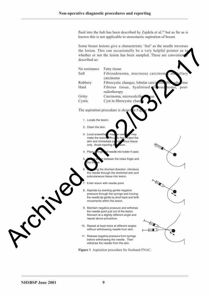
The aspiration procedure is shown in Figure 1.
Figure 1 Aspiration procedure for freehand FNAC.
Aspirate by exerting gentle negativepressure through the syringe and movingthe needle tip gently by short back and forthmovements within the lesion.
8.
9. Maintain negative pressure and withdrawthe needle point just out of the lesion.Reinsert at a slightly different angle andrepeat above procedure.
10. Repeat at least twice at different angleswithout withdrawing needle from skin.
11. Release negative pressure from syringebefore withdrawing the needle. Thenwithdraw the needle from the skin.
1. Locate the lesion.
2. Clean the skin.
Local anaesthetic may be used but maymake the lesion difficult to feel. Inject theskin and immediate subcutaneous tissueonly. Avoid injecting the lesion.
3.
Place syringe and needle into holder if used.4.
Fix the lesion between the index finger andthe thumb.
5.
Choosing the shortest direction, introducethe needle through the stretched skin andsubcutaneous tissue into lesion.
6.
Enter lesion with needle point.7.
Archive
d on 2
2/03/2
017
Non-operative diagnostic procedures and reporting
NHSBSP June 2001 10
The patient is positioned to provide optimal access to the area to be
sampled; this may involve, for example, raising and supporting the
left side for biopsy of lesions situated in the lateral aspect of the left
breast. To sample lesions that are situated in the lateral aspect of the
right breast it may be necessary to turn the patient on the couch so
that a right-handed operator can easily access such lesions using a
lateral approach. An assistant should work from the opposite side of
the couch to the radiologist.
The lesion is demonstrated and the surrounding breast tissue is
immobilised by applying pressure with the palm of the hand holding
the probe. Infi ltration of the skin with local anaesthetic may be carried
out. The FNAC needle, attached by a short connecting tube to a 10-ml
syringe held by the assistant, is introduced into the breast along the line
of the long axis of the ultrasound probe and will be easily visualised if it
is kept parallel to the surface of the probe. The needle tip is guided into
the lesion and an image is taken to record that the needle is correctly
positioned. The needle is then moved back and forth within the lesion
with simultaneous rotation and with negative pressure being applied
by the assistant. Aspiration is continued until material is seen within
the hub of the needle. The aspirate is then delivered onto slides,
and dry and wet preparations made in accordance with guidance
from the pathologist. Two or three separate samples are commonly
obtained in order to increase the chances of obtaining a diagnostic
cellular sample.
Ultrasound jelly may present a problem in interpretation for pathologists
seeing it for the fi rst time and should not be confused with calcium salts
or necrosis. It should not be used during the aspiration procedure and,
if used previously, should be carefully removed.
For FNAC using a stereotactic device with a conventional upright
mammography machine, the patient is seated. A superior approach with
the breast positioned for the craniocaudal view is suitable for most
lesions, but lateromedial, mediolateral or oblique approaches may be
needed for lesions that are inferiorly positioned or are situated laterally
in the axillary tail region. After demonstrating the lesion on a straight
scout fi lm, paired stereotactic views are obtained with the x-ray tube
angled 15° either side of the central straight tube position. The position
of the lesion on the stereotactic views is used to determine the position
of the needle guide in the x- and y-axes so that, when a needle of known
length is introduced through the guide into the breast, the needle tip
will be correctly positioned within the lesion.
The skin is cleaned and superfi cial infi ltration with local anaesthetic
may be carried out. The sampling needle (22G or 23G) is inserted
through the needle guide and into the breast so that the needle hub is
against the guide. Check stereotactic fi lms are taken to ensure correct
positioning of the needle in relation to the target lesion. If the position
is not correct, the needle is repositioned and further check fi lms are
obtained. After checking the position of the needle tip, a point should
be extended through the lesion to allow passage of the needle through
2.3.5 Ultrasound guided
FNAC
2.3.6 Stereotactic FNAC
Archive
d on 2
2/03/2
017
NHSBSP June 2001 11
Non-operative diagnostic procedures and reporting
the lesion during aspiration. When the needle position is correct,
aspiration is carried out by simultaneously rotating and passing the
needle repeatedly in and out of the lesion.
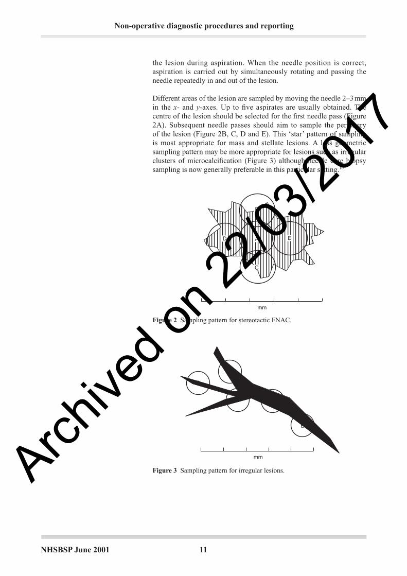
Different areas of the lesion are sampled by moving the needle 2–3 mm
in the x- and y-axes. Up to fi ve aspirates are usually obtained. The
centre of the lesion should be selected for the fi rst needle pass (Figure
2A). Subsequent needle passes should aim to sample the periphery
of the lesion (Figure 2B, C, D and E). This ‘star’ pattern of sampling
is most appropriate for mass and stellate lesions. A less geometric
sampling pattern may be more appropriate for lesions such as irregular
clusters of microcalcifi cation (Figure 3) although needle core biopsy
sampling is now generally preferable in this particular setting.10
mm
B
D A E
C
Figure 2 Sampling pattern for stereotactic FNAC.
Figure 3 Sampling pattern for irregular lesions.
mm
B
D
A
E
C
Archive
d on 2
2/03/2
017
Non-operative diagnostic procedures and reporting
NHSBSP June 2001 12
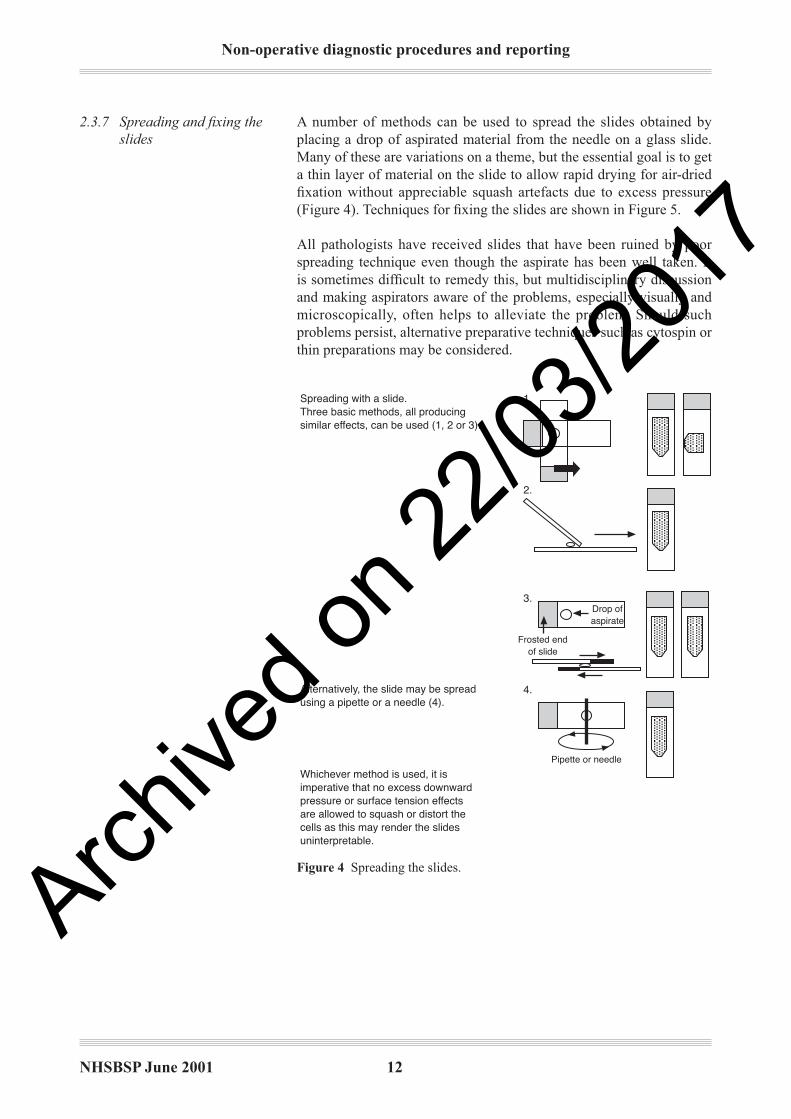
A number of methods can be used to spread the slides obtained by
placing a drop of aspirated material from the needle on a glass slide.
Many of these are variations on a theme, but the essential goal is to get
a thin layer of material on the slide to allow rapid drying for air-dried
fi xation without appreciable squash artefacts due to excess pressure
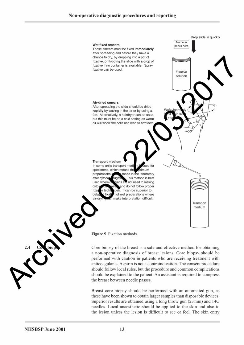
(Figure 4). Techniques for fi xing the slides are shown in Figure 5.
All pathologists have received slides that have been ruined by poor
spreading technique even though the aspirate has been well taken. It
is sometimes diffi cult to remedy this, but multidisciplinary discussion
and making aspirators aware of the problems, especially visually and
microscopically, often helps to alleviate the problem. Should such
problems persist, alternative preparative techniques such as cytospin or
thin preparations may be considered.
Figure 4 Spreading the slides.
2.3.7 Spreading and fi xing the
slides
Spreading with a slide.Three basic methods, all producingsimilar effects, can be used (1, 2 or 3).
Alternatively, the slide may be spreadusing a pipette or a needle (4).
Whichever method is used, it isimperative that no excess downwardpressure or surface tension effectsare allowed to squash or distort thecells as this may render the slidesuninterpretable.
Pipette or needle
Drop ofaspirate
Frosted endof slide
1.
2.
3.
4.
Archive
d on 2
2/03/2
017
NHSBSP June 2001 13
Non-operative diagnostic procedures and reporting
Core biopsy of the breast is a safe and effective method for obtaining
a non-operative diagnosis of breast lesions. Core biopsy should be
performed with caution in patients who are receiving treatment with
anticoagulants. Aspirin is not a contraindication. The consent procedure
should follow local rules, but the procedure and common complications
should be explained to the patient. An assistant is required to compress
the breast between needle passes.
Breast core biopsy should be performed with an automated gun, as
these have been shown to obtain larger samples than disposable devices.
Superior results are obtained using a long throw gun (23 mm) and 14G
needles. Local anaesthetic should be applied to the skin and also to
the lesion unless the lesion is diffi cult to see or feel. The skin entry
Figure 5 Fixation methods.
Drop slide in quickly
Waft vigorouslyin air or in frontof a fan
Transportmedium
Wet fixed smearsThese smears must be fixed immediatelyafter spreading and before they have achance to dry, by dropping into a pot offixative, or flooding the slide with a drop offixative if no container is available. Sprayfixative can be used.
Transport mediumIn some units transport medium is used forspecimens, which means that optimumpreparations can be made in the laboratoryafter cytocentrifugation. This method is bestused where clinicians are not used to makingcytological smears and do not follow properfixation techniques. It can be superior todelayed fixation of wet preparations whereair-drying can make interpretation difficult.
Air-dried smearsAfter spreading the slide should be driedrapidly by waving in the air or by using afan. Alternatively, a hairdryer can be used,but this must be on a cold setting as warmair will 'cook' the cells and lead to artefacts.
Fixativesolution
Name inpencil here
2.42.4 Core biopsy
Archive
d on 2
2/03/2
017
Non-operative diagnostic procedures and reporting
NHSBSP June 2001 14
site should be placed as far away from the nipple as is practicable and
should never be within the areola. A small skin nick that also traverses
the superfi cial fascia should be made with a scalpel blade. This enables
easy insertion of the needle. If the breast tissue is very fi brous, insertion
of the needle in a radial direction makes manipulation easier. The major
complications of breast needle biopsy occur if the pleura is traversed.
To avoid this, the needle should be kept approximately parallel to the
chest wall when fi red. This means that the skin entry site for deep
lesions needs to be further away from the lesion than for superfi cial
lesions. For lesions 10 mm or larger, the tip of the needle should abut
the lesion before fi ring. When biopsying lesions less than 10 mm in
diameter the needle tip should be just short of the lesion before fi ring. It
is the operator’s responsibility to confi rm the patient’s identifi cation and
label the specimen pot before leaving the room.
After the procedure, the biopsy site should be compressed for a
minimum of 5 minutes. The patient should be given written information
concerning where and when she will receive the result and informed
about possible complications of the procedure. The patient should be
advised that mild post-procedure pain and bruising are common and
told not to exercise her upper limbs for the rest of the day. The patient
information sheet should advise the patient to phone the hospital (the
number should be on the patient information sheet) if the breast swells
appreciably or if she becomes short of breath.
The lesion is demonstrated and surrounding breast tissue immobilised
in the same way as for FNAC. Local anaesthetic is infi ltrated both
superfi cially and deeply down to the lesion. For posteriorly placed
lesions, local anaesthetic can be infiltrated posteriorly in order to
displace the lesion anteriorly. A 2- to 3-mm skin incision is made to
allow insertion of the core biopsy needle along the direction of the long
axis of the ultrasound probe. A14G needle is used with an automated
biopsy gun. The needle is advanced until the tip is a few millimetres
proximal to the edge of the lesion. The core biopsy gun is then fi red and
the needle is visualised passing through the lesion. A check image with
the needle through the lesion is recorded. The needle is withdrawn and
the specimen is delivered into fi xative. One or two passes are usually
suffi cient in most cases to obtain diagnostic material from soft tissue
mass lesions. At the end of the procedure fi rm pressure is applied by the
assistant over the site of the biopsy to ensure haemostasis.
Stereotactic guided core biopsy may be carried out with upright
stereotactic apparatus. Targeting of the lesion is carried out in the same
way as for FNAC. The procedure is carried out under local anaesthetic.
After checking the position of the needle tip, the needle is withdrawn
approximately 5 mm so that the needle tip is just proximal to the lesion
before fi ring. The biopsy port will then traverse the lesion on fi ring,
ensuring adequate tissue sampling. It is important to ensure that there
is suffi cient tissue deep to the needle tip following fi ring so that the
needle tip does not hit the surface of the cassette holder. Apparatus is
also available that allows a lateral approach to lesions with the breast
compressed in the craniocaudal position. This allows improved access to
2.4.1 Ultrasound guided core
biopsy
2.4.2 Stereotactic core biopsy
Archive
d on 2
2/03/2
017
NHSBSP June 2001 15
Non-operative diagnostic procedures and reporting
lesions which are in the inferior part of the breast and eliminates the risk
of the needle tip hitting the surface of the x-ray cassette holder.
The breast is passed through a rounded aperture in the table. For lesions
which are very posteriorly positioned or which lie in the region of the
axillary tail, access can be improved by passing the ipsilateral arm and
shoulder girdle through the aperture. Stereotactic views are obtained by
rotating the tube 15° either side of the central position. Digital images
are displayed on a computer screen for targeting and the biopsy needle
holder position is adjusted automatically. Check fi lms are taken during
the procedure to ensure accurate positioning and that the needle has
traversed the lesion. Five or more core samples are usually obtained,
using a 14G needle.
When sampling areas of microcalcifi cation with either conventional
upright stereotactic equipment or with prone stereotactic systems,
radiography of the core samples is carried out to ensure that tissue
containing microcalcifi cation has been obtained.
The Mammotome and MIB vacuum assisted core biopsy systems use
either an 8G, 11G or 14G needle probe. Most published results describe
their use with prone stereotactic apparatus, but recent developments
enable them to be used for ultrasound guided breast biopsy.
The biopsy probe incorporates a vacuum channel, which applies negative
pressure to the biopsy port and thereby sucks the adjacent breast tissue
into the port for sampling. The biopsy probe is introduced into the
breast and positioned using image guidance. Deep local anaesthetic
containing adrenaline is used. The vacuum is activated and sucks breast
tissue into the biopsy port; a rotating cutting cylinder then passes down
within the probe and separates the biopsy material from the surrounding
tissue. The biopsy specimen is then delivered by withdrawing the
cutting cylinder while applying negative pressure and while the main
probe remains within the breast. Multiple specimens are obtained by
rotating the biopsy probe within the breast so that the biopsy port is
applied to different areas of breast tissue.
The potential advantages of this system are the ability to obtain a larger
volume of tissue for histological examination and the rapid evacuation
of any haematoma which collects at the site of biopsy. This ensures that
the specimens obtained are of good quality and are not compromised
by the presence of haematoma.
In cases where the whole or a high proportion of the mammographic
lesion has been removed, a small metal marker clip is introduced
through the biopsy probe and deployed at the biopsy site.
2.4.3 Prone stereotactic core
biopsy
2.52.5 Large volume
sampling techniques
2.5.1 Vacuum assisted core
biopsy
Archive
d on 2
2/03/2
017

Non-operative diagnostic procedures and reporting
NHSBSP June 2001 16
Both of these devices involve insertion, using prone stereotactic imaging
guidance, of a large bore cutting cylinder which cuts a core of tissue
containing the mammographic abnormality. The diameter of the ABBI
sampling device is up to 20 mm, that of the PBB device 15 mm. The
PBB device differs from the ABBI system in having a cutting blade at
the leading edge of the biopsy probe, allowing it to be placed against
the proximal edge of the lesion before sampling. These techniques are
more invasive than standard core biopsy or vacuum assisted core biopsy
and require a 20-mm skin incision for insertion of the biopsy device,
and for these reasons their use is limited.
FNAC and core biopsy are remarkably complication free; however,
certain rare problems should be considered.
Pain is common on fi ne needle aspiration but is transitory and is not
usually severe. Aspiration from painful areas of benign breast change
is sometimes associated with some pain when the needle comes into
contact with the painful area. Carcinomas, particularly those with
abundant fi broelastotic stroma, are often also painful, and this can be a
guide to the aspirator that the needle has hit the lesion.
All imaging investigations should be complete before sampling is
performed as haematoma formation, if it occurs, can cause confusion
on subsequent mammography or ultrasound. Haematomas may cause
problems, especially if a larger vessel is punctured. In some cases, the
procedure may have to be abandoned and reaspiration delayed until
a later clinic visit because the lesion may no longer be localisable
if a large haematoma has formed. Haematoma formation may not
be painful for the patient at the time but often becomes so later,
and a recommendation for light analgesia, eg paracetamol, may be
appropriate.
This is a rare complication21 found mainly in women with small breasts,
in medial lesions or when sampling axillary nodes. It is not a problem
with image guidance such as stereotactic or perforated plate and should
not occur with an experienced ultrasound operator as the position of the
needle can be seen. It is therefore most likely to occur with palpable
lesions. It is more likely to occur with an experienced aspirator as
novices tend to be rather circumspect and, if anything, do not probe
to suffi cient depth. Large pneumothoraces should be obvious, but the
problem may go undetected if the pneumothorax is small. Clues such as
sharp pain, coughing or a hiss of air on withdrawing the needle without
evidence of air in the syringe may occur.
This complication has occasionally occurred during sampling. It is
of special signifi cance during upright stereotactic or perforated plate
examinations where the patient has to be released from the machine and
laid fl at. The procedure usually has to be abandoned.
Small lesions, including foci of microcalcifi cation, may, particularly if
extensively sampled, be removed by core biopsy. This risk increases
when a large number of core samples are taken or larger bore sampling
2.62.6 Complications of
FNAC and core biopsy
2.6.1 Pain
2.6.2 Haematoma
2.6.3 Pneumothorax
2.6.4 Fainting
2.6.5 Removal of lesion by
core biopsy
2.5.2 The ABBI and Site
Select PBB biopsy
devices
Archive
d on 2
2/03/2
017
NHSBSP June 2001 17
Non-operative diagnostic procedures and reporting
techniques such as Mammotome or ABBI are used. This problem has
been reported more frequently from the USA, where there is more
aggressive use of core biopsy than is presently undertaken in the UK.22
It is recommended that clips or ink be inserted at the site of biopsy by
the radiologist at the time to ensure that the site can be identifi ed for
subsequent localisation biopsy and pathological assessment.
On occasions, a small invasive focus in a predominant ductal carcinoma
in situ (DCIS) lesion may be sampled and removed by core with no
further foci of invasion remaining in the excision specimen, despite
thorough examination. In such circumstances, the core biopsy sample
can be used to provide information on tumour differentiation and
type.
Seeding of malignant cells has become increasingly recognised as a result
of the increased use of core biopsy.23 This process is also, uncommonly,
seen after FNAC.24 Rarely, this may cause histopathological diagnostic
diffi culties in the subsequent excision. Islands of cells (sometimes
showing degenerative features) are seen outside the main lesion, often
within a fibroblastic and histiocyte tissue response indicating the
previous sampling site. Seeding is rarely recognised more than a few
millimetres from the source of the cells, and the correct identifi cation
is usually straightforward. Cell groups may be seeded from papillary
lesions or DCIS mimicking invasive carcinoma. The associated signs
of trauma from non-operative sampling should be sought. The clinical
signifi cance of this phenomenon is not yet clear.
2.6.6 Seeding of tumour
Archive
d on 2
2/03/2
017

Non-operative diagnostic procedures and reporting
NHSBSP June 2001 18
3. FNAC REPORTING GUIDELINES
This chapter is designed to assist classifi cation and reporting of FNAC
samples. The terminology and diagnostic entities referred to are
described in more detail in Pathology Reporting in Breast Cancer
Screening (2nd edition) (NHSBSP Publication No 3).5
The cytopathology core biopsy reporting forms used may be the separate
reporting form (Figure 6) or the form generated specifi cally by the
national breast screening computer system (Figure 7), which comes
with the patient details automatically fi lled in by the computer. Both
forms request essentially the same information, although the computer
generated form has spaces for radiographic information such as kV,
mAs, side and type of localisation (palpable, ultrasound, stereotactic
or other x-ray guided procedure). The way in which the national
breast screening system treats this information is shown in Appendix
1. Information on the nature of the mammographic abnormality and
clinical characteristics should be provided by the breast screening
radiologist requesting the pathology examination. It may not be possible
to enter some details, such as name of aspirator and type of procedure, if
the pathologist completing the form did not perform the aspirate or was
not provided with this information on a request form.
Figure 6 Example of a cytopathology reporting form.
3.13.1 Using the
cytopathology
reporting form
Reporting forms
BREAST SCREENING CYTOPATHOLOGY
Surname Forenames Date of birth
Screening no. Hospital no. Centre Report no.
Case for review ?
Side
Specimen type
Localisation technique
Opinion
Right Left
FNA (solid lesion) FNA (cyst) Nipple discharge Nipple or skin scrapings
Palpation X-ray guided Ultrasound guided Stereotaxis
Comment1 Unsatisfactory
2 Benign
3 Atypia probably benign
4 Suspicious of malignancy
5 Malignant
NAME OF ASPIRATOR DATEPATHOLOGISTArchive
d on 2
2/03/2
017

NHSBSP June 2001 19
Non-operative diagnostic procedures and reporting
Centre/location
Give the name of the assessment centre, clinic, department, etc where
the specimen was obtained.
Side
Indicate right or left. For specimens from both sides use a separate
form for each side.
Specimen type
Please choose one of the following terms:
FNA (solid lesion) Fine needle aspiration of a solid lesion
FNA (cyst) Fine needle aspiration of a cyst subjected to
cytological examination
Nipple discharge Cytological preparation of a nipple
discharge
Nipple or skin scrapings Cytological preparation of scrapings from
the nipple or skin
Comment (max. 132 characters)
Comment (max. 65 characters)
Pathologist:
Aspirator:Location:
Cyst aspiration without cytology:
BSS: FOLLOW-UP FNA FORM Sx number: Name:
NHS number:
Date of diagnosis:
Surname:Forenames:
Title:Date of birth:
FNA CYTOLOGY
FNADate performed:
Side:
LOCALISATION TYPE:kV:
Total mAs:Total exposures:
Total films:
Date reported:Specimen number:
Specimen type:
AS FNA (solid)ND Nipple dischargeNS Nipple or skin scraping
AC FNA (cyst)
Cytological opinion:
C1 UnsatisfactoryC2 Benign
C3 Atypia, probably benignC4 Suspicious of malignancy
C5 Malignant
Figure 7 Example of a computer generated FNAC reporting form.
3.23.2 Recording basic
information
Archive
d on 2
2/03/2
017
Non-operative diagnostic procedures and reporting
NHSBSP June 2001 20
Localisation technique
Please choose one of the following terms:
Palpation FNA guided by palpation
Ultrasound guided FNA guided by ultrasound
X-ray guided FNA guided by x-ray examination
(perforated or fenestrated plate)
Stereotactic FNA guided by stereotaxis
Pathologist
The name of the pathologist giving the cytological opinion, who must
be registered at the screening offi ce, should be entered.
Aspirator
Enter the name of the person performing the fi ne needle aspiration.
Date
Enter the date of reporting the slides.
Case for review
This is a fi eld to indicate that a specimen has been sent for a further
opinion or that the case is a particularly interesting example.
Recording the cytology opinion
See section 3.3 below.
Comment fi eld
This free text fi eld is included for extra information to be recorded.
In ideal circumstances, one should aim for a defi nitive diagnosis of
malignancy or benignity. The proportion of cases in which this is possible
will increase with experience of both the pathologist and aspirator.
However, there are always cases in which an inadequate sample or cell
morphology make such a clear distinction impossible.
C1 (inadequate)
The designation of an aspirate as ‘inadequate’ is to some extent a
subjective matter and may depend on the experience of the aspirator
and/or the interpreter. It is generally based on the presence of suffi cient
numbers of epithelial cells to provide a sample adequate for confi dent
assessment. There are a number of reasons for labelling a smear as
inadequate. These fall into three main groups:
1. hypocellularity
2. error in aspiration, spreading or staining
3. excessive blood.
In some cases diagnostic information may be present and may be
conveyed in the accompanying text description, for example adipose
tissue fragments could support a clinical diagnosis of lipoma. Aspirates
from some lesions, such as cysts, abscesses, fat necrosis and nipple
3.33.3 Reporting categories
Archive
d on 2
2/03/2
017

NHSBSP June 2001 21
Non-operative diagnostic procedures and reporting
discharge specimens, may not contain epithelial cells but should clearly
not be classifi ed as inadequate.
Preparative artefacts include:
• crush, when too much pressure is used during smearing
• drying, when the dry smears are allowed to dry too slowly (dry
smears should be dried quickly; wafting in the air can speed up
drying) or when the wet fi xed smears have been allowed to dry
out before fi xation
• thickness of smear, when an overlay of blood, protein rich fl uid or
cells is obscuring the picture, making assessment impossible.
It is often helpful to make a comment as to the cause of inadequate
specimens in the comment box on the reporting form.
C2 (benign)
This category indicates an adequate sample showing no evidence of
significant atypia or malignancy and, if representative, a negative
report. Alternatively, an aspirate may be poorly to moderately cellular
and consist mainly of regular duct epithelial cells. These are generally
arranged as monolayers and the cells have the characteristic benign
cytological features. The background is usually composed of dispersed
individual and paired naked nuclei. If cystic structures are a component
of the aspirated breast, then a mixture of foamy macrophages and
regular apocrine cells may be part of the picture. Fragments of fi brofatty
and/or fatty tissue are common fi ndings.
A positive diagnosis of specifi c conditions, for example fi broadenoma,
fat necrosis, granulomatous mastitis, breast abscess or lymph node,
may be suggested if suffi cient specifi c features are present to establish
the diagnosis with confi dence and may be helpful in multidisciplinary
correlation.
C3 (atypia probably benign)
The aspirate here can have all the characteristics of a benign aspirate as
described above. However, in addition, certain features not commonly
seen in benign aspirates may be present.25 These could be any, or a
combination, of the following:
1. nuclear pleomorphism
2. some loss of cellular cohesiveness
3. nuclear and cytoplasmic changes resulting from, for example,
hormonal (pregnancy, pill, hormone replacement therapy) or
treatment infl uences (see Chapter 4)
4. increased cellularity accompanying the above features.
C4 (suspicious of malignancy)
This category should be used for those aspirates with highly atypical
features, such that the pathologist is almost certain that they come from
a malignant lesion although a confi dent diagnosis cannot be made.
There may be for three main reasons for this:
Archive
d on 2
2/03/2
017
Non-operative diagnostic procedures and reporting
NHSBSP June 2001 22
1. The specimen is scanty, poorly preserved or poorly prepared, but
some cells with features of malignancy are present.
2. The sample may show some malignant features in the absence
of overtly malignant cells. The degree of abnormality should be
more severe than in the previous category.
3. The sample has an overall benign pattern with large numbers of
naked nuclei and/or cohesive sheets of cells but with occasional
cells showing distinct malignant features.
Defi nitive therapeutic surgery should not be undertaken as a result
of a C3 or C4 diagnosis.
C5 (malignant)
This category indicates an adequate sample containing cells characteristic
of carcinoma, or other malignancy. The interpreter should feel at ease
in making such a diagnosis. Malignancy should not be diagnosed on the
basis of a single criterion. Combination of the features listed in Table 3
will be necessary to achieve this diagnosis.
Table 3 General diagnostic criteria for the recognition of benign and malignant conditions
Criterion Benign Malignant
Cellularity Usually poor or moderate Usually high
Cell to cell cohesion Good with large defi ned clusters of cells Poor with cell separation resulting in dissociated cells
with cytoplasm or small groups of intact cells
Cell arrangement Even, usually in fl at sheets (monolayers) Irregular with overlapping and three-dimensional
arrangement
Cell types Mixtures of epithelial, myoepithelial and Usually uniform cell population
other cells with fragments of stroma
Bipolar (elliptical) bare nuclei Present, often in high numbers Not conspicuous
Background Generally clean except in infl ammatory Occasionally with necrotic debris and sometimes
conditions infl ammatory cells including macrophages
Nuclear characteristics
Size (in relation to red blood Small Variable, often large, depending on tumour type
cell (RBC) diameter)
Pleomorphism Rare Common
Nuclear membranes Smooth Irregular with indentations
(Pap stain)
Nucleoli (Pap stain) Indistinct or small and single Variable but may be prominent, large and multiple
Chromatin (Pap stain) Smooth or fi ne Clumped and may be irregular
Additional features Apocrine metaplasia, foamy Mucin, intracytoplasmic lumina
macrophages
Archive
d on 2
2/03/2
017

NHSBSP June 2001 23
Non-operative diagnostic procedures and reporting
3.43.4 Calcifi cation in FNAC
3.53.5 General diagnostic
patterns
It is very useful for the radiologist if the pathologist reports the presence
of calcifi cation within specimens taken from stereotactic or perforated
plate guided FNAC when the abnormality is one of mammographic
microcalcifi cation. If calcifi cation is present in these circumstances,
the radiologist or multidisciplinary team can be more certain that the
lesion has been sampled accurately and that the likelihood of a false
negative due to an aspiration miss is lower. Calcifi cation alone does not
discriminate between benign and malignant conditions.
The essential role of cytological diagnosis is to distinguish benign from
malignant processes. The common general criteria used are illustrated
in Table 3. It is important to bear in mind that the morphological and
histological patterns seen in both benign and malignant breast disease
are quite varied, and this is refl ected in the cytological appearances.
For this reason, it is useful to have a working understanding of breast
histology before approaching breast fi ne needle aspiration cytology.
This knowledge can improve recognition of rare lesions and reduce
numbers of false positive and negative diagnoses.
Archive
d on 2
2/03/2
017
Non-operative diagnostic procedures and reporting
NHSBSP June 2001 24
4. DIAGNOSTIC PITFALLS
IN INTERPRETATION OF
BREAST FNAC
Often, smears from fi broadenoma may give very worrisome appearances
with marked pleomorphism and some dissociation. Fortunately, this
usually happens in actively growing lesions in teenage women rather
than in women in the screening age range. The clue to the diagnosis is
the presence of ‘stripped’ bipolar nuclei. Smears containing these in
signifi cant numbers should not be diagnosed as malignant unless there
are clear features of a benign epithelial lesion (with benign epithelial
clumps) and also malignant clumps and dissociated malignant cells
recognisable as a distinctly separate cell population. These smears,
obtained from samples in which the needle has passed through both a
benign and a malignant lesion, may be very diffi cult to interpret, but the
two distinct populations of epithelial cells should aid their recognition.
Smears from some malignant tumours contain bare nuclei. These bare
or stripped nuclei are not bipolar and have obvious malignant features
identical to coexisting intact tumour cells. Often, in fi broadenomas
two cell types can be recognised in the cell clumps, even in the rather
pleomorphic examples.
Apocrine cells in smears may appear rather pleomorphic and may
dissociate. Degenerate apocrine cells in cyst fl uids may also have a
rather worrisome appearance. Recognition of the dusty blue cytoplasm,
with or without cytoplasmic granules with Giemsa stains or pink
cytoplasm on Papanicolaou or haematoxylin and eosin stains coupled
with a prominent central nucleolus is the key to identifying cells as
apocrine. Awareness of the marked pleomorphism that may occur in
degenerate apocrine cells and careful assessment of the cellularity
and chromatin pattern should allow the distinction from the rare
apocrine carcinoma. If there is doubt about the nature of apocrine
cells it is better to err on the side of caution and give a suspicious
or atypical report.
One particularly diffi cult lesion is atypical apocrine change in sclerosing
adenosis,26 especially if this is associated, as it often is, with a complex
sclerosing lesion or radial scar giving a mammographically worrying
appearance. In this case, the highly pleomorphic apocrine cells may
not always appear obviously apocrine in smears. Features that may be
helpful are the abundant cytoplasm with granules and the absence of
necrosis. Spindling of cells in the centre of the clumps (myoepithelial
cells from the sclerosing adenosis) surrounded by or intermingled with
the atypical apocrine cells may be seen.
4.14.1 Common conditions
resulting in a false
positive diagnosis
4.1.1 Fibroadenoma
4.1.2 Apocrine cells
Archive
d on 2
2/03/2
017

NHSBSP June 2001 25
Non-operative diagnostic procedures and reporting
Excessive pressure during spreading of slides may produce dissociation
of cells from benign clumps. If the cells within these clumps are also
somewhat pleomorphic as a result of degenerative or atypical changes,
then the dissociation may cause the cells to resemble dissociated
malignant cells. The clue to this is often the fi nding of nuclear lysis and
trails of chromatin due to the overspreading artefact. Fibroadenomata are
the most likely lesions to produce these problems when overspread.
Aspiration of papillomas usually produces cellular aspirates with
‘staghorn’ or ‘antler horn’ clusters of cells similar on low power
appearance to those seen in fi broadenomas, although they may appear
three-dimensional.19 In some cases, connective tissue cores may be seen
within these clusters. These may be diagnostic of papillomas but are
not a common feature. Fibroadenomas do not contain large numbers of
foam cells. Bare nuclei are seen in papillomas, but there are generally
not as many as in fi broadenomas. Apocrine metaplasia may also be
present. Although it is important clinically to distinguish papillomas
from intracystic papillary carcinoma, this may not be possible on
cytological grounds. Some features of malignancy, such as nuclear
pleomorphism, increased nuclear to cytoplasm ratio and cellular
crowding or overlapping, may occur with some benign forms of
papilloma. No single feature can differentiate the two conditions.
It is not possible to distinguish atypical lobular hyperplasia (ALH),
lobular carcinoma in situ (LCIS) and even invasive lobular carcinoma
reliably on fi ne needle aspiration smears alone. The difference between
lobular carcinoma in situ and atypical lobular hyperplasia is one of
extent of lobule involvement seen in histological sections and is not
based on the cytological appearances of the cell. The cells are similar
or identical in morphology. The cytological features of ALH have
been well described.27 Cytologically dissociated small epithelial cells
with rounded or squared-off nuclei are seen. These are present singly
or in small groups with nuclear moulding. The cells may contain
intracytoplasmic lumina (private acini) seen best on mucin staining,
when they appear like a ‘bull’s-eye’ with an Alcian blue stained
microvillous membrane and a periodic acid–Schiff (PAS) stained mucin
droplet in the centre. Atypical lobular hyperplasia and LCIS are usually
seen as a chance fi nding in association with another lesion, which can
result in complex appearances in fi ne needle aspiration smears.
Atypical ductal hyperplasia is another lesion for which the diagnosis
mainly depends on the architectural features and extent of the lesion
seen on histology. It is defi ned as an intraluminal lesion in which some
but not all of the features of ductal carcinoma in situ are present. As it
can be diffi cult to distinguish atypical ductal hyperplasia from ductal
carcinoma in situ on histological grounds, it is not surprising that it may
be diffi cult or impossible cytologically.
Most cases of ductal carcinoma in situ detected by breast screening are
of the ‘comedo’ or large cell type, and these do not present a problem
as, if they are aspirated, the characteristic features of malignant cells
are present along with necrosis and dissociation. The diffi culty comes
4.1.3 Spreading artefacts
4.1.4 Papilloma
4.1.5 Atypical lobular
hyperplasia and lobular
carcinoma in situ
4.1.6 Atypical ductal
hyperplasia
Archive
d on 2
2/03/2
017
Non-operative diagnostic procedures and reporting
NHSBSP June 2001 26
in the distinction of small cell ductal carcinoma in situ of cribriform
or micropapillary type from atypical ductal hyperplasia. Cribriform
or micropapillary ductal carcinoma in situ does not produce necrosis
or large numbers of dissociated cells and is mainly recognised by its
architectural pattern within the cell clusters. Atypical ductal hyperplasia
is similar but, unlike the monotony of the cell clusters in cribriform
ductal carcinoma in situ, the clusters of atypical ductal hyperplasia still
show a biphasic pattern, at least in part. They differ from the cell groups
found in benign breast lesions in that they have a three-dimensional
appearance and usually show some cytological atypia, which may
be severe in some cases.
Bibbo et al28 developed a scoring system for the diagnosis of signifi cant
atypical hyperplasia in smears based on four criteria:
• myoepithelial cells
• cellular arrangement (monolayer, overlapping, cluster)
• cellular composition of groups (heterogeneous, variable, homo-
geneous)
• chromatin pattern (regular fi ne, regular coarse, irregular coarse).
Abendroth et al29 felt that architectural features were most helpful
in distinguishing atypical ductal hyperplasia from ductal carcinoma
in situ. They cited the presence of fl at sheets of atypical epithelium,
myoepithelial cells and distinct cell borders as distinguishing features.
Large numbers of single atypical epithelial cells, dissociation and an
infl ammatory background were said to be features of carcinoma in situ.
Sneige and Staerkel30 did however try to make the distinction between
ADH and low grade ductal carcinoma in situ with limited success.
Although commendable observations, the practical application of
these recommendations is diffi cult, and in the majority of cases in
which an atypical low grade small cell epithelial proliferation is seen,
basic principles of FNA cytology diagnosis should be applied to allow
classifi cation as C5, C4 or C3 depending on the characteristics present.
It is also necessary to remember that atypical ductal hyperplasia
may coexist with fibrocystic change, papilloma or radial scar and
therefore the radiological appearances are generally dependent on
the associated lesion.
This may produce dissociation, and some authors have noted that
the cells may resemble lobular carcinoma cells. Some of the cells
are columnar in nature, resembling bronchial epithelial cells (see
also section 4.1.5).
Focal lactational changes can occur even among women in the screening
age group. They are uncommon but can produce occasional dissociated
cells within an otherwise benign-appearing smear. The dissociated
cells may possess nucleoli and have larger nuclei than the surrounding
benign cells. They do, however, have a moderate quantity of pale blue
cytoplasm on Giemsa staining with lipid droplets in the cytoplasm.
Caution in interpreting occasional dissociated cells in an otherwise
benign pattern should be exercised even in women in the screening age
4.1.7 Columnar cell change
within lobules (‘blunt
duct adenosis’)
4.1.8 Lactational changeArchive
d on 2
2/03/2
017
NHSBSP June 2001 27
Non-operative diagnostic procedures and reporting
range, and the question ‘Could these be lactational/secretory cells?’
should be specifically asked in these cases. In women outside the
screening age range, a history of pregnancy/lactation should always
be sought and clinicians should always tell the pathologist of lactation
or pregnancy.
These can lead to a false positive cytological diagnosis, especially
when the history of previous irradiation is not provided. The aspirate,
however, is usually not very cellular, and the interpretation of poorly
cellular smears, especially with a history of irradiation, should be
undertaken with caution, as described in section 4.1.3. Irradiation can
cause marked nuclear pleomorphism and dissociation. Mammography
may also not be helpful or even false positive in this situation, which
may lead to an inaccurate clinical impression.
This has been described as a pitfall by Oertel and Galblum.31 Again,
the smears are not very cellular and haemosiderin can be interpreted
as melanin, leading to an erroneous diagnosis of metastatic melanoma.
Problems can be encountered in aspirates following a previous
aspiration shortly before. This is due to activated macrophages and
fibroblasts involved in the repair process. Reaspiration should not
be performed until 2–3 weeks after a previous aspirate in order to
let this reaction settle.
These should not cause a problem if the pathologist recognises the
cells as lymphoid. Awareness that these can occur and can be aspirated
should be enough to avoid an error. Lymphomas may be more diffi cult
to distinguish from carcinoma, but the lack of clumps should suggest
the possibility. Careful assessment including immunocytochemistry
should distinguish the occasional carcinoma that shows almost complete
dissociation with a rather plasmacytoid appearance. Examples of bone
marrow in aspirates of lesions stated to be in the breast are rarely seen;
the origin of these is assumed to be rib or myelolipoma.
Degeneration of cells within cysts or nipple discharge specimens
can give pleomorphic appearances, especially when these are larger
apocrine cells. Cautious interpretation of cells within degenerate
cysts is advised.
The amorphous appearance of the gel in the background of the smear
may suggest necrosis, and if the preparation is rather cellular, with
some cellular pleomorphism, this may lead the unwary to a false
positive diagnosis. The problem may be compounded if the gel
produces cell lysis.
4.1.9 Radiotherapy changes
4.1.10 Organising haematoma
4.1.11 Intramammary lymph
nodes
4.1.12 Degenerate cells in cyst
fl uids
4.1.13 Ultrasound gel
Archive
d on 2
2/03/2
017
Non-operative diagnostic procedures and reporting
NHSBSP June 2001 28
Epithelioid macrophages in granulomatous mastitis and fat necrosis can
mimic carcinoma cells. They are associated with other infl ammatory
cells in the smear, and numerous macrophages may be seen. The
smear is also very cellular. In the presence of infl ammation and a
cellular smear, the fi nding of multinucleate macrophages should alert
the observer to the possibility of granulomatous mastitis. The rare
cribriform carcinomas with multinucleate giant cells do not usually
contain other infl ammatory cells and are therefore distinguishable from
granulomatous mastitis by their dimorphic picture of small malignant
cells in clumps and singly and more basophilic ‘osteoclast-like’ giant
cells with larger nuclei and prominent nucleoli. Mononuclear forms of
the multinucleate cells may also be present.
This can present a worrisome appearance in smears. There is marked
dissociation of rather pink cells which, although they have small nuclei
generally, may contain occasional larger nuclei, giving a pleomorphic
appearance. The cells, however, do not look epithelial, and benign
epithelial clumps are seen between the dissociated cells of the tumour.
The cells have eosinophilic granular cytoplasm on Papanicolaou or
haematoxylin and eosin staining and a rather mottled pale mauve
cytoplasm on Giemsa stains, looking rather similar to apocrine cells.
These diffi cult and as yet incompletely understood lesions can show
malignant cytological features because of dissociation of rather
pleomorphic cells that are in fact myoepithelial. However, obvious
benign clumps and bipolar bare nuclei are present.
This lesion produces rounded globules staining a granular purple
colour on Giemsa stains with surrounding spindle cells.32,33 There is a
resemblance to adenoid cystic carcinoma, with which the lesion can
be confused. The globules can also be seen in papilloma and ductal
adenoma. Biopsy in these rare conditions is advised.
This is reported as being a potential problem in diagnosis.34
The most common cause of false negative cytological diagnosis is an
aspiration miss. There are, however, types of carcinoma35 which, by
their nature, may produce a false negative diagnosis. The most common
of these are as follows.
Tubular carcinoma cells often have much in common with benign
breast epithelial cells, including uniformity, nuclear size and, often, the
absence of immediately obvious nuclear abnormalities.36 Knowledge of
the mammographic fi ndings, a lack of bare nuclei, individual cells with
cytoplasm and occasional tubular profi les are pointers to the diagnosis.
Paradoxically, the nuclei are often more regular and orderly than benign
4.24.2 Uncommon lesions
causing a false positive
diagnosis
4.2.1 Infl ammatory lesions –
granulomatous mastitis
and fat necrosis
4.2.2 Granular cell tumour
4.2.3 Adenomyoepithelial
lesions
4.2.4 Collagenous spherulosis
4.2.5 Microglandular
adenosis
4.34.3 Conditions causing a
false negative diagnosis
4.3.1 Tubular carcinomaArchive
d on 2
2/03/2
017
NHSBSP June 2001 29
Non-operative diagnostic procedures and reporting
ductal epithelium and there is a single cell population in the clumps.
Often it is not possible to give an unequivocal diagnosis, but care should
always be taken in interpreting smears from stellate opacities to avoid
false negative results from this type of tumour. It should be noted that
tubules can occasionally be obtained from benign lesions, including
radial scars, tubular adenomas and fi broadenomas.
Aspirates from this type of carcinoma are often diffi cult to interpret.
The cellularity of these specimens is usually less than that seen in
‘ductal’ carcinoma.27,35 A number of patterns can be observed, ranging
in cytological appearance from benign-looking uniform cells to atypical
cells not dissimilar to those seen in invasive ‘ductal’ carcinoma.
The presence of small three-dimensional collections of cells with
only slightly enlarged nuclei is helpful. A large number of cells with
intracytoplasmic lumina (private acini) in association with the above
features is an indication of lobular carcinoma, although not specifi c.
Nuclear irregularities and small protrusions from the nucleus (‘noses’)
may also be seen.
This rare type of carcinoma produces cellular smears. Diffi culty in
interpretation is related to the subtle appearance of the neoplastic
apocrine cells and their resemblance to benign apocrine cells with
degenerative changes. Clustering of cells and papillary formations are
seen in benign as well as malignant lesions and are of little help. The
key features of a malignant aspirate are the uniform cell population
with nuclear atypia, which one should not confuse with degenerative
changes. Necrosis is also a helpful feature. Until one is aware
of the marked atypical changes associated with apocrine cells in
fi brocystic change the diagnosis of apocrine carcinoma should always
be approached with caution.
It should be noted that ductal carcinoma in situ and invasive ‘ductal’
carcinoma cannot be distinguished accurately by cytology alone. Some
features have been identifi ed which may help to distinguish in some
cases but these are not uniformly present.19 While some of the cases
of ductal carcinoma in situ are overtly malignant, the small cell type
may present a diagnostic dilemma. The cellularity of these samples is
moderate; they are never as cellular as frank carcinomas. One should
be guided by the increased nuclear/cytoplasmic ratio in the presence of
normal size cells. The abnormal nuclear chromatin pattern is a clue to
the real nature of the lesion. The presence of some necrotic debris in
the background should alert the interpreter to the possible malignant
nature of the lesion. A clue in some cases can be obtained from the
architectural pattern within the rather rigid and monomorphic clumps. In
some cases a report of intraductal proliferation (atypical or suspicious)
may be all that can be given, and in such cases biopsy may be the only
way to resolve the problem.
These tumours may give sparsely cellular smears, which can lead to
diffi culties in diagnosis. Often it is not possible to be defi nitive, and
the need for caution in the interpretation of poorly cellular smears
is again emphasised.
4.3.2 Lobular carcinoma
4.3.3 Apocrine carcinoma
4.3.4 Ductal carcinoma in situ
4.3.5 Carcinoma with
extensive fi broelastosis
Archive
d on 2
2/03/2
017
Non-operative diagnostic procedures and reporting
NHSBSP June 2001 30
This may occasionally be problematic because of cell dissociation, but
the appearances are made easier with the recognition of multinucleate
cells and oil or silicone droplets in the cytoplasm of the macrophages.
Clinical data are helpful here, and clinicians should understand the
need to supply the pathologist with proper clinical information on all
breast lumps sampled by FNAC.
These lesions are occasionally aspirated when they produce an irregular
mass on mammography or palpation. One of the more usual lesions
to be mistaken for carcinoma radiologically is fi bromatosis. Nodular
fasciitis may, however, also be sampled. On aspiration there are small
numbers of stromal cells which are dissociated from each other. The cells
are spindle shaped and have regular nuclear characteristics.
The benign variants of phyllodes tumour may not be recognised as such
on fi ne needle aspiration and may give a picture similar to fi broadenoma.
Clues to the diagnosis include the presence of intact stromal cells,
occasionally with nuclear abnormalities, and the fi nding of pieces of
cellular mucoid connective tissue in the aspirate. Fibroadenomas
can also show both these features however, and the recognition of
benign phyllodes tumours often depends on clinical and monographic
features. Occasionally phyllodes tumours can also produce false positive
diagnosis of malignancy. Malignant phyllodes tumours show a pattern
of benign-appearing epithelial clumps with spindle cells showing
obvious malignant nuclear features.
Metastatic tumours in the breast should always be considered in FNAC
samples in which a peculiar pattern unusual for breast tumours is seen.37
Melanoma and oat cell carcinoma are the most common. In melanoma,
pigment and large intranuclear cytoplasmic inclusions may be visible.
Ovarian metastases are often papillary with psammoma bodies (an
uncommon feature of breast tumours), large clear cells full of glycogen
may suggest a renal metastasis, squamous carcinoma cells may be from
a primary breast lesion but may also be from a metastatic lesion, etc.
The triple approach may often resolve this problem.
The recognition of the lymphoid nature of an apparent primary breast
tumour depends on the recognition of the spectrum of lymphoid cell
types and the absence of clumps of cells. Immunocytochemistry may
be necessary in some cases.
The commonest sarcoma to be aspirated from the breast is the
angiosarcoma. This can show variable cytological features but is
often accompanied by a large amount of blood. Clumps of cells may
occasionally be seen but the pattern is often that of malignant-appearing
spindle or ovoid cells. Malignant fi brous histiocytoma also gives a
picture of dissociated malignant spindle cells. The major diagnostic
dilemma is between spindle cell carcinoma and sarcoma and, when
this is a problem, immunocytochemistry for epithelial markers may
be necessary.
4.44.4 Other unusual lesions
4.4.1 Silicone, soya oil or
paraffi n granuloma
4.4.2 Benign stromal lesions
4.4.3 Phyllodes tumours
4.4.4 Metastatic tumours
4.4.5 Lymphoma
4.4.6 Malignant stromal
tumoursArchive
d on 2
2/03/2
017
NHSBSP June 2001 31
Non-operative diagnostic procedures and reporting
Currently, breast FNAC samples are used virtually only for diagnostic
purposes. In some centres, however, research studies have indicated that
additional evaluation may be possible on this type of sample.
1. Oestrogen and progesterone receptor immunocytochemical
assays can be performed on direct smears or cytocentrifuge
preparations.38 It must be borne in mind that this is a labile
soluble antigen and preparative methods are critical. Direct
smears can be adequately fi xed for this test by immersion in
formalin while still wet. Drying of the smear can abolish staining
and give a false negative result.
2. Grading systems using microscopic or image cytometric methods
can be applied to cytological samples.39,40,41 These studies have
indicated that morphological characteristics of tumour cells can
be used to provide information that is related to histological
grade or to overall prognosis.
3. While the histological type of tumours may be refl ected in the
cytological appearances, one must bear in mind that the stringent
criteria used to defi ne many histological types are based on purity
of tumour and architectural features. For these reasons, although
sometimes a type can be suggested, consistent typing of breast
tumours is not, as yet, possible.
4. Calculation of the standard deviation of nuclear area has been
evaluated as a method of increasing sensitivity of diagnosis42 but
is not generally used at present. It is thought by some workers to
have prognostic signifi cance.
Information on prognosis or receptors is used in some centres to
determine optimum primary therapy where choices between surgery,
primary chemotherapy and primary hormone therapy are being
made. At present, these types of investigation should be regarded
as developmental, and their efficacy in routine clinical practice is
still to be determined.
4.54.5 Prognostic information
Archive
d on 2
2/03/2
017
Non-operative diagnostic procedures and reporting
NHSBSP June 2001 32
5. CORE BIOPSY REPORTING
GUIDELINES
This section of this document is designed to assist in classifi cation of
needle core biopsy samples. The diagnostic terminology and entities
referred to are described in more detail in NHSBSP Publication No 3.5
Note that the term ‘wide bore needle (WBN) biopsy’ is used on the
reporting forms rather than the term ‘core biopsy’.
Proper interpretation of core biopsies requires knowledge of details
of both clinical and mammographic fi ndings, and this information
should be provided on the request form. The completed request form
should include clinical details specifying the radiographic sign and
the site of biopsies.
1. Biopsies performed from microcalcifi cations should be x-rayed
to determine the presence of calcium. Whenever possible, a
radiological comment regarding the presence of representative
microcalcifi cation of the mammographic lesion in the sample
should be provided along with the specimen x-ray. Examination
of further levels should be performed if the calcifi cation is not
immediately apparent on histological examination.
2. Optimal fi xation is paramount. Biopsies should be placed in
fi xative solution immediately and sent promptly to the laboratory.
Ideally, biopsies should be fi xed routinely for a minimum of 6 h,
although specimens may be fi xed rapidly with the aid of heat
or microwave techniques.
3. After processing, haematoxylin and eosin stained sections
from one level are usually sufficient for core biopsies from
mass lesions, but core biopsies taken for the investigation
of microcalcifi cation should have a minimum of three levels
examined. In practice, most laboratories choose to examine
all core biopsies from screen detected lesions on at least
three levels initially. In problematic cases, further levels and
immunohistochemical studies may be helpful.
The core biopsy (WBN) reporting forms used may be the separate
reporting form (Figure 8) or the form generated specifi cally by the
National Breast Screening System (Figure 9), which comes with the
patient details already fi lled in by the computer. These both request
essentially the same information, although the computer generated form
has spaces for radiographic information such as kV, mAs, side and
type of localisation (palpable, ultrasound, stereotactic or other x-ray
guided procedure) in the upper portion. The way in which the National
Breast Screening System treats this information is shown in Appendix
1. Information on the nature of the mammographic abnormality and
clinical characteristics should be provided by the breast screening
radiologist requesting the pathology examination.
5.15.1 Core biopsy specimen
information and
handling
5.25.2 Using the core biopsy
(WBN) reporting form
Archive
d on 2
2/03/2
017

NHSBSP June 2001 33
Non-operative diagnostic procedures and reporting
Centre/location
Give the name of the assessment centre, clinic, department, etc where
the specimen was obtained.
Side
Indicate right or left. For specimens from both sides use a separate
form for each side.
Number of cores
If known, indicate the number of core biopsy samples taken.
Calcifi cation present on specimen x-ray?
Indicate whether there are calcifi cation type opacities visible on the
specimen radiograph if available.
Histological calcifi cation
Indicate whether calcifi cation has been identifi ed in the sample and, if
present, whether it is associated with benign or malignant disease.
Figure 8 Example of a WBN reporting form.
5.35.3 Recording basic
information
Surname
NHS no.
Centre
Forenames
Screening no.
Report no.
Date of birth
Hospital no.
Number of coresSide Right Left
Calcification present on specimen x-ray?
Histological calcification
Localisation technique
Opinion
Yes No Radiograph not seen
Absent
Palpation
Benign
X-ray guided
Malignant
Ultrasound guided
Both
Stereotactic
B1. Unsatisfactory/Normal tissue only
B2. Benign
B3. Lesion of uncertain malignant potential
B4. Suspicion of malignancy
B5. Malignant a.
b.
c.
In-situ
Invasive
Not assessable
BREAST SCREENING WIDE BORE NEEDLE BIOPSY FORM
PATHOLOGIST
Date
Comment
Operator taking biopsy
Archive
d on 2
2/03/2
017
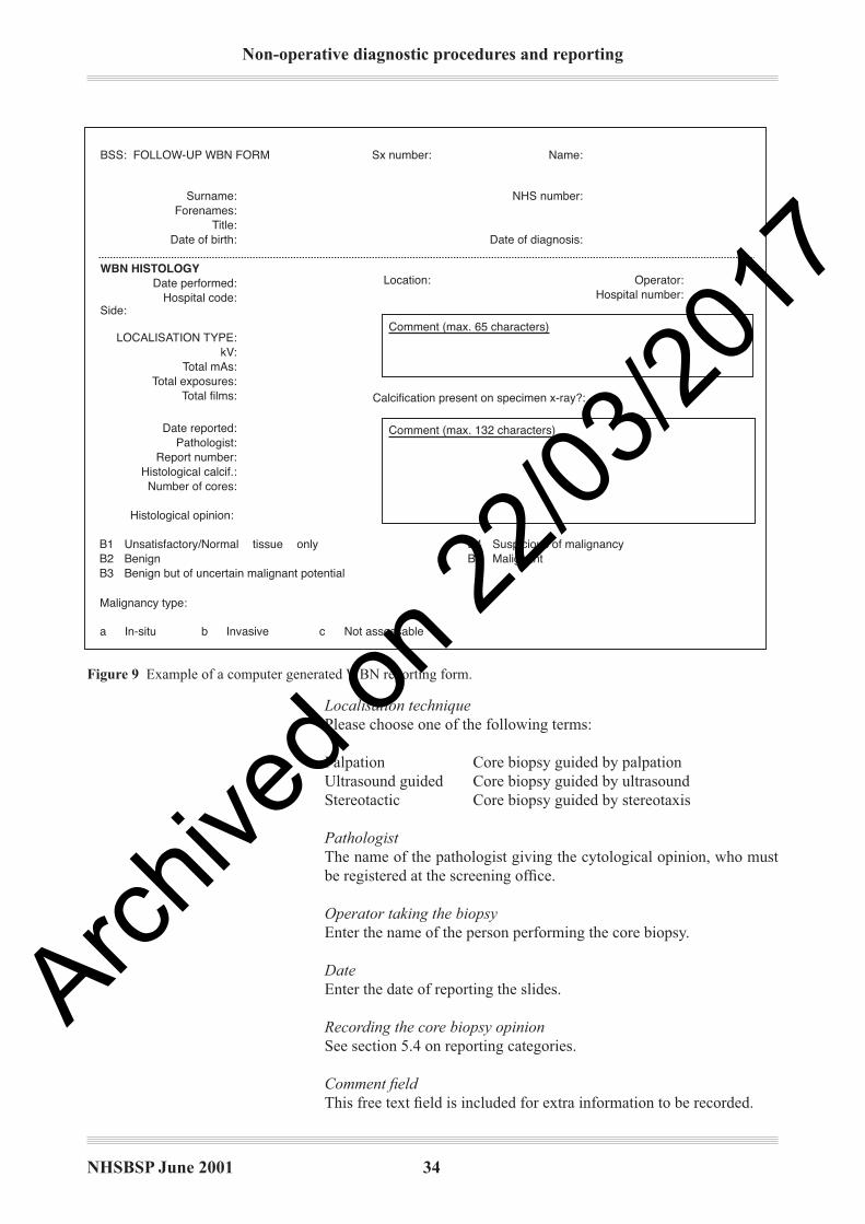
Non-operative diagnostic procedures and reporting
NHSBSP June 2001 34
Localisation technique
Please choose one of the following terms:
Palpation Core biopsy guided by palpation
Ultrasound guided Core biopsy guided by ultrasound
Stereotactic Core biopsy guided by stereotaxis
Pathologist
The name of the pathologist giving the cytological opinion, who must
be registered at the screening offi ce.
Operator taking the biopsy
Enter the name of the person performing the core biopsy.
Date
Enter the date of reporting the slides.
Recording the core biopsy opinion
See section 5.4 on reporting categories.
Comment fi eld
This free text fi eld is included for extra information to be recorded.
Figure 9 Example of a computer generated WBN reporting form.
WBN HISTOLOGY
Comment (max. 132 characters)
Comment (max. 65 characters)
Operator:Hospital number:
Location:
Calcification present on specimen x-ray?:
BSS: FOLLOW-UP WBN FORM Sx number: Name:
NHS number:
Date of diagnosis:
Surname:Forenames:
Title:Date of birth:
Date performed:Hospital code:
Side:
LOCALISATION TYPE:kV:
Total mAs:Total exposures:
Total films:
Date reported:Pathologist:
Report number:Histological calcif.:Number of cores:
Malignancy type:
Histological opinion:
B1 Unsatisfactory/Normal tissue onlyB2 BenignB3 Benign but of uncertain malignant potential
B4 Suspicious of malignancyB5 Malignant
a In-situ b Invasive c Not assessable
Archive
d on 2
2/03/2
017

NHSBSP June 2001 35
Non-operative diagnostic procedures and reporting
It is important to remember that histological examination of core biopsy
samples is performed to fulfi l the assessment process role by giving a
pathology category classifi cation (B1–5). It is not designed to give
a definitive diagnosis, although this is possible in the majority of
cases. Thus, while most core biopsy samples can be readily categorised
as normal, benign, or malignant, it must be recognised that a small
proportion (probably less than 10%) of samples cannot. The following
reporting guidelines have been devised in recognition of this and should
be used for all screen detected lesions (microcalcifi cation, architectural
deformities and mass lesions). It is recommended that this approach
should also be adopted for symptomatic practice. It is also important
to remember that, although there are fi ve reporting categories, similar
to those used in fi ne needle aspiration cytology (FNAC), these are
not equivalent.
These categories are designed to take account purely of the histological
nature of the specimen and not the clinical or imaging characteristics.
Similarly, it is not feasible for pathology interpretation to judge
independently whether a sample is adequate from the mammographic
lesion. This judgement requires multidisciplinary discussion. For
these reasons there is no inadequate biopsy category for core biopsy
specimens.
In some situations, particularly epithelial proliferation and papillary
lesions, the choice of diagnostic category should be based on the
features present in the sample, and additional guidance is given in
Table 4 and Figure 10.
B1 (normal tissue)
This indicates a core of normal tissue whether or not breast parenchymal
structures are present; thus this category is equally appropriate for a core
including normal breast ducts and lobules or mature adipose tissue or
stroma only. A B1 report should include a description of the components
present, and comment should be made regarding the presence of breast
epithelial structures.
Normal histology may indicate that the lesion has not been sampled. This
is, however, not necessarily so. In the case of certain benign lesions, such
as hamartomas and lipomas, apparently normal histological features
would be expected on core biopsy. Minor architectural distortions seen
mammographically may also result in minimal changes such as a slight
increase in stromal fi brosis on biopsy.
Cores with B1 diagnoses may contain microcalcifi cation, for example
within involutional lobules. It is important in these cases that discussion
between pathology and radiology colleagues is undertaken to confi rm the
appropriateness of the microcalcifi cation in the histological specimen.
Small foci of calcifi cation within involuted lobules are common and
frequently too small to be visible mammographically. Thus, a report
that merely records the presence of this calcifi cation without additional
comment on its nature, size and site may be misleading and lead to
false reassurance. It is evident that microcalcifi cation, either singly or in
clusters, less than 100 µm in diameter is not visible radiologically.43
5.45.4 Reporting categories
Archive
d on 2
2/03/2
017
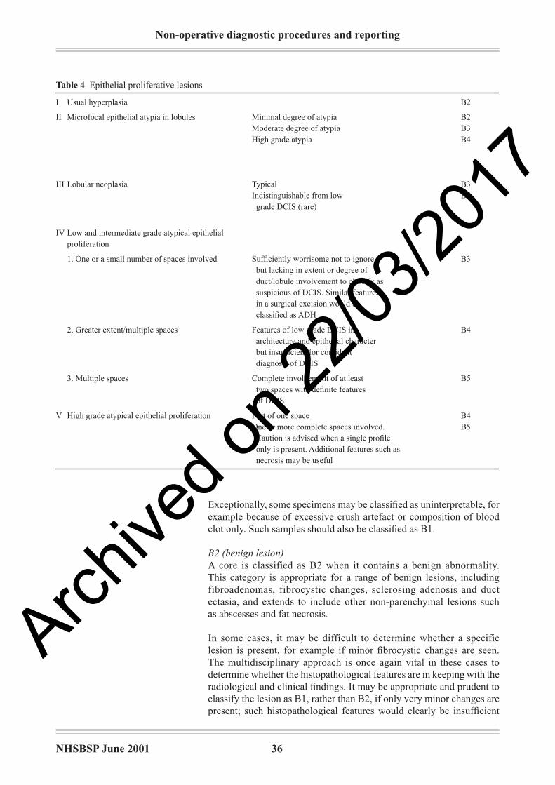
Non-operative diagnostic procedures and reporting
NHSBSP June 2001 36
Exceptionally, some specimens may be classifi ed as uninterpretable, for
example because of excessive crush artefact or composition of blood
clot only. Such samples should also be classifi ed as B1.
B2 (benign lesion)
A core is classified as B2 when it contains a benign abnormality.
This category is appropriate for a range of benign lesions, including
fibroadenomas, fibrocystic changes, sclerosing adenosis and duct
ectasia, and extends to include other non-parenchymal lesions such
as abscesses and fat necrosis.
In some cases, it may be difficult to determine whether a specific
lesion is present, for example if minor fi brocystic changes are seen.
The multidisciplinary approach is once again vital in these cases to
determine whether the histopathological features are in keeping with the
radiological and clinical fi ndings. It may be appropriate and prudent to
classify the lesion as B1, rather than B2, if only very minor changes are
present; such histopathological features would clearly be insuffi cient
Table 4 Epithelial proliferative lesions
I Usual hyperplasia B2
II Microfocal epithelial atypia in lobules Minimal degree of atypia B2
Moderate degree of atypia B3
High grade atypia B4
III Lobular neoplasia Typical B3
Indistinguishable from low B5
grade DCIS (rare)
IV Low and intermediate grade atypical epithelial
proliferation
1. One or a small number of spaces involved Suffi ciently worrisome not to ignore, B3
but lacking in extent or degree of
duct/lobule involvement to classify as
suspicious of DCIS. Similar features
in a surgical excision would be
classifi ed as ADH
2. Greater extent/multiple spaces Features of low grade DCIS in B4
architecture and epithelial character
but insuffi cient for confi dent
diagnosis of DCIS
3. Multiple spaces Complete involvement of at least B5
two spaces with defi nite features
of DCIS
V High grade atypical epithelial proliferation Part of one space B4
One or more complete spaces involved. B5
Caution is advised when a single profi le
only is present. Additional features such as
necrosis may be useful
Archive
d on 2
2/03/2
017

NHSBSP June 2001 37
Non-operative diagnostic procedures and reporting
Figure 10 Papillary lesions.
to explain a well-defi ned mass lesion and classifi cation as B2 would
be inappropriate.
B3 (lesion of uncertain malignant potential)
This category mainly consists of lesions which may provide benign
histology on core biopsy but are known to show heterogeneity or to have
an increased risk (albeit low) of associated malignancy.
Atypical epithelial proliferations (atypical ductal hyperplasia, ADH)
Included under a B3 classifi cation are atypical epithelial hyperplastic
lesions in which a uniform population of cells arranged in an appropriate
manner involves one space or partially involves two or more spaces.
These appearances should be sufficiently structured to raise the
possibility of low grade DCIS but insuffi cient in the tissue available
to fulfi l the diagnostic criteria.5 There is a range of severity from those
which are insufficient for a definite diagnosis of DCIS but highly
suspicious to those which only show a minor degree of atypia, normally
architectural, which requires further assessment, and judgement of
appropriate categorisation B3 or B4 is required.
The defi nition of atypical ductal hyperplasia is derived from surgical
resection specimens and relies on a combination of histological,
morphological and size extent criteria. For this reason, accurate
diagnosis of ADH is not possible on core biopsy. It has, however,
been shown that core biopsy samples which include atypical epithelial
proliferative foci of insuffi cient extent for classifi cation as DCIS may,
B2
Benign papilloma (part of) withno epithelial proliferation
Associated epithelial proliferation
It is anticipated that the majority of papillary lesions will be designated B3.
Lesion only partly sampled
Usual hyperplasia
Uniform epithelialproliferationAssess degree ofatypia and extent
Unequivocal DCIS
Unequivocal papillarycarcinoma in situ
Could be considered appropriateif lesion small and/or an incidentalfinding or believed to be adequatelyor fully sampled
B3
B3
B4
B5
Archive
d on 2
2/03/2
017
Non-operative diagnostic procedures and reporting
NHSBSP June 2001 38
on subsequent surgical resection, form part of an established in situ
neoplastic lesion with or without associated invasion. This view is
based on several studies that describe the subsequent surgical diagnoses
in cases described as ADH in non-operative core biopsy. In over 50%
of cores, surgical excision biopsy has shown either in situ or invasive
carcinoma.44 This is not surprising as ADH is basically defi ned as an
epithelial proliferation showing the features of low grade DCIS, but
in fewer than two spaces or less than 3 mm in diameter. The limited
tissue sampling that can be undertaken by core biopsy guns (often by
stereotactic methods for foci of microcalcifi cation) may thus provide
insuffi cient material for defi nitive diagnosis of low grade DCIS if
only a few spaces are obtained. In these cases a diagnosis of atypical
intraductal epithelial proliferation and a classifi cation of B3 of uncertain
malignant potential or B4 suspicious of malignancy should be made
dependent on the severity and extent of the lesion (see Table 4).
Lobular neoplasia
A small cell regular epithelial proliferation within lobules that is
considered by the pathologist to represent lobular neoplasia (ALH/LCIS)
should be classifi ed as B3. In common with ADH, the criteria used to
distinguish between atypical lobular hyperplasia (ALH) and lobular
carcinoma in situ (LCIS) are derived from surgically resected tissue.
Accurate distinction may not be possible in a limited core biopsy
sample. This process does not have the same management implications
as a diagnosis of DCIS or invasive malignancy and does not per se
require therapeutic excision. Lobular neoplasia is, however, most
frequently a coincidental fi nding in a core biopsy from a screen detected
lesion, and multidisciplinary discussion is essential, as the abnormality
identifi ed radiologically may not be represented. These cases must
be managed cautiously.
On occasion it may be impossible to classify a small cell epithelial
proliferation in lobules and/or ducts as either lobular neoplasia or low
grade DCIS, and in these circumstances a numerically higher category
(B4 or B5) is prudent and should be considered.
Phyllodes tumour
Fibroadenomatoid lesions with cellular stroma, stromal overgrowth and
possibly some mitotic activity suggesting a phyllodes tumour should
also be designated B3. Thus, the presence of a cellular stroma within
a fibroepithelial lesion should prompt a search for other features
that may aid in discrimination from a fibroadenoma. In practice,
however, this distinction is often impossible, and careful appraisal of
the entire clinical picture will usually allow appropriate management
to be undertaken.
Papillary lesions
Papillary lesions may also show signifi cant intralesional heterogeneity,
and the limited sampling achieved with core biopsy may miss areas
of in situ cancer. Thus, the majority of these lesions should also be
designated B3 of uncertain malignant potential. On rare occasions
when a small lesion has been very widely sampled and submitted for
Archive
d on 2
2/03/2
017

NHSBSP June 2001 39
Non-operative diagnostic procedures and reporting
pathological examination, a benign B2 classifi cation may be considered.
Conversely, when a sample of a papillary lesion in a core biopsy
shows atypia, for example strongly suspicious of papillary carcinoma
in situ, a B4 designation may occasionally be more appropriate (see
Figure 10).
Radial scar/complex sclerosing lesion
Biopsies which show features of a radial scar/complex sclerosing lesion
such as areas of hyalinisation, elastosis or tubular entrapment with
epithelial proliferation should be categorised as B3. Although still a
matter of debate, many authorities believe that a proportion of these
lesions are associated with malignancy. Thus, unless the sclerosing
lesion is very widely sampled, the process should be designated B3
as the presence of an associated area of DCIS or invasive carcinoma
cannot be excluded.
B4 (suspicious)
Technical problems such as crushed or poorly fi xed cores that contain
probable carcinoma but cannot provide the defi nitive diagnosis are
best included as B4. Similarly, apparently neoplastic cells contained
within blood clot or adherent to the outer aspect of the sample should
be classifi ed as B4 suspicious. Very small foci of invasive carcinoma
in which there is insuffi cient material to allow immunocytochemical
studies may also reasonably be assigned to this category.
A complete single duct space bearing an unequivocal high grade atypical
epithelial proliferative process can be classified as B5 malignant.
However, care must be taken if one or only part of a duct space is seen
to contain a highly atypical epithelial process (particularly if no necrosis
is present); this may be regarded as suspicious rather than defi nitively
malignant. In particular, great care should be taken if the epithelial cells
show any features of an apocrine phenotype, which may represent an
atypical apocrine proliferation rather than DCIS.
Another lesion that can be allocated to this category is a non-high grade
intraductal proliferation with a signifi cant degree of atypia probably
representing intermediate or low grade DCIS; in this case, relatively
few duct spaces are involved. A pragmatic approach is usually required
by reporting an atypical intraductal proliferation and qualifying this
according to the degree of suspicion, ie ‘at least ADH, probably
low grade DCIS’, and on the basis of extent or severity of atypia
allocating the case either to the B3 or to B4 category (see also B3
above and Figure 10).
The management of cases classified as B4 will usually be either
diagnostic excision biopsy of the area or repeat core biopsy sampling to
obtain defi nitive diagnosis. Defi nitive therapeutic surgery should not
be undertaken as a result of a B3 or B4 core biopsy diagnosis.
B5 (malignant)
This category is appropriate for cases of unequivocal malignancy on
core biopsy. Further categorisation into in situ and invasive malignancy
Archive
d on 2
2/03/2
017
Non-operative diagnostic procedures and reporting
NHSBSP June 2001 40
should be undertaken whenever possible. Other forms of malignancy
such as malignant lymphoma should also be classifi ed as B5.
Ductal carcinoma in situ
One of the benefi ts of core biopsy is that it can allow distinction between
in situ and invasive carcinoma. It should, however, be borne in mind
that, as a result of sampling error, the exclusive presence of DCIS in
the core does not exclude the possibility of an invasive focus being
present. In approximately 20% of cases sampled by standard methods
coexisting invasive carcinoma will be identifi ed in the subsequent
surgical excision specimen.45
The nuclear grade, architecture and the presence of necrosis of the DCIS
can be indicated on the core biopsy report. In particular, the presence
of associated calcifi cation should be recorded.
Lobular neoplasia
Lobular neoplasia is included in the B3 category, as it does not have
the same management implications as a diagnosis of DCIS or invasive
malignancy. In exceptional circumstances, lobular neoplasia may
be impossible to distinguish from small cell solid DCIS and may be
classifi ed as B5 (see also B3 above).
Invasive carcinoma
An advantage of core biopsy over FNAC is the ability to diagnose
invasion positively. Invasive mammary carcinoma can be unequivocally
identifi ed in core biopsy with a positive predictive value of 98%.45 As
noted above, however, the negative predictive value for invasion is only
80% when only DCIS is identifi ed.
All cores taken for microcalcification should undergo immediate
specimen x-ray, enabling the operator to repeat the biopsy if no
calcifi cation is seen. While being x-rayed, cores may be moistened
with saline to prevent desiccation, but this should be blotted prior
to fi lm exposure or resolution may be adversely affected. Cores in
which microcalcifi cation is detected may be marked with a vital dye or,
preferably, sent to the laboratory in a separate specimen pot, allowing
‘targeted’ examination to deeper levels should calcifi cation not be
detected in the initial series of sections. It is important to remember that
calcium oxalate crystals are often indistinct on routine haematoxylin
and eosin sections but are easily seen on polarisation. See also section
5.4 on reporting and multidisciplinary discussion of calcifi cation in
B1 (normal tissue).
Diagnostic pitfalls and problems in diagnosis in core biopsy include
many of the lesions that cause diffi culties in FNAC diagnosis (see page
24). Other lesions, however, may present particular diagnostic problems
in core samples (see Table 5).
Mild atypia of epithelium within lobular units is one of the commonest
problems encountered in core biopsy samples. Care must be taken not
to overdiagnose such minimal degrees of atypia, which may represent
5.55.5 Calcifi cation in core
biopsy
5.65.6 Problems and pitfalls
in diagnosis
5.6.1 Minor degrees of
epithelial atypia
Archive
d on 2
2/03/2
017
NHSBSP June 2001 41
Non-operative diagnostic procedures and reporting
usual epithelial hyperplasia, apocrine change or reactive changes (for
example adjacent to a previous sampling procedure). Conversely,
more severe degrees of atypia must be sought, as these may refl ect
cancerisation of lobules by high grade DCIS. The degree of atypia
should be helpful in distinguishing the process, and the nuclear
chromatin and presence of mitoses (although rarely seen) may aid
in the diagnosis.
Similarly, usual epithelial hyperplasia (UEH) and other forms of benign
hyperplasia such as that of gynaecomastoid type are commonly seen
in cores from benign fi broadenomas. Such cores often show apparent
discohesion due to the trauma of the core biopsy sampling process, and
‘telescoping’ of epithelium is seen within the duct spaces; thus, UEH
may resemble a hyperplastic process. As with UEH in surgical excision
specimens, the lack of uniformity and distribution/streaming of the
epithelial cells with bland nuclear features and paucity of mitoses
is of assistance in reaching a diagnosis. Atypical ductal hyperplasia
should not be diagnosed in these cases unless uniformity of nuclear size
and shape and regular, evenly placed nuclei are seen. Usual epithelial
hyperplasia of gynaecomastoid type with a micropapillary architecture
should not be mistaken for micropapillary ADH/DCIS.
Apocrine atypia, particularly in association with a sclerosing lesion
such as sclerosing adenosis (so-called ‘apocrine adenosis’), may be
especially diffi cult to identify correctly in non-operative diagnostic
samples. In core biopsy, large nuclei, often with prominent nucleoli,
may be mistaken for DCIS if pleomorphism is also present. The typical
granular eosinophilic cytoplasmic appearance of apocrine cells should
be sought. Pure apocrine DCIS is relatively rare, and when an apocrine
proliferation is seen within ducts in a core biopsy, additional features
of malignancy such as signifi cant atypia, intraluminal necrosis and the
presence of mitoses as well as multiple duct involvement should be
sought for confi rmatory evidence. Multiple duct involvement indicating
a more extensive lesion may provide further supportive evidence. Mild
or moderate degrees of apocrine proliferation with atypical features in
a duct space should be assessed with caution, and it may be prudent
not to record a defi nite diagnosis but to classify such a process as B3
(uncertain malignant potential). Conversely, papillary apocrine change
should not be mistakenly classifi ed as other than benign.
Focal lactational change may be seen in women who are neither lactating
nor pregnant and, indeed, are nulliparous and/or post-menopausal.
The involved acini are usually lined by plump vacuolated cells with
a ‘hobnail’ architecture but may, less frequently, appear atypical with
irregular, large or pyknotic nuclei. The epithelial cells may appear
degenerative, and rarely the benign nature of the process may be
mistaken for cancerisation of lobules by DCIS. The recognition of the
vacuolation of the cytoplasm and the typical hobnail architecture will
enable the correct diagnosis to be established.
5.6.2 Apocrine atypia and
apocrine DCIS
5.6.3 Lactational change
Archive
d on 2
2/03/2
017
Non-operative diagnostic procedures and reporting
NHSBSP June 2001 42
There is a risk of overdiagnosis of invasive carcinoma when confronted
by sclerosing adenosis in a core biopsy, particularly as the normal
lobular arrangement may be less apparent than on an excision biopsy
specimen. Immunohistochemical staining with collagen IV, laminin
and/or smooth muscle actin to demonstrate the presence of a basement
membrane and a dual epithelial/myoepithelial layer respectively can be
extremely useful in this situation.
The stromal appearances may be helpful in achieving a correct
diagnosis; sclerosing lesions do not induce the fi broblastic/desmoplastic
reaction generally seen in an invasive carcinoma. Radial scars/complex
sclerosing lesions have an eosinophilic fi brotic and elastotic central focus
with entrapped tubules. These elements have, however, a surrounding
myoepithelial layer. In diffi cult cases, immunohistochemistry for smooth
muscle actin may be invaluable; the absence of the myoepithelial
component in tubular carcinomas can be confi rmed.
Stromal proliferations may cause diffi culties in diagnosis in core biopsy
samples. Occasionally, a second biopsy sample taken from a patient
may show fi broblastic proliferation; this may represent the target lesion
but could also refl ect tissue reaction and repair at the previous biopsy
site. If the lesion represents the core site, an associated histiocyte
reaction or indeed fat necrosis may be present and haemosiderin-laden
macrophages can be seen. Sometimes a fibroblastic stroma may
be identified in a sample from a patient who has not undergone
previous FNAC or core biopsy and which may represent a spindle cell
proliferation such as a fi bromatosis or part of a spindle cell tumour such
as a nerve sheath tumour or myofi broblastoma. A stromal proliferation
may also be seen in phyllodes tumours and evidence for an epithelial
component should be sought, for example by performing additional
levels. Metaplastic carcinomas or rarely primary sarcomas (see section
5.7) may also mimic stromal proliferations, and a high index of
suspicion may enable confi rmatory diagnosis by immunohistochemical
examination with a range of anti-cytokeratin antibodies (at least one
broad spectrum and a high molecular weight cytokeratin, such as
MNF116 and LP34). Immunohistochemistry may prove unhelpful
and the multidisciplinary approach must be applied to the clinical,
radiological and histopathological features. When a definitive
histological diagnosis cannot be made the abnormality should be
reported as a spindle cell lesion of uncertain histogenesis or nature
and classifi ed as B3.
As noted above, phyllodes tumours may rarely be diffi cult to distinguish
from other stromal lesions. More commonly, the differential diagnosis
lies between a cellular benign fi broadenoma and a phyllodes tumour.
Features including stromal atypia, if present, can be useful, but the
degree of cellularity of the stroma is the most valuable feature to assess.
In rare cases it is not possible to distinguish between these two lesions
and the sample should be reported as a ‘fi broepithelial lesion’ and
classifi ed as B3, to avoid underdiagnosis of a phyllodes tumour. These
cases should be discussed at multidisciplinary meetings.
5.6.5 Stromal proliferations
and spindle cell lesions
5.6.6 Fibroepithelial tumours
5.6.4 Sclerosing lesions/
tubular carcinoma
Archive
d on 2
2/03/2
017
NHSBSP June 2001 43
Non-operative diagnostic procedures and reporting
Radiotherapy changes to the breast may be diffi cult to differentiate
from foci of recurrent or residual carcinoma, both in situ and invasive.
The radiation induces a degree of atypia of the breast epithelium but
also in the histiocyte population, which is prominent as a result of the
radiotherapy and also recent surgery. The macrophages may also show
degenerative features. Thus, carcinoma cells can conversely mimic
macrophages. Immunocytochemistry can be helpful in diffi cult cases
as irradiated neoplastic cells retain cytokeratin expression whereas
macrophages demonstrate a histiocytic phenotype, for example CD68
reactivity.
Small foci of invasive lobular carcinoma can be missed in histological
sections and be dismissed as chronic infl ammation or stromal cells. The
targetoid infi ltrative pattern of classical lobular carcinoma may be of
assistance, but a reactive lymphocyte process can also have a periductal
or perilobular distribution. Cytokeratin immunohistochemistry, to
demonstrate the neoplastic cells, is of value in difficult cases, but
recognition of the abnormal cell proliferation requires vigilance as
the features can be subtle.
As noted above, malignant lymphoma may rarely be identifi ed in core
biopsies and should be classifi ed as B5 malignant.
The majority of these lesions are of high grade B-cell morphology, and
these may mimic epithelial malignancy. As in other organs, the cells
frequently show less cohesion and a higher nuclear to cytoplasmic ratio
and do not demonstrate the architectural features of carcinoma, such
as tubule formation. To avoid misclassifi cation as carcinoma, however,
the correct diagnosis must be considered and can be supported by
immunohistochemistry (CD45, CD20, CD3, CD30, etc) to demonstrate
the appropriate phenotype.
5.6.7 Radiation induced
changes
5.6.8 Infi ltrating lobular
carcinoma
Table 5 Common causes of false positive and negative diagnoses
Common causes of false positive diagnosis
Sclerosing adenosis or radial scar/CSL mistakenly diagnosed as tubular carcinoma
Apocrine atypia in lobules, ducts or sclerosing lesions mistakenly diagnosed as DCIS
Chronic infl ammation mistakenly diagnosed as infi ltrating lobular carcinoma
Invasion mistakenly diagnosed in DCIS
Radiotherapy effect mistakenly diagnosed as carcinoma
Common causes of false negative diagnosis
Tubular carcinoma mistakenly diagnosed as sclerosing adenosis or radial scar/CSL
Infi ltrating lobular carcinoma mistakenly interpreted as chronic infl ammation or
missed
Radiotherapy effect with missed foci of carcinoma
Metaplastic carcinoma mistakenly diagnosed as a stromal proliferation/fi broblastic
scar
5.75.7 Rare lesions
5.7.1 Lymphoma
Archive
d on 2
2/03/2
017
Non-operative diagnostic procedures and reporting
NHSBSP June 2001 44
Low grade lymphomas may be more diffi cult to distinguish, mimicking
a chronic infl ammatory process. Infi ltration of the lobular epithelium
should be sought, and the degree of lymphoid infi ltrate, if high, should
raise the possibility of a neoplastic process. A panel of lymphoid
markers may be necessary to demonstrate the immunophenotype of the
cells present and to allow correct diagnosis.
Metastasis to the breast from malignancies derived elsewhere is well
recognised although rarely biopsied. A full clinical history is essential
to avoid misdiagnosis of a metastatic adenocarcinoma as a primary
breast carcinoma. Lesions which are recognised as metastasising to the
breast include lung, ovarian and renal carcinomas, but non-epithelial
malignancies such as melanoma, myelomas and rhabdomyosarcomas
may also be seen.
A panel of antibodies frequently allows identifi cation of the likely
site of a metastatic adenocarcinoma and enables appropriate clinical
investigation/management. Breast carcinomas usually express
cytokeratin 7 and 18 (and not cytokeratin 20), epithelial membrane
antigen and carcinoembryonic antigen (CEA)/non-specific cross-
reacting antigen (NCA), and approximately 80% will express oestrogen
receptor.
Primary breast sarcomas are rare. They most commonly originate in
association with phyllodes tumours, but in core biopsy specimens an
epithelial component is often not present. The most common phyllodes
associated sarcomas seen are liposarcoma and fi brosarcoma, although
other differentiation, including osteosarcoma, chondrosarcoma and
rhabdomyosarcomas, can be identifi ed. Angiosarcomas may be a cause
of false negative diagnosis as they may be relatively subtle and bland
and may be mistaken for radiotherapy changes, particularly when they
occur in this situation in the treated breast. Primary leiomyosarcoma
(and leiomyoma) may be found in the breast; the latter most commonly
in a retroareolar site. All these lesions can be diffi cult to diagnose
defi nitively in core samples. A high index of suspicion and judicious
use of immunohistochemistry can facilitate or support a diagnosis, but
non-diagnostic classifi cation as B3 or B4 is often prudent.
Grading on core biopsy can be performed and is reasonably accurate.
Current evidence suggests that concordance between grade on core
biopsy and that in the defi nitive excision specimen can be achieved
in approximately 75% of cases. It should, however, be made clear to
the clinicians that the grade may differ (almost invariably by only
one level) from that in the subsequent resection specimen. Mitotic
count in particular may be lower in the core biopsy than in the excision
specimen, therefore leading to underscore on the core. Tumours may
also often be typed according to the most common categories, such as
ductal/no specifi c type (NST) or classical lobular carcinoma. Invasive
carcinoma of special type, however, cannot be accurately predicted
although this may be suggested with some degree of accuracy in the
core biopsy report.
5.7.2 Metastasis to the breast
5.7.3 Sarcomas
5.85.8 Assessment of
prognostic information
Archive
d on 2
2/03/2
017

NHSBSP June 2001 45
Non-operative diagnostic procedures and reporting
ER assessment on core biopsies has been shown to correlate with the
subsequent surgical excision specimen46 and also to predict response
to hormone therapy.47 As with determination on excision biopsy
samples, a standard protocol and method of assessment should be
used (see Appendix 2).
5.95.9 Oestrogen receptor
(ER) assessment
Archive
d on 2
2/03/2
017
Non-operative diagnostic procedures and reporting
NHSBSP June 2001 46
6. QUALITY ASSURANCE
The defi nitions shown in Table 6 are intended to relate to the clinical
evaluation of the effectiveness of FNAC or core biopsy rather than
specifi cally to evaluation of laboratory performance. Thus, inadequate
(C1) FNAC or normal (B1) core biopsy results are not excluded from the
calculations as in some evaluations in the literature. Pathologists wishing
to evaluate their statistics purely to see their own accuracy in diagnosis
may wish to calculate the fi gures slightly differently.
Table 6 Defi nition of quality assurance measures for cytology and core biopsy
Absolute sensitivity The number of carcinomas diagnosed as such (C5 or B5) expressed as a percentage of the total number
of carcinomas sampled
Complete sensitivity The number of carcinomas that were not defi nitely negative or inadequate on FNAC or core expressed as
a percentage of the total number of carcinomas
Specifi city (full) The number of correctly identifi ed benign lesions (the number of C2 or B2 results minus the number of
false negatives) expressed as a percentage of the total number of benign lesions aspirated
Positive predictive The number of correctly identifi ed cancers (number of C5 or B5 results minus the number of false positive
value of a C5/B5 results) expressed as a percentage of the total number of positive results (C5 or B5)
diagnosis
Positive predictive The number of cancers identifi ed as suspicious (number of C4 or B4 results minus the number of false
value of a C4/B4 suspicious results) expressed as a percentage of the total number of suspicious results (C4 or B4)
diagnosis
Positive predictive The number of cancers identifi ed as atypia (number of C3 or B3 results minus the number of benign
value of a C3/B3 atypical results) expressed as a percentage of the total number of atypical results (C3 or B3)
diagnosis
False negative case A case that turns out (within a period of 3 years) to be carcinoma despite a negative cytology or core
result (this will by necessity include some patients in whom an area different from the lesion was sampled
but who present with an interval cancer)
False positive case A case that was given a C5 or B5 result and which turns out at open surgery to be a benign lesion
(including atypical hyperplasia)
False negative rate The number of false negative results expressed as a percentage of the total number of carcinomas sampled
False positive rate The number of false positive results expressed as a percentage of the total number of carcinomas sampled
Inadequate rate for The number of inadequate FNAC specimens expressed as a percentage of the total number of cases
FNAC aspirated
6.26.2 How to calculate
quality assurance
statistics
6.16.1 Defi nitions
Most of the computer systems in use in the National Health Service
Breast Cancer Screening Programme (NHSBSP) can calculate quality
assurance (QA) statistics automatically from the data recorded in the
screening database, cross-referenced with the histology or subsequent
outcome. A software routine (CQA in the case of cytology, BQA in the
case of core biopsy) can generate reports for quality assurance purposes.
Examples of the reports are shown in Tables 7 and 8. There is also a
further routine (NDQA) that combines the two routines, and calculates
performance measures for non-operative diagnostic procedures. This
routine takes the worst diagnosis of the two techniques where both have
been performed on the same patient and calculates the same parameters
as the CQA and BQA routines.
Archive
d on 2
2/03/2
017

NHSBSP June 2001 47
Non-operative diagnostic procedures and reporting
6.2.1 Further rules used in
deriving quality
assurance statistics
Table 7 Cytology /core biopsy quality assurance standard report
TOTAL CASES SCREENED IN PERIOD ……
TOTAL ASSESSED ……
TOTAL FNA (WBN) PERFORMED ……
Cytology
FNAC C5 C4 C3 C2 C1 Total
WBN B5 B4 B3 B2 B1 Total
Histology
Total malignant Box 1 Box 2 Box 3 Box 4 Box 5 Box 6
Invasive Box 7 Box 8 Box 9 Box 10 Box 11 Box 12
Non-invasive Box 13 Box 14 Box 15 Box 16 Box 17 Box 18
Total benign Box 19 Box 20 Box 21 Box 22 Box 23 Box 24
No histology Box 25 Box 26 Box 27 Box 28 Box 29 Box 30
Total C/B results Box 31 Box 32 Box 33 Box 34 Box 35 Box 36
Cases with both a non-invasive cancer and an invasive cancer in the
pathology database should count as invasive unless they are on opposite
sides, in which case they should be counted twice (once in each).
In cases with a malignant diagnosis the malignant fi ndings override
benign fi ndings unless they are on opposite sides.
Cases with open episodes are listed at the bottom of the report.
Tables 7 and 8 can be produced for internal QA purposes for all
clients, all tests, all cases/tests performed by any one aspirator, all
cases/tests reported by any one pathologist and all cases/tests by any
one localisation method (palpable, ultrasound, x-ray guided (perforated
plate) or stereotactic). The tables can also be produced for any date range,
the date taken being the date of needling (or, if that is not available, the
date of reporting) and for any geographic location.
The tables can be produced for any or all of the radiological appearances:
spiculated mass, rounded opacity, microcalcifi cation, stellate lesion
or asymmetrical density.
It is possible to request a report that lists the screening numbers of clients
involved in any of the cells in Table 7. For example, a list of cases in cell
no 26 is produced with the title: ‘Cases with C4 cytology (B4 results)
not biopsied but with closed episodes – please check’.
Note that all cases in box 25 are regarded as malignant and that all cases
in box 27 are regarded as benign.
Archive
d on 2
2/03/2
017
Non-operative diagnostic procedures and reporting
NHSBSP June 2001 48
The entry in each box (numbered 1 to 36) in Table 7 is calculated
either from the numbers of FNA with a C code (C1, C2, etc) cross-
referenced with the worst histology diagnosis or from the numbers of
WBN with a B code ( B1, B2, etc) cross-referenced with the worst
histology diagnosis.
The table should be produced for all FNA tests (headed ‘all tests’)
and also for all patients (headed ‘all patients’): if two FNA or WBN
records are present, the highest C or B number is taken. Only closed
episodes should be used.
The fi gures in the tables are then used to calculate values for each of the
QA measures in the cytology document. The calculations are shown in
Table 8 (numbers in bold correspond to box numbers in Table 7).
It is recognised that the specificities and false negative rates are
approximate and will be more accurate the longer the date range of the
analysis is from the date printed.
Suggested thresholds for cytology and core biopsy performance are
shown in Tables 9 and 10 respectively. These fi gures will obviously
depend on sampling techniques and the experience and care of the
aspirator48 and will vary widely between units.
The previous version of the Guidelines for Cytology Procedures and
Reporting in Breast Cancer Screening,3 now incorporated in this text,
gave fi gures for performance of cytology drawn from the literature and
reviewed also in the European Journal of Cancer.49 Since that time there
have been two published surveys of performance of, fi rstly, cytology50
and, subsequently, both cytology and needle core biopsy.51 Currently,
a further study of performance is under way, and provisional fi gures of
current performance are given in Tables 9 and 10.
The performance measures are interrelated, and strategy to improve one
aspect of performance will affect others. Thus, an attempt to reduce the
inadequate rate will often increase the number of suspicious reports,
attempts to improve the sensitivity are likely to increase the false
positive rate, attempts to improve the specifi city will increase the false
negative rate, and so on. Also, attempts to reduce the benign biopsy rate
by not biopsying the majority of lesions called benign on cytology will
reduce the specifi city where this is based on benign histology results
rather than on all aspirated cases.
The most common problem encountered in the NHSBSP surveys50,51
appears to be low sensitivities combined with high false negative rates
and high inadequate rates from lesions that subsequently turn out to
be cancer. This combination of statistics suggests a problem with the
accurate localisation of lesions for aspiration.49 A signifi cant proportion
of these lesions will have been palpable or thought to be palpable as
an area of thickening; aspiration of these areas without radiological
guidance may account for some of the problems. It is of interest to note
that, in centres where cytology has not been as useful in non-operative
6.36.3 Cytology and core
biopsy performance
fi gures
6.46.4 How to interpret the
results
Archive
d on 2
2/03/2
017

NHSBSP June 2001 49
Non-operative diagnostic procedures and reporting
Table 8 Calculation of cytology performance measures
Absolute sensitivity (this assumes that all unbiopsied
C5 or B5 results are carcinoma and are treated with
primary chemotherapy or hormonal therapy)
Complete sensitivity
Specifi city (biopsy cases only)
Specifi city (full) (this assumes that all cases of atypia (C3
or B3) which are not biopsied are benign)
Positive predictive value (C5/B5 diagnosis)
Positive predictive value (C4/B4 diagnosis)
Positive predictive value (C3/B3 diagnosis)
Negative predictive value (C2/B2) (at present this
parameter is not calculated by the various routines)
False negative rate (this excludes inadequate results)
False positive rate
Inadequate FNA rate and B1 core biopsy rate
Inadequate FNA rate and B1 core biopsy rate from
cancers
Suspicious rate
Core biopsy miss rate from cancers Sum of false negative rate and
B1 core biopsy rate from cancers
1 25
6 25
+
+¥100%
1 2 3 25
6 25
+ + +
+¥100%
22
24¥100%
22 28
24 27 28 29
+
+ + +¥100%
31 19
31
-¥100%
2
32 26-¥100%
3
33¥100%
34 4
34
-¥100%
19
6 25+¥100%
4
6 25+¥100%
35
36¥100%
5
6 25+¥100%
32 33
36
+¥100%
Archive
d on 2
2/03/2
017

Non-operative diagnostic procedures and reporting
NHSBSP June 2001 50
Table 9 Suggested thresholds for cytology performance (where therapy is
partially based on FNAC)
Minimum Preferred Current
(%) (%) median (%)
Absolute sensitivity (AS) > 60 > 70 57.1
Complete sensitivity (CS) > 80 > 90 81.5
Specifi city (full) (SPEC) >55 >65 58.4
(including non-biopsied
cases) (as calculated above)
Positive predictive value >98 >99 99.6
(+PV)
False negative rate (F–) < 6 < 4 6.3
False positive rate (F+) < 1 < 0.5 0.2
Inadequate rate (INAD) < 25 < 15 23.4
Inadequate rate from cancers < 10 < 5 9.8
Suspicious rate < 20 < 15 15.8
Table 10 Suggested thresholds for core biopsy performance
Minimum Preferred Current
(%) (%) median (%)
Absolute sensitivity (AS) > 70 > 80 76.4
Complete sensitivity (CS) > 80 > 90 84.5
Specifi city (full) (SPEC) > 75 > 85 81.2
(including non-biopsied
cases) (as calculated above)
Positive predictive value > 99 > 99.5 100
(+PV)
False positive rate (F+) < 0.5 < 0.1 0
Miss rate (B1 + B2) from <15 <10% 15.1
cancer
Suspicious rate < 10 < 5 4.8
diagnosis, there has been a swing towards the use of core biopsy,52 as
is commonly reported in publications from the United States.53 Audit
of core biopsy in the NHSBSP shows similar variability in practice.51
Some units are using the two techniques to complement each other and
are achieving higher non-operative diagnosis rates in diffi cult cases.54
This can be especially useful in lobular and tubular carcinomas where
cytology is less able to give an unequivocal diagnosis.36,51,55
In general, the cytological performance of pathologists as assessed by
the positive predictive values is good, although some pathologists are
clearly being cautious in diagnosis. This caution can be inferred from
the statistics in the units with high positive predictive values for C4
and C3 diagnoses and also in units which have a high suspicious rate. It
Archive
d on 2
2/03/2
017
NHSBSP June 2001 51
Non-operative diagnostic procedures and reporting
6.56.5 Education and
training
should be noted that caution by pathologists may be a function of their
experience in the technique, previous experience of false positive and
false negative results, poorly cellular samples or local differences in
treatment protocols when faced with a C5 diagnosis.
High inadequate rates without a corresponding increase in the inadequate
rate from cancers can be seen in occasional units. In these units, a
high proportion of the women who were recalled for assessment had
a needling procedure. This suggests that, in these units, the clinicians
were needling lesions with a low predictive value for malignancy in
order to reassure either the patient or themselves. This is not necessarily
a problem, and therefore the crude inadequate rate may not be a good
measure of aspiration technique. A better measure appears to be the
inadequate rate from lesions which turn out subsequently to be cancer
or, for core biopsy, the miss rate (see above).
The Training and Education Subgroup of the National Coordinating
Group for Breast Screening Pathology is responsible for organising
breast screening pathology courses including a biannual non-operative
diagnosis course. Information on these courses can be obtained from
Nottingham International Breast Education Centre. In addition, the
British Society for Cervical Cytology (BSCC) and other specialist
centres organise workshops or courses in fi ne needle aspiration cytology
in Edinburgh, Guildford, Nottingham and at the Royal Marsden
Hospital. Additional experience may be gained by secondment to
neighbouring centres of expertise and participating in external quality
assurance (EQA) schemes. It is recognised that courses can only
provide baseline knowledge, and acceptable levels of performance,
particularly in cytological diagnosis, can only be realistically achieved
by experience in routine practice. Regular self-audit of non-operative
diagnosis results should be undertaken and is of educational value.
Archive
d on 2
2/03/2
017
Non-operative diagnostic procedures and reporting
NHSBSP June 2001 52
APPENDIX 1: HOW THE NATIONAL
SCREENING SYSTEM TREATS
CYTOLOGY DATA
Please note that, owing to a conversion error in the National Breast
Screening System, some FNAC results recorded before January 1991
may be miscoded. See below for further details.
The National Breast Screening Computer System (NBSS), designed in
Oxford and used in most regions for the recording of breast screening,
stores fi ne needle aspiration results as a diagnostic set (FINE) and
core biopsy results as a diagnostic set (WIDE). These diagnostic sets
are part of the assessment module. Specimen type and FNAC/core
biopsy diagnosis are recorded with the comment fi eld, and radiological
information is also recorded. The method of localisation of the lesion,
whether by clinical, ultrasound, perforated plate or stereotactic, is
recorded and the radiation dosage (number of exposures, fi lms, kV
and mAs) is similarly recorded. The system records an FNAC/core
biopsy opinion (C1/B1, C2/B2, etc) and also an overall opinion after
full assessment, which is entered after a multidisciplinary decision
is taken. This opinion then forms the basis for an action, which may
range from routine recall (RR) to surgical treatment (ST) depending on
the outcome of assessment. The date of the sampling and report, the
pathologist, operator and specimen number are also recorded.
The FNAC/core biopsy opinion forms a part of a fi eld in the software,
which is called the ‘episode endpoint’. In this FNAC/core biopsy is
not treated the same as in the program itself, being separated from
assessment. Examples of the coding of this fi eld would be S+, Aabn,
C–, W–, H– where a lesion was thought positive on screening fi lms
(S), was thought benign in nature after assessment (A), was thought
overall to be benign after cytology (note that this does not necessarily
mean that cytology was benign) (C), was thought to be benign after
core biopsy (W) (this does not necessarily mean that the core was
benign) and was histologically benign (H), or S+, A+, C+, W+, H+
for a malignancy identified as such by screening (S), assessment
(A), cytology (C) (this does not mean that cytology was necessarily
malignant), core biopsy (W) (this does not necessarily mean that
core biopsy was malignant) and histology (H). The system uses
bespoke downloads (‘fi le of episodes’, ‘fi le of assessment details’,
etc) or the ‘Co-Writer’™ report generator to produce custom reports
on request.
Because of a problem on conversion of the database from the version 2.0
to version 2.1 whereby the assessment and cytology record structures
were changed, a number of cytology results that were originally ‘benign’
on the system were recoded as ‘suspicious probably benign (C3)’. This
error occurred at date of conversion in each unit, which generally varies
between units from August 1990 and December 1990, although in some
units may be even later than this. Records entered before that date may
Archive
d on 2
2/03/2
017

NHSBSP June 2001 53
Non-operative diagnostic procedures and reporting
be subject to this error, and data obtained from the computer before
1991 may contain a high number of suspicious results because of this
data conversion. It is therefore recommended that data downloaded
or patient episodes reviewed from the national system pertaining to
results entered before 1991 are treated with caution. Also, some of
these records may be lacking certain data items that were not part of
the previous software. It is not possible to run a general correction for
these results as the dates at which the conversion was done varies from
unit to unit, but it is possible to run an FNAC program that will list all
FNACs performed, with their dates, to allow correction of these. If this
is required then information may be obtained from the local Oxford
Level Two Support Team. This error does not apply to other systems, but
information on those may be obtained from the appropriate computer
management teams.
In version 3 of the software, core biopsy results were removed from the
biopsy and treatment records (BT) and converted into WBN results in
the new diagnostic set WIDE. Because the facility to code results as
B3 or B4 was not possible in the old BT record, some early suspicious
core biopsy results may be miscoded as malignant (B5). This can be
investigated as a possible cause if a high false positive rate is noted, but
many units were not entering core biopsy results onto the system at that
time. These converted records will also lack some data items such as
operator, number of cores, etc.
Archive
d on 2
2/03/2
017
Non-operative diagnostic procedures and reporting
NHSBSP June 2001 54
APPENDIX 2: IMMUNOHISTO-
CHEMICAL DETECTION OF STEROID
RECEPTORS IN BREAST CANCER
A WORKING PROTOCOL PRODUCED
BY THE UK RECEPTOR GROUP, UK
NEQAS, THE SCOTTISH BREAST CANCER
PATHOLOGY GROUP AND THE RECEPTOR
AND BIOMARKER STUDY GROUP OF THE
EORTC
The protocol described in this paper refl ects an overall method that is
currently working well in a number of British and mainland European
laboratories. Using this methodology, these laboratories fi nd that results
(obtained with the attached scoring system) are highly reproducible.
Such a scoring system has also been shown to correlate well with the
old biochemical assays and to give at least as signifi cant predictive
and prognostic information.56
To keep loss of antigenicity to a minimum, excised tissue should
be incised (sliced) and placed into an adequate volume of fi xative
(preferably formol saline or neutral buffered formalin) for about 24 h
(the slicing allows the necessary rapid and even penetration of fi xative
– time of fi xation can vary according to the size of tissue slices but
should be judged on achievement of good morphological appearance).
Wherever possible, adjacent sections of normal parenchyma should be
taken to act as internal positive control. Localisation biopsies should be
handled according to the published guidelines (eg NHSBSP Publication
No 35). Core biopsies may be fi xed for a period shorter than 24 h.
Sections should be heated onto slides that are either commercially
precoated or hand coated with a strong adhesive such as APES –
to avoid sections becoming detached from the slide during antigen
retrieval. Antigen retrieval is most reliably achieved with 1–2 min in a
pressure cooker using 0.01 mol/l citrate buffer pH 6.0 (though properly
controlled microwaving is acceptable and detailed methodology for
antigen retrieval, buffer selection etc is available from UK NEQAS-ICC
– address below). The anti-receptor antibodies selected should be those
that have been validated and have a good record in published work,
eg clone 1D5 (available from Dako and others) or clone 6F11 from
Novocastra for oestrogen receptor, and clones 1A6 (Novocastra) or
PR88 (Biogenix) for progesterone receptor (PR). The use of surfactants
in buffers prior to the application of the anti-receptor antibody can
reduce the detection of the receptor signifi cantly (shown, for example,
for 1D5) and should, therefore, be avoided.
Archive
d on 2
2/03/2
017
NHSBSP June 2001 55
Non-operative diagnostic procedures and reporting
In-house controls should come from a composite block containing
receptor rich, receptor poor and receptor negative components. If, as
recommended, normal breast epithelial cells in adjacent tissue have been
taken, these will provide a good internal, positive control.
Staining of sections by hand can be more reliable for low, positive
receptor expression. Where automatic stainers are used, it is essential to
check the equivalence of the sensitivity with that produced by manual
methods. Experienced users of automatic stainers get good results even
for low positive receptor expression, so long as surfactants are avoided.
A sensitive method of detection, such as ABC (for example, the Dako
Duet K0492 gives consistently good results but others will work equally
well), should be used. Counterstaining should not obscure weak, positive
staining, but if this is avoided can be valuable. Excessive counterstaining
does occur in some automatic stainers though adjustment should be
possible. True receptor staining is taken to be nuclear and cytoplasmic
staining should be ignored.
All laboratories must take part in an external quality assurance scheme:
an appropriate one is run by UK NEQAS (address below).
In reporting the results of steroid receptor immunohistochemistry,
simple scoring systems are found to work the best (see reference 57).
These use either a direct count of the proportion of epithelial nuclei that
take up stain or a simple combination of the percentage cells staining
plus a measure of intensity of stain, eg the Quick Score.
The following scoring system is a simple, additive system giving a
range from 0 to 8 and has proven to be highly reproducible among
readers:
Score for proportion staining Score for staining intensity
0 = no nuclear staining 0 = no staining
1 = <1% nuclei staining 1 = weak staining
2 = 1–10% nuclei staining 2 = moderate staining
3 = 10–33% nuclei staining 3 = strong staining
4 = 33–66% nuclei staining
5 = 66–100% nuclei staining
Adding the two scores together gives a maximum score of 8.
Experience to date suggests that (see also reference 56), using such
a simple scoring system, appropriate cut-off values for treatment of
advanced disease are as follows:
• a score of zero indicates that endocrine therapies will defi nitely
not work
• a score of 2–3 indicates a small (20%) chance of response to
endocrine therapies
• a score of 4–6 indicates an even (50%) chance of response
• a score of 7–8 indicates a good (75%) chance of response.
Archive
d on 2
2/03/2
017
Non-operative diagnostic procedures and reporting
NHSBSP June 2001 56
Where progesterone receptor content has also been determined,
endocrine therapy is deemed worthwhile in patients with low ER but
high progesterone receptor (PR).58
In elderly patients, some tumour shrinkage may be seen after endocrine
therapy, even though the ER staining is low.58
Perhaps the single most valuable observation is that for patients with a
breast cancer that is zero on the Quick Score the chances of response to
endocrine therapy are essentially nil and these patients should receive
an alternative therapy as fi rst-line therapy.
The appropriate cut-off values for adjuvant therapy using steroid
receptor determination by immunohistochemistry have yet to be
determined, though Harvey et al56 reported that, using a similar scoring
system but slightly different protocol, a value > 2 was the optimal cut-off
point for predicting improved outcome (P < 0.0001).
Authors
Written on behalf of the associated groups by Robin Leake (Biochemistry,
Glasgow), Diana Barnes (Pathology, Guy’s Hospital, London), Sarah
Pinder and Ian Ellis (Pathology, Nottingham), Liz Anderson (Clinical
Research, Christie Hospital, Manchester), Tom Anderson (Pathology,
Edinburgh), Ruth Adamson (Pathology, Kilmarnock), Tony Rhodes
and Keith Miller (UK NEQAS) and Rosemary Walker (Pathology,
Leicester).
Technical advice can be obtained from
UK NEQAS for Immunohistochemistry
Department of Histopathology
UCL Medical School
University Street
London WC1E 6JJ
Phone +44 (0) 20 7209 6049 Fax +44 (0) 20 7387 3674
Reproduced with permission from Journal of Clinical Pathology 2000,
53: 634–635.
Archive
d on 2
2/03/2
017

NHSBSP June 2001 57
Non-operative diagnostic procedures and reporting
REFERENCES
1. Royal College of Pathologists Working Group for Breast Screening Pathology.
Guidelines for Pathologists. Sheffi eld, NHS Breast Screening Programme, 1989
(NHSBSP Publication No 2).
2. Royal College of Pathologists Working Group. Pathology reporting in breast
cancer screening. Journal of Clinical Pathology 1989, 44: 710–725.
3. Cytology Sub-group of the National Coordinating Committee for Breast Cancer
Screening Pathology. Guidelines for Cytology Procedures and Reporting in
Breast Cancer Screening. Sheffi eld, NHS Breast Screening Programme and
Royal College of Pathologists, 1993 (NHSBSP Publication No 22).
4. European Commission. European Guidelines for Quality Assurance in
Mammography Screening, 2nd edn. Luxembourg, Offi ce for Offi cial Publications
of the European Communities, 1996.
5. Royal College of Pathologists Working Group for Breast Screening Pathology.
Pathology Reporting in Breast Cancer Screening (2nd edition). Sheffi eld, NHS
Breast Screening Programme, 1997 (NHSBSP Publications No 3).
6. National Coordinating Group for Breast Screening Pathology. Guidelines for
Breast Pathology Services. Sheffi eld, NHS Breast Screening Programme, 1997
(NHSBSP Publication No 2).
7. Lamb J, Anderson TJ, Dixon MJ, Levack P. Role of fi ne needle aspiration
cytology in breast cancer screening. Journal of Clinical Pathology 1987, 40:
705–709.
8. Zajdela A, Chossein NA, Pillerton JP. The value of aspiration cytology in the
diagnosis of breast cancer. Cancer 1975, 35: 499–506.
9. Azavedo E, Svane G, Auer G. Stereotactic fi ne needle biopsy in 2594
mammographically detected non-palpable lesions. Lancet 1989, 1: 1033–1035.
10. Wilson ARM, Asbury D, Cooke J et al (eds). Clinical Guidelines for Breast
Cancer Screening Assessment. Sheffi eld, NHS Cancer Screening Programmes,
2001 (NHSBSP Publication No 49).
11. Britton P. Fine needle aspiration or core biopsy. The Breast 1999, 8: 1–4.
12. Philpotts LE, Shaheeh NA, Carter D et al. Comparison of rebiopsy rates after
stereotactic core needle biopsy of the breast with 11-gauge vacuum suction probe
versus 14-gauge needle and automatic gun. American Journal of Roentgenology
1999, 172: 683–687.
13. Warren R. Team learning and breast cancer screening. Lancet 1991, 338: 514.
14. Kaminsky DB. Aspiration biopsy in the context of the new Medicare fi scal policy.
Acta Cytologica 1984, 28: 333–337.
15. Kocjan G. Evaluation of the cost effectiveness of establishing a fi ne needle
aspiration cytology clinic in a hospital out-patient department. Cytopathology
1991, 2: 13–18.
16. McKee G, Thomas B, Cooke J. Stereotactic cytology – results of the fi rst 250
cases. In: 3rd Annual BSCC Meeting, Sheffi eld. 1991. BSCC.
17. Burkhardt JH, Sunshine JH. Core-needle and surgical breast biopsy: Comparison
of three methods of assessing cost. Radiology 1999, 212: 181–188.
18. Liberman L, Feng TL, Dershall DD. US-guided core breast biopsy: use and
cost-effectiveness. Radiology 1998, 208: 717–723.
19. Zakhour H, Wells C. Diagnostic Cytopathology of the Breast. London, Churchill
Livingstone, 1999.
20. Zajdela A, Zillhardt P, Voillemot N. Cytological diagnosis by fi ne needle
sampling without aspiration. Cancer 1987, 59: 1201–1205.
21. Catania S, Boccato P, Bono A et al. Pneumothorax: a rare complication of fi ne
needle aspiration of the breast. Acta Cytologica 1989, 33: 140.
Archive
d on 2
2/03/2
017
Non-operative diagnostic procedures and reporting
NHSBSP June 2001 58
22. Liberman L, Smolkin JH, Dershaw DD et al. Calcifi cation retrieval at
stereotactic, 11-gauge, directional, vacuum-assisted breast biopsy. Radiology
1998, 208: 251–260.
23. Liberman L, Vuolo M, Dershaw DD et al. Epithelial displacement after
stereotactic 11-gauge directional vacuum-assisted breast biopsy. American
Journal of Roentgenology 1999, 172: 677–681.
24. Preece PE, Hunter SM, Duguid HLD, Wood RAB. Cytodiagnosis and other
methods of biopsy in the modern management of breast cancer. Seminars in
Surgical Oncology 1989, 5: 69–81.
25. Peterse JL, Koolman-Schellekens MA, van-de-Peppel-van-de-Ham T, van-
Heerde P. Atypia in fi ne-needle aspiration cytology of the breast: a histological
follow-up study of 301 cases. Seminars in Diagnostic Pathology 1989, 6:
126–134.
26. Simpson JF, Page DL, Dupont WD. Apocrine adenosis – a mimic of mammary
carcinoma. Surgical Pathology 1990, 3: 289–299.
27. Salhany KE, Page DL. Fine needle aspiration of mammary lobular carcinoma
in situ and atypical lobular hyperplasia. American Journal of Clinical Pathology
1989, 92: 22–26.
28. Bibbo M, Scheiber M, Cajulis R et al. Stereotaxic fi ne needle aspiration cytology
of clinically occult malignant and premalignant breast lesions. Acta Cytologica
1988, 32: 193–201.
29. Abendroth CS, Wang HH, Ducatman BS. Comparative features of carcinoma in
situ and atypical ductal hyperplasia of the breast on fi ne-needle aspiration biopsy
specimens. American Journal of Clinical Pathology 1991, 96: 654–659.
30. Sneige N, Staerkel GA. Fine needle aspiration cytology of ductal hyperplasia
with and without atypia and ductal carcinoma in situ. Human Pathology 1994,
25: 485–492.
31. Oertel YC, Galblum LI. Fine needle aspiration of the breast: diagnostic criteria.
Pathology Annual 1983, 18 Pt 1.
32. Wells CA, Wells CW, Yeomans P et al. Spherical connective tissue inclusions
in epithelial hyperplasia of the breast (‘collagenous spherulosis’). Journal of
Clinical Pathology 1990, 43: 905–908.
33. Tyler X, Coghill SB. Fine needle aspiration cytology of collagenous spherulosis
of the breast. Cytopathology 1991, 2: 159–162.
34. Evans AT, Hussein KAH. A microglandular adenosis like lesion simulating
tubular adenocarcinoma. Cytopathology 1990, 1: 311–316.
35. Lamb J, Anderson TJ. Infl uence of cancer histology on the success of fi ne needle
aspiration of the breast. Journal of Clinical Pathology 1989, 42: 733–735.
36. Bondesan L, Lindholm K. Aspiration cytology of tubular breast carcinoma. Acta
Cytologica 1990, 34: 15–20.
37. Sneige N, Zachariah S, Fanning TV et al. Fine needle aspiration cytology of
metastatic neoplasms in the breast. American Journal of Clinical Pathology 1989,
92: 27–35.
38. Redard M, Vassilakos P, Weintraub J. A simple method for oestrogen receptor
antigen preservation in cytologic specimens containing breast carcinoma cells.
Cytopathology 1989, 5: 188–193.
39. Hunt CM, Ellis IO, Elston CW et al. Cytological grading of breast carcinoma – a
feasible proposition? Cytopathology 1990, 1: 287–295.
40. Thomas JSJ, Mallon EA, George WD. Semi-quantitative analyses of fi ne needle
aspirates from benign and malignant breast lesions. Journal of Clinical Pathology
1989, 42: 28–34.
41. Robinson IA, McKee G, Nicholson A et al. Prognostic value of cytological
grading of fi ne-needle aspirates from breast carcinomas. Lancet 1994, 343:
947–949.
42. Mapstone NP, Zakhour HD. Morphometric analysis of fi ne needle aspirates from
breast lesions. Cytopathology 1990, 1: 349–355.
Archive
d on 2
2/03/2
017

NHSBSP June 2001 59
Non-operative diagnostic procedures and reporting
43. Dahlstrom JE, Sutton S, Jain S. Histologic–radiologic correlation of
mammographically detected microcalcifi cation in stereotactic core biopsies.
American Journal of Surgical Pathology 1998, 22: 256–259.
44. Liberman L, Cohen MA, Dershaw DD et al. Atypical ductal hyperplasia
diagnosed at stereotaxic core biopsy of breast lesions: an indication for surgical
biopsy. American Journal of Roentgenology 1995, 164: 1111–1113.
45. Liberman L, Dershaw DD, Rosen PP et al. Stereotaxic core biopsy of breast
carcinoma: accuracy at predicting invasion. Radiology 1995, 194: 379–381.
46. Zidan A, Brown JSC, Peston D, Shousha S. Oestrogen and progesterone receptor
assessment in core biopsy specimens of breast carcinoma. Journal of Clinical
Pathology 1997, 50: 27–29.
47. Goulding H, Pinder S, Cannon P et al. A new immunohistochemical antibody
for the assessment of estrogen receptor status on routine formalin-fi xed tissue
samples. Human Pathology 1995, 26: 291–294.
48. Barrows GH, Anderson TJ, Lamb JL, Dixon JM. Fine needle aspiration of
breast cancer – relationship of clinical factors to cytology results in 689 primary
malignancies. Cancer 1986, 58: 1493–1498.
49. Wells CA. Quality assurance in breast cancer screening cytology: A review of the
literature and a report on the UK National Cytology Scheme. European Journal
of Cancer 1995, 31A: 273–280.
50. Wells CA, Perera R, White FE, Domizio P. Fine needle aspiration cytology in
the UK Breast Screening Programme – a national audit of results. The Breast
1999, 8: 261–266.
51. Britton PD, McCann J. Needle biopsy in the NHS Breast Screening Programme
1996/1997: How much and how accurate? The Breast 1999, 8: 5–11.
52. Crotch-Harvey MA, Loughran CV. Combined stereotactic wide core needle
biopsy and fi ne needle aspiration cytology in the assessment of impalpable
mammographic abnormalities detected in a breast screening programme. The
Breast 1996, 5: 48–49.
53. Meyer JE. Value of large core biopsy of occult breast lesions. American Journal
of Roentgenology 1992, 158: 991–992.
54. Yeoman LJ, Michel MJ, Humphreys S et al. Radiographically guided fi ne needle
aspiration cytology and core biopsy in the assessment of impalpable breast
lesions. The Breast 1996, 5: 41–47.
55. Sadler GP, McGee S, Dallimore NS et al. Role of fi ne needle aspiration cytology
and needle core biopsy in the diagnosis of lobular carcinoma of the breast. British
Journal of Surgery 1994, 81: 1315–1317.
56. Harvey JM, Clark GM, Osborne CK, Allred DC. Oestrogen receptor status by
immunohistochemistry is superior to the ligand-binding assays for predicting
response to adjuvant therapy in breast cancer. Journal of Clinical Oncology 1999,
17: 1474–1485.
57. Barnes DM, Millis RR, Beex L et al. Increased use of immunohistochemistry
for oestrogen receptor measurement in mammary carcinoma: the need for quality
assurance. European Journal of Cancer 1998, 34: 1677–1682.
58. EBCTCG. Tamoxifen for early breast cancer: an overview of randomised trials.
Lancet 1998, 351: 1451–1467.
Archive
d on 2
2/03/2
017

Non-operative diagnostic procedures and reporting
NHSBSP June 2001 60
Archive
d on 2
2/03/2
017