guidelines for histopathological evaluation of …€¦ · guidelines for histopathological...
TRANSCRIPT

IIVS Abridged Guidelines for BCOP Histopathology 1
GUIDELINES FOR HISTOPATHOLOGICAL EVALUATION OF
BOVINE CORNEAS AS AN ENDPOINT OF THE BOVINE CORNEAL
OPACITY AND PERMEABILITY (BCOP) ASSAY
INSTITUTE FOR IN VITRO SCIENCES, INC.
30 W. Watkins Mill Road, Suite 100
Gaithersburg, Maryland 20878 USA
December 2016

IIVS Abridged Guidelines for BCOP Histopathology 2
TABLE OF CONTENTS
1. Brief Introduction of the Bovine Corneal Opacity and Permeability Assay ................................ 3
2. Depth of Injury as a Predictor of Degree and Duration of Ocular Injury ..................................... 5
3. Application of Histopathology to the Determination of Ocular Irritation Potential ..................... 4
4. Overview of the Histology Procedures Used at the Institute for In Vitro Sciences (IIVS) .......... 5
4.1 Corneal Accession Numbers ................................................................................................... 5
4.2 Fixation of the Corneas ............................................................................................................ 5
4.3 Preparation of the Slides .......................................................................................................... 5
5. Evaluating the Corneal Histology ................................................................................................. 7
5.1 Evaluation of the Corneal Sections (Overview) ...................................................................... 7
5.2 Evaluation of the Quality and Acceptability of the Corneal Sections ..................................... 7
5.3 Recording Observations......................................................................................................... 11
5.3.1 Evaluating the Negative Control-Treated Corneas .......................................................... 11
5.3.2 Evaluating the Corneal Histologic changes ..................................................................... 11
5.4 Preparation of Photomicrographs .......................................................................................... 14
6. A short compendium of micrographs to illustrate negative control-treated (normal) and
select histologic changes in bovine corneal tissue ........................................................................... 14
6.1 Negative Control Corneas....................................................................................................... 14
6.2 Epithelial Damage .................................................................................................................. 17
6.3 Stromal Histologic changes .................................................................................................... 24
6.4 Endothelial Cell Histologic changes ....................................................................................... 26
7. References................................................................................................................................... 28
IIVS Abridged Guidelines for BCOP Histopathology 3
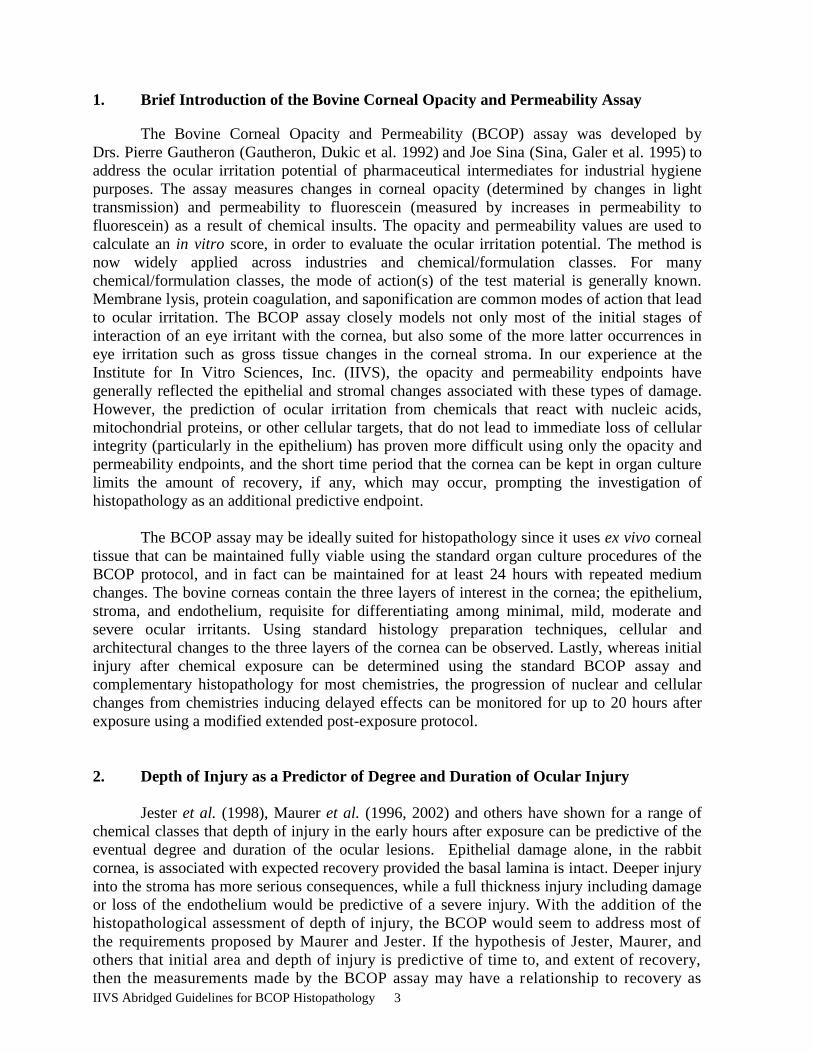
1. Brief Introduction of the Bovine Corneal Opacity and Permeability Assay
The Bovine Corneal Opacity and Permeability (BCOP) assay was developed by
Drs. Pierre Gautheron (Gautheron, Dukic et al. 1992) and Joe Sina (Sina, Galer et al. 1995)
to
address the ocular irritation potential of pharmaceutical intermediates for industrial hygiene
purposes. The assay measures changes in corneal opacity (determined by changes in light
transmission) and permeability to fluorescein (measured by increases in permeability to
fluorescein) as a result of chemical insults. The opacity and permeability values are used to
calculate an in vitro score, in order to evaluate the ocular irritation potential. The method is
now widely applied across industries and chemical/formulation classes. For many
chemical/formulation classes, the mode of action(s) of the test material is generally known.
Membrane lysis, protein coagulation, and saponification are common modes of action that lead
to ocular irritation. The BCOP assay closely models not only most of the initial stages of
interaction of an eye irritant with the cornea, but also some of the more latter occurrences in
eye irritation such as gross tissue changes in the corneal stroma. In our experience at the
Institute for In Vitro Sciences, Inc. (IIVS), the opacity and permeability endpoints have
generally reflected the epithelial and stromal changes associated with these types of damage.
However, the prediction of ocular irritation from chemicals that react with nucleic acids,
mitochondrial proteins, or other cellular targets, that do not lead to immediate loss of cellular
integrity (particularly in the epithelium) has proven more difficult using only the opacity and
permeability endpoints, and the short time period that the cornea can be kept in organ culture
limits the amount of recovery, if any, which may occur, prompting the investigation of
histopathology as an additional predictive endpoint.
The BCOP assay may be ideally suited for histopathology since it uses ex vivo corneal
tissue that can be maintained fully viable using the standard organ culture procedures of the
BCOP protocol, and in fact can be maintained for at least 24 hours with repeated medium
changes. The bovine corneas contain the three layers of interest in the cornea; the epithelium,
stroma, and endothelium, requisite for differentiating among minimal, mild, moderate and
severe ocular irritants. Using standard histology preparation techniques, cellular and
architectural changes to the three layers of the cornea can be observed. Lastly, whereas initial
injury after chemical exposure can be determined using the standard BCOP assay and
complementary histopathology for most chemistries, the progression of nuclear and cellular
changes from chemistries inducing delayed effects can be monitored for up to 20 hours after
exposure using a modified extended post-exposure protocol.
2. Depth of Injury as a Predictor of Degree and Duration of Ocular Injury
Jester et al. (1998), Maurer et al. (1996, 2002) and others have shown for a range of
chemical classes that depth of injury in the early hours after exposure can be predictive of the
eventual degree and duration of the ocular lesions. Epithelial damage alone, in the rabbit
cornea, is associated with expected recovery provided the basal lamina is intact. Deeper injury
into the stroma has more serious consequences, while a full thickness injury including damage
or loss of the endothelium would be predictive of a severe injury. With the addition of the
histopathological assessment of depth of injury, the BCOP would seem to address most of
the requirements proposed by Maurer and Jester. If the hypothesis of Jester, Maurer, and
others that initial area and depth of injury is predictive of time to, and extent of recovery,
then the measurements made by the BCOP assay may have a relationship to recovery as

IIVS Abridged Guidelines for BCOP Histopathology 4
well.
Since test materials are applied topically on the outer corneal epithelium of the
bovine cornea in the BCOP assay, a top-down evaluation follows the potential for lesions
to occur dependent upon the penetration of the test chemical into the cornea, and the
potential for toxic effects to be induced. Therefore, the evaluation of changes in the treated
corneas would be performed starting superficially with the squamous epithelium at the site
of test material exposure, and progressing deep into the cornea to the endothelium.
Redden et al. (2009) presented on an evaluation of the BCOP assay with corneal
histopathology for predicting the eye irritation potential of a series of anti-microbial products
with certain cleaning claims which had been previously classified in vivo, where the depth of
injury model was fundamental to the in vitro classifications. The BCOP histopathology
findings showed that anti-microbial cleaning products predicted in vivo to have an EPA
category IV classification (approximately consistent with a Globally Harmonized System
(GHS) No Label classification) generally induced cellular damage or corneal changes in the in
vitro assay no deeper than midway through the epithelium. Furthermore, those products
predicted in vivo to have an EPA category III classification (approximately consistent with a
GHS 2B classification) generally induced cellular damage or corneal changes in vitro in the
corneal epithelium and extending no deeper than the upper third of the stroma. Those products
predicted in vivo to have an EPA category II classification (approximately consistent with a
GHS 2A classification) generally induced corneal changes in vitro extending no deeper than
two thirds of the stroma. Finally, those products predicted in vivo to have an EPA category I
classification (approximately consistent with a GHS 1 classification) generally induced corneal
changes in vitro extending into the lower third of the stroma.
3. Application of Histopathology to the Determination of Ocular Irritation Potential
Not all BCOP studies require histopathological evaluation. Where the mode of action
of the active chemical(s) on the cornea is well understood, the opacity and permeability
endpoints typically provide sufficient predictive capacity to determine ocular irritation
potential. However, histopathology may be included in the BCOP study protocol for several
reasons. For hazard assessments, histopathology is performed to determine whether any test
material-induced changes in the treated corneas are evident, relative to the negative control-
treated corneas, and in the presence of such changes, the depth and degree of these changes are
reported. Histopathology can either complement the BCOP assay by elucidating the cellular
and architectural changes associated with the increases in opacity and fluorescein permeability,
or it could provide evidence of corneal injury not revealed by the BCOP endpoints alone. In
product development and research settings, histopathology may be helpful to understand the
types of lesions that a material might induce, or to elucidate modes of action.
The inclusion of chemically-relevant concurrent benchmark formulations and
chemistries may help facilitate interpretation of the study data. When formulations contain
reactive chemicals (e.g., peroxides, bleaches, etc.) where immediate changes in corneal
transparency or barrier function may not be evident, histopathology is highly recommended.
Maurer, Molai et al. (2001) have shown that oxidizing agents induce a delayed toxicity in vivo
and act more profoundly on the stromal keratocytes. Our experience has shown that peroxide-
containing formulations require histopathological assessment to elucidate the full depth of

IIVS Abridged Guidelines for BCOP Histopathology 5
injury (Swanson, White et al. 2003) since it is the cellular/nuclear changes observed
histologically that are predictive of downstream corneal degeneration. Accordingly, as a rule,
new chemistries where the mode of action cannot be predicted, or when “reactive chemistries”
may be expected, may require including a modified extended post-exposure protocol and
histopathology (Curren, Evans et al. 2000) to the standard BCOP protocol. Finally, positive
and negative control corneas are always used (Cuellar, Merrill et al. 2002). The positive
controls provide a measure of reproducibility of the test system and assay execution, while the
negative controls provide the baseline against which histological changes are compared.
4. Overview of the Histology Procedures Used at IIVS
4.1 Corneal Accession Numbers
During the normal course of a BCOP assay, each cornea is identified by the
individual corneal holder number. For those corneas requiring histology, each cornea is
further assigned a unique histology accession number, which relates the corneal holder
number to the accession number, test treatment or control designation, exposure time,
harvest date, and any relevant comments. The documentation is maintained in the study
notebook and in the histology logbook.
4.2 Fixation of the Corneas
The corneas are fixed after the completion of the fluorescein penetration step of the
BCOP assay to allow the histological evaluation to be performed on the same corneas from
which the opacity and permeability values were taken. In-house evaluation has shown that the
fluorescein exposure does not impact tissue morphology. Once the posterior chamber of the
corneal holder has been sampled for fluorescein penetration, the remaining fluids are
removed and the corneal holder is dismantled. Each cornea is placed into a tissue cassette that
has been pre-labeled with its accession number, and which ash been fitted with a “histology
sponge” to help protect the endothelial surface. The cornea will be placed onto the sponge with
the epithelium facing up. The tissue handling should be conducted carefully but expediently to
prevent the cornea from drying during the transfer process. The cassette is closed and
immersed in 10% neutral buffered formalin. It is important to assure that the tissue is fully
submerged in the formalin. Approximately 20 cassettes are fixed in a volume of 300 mL.
Corneas should be fixed at room temperature for at least 24 hours before processing.
Whereas the use of a Davidson's fixative is frequently recommended for the rapid
fixation of whole globe eye tissue, in-house comparisons of excised bovine corneas fixed
in Davidson's fixative and 10% neutral buffered formalin have shown that the latter has
provided a better fixation of the tissues.
4.3 Preparation of the Slides
Corneas are trimmed, embedded, sectioned and stained by a qualified histology
laboratory. The following general guidance is provided to the histology laboratory:
The actual size of the cornea and the treatment area are shown below. The center
shows the area treated and the area of interest for histological examination.

IIVS Abridged Guidelines for BCOP Histopathology 6
Treatment area
Crush zone
Cornea shown during disassembly of the
corneal holder
The fixed corneas are transferred to the histology laboratory. Placement paperwork will
be sent with the corneas. Upon receipt by the histology lab, the samples will be received into
their tissue accession system.
The corneas may be trimmed to remove some of the excess scleral tissue outside
of the outer crush zone if necessary; however, the cornea will be infiltrated whole. Before
the corneas are placed on the tissue infiltration machine, a second sponge will be placed on the
anterior (epithelial) surface of the cornea. This second sponge is intended to reduce the
chances of tissue warping during infiltration. Corneas are always mounted in the cassette
at IIVS with the anterior surface facing the upper lid. Elevated temperatures should not be
used with the infiltrating solvents.
Once infiltrated with paraffin, the cornea will be bisected so
that the two halves of the cornea can be embedded in the same
block. The cornea will have some wrinkles and so it is often
helpful to cut across the wrinkles (if they fall in a particular
orientation) so that a good cross section can be obtained when the
tissue is sectioned.
The microtome cuts must produce as close to true cross sections (anterior to posterior)
of the cornea as possible. The true cross section allows us to better measure increases in the
thickness of the corneas (expansion as a result of test material exposure) relative to the
thickness of the negative control-treated corneas. Large, deep molds must be used and great
care in orienting the tissue is required. The two halves of the cornea are placed with the cut
side down in the mold and aligned vertically and with their long axis in parallel with the long
axis of the mold.
If the tissue is too long to fit into the mold, the outer edges (shown as the dark outer
area above) may be trimmed. Ideally, one would like to see some of the dark “crushed”
tissue in the section so as to be assured that one is evaluating the whole cross section of the
cornea. Having the two tissue strips oriented in parallel along the long axis of the slide (once
they are cut) makes scoring much easier.
The tissues are sectioned to approximately 5 microns. Two tissue sections (one
section from each half of a cornea) are mounted on a slide. Sectioning the tissue requires
that the area damaged by the bisecting cut be trimmed away (with the microtome) so that
the artifacts introduced by the cutting are not mistaken for changes associated with the test
material exposure. The sections must include the full cross section of the cornea (epithelium,
Bisected cornea in paraffin block

IIVS Abridged Guidelines for BCOP Histopathology 7
stroma, and endothelium). The corneal stroma is quite delicate and prone to artifacts from
over-stretching of the sections on the water bath. In addition, over-stretching will induce
breaks between the epithelium and stroma that might be mistaken for test material-induced
damage. Finally, the tissue sections are stained with hematoxylin and eosin (H&E).
5. Evaluating the Corneal Histology
5.1 Evaluation of the Corneal Sections (Overview)
The goal of the histopathology is to determine whether any changes from the control
corneas are evident, and to determine the nature, depth and degree of the observed histologic
changes within the treated corneas. The three tissue layers of the cornea are evaluated for
histologic changes. Typically, the evaluations are conducted top-down, starting with the upper
epithelium and progressing through the epithelial layers, through the stroma, and down to the
corneal endothelium. Since test materials are applied topically on the outer corneal epithelium,
the top-down evaluation follows the potential for histologic changes to occur dependant upon
the penetration of the test chemical into the cornea, and the potential for toxic effects to be
induced.
In some cases, quite evident or notable changes are observed in the treated corneas. For
example, gross changes or erosion of the corneal architecture, whether limited in depth of
injury or as a full corneal thickness injury would be readily apparent. Other lesions may be a
bit more subtle, and may simply be limited to changes in nuclear staining, perhaps as an
apoptotic event. In such cases, these changes may be expected to result in downstream loss of
corneal function, which may not be evident by the opacity and permeability endpoints in the
standard short-term BCOP assay. While in general, the depth of observed changes in the
bovine cornea can be reported, the degree and impact of many of the observed changes may
not be fully assessed or known. For example, the observation of abnormal nuclear staining in
stromal keratocytes is not readily described in terms of a scale or degree of abnormality.
Rather, an attempt is made to present the relative frequency of such observed changes relative
to the negative control corneas. As another example, stromal edema may be presented in terms
of the depth of the observed stromal expansion, but should be characterized by the relative
degree of stromal expansion in the upper, mid and lower stroma. Furthermore, stromal edema,
resulting from loss of epithelial barrier function, in and of itself may not have the same
consequences to corneal recovery as stromal edema with loss of keratocytes, or stromal edema
occurring in concert with stromal protein precipitation. Therefore, the interpretation of the
histopathology should allow for an integration of all of the observed changes, prior to the
definitive assessment.
5.2 Evaluation of the Quality and Acceptability of the Corneal Sections
Negative control corneas were processed with the test article-treated corneas as a
common histology batch process. The histology of the negative control corneas may thus
be used to evaluate the quality/acceptability of the slides within the processing batch. Prior
to conducting the evaluation of test article-induced histopathology, the quality of the H&E
stained corneal sections must be evaluated, so that the nature and degree of the artifacts of
both the BCOP assay and the histology processing can be assessed. To this end, the
negative control slides are used to detect artifacts at the batch level. They are also used to
assess “normal” staining (degree of hematoxylin or eosin in each layer/cell type), tissue

IIVS Abridged Guidelines for BCOP Histopathology 8
architecture and general thickness. Slides are normally stained with hematoxylin and eosin
(H&E) although other stains may be requested. The following photographs are of negative
control (tissue culture-grade deionized water) treated corneas.
Slides are thoroughly examined by microscopic evaluation. Each cross section of each
cornea within a treatment group is observed first under low magnification for an overall
assessment of the quality of the tissue sections for conducting the histopathology. Corneal
sections are evaluated across the entire section from one crush zone to the other (the crush
zone is the outer corneal perimeter where the cornea was mounted against an O-ring within the
corneal chamber, and is readily apparent in the histology sections). It is not uncommon for
areas within a tissue section to have aberrations or artifacts of processing which preclude those
areas from being used in the histopathology. Furthermore, occasional processing artifacts may
render an entire tissue section, or both sections from a cornea, unacceptable for use in the
histopathology.
In the ideal, the corneal sections are prepared as true cross sections rather than
tangential sections. However, some fraction of the sections (or portions of a section) will not
be true cross sections and so the overall thickness and tissue architecture will be distorted from
the ideal. There may also be some fields in a section that are tangential even when most of the
section is a good cross section. In control or minimally damaged corneas, stromal thickness
provides a good indication of how true the cross section is. In addition, the thickness of
Descemet’s Membrane may provide a good measure of a true cross section. The Descemet’s
Membrane in a good section of a control cornea can be used to compare with treated corneas.
This measure can be especially helpful in sections of treated corneas where collagen matrix
expansion and stromal swelling are evident.
Bovine cornea treated with tissue culture grade deionized water cut on a tangent such that Descemet’s
Membrane has two distinct sections.
It is essential that all layers of the cornea be included in the section. Poorly trimmed
blocks may produce sections where the full depth of the cornea is not present (e.g., the lower
stroma and endothelium missing). It may be necessary to request recuts of such slides.

IIVS Abridged Guidelines for BCOP Histopathology 9
Bovine cornea treated with tissue culture grade deionized water with lower stroma and endothelium
missing (left figure), and epithelium missing (right figure).
Slides are usually stained with an automated slide stainer. The intensity of staining
depends on several factors and may vary slightly across studies. Decreased hematoxylin or
eosin staining can markedly compromise interpretation and photography. Such slides should be
returned for restaining or recutting. Note that highly swollen stromal collagen will appear to be
poorly stained but, in fact, there is a great deal of empty space between the fibers that is not
stained.
Bovine cornea treated with tissue culture grade deionized water with hypoeosinophilic staining of the
epithelium (left figure) and areas of hypereosinophilic staining of the stroma (right figure).
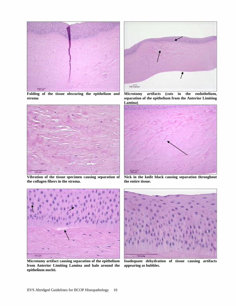
There are many other types of histological artifacts that are not presented here. Below
is a selection of figures from tissue culture grade deionized water-treated bovine corneas which
show histological artifacts which result in difficulty interpreting the test material-induced
damage. These artifacts are, but not limited to, folding of the tissue, vibration of the blade,
nick in the knife blade, and inadequate dehydration.
IIVS Abridged Guidelines for BCOP Histopathology 10
Folding of the tissue obscuring the epithelium and
stroma
Microtomy artifacts (cuts in the endothelium,
separation of the epithelium from the Anterior Limiting
Lamina)
Vibration of the tissue specimen causing separation of
the collagen fibers in the stroma.
Nick in the knife black causing separation throughout
the entire tissue.
Microtomy artifact causing separation of the epithelium
from Anterior Limiting Lamina and halo around the
epithelium nuclei.
Inadequate dehydration of tissue causing artifacts
appearing as bubbles.

IIVS Abridged Guidelines for BCOP Histopathology 11
5.3 Recording Observations
Observations of treated corneas are recorded. The data include the treatment group,
slide (cornea accession) numbers, exposure time, post-exposure expression time, date of test
material application, observations on each corneal tissue layer, and the related figure numbers
(where appropriate). Once the observations are completed they are formalized in the
Histology Report. The observations are typically peer reviewed, and upon consensus the
Histology Report is signed and dated by both the Principle Investigator/Lead
Histopathologist and the Peer Reviewer.
5.3.1 Evaluating the Negative Control-Treated Corneas
Negative control corneas are treated with sterile, deionized water or saline in
parallel with the positive control and test material-treated corneas. The overall thickness
of the bovine cornea is generally between 850-1000 µm.
Epithelium: The negative control-treated epithelium is composed of three layers. The
basal cell layer is a well-formed, columnar-cell region directly attached to
the basement membrane above the Anterior Limiting Lamina. The basal
cells were always tightly attached to each other. Several layers of wing
cells covered the columnar basal layer. In both of these layers, the cell
nuclei showed diffuse chromatin without clear nucleoli. Rare mitotic
figures were seen in the basal layer. The squamous layer was flattened
with limited cytoplasm and highly condensed nuclei
Stroma: The stromal elements begin with the Anterior Limiting Lamina and are
composed of well-organized collagen matrix fibers with dispersed
keratocytes. Keratocyte nuclei show a range of morphologies from
moderate sized (smaller than the epithelial nuclei) with diffuse basophilic
staining to narrow, elongated and condensed with dark basophilic staining.
Cytoplasmic staining, when visible, is moderately basophilic. Rarely, cells
with eosinophilic cytoplasmic staining may be observed. Collagen bundles
are generally parallel and well ordered. The Descemet's Membrane is
prominent and forms the bottom of the stroma. The overall thickness of the
stroma is approximately one 20x field when a good cross section is
obtained.
Endothelium: The endothelium is a single layer of flattened cells attached to the
basal surface of the Descemet’s Membrane. Nuclei are elongated and
flattened. In a cross section, little cytoplasm is visible. Generally, the cells
are firmly attached to the Descemet’s Membrane but in some areas (or
fields), they may be detached or lost through mechanical damage.
5.3.2 Evaluating the Corneal Histologic Changes
The goal in scoring the corneal damage and changes is to record the nature, degree and
depth of the changes in each tissue layer. In most cases, the individual corneas in a treatment
group will not be reported separately but rather they will be “averaged” to highlight the
predominant lesions. The opacity and permeability values should be reviewed before scoring
IIVS Abridged Guidelines for BCOP Histopathology 12
the slides. If there are wide variations among the corneas in either the indirect measures or
histological changes, it may be necessary to report on some individual corneas within the
treatment group.
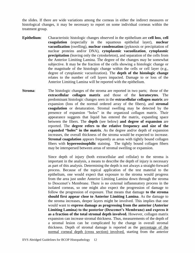
Epithelium: Characteristic histologic changes observed in the epithelium are cell loss, cell
coagulation (especially in the squamous epithelial layer), nuclear
vacuolization (swelling), nuclear condensation (pyknosis or precipitation of
nuclear proteins and/or DNA), cytoplasmic vacuolization, cytoplasmic
precipitation (leaving only the cytoskeleton), and separation of the cells from
the Anterior Limiting Lamina. The degree of the changes may be somewhat
subjective. It may be the fraction of the cells showing a histologic change or
the magnitude of the histologic change within the cells or cell layer (e.g.,
degree of cytoplasmic vacuolization). The depth of the histologic change
relates to the number of cell layers impacted. Damage to or loss of the
Anterior Limiting Lamina will be reported with the epithelium.
Stroma: The histologic changes of the stroma are reported in two parts; those of the
extracellular collagen matrix and those of the keratocytes. The
predominant histologic changes seen in the extracellular collagen matrix are
expansion (loss of the normal ordered array of the fibers), and stromal
coagulation or denaturation. Stromal swelling may be detected by the
presence of expansion “holes” in the organized collagen matrix. Their
appearance suggests that liquid has entered the matrix, expanding space
between the fibers. The depth (see below) and degree of expansion are
reported. The degree refers to the relative frequency and size of the
expanded “holes” in the matrix. As the degree and/or depth of expansion
increases, the overall thickness of the stroma would be expected to increase.
Stromal coagulation appears frequently as areas with tightly bound collagen
fibers with hypereosinophilic staining. The tightly bound collagen fibers
may be interspersed between areas of stromal swelling or expansion.
Since depth of injury (both extracellular and cellular) to the stroma is
important in the analysis, a means to describe the depth of injury is necessary
as part of this analysis. Determining the depth is not always a straight-forward
process. Because of the topical application of the test material to the
epithelium, one would expect that exposure to the stroma would progress
from the area just under Anterior Limiting Lamina down through the stroma
to Descemet’s Membrane. There is no external inflammatory process in the
isolated corneas, so one might also expect the progression of damage to
follow the progression of exposure. That means that damage to the stroma
should first appear close to Anterior Limiting Lamina. As the damage to
the stroma increases, deeper layers might be involved. This implies that one
would want to express damage as progressing from the anterior (Anterior
Limiting Lamina) to the posterior (Descemet’s Membrane) and express it
as a fraction of the total stromal depth involved. However, collagen matrix
expansion can increase stromal thickness. Thus, measurements of the depth of
a stromal lesion can be complicated by the change in overall stromal
thickness. Depth of stromal damage is reported as the percentage of the
normal corneal depth (cross section) involved, starting from the anterior

IIVS Abridged Guidelines for BCOP Histopathology 13
border (Anterior Limiting Lamina). However, to account for stromal swelling
or expansion, this depth is actually estimated from the percentage of the
stromal cross section that remained undamaged (starting at the posterior
border). For example, a cornea reported to show collagen matrix expansion to
30% depth would mean that 70% of the cross section of that cornea (starting
at Descemet’s Layer) did not show expansion.
An exception to the anterior to posterior progression of stromal expansion is
caused by the loss of the endothelial cell layer. Since the endothelium is
responsible for maintaining balanced hydration in the lower stroma, its
loss (either through mechanical damage or test material toxicity) can lead
to appreciable deep stromal swelling. It is important to differentiate
between endothelial damage and expansion caused by the test material
exposure and damage from other sources (e.g., mechanical). In the case of
mechanical damage, the deep swelling can occur in the absence of expansion
in the anterior stroma. Test material-induced damage should progress
through the cornea and be manifested in both the anterior and posterior
stroma. Sections or portions of sections where the endothelium is lost and
posterior stromal swelling (collagen matrix expansion) is observed without
similar anterior stromal swelling are likely the result of mechanical damage to
the endothelium that occurred early in the assay (incubation). An effort should
be made to score corneal sections that do not show such damage.
Histologic changes in the keratocytes are manifested in both the cytoplasm
and nucleus. Rapid necrotic cell degeneration, as might follow exposure to a
strong alkaline, organic solvent or surfactant, is quite apparent because the
cellular components rapidly breakdown. Oxidative damage or DNA alkylation
might produce more subtle damage (initially) but could also lead to cell death
(delayed) and release of inflammatory mediators. Nuclear changes (pyknosis
or karyorrhexis) are signs of this process. Progressive nuclear pyknosis or
complete destruction are also signs of this process. Cytoplasmic changes
include vacuole formation or loss of basic elements (mRNA for example)
that are also indicative of the beginning of the degenerative process. The cell
cytoplasm normally stains with both basophilic (hematoxylin) and acidophilic
(eosin) stains. When the basic elements are lost, eosinophilic staining
predominates. This change is termed keratocyte eosinophilia.
Endothelium: Histologic changes in this layer include cell loss and cytoplasmic
degeneration (vacuolization). Since this layer is only one cell thick,
mechanical damage has the potential to confound the evaluation. Where there
is endothelial cell loss, it is important to evaluate surrounding fields for the
presence of normal endothelium. Since the whole corneal surface is treated, a
lack of a uniform changes to most of the endothelium would suggest
mechanical damage to isolated patches rather than test material-induced
damage. When mechanical damage occurs late in the assay or after fixation
(e.g., during processing), little or no deep stromal swelling or expansion is
expected.

IIVS Abridged Guidelines for BCOP Histopathology 14
5.4 Preparation of the Photomicrographs
Photomicrographs of the histologic changes are made to be “representative” of the
observations and the degree of damage at the indicated depth observed in the treatment group.
They are not intended to document damage, or be considered raw data. Images are prepared
using a Spot Insight Digital Camera and Spot 4.0.8 software (Diagnostic Instruments, Inc.,
Sterling Heights, MI). The color balance of the images is sometimes corrected to better
represent the colors that would be seen through the microscope. Each photomicrograph is
documented in a study-associated digital image log. Once finalized, the image log for the study
is printed, signed and dated by the scientist responsible. The finalized copy is placed into the
study notebook. The photomicrographs are “pasted” electronically into the Histology Report.
6. A Short Compendium of Micrographs to Illustrate Negative Control-
Treated (Normal) and Select Histologic Changes in Bovine Corneal Tissue
The following series of photomicrographs are intended to illustrate normal bovine
corneal morphology and provide examples of the types of histologic changes that might be
observed in the epithelium, stroma, and endothelium. It is by no means a complete listing of all
histologic changes, but is intended to illustrate the types of changes mentioned in the
discussion of corneal lesions. In some figures, the chemical and exposure are provided. When
photomicrographs have been taken from unpublished client studies, test material information is
omitted.
6.1 Negative Control Corneas
Figure 1 Negative control cornea (sterile, deionized water, 10-minute exposure, 2 hour post exposure) –
Full thickness (4X)
IIVS Abridged Guidelines for BCOP Histopathology 15
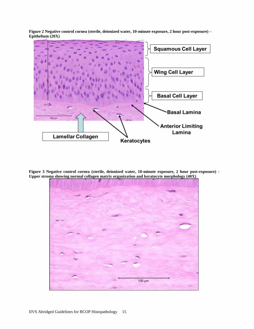
Figure 2 Negative control cornea (sterile, deionized water, 10-minute exposure, 2 hour post-exposure) –
Epithelium (20X)
Figure 3 Negative control cornea (sterile, deionized water, 10-minute exposure, 2 hour post-exposure) -
Upper stroma showing normal collagen matrix organization and keratocyte morphology (40X)

IIVS Abridged Guidelines for BCOP Histopathology 16
Figure 4 Negative control cornea (sterile, deionized water, 10-minute exposure, 2-hour post-exposure) -
Deep stroma and endothelium (40X)

IIVS Abridged Guidelines for BCOP Histopathology 17
6.2 Epithelial Damage
Figure 5 Sodium Lauryl Sulfate (1% SLS, 30-minute exposure) - Epithelial cell loss induced by surfactant
exposure (40X)
Opacity 3.2 Permeability 0.772 In Vitro Score 14.7

IIVS Abridged Guidelines for BCOP Histopathology 18
Figure 6 Sodium Lauryl Sulfate (5% SLS, 30-minute exposure) – Epithelial cell loss induced by surfactant
exposure (40X)
Opacity 37.7 Permeability 2.538 In Vitro Score 45.7
Figure 7 Ethanol (neat ETOH), 10-minute exposure, 2-hr post-exposure – Squamous layer coagulation and
cytoplasmic and nuclear vacuolization in the wing and basal layers (20X)
Opacity 24.8 Permeability 1.476 In Vitro Score 47.0

IIVS Abridged Guidelines for BCOP Histopathology 19
Figure 8 Acid formulation “A” (neat), 3-minute exposure, 2-hr post-exposure. Moderate coagulation,
abnormal chromatin condensation and cytoplasmic vacuolization in the squamous and upper wing cell
layers (40X)
Opacity 53.3 Permeability 0.533 In Vitro Score 61.3
Figure 9 Acid formulation “B” (neat), 3-minute exposure, 2-hr post-exposure. Marked squamous cell
coagulation, abnormal chromatin condensation and breakdown of basal cell adhesion to the basal lamina
(40X)
Opacity 34.0 Permeability 0.911 In Vitro Score 47.7

IIVS Abridged Guidelines for BCOP Histopathology 20
Figure 10 Acid formulation ”B” (neat), 10-minute exposure, 2-hr post-exposure. Severe squamous cell
coagulation, abnormal chromatin condensation (precipitation) and marked cytoplasmic eosinophilia (40X)
Opacity 46.7 Permeability 1.682 In Vitro Score 71.9
Figure 11 (neat) 10-minute exposure, 2-hr post-exposure. Hypo-chromic staining in the upper epithelium
and marked nuclear pyknosis and upper hyper-eosinophilia in the deep epithelium (20X)
Opacity 15.2 Permeability 0.512 In Vitro Score 22.9
IIVS Abridged Guidelines for BCOP Histopathology 21
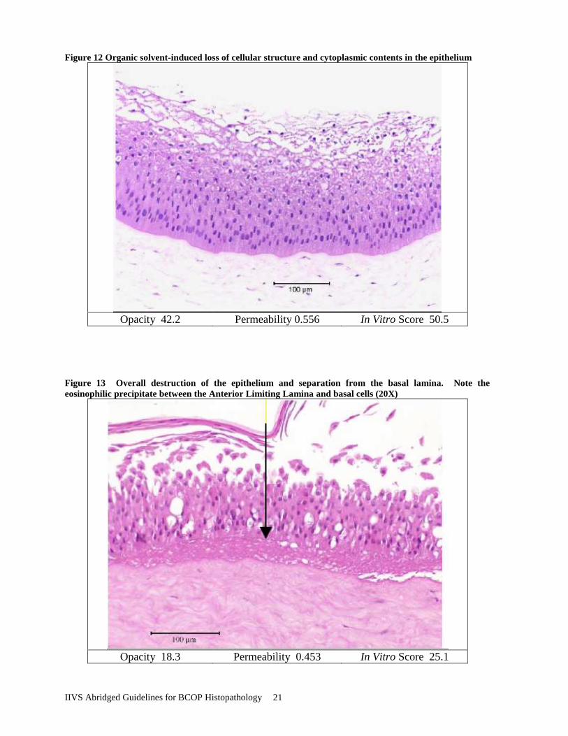
Figure 12 Organic solvent-induced loss of cellular structure and cytoplasmic contents in the epithelium
Opacity 42.2 Permeability 0.556 In Vitro Score 50.5
Figure 13 Overall destruction of the epithelium and separation from the basal lamina. Note the
eosinophilic precipitate between the Anterior Limiting Lamina and basal cells (20X)
Opacity 18.3 Permeability 0.453 In Vitro Score 25.1

IIVS Abridged Guidelines for BCOP Histopathology 22
Figure 14 Damage to the epithelium and upper stroma resulting in a complete loss of viability and reduced
staining.
Opacity Permeability In Vitro Score
Figure 15 Bleach Mixture (neat), 10-minute exposure, 2-hour post exposure. Epithelium showing complete
loss of nuclear and cytoplasmic staining in the squamous cell layer and upper wing layer.
Opacity 14.7 Permeability 0.082 In Vitro Score 15.9

IIVS Abridged Guidelines for BCOP Histopathology 23
Figure 16 Bleaching Agent (neat), 3-minute exposure, 2-hr post-exposure. Epithelium showing
microvacuolation of cellular structure and cytoplasm
Opacity 89.2 Permeability 2.145 In Vitro Score 121.3
IIVS Abridged Guidelines for BCOP Histopathology 24
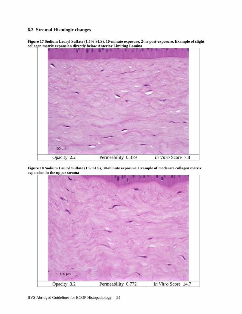
6.3 Stromal Histologic changes
Figure 17 Sodium Lauryl Sulfate (1.5% SLS), 10-minute exposure, 2-hr post-exposure. Example of slight
collagen matrix expansion directly below Anterior Limiting Lamina
Opacity 2.2 Permeability 0.379 In Vitro Score 7.8
Figure 18 Sodium Lauryl Sulfate (1% SLS), 30-minute exposure. Example of moderate collagen matrix
expansion in the upper stroma
Opacity 3.2 Permeability 0.772 In Vitro Score 14.7
IIVS Abridged Guidelines for BCOP Histopathology 25
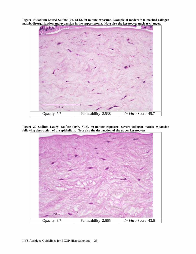
Figure 19 Sodium Lauryl Sulfate (5% SLS), 30-minute exposure. Example of moderate to marked collagen
matrix disorganization and expansion in the upper stroma. Note also the keratocyte nuclear changes.
Opacity 7.7 Permeability 2.538 In Vitro Score 45.7
Figure 20 Sodium Lauryl Sulfate (10% SLS), 30-minute exposure. Severe collagen matrix expansion
following destruction of the epithelium. Note also the destruction of the upper keratocytes
Opacity 3.7 Permeability 2.665 In Vitro Score 43.6

IIVS Abridged Guidelines for BCOP Histopathology 26
6.4 Endothelial Cell Histologic changes
Figure 21 Quinacrine (20% Quinacrine in MEM, 4-hour exposure) Endothelial cell vacuolation (severe)
Opacity 10.5 Permeability -0.040 In Vitro Score 9.9
Figure 22 Bleach Mixture, 10-minute exposure, 2-hour post exposure. Damage to the endothelial cell layer,
cytoplasmic, and deep stromal collagen matrix expansion (severe).
Opacity 14.7 Permeability 0.082 In Vitro Score 15.9

IIVS Abridged Guidelines for BCOP Histopathology 27
Figure 23 Ethanol (neat ETOH), 10-minute exposure, 2-hr post-exposure) Severe damage to the epithelium
and expansion of the upper stroma. The endothelium was intact and little expansion was observed in the
deep stroma.
Opacity 33.0 Permeability 0.978 In Vitro Score 47.7
Figure 24 Example of massive stromal swelling resulting from the loss of both the epithelium and
endothelium (severe).

IIVS Abridged Guidelines for BCOP Histopathology 28
7. References
Araki, K., Y. Ohahsi, S. Knoshita, K. Hayahsi, Y. Kuwayama and Y. Tano (1994). "Epithelial
Wound Healing in the Denervated Cornea." Current Eye Research 13: 203-211.
Baldwin, H. A., T. O. McDonald and C. H. Beasley (1973). "Slit examination of experimental
animals eyes. II Grading scales and photographic evaluation of induced pathological
conditions." J Society of Cosmetic Chemistry 24: 181-195.
Butler, J. M. and B. R. Hammond (1980). "The Effects of Sensory Denervation on the
Response of the Rabbit Eye to prostaglandin E1, bradykinin and substance P." British
Journal of Pharmacology 69: 495-502.
Chan, P. K. and A. W. Hayes (1994). "Acute Toxicity and Eye Irritancy." In Principles and
Methods of Toxicology, 3rd Edition (ed Hayes A.W.): 579-647.
Cuellar, N., J. Merrill, M. Clear, G. Mun and J. W. Harbell (2002). "The Application of
Benchmarks for the Evaluation of the Potential Ocular Irritancy of Aerosol
Fragrances." The Toxicologist 66(1): 243-244.
Curren, D. R., M. G. Evans, H. Raabe, R. R. Ruppalt and J. Harbell (2000). "Correlation of
Histopathology, Opacity, and Permeability of Bovine corneas Exposed In Vitro to
Known Ocular Irritants." Veterinary Pathology 37(5): 557.
Dearman, R. J., M. Cumberbatch and I. Kimber (2003). "Cutaneous Cytokine Expression:
Induction by Chemical Allergen and Paracrine Regulation." Journal of Toxicology-
Cutaneous and Ocular Toxicology 22: 69-86.
Dua, H. S., J. A. P. Gomes and A. Singh (1994). "Corneal Epithelium Wound Healing." British
Journal of Ophthalmology 78: 401-408.
Eurell, T. E., J. M. Sinn, P. A. Gerding and C. L. Alden (1991). "In Vitro Evaluation of Ocular
Irritants Using Corneal Protein Profiles." Toxicology and Applied Pharmacology 108:
374-378.
Farquhar, M. G. and G. E. Palade (1963). "Junctional complexes in Various Epithelia. ." J Cell
Biology 17: 375-412.
Gautheron, P., M. Dukic, D. Alix and J. F. Sina (1992). "Bovine Corneal Opacity and
Permeability Test: An In Vitro Assay of Ocular Irritancy." Fundam Appl Toxicol 18(3):
442-9.
Hackett, R. B. and T. O. McDonald (1994). "Mechanisms of Ocular Response to Irritants."
Dermatotoxicology, 5th Editions, (ed Marzulli F.N, Maibach H.I.): 299-306.
Hogan, M. J. and L. E. Zimmerman (1962). "Ophthalmic Pathology: An Atlas and Textbook,
2nd edition." Philadelphia: W.B. Saunders.
Hubert, F. (1992). "The Eye (Rabbit/Human): Parameters to be Measured in the Field of
Ocular Irritation." ATLA 20: 476-479.
Jester, JV, Li, HF, Petroll, WM, Parker, RD, Cavanaugh, HD, Carr, GJ, Smith, B, and Maurer,
JK. (1998) Area and depth of surfactant-induced corneal injury correlates with cell
death. Investigative Ophthalmology & Visual Science 39(6):922-936.
Jacobs, G. A. and M. A. Martens (1990). "Quantification of Eye Irritation Based Upon In Vitro
Changes of Corneal Thickness." ATLA 17: 255-262.
Katahira, J. H., H. Sugiyama, N. Inoue, Y. Horiguchi, M. Matsuda and N. Sugimoto (1997).
"Clostridium Perfringens Enterotoxin Utilizes Two Structurally Related Membrane
Proteins as Functional Receptors in Vivo." J Biological Chemistry 272: 26652-26658.
Klyce, S. D. and R. W. Beuerman (1988). "Structure and Function of the Cornea." The Cornea:
3-23.

IIVS Abridged Guidelines for BCOP Histopathology 29
Mamalis, N., H. F. Edelhauser, D. G. Dawson, J. Chew, R. M. LeBoyer and L. Werner (2006).
"Toxic Anterior Segment Syndrome." J Cataract Refract Surg 32(2): 324-332.
Maurer, J. K., A. Molai, R. D. Parker, L. Li, G. J. Carr, W. M. Petroll, et al. (2001). "Pathology
of Ocular Irritation with Bleaching Agents in the Rabbit Low-volume Eye Test."
Toxicol Pathol 29(3): 308-319.
Maurer, J. K. and R. D. Parker (1996). "Light Microscope Comparison of Surfactant Induced
Eye Irritation in Rabbits and Rats at Three Hours and Recovery/day 35." Toxicologic
Pathology 24(4): 403-411.
Maurer, J. K., R. D. Parker and J. V. Jester (2002). "Extent of Initial Corneal Injury as the
Mechanistic Basis for Ocular Irritation: Key Findings and Recommendations for the
Development of Alternative Assays." Regul Toxicol Pharmacol 36(1): 106-117.
Redden, J., Perry, M.J., Leighton, T., Chen, J., and McMahon, T., (2009). Voluntary Pilot
Program to Evaluate Use of a Non-Animal Testing Approach to EPA Labeling For Eye
Irritation For Certain Antimicrobial Products With Cleaning Claims.
Sina, J. F., D. M. Galer, R. G. Sussman, P. D. Gautheron, E. V. Sargent, B. Leong, et al.
(1995). "A Collaborative Evaluation of Seven Alternatives to the Draize Eye Irritation
Test Using Pharmaceutical Intermediates." Fundamental and Applied Toxicology 26:
20-31.
Swanson, J. E., B. T. White, B. P. Gran, J. Merrill and J. Harbell (2003). "Evaluating
Oxidizing/reactive Cleaning Products in the Bovine Corneal Opacity and Permeability
(BCOP) Assay." The Toxicologist 72: 220-221.
Van Meer, G., W. van Hof and I. and Genderen (1992). "Tight Junctions and Polarity of Lipids
" Tight Junctions (ed Cereijido M.): 187-201.
Wilhelmus, K. R. (2001). "The Draize Eye Test." Surveys in Ophthalmology 45: 493-515.