guidelines and standards...mates ranging from 0.11:100,000 athlete-years in high school stu-dents in...
TRANSCRIPT
GUIDELINES AND STANDARDS
From Massachus
R.B.W.); Universit
C.M.K.); Universi
Children’s Health
Georgia (W.L.B.)
Health, Morristo
University of Col
Vascular Institute
Clinic Foundation
(E.W.).
The following auth
to this document:
Beaver, MD, FAS
MD, Jennifer H.
MD, PhD, FASE, T
authors reported
Douglas, MD, FAS
63B; Christopher
Recommendations on the Use of MultimodalityCardiovascular Imaging in Young AdultCompetitive Athletes: A Report from theAmerican Society of Echocardiography in
Collaboration with the Society of CardiovascularComputed Tomography and the Society for
Cardiovascular Magnetic Resonance
Aaron L. Baggish, MD, (Chair), Robert W. Battle, MD, Timothy A. Beaver, MD, FASE,William L. Border, MBChB, MH, FASE, Pamela S. Douglas, MD, FASE, Christopher M. Kramer, MD,
Matthew W. Martinez, MD, Jennifer H. Mercandetti, BS, RDCS (AE/PE), ACS, FASE, Dermot Phelan, MD,PhD, FASE, Tamanna K. Singh,MD, Rory B.Weiner,MD, FASE, and EricWilliamson,MD, Boston,Massachusetts;
Charlottesville, Virginia; Kansas City, Kansas; Atlanta, Georgia; Durham and Charlotte, North Carolina;Morristown, New Jersey; Denver, Colorado; Cleveland, Ohio; Rochester, Minnesota
Keywords: Athlete, Athlete’s heart, Pre-participation screening, Echocardiography, Cardiac computedtomography, Cardiac magnetic resonance
In addition to the collaborating societies listed in the title, this document is endorsed by the followingAmerican Society
of Echocardiography International Alliance Partners: Argentine Federation of Cardiology, Argentine Society ofCardiology, Asian-Pacific Association of Echocardiography, Australasian Sonographers Association, BrazilianDepartment of Cardiovascular Imaging, Canadian Society of Echocardiography, Cardiovascular and Thoracic Societyof Southern Africa, Cardiovascular Imaging Society of the Interamerican Society of Cardiology, Chinese Society ofCardiothoracic and Vascular Anesthesiology, Chinese Society of Echocardiography, Cuban Society of CardiologyEchocardiography Section, Indian Academy of Echocardiography, Indian Association of Cardiovascular Thoracic
Anaesthesiologists, Iranian Society of Echocardiography, IsraelWorkingGrouponEchocardiography, Japanese Societyof Echocardiography, Korean Society of Echocardiography,Mexican Society of Echocardiography andCardiovascularImaging, National Association of Cardiologists of Mexico, Philippine Society of Echocardiography, Saudi ArabianSociety of Echocardiography, Thai Society of Echocardiography, and Vietnamese Society of Echocardiography.
TABLE OF CONTENTS
General Considerations 524I. Introduction 524A. The CA in Cardiovascular Practice 524
II. Integrated Multimodality Imaging 525A. Transthoracic Echocardiography (TTE) 525
etts General Hospital, Boston, Massachusetts (A.L.B. and
y of Virginia Health System, Charlottesville, Virginia (R.W.B. and
ty of Kansas Medical Center, Kansas City, Kansas (T.A.B.);
care of Atlanta, Emory University School of Medicine, Atlanta,
; Duke University, Durham, North Carolina (P.S.D.); Atlantic
wn Medical Center, Morristown, New Jersey (M.W.M.);
orado Hospital, Denver, Colorado (J.H.M.); Sanger Heart and
in Atrium Health, Charlotte, North Carolina (D.P.); Cleveland
, Cleveland, Ohio (T.K.S.); Mayo Clinic, Rochester, Minnesota
ors reported no actual or potential conflicts of interest in relation
Aaron L. Baggish, MD (Chair), Robert W. Battle, MD, Timothy A.
E, William L. Border, MBChB, MH, FASE, Matthew W. Martinez,
Mercandetti, BS, RDCS (AE/PE), ACS, FASE, Dermot Phelan,
amanna K. Singh, MD, Rory B. Weiner, MD, FASE. The following
relationships with one or more commercial interests: Pamela S.
E owns stock in UpToDate/Kluwer and is DSMB for REAL TIMI
M. Kramer, MD received grant support from Regeneron and is
B. Cardiac Magnetic Resonance Imaging (CMR) 525C. Cardiac Computed Tomography Angiography (CTA) 526
III. Exercise-Induced Cardiac Remodeling (EICR) 527A. Basic Exercise Physiology 527B. Determinants of EICR 527
a consultant for Cytokinetics; Eric Williamson, MD is an unpaid consultant for
Siemens Medical and is the recipient of an investigator-initiated research grant
from GE Healthcare.
Reprint requests: American Society of Echocardiography, Meridian Corporate
Center, 2530 Meridian Parkway, Suite 450, Durham, NC 27713 (Email: ase@
asecho.org).
Attention ASE Members:
Visit www.aseuniversity.org to earn free continuing medical education credit
through an online activity related to this article. Certificates are available for im-
mediate access upon successful completion of the activity. Nonmembers will
need to join the ASE to access this great member benefit!
0894-7317/$36.00
Copyright 2020 by the American Society of Echocardiography.
https://doi.org/10.1016/j.echo.2020.02.009
523
Abbreviations
CA = Competitive athlete(s)
CMR = Cardiac magnetic resonance imaging
CTA = Computed tomography angiography
CVD = Cardiovascular disease
EICR = Exercise-induced cardiac remodeling
LA = Left atrium/left atrial
LV = Left ventricle/left ventricular
RA = Right atrium/right atrial
RV = Right ventricle/right ventricular
SCD = Sudden cardiac death
TTE = Transthoracic echocardiography/echocardiogram
524 Baggish et al Journal of the American Society of EchocardiographyMay 2020
C. Left Ventricular Adaptations 529D. Right Ventricular Adaptations 530E. Atrial Adaptations 531F. Aortic Adaptations 531
IV. Differentiating EICR from Pathology 533A. Left Ventricular Wall Thickening 534B. Left Ventricular Dilation 535C. Right Ventricular Dilation 536D. Hypertrabeculation 538
V. Pre-participation Cardiovascular Screening 539A. Contemporary Standard of Care 539B. Imaging During PPCS 540
VI. The Symptomatic Competitive Athlete 540A. Exertional Chest Discomfort 540B. Syncope 541C. Palpitations and Arrhythmias 541D. Inappropriate Exertional Dyspnea 542E. Athletic Performance Decrement 542
VII. Additional Considerations 543A. Masters Level CA 543B. Pediatric CA 543C. Congenital Heart Disease 544
VIII. Conclusions and Future Directions 544Reference 545
GENERAL CONSIDERATIONS
Competitive athletes (CA) are a rapidly growing population world-wide. Habitual vigorous exercise, a defining characteristic of this pop-ulation, is a potent stimulus for adaptive structural and functionalcardiac remodeling and is an effective way to reduce the risk of car-diovascular disease (CVD). Manifestations of CVD in CA are highlyvariable ranging from subtle findings during pre-participation cardio-vascular screening (PPCS) to collapse or cardiac arrest during exer-cise. In general, young CA are most commonly affected bycongenital and genetic conditions while older CA most commonlyharbor acquired CVD. Cardiac imaging using transthoracic echocar-diography (TTE), cardiac computed tomography angiography(CTA), and cardiacmagnetic resonance imaging (CMR) plays a funda-mental role in the care of CA. This document was created to provideclinical imaging specialists with a comprehensive guide for the perfor-mance of multimodality imaging in CA.
I. INTRODUCTION
The interpretation of imaging data obtained in the care of competitiveathletes (CA) requires a comprehensive understanding of exercise-induced cardiac remodeling (EICR) and an ability to distinguish thecharacteristics of this process from findings suggestive of pathology.Accordingly, cardiovascular specialists are best positioned to provideeffective care for CA if they possess the ability to integrate and inter-pret multimodality diagnostic imaging as required on an individualcase-by-case basis. This document was designed to provide a frame-work for the use of multimodality imaging in the assessment of CA.Throughout this document, emphasis will be placed on the use ofappropriately selectedmultimodality imaging as required to diagnose,exclude, and manage clinically relevant CVD in CA.
The definition of a CA, as endorsed by the American College ofCardiology and American Heart Association, has remained un-changed for more than thirty years. As described at the initial 36th
Bethesda Conference Proceedings in 1985,1 and each subsequent up-date,2,3 a competitive athlete is defined as an individual ‘‘who partic-ipates in an organized team or individual sport that requires regularcompetition against others as a central component, places a high pre-mium on excellence and achievement, and requires some form of sys-tematic (and usually intense) training’’. Accordingly, CA adhere to adistinctive lifestyle characterized by routine individualized exercisetraining geared toward preparation for competition.4 This documentwill focus on the assessment of young CA as defined by the age rangebeginning with the cessation of puberty and ending at age 35.However, key issues relevant to imaging in pediatric and aging or‘‘masters level’’ CA will also be addressed.
A. The CA in Cardiovascular Practice
CA represent one of the healthiest segments of the general popula-tion and are often mistakenly viewed as being ‘‘immune’’ fromCVD. However, vigorous physical activity, particularly in CAwith un-derlying cardiovascular disease (CVD), transiently increases the risk ofadverse events, including sudden cardiac death (SCD).5,6 The trueincidence of SCD in CA 35 years or younger is unknown, with esti-mates ranging from 0.11:100,000 athlete-years in high school stu-dents in Minnesota,7 to 1.9:100,000 athlete-years in NationalCollegiate Athletic Association athletes.8 Available data suggest thatSCD among CA occurs more commonly in men than women,more commonly in CA of African-American descent comparedto Caucasians, and in CA participating in all sports with basketballand American-style football representing relatively high-riskdisciplines.9-11 There are numerous cardiovascular causes of SCD inCA, including genetic and acquired diseases of the heart muscle,valves, electrical system, and coronary arteries (Table 1).12
Historically, hypertrophic cardiomyopathy (HCM) was consideredthe most common cause of SCD in CA based on pioneering datafrom The United States (US) National Registry of Sudden Death inAthletes, in which one-third of deaths in CA were attributable tothis cause.9 More recently, several independent publications sug-gested that hypertrophic cardiomyopathy accounts for a lower pro-portion of SCD than previously reported, with the majority of SCDoccurring in CA with structurally normal hearts on autopsy.11,13
Additional important causes of SCD in young CA include other formsof genetic heart muscle disease (e.g., arrhythmogenic, dilated, andnoncompaction cardiomyopathy), acquired heart muscle disease(e.g., myocarditis, toxic cardiomyopathy attributable to illicit perfor-mance enhancing drugs), genetic channelopathies (e.g., long QT
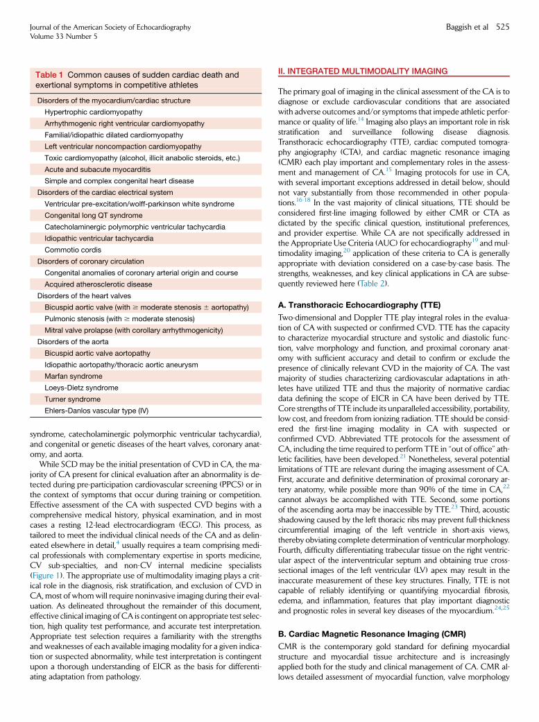
Table 1 Common causes of sudden cardiac death andexertional symptoms in competitive athletes
Disorders of the myocardium/cardiac structure
Hypertrophic cardiomyopathy
Arrhythmogenic right ventricular cardiomyopathy
Familial/idiopathic dilated cardiomyopathy
Left ventricular noncompaction cardiomyopathy
Toxic cardiomyopathy (alcohol, illicit anabolic steroids, etc.)
Acute and subacute myocarditis
Simple and complex congenital heart disease
Disorders of the cardiac electrical system
Ventricular pre-excitation/wolff-parkinson white syndrome
Congenital long QT syndrome
Catecholaminergic polymorphic ventricular tachycardia
Idiopathic ventricular tachycardia
Commotio cordis
Disorders of coronary circulation
Congenital anomalies of coronary arterial origin and course
Acquired atherosclerotic disease
Disorders of the heart valves
Bicuspid aortic valve (with $ moderate stenosis 6 aortopathy)
Pulmonic stenosis (with $ moderate stenosis)
Mitral valve prolapse (with corollary arrhythmogenicity)
Disorders of the aorta
Bicuspid aortic valve aortopathy
Idiopathic aortopathy/thoracic aortic aneurysm
Marfan syndrome
Loeys-Dietz syndrome
Turner syndrome
Ehlers-Danlos vascular type (IV)
Journal of the American Society of EchocardiographyVolume 33 Number 5
Baggish et al 525
syndrome, catecholaminergic polymorphic ventricular tachycardia),and congenital or genetic diseases of the heart valves, coronary anat-omy, and aorta.
While SCD may be the initial presentation of CVD in CA, the ma-jority of CA present for clinical evaluation after an abnormality is de-tected during pre-participation cardiovascular screening (PPCS) or inthe context of symptoms that occur during training or competition.Effective assessment of the CA with suspected CVD begins with acomprehensive medical history, physical examination, and in mostcases a resting 12-lead electrocardiogram (ECG). This process, astailored to meet the individual clinical needs of the CA and as delin-eated elsewhere in detail,4 usually requires a team comprising medi-cal professionals with complementary expertise in sports medicine,CV sub-specialties, and non-CV internal medicine specialists(Figure 1). The appropriate use of multimodality imaging plays a crit-ical role in the diagnosis, risk stratification, and exclusion of CVD inCA,most of whomwill require noninvasive imaging during their eval-uation. As delineated throughout the remainder of this document,effective clinical imaging of CA is contingent on appropriate test selec-tion, high quality test performance, and accurate test interpretation.Appropriate test selection requires a familiarity with the strengthsand weaknesses of each available imaging modality for a given indica-tion or suspected abnormality, while test interpretation is contingentupon a thorough understanding of EICR as the basis for differenti-ating adaptation from pathology.
II. INTEGRATED MULTIMODALITY IMAGING
The primary goal of imaging in the clinical assessment of the CA is todiagnose or exclude cardiovascular conditions that are associatedwith adverse outcomes and/or symptoms that impede athletic perfor-mance or quality of life.14 Imaging also plays an important role in riskstratification and surveillance following disease diagnosis.Transthoracic echocardiography (TTE), cardiac computed tomogra-phy angiography (CTA), and cardiac magnetic resonance imaging(CMR) each play important and complementary roles in the assess-ment and management of CA.15 Imaging protocols for use in CA,with several important exceptions addressed in detail below, shouldnot vary substantially from those recommended in other popula-tions.16-18 In the vast majority of clinical situations, TTE should beconsidered first-line imaging followed by either CMR or CTA asdictated by the specific clinical question, institutional preferences,and provider expertise. While CA are not specifically addressed inthe Appropriate Use Criteria (AUC) for echocardiography19 and mul-timodality imaging,20 application of these criteria to CA is generallyappropriate with deviation considered on a case-by-case basis. Thestrengths, weaknesses, and key clinical applications in CA are subse-quently reviewed here (Table 2).
A. Transthoracic Echocardiography (TTE)
Two-dimensional and Doppler TTE play integral roles in the evalua-tion of CA with suspected or confirmed CVD. TTE has the capacityto characterize myocardial structure and systolic and diastolic func-tion, valve morphology and function, and proximal coronary anat-omy with sufficient accuracy and detail to confirm or exclude thepresence of clinically relevant CVD in the majority of CA. The vastmajority of studies characterizing cardiovascular adaptations in ath-letes have utilized TTE and thus the majority of normative cardiacdata defining the scope of EICR in CA have been derived by TTE.Core strengths of TTE include its unparalleled accessibility, portability,low cost, and freedom from ionizing radiation. TTE should be consid-ered the first-line imaging modality in CA with suspected orconfirmed CVD. Abbreviated TTE protocols for the assessment ofCA, including the time required to perform TTE in ‘‘out of office’’ ath-letic facilities, have been developed.21 Nonetheless, several potentiallimitations of TTE are relevant during the imaging assessment of CA.First, accurate and definitive determination of proximal coronary ar-tery anatomy, while possible more than 90% of the time in CA,22
cannot always be accomplished with TTE. Second, some portionsof the ascending aorta may be inaccessible by TTE.23 Third, acousticshadowing caused by the left thoracic ribs may prevent full-thicknesscircumferential imaging of the left ventricle in short-axis views,thereby obviating complete determination of ventricular morphology.Fourth, difficulty differentiating trabecular tissue on the right ventric-ular aspect of the interventricular septum and obtaining true cross-sectional images of the left ventricular (LV) apex may result in theinaccurate measurement of these key structures. Finally, TTE is notcapable of reliably identifying or quantifying myocardial fibrosis,edema, and inflammation, features that play important diagnosticand prognostic roles in several key diseases of the myocardium.24,25
B. Cardiac Magnetic Resonance Imaging (CMR)
CMR is the contemporary gold standard for defining myocardialstructure and myocardial tissue architecture and is increasinglyapplied both for the study and clinical management of CA. CMR al-lows detailed assessment of myocardial function, valve morphology
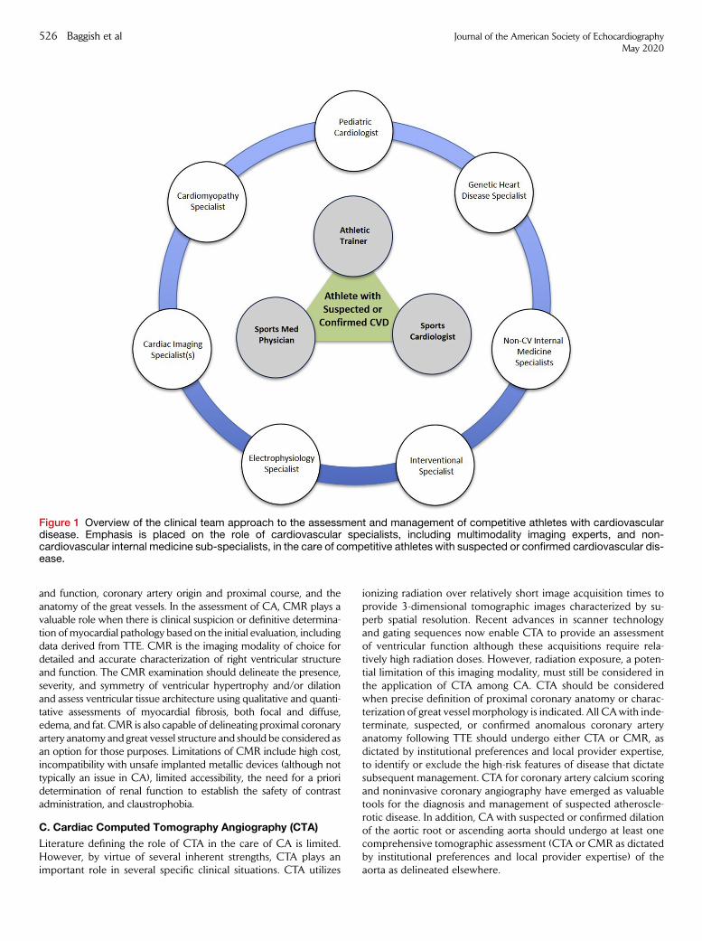
Figure 1 Overview of the clinical team approach to the assessment and management of competitive athletes with cardiovasculardisease. Emphasis is placed on the role of cardiovascular specialists, including multimodality imaging experts, and non-cardiovascular internal medicine sub-specialists, in the care of competitive athletes with suspected or confirmed cardiovascular dis-ease.
526 Baggish et al Journal of the American Society of EchocardiographyMay 2020
and function, coronary artery origin and proximal course, and theanatomy of the great vessels. In the assessment of CA, CMR plays avaluable role when there is clinical suspicion or definitive determina-tion of myocardial pathology based on the initial evaluation, includingdata derived from TTE. CMR is the imaging modality of choice fordetailed and accurate characterization of right ventricular structureand function. The CMR examination should delineate the presence,severity, and symmetry of ventricular hypertrophy and/or dilationand assess ventricular tissue architecture using qualitative and quanti-tative assessments of myocardial fibrosis, both focal and diffuse,edema, and fat. CMR is also capable of delineating proximal coronaryartery anatomy and great vessel structure and should be considered asan option for those purposes. Limitations of CMR include high cost,incompatibility with unsafe implanted metallic devices (although nottypically an issue in CA), limited accessibility, the need for a prioridetermination of renal function to establish the safety of contrastadministration, and claustrophobia.
C. Cardiac Computed Tomography Angiography (CTA)
Literature defining the role of CTA in the care of CA is limited.However, by virtue of several inherent strengths, CTA plays animportant role in several specific clinical situations. CTA utilizes
ionizing radiation over relatively short image acquisition times toprovide 3-dimensional tomographic images characterized by su-perb spatial resolution. Recent advances in scanner technologyand gating sequences now enable CTA to provide an assessmentof ventricular function although these acquisitions require rela-tively high radiation doses. However, radiation exposure, a poten-tial limitation of this imaging modality, must still be considered inthe application of CTA among CA. CTA should be consideredwhen precise definition of proximal coronary anatomy or charac-terization of great vessel morphology is indicated. All CAwith inde-terminate, suspected, or confirmed anomalous coronary arteryanatomy following TTE should undergo either CTA or CMR, asdictated by institutional preferences and local provider expertise,to identify or exclude the high-risk features of disease that dictatesubsequent management. CTA for coronary artery calcium scoringand noninvasive coronary angiography have emerged as valuabletools for the diagnosis and management of suspected atheroscle-rotic disease. In addition, CA with suspected or confirmed dilationof the aortic root or ascending aorta should undergo at least onecomprehensive tomographic assessment (CTA or CMR as dictatedby institutional preferences and local provider expertise) of theaorta as delineated elsewhere.
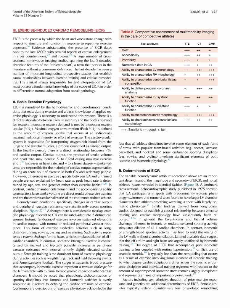
Table 2 Comparative assessment of multimodality imagingin the care of competitive athletes
Test attribute TTE CT CMR
Cost +++ ++ +
Accessibility +++ ++ +
Portability +++ + +
Normative data in CA +++ + ++
Ability to characterize LV morphology ++ +++ +++
Ability to characterize RV morphology + ++ +++
Ability to characterize ventricular tissuecomposition
+ + +++
Ability to define proximal coronary
anatomy
+ +++ ++
Journal of the American Society of EchocardiographyVolume 33 Number 5
Baggish et al 527
III. EXERCISE-INDUCED CARDIAC REMODELING (EICR)
EICR is the process by which the heart and vasculature change withrespect to structure and function in response to repetitive exerciseexposure.26 Evidence substantiating the presence of EICR datesback to the late 1800’s with seminal reports of cardiac enlargementin cross country skiers,27 and rowers.28 A large number of cross-sectional noninvasive imaging studies, spanning the last 5 decades,chronicle features of the ‘‘athlete’s heart’’, a term that persists in theliterature without a consensus definition. The last decade has seen anumber of important longitudinal prospective studies that establishcausal relationships between exercise training and cardiac remodel-ing. The clinical imager responsible for test interpretation of CAmust possess a fundamental knowledge of the scope of EICR in orderto differentiate normal adaptation from occult pathology.
Ability to characterize LV systolic
function
+++ ++ ++
Ability to characterize LV diastolic
function
+++ + +
Ability to characterize aortic morphology ++ +++ +++
Ability to characterize valve function and
morphology
+++ ++ ++
+++, Excellent; ++, good; +, fair.
A. Basic Exercise Physiology
EICR is stimulated by the hemodynamic and neurohumoral condi-tions that exist during exercise and a basic knowledge of applied ex-ercise physiology is necessary to understand this process. There is adirect relationship between exercise intensity and the body’s demandfor oxygen. Increasing oxygen demand is met by increasing oxygenuptake ð _VO2Þ. Maximal oxygen consumption (Peak _VO2) is definedas the amount of oxygen uptake that occurs at an individual’smaximal volitional intensity or effort of exercise. The cardiovascularsystem is responsible for transporting oxygen-rich blood from thelungs to the skeletal muscles, a process quantified as cardiac output.In the healthy person, there is a direct relationship between _VO2
and cardiac output. Cardiac output, the product of stroke volumeand heart rate, may increase 5- to 6-fold during maximal exerciseeffort.29 Increases in heart rate, and – to a lesser degree – stroke vol-ume, are responsible for the majority of cardiac output augmentationduring an acute bout of exercise in both CA and sedentary people.However, differences in exercise capacity between CA and untrainedpeople are not explained by heart rate as peak heart rate is deter-mined by age, sex, and genetics rather than exercise habits.30,31 Incontrast, cardiac chamber enlargement and the accompanying abilityto generate a large stroke volume are direct results of exercise trainingand are the cardiovascular hallmarks of the endurance-trained athlete.
Hemodynamic conditions, specifically changes in cardiac outputand peripheral vascular resistance, vary significantly across sportingdisciplines (Figure 2).32 Although there is considerable overlap, exer-cise physiology relevant to CA can be subdivided into 2 distinct cat-egories. Isotonic (endurance) exercise involves sustained elevationsin cardiac output, with normal or reduced peripheral vascular resis-tance. This form of exercise underlies activities such as long-distance running, rowing, cycling, and swimming. Such activity repre-sents a volume challenge for the heart, which stimulates dilation of all 4cardiac chambers. In contrast, isometric (strength) exercise is charac-terized by marked and typically pulsatile increases in peripheralvascular resistance with normal or only slightly elevated cardiacoutput. Strength training is the dominant form of exercise physiologyduring activities such as weightlifting, track and field throwing events,and American-style football. The surges in systemic blood pressurethat accompany isometric exercise represent a pressure challenge forthe left ventricle with minimal hemodynamic impact on other cardiacchambers. It should be noted that physiologic dichotomization ofsporting disciplines into isotonic and isometric activities is overlysimplistic as it relates to defining the cardiac stressors of exercise.Contemporary descriptions of exercise physiology acknowledge the
fact that all athletic disciplines involve some element of each formof stress, with popular team-based activities (e.g., soccer, lacrosse,basketball, and hockey) and some endurance sporting disciplines(e.g., rowing and cycling) involving significant elements of bothisotonic and isometric physiology.32
B. Determinants of EICR
The variable hemodynamic attributes described above are an impor-tant determinant of the magnitude and geometry of EICR, and not allathletes’ hearts remodel in identical fashion (Figure 3). A landmarkcross-sectional echocardiographic study published in 1975 showedthat CA participating in sports with predominantly isotonic physi-ology (swimmers and runners) were found to have larger LV chamberdiameters than athletes practicing wrestling, a sport with largely iso-metric physiology.33 Similar findings derived from longitudinalstudies designed to establish a causal relationship between exercisetraining and cardiac morphology have subsequently been re-ported.34-36 In general, the biventricular and biatrial volumechallenge inherent in isotonic or endurance-based sporting activitystimulates dilation of all 4 cardiac chambers. In contrast, isometricor strength-based sporting activity may lead to mild thickening ofthe LV walls without chamber dilation. Limited available data suggestthat the left atrium and right heart are largely unaffected by isometrictraining.34 The degree of EICR that accompanies pure isometrictraining, unless coupled with resting hypertension,37 or illicit use ofanabolic steroids,38 is typically less than the remodeling that occursas a result of exercise involving some element of isotonic training.To what degree cardiac adaptations differ across the specific endur-ance disciplines and individual training regimens with respect to theamount of superimposed isometric stress remains largely unexploredand represents an area of important ongoing work.39
Factors including sex, ethnicity, duration of prior exercise expo-sure, and genetics are additional determinants of EICR. Female ath-letes typically exhibit quantitatively less physiologic remodeling
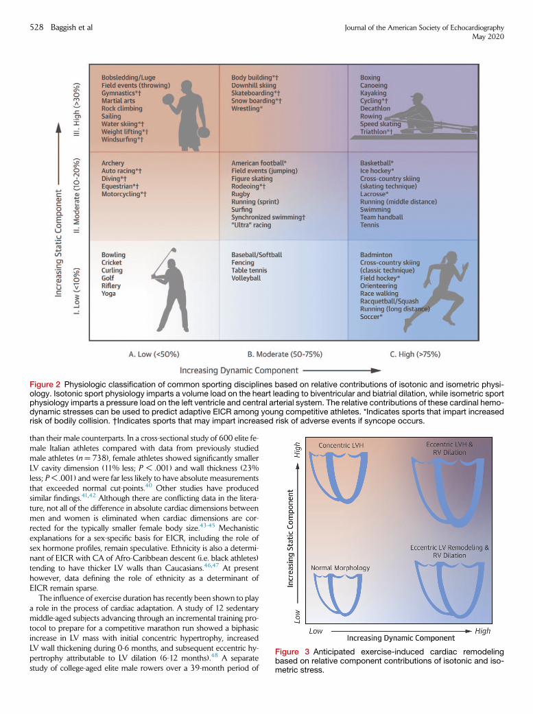
Figure 3 Anticipated exercise-induced cardiac remodelingbased on relative component contributions of isotonic and iso-metric stress.
Figure 2 Physiologic classification of common sporting disciplines based on relative contributions of isotonic and isometric physi-ology. Isotonic sport physiology imparts a volume load on the heart leading to biventricular and biatrial dilation, while isometric sportphysiology imparts a pressure load on the left ventricle and central arterial system. The relative contributions of these cardinal hemo-dynamic stresses can be used to predict adaptive EICR among young competitive athletes. *Indicates sports that impart increasedrisk of bodily collision. †Indicates sports that may impart increased risk of adverse events if syncope occurs.
528 Baggish et al Journal of the American Society of EchocardiographyMay 2020
than their male counterparts. In a cross-sectional study of 600 elite fe-male Italian athletes compared with data from previously studiedmale athletes (n = 738), female athletes showed significantly smallerLV cavity dimension (11% less; P < .001) and wall thickness (23%less; P < .001) and were far less likely to have absolute measurementsthat exceeded normal cut-points.40 Other studies have producedsimilar findings.41,42 Although there are conflicting data in the litera-ture, not all of the difference in absolute cardiac dimensions betweenmen and women is eliminated when cardiac dimensions are cor-rected for the typically smaller female body size.43-45 Mechanisticexplanations for a sex-specific basis for EICR, including the role ofsex hormone profiles, remain speculative. Ethnicity is also a determi-nant of EICR with CA of Afro-Caribbean descent (i.e. black athletes)tending to have thicker LV walls than Caucasians.46,47 At presenthowever, data defining the role of ethnicity as a determinant ofEICR remain sparse.
The influence of exercise duration has recently been shown to playa role in the process of cardiac adaptation. A study of 12 sedentarymiddle-aged subjects advancing through an incremental training pro-tocol to prepare for a competitive marathon run showed a biphasicincrease in LV mass with initial concentric hypertrophy, increasedLV wall thickening during 0-6 months, and subsequent eccentric hy-pertrophy attributable to LV dilation (6-12 months).48 A separatestudy of college-aged elite male rowers over a 39-month period of

Table 3 Selected studies providing normative data for left ventricular end-diastolic internal dimension in competitive athletes
Authorref. Imaging tech. Athlete type Age (y) W/M, (n) Women, mean 6 SD (max.) Men, mean 6 SD (max.)
Spirito53 TTE C (Italian, national) 22 6 N/A 209/738 – 53 6 5 (66)*
Finocchiaro54 TTE C (British, regional/
national)
22 6 6 600/644 49 6 4 (–) 54 6 5 (–)
Pelliccia40 TTE C (Italian, national) 21 6 5 600/738 49 6 4 (11) 54 6 4 (66)
Weiner21 TTE C (American, university) 19 6 3 197/300 47 6 7 (55) 54 6 6 (62)
Pelliccia55 TTE C (Italian, national) 24 6 N/A 352/957 48 (66) 55 (70)
Pluim56 TTE S (meta-analysis) 18-40 –/544 – 52 6 1 (–)
Pluim56 TTE E (meta-analysis) 18-40 –/413 – 54 6 1 (–)
Engel57 TTE C (American,pro-basketball)
26 6 4 –/526 – 57 6 0.2 (71)
Krysztofiak58 TTE C (Polish, pediatric) 12 6 5 327/464 – 44 6 1 (60)*
Makan59 TTE C (British, pediatric) 16 6 1 236/664 – 51 6 4 (60)*
Weiner37 TTE C (American, university,
football)
19 6 1 –/113 – 53 6 4 (60)
Prakken60 CMR† E (Dutch, national) 26 6 6 33/46 55 6 4 (63) 60 6 4 (68)
Luijkx61 CMR‡ E (Dutch, national) 27 6 5 24/57 55 6 3.9 (63) 60 6 4 (67)
C, Combined endurance and strength training physiology;CMR, Cardiac magnetic resonance imaging; E, endurance training physiology;M, Men;
Max., Maximal values recorded; S, strength training physiology; TTE, transthoracic echocardiography; W, women.Data reflect measurements of the left ventricular end-diastolic major dimension (mm) obtained from a parasternal long-axis transthoracic echocar-
diographic view unless otherwise specified.
*Denotes cardiac data inclusive of both men and women.†Data reflect maximum end-diastolic dimension as measured from a 4-chamber view.‡Data reflect maximal end-diastolic dimension as measured from a short-axis view.
Journal of the American Society of EchocardiographyVolume 33 Number 5
Baggish et al 529
high-intensity and high-volume team-based training showed a phasicremodeling response with distinct acute adaptations, including in-creases in LV chamber size, early diastolic function, and systolic twistfollowed by a chronic phase of adaptation characterized by increasingwall thickness and regression in LV twist.49 Finally, genetics appear tobe a determinant of EICR. Studies examining polymorphisms withingenes coding for proteins of the renin-angiotensin-aldosterone axisfound that the angiotensin converting enzyme-deletion/deletion(DD) polymorphism was associated with more LV hypertrophythan the insertion/insertion (II) polymorphism during 10 weeks of ex-ercise training in military recruits.50 Specific polymorphisms of the an-giotensinogen gene have also been associated with LV remodeling.51
In addition, familial hypertension, a complex polygenic trait, wasshown to be associated with both the magnitude and geometry ofexercise-induced concentric LVremodeling in youthful normotensiveendurance-trained athletes.52 The integration of prior exercise expo-sure and genetics into the interpretation of clinical imaging willrequire further studies clarifying their impact on myocardial structureand function.
C. Left Ventricular Adaptations
Dilation of the LV is common and should be considered as a normalfinding in endurance CA (Table 3). LV end-diastolic dimensions, asmeasured by TTE, in a large group (n = 1,309) of Italian elite CA rep-resenting 38 different sports varied from 38 to 66 mm in women(mean = 48 mm) and from 43 to 70 mm in men(mean = 55 mm).55 LV end-diastolic diameter was $55 mm in45% and$60mm in 14% of this cohort. Thus, use of the recommen-ded upper limits of normality of 55-58 mm62 would render approx-imately 40% of male CA in this study as abnormal. In a US-based
study of approximately 500 university CA, approximately 25% ex-ceeded sex-specific recommended limits for LV end-diastolic diam-eter.21 However, it is noteworthy that the majority of CA in thesetwo studies had LV chamber dimensions within normal limits, indi-cating that not all CA demonstrate LV dilation. Thus, the use of‘‘cut-off’’ values for LV end-diastolic diameter or volume, to eitherestablish or exclude the presence of pathologic cardiomyopathy, isnot recommended.
Mild thickening of LV walls, either with or without concomitantLV chamber dilation, may develop in CA (Table 4). Balanced LVwall thickening and chamber dilation (i.e., eccentric LV hypertro-phy as defined by increased LV mass and a relative wall thickness<0.42) is common among CAs who engage in endurance sportswith concomitantly highly levels of isotonic and isometric loadssuch as rowing and cycling (Figure 4). In contrast, mild isolatedLV wall thickening (i.e., concentric LVH as defined by increasedLV mass and a relative wall thickness $0.42) may be seen in ath-letes who participate in strength-based activities with no significantisotonic component, including weight lifting and American-stylefootball (Figure 5).33,34 Functional implications of concentricLVH stimulated by EICR, the same variant of hypertrophy that ac-companies pathologic forms of ventricular pressure overload suchas hypertension and aortic stenosis, remain incompletely under-stood. Longitudinal studies of male American-style football playersshowed that the development of concentric LVH was associatedwith relative impairments both of early diastolic relaxation veloc-ity,34 and systolic function,67 raising uncertainty about the adaptivenature of this form of EICR. LV wall thickening attributable toEICR, regardless of whether it occurs in an eccentric or concentricmorphology, rarely leads to measurements that exceed 12-13 mmin Caucasian CA. In 947 elite Italian CA, only a small number

Table 4 Selected studies providing normative data for left ventricular wall thickness in competitive athletes
Authorref. Imaging tech. Athlete type Age (y) W/M, (n) Women, mean 6 SD (max.) Men, mean 6 SD (max.)
Spirito53 TTE C (Italian, national) 22 209/738 – 10 6 1 (16)*
Finocchiaro54 TTE C (British, regional/national) 22 6 6 600/644 8.4 6 1.2 (–) 9.6 6 1.2 (–)
Pelliccia40 TTE C (Italian, national) 21 6 5 600/738 7.8 6 0.9 (11) 9.4 6 0.9 (13)
Weiner21 TTE C (American, university) 19 6 3 197/300 9.2 6 1.4 (12) 10.5 6 1.5 (14)
D’Andrea35 TTE S (Italian, ‘‘highly trained’’) 29 6 10 120/160 – 11.3 6 2.4 (–)*
D’Andrea35 TTE E (Italian, ‘‘highly trained’’) 28 6 10 160/210 – 9.7 6 3.1 (–)*
Pluim56 TTE S (meta-analysis) 18-40 –/413 – 10.3 6 0.3 (–)
Pluim56 TTE E (meta-analysis) 18-40 –/526 – 11.0 6 0.8 (–)
Engel57 TTE C (American, pro-basketball) 26 6 4 –/791 – 11.0 6 0.1 (15)
Krysztofiak58 TTE C (Polish, pediatric) 12 6 5 327/464 – 8.0 6 0.2 (12)*
Makan59 TTE C (British, pediatric) 16 6 1 236/664 – 9.6 6 1.3 (14)*
Lee63 CMR C (British, military recruits) 20 6 2 –/309 – 10.7 6 1.4 (14.1)
Baggish64 TTE E (American, olympic, rowing) 25 6 3 –/20 – 12.7 6 1.5 (15)
Weiner37 TTE C (American, university,
football)
19 6 1 –/113 – 10.6 6 1.0 (13.9)
Prakken60 CMR† E (Dutch, national) 26 6 6 33/46 11 6 1.4 (14) 9.0 6 1.3 (12)
Luijkx61 CMR‡ E (Dutch, national) 27 6 5 24/57 11 6 1.5 (14) 9.3 6 1.3 (12)
Scharf65 CMR§ E (German, national, triathlon) 28 6 4 –/26 – 9.8 6 1.0 (11.6)
Scharf66 CMR§ C (German, professional,
soccer)
27 6 4 –/29 – 9.4 6 0.9 (11.6)
C, Combined endurance and strength training physiology; CMR, cardiac magnetic resonance imaging; E, endurance training physiology;M, Men;
Max., Maximal values recorded; S, strength training physiology; TTE, transthoracic echocardiography; W, women.
Data reflectmeasurements of the posterolateral left ventricular wall (mm) as obtained from a parasternal long-axis transthoracic echocardiographicview unless otherwise specified.
*Denotes cardiac data inclusive of both men and women.†Data reflect maximum end-diastolic septal thickness as measured from a 4-chamber view.‡Data reflect maximal end-diastolic septal thickness as measured from a short-axis view.§Data reported reflect average of 6 segment thickness measurements (anterior, anterolateral, anteroseptal, inferior, inferolateral, inferoseptal) as
measured from a short-axis view.
530 Baggish et al Journal of the American Society of EchocardiographyMay 2020
(1.7%) had LV wall thicknesses $13 mm.68 Similarly, a low preva-lence (0.4%) of LV wall thickness >12 mm was observed in 720elite British junior CA,69 and in nearly 500 collegiate AmericanCA, not a single participant had LV wall thickness >14 mm.21
While uncommon, EICR may lead to LV wall thicknesses of 13-15 mm in CA participating in high isotonic/high isometric sportingdisciplines or in CA with large body size or Afro-Caribbeandescent.70-72 In contrast, an LV wall thickness >15 mm shouldraise suspicion for pathology and should stimulate further testingto exclude or confirm an explanatory cardiomyopathy.
EICR leads to preservation or enhancement of LV diastolic func-tion with normal noninvasive estimates of left atrial pressure.72-74
Endurance-trained CA with eccentric LV hypertrophy typicallydemonstrate supranormal indices of diastolic function under restingconditions (Figure 6). In contrast, strength trained athletes withconcentric LV hypertrophy may demonstrate mildly impaired TTEindices of diastolic function (Figure 5). Abnormal transmitral fillingprofiles and reductions in early diastolic tissue velocities should raisesuspicion for pathology in young CA and also older CA who do notshow typical age-related diastolic changes.75 LV ejection fraction inCA is generally in the normal range despite variable amounts of LVchamber dilation and wall thickening.76,77 However, trained endur-ance CAwith substantial LV dilation, when imaged under resting con-ditions, may demonstrate an LV ejection fraction at or slightly belowthe lower limits of normal.78 This reflects the fact that stroke volume,
not ejection fraction, is physiologically regulated with large ventriclesejecting a lower fraction of end-diastolic volume than smaller ventri-cles under resting conditions. However, studies using pulsed-waveDoppler, tissue Doppler, and speckle-tracking echocardiographyhave shown that endurance CA typically demonstrate preserved orenhanced systolic function.64,72,79-81
D. Right Ventricular Adaptations
EICR is not confined to the LV. Endurance exercise requires both theLV and right ventricle (RV) to accept and eject relatively large quanti-ties of blood. For the comparatively thin-walled RV, remodeling typi-cally takes the form of mild to moderate RV dilation withoutsignificant hypertrophy. In an echocardiographic study of 102 endur-ance CA, RV chamber dimensions were larger than ‘‘normal’’ valuesin over one-half of the athletes and 28% had an RV outflow tractdimension that met the proposed major size criteria for the diagnosisof arrhythmogenic RV cardiomyopathy (ARVC).82 In a similar studyof Italian CA, 15.6% exceeded a basal RV dimension of 40 mm.36
CMR has similarly demonstrated that RV enlargement is commonamong endurance athletes.83 Differentiating exercise-induced RVchanges from the diagnosis of ARVC is one of the most importantclinical challenges faced by the clinical imager. RV dilation in theendurance-trained CA should be associated with concomitant LV re-modeling (dilation), and the finding of isolated RV enlargementshould raise suspicion of a pathologic process. Furthermore,
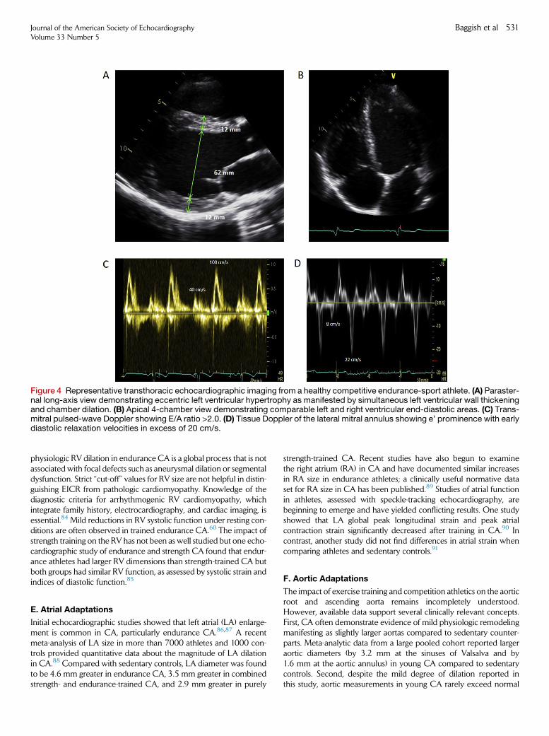
Figure 4 Representative transthoracic echocardiographic imaging from a healthy competitive endurance-sport athlete. (A) Paraster-nal long-axis view demonstrating eccentric left ventricular hypertrophy as manifested by simultaneous left ventricular wall thickeningand chamber dilation. (B) Apical 4-chamber view demonstrating comparable left and right ventricular end-diastolic areas. (C) Trans-mitral pulsed-wave Doppler showing E/A ratio >2.0. (D) Tissue Doppler of the lateral mitral annulus showing e’ prominence with earlydiastolic relaxation velocities in excess of 20 cm/s.
Journal of the American Society of EchocardiographyVolume 33 Number 5
Baggish et al 531
physiologic RV dilation in endurance CA is a global process that is notassociated with focal defects such as aneurysmal dilation or segmentaldysfunction. Strict ‘‘cut-off’’ values for RV size are not helpful in distin-guishing EICR from pathologic cardiomyopathy. Knowledge of thediagnostic criteria for arrhythmogenic RV cardiomyopathy, whichintegrate family history, electrocardiography, and cardiac imaging, isessential.84 Mild reductions in RV systolic function under resting con-ditions are often observed in trained endurance CA.60 The impact ofstrength training on the RV has not been as well studied but one echo-cardiographic study of endurance and strength CA found that endur-ance athletes had larger RV dimensions than strength-trained CA butboth groups had similar RV function, as assessed by systolic strain andindices of diastolic function.85
E. Atrial Adaptations
Initial echocardiographic studies showed that left atrial (LA) enlarge-ment is common in CA, particularly endurance CA.86,87 A recentmeta-analysis of LA size in more than 7000 athletes and 1000 con-trols provided quantitative data about the magnitude of LA dilationin CA.88 Compared with sedentary controls, LA diameter was foundto be 4.6 mm greater in endurance CA, 3.5 mm greater in combinedstrength- and endurance-trained CA, and 2.9 mm greater in purely
strength-trained CA. Recent studies have also begun to examinethe right atrium (RA) in CA and have documented similar increasesin RA size in endurance athletes; a clinically useful normative dataset for RA size in CA has been published.89 Studies of atrial functionin athletes, assessed with speckle-tracking echocardiography, arebeginning to emerge and have yielded conflicting results. One studyshowed that LA global peak longitudinal strain and peak atrialcontraction strain significantly decreased after training in CA.90 Incontrast, another study did not find differences in atrial strain whencomparing athletes and sedentary controls.91
F. Aortic Adaptations
The impact of exercise training and competition athletics on the aorticroot and ascending aorta remains incompletely understood.However, available data support several clinically relevant concepts.First, CA often demonstrate evidence of mild physiologic remodelingmanifesting as slightly larger aortas compared to sedentary counter-parts. Meta-analytic data from a large pooled cohort reported largeraortic diameters (by 3.2 mm at the sinuses of Valsalva and by1.6 mm at the aortic annulus) in young CA compared to sedentarycontrols. Second, despite the mild degree of dilation reported inthis study, aortic measurements in young CA rarely exceed normal
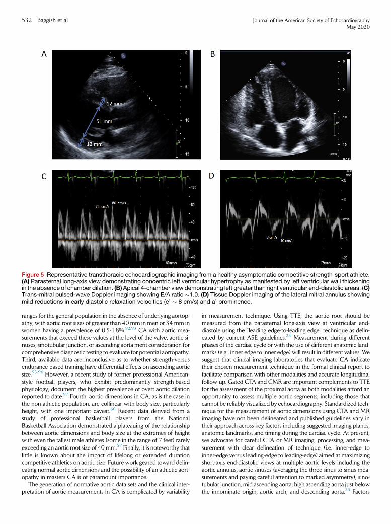
Figure 5 Representative transthoracic echocardiographic imaging from a healthy asymptomatic competitive strength-sport athlete.(A) Parasternal long-axis view demonstrating concentric left ventricular hypertrophy as manifested by left ventricular wall thickeningin the absence of chamber dilation. (B)Apical 4-chamber view demonstrating left greater than right ventricular end-diastolic areas. (C)Trans-mitral pulsed-wave Doppler imaging showing E/A ratio �1.0. (D) Tissue Doppler imaging of the lateral mitral annulus showingmild reductions in early diastolic relaxation velocities (e’ � 8 cm/s) and a’ prominence.
532 Baggish et al Journal of the American Society of EchocardiographyMay 2020
ranges for the general population in the absence of underlying aortop-athy, with aortic root sizes of greater than 40mm inmen or 34mm inwomen having a prevalence of 0.5-1.8%.92,93 CA with aortic mea-surements that exceed these values at the level of the valve, aortic si-nuses, sinotubular junction, or ascending aorta merit consideration forcomprehensive diagnostic testing to evaluate for potential aortopathy.Third, available data are inconclusive as to whether strength-versusendurance-based training have differential effects on ascending aorticsize.93-96 However, a recent study of former professional American-style football players, who exhibit predominantly strength-basedphysiology, document the highest prevalence of overt aortic dilationreported to date.97 Fourth, aortic dimensions in CA, as is the case inthe non-athletic population, are collinear with body size, particularlyheight, with one important caveat.60 Recent data derived from astudy of professional basketball players from the NationalBasketball Association demonstrated a plateauing of the relationshipbetween aortic dimensions and body size at the extremes of heightwith even the tallest male athletes (some in the range of 7 feet) rarelyexceeding an aortic root size of 40mm.57 Finally, it is noteworthy thatlittle is known about the impact of lifelong or extended durationcompetitive athletics on aortic size. Future work geared toward delin-eating normal aortic dimensions and the possibility of an athletic aort-opathy in masters CA is of paramount importance.
The generation of normative aortic data sets and the clinical inter-pretation of aortic measurements in CA is complicated by variability
in measurement technique. Using TTE, the aortic root should bemeasured from the parasternal long-axis view at ventricular end-diastole using the ‘‘leading edge-to-leading edge’’ technique as delin-eated by current ASE guidelines.23 Measurement during differentphases of the cardiac cycle or with the use of different anatomic land-marks (e.g., inner edge to inner edge) will result in different values. Wesuggest that clinical imaging laboratories that evaluate CA indicatetheir chosen measurement technique in the formal clinical report tofacilitate comparison with other modalities and accurate longitudinalfollow-up. Gated CTA and CMR are important complements to TTEfor the assessment of the proximal aorta as both modalities afford anopportunity to assess multiple aortic segments, including those thatcannot be reliably visualized by echocardiography. Standardized tech-nique for the measurement of aortic dimensions using CTA and MRimaging have not been delineated and published guidelines vary intheir approach across key factors including suggested imaging planes,anatomic landmarks, and timing during the cardiac cycle. At present,we advocate for careful CTA or MR imaging, processing, and mea-surement with clear delineation of technique (i.e. inner-edge toinner-edge versus leading-edge to leading-edge) aimed at maximizingshort-axis end-diastolic views at multiple aortic levels including theaortic annulus, aortic sinuses (averaging the three sinus-to-sinus mea-surements and paying careful attention to marked asymmetry), sino-tubular junction, mid ascending aorta, high ascending aorta just belowthe innominate origin, aortic arch, and descending aorta.23 Factors
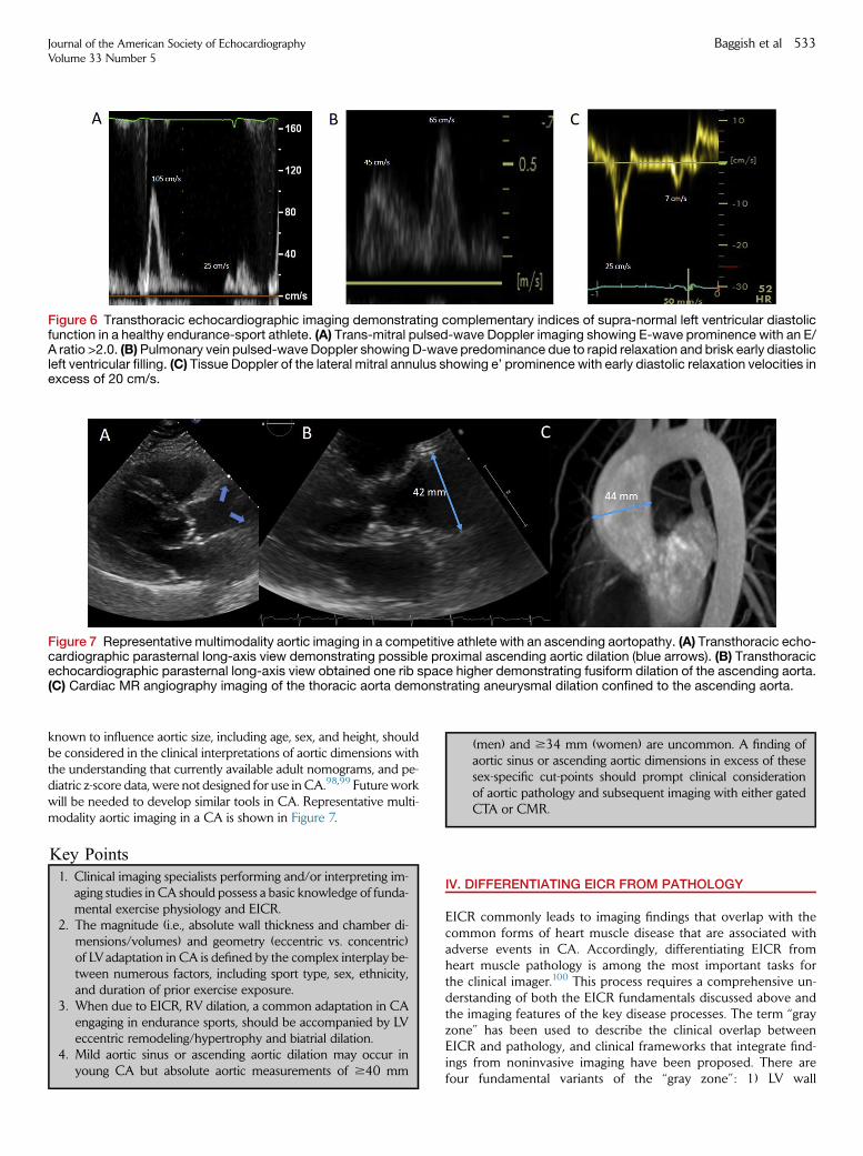
Figure 6 Transthoracic echocardiographic imaging demonstrating complementary indices of supra-normal left ventricular diastolicfunction in a healthy endurance-sport athlete. (A) Trans-mitral pulsed-wave Doppler imaging showing E-wave prominence with an E/A ratio >2.0. (B)Pulmonary vein pulsed-wave Doppler showing D-wave predominance due to rapid relaxation and brisk early diastolicleft ventricular filling. (C) Tissue Doppler of the lateral mitral annulus showing e’ prominence with early diastolic relaxation velocities inexcess of 20 cm/s.
Figure 7 Representative multimodality aortic imaging in a competitive athlete with an ascending aortopathy. (A) Transthoracic echo-cardiographic parasternal long-axis view demonstrating possible proximal ascending aortic dilation (blue arrows). (B) Transthoracicechocardiographic parasternal long-axis view obtained one rib space higher demonstrating fusiform dilation of the ascending aorta.(C) Cardiac MR angiography imaging of the thoracic aorta demonstrating aneurysmal dilation confined to the ascending aorta.
Journal of the American Society of EchocardiographyVolume 33 Number 5
Baggish et al 533
known to influence aortic size, including age, sex, and height, shouldbe considered in the clinical interpretations of aortic dimensions withthe understanding that currently available adult nomograms, and pe-diatric z-score data, were not designed for use in CA.98,99 Future workwill be needed to develop similar tools in CA. Representative multi-modality aortic imaging in a CA is shown in Figure 7.
1. Clinical imaging specialists performing and/or interpreting im-aging studies in CA should possess a basic knowledge of funda-
Key Points
mental exercise physiology and EICR.2. The magnitude (i.e., absolute wall thickness and chamber di-
mensions/volumes) and geometry (eccentric vs. concentric)of LVadaptation in CA is defined by the complex interplay be-tween numerous factors, including sport type, sex, ethnicity,and duration of prior exercise exposure.
3. When due to EICR, RV dilation, a common adaptation in CAengaging in endurance sports, should be accompanied by LVeccentric remodeling/hypertrophy and biatrial dilation.
4. Mild aortic sinus or ascending aortic dilation may occur inyoung CA but absolute aortic measurements of $40 mm
(men) and $34 mm (women) are uncommon. A finding ofaortic sinus or ascending aortic dimensions in excess of thesesex-specific cut-points should prompt clinical considerationof aortic pathology and subsequent imaging with either gatedCTA or CMR.
IV. DIFFERENTIATING EICR FROM PATHOLOGY
EICR commonly leads to imaging findings that overlap with thecommon forms of heart muscle disease that are associated withadverse events in CA. Accordingly, differentiating EICR fromheart muscle pathology is among the most important tasks forthe clinical imager.100 This process requires a comprehensive un-derstanding of both the EICR fundamentals discussed above andthe imaging features of the key disease processes. The term ‘‘grayzone’’ has been used to describe the clinical overlap betweenEICR and pathology, and clinical frameworks that integrate find-ings from noninvasive imaging have been proposed. There arefour fundamental variants of the ‘‘gray zone’’: 1) LV wall
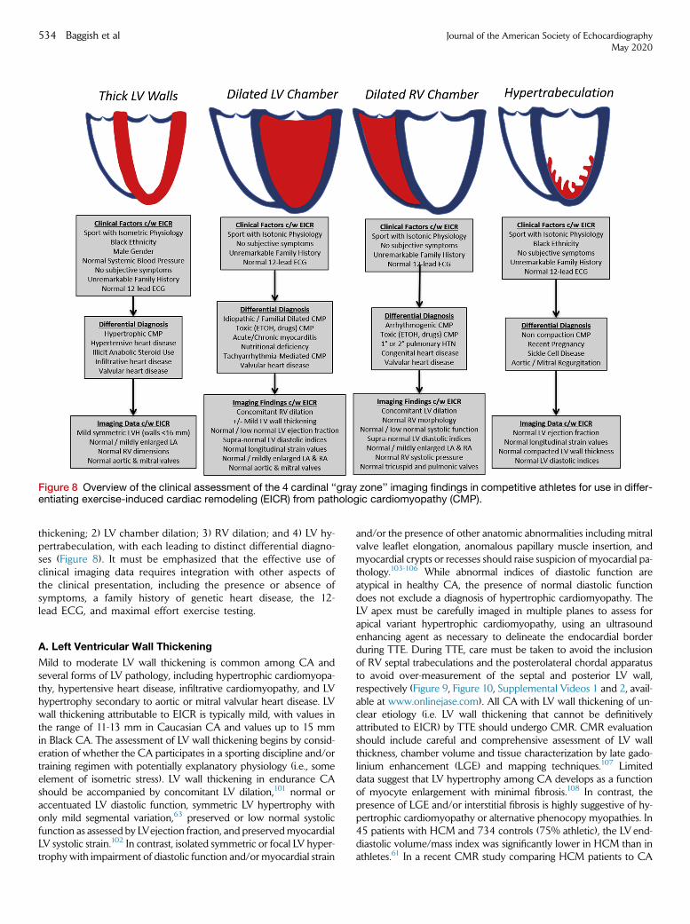
Figure 8 Overview of the clinical assessment of the 4 cardinal ‘‘gray zone’’ imaging findings in competitive athletes for use in differ-entiating exercise-induced cardiac remodeling (EICR) from pathologic cardiomyopathy (CMP).
534 Baggish et al Journal of the American Society of EchocardiographyMay 2020
thickening; 2) LV chamber dilation; 3) RV dilation; and 4) LV hy-pertrabeculation, with each leading to distinct differential diagno-ses (Figure 8). It must be emphasized that the effective use ofclinical imaging data requires integration with other aspects ofthe clinical presentation, including the presence or absence ofsymptoms, a family history of genetic heart disease, the 12-lead ECG, and maximal effort exercise testing.
A. Left Ventricular Wall Thickening
Mild to moderate LV wall thickening is common among CA andseveral forms of LV pathology, including hypertrophic cardiomyopa-thy, hypertensive heart disease, infiltrative cardiomyopathy, and LVhypertrophy secondary to aortic or mitral valvular heart disease. LVwall thickening attributable to EICR is typically mild, with values inthe range of 11-13 mm in Caucasian CA and values up to 15 mmin Black CA. The assessment of LV wall thickening begins by consid-eration of whether the CA participates in a sporting discipline and/ortraining regimen with potentially explanatory physiology (i.e., someelement of isometric stress). LV wall thickening in endurance CAshould be accompanied by concomitant LV dilation,101 normal oraccentuated LV diastolic function, symmetric LV hypertrophy withonly mild segmental variation,63 preserved or low normal systolicfunction as assessed by LVejection fraction, and preservedmyocardialLV systolic strain.102 In contrast, isolated symmetric or focal LV hyper-trophy with impairment of diastolic function and/or myocardial strain
and/or the presence of other anatomic abnormalities including mitralvalve leaflet elongation, anomalous papillary muscle insertion, andmyocardial crypts or recesses should raise suspicion of myocardial pa-thology.103-106 While abnormal indices of diastolic function areatypical in healthy CA, the presence of normal diastolic functiondoes not exclude a diagnosis of hypertrophic cardiomyopathy. TheLV apex must be carefully imaged in multiple planes to assess forapical variant hypertrophic cardiomyopathy, using an ultrasoundenhancing agent as necessary to delineate the endocardial borderduring TTE. During TTE, care must be taken to avoid the inclusionof RV septal trabeculations and the posterolateral chordal apparatusto avoid over-measurement of the septal and posterior LV wall,respectively (Figure 9, Figure 10, Supplemental Videos 1 and 2, avail-able at www.onlinejase.com). All CA with LV wall thickening of un-clear etiology (i.e. LV wall thickening that cannot be definitivelyattributed to EICR) by TTE should undergo CMR. CMR evaluationshould include careful and comprehensive assessment of LV wallthickness, chamber volume and tissue characterization by late gado-linium enhancement (LGE) and mapping techniques.107 Limiteddata suggest that LV hypertrophy among CA develops as a functionof myocyte enlargement with minimal fibrosis.108 In contrast, thepresence of LGE and/or interstitial fibrosis is highly suggestive of hy-pertrophic cardiomyopathy or alternative phenocopy myopathies. In45 patients with HCM and 734 controls (75% athletic), the LVend-diastolic volume/mass index was significantly lower in HCM than inathletes.61 In a recent CMR study comparing HCM patients to CA
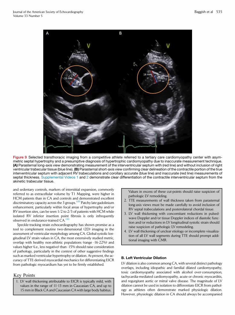
Figure 9 Selected transthoracic imaging from a competitive athlete referred to a tertiary care cardiomyopathy center with asym-metric septal hypertrophy and a presumptive diagnosis of hypertrophic cardiomyopathy due to inaccurate measurement technique.(A) Parasternal long-axis view demonstrating measurement of the interventricular septum with (red line) and without inclusion of rightventricular trabecular tissue (blue line). (B)Parasternal short-axis view confirming clear delineation of the contractile portion of the trueinterventricular septum with adjacent RV trabeculations and corollary accurate (blue line) and inaccurate (red line) measurements ofseptal thickness. Supplemental Videos 1 and 2 demonstrate clear differentiation of the contractile interventricular septum from theakinetic trabecular tissue.
Journal of the American Society of EchocardiographyVolume 33 Number 5
Baggish et al 535
and sedentary controls, markers of interstitial expansion, commonlyreferred to as extracellular volume by T1 Mapping, were higher inHCM patients than in CA and controls and demonstrated excellentdiscriminatory capacity across the 3 groups.109 Patchy late gadoliniumenhancement, particularly within focal areas of hypertrophy and/orRV insertion sites, can be seen 1/2 to 2/3 of patients with HCMwhileisolated RV inferior insertion point fibrosis is only infrequentlyobserved in endurance-trained CA.110
Speckle-tracking strain echocardiography has shown promise as atool to complement routine two-dimensional (2D) imaging in theassessment of ventricular morphology among CA. Global systolic lon-gitudinal LV strain values in CA, the most extensively studied metric,overlap with healthy non-athletic populations (range -16-22%) andvalues higher (i.e., less negative) than -15% should raise considerationof pathology, particularly in the context of other suggestive findingssuch asmarked ventricular hypertrophy or dilation. At present, the ac-curacy of TTE-derived myocardial mechanics for differentiating EICRfrom pathologic myocardium has yet to be firmly established.
Key Points
1. LV wall thickening attributable to EICR is typically mild, withvalues in the range of 11-13 mm in Caucasian CA, and up to
15mm in Black CA andCaucasian CAwith large body habitus.Values in excess of these cut-points should raise suspicion ofpathologic LV remodeling.
2. TTE measurements of wall thickness taken from parasternallong-axis views must be made carefully to avoid inclusion ofRV septal trabeculations and posterolateral chordal tissue.
3. LV wall thickening with concomitant reductions in pulsed-wave Doppler and/or tissue Doppler indices of diastolic func-tion and/or reductions in LV longitudinal systolic strain shouldraise suspicion of pathologic LV remodeling.
4. LV wall thickening of unclear etiology or incomplete visualiza-tion of all LV wall segments during TTE should prompt addi-tional imaging with CMR.
B. Left Ventricular Dilation
LV dilation is also common amongCA,with several distinct pathologyoverlaps, including idiopathic and familial dilated cardiomyopathy,toxic cardiomyopathy associated with alcohol over-consumption,tachycardia-mediated cardiomyopathy, acute or chronic myocarditis,and regurgitant aortic or mitral valve disease. The magnitude of LVdilation cannot be used in isolation to differentiate EICR from pathol-ogy as athletes often demonstrate marked physiologic dilation.However, physiologic dilation in CA should always be accompanied
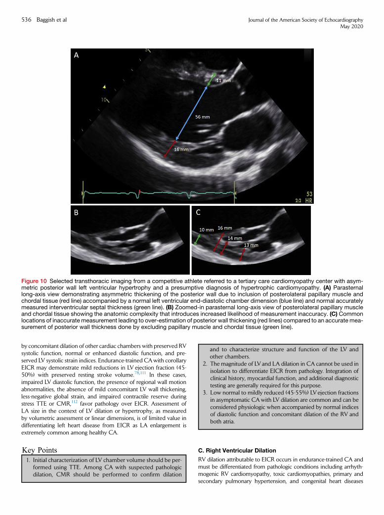
Figure 10 Selected transthoracic imaging from a competitive athlete referred to a tertiary care cardiomyopathy center with asym-metric posterior wall left ventricular hypertrophy and a presumptive diagnosis of hypertrophic cardiomyopathy. (A) Parasternallong-axis view demonstrating asymmetric thickening of the posterior wall due to inclusion of posterolateral papillary muscle andchordal tissue (red line) accompanied by a normal left ventricular end-diastolic chamber dimension (blue line) and normal accuratelymeasured interventricular septal thickness (green line). (B) Zoomed-in parasternal long-axis view of posterolateral papillary muscleand chordal tissue showing the anatomic complexity that introduces increased likelihood of measurement inaccuracy. (C) Commonlocations of inaccuratemeasurement leading to over-estimation of posterior wall thickening (red lines) compared to an accuratemea-surement of posterior wall thickness done by excluding papillary muscle and chordal tissue (green line).
536 Baggish et al Journal of the American Society of EchocardiographyMay 2020
by concomitant dilation of other cardiac chambers with preserved RVsystolic function, normal or enhanced diastolic function, and pre-served LV systolic strain indices. Endurance-trained CAwith corollaryEICR may demonstrate mild reductions in LV ejection fraction (45-50%) with preserved resting stroke volume.78,111 In these cases,impaired LV diastolic function, the presence of regional wall motionabnormalities, the absence of mild concomitant LV wall thickening,less-negative global strain, and impaired contractile reserve duringstress TTE or CMR,112 favor pathology over EICR. Assessment ofLA size in the context of LV dilation or hypertrophy, as measuredby volumetric assessment or linear dimensions, is of limited value indifferentiating left heart disease from EICR as LA enlargement isextremely common among healthy CA.
Key Points
1. Initial characterization of LV chamber volume should be per-formed using TTE. Among CA with suspected pathologic
dilation, CMR should be performed to confirm dilationand to characterize structure and function of the LV andother chambers.
2. The magnitude of LV and LA dilation in CA cannot be used inisolation to differentiate EICR from pathology. Integration ofclinical history, myocardial function, and additional diagnostictesting are generally required for this purpose.
3. Low normal to mildly reduced (45-55%) LVejection fractionsin asymptomatic CAwith LV dilation are common and can beconsidered physiologic when accompanied by normal indicesof diastolic function and concomitant dilation of the RV andboth atria.
C. Right Ventricular Dilation
RV dilation attributable to EICR occurs in endurance-trained CA andmust be differentiated from pathologic conditions including arrhyth-mogenic RV cardiomyopathy, toxic cardiomyopathies, primary andsecondary pulmonary hypertension, and congenital heart diseases

Figure 11 Representative multimodality imaging comparing CA with physiologic RV dilation (A & B) and a CA found to havean abnormal ECG during pre-participation ultimately diagnosed with gene-positive arrhythmogenic right ventricular cardio-myopathy (C & D). (A) Apical 4-chamber transthoracic echocardiographic view demonstrating comparable left and rightventricular end-diastolic areas and normal biventricular systolic function (Supplemental Video 3, available at www.onlinejase.com). (B) Steady-state free precession 4-chamber cardiac MR image demonstrating mild biventricular dilationand a smooth minimally trabeculated right ventricular free wall. (C) Apical 4-chamber transthoracic echocardiographicview with incomplete visualization of the RV but suggestion of RV hypokinesis (Supplemental Video 4, available at www.onlinejase.com). (D) Steady-state free precession 4-chamber cardiac MR image at end-systole demonstrating isolated rightventricular dilation with sacculation and focal aneurysmal dilation of the mid to distal RV free wall.
Journal of the American Society of EchocardiographyVolume 33 Number 5
Baggish et al 537
with chronic right heart volume overload. Physiologic RV dilationshould be accompanied by concomitant LV dilation, preserved oronly slightly reduced systolic function without focal wall motion de-fects, and the absence of anatomic RVabnormalities including saccula-tions, aneurysms, and focal thinning (Supplemental Video 3, availableat www.onlinejase.com). Chamber size in isolation is of limited value indifferentiating EICR from pathology. While TTE is capable of establish-ing RV dilation, it has important limitations for characterizing the rela-tively complex anatomy of the RV (Supplemental Video 4, available atwww.onlinejase.com). Therefore, CMR is required for most athleteswith RV dilation of unclear etiology as it provides superior diagnostic
accuracy for identifying morphological abnormalities of the RV. Aswith the LV, mild reductions in RV systolic function in CA should beaccompanied by significant contractile reserve during stress imaging.113
Speckle-tracking strain echocardiography has recently emerged as apromising tool to differentiate RV EICR from pathology.114-116
However, the limited available data and lack of consensus regardingnormal ranges represent current limitations to its implementation inclinical practice. Representative multimodality imaging comparing aCA with physiologic RV dilation with a CA diagnosed witharrhythmogenic right ventricular cardiomyopathy is shown inFigure 11.
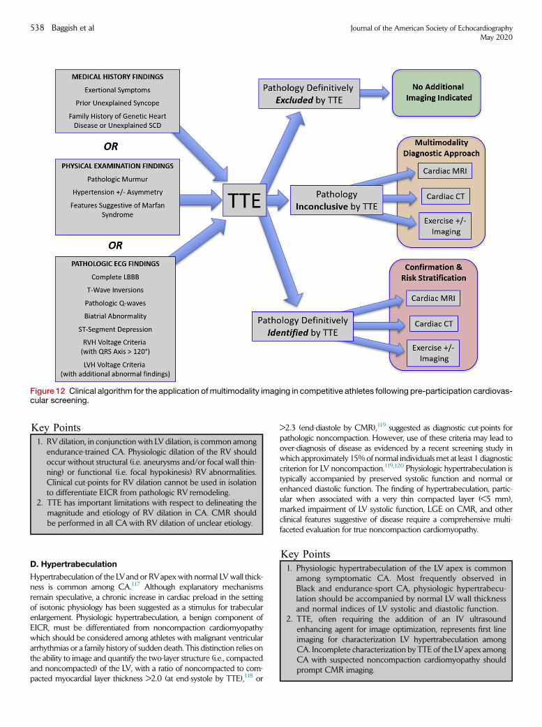
Figure 12 Clinical algorithm for the application ofmultimodality imaging in competitive athletes following pre-participation cardiovas-cular screening.
538 Baggish et al Journal of the American Society of EchocardiographyMay 2020
Key Points1. RV dilation, in conjunction with LV dilation, is common among
endurance-trained CA. Physiologic dilation of the RV should
occur without structural (i.e. aneurysms and/or focal wall thin-ning) or functional (i.e. focal hypokinesis) RV abnormalities.Clinical cut-points for RV dilation cannot be used in isolationto differentiate EICR from pathologic RV remodeling.2. TTE has important limitations with respect to delineating themagnitude and etiology of RV dilation in CA. CMR shouldbe performed in all CA with RV dilation of unclear etiology.
D. Hypertrabeculation
Hypertrabeculation of the LVand or RVapex with normal LVwall thick-ness is common among CA.117 Although explanatory mechanismsremain speculative, a chronic increase in cardiac preload in the settingof isotonic physiology has been suggested as a stimulus for trabecularenlargement. Physiologic hypertrabeculation, a benign component ofEICR, must be differentiated from noncompaction cardiomyopathywhich should be considered among athletes with malignant ventriculararrhythmias or a family history of sudden death. This distinction relies onthe ability to image and quantify the two-layer structure (i.e., compactedand noncompacted) of the LV, with a ratio of noncompacted to com-pacted myocardial layer thickness >2.0 (at end-systole by TTE),118 or
>2.3 (end-diastole by CMR),119 suggested as diagnostic cut-points forpathologic noncompaction. However, use of these criteria may lead toover-diagnosis of disease as evidenced by a recent screening study inwhich approximately 15%of normal individualsmet at least 1 diagnosticcriterion for LV noncompaction.119,120 Physiologic hypertrabeculation istypically accompanied by preserved systolic function and normal orenhanced diastolic function. The finding of hypertrabeculation, partic-ular when associated with a very thin compacted layer (<5 mm),marked impairment of LV systolic function, LGE on CMR, and otherclinical features suggestive of disease require a comprehensive multi-faceted evaluation for true noncompaction cardiomyopathy.
Key Points1. Physiologic hypertrabeculation of the LV apex is common
among symptomatic CA. Most frequently observed in
Black and endurance-sport CA, physiologic hypertrabecu-lation should be accompanied by normal LV wall thicknessand normal indices of LV systolic and diastolic function.2. TTE, often requiring the addition of an IV ultrasoundenhancing agent for image optimization, represents first lineimaging for characterization LV hypertrabeculation amongCA. Incomplete characterization by TTE of the LVapex amongCA with suspected noncompaction cardiomyopathy shouldprompt CMR imaging.
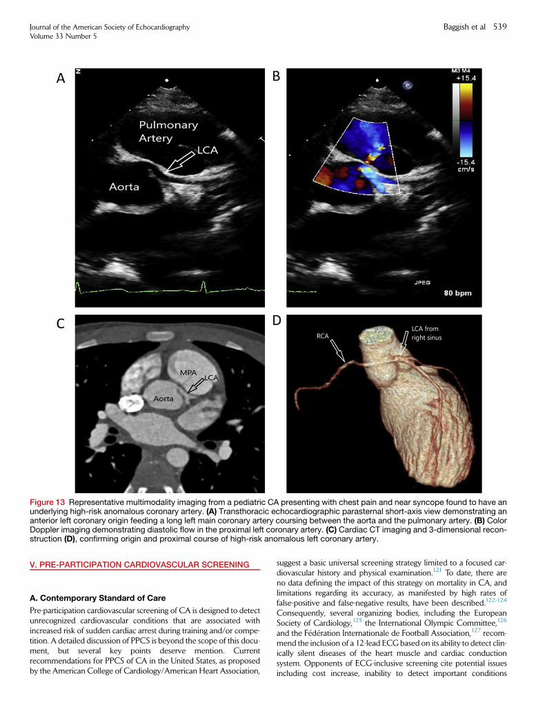
Figure 13 Representative multimodality imaging from a pediatric CA presenting with chest pain and near syncope found to have anunderlying high-risk anomalous coronary artery. (A) Transthoracic echocardiographic parasternal short-axis view demonstrating ananterior left coronary origin feeding a long left main coronary artery coursing between the aorta and the pulmonary artery. (B) ColorDoppler imaging demonstrating diastolic flow in the proximal left coronary artery. (C) Cardiac CT imaging and 3-dimensional recon-struction (D), confirming origin and proximal course of high-risk anomalous left coronary artery.
Journal of the American Society of EchocardiographyVolume 33 Number 5
Baggish et al 539
V. PRE-PARTICIPATION CARDIOVASCULAR SCREENING
A. Contemporary Standard of Care
Pre-participation cardiovascular screening of CA is designed to detectunrecognized cardiovascular conditions that are associated withincreased risk of sudden cardiac arrest during training and/or compe-tition. A detailed discussion of PPCS is beyond the scope of this docu-ment, but several key points deserve mention. Currentrecommendations for PPCS of CA in the United States, as proposedby the American College of Cardiology/American Heart Association,
suggest a basic universal screening strategy limited to a focused car-diovascular history and physical examination.121 To date, there areno data defining the impact of this strategy on mortality in CA, andlimitations regarding its accuracy, as manifested by high rates offalse-positive and false-negative results, have been described.122-124
Consequently, several organizing bodies, including the EuropeanSociety of Cardiology,125 the International Olympic Committee,126
and the F�ed�eration Internationale de Football Association,127 recom-mend the inclusion of a 12-lead ECG based on its ability to detect clin-ically silent diseases of the heart muscle and cardiac conductionsystem. Opponents of ECG-inclusive screening cite potential issuesincluding cost increase, inability to detect important conditions

540 Baggish et al Journal of the American Society of EchocardiographyMay 2020
including congenital anomalous coronary artery anatomy, unaccept-able rates of false-positive findings, and lack of widespread expertisein the interpretation of the CA ECG.128 The sensitivity and specificityof ECG-inclusive screening depends on the criteria used to definewhat is normal and what is not,129-131 and on the specificpopulation being evaluated.132 Criteria designed for ECG interpreta-tion in athletes, first proposed in a formal fashion in 2005,133 havesince undergone numerous revisions,134-136 with corollaryimprovements in diagnostic accuracy.137-139 Accordingly, currentUnited States-based consensus documents now acknowledge the po-tential value of the ECG during PPCS but provide important stipula-tions for this approach, including financial resources and clinicalexpertise.140
B. Imaging During PPCS
At present, the use of noninvasive imaging is not recommended as afirst-line strategy during PPCS by any professional societies.Nonetheless, some organizations, including large universities, nationalteams, professional teams, and charitable groups have elected toincorporate some form of imaging into routine PPCS. The impactof this strategy has not been rigorously assessed to date and wecaution against its use outside of carefully controlled settings thatare resourced with expertise in sports cardiology and the comprehen-sive noninvasive-imaging resources required to evaluate positive orequivocal findings. Proponents of an imaging-inclusive PPCS strategy,most often the use of TTE, cite potential advantages, includingenhanced sensitivity to detect asymptomatic cardiovascular disordersand a reduction in the rate of temporary disqualifications that requirefuture imaging for definitive clarification.141 Opponents of animaging-inclusive PPCS strategy justify this stance based on the poten-tial for false-positive findings generated by inconclusive imaging andfalse-negative findings for certain conditions that may not be readilydetectable by some forms of imaging, and the additional require-ments of money, clinical expertise, and time. ‘‘Limited’’ TTE duringPPCS, a term used to describe abbreviated examinations designedto detect specific high-risk conditions such as hypertrophic cardiomy-opathy, have been proposed.142 To date, the diagnostic accuracy ofthis approach and its impact on outcomes has not been adequatelystudied to justify definitive endorsement. The use of advanced imag-ing, including CMR and CTA as components of PPCS, are similarlydiscouraged due to issues including high cost, unnecessary radiationexposure for CTA, need for administration of intravenous contrast,and little additional diagnostic yield. Finally, stress echocardiographyis not recommended for use during PPCS due to the exceedinglylow prevalence of atherosclerotic coronary disease in young CAand its limited diagnostic yield in patients with anomalous coronaryanatomy.
Despite its limited role in the setting of PPCS, noninvasive imag-ing is often required as part of the downstream testing followingPPCS (Figure 12). Common abnormal findings that merit consider-ation of imaging include historical issues such as unexplained priorsyncope or a family history of sudden cardiac death in a first-degree relative, subjective report by a CA of exertional symptomsincluding chest discomfort or inappropriately labored breathing,and ‘‘training-unrelated’’ ECG finding as proposed by currentInternational recommendations.136 Athletes who demonstratesuch abnormalities during PPCS should undergo a targeted diag-nostic assessment that is capable of excluding the relevant formsof suggested pathology. In the majority of cases, this evaluation
will include one or more imaging tests to document valvemorphology, valve function, myocardial structure, myocardial func-tion, and proximal coronary artery anatomy. Given its cost andunparalleled accessibility, TTE should be considered the first-line im-aging modality in most athletes following PPCS. Limited data sug-gest that on-site access to TTE during PPCS is feasible and mayobviate the need for further off-site testing.21 Regardless of wherethe TTE is conducted, yield following PPCS will be optimized ifthe referring clinicians communicate with the imaging team priorto testing to confirm the clinical question and corollary differentialdiagnosis that arose during PPCS. Regardless, post-PPCS imagingshould routinely include careful quantitative assessment of biven-tricular structure and function with emphasis on the characterizationof LV wall thickness and symmetry, right ventricular morphologyand function, valvular morphology and function, ascending aorticgeometry and dimensions, and right and left coronary origin andproximal course. In all cases, the clinical report should indicateany inconclusive TTE data such as the inability to visualize all ven-tricular wall segments or complete proximal coronary anatomy tofacilitate the determination of the need for additional imaging.The use of CMR, CTA, and exercise testing often complementsTTE in cases of diagnostic uncertainty or during risk stratificationfollowing a definitive diagnosis.
1. Routine PPCS of young CA should include a focused personaland medical history and physical examination. The addition of
Key Points
a 12-lead ECG may be considered in situations with adequatefinancial resources and clinical expertise.
2. The use of noninvasive imaging including comprehensive andlimited TTE, CTA, and CMR is not recommended as a first-linestrategy during PPCS.
3. PPCS programs should ensure timely access to clinical centerswith sports cardiology and clinical imaging expertise to facili-tate the comprehensive multimodality imaging required toevaluate findings detected during PPCS.
VI. THE SYMPTOMATIC COMPETITIVE ATHLETE
A. Exertional Chest Discomfort
Chest discomfort is a common reason why CA require clinical eval-uation. Cardiac and non-cardiac etiologies both account for chestdiscomfort, with the latter being responsible for the vast majorityof causal diagnoses.141 Although CV etiologies account for only�5% of chest discomfort diagnoses, their presence is typically asso-ciated with adverse outcomes. The evaluation of exertional chestdiscomfort in a CA begins with a detailed medical history, physicalexamination, and resting 12-lead electrocardiogram. These basicsteps often identify causal musculoskeletal issues and other non-cardiac etiologies, thereby obviating the need for further cardiovas-cular diagnostics. When a cardiac etiology is either suggested or notsufficiently excluded, exercise testing and noninvasive imagingrepresent the next steps in the evaluation. A comprehensive discus-sion of exercise testing for CA is beyond the scope of this docu-ment but several key issues deserve mention. First, exercise
Journal of the American Society of EchocardiographyVolume 33 Number 5
Baggish et al 541
testing should be conducted using a protocol and modality thatbest approximate the physiology responsible for the presentingsymptoms and should be designed to reproduce it.142 Second, ex-ercise testing, both as a stand-alone test and when coupled with im-aging, should never be terminated at a predetermined heart rate(e.g., 85% maximal age-sex predicted) as is common in some labo-ratories, but instead should be terminated by exhaustion or otherhigh-risk findings, as some patients will manifest symptoms and cor-ollary ischemia only at very high workloads. Third, the need foradjunctive imaging during diagnostic exercise testing should bedetermined based on current guidelines.143 Finally, inducibleischemia in CA often resolves very rapidly during exercise recov-ery, thereby necessitating rapid imaging when post-exercise echo-cardiography is used to assess for dynamic wall motion defects toavoid false-negative results.
This writing group recommends TTE as the initial noninvasiveimaging test for CA presenting with possible or probable cardiacchest pain. In this context, the TTE examination should focus ondelineating or excluding the key cardiac causes of exertionalchest discomfort, including anomalous coronary anatomy withhigh-risk features, hypertrophic cardiomyopathy with or withoutobstructive physiology, and congenital or acquired aortic valveor pulmonic valve stenosis. However, a complete echocardio-graphic assessment should be performed in all cases and addi-tional imaging is often of considerable value. In CA withsuspected or confirmed myocardial pathology such as hypertro-phic cardiomyopathy or arrhythmogenic right ventricular cardio-myopathy, CMR is indicated after TTE in most cases as it affordssuperior diagnostic and prognostic accuracy. In athletes with sus-pected anomalous coronary circulation by TTE or in whomnormal proximal anatomy cannot be confirmed by TTE, CTAor CMR imaging is indicated as dictated by institutional prefer-ences. The primary goals with tomographic imaging includeconfirmation of coronary origin and proximal course and thedelineation of other high-risk features including ostial anatomy,intramural involvement, proximal aneurysmal disease reflectingprior vasculitis, and distal vessel caliber. Representative multimo-dality imaging in an athlete found to have a high-risk anomalouscoronary artery is shown in Figure 13 and Supplemental Videos5 and 6, available at www.onlinejase.com.
Key Points
1. TTE should be performed as the initial noninvasive imagingtest in CA presenting with possible or probable cardiac chest
pain.2. CA presenting with possible or probable cardiac chest painshould undergo maximal effort-limited exercise testing withor without adjunctive imaging as dictated by current guide-lines. Exercise stress echocardiography with immediate post-exercise imaging should be applied with caution as rapid heartrate recovery in CA often renders imaging non-diagnostic forthe evaluation of ischemia.
3. TTE imaging in CA with possible or probable cardiac chestpain should include careful and definitive delineation ofboth the left and right coronary origins and proximal courseto exclude anomalous coronary circulation. Failure of TTE toconfirm normal coronary anatomy requires additional imagingwith CMR or gated CTA as dictated by patient, provider, andinstitutional preferences.
B. Syncope
Syncope, a transient loss of consciousness followed by sponta-neous and complete recovery, is common among CA.144,145 Inone study of CA undergoing PPCS, 474 out of 7,568 (6.2%)CA reported at least one episode of syncope during the preced-ing five years.146 The vast majority of syncope in CA is attribut-able to neural mechanisms that carry a benign prognosis.Neurally-mediated syncope manifests as classic ‘‘vasovagal’’ epi-sodes that are unrelated to exercise, or post-exertional syncopein which fainting that typically occurs within seconds to minutesof abrupt exercise termination. In contrast, syncope that occursduring exercise, often characterized by abrupt loss of conscious-ness with dramatic collapse and musculoskeletal injury, shouldbe attributed to underlying CV etiology and should prompt athorough evaluation. Common cardiovascular causes of syncopeinclude obstructive valve and outflow tract pathology andarrhythmia arising from inducible ischemia, myocardial scar, orprimary disorders of electrical conduction. A comprehensivemedical history, physical examination, and resting 12-lead elec-trocardiogram should be performed for all CA who presentfollowing syncope. In the majority of cases, these initial diag-nostic steps will be sufficient to confirm neurally-mediated phys-iology thereby obviating the need for additional evaluation.When one or more aspects of this initial assessment is inconclu-sive or suggestive of underlying CVD, further evaluationincluding exercise testing and noninvasive imaging is warranted.This writing group recommends TTE as the initial imaging test inCA with prior syncope and a possible or probable cardiac etiol-ogy. The TTE assessment should be geared toward the evalua-tion of key causes of syncope including obstructive valvularand ventricular outflow pathology, cardiomyopathies witharrhythmic and/or ischemic propensity, and high-risk anomalouscoronary anatomy. The use of alternative forms of imagingshould be reserved for further evaluation and risk stratificationof pathology as suggested or incompletely excluded by echocar-diography.
Key Points1. Neurally-mediated syncope in the post-exercise period or in
situations unrelated to exercise is common among CA and
does not require evaluation with any noninvasive imaging mo-dality.2. CA presenting with syncope of unclear etiology, particularlysyncope during exercise, should undergo comprehensive mul-timodality imaging beginning with TTE and extending, on acase-by-case basis, to CTA or CMR to exclude structural andvalvular heart disease as part of a comprehensive evaluation.
3. CA presenting with syncope of unclear etiology should un-dergo maximal effort-limited exercise testing with or withoutadjunctive imaging as dictated by current guidelines.
C. Palpitations and Arrhythmias
The evaluation of the CAwith palpitations or subjective arrhythmiabegins with a comprehensive medical history, physical examina-tion, and resting 12-lead ECG. Palpitations under resting conditionsoften arise from benign premature atrial and/or ventricular beats,are common among bradycardic CA, and are typically benign. In
542 Baggish et al Journal of the American Society of EchocardiographyMay 2020
contrast, palpitations that emerge or intensify during exerciseshould be considered pathologic until proven otherwise therebyprompting a comprehensive evaluation including non-invasive im-aging. All CA with suspected arrhythmia should undergo electro-cardiographic capture of the arrhythmia with provocative exercisetesting, ambulatory rhythm monitoring or implantable monitoring,and in limited cases drug infusion and invasive electrophysiologystudy. The primary objective of ambulatory rhythm monitoring isto differentiate malignant ventricular arrhythmias from the morebenign variants that arise from the atria or atrioventricular junction.The writing panel suggests a comprehensive TTE examination forCA presenting with palpitations or subjective arrhythmias thatoccur or intensify during exercise. The role of imaging in athleteswith suspected arrhythmia is to diagnose or exclude an underlyingcausal cardiac abnormality, particularly in the setting of confirmedor suspected ventricular arrhythmias, as arrhythmias may arisefrom abnormal myocardial substrate including genetic, congenitalor acquired cardiomyopathy, edema and inflammation in thesetting of acute myocarditis, or scar from prior myocardial injury.Alternatively, arrhythmias may be caused by subendocardialischemia from obstructive atherosclerotic disease, congenital coro-nary anomalies, or pressure/volume overload in the context ofvalvular pathology. Finally, arrhythmias in CA may be attributableto primary disorders of electrical conduction, including ventricularpre-excitation and genetic channelopathies. Ventricular pre-excitation on the 12-lead electrocardiogram, either detected duringPPCS in an asymptomatic CA or in a CA presenting witharrhythmic symptoms, should prompt imaging to exclude associ-ated cardiac conditions including Ebstein’s anomaly,147 PRKAG2gene-mediated hypertrophic cardiomyopathy,148 and complexforms of congenital heart disease. The primary channelopathies/in-herited arrhythmia syndromes, including long QT syndromes, cate-cholaminergic polymorphic ventricular tachycardia, Brugadasyndrome, and idiopathic ventricular tachycardia are not associatedwith underlying myocardial, valvular, or coronary abnormalitiesthat can be detected with contemporary imaging. Nonetheless,this writing group recommends a comprehensive TTE to excludealternative diagnoses,149 when history or 12-lead ECG suggestsone of these entities. CTA and CMR imaging in CAwith suspectedarrhythmia should be considered second line and be performed asdictated by suspected or confirmed pathology following TTE.Specifically, CMR should be performed following a normal TTE ex-amination among CA with documented complex ventriculararrhythmia to evaluate for underlying myocardial scar.
Key Points
1. CA presenting with palpitations or subjective arrhythmias thatoccur or intensify during exercise should undergo TTE to diag-
nose or exclude underlying structural disease. Additional imag-ing with CMR may be appropriate on a case-by-case basis.2. CA presenting with palpitations or subjective arrhythmias thatoccur or intensify during exercise should undergo maximaleffort-limited exercise testing with or without adjunctive imag-ing as dictated by current guidelines.
3. All CA undergoing evaluation for ventricular pre-excitation,both asymptomatic and symptomatic, should undergo TTEto exclude concomitant Ebstein’s anomaly, PRKAG2 gene-mediated hypertrophic cardiomyopathy, and complex formsof congenital heart disease.
D. Inappropriate Exertional Dyspnea
Perceived breathlessness during exertion is common among CA andmay be a manifestation of underlying CVD.150,151 The neurophysi-ology of dyspnea is not well understood but the sensation can begenerated by neural feedback mechanisms from the cardiovascular,respiratory, and musculoskeletal systems.152 A careful historyfocusing on the duration and intensity of exercise is often adequateto determine if the corollary sensation of breathlessness is appropriateor inappropriate for that individual athlete. Subjective breathlessnessis typically appropriate as CA approach their physiological upperlimits of exercise capacity as often occurs during training escalationor after prolonged deconditioning. In contrast, dyspnea at previouslytolerable exercise intensities and new-onset dyspnea without changein training regimen or competition should be considered inappro-priate and thus trigger further evaluation. Associated symptomsincluding stridor, wheezing, chest tightness, chest pain, palpitations,and presyncope/syncope are useful in dictating the subsequent eval-uation. Non-cardiac etiologies, including reactive airway disease withor without exercise-induced bronchospasm, paradoxical vocal foldmovement disorder, upper respiratory infection, allergic and non-allergic rhinitis, and dysfunctional breathing account for the majorityof inappropriate exertional dyspnea in young CA.153 CA presentingwith history and physical examination consistent with one of these di-agnoses do not require any form of cardiac imaging prior to empirictreatment. However, failure to respond adequately to medical ther-apy, particularly in CA with suspected exercise-induced broncho-spasm, should raise consideration of alternative, possibly cardiacetiologies.
Most forms of underlying CV disease can cause inappropriate ex-ertional dyspnea. This writing group recommends a comprehensiveTTE in CA presenting with inappropriate exertional dyspnea, andin CA with previously diagnosed non-cardiac cause of inappropriateexertional dyspnea who do not demonstrate adequate response totherapy. Exercise testing, with or without concomitant imaging,should also be performed in both situations. Assessment of gas ex-change, in the form of both pulmonary function testing and cardiopul-monary exercise testing, is often valuable in patients withinappropriate exertional dyspnea. The role of tomographic imagingin CA with inappropriate exertional dyspnea should be consideredsecond line and be performed as dictated by suspected or confirmedpathology following TTE.
Key Points1. Inappropriate exertional dyspnea may occur in the context of
numerous cardiovascular diseases. Comprehensive TTE
should be performed in CA presenting with inappropriate ex-ertional dyspnea, and in CAwith a previously diagnosed non-cardiac cause of inappropriate exertional dyspnea who do notdemonstrate adequate response to therapy.2. Patients presenting with Inappropriate exertional dyspneashould undergo maximal effort-limited exercise testing withor without adjunctive imaging as dictated by current guide-lines.
E. Athletic Performance Decrement
CA presenting for clinical evaluation in the context of performancedecrement pose a difficult diagnostic dilemmawith a broad differentialdiagnosis. A comprehensive history and physical should include
Journal of the American Society of EchocardiographyVolume 33 Number 5
Baggish et al 543
assessment of dietary intake to ensure adequate energy balance, sleeppatterns, mood disturbances, and evaluation for signs and symptoms ofcommon organic pathologies including endocrine disorders (such asthyroid or adrenal disorders and diabetes), infectious etiologies (suchas glandular fever or hepatitis), inflammatory diseases, electrolyte defi-ciencies, and anemia. Training in unfamiliar conditions (altitude, heat orcold) or a significant increase in training load or change in training pat-terns should be considered. Overtraining syndrome is common and af-fects �60% of elite runners and 33% of non-elite runners, but shouldbe considered as a diagnosis of exclusion.154 Evaluation is best per-formed by an experienced sports cardiologist or in combination witha sports medicine physician, and may ultimately require involvementofmultiple other specialties. Testing followingmedical history and phys-ical examination should be predicated on clinical suspicion. It is reason-able but generally of low yield to obtain a 12-lead ECG on all CAwithunexplained performance decrement. The writing committee advo-cates for the use of TTE in CAwith some historical, physical examina-tion, or electrocardiographic finding suggestive of myocardial,coronary, or valvular pathology or in cases where no conclusive expla-nation is determined during initial evaluation. The use of CTA or MRIimaging should be reserved for individual situations as dictated by sus-pected or confirmed pathology.
Key Points1. TTE should be performed for CA presenting with athletic per-
formance decrement when medical history, physical examina-
tion, 12-lead electrocardiography, or routine bloodwork yieldone or more findings suggestive of myocardial, coronary, orvalvular pathology or when no clear explanation can be ascer-tained from the initial evaluation.2. The use of exercise testing and CTA/MRI imaging should beconsidered on a case-by-case basis among CA presentingwith athletic performance as dictated by suspected orconfirmed pathology.
VII. ADDITIONAL CONSIDERATIONS
A. Masters Level CA
CA span the entire age range and masters level CA, competitivesports participants older than 35 or 40 years as dictated by specificsporting disciplines, are a rapidly growing population worldwide.Masters level CA, often presenting with the same cardinal manifesta-tions of disease discussed above, are less likely to have congenital orgenetic heart disease and more likely to be experiencing symptomsattributable to acquired diseases of the heart muscle, electrical system,and coronary arteries. The most common cardiac causes of morbidityand mortality in masters level CA are atherosclerotic coronary dis-ease, atrial tachyarrhythmias (particularly atrial fibrillation), degenera-tive aortic and mitral valve disease, and hypertensive heart disease. Adetailed discussion of each condition has been previously publishedand is beyond the scope of this document.4 However, several prac-tical considerations for the cardiovascular imager are noteworthy.
Atherosclerotic coronary disease is the most common cause of car-diac arrest during exercise in masters level CA.155 The diagnosis ofatherosclerotic coronary disease in this population requires a high in-dex of suspicion in the setting of exertional symptoms and is often but
not always accompanied by the presence of traditional atheroscleroticrisk factors including hypertension, dyslipidemia, family history of dis-ease, and prior/ongoing tobacco use. Exercise stress testing and coro-nary imaging with invasive or CT coronary angiography are keycomponents of this diagnostic evaluation. This writing group recom-mends CTA following exercise testing in patients judged to be atlow or intermediate risk of obstructive disease. In higher-risk CA,traditional coronary angiography in lieu of CTA may be an appro-priate next step, as dictated both by patient and provider preference.The utility of coronary artery calcium (CAC) scoring in masters levelCA remains controversial. Several studies document a higher CACburden in this population than can be explained by traditional risk fac-tors and a tendency for CA to have calcified versus non-calcified pla-que.156,157 While emerging data suggest that CAC in CA may carry arelatively benign prognosis,158,159 the full prognostic implications ofCAC and its relationshipwith obstructive forms of disease responsiblefor symptoms and attendant risk in CA have not been firmly estab-lished. Consequently, this writing group does not recommend theuse of CAC scoring in isolation in asymptomatic or symptomatic mas-ters level CA.
Atrial tachyarrhythmias, including atrial fibrillation and atrial flutter,are common among masters level CA and their incidence appears tobe associated with lifelong duration and intensity of sport participa-tion.160 Although the diagnosis of these arrhythmias does not requirenoninvasive imaging, the writing group recommends comprehensiveTTE for all CA with these arrhythmias to exclude concomitanttachycardia-mediated cardiomyopathy and to exclude valvular etiol-ogies of arrhythmia. The use of tomographic imaging in masters levelCA with atrial arrhythmias should be confined to use for anatomicmapping once an invasive strategy utilizing catheter-based ablationhas been established.
Hypertension is common, often under-diagnosed, and routinelyundertreated in masters level CA. While routine vigorous physical ex-ercise has some beneficial effects on blood pressure, its impact is typi-cally inadequate to prevent or treat hypertension in predisposed CA.Hypertension in CA may be diagnosed by serial resting measure-ments, ideally obtained during ambulatory monitoring, and isfrequently suggested by hypertensive blood pressure response to ex-ercise testing. This writing committee recommends comprehensiveTTE in CAwith newly established hypertension to assess for acquiredhypertensive LV hypertrophy, a phenotype that can be differentiatedfrom EICR based on assessment of diastolic function, as delineated bycurrent clinical guidelines.161
B. Pediatric CA
Children and adolescents represent a significant segment of theoverall CA population. Many of the key cardiac conditions associ-ated with an increased risk of sudden death during sport becomeclinically apparent during this early life period. To date, there aresparse data documenting EICR in pre- and early-adolescent CA.Available data suggest that EICR, including LV hypertrophy andchamber dilation, may occur but at a lesser magnitude than thattypical of adult CA.162 Independent of sport however, pediatricCA are in a period characterized by rapid growth of the heart andblood vessels, thereby making it difficult to differentiate normalgrowth-related changes from changes due to EICR or fromemerging pathology. Challenges in differentiating normal physi-ology from pathology are further compounded by standardpediatric-practice use of body surface area-normalized cardiovascu-lar measurements and by the relative dearth of age-specific
544 Baggish et al Journal of the American Society of EchocardiographyMay 2020
normative data in pediatric CA. In a recent study presenting norma-tive echocardiographic data in Polish pediatric CA (n = 791, age13 6 5 years), mean values of cardiac dimensions were highercompared to general pediatric population estimates but in mostcases, the upper limit of normality in the athletes was confinedwithin the upper limit of the general population.58 One cross-sectional study of 140 pediatric athletes utilizing TTE and deforma-tion imaging revealed geometric and functional differences acrossfive sporting disciplines. Swimmers, the only endurance disciplinestudied, had larger LV chambers and more eccentric hypertrophythan other team and individual sporting disciplines.163
This writing group recommends that the use of imaging in pediatricCA be pursued in a manner analogous to that delineated above foradult athletes, under the guidance of pediatric imaging experts. Inaddition, the writing group recommends the use of standard z-scoresduring interpretation with the acknowledgement that some pediatricCA, particularly those that are nearest chronologically to adulthoodand those that engage in high levels of endurance sports, may demon-strate physiologic remodeling resulting in abnormal measurements.Such cases may require a collaborative approach involving inputfrom pediatric cardiologists and imagers with expertise in the careof athletes with input from adult sports cardiologists and imaging spe-cialists. The development of more data delineating the extent of phys-iologic remodeling that can occur during early life and its corollarynormative data for pediatric CA represent important areas of futurework.
C. Congenital Heart Disease
Dramatic advances in the management of patients with congenitalheart disease, with increasing recognition of the health benefits ofroutine exercise, have led to an increase in the number of CA withestablished congenital cardiac conditions. However, the safety ofcompetitive sport participation in people with congenital heart de-fects has not been rigorously studied. Almost certainly, the overallrisk-benefit balance is determined by several key factors, includingthe type of congenital malformation, severity of the malformation,prior and ongoing therapeutic interventions, and the volume and in-tensity of sport participation. Current competitive sport eligibility rec-ommendations for people with congenital heart disease provide auseful template for use in clinical care, with data derived from nonin-vasive imaging factoring prominently in recommendations.164 Itshould, however, be noted that these recommendations reflect theabsence of rigorous supportive data and represent expert opinion.At present, there remains wide variation in the implementation ofthese guidelines in clinical practice and individualized recommenda-tions for patients, including CA, is common.165 There are several mul-timodality imaging guidelines geared toward specific congenital heartlesions that are relevant when tailoring the imaging approach to theCAwith congenital heart disease.166,167While exercise stress echocar-diography has recently been used to examine myocardial reserve inchildren with congenital heart disease, its role in the risk assessmentof CA remains uncertain.168-170
This writing committee identifies competitive sport participation inyoung people with congenital heart disease, including but not limitedto the role of noninvasive testing, as an important area of activeresearch. In the interim, we recommend comprehensive assessmentof all CA diagnosed with congenital heart disease that is more thanminor in severity to include functional assessment with maximaleffort-limited exercise testing, multimodality imaging as dictated bylesion type and severity, a shared decision-making approach to
eligibility recommendations, and close surveillance as determinedon a patient-by-patient basis.
VIII. CONCLUSIONS AND FUTURE DIRECTIONS
This document provides an overview of the fundamental role ofnoninvasive cardiovascular imaging in the care of young CA. Theprinciples contained in this document permit clinicians to apply andinterpret noninvasive imaging with accuracy and cost-effectiveness.A carefully constructed multimodality imaging strategy has the poten-tial to diagnose and risk-stratify athletes with CVD and to exclude thepresence of disease to permit safe unrestricted competition. Optimaluse of multimodality imaging in CA requires both an understandingof EICR and the strengths and weaknesses of available imaging tech-niques. TTE should be considered the first-line imaging modality inCAwith suspected pathology. CTA, CMR, and exercise with imagingprovide complementary data and their use should be considered incases of suspected or confirmed pathology. Differentiating EICRfrom mild forms of pathology remains challenging in clinical practiceand corollary data in specific groups, including children and peoplewith congenital heart disease, are sparse. Further acquisition ofnormative data, particularly data derived from CMR and CTA, mayfurther address these important contemporary clinical challenges.
NOTICE AND DISCLAIMER: This report is made available byASE as a courtesy reference source for members. This report containsrecommendations only and should not be used as the sole basis tomake medical practice decisions or for disciplinary action againstany employee. The statements and recommendations contained inthis report are primarily based on the opinions of experts, ratherthan on scientifically-verified data. ASE makes no express or impliedwarranties regarding the completeness or accuracy of the informationin this report, including the warranty of merchantability or fitness for aparticular purpose. In no event shall ASE be liable to you, your pa-tients, or any other third parties for any decision made or action takenby you or such other parties in reliance on this information. Nor doesyour use of this information constitute the offering of medical adviceby ASE or create any physician-patient relationship between ASE andyour patients or anyone else.
Reviewers: This document was reviewed by members of the ASEGuidelines and Standards Committee, ASE Board of Directors, ASEExecutive Committee, and designated representatives from theSociety of Cardiovascular Computed Tomography and Society forCardiovascular Magnetic Resonance. Reviewers included AzinAlizadehasl, MD, FASE, Alicia Armour, MA, BS, RDCS, FASE, ScottD. Choyce, RDCS, RVT, RDMS, FASE, Maros Ferencik, MD, PhD,MCR, Craig Fleishman, MD, FASE, Benjamin Freed, MD, FASE,Edward Gill, MD, FASE, Suzanne Golz, MS, RDCS, FASE, DavidHarrild, MD, FASE, Renuka Jain, MD, FASE, Pei-Ni Jone, MD,FASE, William Katz, MD, FASE, Stephen H. Little, MD, FASE,Jonathan R. Lindner, MD, FASE, Viviana Maestrini, MD, PhD,Yasdet Maldonado, MD, Anuj Mediratta, MD, FASE, DavidOrsinelli, MD, FASE, Alan S. Pearlman, MD, FASE, Andrew Pellett,PhD, RDCS, FASE, Geoffrey A. Rose, MD, FASE, David H. Wiener,MD, FASE, Megan Yamat, RDCS, RCS, FASE.
SUPPLEMENTARY DATA
Supplementary data to this article can be found online at https://doi.org/10.1016/j.echo.2020.02.009.

Journal of the American Society of EchocardiographyVolume 33 Number 5
Baggish et al 545
REFERENCES
1. Mitchell JH, Maron BJ, Epstein SE. 16th Bethesda Conference: cardiovas-cular abnormalities in the athlete: recommendations regarding eligibilityfor competition. October 3-5, 1984. J Am Coll Cardiol 1985;6:1186-232.
2. Maron BJ, Mitchell JH. Revised eligibility recommendations for compet-itive athletes with cardiovascular abnormalities. J Am Coll Cardiol 1994;24:848-50.
3. Maron BJ, Zipes DP. Introduction: eligibility recommendations forcompetitive athletes with cardiovascular abnormalities-general consider-ations. J Am Coll Cardiol 2005;45:1318-21.
4. Baggish AL, Battle RW, Beckerman JG, Bove AA, Lampert RJ, Levine BD,et al. Sports cardiology: core curriculum for providing cardiovascular careto competitive athletes and highly active people. J AmColl Cardiol 2017;70:1902-18.
5. Siscovick DS, Weiss NS, Fletcher RH, Lasky T. The incidence of primarycardiac arrest during vigorous exercise. N Engl J Med 1984;311:874-7.
6. Albert CM, Mittleman MA, Chae CU, Lee IM, Hennekens CH,Manson JE. Triggering of sudden death from cardiac causes by vigorousexertion. N Engl J Med 2000;343:1355-61.
7. Roberts WO, Stovitz SD. Incidence of sudden cardiac death inMinnesotahigh school athletes 1993-2012 screened with a standardized pre-participation evaluation. J Am Coll Cardiol 2013;62:1298-301.
8. Harmon KG, Asif IM, Maleszewski JJ, Owens DS, Prutkin JM, Salerno JC,et al. Incidence, cause, and comparative frequency of sudden cardiacDeath in National Collegiate Athletic Association athletes: a decade inreview. Circulation 2015;132:10-9.
9. Maron BJ, Doerer JJ, Haas TS, TierneyDM,Mueller FO. Sudden deaths inyoung competitive athletes: analysis of 1866 deaths in the United States,1980-2006. Circulation 2009;119:1085-92.
10. CorradoD, Basso C, Pavei A,Michieli P, SchiavonM, ThieneG. Trends insudden cardiovascular death in young competitive athletes after imple-mentation of a preparticipation screening program. JAMA 2006;296:1593-601.
11. Harmon KG, Asif IM, Klossner D, Drezner JA. Incidence of sudden car-diac death in national collegiate athletic association athletes. Circulation2011;123:1594-600.
12. Baggish AL, Wood MJ. Athlete’s heart and cardiovascular care of theathlete: scientific and clinical update. Circulation 2011;123:2723-35.
13. Finocchiaro G, Papadakis M, Robertus JL, Dhutia H, Steriotis AK,Tome M, et al. Etiology of sudden death in sports: insights from a UnitedKingdom regional registry. J Am Coll Cardiol 2016;67:2108-15.
14. La Gerche A, Baggish AL, Knuuti J, Prior DL, Sharma S, Heidbuchel H,et al. Cardiac imaging and stress testing asymptomatic athletes to identifythose at risk of sudden cardiac death. JACC Cardiovasc Imaging 2013;6:993-1007.
15. La Gerche A, Taylor AJ, Prior DL. Athlete’s heart: the potential for multi-modality imaging to address the critical remaining questions. JACC Car-diovasc Imaging 2009;2:350-63.
16. Mitchell C, Rahko PS, Blauwet LA, Canaday B, Finstuen JA, Foster MC,et al. Guidelines for performing a comprehensive transthoracic echocar-diographic examination in adults: recommendations from the AmericanSociety of Echocardiography. J Am Soc Echocardiogr 2019;32:1-64.
17. Kramer CM, Barkhausen J, Flamm SD, Kim RJ, Nagel E, Society for Car-diovascular Magnetic Resonance Board of Trustees Task Force on Stan-dardized Protocols. Standardized cardiovascular magnetic resonance(CMR) protocols 2013 update. J Cardiovasc Magn Reson 2013;15:91.
18. Abbara S, Blanke P, Maroules CD, CheezumM, Choi AD, Han BK, et al.SCCT guidelines for the performance and acquisition of coronarycomputed tomographic angiography: a report of the society of Cardio-vascular Computed Tomography Guidelines Committee: endorsed bythe North American Society for Cardiovascular Imaging (NASCI). J Car-diovasc Comput Tomogr 2016;10:435-49.
19. Douglas PS, GarciaMJ, Haines DE, LaiWW,ManningWJ, Patel AR, et al.,ACCF/ASE/AHA/ASNC/HFSA/HRS/SCAI/SCCM/SCCT/SCMR2011 Appropriate Use Criteria for Echocardiography. A report of the
American College of Cardiology Foundation Appropriate UseCriteria Task Force, American Society of Echocardiography, Amer-ican Heart Association, American Society of Nuclear Cardiology,Heart Failure Society of America, Heart Rhythm Society, Societyfor Cardiovascular Angiography and Interventions, Society of CriticalCare Medicine, Society of Cardiovascular Computed Tomography,Society for Cardiovascular Magnetic Resonance American Collegeof Chest Physicians. J Am Soc Echocardiogr 2011;24:229-67.
20. Writing Group M, Doherty JU, Kort S, Mehran R, Schoenhagen P,Soman P, et al., ACC/AATS/AHA/ASE/ASNC/HRS/SCAI/SCCT/SCMR/STS 2019 appropriate use criteria for multimodality imaging inthe assessment of cardiac structure and function in nonvalvular heart dis-ease: a report of the American College of Cardiology Appropriate UseCriteria Task Force, American Association for Thoracic Surgery, Amer-ican Heart Association, American Society of Echocardiography, Amer-ican Society of Nuclear Cardiology, Heart Rhythm Society, Society forCardiovascular Angiography and Interventions, Society of Cardiovascu-lar Computed Tomography, Society for Cardiovascular Magnetic Reso-nance, and the Society of Thoracic Surgeons. J Am Soc Echocardiogr2019;32:553-79.
21. Weiner RB, Wang F, Hutter AM Jr., Wood MJ, Berkstresser B,McClanahan C, et al. The feasibility, diagnostic yield, and learning curveof portable echocardiography for out-of-hospital cardiovascular diseasescreening. J Am Soc Echocardiogr 2012;25:568-75.
22. Hoyt WJ, Dean PN, Schneider DS, Conaway MR, Kramer CM,Battle RW. Coronary artery evaluation by screening echocardiogram inintercollegiate athletes. Med Sci Sports Exerc 2017;49:863-9.
23. Goldstein SA, Evangelista A, Abbara S, Arai A, Asch FM, Badano LP, et al.Multimodality imaging of diseases of the thoracic aorta in adults: fromthe American Society of Echocardiography and the European Associa-tion of Cardiovascular Imaging: endorsed by the Society of Cardiovascu-lar Computed Tomography and Society for Cardiovascular MagneticResonance. J Am Soc Echocardiogr 2015;28:119-82.
24. Chan RH, Maron BJ, Olivotto I, Pencina MJ, Assenza GE, Haas T, et al.Prognostic value of quantitative contrast-enhanced cardiovascular mag-netic resonance for the evaluation of sudden death risk in patientswith hypertrophic cardiomyopathy. Circulation 2014;130:484-95.
25. Friedrich MG, Strohm O, Schulz-Menger J, Marciniak H, Luft FC,Dietz R. Contrast media-enhanced magnetic resonance imaging visual-izes myocardial changes in the course of viral myocarditis. Circulation1998;97:1802-9.
26. Weiner RB, Baggish AL. Exercise-induced cardiac remodeling. Prog Car-diovasc Dis 2012;54:380-6.
27. Henschen S. Skidlauf und Skidwettlauf. Eine medizinische Sportstudie.Mitt Med Klin Upsala 1899;2.
28. Darling EA. The effects of training. A study of the Harvard Universitycrews. Boston Med Surg J 1899;CXLI:229-33.
29. Astrand PO, Cuddy TE, Saltin B, Stenberg J. Cardiac output during sub-maximal and maximal work. J Appl Physiol 1964;19:268-74.
30. Jose AD, Collison D. The normal range and determinants of the intrinsicheart rate in man. Cardiovasc Res 1970;4:160-7.
31. Uusitalo AL, Uusitalo AJ, Rusko HK. Exhaustive endurance training for 6-9 weeks did not induce changes in intrinsic heart rate and cardiac auto-nomic modulation in female athletes. Int J Sports Med 1998;19:532-40.
32. Levine BD, Baggish AL, Kovacs RJ, Link MS, Maron MS, Mitchell JH,et al. Eligibility and disqualification recommendations for competitiveathletes with cardiovascular abnormalities: task force 1: classification ofsports: dynamic, static, and impact: a scientific statement from the Amer-ican Heart Association and American College of Cardiology. Circulation2015;132:e262-6.
33. Morganroth J, Maron BJ, Henry WL, Epstein SE. Comparative left ven-tricular dimensions in trained athletes. Ann Intern Med 1975;82:521-4.
34. Baggish AL, Wang F, Weiner RB, Elinoff JM, Tournoux F, Boland A, et al.Training-specific changes in cardiac structure and function: a prospectiveand longitudinal assessment of competitive athletes. J Appl Physiol(1985) 2008;104:1121-8.
546 Baggish et al Journal of the American Society of EchocardiographyMay 2020
35. D’Andrea A, Limongelli G, Caso P, Sarubbi B, Della Pietra A,Brancaccio P, et al. Association between left ventricular structure and car-diac performance during effort in two morphological forms of athlete’sheart. Int J Cardiol 2002;86:177-84.
36. D’Andrea A, Riegler L, Golia E, Cocchia R, Scarafile R, Salerno G, et al.Range of right heart measurements in top-level athletes: the trainingimpact. Int J Cardiol 2013;164:48-57.
37. Weiner RB, Wang F, Isaacs SK, Malhotra R, Berkstresser B, Kim JH, et al.Blood pressure and left ventricular hypertrophy during American-stylefootball participation. Circulation 2013;128:524-31.
38. Baggish AL, Weiner RB, Kanayama G, Hudson JI, Lu MT, Hoffmann U,et al. Cardiovascular toxicity of illicit anabolic-androgenic steroid use.Circulation 2017;135:1991-2002.
39. Wasfy MM, Weiner RB, Wang F, Berkstresser B, Lewis GD, DeLuca JR,et al. Endurance exercise-induced cardiac remodeling: not all sportsare created equal. J Am Soc Echocardiogr 2015;28:1434-40.
40. Pelliccia A, Maron BJ, Culasso F, Spataro A, Caselli G. Athlete’s heart inwomen. Echocardiographic characterization of highly trained elite fe-male athletes. Jama 1996;276:211-5.
41. George KP, Wolfe LA, Burggraf GW, Norman R. Electrocardiographicand echocardiographic characteristics of female athletes. Med Sci SportsExerc 1995;27:1362-70.
42. Whyte GP, George K, Nevill A, Shave R, Sharma S, McKenna WJ. Leftventricular morphology and function in female athletes: a meta-analysis.Int J Sports Med 2004;25:380-3.
43. Sun B, Ma JZ, Yong YH, Lv YY. The upper limit of physiological cardiachypertrophy in elitemale and female athletes in China. Eur J Appl Physiol2007;101:457-63.
44. Whyte GP, George K, Sharma S, Firoozi S, Stephens N, Senior R, et al.The upper limit of physiological cardiac hypertrophy in elite male andfemale athletes: the British experience. Eur J Appl Physiol 2004;92:592-7.
45. Howden EJ, Perhonen M, Peshock RM, Zhang R, Arbab-Zadeh A,Adams-Huet B, et al. Females have a blunted cardiovascular responseto one year of intensive supervised endurance training. J Appl Physiol(1985) 2015;119:37-46.
46. Basavarajaiah S, Boraita A, Whyte G, Wilson M, Carby L, Shah A, et al.Ethnic differences in left ventricular remodeling in highly-trained athletesrelevance to differentiating physiologic left ventricular hypertrophy fromhypertrophic cardiomyopathy. J Am Coll Cardiol 2008;51:2256-62.
47. Rawlins J, Carre F, Kervio G, Papadakis M, Chandra N, Edwards C, et al.Ethnic differences in physiological cardiac adaptation to intense physicalexercise in highly trained female athletes. Circulation 2010;121:1078-85.
48. Arbab-Zadeh A, Perhonen M, Howden E, Peshock RM, Zhang R,Adams-Huet B, et al. Cardiac remodeling in response to 1 year of inten-sive endurance training. Circulation 2014;130:2152-61.
49. Weiner RB, DeLuca JR, Wang F, Lin J, Wasfy MM, Berkstresser B, et al.Exercise-induced left ventricular remodeling among competitive ath-letes: a phasic phenomenon. Circ Cardiovasc Imaging 2015;8:1-9.
50. Montgomery HE, Clarkson P, Dollery CM, Prasad K, Losi MA,Hemingway H, et al. Association of angiotensin-converting enzymegene I/D polymorphism with change in left ventricular mass in responseto physical training. Circulation 1997;96:741-7.
51. Karjalainen J, Kujala UM, Stolt A, Mantysaari M, Viitasalo M,Kainulainen K, et al. Angiotensinogen gene M235T polymorphism pre-dicts left ventricular hypertrophy in endurance athletes. J Am Coll Car-diol 1999;34:494-9.
52. Baggish AL, Weiner RB, Yared K, Wang F, Kupperman E, Hutter AM Jr,et al. Impact of family hypertension history on exercise-induced cardiacremodeling. Am J Cardiol 2009;104:101-6.
53. Spirito P, Pelliccia A, Proschan MA, Granata M, Spataro A, Bellone P,et al. Morphology of the "athlete’s heart" assessed by echocardiogra-phy in 947 elite athletes representing 27 sports. Am J Cardiol 1994;74:802-6.
54. Finocchiaro G, Dhutia H, D’Silva A, Malhotra A, Steriotis A, Millar L,et al. Effect of sex and sporting discipline on LV adaptation to exercise.JACC Cardiovasc Imaging 2017;10:965-72.
55. Pelliccia A, Culasso F, Di Paolo FM,Maron BJ. Physiologic left ventricularcavity dilatation in elite athletes. Ann Intern Med 1999;130:23-31.
56. Pluim BM, Zwinderman AH, van der Laarse A, van derWall EE. The ath-lete’s heart. Ameta-analysis of cardiac structure and function. Circulation2000;101:336-44.
57. Engel DJ, Schwartz A, Homma S. Athletic cardiac remodeling in US pro-fessional basketball players. JAMA Cardiology 2016;1:80-7.
58. Krysztofiak H, Malek LA, Mlynczak M, Folga A, Braksator W. Compari-son of echocardiographic linear dimensions for male and female childand adolescent athletes with published pediatric normative data. PLoSOne 2018;13:e0205459.
59. Makan J, Sharma S, Firoozi S, Whyte G, Jackson PG, McKenna WJ. Phys-iological upper limits of ventricular cavity size in highly trained adoles-cent athletes. Heart 2005;91:495-9.
60. Prakken NH, Velthuis BK, Teske AJ, Mosterd A, Mali WP, Cramer MJ.Cardiac MRI reference values for athletes and nonathletes correctedfor body surface area, training hours/week and sex. Eur J CardiovascPrev Rehabil 2010;17:198-203.
61. Luijkx T, Cramer MJ, Buckens CF, Zaidi A, Rienks R, Mosterd A, et al.Unravelling the grey zone: cardiac MRI volume to wall mass ratio todifferentiate hypertrophic cardiomyopathy and the athlete’s heart. Br JSports Med 2015;49:1404-9.
62. Lang RM, Badano LP,Mor-Avi V, Afilalo J, ArmstrongA, Ernande L, et al.Recommendations for cardiac chamber quantification by echocardiogra-phy in adults: an update from the American Society of Echocardiographyand the European Association of Cardiovascular Imaging. J Am SocEchocardiogr 2015;28:1-39.e14.
63. Lee PT, Dweck MR, Prasher S, Shah A, Humphries SE, Pennell DJ, et al.Left ventricular wall thickness and the presence of asymmetric hypertro-phy in healthy young army recruits: data from the LARGE heart study.Circ Cardiovasc Imaging 2013;6:262-7.
64. Baggish AL, Yared K,Weiner RB,Wang F, Demes R, PicardMH, et al. Dif-ferences in cardiac parameters among elite rowers and subelite rowers.Med Sci Sports Exerc 2010;42:1215-20.
65. Scharf M, Brem MH, Wilhelm M, Schoepf UJ, Uder M, Lell MM. Atrialand ventricular functional and structural adaptations of the heart in elitetriathletes assessed with cardiac MR imaging. Radiology 2010;257:71-9.
66. Scharf M, BremMH,WilhelmM, Schoepf UJ, Uder M, Lell MM. Cardiacmagnetic resonance assessment of left and right ventricular morphologicand functional adaptations in professional soccer players. Am Heart J2010;159:911-8.
67. Lin J, Wang F, Weiner RB, DeLuca JR, Wasfy MM, Berkstresser B, et al.Blood pressure and LV remodeling among American-Style footballplayers. JACC Cardiovasc Imaging 2016;9:1367-76.
68. Pelliccia A, Maron BJ, Spataro A, ProschanMA, Spirito P. The upper limitof physiologic cardiac hypertrophy in highly trained elite athletes. N EnglJ Med 1991;324:295-301.
69. Sharma S, Maron BJ, Whyte G, Firoozi S, Elliott PM, McKenna WJ. Phys-iologic limits of left ventricular hypertrophy in elite junior athletes: rele-vance to differential diagnosis of athlete’s heart and hypertrophiccardiomyopathy. J Am Coll Cardiol 2002;40:1431-6.
70. Sheikh N, Papadakis M, Carre F, Kervio G, Panoulas VF, Ghani S, et al.Cardiac adaptation to exercise in adolescent athletes of African ethnicity:an emergent elite athletic population. Br J Sports Med 2013;47:585-92.
71. Di Paolo FM, Schmied C, Zerguini YA, Junge A, Quattrini F, Culasso F,et al. The athlete’s heart in adolescent Africans: an electrocardiographicand echocardiographic study. J Am Coll Cardiol 2012;59:1029-36.
72. D’AndreaA, Cocchia R, Riegler L, Scarafile R, SalernoG, Gravino R, et al.Left ventricular myocardial velocities and deformation indexes in top-level athletes. J Am Soc Echocardiogr 2010;23:1281-8.
73. Levine BD, Lane LD, Buckey JC, Friedman DB, Blomqvist CG. Left ven-tricular pressure-volume and Frank-Starling relations in endurance ath-letes. Implications for orthostatic tolerance and exercise performance.Circulation 1991;84:1016-23.
74. Zilinski JL, Contursi ME, Isaacs SK, Deluca JR, Lewis GD, Weiner RB,et al. Myocardial adaptations to recreational marathon training amongmiddle-aged men. Circ Cardiovasc Imaging 2015;8:1-9.
Journal of the American Society of EchocardiographyVolume 33 Number 5
Baggish et al 547
75. Finocchiaro G, Dhutia H, D’Silva A, Malhotra A, Sheikh N, Narain R,et al. Role of Doppler diastolic parameters in differentiating physiologicalleft ventricular hypertrophy from hypertrophic cardiomyopathy. J AmSoc Echocardiogr 2018;31:606-13.e1.
76. Bar-Shlomo BZ, Druck MN, Morch JE, Jablonsky G, Hilton JD,Feiglin DH, et al. Left ventricular function in trained and untrainedhealthy subjects. Circulation 1982;65:484-8.
77. Douglas PS, O’Toole ML, Hiller WD, Reichek N. Left ventricular struc-ture and function by echocardiography in ultraendurance athletes. AmJ Cardiol 1986;58:805-9.
78. Abergel E, Chatellier G, Hagege AA, Oblak A, Linhart A,Ducardonnet A, et al. Serial left ventricular adaptations in world-classprofessional cyclists: implications for disease screening and follow-up. JAm Coll Cardiol 2004;44:144-9.
79. Baggish AL, Yared K,Wang F,Weiner RB, Hutter AM Jr., PicardMH, et al.The impact of endurance exercise training on left ventricular systolic me-chanics. Am J Physiol Heart Circ Physiol 2008;295:H1109-16.
80. Nottin S, Doucende G, Schuster-Beck I, Dauzat M, Obert P. Alteration inleft ventricular normal and shear strains evaluated by 2D-strain echocar-diography in the athlete’s heart. J Physiol 2008;586:4721-33.
81. Weiner RB, Hutter AM Jr., Wang F, Kim J, Weyman AE, Wood MJ, et al.The impact of endurance exercise training on left ventricular torsion.JACC Cardiovasc Imaging 2010;3:1001-9.
82. Oxborough D, Sharma S, Shave R, Whyte G, Birch K, Artis N, et al. Theright ventricle of the endurance athlete: the relationship betweenmorphology and deformation. J Am Soc Echocardiogr 2011;25:263-71.
83. Scharhag J, Schneider G, Urhausen A, Rochette V, Kramann B,Kindermann W. Athlete’s heart: right and left ventricular mass and func-tion in male endurance athletes and untrained individuals determined bymagnetic resonance imaging. J Am Coll Cardiol 2002;40:1856-63.
84. Marcus FI, McKennaWJ, Sherrill D, Basso C, Bauce B, BluemkeDA, et al.Diagnosis of arrhythmogenic right ventricular cardiomyopathy/dysplasia: proposed modification of the Task Force Criteria. Eur Heart J2010;31:806-14.
85. Pagourelias ED, Kouidi E, Efthimiadis GK, Deligiannis A, Geleris P,Vassilikos V. Right atrial and ventricular adaptations to training in maleCaucasian athletes: an echocardiographic study. J Am Soc Echocardiogr2013;26:1344-52.
86. Pelliccia A, Maron BJ, Di Paolo FM, Biffi A, Quattrini FM, Pisicchio C,et al. Prevalence and clinical significance of left atrial remodeling incompetitive athletes. J Am Coll Cardiol 2005;46:690-6.
87. D’Andrea A, Riegler L, Cocchia R, Scarafile R, SalernoG, Gravino R, et al.Left atrial volume index in highly trained athletes. Am Heart J 2010;159:1155-61.
88. Iskandar A,MujtabaMT, Thompson PD. Left atrium Size in Elite athletes.JACC Cardiovasc Imaging 2015;8:753-62.
89. Grunig E, Henn P, D’Andrea A, Claussen M, Ehlken N, Maier F, et al.Reference values for and determinants of right atrial area in healthyadults by 2-dimensional echocardiography. Circ Cardiovasc imaging2013;6:117-24.
90. D’Ascenzi F, Pelliccia A, Natali BM, Zaca V, Cameli M, Alvino F, et al.Morphological and functional adaptation of left and right atria inducedby training in highly trained female athletes. Circ Cardiovasc Imaging2014;7:222-9.
91. McClean G, George K, Lord R, Utomi V, Jones N, Somauroo J, et al.Chronic adaptation of atrial structure and function in elite male athletes.Eur Heart J Cardiovasc Imaging 2015;16:417-22.
92. Iskandar A, Thompson PD. Ameta-analysis of aortic root size in elite ath-letes. Circulation 2013;127:791-8.
93. Boraita A, Heras ME, Morales F, Marina-Breysse M, Canda A,Rabadan M, et al. Reference values of aortic root in male and femalewhite elite athletes according to sport. Circ Cardiovasc Imaging 2016;9:1-9.
94. D’Andrea A, Cocchia R, Riegler L, Salerno G, Scarafile R, Citro R, et al.Aortic stiffness and distensibility in top-level athletes. J Am Soc Echocar-diogr 2012;25:561-7.
95. D’AndreaA, Cocchia R, Riegler L, Scarafile R, SalernoG, Gravino R, et al.Aortic root dimensions in elite athletes. Am J Cardiol 2010;105:1629-34.
96. Kasikcioglu E, Kayserilioglu A, Oflaz H, Akhan H. Aortic distensibilityand left ventricular diastolic functions in endurance athletes. Int J SportsMed 2005;26:165-70.
97. Gentry JL 3rd, Carruthers D, Joshi PH, Maroules CD, Ayers CR, deLemos JA, et al. Ascending aortic dimensions in FormerNational FootballLeague athletes. Circ Cardiovasc Imaging 2017;10: pii: e006852.
98. Campens L, Demulier L, De Groote K, Vandekerckhove K, De Wolf D,Roman MJ, et al. Reference values for echocardiographic assessment ofthe diameter of the aortic root and ascending aorta spanning all age cat-egories. Am J Cardiol 2014;114:914-20.
99. RomanMJ, Devereux RB, Kramer-Fox R, O’Loughlin J. Two-dimensionalechocardiographic aortic root dimensions in normal children and adults.Am J Cardiol 1989;64:507-12.
100. Quarta G, Papadakis M, Donna PD, Maurizi N, Iacovoni A, Gavazzi A,et al. Grey zones in cardiomyopathies: defining boundaries between ge-netic and iatrogenic disease. Nat Rev Cardiol 2017;14:102-12.
101. Caselli S, Maron MS, Urbano-Moral JA, Pandian NG, Maron BJ,Pelliccia A. Differentiating left ventricular hypertrophy in athletes fromthat in patients with hypertrophic cardiomyopathy. Am J Cardiol 2014;114:1383-9.
102. Afonso L, Kondur A, Simegn M, Niraj A, Hari P, Kaur R, et al. Two-dimensional strain profiles in patients with physiological and pathologicalhypertrophy and preserved left ventricular systolic function: a compara-tive analyses. BMJ Open 2012;2: pii: e001390.
103. MaronMS, Maron BJ, Harrigan C, Buros J, Gibson CM, Olivotto I, et al.Hypertrophic cardiomyopathy phenotype revisited after 50 yearswith cardiovascular magnetic resonance. J Am Coll Cardiol 2009;54:220-8.
104. Harrigan CJ, Appelbaum E, Maron BJ, Buros JL, Gibson CM, Lesser JR,et al. Significance of papillary muscle abnormalities identified by cardio-vascular magnetic resonance in hypertrophic cardiomyopathy. Am J Car-diol 2008;101:668-73.
105. Teo EP, Teoh JG, Hung J. Mitral valve and papillary muscle abnormalitiesin hypertrophic obstructive cardiomyopathy. Curr Opin Cardiol 2015;30:475-82.
106. Maron MS, Rowin EJ, Lin D, Appelbaum E, Chan RH, Gibson CM, et al.Prevalence and clinical profile of myocardial crypts in hypertrophic car-diomyopathy. Circ Cardiovasc Imaging 2012;5:441-7.
107. Messroghli DR, Moon JC, Ferreira VM, Grosse-Wortmann L, He T,Kellman P, et al. Clinical recommendations for cardiovascular magneticresonance mapping of T1, T2, T2* and extracellular volume: a consensusstatement by the Society for Cardiovascular Magnetic Resonance(SCMR) endorsed by the European Association for Cardiovascular Imag-ing (EACVI). J Cardiovasc Magn Reson 2017;19:75.
108. McDiarmid AK, Swoboda PP, Erhayiem B, Lancaster RE, Lyall GK,Broadbent DA, et al. Athletic cardiac adaptation in males is a conse-quence of elevated myocyte mass. Circ Cardiovasc Imaging 2016;9:e003579.
109. Swoboda PP, McDiarmid AK, Erhayiem B, Broadbent DA, Dobson LE,Garg P, et al. Assessing myocardial extracellular volume by T1 mappingto distinguish hypertrophic cardiomyopathy from athlete’s heart. J AmColl Cardiol 2016;67:2189-90.
110. Wilson M, O’Hanlon R, Prasad S, Deighan A, Macmillan P,Oxborough D, et al. Diverse patterns of myocardial fibrosis in lifelong,veteran endurance athletes. J Appl Physiol 2011;110:1622-6.
111. Boraita A, Sanchez-Testal MV, Diaz-Gonzalez L, Heras ME, Alcocer-Ayuga M, de la Rosa A, et al. Apparent ventricular dysfunction in eliteyoung athletes: another form of cardiac adaptation of the athlete’s heart.J Am Soc Echocardiogr 2019;32:987-96.
112. Claessen G, Schnell F, Bogaert J, Claeys M, Pattyn N, De Buck F, et al. Ex-ercise cardiac magnetic resonance to differentiate athlete’s heart fromstructural heart disease. Eur Heart J Cardiovasc Imaging 2018;19:1062-70.
113. La Gerche A, Burns AT, D’Hooge J, Macisaac AI, Heidbuchel H, Prior DL.Exercise strain rate imaging demonstrates normal right ventricular

548 Baggish et al Journal of the American Society of EchocardiographyMay 2020
contractile reserve and clarifies ambiguous restingmeasures in enduranceathletes. J Am Soc Echocardiogr 2012;25:253-62.e1.
114. Teske AJ, Prakken NH, De Boeck BW, Velthuis BK, Martens EP,Doevendans PA, et al. Echocardiographic tissue deformation imagingof right ventricular systolic function in endurance athletes. Eur Heart J2009;30:969-77.
115. Teske AJ, Cox MG, Te Riele AS, De Boeck BW, Doevendans PA,Hauer RN, et al. Early detection of regional functional abnormalities inasymptomatic ARVD/C gene carriers. J Am Soc Echocardiogr 2012;25:997-1006.
116. Sarvari SI, Haugaa KH, Anfinsen OG, Leren TP, Smiseth OA,Kongsgaard E, et al. Right ventricular mechanical dispersion is relatedto malignant arrhythmias: a study of patients with arrhythmogenic rightventricular cardiomyopathy and subclinical right ventricular dysfunction.Eur Heart J 2011;32:1089-96.
117. Caselli S, Attenhofer Jost CH, Jenni R, Pelliccia A. Left ventricular non-compaction diagnosis and management relevant to pre-participationscreening of athletes. Am J Cardiol 2015;116:801-8.
118. Jenni R, Oechslin E, Schneider J, Attenhofer Jost C, Kaufmann PA. Echo-cardiographic and pathoanatomical characteristics of isolated left ventric-ular non-compaction: a step towards classification as a distinctcardiomyopathy. Heart 2001;86:666-71.
119. Petersen SE, Selvanayagam JB, Wiesmann F, Robson MD, Francis JM,Anderson RH, et al. Left ventricular non-compaction: insights from car-diovascular magnetic resonance imaging. J Am Coll Cardiol 2005;46:101-5.
120. Kawel N, Nacif M, Arai AE, Gomes AS, HundleyWG, JohnsonWC, et al.Trabeculated (noncompacted) and compact myocardium in adults: themulti-ethnic study of atherosclerosis. Circ Cardiovasc Imaging 2012;5:357-66.
121. Maron BJ, Levine BD, Washington RL, Baggish AL, Kovacs RJ,Maron MS, et al. Eligibility and disqualification recommendations forcompetitive athletes with cardiovascular abnormalities: task force 2: pre-participation screening for cardiovascular disease in competitive athletes:a scientific statement from the American Heart Association and Amer-ican College of Cardiology. Circulation 2015;132:e267-72.
122. Harmon KG, Zigman M, Drezner JA. The effectiveness of screening his-tory, physical exam, and ECG to detect potentially lethal cardiac disor-ders in athletes: a systematic review/meta-analysis. J Electrocardiol2015;48:329-38.
123. Williams EA, Pelto HF, Toresdahl BG, Prutkin JM, Owens DS, Salerno JC,et al. Performance of the American Heart Association (AHA) 14-pointevaluation versus electrocardiography for the cardiovascular screeningof high school athletes: a prospective study. J Am Heart Assoc 2019;8:e012235.
124. WilsonMG, Basavarajaiah S,Whyte GP, Cox S, LoosemoreM, Sharma S.Efficacy of personal symptom and family history questionnaires whenscreening for inherited cardiac pathologies: the role of electrocardiog-raphy. Br J Sports Med 2008;42:207-11.
125. Pelliccia A, Fagard R, Bjornstad HH, Anastassakis A, Arbustini E,Assanelli D, et al. Recommendations for competitive sports participationin athletes with cardiovascular disease: a consensus document from theStudy Group of Sports Cardiology of the Working Group of CardiacRehabilitation and Exercise Physiology and the Working Group ofMyocardial and Pericardial Diseases of the European Society of Cardiol-ogy. Eur Heart J 2005;26:1422-45.
126. Ljungqvist A, Jenoure P, Engebretsen L, Alonso JM, Bahr R, Clough A,et al. The International Olympic Committee (IOC) Consensus State-ment on periodic health evaluation of elite athletes March 2009. Br JSports Med 2009;43:631-43.
127. Dvorak J, Kramer EB, Schmied CM, Drezner JA, Zideman D, Patricios J,et al. The FIFA medical emergency bag and FIFA 11 steps to prevent sud-den cardiac death: setting a global standard and promoting consistentfootball field emergency care. Br J Sports Med 2013;47:1199-202.
128. Hamilton B, Levine BD, Thompson PD, Whyte GP, WilsonMG. Debate:challenges in sports cardiology; US versus European approaches. Br JSports Med 2012;46(Suppl 1):i9-14.
129. Baggish AL, Hutter AM Jr., Wang F, Yared K, Weiner RB, Kupperman E,et al. Cardiovascular screening in college athletes with and without elec-trocardiography: a cross-sectional study. Ann Intern Med 2010;152:269-75.
130. Magalski A, McCoy M, Zabel M, Magee LM, Goeke J, Main ML, et al.Cardiovascular screening with electrocardiography and echocardiogra-phy in collegiate athletes. Am J Med 2011;124:511-8.
131. Malhotra R, West JJ, Dent J, Luna M, Kramer CM, Mounsey JP, et al. Costand yield of adding electrocardiography to history and physical inscreening Division I intercollegiate athletes: a 5-year experience. HeartRhythm 2011;8:721-7.
132. Riding NR, Salah O, Sharma S, Carre F, George KP, Farooq A, et al. ECGand morphologic adaptations in Arabic athletes: are the European Soci-ety of Cardiology’s recommendations for the interpretation of the 12-lead ECG appropriate for this ethnicity? Br J Sports Med 2014;48:1138-43.
133. Corrado D, Pelliccia A, Bjornstad HH, Vanhees L, Biffi A, Borjesson M,et al. Cardiovascular pre-participation screening of young competitiveathletes for prevention of sudden death: proposal for a common Euro-pean protocol. Consensus Statement of the Study Group of Sport Cardi-ology of the Working Group of Cardiac Rehabilitation and ExercisePhysiology and the Working Group of Myocardial and Pericardial Dis-eases of the European Society of Cardiology. Eur Heart J 2005;26:516-24.
134. Drezner JA, Ackerman MJ, Anderson J, Ashley E, Asplund CA,Baggish AL, et al. Electrocardiographic interpretation in athletes: the ’Se-attle criteria’. Br J Sports Med 2013;47:122-4.
135. Corrado D, Pelliccia A, Heidbuchel H, Sharma S, Link M, Basso C, et al.Recommendations for interpretation of 12-lead electrocardiogram in theathlete. Eur Heart J 2010;31:243-59.
136. Sharma S, Drezner JA, Baggish A, Papadakis M, Wilson MG, Prutkin JM,et al. International recommendations for electrocardiographic interpreta-tion in athletes. Eur Heart J 2018;39:1466-80.
137. BrosnanM, LaGerche A, Kalman J, LoW, Fallon K,MacIsaac A, et al. TheSeattle Criteria increase the specificity of preparticipation ECG screeningamong elite athletes. Br J Sports Med 2014;48:1144-50.
138. Wasfy MM, DeLuca J, Wang F, Berkstresser B, Ackerman KE, Eisman A,et al. ECG findings in competitive rowers: normative data and the prev-alence of abnormalities using contemporary screening recommenda-tions. Br J Sports Med 2015;49:200-6.
139. McClean G, Riding NR, Pieles G, Watt V, Adamuz C, Sharma S, et al.Diagnostic accuracy and Bayesian analysis of new international ECG rec-ommendations in paediatric athletes. Heart 2019;105:152-9.
140. Maron BJ, Levine BD, Washington RL, Baggish AL, Kovacs RJ,Maron MS. Eligibility and disqualification recommendations for compet-itive athletes with cardiovascular abnormalities: task force 2: prepartici-pation screening for cardiovascular disease in competitive athletes: ascientific statement from the American Heart Association and AmericanCollege of Cardiology. J Am Coll Cardiol 2015;66:2356-61.
141. Singh AM, McGregor RS. Differential diagnosis of chest symptoms in theathlete. Clin Rev Allergy Immunol 2005;29:87-96.
142. Churchill TW, Disanto M, Singh TK, Groezinger E, Loomer G,Contursi M, et al. Diagnostic yield of customized Exercise ProvocationFollowing routine Testing. Am J Cardiol 2019;123:2044-50.
143. Gibbons RJ, Balady GJ, Bricker JT, Chaitman BR, Fletcher GF,Froelicher VF, et al. ACC/AHA 2002 guideline update for exercisetesting: summary article: a report of the American College of Cardiol-ogy/American Heart Association Task Force on Practice Guidelines(Committee to update the 1997 Exercise Testing Guidelines). Circulation2002;106:1883-92.
144. Task Force for the Diagnosis and Management of Syncope, EuropeanSociety of Cardiology, European Heart Rhythm Association, HeartFailure Association, Heart Rhythm Society, et al. Guidelines for thediagnosis and management of syncope (version 2009). Eur Heart J2009;30:2631-71.
145. Freeman R,WielingW, Axelrod FB, Benditt DG, Benarroch E, Biaggioni I,et al. Consensus statement on the definition of orthostatic hypotension,
Journal of the American Society of EchocardiographyVolume 33 Number 5
Baggish et al 549
neurally mediated syncope and the postural tachycardia syndrome. Au-ton Neurosci 2011;161:46-8.
146. Colivicchi F, Ammirati F, Biffi A, Verdile L, Pelliccia A, Santini M. Exer-cise-related syncope in young competitive athletes without evidence ofstructural heart disease. Clinical presentation and long-term outcome.Eur Heart J 2002;23:1125-30.
147. Zuberbuhler JR, Allwork SP, Anderson RH. The spectrum of Ebstein’sanomaly of the tricuspid valve. J Thorac Cardiovasc Surg 1979;77:202-11.
148. Porto AG, Brun F, Severini GM, Losurdo P, Fabris E, Taylor MRG, et al.Clinical Spectrum of PRKAG2 Syndrome. Circ Arrhythm Electrophysiol2016;9:e003121.
149. Weissler-Snir A, GollobMH, Chauhan V, CareM, Spears DA. Evaluationof Prolonged QT interval: Structural Heart Disease mimicking long QTSyndrome. Pacing Clin Electrophysiol 2017;40:417-24.
150. Rundell KW, Im J, Mayers LB, Wilber RL, Szmedra L, Schmitz HR. Self-reported symptoms and exercise-induced asthma in the elite athlete.Med Sci Sports Exerc 2001;33:208-13.
151. Wilber RL, Rundell KW, Szmedra L, Jenkinson DM, Im J, Drake SD. Inci-dence of exercise-induced bronchospasm in Olympic winter sport ath-letes. Med Sci Sports Exerc 2000;32:732-7.
152. Parshall MB, Schwartzstein RM, Adams L, Banzett RB, Manning HL,Bourbeau J, et al. An official American Thoracic Society statement: up-date on the mechanisms, assessment, and management of dyspnea.Am J Respir Crit Care Med 2012;185:435-52.
153. Boulet LP. Cough and upper airway disorders in elite athletes: a criticalreview. Br J Sports Med 2012;46:417-21.
154. Meeusen R, Duclos M, Foster C, Fry A, Gleeson M, Nieman D, et al. Pre-vention, diagnosis, and treatment of the overtraining syndrome: jointconsensus statement of the European College of Sport Science and theAmerican College of Sports Medicine. Med Sci Sports Exerc 2013;45:186-205.
155. Kim JH, Malhotra R, Chiampas G, d’Hemecourt P, Troyanos C, Cianca J,et al. Cardiac arrest during long-distance running races. N Engl J Med2012;366:130-40.
156. Aengevaeren VL, Mosterd A, Braber T, Prakken N, Doevendans P,Grobbee D, et al. The relationship between lifelong exercise volumeand coronary atherosclerosis in athletes. Circulation 2017;136:138-48.
157. Merghani A, Maestrini V, Rosmini S, Cox AT, Dhutia H, Bastiaenan R,et al. Prevalence of subclinical coronary artery disease in masters endur-ance athletes with a low atherosclerotic risk profile. Circulation 2017;136:126-37.
158. DeFina LF, Radford NB, Barlow CE, Willis BL, Leonard D, Haskell WL,et al. Association of all-cause and cardiovascular mortality with highlevels of physical activity and concurrent coronary artery calcification.JAMA Cardiol 2019;4:174-81.
159. Radford NB, DeFina LF, Leonard D, Barlow CE, Willis BL, Gibbons LW,et al. Cardiorespiratory fitness, coronary artery calcium, and cardiovascu-
lar disease events in a cohort of generally healthymiddle-agemen: resultsfrom the Cooper Center Longitudinal Study. Circulation 2018;137:1888-95.
160. Andersen K, Farahmand B, Ahlbom A, Held C, Ljunghall S,Michaelsson K, et al. Risk of arrhythmias in 52 755 long-distancecross-country skiers: a cohort study. Eur Heart J 2013;34:3624-31.
161. Marwick TH, Gillebert TC, Aurigemma G, Chirinos J, Derumeaux G,Galderisi M, et al. Recommendations on the use of echocardiographyin adult hypertension: a report from the European Association of Cardio-vascular Imaging (EACVI) and the American Society of Echocardiogra-phy (ASE). J Am Soc Echocardiogr 2015;28:727-54.
162. Rowland T. Morphologic features of the ‘‘athlete’s heart’’ in children: acontemporary review. Pediatr Exerc Sci 2016;28:345-52.
163. Binnetoglu FK, Babaoglu K, Altun G, Kayabey O. Effects that differenttypes of sports have on the hearts of children and adolescents and thevalue of two-dimensional strain-strain-rate echocardiography. PediatrCardiol 2014;35:126-39.
164. Van Hare GF, Ackerman MJ, Evangelista JK, Kovacs RJ, Myerburg RJ,Shafer KM, et al. Eligibility and disqualification recommendations forcompetitive athletes with cardiovascular abnormalities: task force 4:congenital heart disease: a scientific statement from the American HeartAssociation and American College of Cardiology. J Am Coll Cardiol2015;66:2372-84.
165. Dean PN, Battle RW. Congenital heart disease and the athlete: what weknow and what we do not know. Cardiol Clin 2016;34:579-89.
166. CohenMS, EidemBW,Cetta F, FogelMA, Frommelt PC, Ganame J, et al.Multimodality imaging guidelines of patients with transposition of thegreat arteries: a report from the American Society of Echocardiographydeveloped in collaboration with the Society for Cardiovascular MagneticResonance and the Society of Cardiovascular Computed Tomography. JAm Soc Echocardiogr 2016;29:571-621.
167. Valente AM, Cook S, Festa P, Ko HH, Krishnamurthy R, Taylor AM, et al.Multimodality imaging guidelines for patients with repaired tetralogy offallot: a report from the American Society of Echocardiography: devel-oped in collaborationwith the Society for CardiovascularMagnetic Reso-nance and the Society for Pediatric Radiology. J Am Soc Echocardiogr2014;27:111-41.
168. Cifra B, Dragulescu A, Border WL, Mertens L. Stress echocardiographyin paediatric cardiology. Eur Heart J Cardiovasc Imaging 2015;16:1051-9.
169. Roche SL, Grosse-Wortmann L, Friedberg MK, Redington AN,Stephens D, Kantor PF. Exercise echocardiography demonstrates biven-tricular systolic dysfunction and reveals decreased left ventricular con-tractile reserve in children after tetralogy of Fallot repair. J Am SocEchocardiogr 2015;28:294-301.
170. ChenCK, Cifra B,Morgan GJ, Sarkola T, SlorachC,Wei H, et al. Left ven-tricular myocardial and hemodynamic response to exercise in young pa-tients after endovascular stenting for aortic coarctation. J Am SocEchocardiogr 2016;29:237-46.