guided bone regeneration using neogen ti-reinforced
TRANSCRIPT
May-June 2017 | No. 3, Vol. 7PUBLISHED IN DUBAI www.dental-tribune.me
ÿPage D2
Guided Bone Regeneration using NeoGen Ti-Reinforced Membranes: Case Reports
By Neoss Ltd, Cases by Dr. Norbert Hassfurther, Germany
Membranes are used in Guided Bone Regeneration (GBR) to aid in the re-generative healing of bone defects. The membrane is surgically placed under the oral mucosa. It stops the soft tissue from growing into the de-fect and creates space for complete fill of the defect with regenerated bone.
In many cases where substantial bone regeneration is required, such as vertical bone augmentation, a titanium-reinforced non-resorbable membrane is required to achieve predictable results.
NeoGen Ti-reinforced Membrane is a new generation of non-resorbable titanium-reinforced membrane combining the handling and tissue interactions of expanded PTFE with the enhanced barrier function of-fered by dense PTFE. The membrane has a three-layer design. The outer, soft tissue friendly, PTFE layer has a tight texture that is impermeable to bacteria; the middle layer is a strong and highly shapeable titanium mesh that retains its shape throughout the healing period; and the inner PTFE layer has an expanded texture that enables predictable hard tissue inte-gration. This combination results in a membrane that is easy to handle
and protects the augmentation site in a predictable manner.
This article describes three cases of GBR using a Ti-reinforced PTFE membrane and simultaneously placed dental implants without the use of bone substitutes.
Case 1Vertical ridge augmentation of severely resorbed mandibleA 52 year old male was referred to the clinic with a severely resorbed ante-rior mandible due to a failed bone graft after removal of a large cyst (Figure 1). Pre-treatment radiograph-ic assessment (Figure 2) showed that
the bone height was inadequate to properly house implants. It was decided to perform a vertical ridge augmentation using NeoGen™ Ti-Reinforced Membrane and simulta-neous placement of Neoss ProActive Straight Implants.
A full thickness flap with releas-ing incisions was opened and four Neoss ProActive Straight implants were placed; two anterior and two posterior. The vertical defect be-tween the two anterior implants was 5-6 mm (Figure 3). Autogenous bone cylinders (3.4 x 4-5 mm) were harvested from the oblique line of the mandible in the molar region and placed between the two ante-
rior implants to accelerate regen-eration and to act as space fillers. A NeoGen™ Ti-Reinforced Membrane Large was trimmed, shaped, and fit-ted at the surgical site and secured buccally with two tacks (Figure 4). A stable membrane configuration was achieved using the implants as tent posts (Figure 5). Stress free flap clo-sure was achieved by releasing the periosteum on the buccal side. The soft tissue healing was uneventful (Figure 6).
After 4-5 months, second stage sur-gery was performed. A mid-crestal incision was used to lift a flap and ex-
Fig 1
Fig 5
Fig 9
Fig 13
Fig 3
Fig 7
Fig 11
Fig 15
Fig 2
Fig 6
Fig 10
Fig 14
Fig 4
Fig 8
Fig 12
Fig 16
◊Page D1
Dental Tribune Middle East & Africa Edition | 3/2017 IMPLANT TRIBUNED2
Fig 17
Fig 21
Fig 25
Fig 29
Fig 33
Fig 19
Fig 23
Fig 27
Fig 31
Fig 18
Fig 22
Fig 26
Fig 30
Fig 20
Fig 24
Fig 28
Fig 32
pose the membrane. The membrane was removed, excess bone removed and PEEK healing abutments were connected to the implants. As seen in figure 7, the implants were totally enclosed in newly formed bone, and the ridge had been regenerated to the desired height.
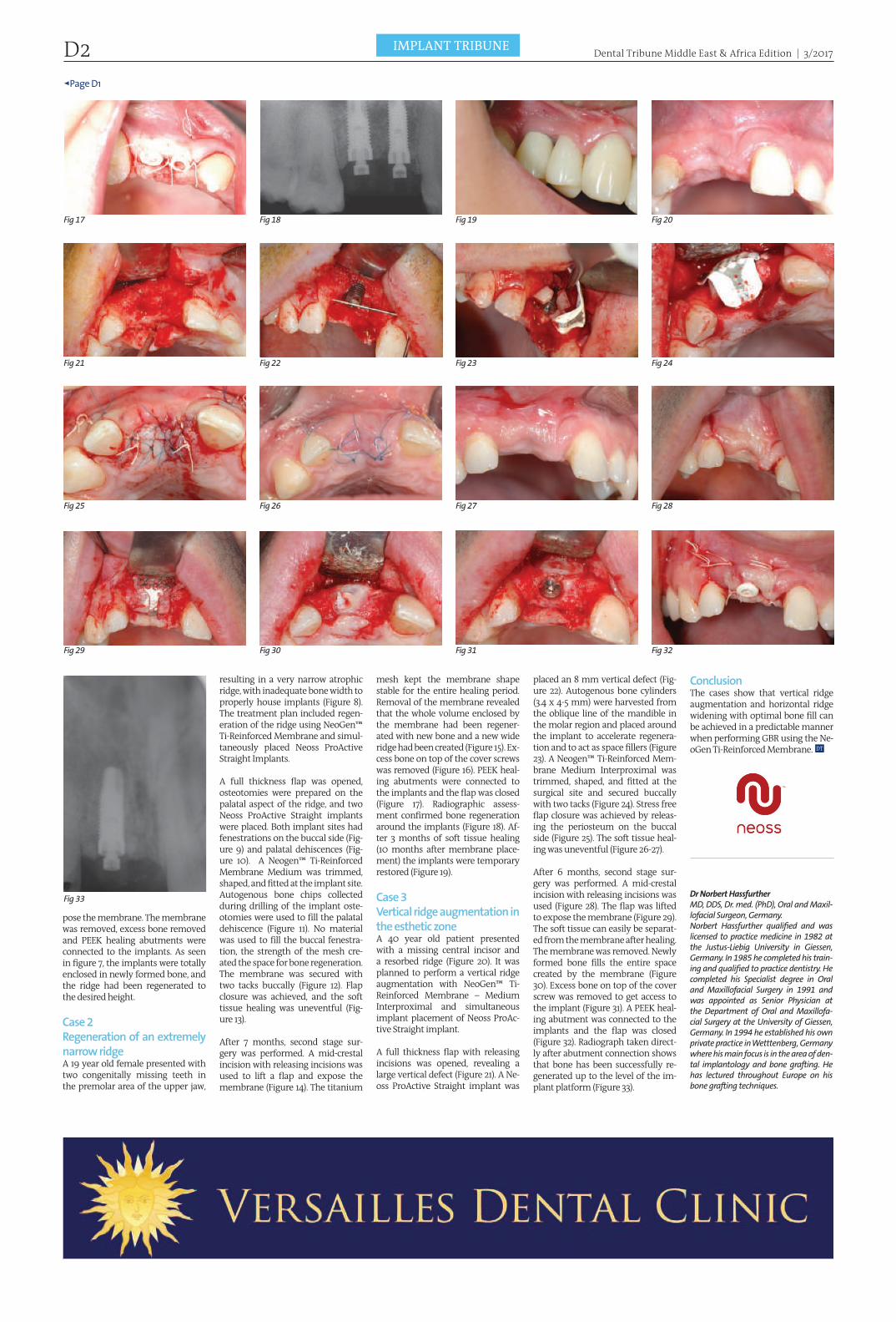
Case 2Regeneration of an extremely narrow ridgeA 19 year old female presented with two congenitally missing teeth in the premolar area of the upper jaw,
resulting in a very narrow atrophic ridge, with inadequate bone width to properly house implants (Figure 8). The treatment plan included regen-eration of the ridge using NeoGen™ Ti-Reinforced Membrane and simul-taneously placed Neoss ProActive Straight Implants.
A full thickness flap was opened, osteotomies were prepared on the palatal aspect of the ridge, and two Neoss ProActive Straight implants were placed. Both implant sites had fenestrations on the buccal side (Fig-ure 9) and palatal dehiscences (Fig-ure 10). A Neogen™ Ti-Reinforced Membrane Medium was trimmed, shaped, and fitted at the implant site. Autogenous bone chips collected during drilling of the implant oste-otomies were used to fill the palatal dehiscence (Figure 11). No material was used to fill the buccal fenestra-tion, the strength of the mesh cre-ated the space for bone regeneration. The membrane was secured with two tacks buccally (Figure 12). Flap closure was achieved, and the soft tissue healing was uneventful (Fig-ure 13).
After 7 months, second stage sur-gery was performed. A mid-crestal incision with releasing incisions was used to lift a flap and expose the membrane (Figure 14). The titanium
mesh kept the membrane shape stable for the entire healing period. Removal of the membrane revealed that the whole volume enclosed by the membrane had been regener-ated with new bone and a new wide ridge had been created (Figure 15). Ex-cess bone on top of the cover screws was removed (Figure 16). PEEK heal-ing abutments were connected to the implants and the flap was closed (Figure 17). Radiographic assess-ment confirmed bone regeneration around the implants (Figure 18). Af-ter 3 months of soft tissue healing (10 months after membrane place-ment) the implants were temporary restored (Figure 19).
Case 3Vertical ridge augmentation in the esthetic zoneA 40 year old patient presented with a missing central incisor and a resorbed ridge (Figure 20). It was planned to perform a vertical ridge augmentation with NeoGen™ Ti-Reinforced Membrane – Medium Interproximal and simultaneous implant placement of Neoss ProAc-tive Straight implant.
A full thickness flap with releasing incisions was opened, revealing a large vertical defect (Figure 21). A Ne-oss ProActive Straight implant was
placed an 8 mm vertical defect (Fig-ure 22). Autogenous bone cylinders (3.4 x 4-5 mm) were harvested from the oblique line of the mandible in the molar region and placed around the implant to accelerate regenera-tion and to act as space fillers (Figure 23). A Neogen™ Ti-Reinforced Mem-brane Medium Interproximal was trimmed, shaped, and fitted at the surgical site and secured buccally with two tacks (Figure 24). Stress free flap closure was achieved by releas-ing the periosteum on the buccal side (Figure 25). The soft tissue heal-ing was uneventful (Figure 26-27).
After 6 months, second stage sur-gery was performed. A mid-crestal incision with releasing incisions was used (Figure 28). The flap was lifted to expose the membrane (Figure 29). The soft tissue can easily be separat-ed from the membrane after healing. The membrane was removed. Newly formed bone fills the entire space created by the membrane (Figure 30). Excess bone on top of the cover screw was removed to get access to the implant (Figure 31). A PEEK heal-ing abutment was connected to the implants and the flap was closed (Figure 32). Radiograph taken direct-ly after abutment connection shows that bone has been successfully re-generated up to the level of the im-plant platform (Figure 33).
ConclusionThe cases show that vertical ridge augmentation and horizontal ridge widening with optimal bone fill can be achieved in a predictable manner when performing GBR using the Ne-oGen Ti-Reinforced Membrane.
Dr Norbert HassfurtherMD, DDS, Dr. med. (PhD), Oral and Maxil-lofacial Surgeon, Germany.Norbert Hassfurther qualified and was licensed to practice medicine in 1982 at the Justus-Liebig University in Giessen, Germany. In 1985 he completed his train-ing and qualified to practice dentistry. He completed his Specialist degree in Oral and Maxillofacial Surgery in 1991 and was appointed as Senior Physician at the Department of Oral and Maxillofa-cial Surgery at the University of Giessen, Germany. In 1994 he established his own private practice in Wetttenberg, Germany where his main focus is in the area of den-tal implantology and bone grafting. He has lectured throughout Europe on his bone grafting techniques.
ÿPage D4
Considerations for Long Term Success Implants are Never Forever!
By Dr. Shankar Iyer, USA
This article will emphasize the im-portance of factors to consider be-fore treatment planning for full arches with implants. It is not un-common to make misleading prom-ises to patients when presenting im-plants as an option with unfounded claims of 98% success rates. Most of the survival statistics have evaluated implants for full mouth reconstruc-tions through profuse citations of the original Branemark’s work pub-lished in 1981. Repeated citations of this article and the subsequent fol-low up articles have made claims of a high percentage of success with im-plants. While this is partially true, the circumstances under which these implants survived has been incor-rectly extrapolated to other clinical situations. The original Branemark research was done on edentulous arches with hybrid prosthesis op-posing either complete dentures or prosthesis of similar construction.
Patients are now wondering with these highly overstated survival rates, why their implants are ail-ing and need maintenance within a short span. The answer lies in the lack of understanding of biomechan-ics. The connotation that anything works has led to confusion in the field. The diametrically opposite views of short vs long implants, axial vs angled implants, graft vs graftless solutions, regular vs minis, delayed vs immediate, one piece vs two piec-es, guided vs free hand placements and platform switiching concepts have only caused anarchy in the discipline of implant dentistry. Po-dium concepts have gained popular-ity through corporate support and we see opinion leaders vociferously making unsubstantiated claims through limited clinical evidence. A novice finds it very difficult to get in-volved in implant dentistry because the education is being blessed by companies and not through univer-sities or institutions.
After being involved in implants for over 20 years, I find it to be an hum-bling experience with cases that I treatment planned two decades ago returning to me for maintenance. Seeing these cases today, I wish I had this experience at that time so I could have served my patients bet-ter. Today it has taught me a lot in treatment planning. I am able to prognosticate the outcome and its management in the event of an un-toward incident. The lessons in bio-mechanics has complemented the initial biologic responses that can be predicted initially so that the surviv-al of implant therapy is prolonged.
I am a firm believer of long term data and I fear the rapid evolution of products and techniques that claim to be faster and easier. If only I could train my patients osteoblasts to work harder and faster so their bones can heal rapidly, all of the problems can be eliminated and failures can be a thing of the past. The life cycles of cells have been a constant over a mil-lion years and now we are told that implants are appoved for immedi-ate load and the cells can adhere to inanimate objects through unique surfaces. My understanding of cell
biology may be limited but it is com-mon knowledge that behavior of cells cannot be hastened because the mitotic cycle for the DNA takes the programmed time period for turn over. Only in disease this rapid un-controlled proliferation takes place. If this normal cycle is upset then we are look at metaplastic or anaplastic changes according to the turnover rate. Claims made by certain compa-nies that, bone heals faster with their implants is presumptuous. Bone levels are magically maintained with their unique surface modification is also far from the truth. I have used over 16 different implant systems in my practice over the years and in my training programs and I have found that the osteoclasts are notoriously unbiased. There is bone loss with every system and modifying the sur-face or creating morphological shifts does not predictably deter bone loss.
In the courses I teach, I recommend waiting for a period of three years after any new feature or biologic product is introduced into implant dentistry. There is no room for lat-est or newest in clinical practice. If a company is constantly introduc-ing new product lines and changing their designs, there is only one con-clusion – They are having trouble and hence they have to change. A robust system that works seldom needs modification for getting pre-dictable results. Aspirin can never be debunked for its efficacy, be-ing so old and dated. The original Branemark external hex (now made out of type 4 Titanium but designed in 1965) is still very functional and a work horse for hybrid prosthesis. The surfaces have improved much but its basic design and biomechani-cal considerations will be valid for another 50 years. Premature adop-tion of technology or materials is fraught with shortcomings and unknown consequences. Classical examples of potential catastrophic failures include the TPS coatings, HA surface modifications, sintered surfaces, flapless surgeries, guided surgeries, immediate loading, costly BMPs and the list goes on.
The message is very simple – one crawls before they walk and you must learn to walk before you can run. The same is true for implant dentistry. The novice today has a wide choice – you can become a com-plete arch implant specialist with 4 implants and guided surgery over a weekend or spend a year learning the basics and judiciously treatment plan cases with customized solu-tions. Half of the participants of our Maxicourses that we run in the U.S. and overseas have practitioners who have placed hundreds of implants and got their training through cor-porate education. One does not be-come a musician by buying a piano or a musical instrument, nor can you become a pilot by buying a plane. Training in implant dentistry has be-come a fad. Most courses are offered through companies and the com-pany’s sole interest is to sell their sys-tem. There is a whole world of treat-ment plan that is out there before the system can be utilized. Lets not place the cart before the horse. The void is very apparent the time is now for implementing judicious treat-
ment plans. Lets serve our patients with what they need and not what we want them to have.
Iyer’s Top 10 Guidelines for Predict-able Implantolgy1. Diagnose the problem first and don’t treat because you have a tool that you can use.
2. Measure the disease and provide the therapy, don’t sell concepts.3. Leave what’s new and latest to the risk takers, stick with proven and tried systems.4. Implants are the last resort in treat-ment planning – exhaust all conserv-ative, conventional modalities5. Implants should replace missing
teeth not replace teeth.6. Expensive implants don’t mean success rates are better, cheaper does not mean everything works – you get what you pay for. There is no substi-tute for evidence based practice7. Consider every implant as a failing
Fig 1
Fig 3
Fig 5
Fig 7
Fig 2
Fig 4
Fig 6
Fig 8
Fig 9 Fig 10
Dental Tribune Middle East & Africa Edition | 3/2017IMPLANT TRIBUNE D3

◊Page D3
Fig 13Fig 11
Fig 15 Fig 17
Fig 14Fig 12
Fig 16 Fig 18
entity and the trick is to do the best you can to maintain it as long as you can. 8. Select the system that does not change its product line every year.9. There are no short cuts or faster way to get success in life and im-plants are no different.10. The success rates of implants are inversely proportional to the num-ber of years you practice implants.
Case ReportThis case reports will provide a ra-tionale for a sound sequential treat-ment plan in the management of long term failure of dental implants. Judicious use of implants and their treatment planning should have long term considerations. I used to perform subperiosteal implants and blade implants in the past. One of the reasons for not using them now is not because they fail, but because in the long term, in the event of a failure, it can have some irrevers-ible consequences. This case under-scores the importance of over engi-neering cases from the beginning so that when patients live into their 90s they don’t become incapacitat-ed, not being able to chew their food properly and lose the benefi ts of im-plants that they enjoyed for a long period of time.
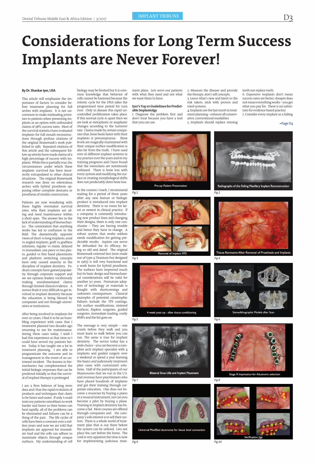
A 78 year old Caucasian female pre-sented to my practice for rehabilita-tion and management of a failing maxillary implant reconstruction. She reported having some implants 27 years ago and it has been trou-bling her with symptoms of sinus infections and movement of the entire maxillary prosthesis (Fig 1). Radiograph revealed bone loss around the unilateral subperiosteal implants and the blade implants in the anterior sextant (Fig 2). After careful examination, it was decided that none the maxillary implants was salvageable. Treatment plan was formulated to stage the case to permit healing of the infl amed soft tissue and resorbed bone.
The entire maxillary frame had to be sectioned and removed piecemeal (Fig 3, 4). An immediate denture was fabricated and the tissues were allowed to heal for a period of two months. (Fig 5) A sterolithographic model was created to assess the con-dition of the remaining bone (Fig 6). A decision was made to reconstruct the maxilla with bilateral sinus aug-mentation. The anterior sextant had bone loss till the anterior nasal spine. Six months following the augmen-tation, nine implants were placed in the augmented bone (Fig 7). Stage II surgery was performed after a heal-ing period of 8 months. Impressions were taken (Fig 8). A Universal modi-fi ed abutment was utilized to bring all of the platforms equi-gingival (Fig
THE TENTH ANNUAL AMERICAN ACADEMY OF IMPLANT DENTISTRYMaxiCours e®- UAE 2017 – 2018 Starts 28 July 2017
A unique opportunity towards becoming a Diplomate of the American Board of Oral
Implantology/Implant Dentistry - AAID is the sponsoring organization of ABOI
In Fulfillme nt o f the Educ ational Re quire me nt for the Examination
for Assoc iate Fe llow Me mbe rship and Fe llowship for the
Ame ric an Ac ade my of Implant De ntistry
Program Includes placement of upto 10 Implants with all surgical and
prosthetic components, all materials for hands – on workshop and
lecture handouts plus one complete surgical instrument Kit.
MaxiCourse ® Advantage: 300 hours of comprehensive lectures, live surgeries,
demonstration and hands-on sessions. In depth review of surgical and prosthetic protocols. Sessions stretch across 5 modules of 6 days. Each
session is always inclusive of a weekend. Curriculun taught by over 18 faculty & speakers from the International Community who are amongst the most distinguished names in implantology.. Certificate of completion awarded by the American Academy of Implant Dentistry.
Non commercial, non sponsored course covering a wide spectrum of implant types and system.
Hands-on patient treatment under direct AAID faculty supervision. Membership for AAID awarded for 2017 – 2018
Dates:
Module 1 July 28th – August 2nd 2017
Module 2 Nov 1st - 6 th 2017
Module 3 Jan 25th – 30th 2018
Module 4 March 30th – April 4th 2018
Module 5 July 5th – 10th 2018
Registration :
Pre-Registration is Mandatory as it is a limited Participation Program.
For further information and registration details visit website: www.maxicourseasia.com or e-mail
Dr. Ninette Banday, Coordinator AAID-MaxiCourse UAE at [email protected]
The Faculty are as follows:
Dr. Shankar Iyer, USA Director, AAID Maxi Course®UAEDiplomate AAIDClinical Assistant Professor,Rutgers School of Dental Medicine.
Dr. Ninette Banday, UAECo-Director AAID Maxicourse- Abu Dhabi, UAEAcademic Associate Fellow AAID
Dr. Amit Vora, USADiplomate of the American Board of PeriodontologyProfessor (partime) ,JFK Hospital and the Veteran Affairs (V.A.) Hospital
Dr. Jaime Lozada , USADirector of the Graduate Program in Implant Dentistry Fellow, American Academy of Implant Dentistry
Dr. William Locante, USADiplomate of ABOIFellow of American Academy of Implant Dentistry
Dr. Robert Horowitz , USADiplomate American Board of Periodontology
Clinical Assistant Professor New York University
Dr. Frank LaMar, Sn USAFellow, American Academy of Implant DentistryDiplomate, American Board of Oral Implantology
Dr. Frank LaMar Jr.Diplomat American Board of Prosthodontist
Dr. John Minichetti, USADiplomat, American Board of Oral ImplantologyHonored Fellow, American Academy of ImplantDentistry
Dr. Kim Gowey , USAPast President – AAID Diplomate ABOI
Dr.Burnee Dunson , USAFellow, American Academy of Implant Dentistry Diplomate ABOI
Dr. Jason Kim, USADiplomate of ABOI
Dr. Ozair Banday, USAProsthodontist
Dr. Stuart Ort on-Jones, UKFounder Member, The Pankey AssociationMember, Alabama Implant Study Group
Dr. Robert Miller, USABoard Certified by the American Board of Oral Implantology/Implant DentistryHonored Fellow American Academy of Implant Dentistry
Dr. Philip T ardeu , FranceFounder and Author, Computer Guided Implantology and the Safe System.
Dr. Natalie Wong, CanadaDiplomate, American Board of Oral ImplantologyFellow, American Academy of Implant Dentistry
Dr. Irfan Kanchwala , IndiaImplant Fellowship ( UMDNJ, USA)Diplomate , American Board of Prosthodontics
Dr. Jihad Abdallah, LebanonDiplomate American Board of Oral ImplantologyFellow AAIDProfessor & Head of Implantology Division, Faculty of Dentistry.Beirut Arab UniversityDirector, AAID Maxi course ®Jordan
2016-2017 Program Accredited by Health Authority Abu Dhabi for 228.5 CME Hours.
2017-2018 Accredition under process.
9). A verifi cation jig was utilized to check for passivity and accuracy of the positions of the abutments (Fig 10). The metal frame was indexed, cast and tried in (Fig 11, 12). Face bow transfer record was obtained for ori-entation relationship. (Fig 13) Porce-
lain overlay for an FP3 prosthesis was processed and inserted (Fig 14, 15 ) A mutually protected occlusal scheme was designed (Fig 16). The patient’s vertical was maintained. The post op radiograph reveals a stable out-come. (Fig 17) The anterior cantile-
vered crowns provide for optimal esthetics in the extremely resorbed anterior maxilla. The post opera-tive outcome provided an esthetic and functional rehabilitation of the failing implant FPD (Fig 18). The provision of pontics enhanced the
outcome in the esthetic zone and in this case it favored the design due to the atrophy that precluded implant placement in the premaxilla. The case has been in function for over 5 years and the patient has been on re-care every 4 months.
Dental Tribune Middle East & Africa Edition | 3/2017 IMPLANT TRIBUNED4