guidance document on the collection of eye tissues …
TRANSCRIPT

Organisation for Economic Co-operation and Development
ENV/JM/MONO(2011)45/REV1
Unclassified English - Or. English
6 July 2018
ENVIRONMENT DIRECTORATE
JOINT MEETING OF THE CHEMICALS COMMITTEE AND THE WORKING PARTY
ON CHEMICALS, PESTICIDES AND BIOTECHNOLOGY
GUIDANCE DOCUMENT ON THE COLLECTION OF EYE TISSUES FOR
HISTOLOGICAL EVALUATION AND COLLECTION OF DATA SERIES ON TESTING AND ASSESMENT
Number 160
(Second Edition)
JT03434402
This document, as well as any data and map included herein, are without prejudice to the status of or sovereignty over any territory, to the
delimitation of international frontiers and boundaries and to the name of any territory, city or area.

2 │ ENV/JM/MONO(2011)45/REV1
Unclassified

ENV/JM/MONO(2011)45/REV1 │ 3
Unclassified
OECD Environment, Health and Safety Publications
Series on Testing and Assessment
No. 160
GUIDANCE DOCUMENT ON THE COLLECTION OF EYE TISSUES FOR HISTOLOGICAL
EVALUATION AND COLLECTION OF DATA
(Second Edition)
Environment Directorate
ORGANISATION FOR ECONOMIC CO-OPERATION AND DEVELOPMENT
Paris 2018

4 │ ENV/JM/MONO(2011)45/REV1
Unclassified
About the OECD
The Organisation for Economic Co-operation and Development (OECD) is an intergovernmental
organisation in which representatives of 35 industrialised countries in North and South America, Europe
and the Asia and Pacific region, as well as the European Commission, meet to co-ordinate and harmonise
policies, discuss issues of mutual concern, and work together to respond to international problems. Most
of the OECD’s work is carried out by more than 200 specialised committees and working groups composed
of member country delegates. Observers from several countries with special status at the OECD, and from
interested international organisations, attend many of the OECD’s workshops and other meetings.
Committees and working groups are served by the OECD Secretariat, located in Paris, France, which is
organised into directorates and divisions.
The Environment, Health and Safety Division publishes free-of-charge documents in twelve different
series: Testing and Assessment; Good Laboratory Practice and Compliance Monitoring; Pesticides;
Biocides; Risk Management; Harmonisation of Regulatory Oversight in Biotechnology; Safety of
Novel Foods and Feeds; Chemical Accidents; Pollutant Release and Transfer Registers; Emission
Scenario Documents; Safety of Manufactured Nanomaterials; and Adverse Outcome Pathways.
More information about the Environment, Health and Safety Programme and EHS publications is available
on the OECD’s World Wide Web site (www.oecd.org/chemicalsafety/).
This publication was developed in the IOMC context. The contents do not necessarily reflect the
views or stated policies of individual IOMC Participating Organizations.
The Inter-Organisation Programme for the Sound Management of Chemicals (IOMC) was
established in 1995 following recommendations made by the 1992 UN Conference on Environment
and Development to strengthen co-operation and increase international co-ordination in the field of
chemical safety. The Participating Organisations are FAO, ILO, UNDP, UNEP, UNIDO, UNITAR,
WHO, World Bank and OECD. The purpose of the IOMC is to promote co-ordination of the policies
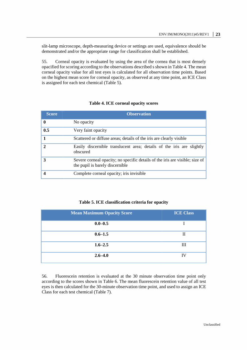
and activities pursued by the Participating Organisations, jointly or separately, to achieve the sound
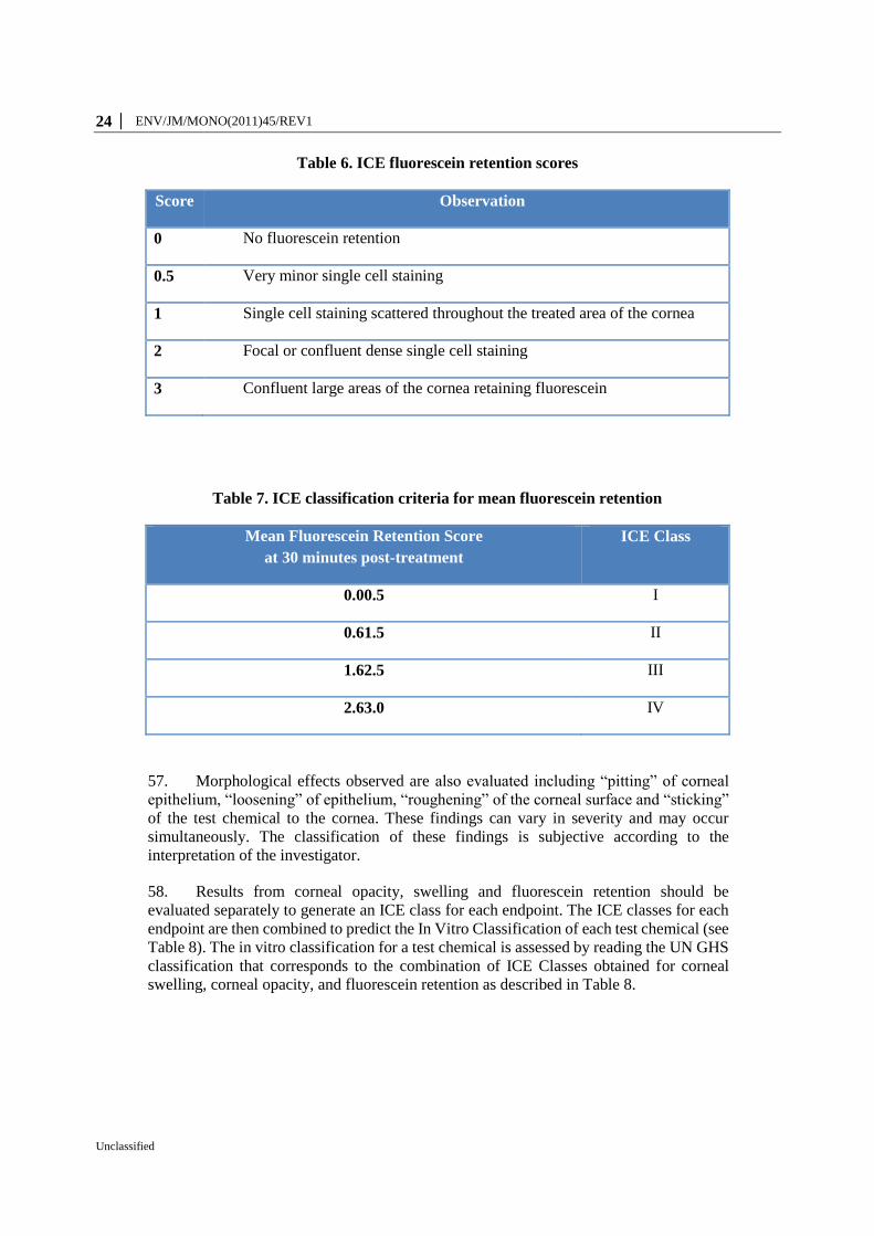
management of chemicals in relation to human health and the environment.

ENV/JM/MONO(2011)45/REV1 │ 5
Unclassified
This publication is available electronically, at no charge.
For this and many other Environment,
Health and Safety publications, consult the OECD’s
World Wide Web site (www.oecd.org/ehs)
or contact:
OECD Environment Directorate,
Environment, Health and Safety Division
2, rue André-Pascal
75775 Paris cedex 16
France
Fax : (33-1) 44 30 61 80
E-mail : [email protected]
© OECD 2018
Applications for permission to reproduce or translate all or part of this material should
be made to: Head of Publications Service, [email protected], OECD, 2 rue André-
Pascal, 75775 Paris Cedex 16, France

6 │ ENV/JM/MONO(2011)45/REV1
Unclassified
FOREWORD
This Guidance Document (GD) was originally developed in 2011 to (i) promote the use of
histopathological evaluation as an additional endpoint for ocular toxicity testing; and (ii)
provide specific guidance on using the TG 437 (BCOP) and TG 438 (ICE) for the purpose
of expanding their respective databases towards optimising their use for identifying all
hazard categories, including the complete recommended decision criteria for both test
methods.
The second edition, dated 2018, reflects increased knowledge on the use of histopathology
especially with the ICE test method including: (i) the recommendation for having an
internal peer-review process when evaluating histopathological effects, (ii) the use of semi-
quantitative scoring systems for e.g. the ICE histopathology, and (iii) inclusion of an Atlas
describing typical ICE histopathological effects. It was further updated in 2018 to be
aligned with the revised TG 438.
The Second Edition of the Guidance Document was approved by the Working Group of
the National Coordinators in 2018. The Joint Meeting of the Chemicals Committee and the
Working Party on Chemicals, Pesticides and Biotechnology agreed to its declassification
on 30 June 2018.
This document is published under the responsibility of the Joint Meeting of the Chemicals
Committee and the Working Party on Chemicals, Pesticides and Biotechnology of the
OECD.
ENV/JM/MONO(2011)45/REV1 │ 7
Unclassified
INTRODUCTION
1. This Guidance Document (GD) accompanies the OECD Test Guideline (TG) 437
on the bovine corneal opacity and permeability (BCOP) test method (OECD 2017a) and
TG 438 on the isolated chicken eye (ICE) test method (OECD 2018a). It provides users
with guidelines for collecting histopathology data for in vitro and/or in vivo ocular safety
test methods. The primary purposes of this GD are: i) to promote the collection of
histopathological data; (ii) to provide guidance on performing histopathological
evaluations; (iii) to support further understanding of the usefulness and limitations of
histopathology as an additional endpoint to improve the accuracy of in vitro ocular safety
test methods; iv) to provide comprehensive protocols on the BCOP and ICE test methods
to promote harmonization of approaches; and v) for those test chemicals (i.e. substances
and mixtures) that are tested as a last resort, in vivo, to provide standard procedures for
enucleating, fixing, and processing eyes from the in vivo rabbit eye studies for
histopathological evaluation. Note that for a full evaluation of eye hazard effects after acute
exposure, the Guidance Document on Integrated Approaches for Testing Assessment
(IATA) should be considered (OECD, 2017b). In particular, the IATA approach includes
the use of recommended testing strategies based on in vitro test methods and on other
information sources before considering testing in living animals (see paragraph 5).
2. Histopathological evaluation may be useful for (i) assessment of the histological
damage of chemical classes or formulations that are not well characterized in the before-
mentioned test methods; (ii) assisting with determination of a mode of action; (iii) assisting
with determination of the likelihood of delayed effects; (iv) evaluation of the depth of
injury, which has been proposed as a measure of reversibility or irreversibility (Maurer et
al. 2002); (v) further characterization of the severity or scope of the damage as needed
(Harbell et al. 2006) (ICCVAM 2010b) (Maurer et al. 2002); (vi) assisting with
discrimination of cases where the response falls along the borderline between two
categories based on the test method decision criteria. Therefore, users are encouraged to
preserve tissues for histopathological evaluation.
3. Histopathological evaluation may also be used to support the development of other
in vitro ocular safety test methods (e.g. Isolated Rabbit Eye test method (ICCVAM 2010a),
Porcine Corneal Opacity and Permeability Assay (Van den Berghe et al. 2005), and 3-
dimensional human corneal tissue constructs (Carrier et al. 2009) including the
Reconstructed human cornea-like Epithelium test methods (OECD TG 492, 2018b).
Furthermore, in cases where an in vivo rabbit eye test is still needed as a last resort,
histopathological evaluation may be used, when relevant, as an additional endpoint to more
thoroughly evaluate the type and extent of ocular damage produced, as well as to provide
a reference against which to compare effects produced in vitro. These additional data may
help in the development of more accurate, mechanism-based in vitro alternatives to the
rabbit eye test. Although the in vivo eye irritation study in rabbits seems to offer the
possibility of performing histopathology of the treated eye in order to provide additional
information on the inflammation process, in normal practice it will not be relevant. After
all, in the standard in vivo rabbit eye irritation test, the rabbits may be sacrificed at the end
of the observation period at which point the eye effects may have reversed. In the event
that rabbits have to be sacrificed prematurely because of the severe nature of the eye
effects, or in the event of persistence of effects in the cornea at the end of the observation
period, sampling of the eyes for histopathology may be useful e.g. for better mechanistic
understanding.

8 │ ENV/JM/MONO(2011)45/REV1
Unclassified
4. This GD describes the general procedures for the collection, preservation, and
preparation of in vitro and in vivo ocular tissues for use in performing histopathological
evaluations. Based on the latest progress on the use of histopathology for the ICE test
method, it provides guidance in performing ICE histopathological evaluations including
the recommendation of having an in-house peer-review system, the use of a semi-
quantitative scoring system to assess histopathological effects, and the use of an Atlas
describing typical histopathological effects. In particular, the use of histopathology has
been accepted as an additional parameter to improve the identification of GHS Category 1
chemicals within the ICE method (TG 438) for the specific applicability domain of non-
extreme pH (2 < pH < 11.5) detergents and surfactants (OECD, 2018a; OECD, 2018c;
Cazelle et al., 2014). A semi-quantitative scoring system and decision criteria have been
developed for the ICE histopathology for non-extreme pH detergents and surfactants (see
paragraphs 39, 62 and 63). Prior to use of this ICE semi-quantitative histopathological
scoring system and/or decision criteria with other test method(s) and/or applicability
domain(s), its/their adequacy to the new test method(s) and/or applicability domain(s)
should be demonstrated first. Finally, in the case of the BCOP or of the in vivo test method,
if differences exist regarding the collection, preservation, preparation, assessment and
interpretation of the corneas or in vivo eyes, laboratories that routinely perform
histopathological evaluations of ocular tissue can employ their existing procedures. When
additional information becomes available, this GD will be updated accordingly.
5. It is currently generally accepted that, in the foreseeable future, no single in vitro
eye irritation test will be able to replace the in vivo Draize eye test to predict across the full
range of irritation for different chemical classes. The IATA for Serious Eye Damage and
Eye Irritation describes several modules which group information sources and analysis
tools, and provides guidance on (i) how to integrate and use existing testing and non-testing
data for the assessment of eye hazard effects and (ii) proposes an approach when further
testing is needed (OECD, 2017b). In particular, strategic combinations of several
alternative test methods within a (tiered) testing strategy may be able to replace the Draize
eye test (OECD, 2017b). For example, the Top-Down approach is designed to be used
when, based on existing information, a chemical is expected to have high irritancy
potential, while the Bottom-Up approach is designed to be used when, based on existing
information, a chemical is expected not to cause sufficient eye irritation to require a
classification (Scott et al., 2010; OECD, 2017b). As described in TG 437 and 438, BCOP
and ICE data are accepted for the hazard classification and labelling of test chemicals
inducing serious eye damage (i.e., UN GHS Category 1) and test chemicals not requiring
classification for eye irritation or serious eye damage (i.e., UN GHS No Category) (OECD
2017a) (OECD 2018a). As a consequence these assays may be used to initiate the Top-
Down and the Bottom-Up approaches at the same time, so that the two tiers of the strategy
recommended in the OECD GD 263 (OECD, 2017b) could be covered with one single in
vitro assay, provided the test chemical fits the applicability domain and does not fall within
the limitations of the test method for each tier. However, since the BCOP has a high
overprediction rate for the test chemicals that do not require classification for eye hazard
(69%), it should not be the first choice to initiate a Bottom-Up approach (OECD, 2017a).
Furthermore, appropriate regulatory authorities should be consulted before using these
assays in a Bottom-Up approach under other classification schemes than the UN GHS.
Finally, even if none of these predictions are obtained, BCOP or ICE data can still be
useful, within an IATA approach in conjunction with other testing and/or non-testing data,
to further evaluate in a weight of evidence approach the potential eye hazard of the test
chemical including moderate and mild irritants (i.e., UN GHS Category 2/2A and 2B)

ENV/JM/MONO(2011)45/REV1 │ 9
Unclassified
(OECD, 2017b). This GD provides further insights on the decision criteria and protocols
of these two assays including the use of histopathology that can be reported in parallel with
other data available.
6. Definitions are provided in Annex 1.
10 │ ENV/JM/MONO(2011)45/REV1
Unclassified
HISTOPATHOLOGICAL EVALUATION IN OCULAR SAFETY TEST
METHODS
1.1. Background
7. With the exception of some research projects (Cuellar et al. 2003) (Kadar et al.
2001) (Maurer et al. 2002), few in vivo eye irritation studies include histopathological
evaluation. The lack of such data has impeded the identification of relevant histopathology
endpoint(s) that can be used in in vivo eye irritation/corrosivity testing, and its use to
develop in vitro ocular safety test methods. While this GD provides examples on the
evaluation and interpretation of histopathological data, it is important to recognize that the
markers of injury in isolated eyes or corneas are different from those observed in eyes
treated in vivo. For example, ex vivo test methods are devoid of an intact inflammatory
response. However, the depth of injury in isolated corneas, as determined by
histopathological evaluation, has been proposed to predict the degree and duration of the
injury (Maurer et al. 2002).
8. To facilitate consideration of histopathological evaluation as a useful endpoint for
in vitro and in vivo ocular safety testing, users are encouraged to submit data and
histopathological specimens generated according to this GD to international validation
organizations (i.e. the US National Toxicology Program Interagency Center for the
Evaluation of Alternative Toxicological Methods [US-NICEATM], the EU European
Union Reference Laboratory for Alternatives to Animal Testing [EURL-ECVAM], or the
Japanese Center for the Validation of Alternative Methods [JaCVAM]).
1.2. Source of Tissue for Histopathological Evaluation
9. The source of tissue to be considered for histopathological evaluation includes
whole eyes or isolated portions of the anterior segment (e.g. cornea), obtained after
completion of an in vitro or in vivo ocular safety test method. All information related to
the type and treatment of a particular tissue sample should be included in the Test Report.
10. All procedures using animal eyes should follow applicable geographical
regulations and the test facility’s procedures for handling animal-derived materials, which
include, but are not limited to, tissues and tissue fluids. Universal laboratory safety
precautions are recommended (Siegel et al. 2007).
1.3. Sample Identification
11. Each sample should be assigned a unique identifier that will allow it to be traced
back to the study from which it was obtained (Billings and Grizzle 2008) (Harbell et al.
2006) (ICCVAM 2010b).
1.4. Tissue Preparation
12. In the case of the in vitro Isolated Chicken Eye test method, treated eyes are
collected after the final examination i.e., four hours after treatment, of the test method
described within the OECD TG 438 (OECD, 2018a). All three eyes treated with a test
chemical, as obtained from the standard ICE test method (OECD, 2018a), are used for

ENV/JM/MONO(2011)45/REV1 │ 11
Unclassified
histopathological evaluation. Histopathology is considered appropriate for the overall
assessment of effects in conjunction with the standard ICE endpoints. Eyes can be incised
almost completely in half with a scalpel just behind the level of the lens and through the
vitreous body, leaving a part of the posterior tissue still attached where eyes can be held
(that will later be discarded) to ensure that the cornea is not damaged during manipulation
by dropping on a surface, whilst at the same time allowing optimal penetration of the
fixation agent (see paragraphs 15 to 19).
13. In the case of the in vitro Bovine Corneal Opacity and Permeability test method,
after completion of the fluorescein permeability endpoint sampling as described within
OECD TG 437 (2017a), remaining fluorescein and medium are removed from the corneal
holders, the holders are carefully disassembled, and the corneas are carefully removed and
transferred to individually labelled tissue cassettes. The corneas are placed endothelial
surface down onto a histology sponge to protect the endothelium. The cassettes are placed
in labelled containers filled with 10% neutral buffered formalin and fixed at room
temperature for a minimum of 24 hours.
14. Corneas to be used for histopathological evaluation following in vivo studies
according to the OECD TG 405, conducted as last resort within the framework of the IATA
for Serious Eye Damage and Eye Irritation (OECD, 2017c), are kept moist with drops of
physiological saline (pre-warmed from 31 to 32°C) applied throughout the dissection
process. Scientists with expertise in performing the dissection have provided details of the
procedure (Jones P, Guest R, personal communications) (ICCVAM 2006a). The nictitating
membrane is deflected away using forceps and the conjunctivae are cut using angled
forceps and curved scissors. The eyeball is removed by applying gentle pressure with
fingers above and below the orbit. The remaining conjunctival tissue, the orbital muscles
and the optic nerve (leaving approximately a 5-10 mm section to prevent loss of intraocular
pressure) are removed and the eyeball is lifted from the orbit. Any tissue adhering to the
globe is then removed by careful dissection, and the eyeball is gently rinsed with a stream
of physiological saline to remove any adherent debris.
1.5. Tissue Preservation
15. Tissue fixatives prevent autolysis by inactivating autolytic enzymes that are
released post-mortem (Banks 1993). Fixation also hardens the tissue thereby allowing thin
sections to be cut without inducing mechanical artefacts (e.g. compression of the tissue).
Factors that affect tissue fixation include time and temperature during incubation, the
volume of the fixative relative to tissue size, the physicochemical properties of the fixative,
and the concentration of the fixative (Banks 1993) (Grizzle et al. 2008). To prevent the
tissues from drying out, which would induce substantial artefacts, they should remain
immersed in fixative before processing and embedding.
16. Tissues should be placed in prelabelled containers filled with fixative. Most
histology protocols recommend a fixative volume at least 5- to 10-fold greater than the size
of the tissue (Billings and Grizzle 2008) (Kiernan 1990) (Samuelson 2007), although
Banks (1993) recommends up to a 30-fold fixative-to-tissue size ratio. In the case of the
ICE test methods, eyes (incised or not) are placed in a container with the fixation agent
(e.g., approximately 20 mL of e.g. 10% formalin (see paragraph 18) for at least 24 hours).
In the case of the BCOP test method, bovine corneas are placed into 10% neutral buffered
formalin (10% NBF) at a rate of approximately 20 corneas per 300 mL.

12 │ ENV/JM/MONO(2011)45/REV1
Unclassified
17. All tissues should be completely immersed in the fixative. Smaller tissues may be
placed into cassettes; however, for consistency in sectioning, care should be taken to orient
them so that the epithelial (anterior) surface faces the top of the cassette (Harbell et al.
2006) (ICCVAM 2010b).
18. The depth of penetration of most fixatives is directly proportional to the square root
of the duration of fixation (t) dependent on the coefficient of diffusibility (k) of the fixative,
which averages to 1 for typically used fixatives. Fixation time thus translates to the square
of the distance the fixative should penetrate. At a rate of 1 mm/hour, the time of fixation
for a 10-mm sphere in neutral buffered formalin (NBF) will be (5)2 or 25 hours of fixation
(Grizzle et al., 2008). Therefore, tissues are typically fixed for at least 24 hours at room
temperature. However, the reported range for fixation is 4 to 48 hours (Kimura et al. 1995)
(Kjellström et al. 2006), and some protocols perform fixation at 4 C (Kjellström et al.
1996) (Maaijwee et al. 2006).
19. The fixatives most commonly used for ocular tissues are 10% NBF and Davidson's
(Bancroft and Cook, 1994) (Spencer and Bancroft, 2008). However, for the ICE test
method, neutral aqueous phosphate buffered 4% solution of formaldehyde (i.e., 10%
formalin) has been generally used for incised eyes (Prinsen, 2011), and Davidson’s fixative
has been suggested in case whole eyes are used due to the rapid penetration into the deeper
tissues by the alcoholic component of the fixative (Latendresse et al. 2002). For the isolated
corneas used in the BCOP test method, extensive experience indicates that fewer artefacts
are induced following fixation with 10% NBF than with Davidson’s fixative (Raabe H,
personal communication). Other fixatives that have been used for ocular tissues include
4% glutaraldehyde (Chen et al. 2008), a mixture of 2.5% glutaraldehyde and 2%
formaldehyde (Kimura et al. 1995) (Zhang and Rao 2005), and 4% paraformaldehyde
(Kjellström et al. 2006) (Maaijwee et al. 2006).
1.6. Post-fixation Tissue Trimming
20. Prior to initiating the tissue-processing step, it may be necessary to trim the fixed
tissues to ensure that they are adequately dehydrated and infiltrated with paraffin wax. Any
post-fixation trimming should be done using a sharp scalpel, scissors, and/or razor blades
to minimize tissue artefacts. In the case of the ICE test method, the fixed eye is trimmed
with scissors in such a way that a thin piece containing the entire cornea and the adjacent
sclera are embedded in the paraffin wax.
1.7. Tissue Processing and Embedding
21. Ocular tissues contain approximately 75% water (Banks 1993) and should be
thoroughly dehydrated prior to embedding. This is most commonly achieved by immersing
the fixed tissue in a graded alcohol series such as ethanol from 60%-70%, 90%-95%, and
100% (Rosa and Green 2008) (Spencer and Bancroft 2008). Lower concentrations, such as
30% ethanol, are recommended for delicate tissue (Spencer and Bancroft 2008). Other
water-miscible solvents have also been used successfully (e.g. n-butanol, dioxane,
isopropanol, propanol, tetrahydrofuran, and tetrahydrofurfuryl alcohol (Banks 1993)
(Fischer et al. 2008) (Kiernan 1990) (Pantcheva et al. 2007). In the case of the ICE test
method an ethanol series of 50%, 70%, 80%, 96%, 100% is generally used.

ENV/JM/MONO(2011)45/REV1 │ 13
Unclassified
22. Because alcohols are not miscible with the paraffin wax used for embedding, a
substance that is miscible with ethanol and paraffin wax in the absence of water should be
used for intermediate clearing. This step also increases the transparency of the resulting
tissue section (i.e. "tissue clearing" (Samuelson 2007) (Spencer and Bancroft 2008)).
Xylene is the most common clearing agent used, although others have been used, including
benzene, chloroform, n-butanol, n-butyl acetate, amyl acetate, ligroin, petroleum solvents
(mainly hexanes), toluene, and trichloroethane, or terpenes such as cedarwood oil,
limonene, and terpineol (Banks 1993) (Fischer et al. 2008) (Kiernan 1990) (Pantcheva et
al. 2007). Many of these solvents may be toxic or potentially carcinogenic, so it is
important to consult the Safety Data Sheets to determine proper handling conditions prior
to use.
23. Because of the damage and resulting morphological artefacts produced by elevated
temperatures (i.e. heating), tissues should ideally be dehydrated and cleared at room to
moderate temperature. For example, in the case of the ICE test method, isolated eyes are
usually dehydrated at 40oC.
24. Ocular tissue is typically embedded in paraffin wax, a polycrystalline mixture of
solid hydrocarbons (Barequet et al. 2007) (Cerven et al. (1996) (Chen et al. 2008) (Harbell
et al. 2006) (ICCVAM 2010b) (Maaijwee et al. 2006). Plastic materials such as glycol
methacrylate have also been used to embed corneal or globe tissue of the rabbit (Kimura
et al. 1995). Plastic embedding has some advantages over paraffin embedding for corneal
disc preparations (e.g. no heat exposure, reduced distortion) (Lee 2002).
25. When processing only the isolated cornea (i.e. when using the BCOP test method
or other isolated corneal models), following infiltration with liquid paraffin, the cornea
should be bisected so that both halves can be embedded in the same block.
26. Processed tissues should be embedded so as to maintain the appropriate orientation
in the hardened tissue block once the paraffin cools. For example, in case of need for
measuring the corneal thickness due to e.g. swelling, true corneal cross-sections (i.e.
anterior to posterior) are usually desired to permit an accurate measurement of the effects
caused by the test chemical relative to the negative control (although this is not applicable
to the ICE test method for which corneal swelling is measured prior to histopathology using
a slit-lamp microscope). In any case, the tissue should be embedded in the block on its
edge in the correct orientation to permit relevant sections to be made according to the
evaluations sought.
27. A routine schedule for processing in vivo eyes with a tissue processor is provided
by Barequet et al. (2007). Enucleated globes that are initially fixed overnight in 10% NBF
are dehydrated in 4% phenol/70% alcohol for 1 hr each. Phenol is added to soften the sclera
and lens. The eyes are then incubated in two separate stations of 95% alcohol (1 hr each),
followed by two separate stations of 100% alcohol (1.5 hr each). Tissue-clearing steps
include incubations in 50% alcohol/50% xylene for 2 hr, followed by two separate stations
of 100% xylene (2 hr/each). Tissue is then infiltrated with liquid paraffin in two separate
2-hr incubations. This schedule may require modification depending on the manufacturer's
specifications and the type of tissue processor used as e.g. described above.

14 │ ENV/JM/MONO(2011)45/REV1
Unclassified
1.8. Tissue Sectioning and Slide Preparation
28. Once embedded, the tissue is usually sectioned using a microtome with a sharpened
steel blade. Depending on the type of microtome used, the thickness of microtome sections
for tissue is generally 3-8 µm (Banks 1993) (Fischer et al. 2008) (Samuelson 2007)
(Spencer and Bancroft 2008) (Lee 2002). In the case of the ICE and BCOP test methods,
longitudinal serial slides are generally sectioned at 4-5 µm, prepared from the central area
of the cornea and further processed with the staining. The microtome should be placed on
a stable surface composed of a dense material that will minimize vibrations (e.g. a marble
desktop). Vibrations can cause substantial tissue artefacts (Harbell et al. 2006) (ICCVAM
2010b) (Spencer and Bancroft 2008).
29. For embedded globes or corneas that have been bisected, tissue sections from each
half of the bisected globe containing adequate corneal tissue or the bisected cornea itself
are cut and placed on a slide for staining (i.e. a series of tissue sections in which the trailing
edge of one section adheres to the trailing edge of the next section are usually floated on
warm water to reduce wrinkles when they are mounted on glass slides) (Banks 1993)
(Harbell et al. 2006) (Kiernan 1990). In the case of the ICE test method, usually one section
per eye is prepared whereas in the case of the BCOP test method (for which the cornea is
bisected), two sections are usually prepared from each cornea. It is important to remove
tissue from the water before it expands and causes artefactual spaces between tissues, cells,
and extracellular fibres (Samuelson 2007) (Spencer and Bancroft 2008). While there is no
standardized length of time for allowing the sections to float, they are typically allowed to
expand to approximately the same dimensions as the block face from which they were cut
for comparison purposes.
30. Poly-L-lysine-coated glass microscope slides are often used to ensure that the tissue
sections adhere to the microscope slide throughout the staining procedures. Alternatively,
gelatine can be added to the water bath (Spencer and Bancroft 2008).
31. Sharp knife blades should always be used; dull blades can cause microtome
artefacts such as compression lines, knife marks or tears, and/or uneven thickness of the
tissue section (Samuelson 2007) (Spencer and Bancroft 2008).
1.9. Staining of the Tissues
32. For routine histopathological evaluations, tissues are most commonly stained with
hematoxylin and eosin (H&E) (Gamble 2008) (Fischer et al. 2008). Additional information
on staining and other aspects of histopathological evaluation are available in the histology
manuals edited by Bancroft and Cook (1994) or Bancroft and Gamble (2008).
33. In the case of the ICE test method it is advised to follow the guidance given in the
manual AFIP Laboratory Methods in Histotechnology (Prophet et al., 1992) using the
Periodic Acid-Schiff (PAS) staining as described previously (Prinsen et al., 2011). Staining
histological slides alternatively with H&E (haematoxylin and eosin) is also possible.
However, a better visibility of the basement membrane can be obtained when PAS is used.
Apart from the effect on the visibility of the basement membrane, both stainings are
suitable for histopathological evaluation of all relevant endpoints in the ICE and BCOP
test methods. The differences in appearance of both types of staining are illustrated in
Annex II.
ENV/JM/MONO(2011)45/REV1 │ 15
Unclassified
1.10. Evaluation of Quality and Acceptability of the Corneal Sections
34. Tissues from animals/samples treated with test chemical should be processed
together with positive and negative control tissues. Concurrent negative control tissues (or,
if applicable, tissues treated with the solvent control) may be used to determine
acceptability of the other slides in a group. They may also be used to evaluate the quality
of the stain, artefacts, tissue architecture, and tissue thickness (Harbell et al. 2006)
(ICCVAM 2010b). Concurrent positive control data allows to confirm that the test has
been conducted adequately and tissues react in an appropriate way. Furthermore, the
existing positive control data from a testing laboratory may be used to develop a database
for ocular damage produced by severe irritants that shall be used to assess the observed
effects of the tested chemicals. Benchmark controls could be used to identify potential
mechanisms of action based on the type of injury produced by a given chemical or product
class (e.g. oxidizer, surfactant). Furthermore benchmark chemicals having similar physical
chemical properties as the tested chemical (e.g. similar colour, state of aggregation,
viscosity etc) might help to evaluate actual adverse effects of a test chemical more
accurately. Hence, the use of appropriate benchmark reference chemicals might be
important and should be assessed on a case-by-case basis.
35. Before using histopathology for regulatory purposes, it is recommended that
laboratories develop an in-house bandwidth of morphological effects based on the negative
controls, as well as a range of induced histopathological changes such as illustrated in
Annex II.

16 │ ENV/JM/MONO(2011)45/REV1
Unclassified
DATA AND REPORTING
1.11. Evaluation of Slides
36. The prepared slides should be maintained for archival purposes. Furthermore, if
feasible, digital slide scans of all tissue sections might be prepared as an additional option
for archival purposes. In the case of the ICE test method, three eyes per test chemical and
one section per eye is considered sufficient. In the case of the BCOP test method, also three
corneas are used for each test chemical, but two sections are usually prepared from each
cornea (see paragraph 29).
37. All histopathological evaluations should be performed by personnel trained to
identify the relevant morphological changes in treated corneas or eyes. Original slides
should preferably be used for assessment.
38. When used for regulatory purposes, consolidated training, transferability and
proficiency appraisal are recommended to ensure harmonized, consistent and reproducible
histopathological observations. Original slides (rather than photomicrographs) need to be
used as some effects require a three-dimensional evaluation of the tissues. Furthermore, an
internal pathology peer review system is recommended especially when histopathology is
needed for a risk assessment or classification and labelling decision, in accordance with
current recommendations (Morton et al., 2010) and in accordance with the OECD
Advisory document n. 16 on GLP requirements for peer review of histopathology (OECD,
2014). In this process, a pathologist (with expertise on the tissues to be evaluated) peer-
reviews a number of slides and pathology data (e.g., 1 out of 3 eyes) to assist the study
pathologist in refining pathology diagnoses and interpretations. Such peer review process
allows to verify and improve the accuracy and quality of pathology diagnoses and
interpretations.
1.12. Scoring system
39. In the case of the ICE, a semi-quantitative scoring system has been developed to
promote harmonized observations of tissue effects and enable comparison of effects caused
by different test chemicals (Prinsen et al., 2011; see also Annex II). Table 1 shows the
typical tissue effects and scores attributed to treated Isolated Chicken Eyes that were fixed,
trimmed, embedded in paraffin wax, sectioned and stained.

ENV/JM/MONO(2011)45/REV1 │ 17
Unclassified
Table 1. Semi-quantitative histopathological scoring system used for isolated
chicken eyes that were fixed, trimmed, embedded in paraffin wax, sectioned and
stained.
Parameter Observation Score Description*
Epithelium: erosion Very slight ½ Few single cells up to the entire
single superficial layer
Slight 1 Up to 3 layers are gone
Moderate 2 Up to 50 % of the epithelial layer
is gone*
Severe 3 Epithelial layer is gone up to the
basement membrane
Epithelium: vacuolation
Separately scored for the top, mid, and
lower parts of the epithelium**
Very slight ½ Single to few scattered cells
Slight 1 Groups of vacuolated cells or
single string of cells with small
vacuoles
Moderate 2 Up to 50% of the epithelium
consists of vacuolated cells
Severe 3 50 – 100% of the epithelium
consists of vacuolated cells
Epithelium:necrosis*** Normal - < 10 necrotic cells†
Very slight ½ 10 – 20 necrotic cells†
Slight 1 20 – 40 necrotic cells†
Moderate 2 Many necrotic cells but < 50% of
the epithelial layer*
Severe 3 50 – 100% of the epithelial layer is
necrotic.
Stroma: pyknotic nuclei ††; †††
In top or bottom region
Normal - < 5 pyknotic nuclei
Slight 1 5 – 10 pyknotic nuclei
Moderate 2 > 10 pyknotic nuclei
Stromal disorder of fibres ††† Present P Irregular appearance of the fibres.
Endothelium:necrosis Present P The endothelium consists of only
one layer, so a grade is not relevant
Note: Annex II displays an Atlas with typical photomicrographs of untreated as well as treated Isolated Chicken
Eyes illustrating the various possible histopathological effects described above.
*Over the entire cornea except in case of test chemicals (e.g. some solid chemicals) causing localized effects
despite of the homogenous application of the test chemical as required within the OECD TG 438. In this case
the evaluation should be based on the localized effects at the site(s) of exposure.

18 │ ENV/JM/MONO(2011)45/REV1
Unclassified
**Top, mid and lower parts represent equal one third parts of the epithelial layer each. If the top layer is missing,
the mid layer does not become the ‘new’ top layer, but is still the mid layer (see Annex II for more details).
***Only necrosis of attached cells/tissues.
† Necrotic cells are counted across the entire length of the cornea (there is no need for a specific fixed length
to report cell counts because the entire length of the cornea is consistent on each slide as there is almost no
variation in the size of the chicken eyes used and in the size of the samples evaluated microscopically). The
scoring system uses absolute cell counts from ‘normal’ to ‘slight’, versus a percentage for ‘moderate’ and
‘severe’. This is due to the way the evaluation is performed by the examiner: necrotic cells are seen as individual
items. If there are more, they are usually scattered. Therefore the examiner counts them to get an impression of
the amount of necrosis. This is in contrast to erosion, for which the first effect the examiner notices is that a
part of the epithelium is missing, so it makes sense to use an estimated percentage of loss.
†† The ICE test method already includes a precise measurement of the thickness of the cornea using a slit lamp
microscope. Therefore, swelling of the stroma is not separately scored during the subsequent histopathological
evaluation.
††† The stromal effects that are scored consist of (1) pyknotic nuclei, which originate from the scoring system
used by Maurer (2001) based on his observations in corneas of rabbits after in vivo exposure (described as
keratocyte loss/necrosis), and of (2) disorder of fibres. Regarding (1), the presence of pyknotic nuclei is
observed only occasionally and the development of pyknotic nuclei is proposed to be dependent on the depth
of injury and/or the inflammation process of the cornea (in vivo). Furthermore, due to the elongated form of
the stromal fibroblasts, normal nuclei could be misleadingly considered as pyknotic nuclei depending on the
section orientation of cells . Regarding (2), the observation and scoring of disorder of fibres may be difficult
because the stromal fibres already show a “natural” disorder. The processing of the cornea for microscopy can
also contribute to an artificial disorder of stromal fibres. In both cases (pyknotic nuclei and disorder of fibres),
these observations coincide with severe corneal effects already observed by the slit-lamp microscope
observations, and with effects observed in the mid and/or lower epithelial layer.
40. The OECD TG 438 requires test chemicals to be homogenously distributed on the
surface of the treated eyes. Based on such exposure, test chemicals usually cause
homogenous effects in the cornea of the isolated chicken eyes, and the mean of
histopathological effects over the entire slide should be scored. However, some test
chemicals may cause focal or multifocal effects confined to certain spots despite their
homogenous application (e.g., as for some solid test chemicals). If (multi)focal effects are
observed during the performance of the ICE test method, the histopathologist should be
informed and the histopathological scoring should be conducted based on the localized
adverse effects observed where exposure to the test chemical occurred. Furthermore if
doubts remain (e.g. a discrepancy between the ICE results and the histopathological
observations is noticed), additional slices may be prepared on other parts of the cornea to
ensure the localized effects are present in the observed section.
41. Only effects that are observed should be scored. No assumptions should be made
(e.g., if the top layer of the epithelium is missing it will not be possible to score for
vacuolation in that layer). Furthermore, effects/changes close to the limbus should be
scored if the tissue architecture is preserved. However, effects/changes occurring within
the limbus should not be scored due to effects not linked to the chemical exposure.
42. It is critical to distinguish treatment-related effects from histopathological artefacts
and/or background morphology, especially for vacuoles (see Annex II). For this purpose
the Atlas presented in Annex II describes both types of effects. Furthermore consolidated
training, transferability and proficiency appraisal are recommended to ensure consistent
histopathological observations (see paragraph 38).

ENV/JM/MONO(2011)45/REV1 │ 19
Unclassified
1.13. Test Report
43. The test report should include the following information, if relevant to the conduct
of the study:
Test Chemical and Control Substances
• Mono-constituent substance: chemical identification, such as IUPAC or Chemical
Abstracts Service (CAS) name(s), CAS registry number(s), SMILES or InChI code,
structural formula, and/or other identifiers;
• Multi-constituent substance, UVCB and mixture: characterization as far as
possible by e.g., chemical identity (see above), purity, quantitative occurrence and
relevant physicochemical properties (see above) of the constituents, to the extent
available;
• Purity, chemical identity of impurities as appropriate and practically feasible;
• Physical state, volatility, pH, stability, chemical class, water solubility, and
additional properties relevant to the conduct of the study, to the extent available;
• Treatment prior to testing, if applicable (e.g. warming, grinding);
• Storage conditions and stability to the extent available.
Information Concerning the Sponsor and the Test Facility
• Name and address of the sponsor, test facility, study director, and study
pathologist;
• Identification of the source of the eyes (e.g. the facility from which they were
collected);
• Storage and transport conditions of eyes (e.g. date and time of eye collection, time
interval prior to initiating testing);
• If available, specific characteristics of the animals from which the eyes were
collected (e.g. age, sex, strain, weight of the donor animal).
Histology Report
• Unique sample identifier;
• Type of tissue analyzed (e.g. cornea, whole eye);
• Tissue species (e.g. bovine, rabbit);
• Time of animal slaughter and/or eye collection and time of tissue fixation;
• Number of tissues analyzed for each test chemical and control (e.g. n=3);
• Peer-review system used if applicable;
• Furthermore, if not included in the e.g. standard operating procedure (SOP), when
available, the following information shall be included:
- Description of consolidated training and transferability;
- Fixative, dehydration and clarifying agents, and protocols used;

20 │ ENV/JM/MONO(2011)45/REV1
Unclassified
- Embedding material, infiltration solvents, and concentrations used;
- Thickness of tissue sections;
- Stain (in report) and the associated staining protocol used;
- Information on instruments used.
Results
• Optional digital images or digital slide scans, if feasible;
• Detailed descriptions of all lesions and artefacts using a semi-quantitative scoring
system or, if not available, standard histopathological terminology;
• If applicable, indication of use of localized effects for histopathological scoring;
• Description of the decision criteria used in the evaluation;
• Individual specimen data tables and if applicable, summary tables.
1.14. Decision Criteria for All Ocular Hazard Categories
44. As described in TG 437 (OECD 2017a) and 438 (OECD 2018a), BCOP and ICE
can be used, under certain circumstances and with specific limitations, to classify
substances and mixtures for eye hazards. They are considered relevant information sources
to be used within an IATA approach before considering testing in living animals (OECD,
2017b). In particular, while not considered valid as a stand-alone replacement for the in
vivo rabbit eye test, both the ICE and BCOP test methods are accepted for the hazard
classification and labelling of test chemicals inducing serious eye damage (i.e., UN GHS
Category 1) and test chemicals not requiring classification for eye irritation or serious eye
damage (i.e., UN GHS No Category) (OECD 2017a) (OECD 2018a).
45. Within the context of the IATA for Serious Eye Damage and Eye Irritation, a
substance or mixture that is not predicted as causing serious eye damage or as not classified
for eye irritation/serious eye damage requires consideration of additional information
sources such as additional testing (in vitro and/or in vivo as a last resort) to establish a
definitive classification. Even if no predictions can be made on the classification based on
the OECD TG 437 and 438, BCOP or ICE data can be useful within an IATA approach, in
conjunction with other testing and/or non-testing data, to further evaluate eye hazard
effects in a weight-of-evidence approach. Therefore, the following detailed decision
criteria are provided to correspond to all current UN GHS hazard categories. These data
can then be reported in parallel with the other data available.
1.15. The BCOP Test Method
46. A detailed protocol for BCOP is provided in Annex V. As described in OECD TG
437 (OECD 2017a), the mean opacity and permeability OD490 values for each treatment
group are combined to calculate an in vitro irritancy score (IVIS) for each treatment group
as follows: IVIS = mean opacity value + (15 x mean OD490 value).
47. A substance or mixture that induces an IVIS > 55 is predicted as inducing serious
eye damage (UN GHS Category 1) and a substance or mixture that has an IVIS 3.0 is
predicted to not require classification according to the UN GHS (No Category). The

ENV/JM/MONO(2011)45/REV1 │ 21
Unclassified
recommended decision criteria for using BCOP to identify other hazard categories are
provided in Table 2.
Table 2. Overall BCOP classification criteria
UN GHS Classification
(OECD TG 437)
In Vitro Prediction* (ICCVAM,
2010a)
IVIS Score
Range
No Category Not Classified 3
No prediction can be
made
Mild > 3; 25
Moderate > 25; 55
Category 1 Severe > 55
Note:
* Adapted according to criteria according to OECD TG 437
48. The ability of the BCOP test method to identify all categories of ocular irritation
potential, as defined by the EPA, EU, and GHS classification systems (EPA 2003a) (EU
2008) (UN 2015), was evaluated by ICCVAM (2010a). Based on the then available BCOP
database (n=211 test chemicals),(ICCVAM 2006b), the overall correct classification
ranged from 49% (91/187) to 55% (102/187) when evaluating the entire database,
depending on the hazard classification system used. Based on these performance statistics,
the BCOP test method is not considered valid as a complete replacement for the in vivo
rabbit eye test.
49. Although not considered valid as a stand-alone replacement for the in vivo rabbit
eye test, the BCOP test method falling within the OECD TG 437 can be used to identify
UN GHS Category 1 chemicals and UN GHS No Category chemicals without further
testing (UN, 2015). If no predictions can be made on the classification based on the OECD
TG 437, the BCOP test data may still be useful within an IATA approach, in conjunction
with other testing and/or non-testing data, to further evaluate eye hazard effects in a weight-
of-evidence approach (OECD, 2017b). In addition, the detailed decision criteria as shown
in Table 2 may be used to further evaluate the usefulness and limitations of the BCOP test
method for identifying all categories of ocular irritation.
50. When such data are generated, the criteria described above may need to be modified
in order to optimize the BCOP test method for identifying moderate and mild irritants (i.e.
UN GHS Categories 2/2A and 2B). Furthermore, the concurrent testing of benchmark
chemicals (as described in paragraph 34) or materials relevant in chemistry and
formulation to the test chemical or material, and for which sufficient and adequate data on
eye hazard classification exist, may provide further support for predicting the test chemical
eye hazard potential in a Weight of Evidence approach.

22 │ ENV/JM/MONO(2011)45/REV1
Unclassified
1.16. The ICE Test Method
51. A detailed protocol for ICE is provided in Annex IV. As described in OECD TG
438 (OECD 2018a), the in vitro classification for a test chemical is assessed by reading the
UN GHS classification that corresponds to the combination of categories obtained for
corneal swelling, corneal opacity, and fluorescein retention (see Table 8). Furthermore
morphological effects are observed.
52. Corneal swelling is determined from corneal thickness measurements made with
an optical pachymeter on a slit-lamp microscope. It is expressed as a percentage and is
calculated from corneal thickness measurements according to the following formula:
53. The mean percentage of corneal swelling for all test eyes is calculated for all
observation time points. Based on the highest mean score for corneal swelling, as observed
at any time point, an ICE Class is assigned for each test chemical (Table 3).
Table 0. ICE classification criteria for corneal thickness
Mean Corneal Swelling (%) ICE Class
0 to 5 I
> 5 to 12 II
> 12 to 18 (>75 minutes after treatment) II
> 12 to 18 (=75 minutes after treatment) III
> 18 to 26 III
> 26 to 32 (>75 minutes after treatment) III
> 26 to 32 (=75 minutes after treatment) IV
> 32 IV
54. The above mean corneal swelling scores are only applicable if thickness is
measured with a Haag-Streit BP900 slit-lamp microscope (or alternatively a Haag-Streit
BQ900 slit-lamp microscope) with depth-measuring device no. I and slit-width setting at
9½, equalling 0.095 mm. Users should be aware that slit-lamp microscopes could yield
different corneal thickness measurements if the slit-width setting is different. If another
ENV/JM/MONO(2011)45/REV1 │ 23
Unclassified
slit-lamp microscope, depth-measuring device or settings are used, equivalence should be
demonstrated and/or the appropriate range for classification shall be established.
55. Corneal opacity is evaluated by using the area of the cornea that is most densely
opacified for scoring according to the observations described s shown in Table 4. The mean
corneal opacity value for all test eyes is calculated for all observation time points. Based
on the highest mean score for corneal opacity, as observed at any time point, an ICE Class
is assigned for each test chemical (Table 5).
Table 4. ICE corneal opacity scores
Score Observation
0 No opacity
0.5 Very faint opacity
1 Scattered or diffuse areas; details of the iris are clearly visible
2 Easily discernible translucent area; details of the iris are slightly
obscured
3 Severe corneal opacity; no specific details of the iris are visible; size of
the pupil is barely discernible
4 Complete corneal opacity; iris invisible
Table 5. ICE classification criteria for opacity
56. Fluorescein retention is evaluated at the 30 minute observation time point only
according to the scores shown in Table 6. The mean fluorescein retention value of all test
eyes is then calculated for the 30-minute observation time point, and used to assign an ICE
Class for each test chemical (Table 7).
Mean Maximum Opacity Score ICE Class
0.0–0.5 I
0.6–1.5 II
1.6–2.5 III
2.6–4.0 IV
24 │ ENV/JM/MONO(2011)45/REV1
Unclassified
Table 6. ICE fluorescein retention scores
Score Observation
0 No fluorescein retention
0.5 Very minor single cell staining
1 Single cell staining scattered throughout the treated area of the cornea
2 Focal or confluent dense single cell staining
3 Confluent large areas of the cornea retaining fluorescein
Table 7. ICE classification criteria for mean fluorescein retention
Mean Fluorescein Retention Score
at 30 minutes post-treatment
ICE Class
0.00.5 I
0.61.5 II
1.62.5 III
2.63.0 IV
57. Morphological effects observed are also evaluated including “pitting” of corneal
epithelium, “loosening” of epithelium, “roughening” of the corneal surface and “sticking”
of the test chemical to the cornea. These findings can vary in severity and may occur
simultaneously. The classification of these findings is subjective according to the
interpretation of the investigator.
58. Results from corneal opacity, swelling and fluorescein retention should be
evaluated separately to generate an ICE class for each endpoint. The ICE classes for each
endpoint are then combined to predict the In Vitro Classification of each test chemical (see
Table 8). The in vitro classification for a test chemical is assessed by reading the UN GHS
classification that corresponds to the combination of ICE Classes obtained for corneal
swelling, corneal opacity, and fluorescein retention as described in Table 8.

ENV/JM/MONO(2011)45/REV1 │ 25
Unclassified
Table 8. Overall ICE classification criteria
UN GHS
Classification
(OECD TG 438,
2018a)
In vitro
Prediction
(OECD GD 188,
2013c)
Combinations of Three Endpoints
No Category
Not Classified2 3 x I
2 x I, 1 x II
2 x II, I x I
No prediction can be
made Mild3 3 x II
2 x II, 1 x III
2xI, 1xIII**
1 x I, 1 x II, 1 x III
Moderate4 3 x III
2 x III, 1 x I
2 x III, 1 x II
2 x III, 1 x IV
2 x I, 1 x IV**
2 x II, 1 x IV**
1xI, 1xII, 1xIV**
1xI, 1xIII, 1xIV**
1 x II, 1 x III, 1 x IV**
Category 1 Severe5 3 x IV
2 x IV, 1 x III
2 x IV, 1 x II**
2 x IV, 1 x I**
Corneal opacity = 3 at 30 min (in at least 2
eyes)
Corneal opacity = 4 at any time point (in at
least 2 eyes)
Severe loosening of the epithelium (in at least
1 eye)
Note: **Combinations less likely to occur.

26 │ ENV/JM/MONO(2011)45/REV1
Unclassified
59. When used to identify chemicals inducing serious eye damage (UN GHS Category
1), the ICE test method (without use of histopathology) was found to have an overall
accuracy of 83% (142/172), a false positive rate of 7% (9/127) and a false negative rate of
47% (21/45) when compared to in vivo rabbit eye test method data classified according to
the UN GHS classification system (OECD GD 188, XXX).
60. When used to identify chemicals that do not require classification for eye irritation
and serious eye damage, the ICE test method has an overall accuracy of 88% (161/184), a
false positive rate of 24% (20/83), and a false negative rate of 3% (3/101), when compared
to in vivo rabbit eye test method data classified according to the UN GHS (OECD GD 188,
XXX). When test chemicals within certain classes (i.e., anti-fouling organic solvent
containing paints) are excluded from the database, the accuracy of the ICE test method is
88% (159/181), the false positive rate 24% (20/83), and the false negative rate of 2% (2/99)
for the UN GHS classification system.
61. To further evaluate the usefulness and limitations of the ICE test method for
identifying all categories of ocular irritation it is recommended that the complete
classification scheme of the ICE test method (see Table 8) be applied and that these data
are reported in parallel with any other data available e.g. within the IATA context (OECD,
2017b). When such data are generated, the criteria described above may need to be
modified in order to optimize the ICE for identifying moderate and mild irritants (i.e. UN
GHS Categories 2/2A and 2B).
62. If histopathology is used for non-extreme pH (2<pH<11.5) detergents and
surfactants, the decision criteria shown in Table 9 should be used. In addition, in case
stromal pyknotic nuclei scores ≥ slight (score 1) in at least 2 out of 3 eyes are observed; or
any endothelium effects are observed in at least 2 out of 3 eyes, such effects should be
noted as observations to give indication on the severity of effects.

ENV/JM/MONO(2011)45/REV1 │ 27
Unclassified
Table 9. Histopathology decision criteria to be used in addition to the standard
validated ICE test method for the identification of UN GHS Category 1 non-extreme
pH detergents (2<pH<11.5) and surfactants
Tissue
layer Effects triggering eye serious damage (GHS Category 1) identification
Epithelium - erosion = moderate (score 2) in at least 2 out of 3 eyes
- and/or, any vacuolation (= very slight, score ½) observed in the mid and/or lower
parts in at least 2 out of 3 eyes
- or, if erosion = moderate (score 2) in 1 out of 3 eyes + vacuolation = very slight
in mid and/or low part (score ½) is observed in at least another eye out of the 3
eyes
- and/or, necrosis = moderate (score 2) observed in at least 2 out of 3 eyes
63. Furthermore, the prediction model shown in Table 10 should be used. It is important
to note that, based on the dataset currently available, histopathology cannot be used in a
stand-alone manner to identify UN GHS Cat. 2 and UN GHS No Cat. test chemicals.
Furthermore, the ICE histopathology criteria and prediction model described in Tables 9
and 10, respectively, are applicable only to identify UN GHS Cat. 1 non-extreme pH
(2<pH<11.5) detergents and surfactants.
Table 10. Prediction model for identification of non-extreme pH (2<pH<11.5)
detergents and surfactants based on ICE histopathology evaluations
Standard ICE ICE histopathology criteria
described in Table 9 UN GHS Classification
No prediction can be made Criteria met UN GHS Cat. 1
Criteria not met No prediction can be made
64. Overall, when histopathology is used as an additional endpoint to identify UN GHS
Cat. 1 non-extreme pH (2 < pH < 11.5) detergents and surfactants, the false negative rate
of the ICE test method and its accuracy (as compared to Draize in vivo data and LVET
Cat. 1 data) are improved (from 64% to 27% false negatives (n=22) and from 53% to 77%
accuracy (n=30)), whilst an acceptable false positive rate is maintained (from 0% to 12.5%
false positives (n=8)) (OECD, 2018c).

28 │ ENV/JM/MONO(2011)45/REV1
Unclassified
1.17. Study Acceptance Criteria
65. For the BCOP and ICE test methods, the study acceptance criteria are outlined in
TG 437 (OECD 2017a) and 438 (OECD 2018a), respectively.
1.18. Test Report
66. For the BCOP and ICE test methods, the information to be included in the test
report is outlined in TG 437 (OECD 2017a) and 438 (OECD 2018a), respectively.

ENV/JM/MONO(2011)45/REV1 │ 29
Unclassified
LITERATURE
1) Adriaens E., Barroso J., Eskes C., Hoffmann S., McNamee P., Alépée N., Bessou-Touya S., De
Smedt A, de Wever B., Pfannenbecker U., Tailhardat M., Zuang V. (2014). Draize test for
serious eye damage / eye irritation: importance of the endpoints evaluated with regard to UN
GHS / EU CLP classification. Archives of Toxicology 88, 701-723.
2) Balls M., Botham P.A., Bruner L.H., Spielmann H. (1995). The EC/HO international validation
study on alternatives to the Draize eye irritation test. Toxicology In Vitro 9, 871-929.
3) Bancroft J.D., Cook H.C. (1994). Manual of Histological Techniques and Their Diagnostic
Application. New York: Churchill Livingstone.
4) Bancroft J.D., Gamble M. (2008). Theory and Practice of Histological Techniques. 6th ed.
(Bancroft & Gamble, eds.) Philadelphia: Churchill Livingstone/Elsevier.
5) Banks W.J. (1993). Applied Veterinary Histology. St. Louis, MO: Mosby-Year Book.
6) Barequet I.S., Zohar H.-W., Lavinsky F., Ziv H., Belkin M., Rosner M. (2007). Effect of fourth-
generation fluoroquinolones on the healing rate of corneal erosions in an animal model. Cornea
26, 606-609.
7) Billings P.E., Grizzle W.E. (2008). The gross room/surgical cutup. In: Theory and Practice of
Histological Techniques. 6th ed. (Bancroft & Gamble, eds.). Philadelphia: Churchill
Livingstone/Elsevier, 75-82.
8) Cazelle E., Eskes C., Hermann M., Jones P., McNamee P., Prinsen M., Taylor H., Wijnands
M.V.W. (2014). Suitability of histopathology as an additional endpoint to the isolated chicken
eye test for classification of non-extreme pH detergent and cleaning products. Toxicology In
Vitro 28, 657-666.
9) Carrier P., Deschambeault A., Audet C., Talbot M., Gauvin R., Giasson C.J., Auger F.A., Guerin
S., Germain L. (2009). Impact of cell source on human cornea reconstructed by tissue
engineering, Inves Ophthal. Vis. Sci. 50, 2645-2652.
10) Cerven, D.R., Arp, L.H., Shah, P.V., Moreno, O.M. (1996), A histopathological and cytological
evaluation of bovine corneas exposed to anionic or cationic surfactants in a bovine corneal
opacity and permeability assay. Poster presented at 35th Society of Toxicology Meeting in
Anaheim, CA.
11) Chamberlain M., Gad S.C., Gautheron P., Prinsen M.K. (1997). Organotypic models for the
assessment/prediction of ocular irritation. Food Chem. Toxicol. 35, 23-27.
12) Chen C.-C., Yeh L.-K., Liu C.-Y., Kao W.W.-Y., Samples J.R., Lin S.-J., Hu F.-R., Wang I.-J.
(2008). Morphological differences between the trabecular meshworks of zebrafish and mammals.
Current Eye Res. 33, 59-72.
13) Cuellar N., Lloyd P.H., Swanson J.E., Merrill J.C., Clear M.L., Mun G., Harbell J.H., Bonnette
K.L. (2003). Evaluating the eye irritancy of solvents in a simple fragrance mixture with the
bovine corneal opacity and permeability (BCOP) assay. The Toxicologist 72, 312.
14) DB-ALM Protocol No. 80 (1994). Chicken enucleated eye test (CEET). Available at:
https://ecvam-dbalm.jrc.ec.europa.eu/methods-and-protocols
15) DB-ALM Protocol No. 124 (1999). Bovine Corneal Opacity and Permeability Assay – SOP of
Microbiological Associates Ltd. Available at: https://ecvam-dbalm.jrc.ec.europa.eu/methods-
and-protocols

30 │ ENV/JM/MONO(2011)45/REV1
Unclassified
16) Doughty M.J., Petrou S., Macmillan H. (1995). Anatomy and morphology of the cornea of
bovine eyes from a slaughterhouse. Can. J. Zool. 73, 2159-2165.
17) EPA (2003a). Label Review Manual. 3rd ed. EPA 735-B-003-001. Washington, DC, U.S.
Environmental Protection Agency.
18) EPA (2003b). Good Laboratory Practice Standards. 40CFR792. Washington, DC U.S.
Environmental Protection Agency.
19) EPA (2003c). Good Laboratory Practice Standards. 40CFR160. Washington, DC U.S.
Environmental Protection Agency.
20) EU (2008). Regulation (EC) No 1272/2008 of the European Parliament and of the Council of 16
December 2008 on classification, labelling and packaging of substances and mixtures, amending
and repealing Directives 67/548/EEC and 1999/45/EC, and amending Regulation (EC) No
1907/2006. Official Journal of the European Union L353, 1-1355.
21) FDA (2003). Good laboratory practice for nonclinical laboratory studies. 21CFR58. Washington,
DC, U.S. Food and Drug Administration.
22) Fischer A.H., Jacobson K.A., Rose J., Zeller R. (2008). Paraffin Embedding Tissue Samples for
Sectioning. CSH Protocols: doi: 10.1101/pdb.prot4989.
23) Gamble M. (2008). The hematoxylins and eosin. In: Theory and Practice of Histological
Techniques. 6th ed. (Bancroft & Gamble, eds.). Philadelphia, Churchill Livingstone/Elsevier,
121-134.
24) Gautheron P., Giroux J., Cottin M., Audegond L., Morilla A., Mayordomo-Blanco L., Tortajada
A., Haynes G., Vericat J.A., Pirovano R., Tos E.G. Hagemann C., Vanparys P., Deknudt G.,
Jacobs G., Prinsen M., Kalweit S., Spielmann H. (1994). Interlaboratory assessment of the
bovine corneal opacity and permeability (BCOP) assay. Toxicol. In Vitro 8, 381-392.
25) Grizzle W.E., Fredenburgh J.L., Myers R.B. (2008). Fixation of tissues. In: Theory and Practice
of Histological Techniques. 6th ed. (Bancroft & Gamble, eds.). Philadelphia: Churchill
Livingstone/Elsevier, 53-74.
26) Harbell J.W., Mun G., Curren R.D. (2006). Application of histological evaluation to enhance the
bovine opacity and permeability (BCOP) assay. Toxicologist 90, 326. Available at:
http://www.iivs.org/pages/publication_view.php?doc_id=135
27) ICCVAM (2006a). Background Review Document: Current Status of In Vitro Test Methods for
Identifying Ocular Corrosives and Severe Irritants: Isolated Rabbit Eye Test Method. NIH
Publication No. 06-4514. Research Triangle Park, NC: National Institute of Environmental
Health Sciences, A5-A14. Available:
http://iccvam.niehs.nih.gov/methods/ocutox/ivocutox/ocu_brd_ice.htm
28) ICCVAM (2006b). ICCVAM Test Method Evaluation Report: In Vitro Ocular Toxicity Test
Methods for Identifying Severe Irritants and Corrosives. NIH Publication No.: 07-4517.
Research Triangle Park, NC:National Institute of Environmental Health Sciences. Available:
http://iccvam.niehs.nih.gov/docs/ocutox_docs/OTeval/OTevalrpt.pdf.
29) ICCVAM (2010a). ICCVAM Test Method Evaluation Report: Current Validation Status of In
Vitro Test Methods Proposed for Identifying Eye Injury Hazard Potential of Chemicals and
Products. NIH Publication No. 10-7553. Research Triangle Park, NC:National Institute of
Environmental Health Sciences. Available at: https://ntp.niehs.nih.gov/pubhealth/evalatm/test-
method-evaluations/ocular/in-vitro-test-methods/tmer/index.html#Correction-to-the-Report-
October-2011-.

ENV/JM/MONO(2011)45/REV1 │ 31
Unclassified
30) ICCVAM (2010b). Guidelines for Histopathological Evaluation of Bovine Corneas as an
Endpoint of the Bovine Corneal Opacity and Permeability (BCOP) Assay (provided by the
Institute for In Vitro Sciences, Gaithersburg, MD), In: ICCVAM Test Method Evaluation
Report: Current Validation Status of a Proposed In Vitro Testing Strategy for U.S.
Environmental Protection Agency Ocular Hazard Classification and Labeling of Antimicrobial
Cleaning Products. NIH Publication No. 10-7513. Research Triangle Park, NC:National Institute
of Environmental Health Sciences. Available at (see Annex G):
https://ntp.niehs.nih.gov/pubhealth/evalatm/test-method-evaluations/ocular/amcp/index.html.
31) Kadar T., Turetz J., Fishbine E., Sahar R., Chapman S., Amir A. (2001). Characterization of
acute and delayed ocular lesions induced by sulfur mustard in rabbits. Currrent Eye Research 22,
42-53.
32) Kiernan J.A. (1990). Histological and Histochemical Methods: Theory and Practice (ED.
Kiernan). New York: Pergamon Press.
33) Kimura H., Sakamoto T., Hinton D.R., Spee C., Ogura Y., Tabata Y., Ikada Y., Ryan S.J. (1995).
A new model of subretinal neovascularization in the rabbit. Inv. Ophthalmol. & Vis. Sci. 36,
2110-2119.
34) Kjellström, S., Bruun A., Isaksson B., Eriksson T., Andréasson S., Ponjavic V. (2006). Retinal
function and histopathology in rabbits treated with Topiramate. Doc. Ophthalmol. 113, 179-186.
35) Latendresse J.R., Warbrittion, A.R., Jonassen, H., Creasy D.M. (2002). Fixation of testes and
eyes using a modified Davidson’s Fluid: Comparison with Bouin’s Fluid and Conventional
Davidson’s Fluid. Toxicologic. Pathol. 30, 524-533.
36) Lee W.R. (2002). Ophthalmic Histopathology. 2nd ed. London: Springer-Verlag.
37) Maaijwee K.J.M., van Meurs J.C., Kirchhof B., Mooij C.M., Fischer J.H., Mackiewicz J.,
Kobuch K., Joussen A.M. (2006). Histological evidence for revascularization of an autologous
retinal pigment epithelium-choroid graft in the pig. Br. J. Ophthalmol. 91, 546-550.
38) Maurer J.K., Parker R.D., Jester J.V. (2002). Extent of initial corneal injury as the mechanistic
basis for ocular irritation: key findings and recommendations for the development of alternative
assays. Reg. Tox. Pharmacol. 36, 106-117.
39) Morton D., Sellers R.S., Barale-Thomas E., Bolon B., George C., Hardisty J.F., Irizarry A.,
McKay J.S., Odin M., Teranishi M. (2010). Recommendations for Pathology Peer Review.
Toxicol. Pathol. 38, 1118-1127.
40) OECD (1998). OECD principles on Good Laboratory Practice. OECD Series on Principles of
Good Laboratory Practice and Compliance Monitoring Number 1, ENV/MC/CHEM(98)17,
OECD, Paris. Available at:
http://www.oecd.org/chemicalsafety/testing/oecdseriesonprinciplesofgoodlaboratorypracticeglpa
ndcompliancemonitoring.htm. .
41) OECD (2005). Series on Testing and Assessment No. 34: Guidance Document on the Validation
and International Acceptance of New or Updated Test Methods for Hazard Assessment.
Organisation for Economic Cooperation and Development, Paris. Available at:
http://www.oecd.org/env/testguidelines.
42) OECD (2014). Advisory document n. 16. Guidance on the good laboratory practice (GLP)
Requirements for Peer Review of Histopathology. OECD Series on principles of good laboratory
practice and compliance monitoring. Advisory document of the working group on GLP.
ENV/JM/MONO(2014)30. Organisation for Economic Cooperation and Development, Paris.
32 │ ENV/JM/MONO(2011)45/REV1
Unclassified
Available at:
http://www.oecd.org/chemicalsafety/testing/oecdseriesonprinciplesofgoodlaboratorypracticeglpa
ndcompliancemonitoring.htm.
43) OECD (2017a). Guidelines for Testing of Chemicals No. 437. Bovine Corneal Opacity and
Permeability Test Method for Identifying i) Chemicals Inducing Serious Eye Damage.
Organisation for Economic Cooperation and Development, Paris. Available at:
http://www.oecd.org/env/testguidelines.
44) OECD (2017b). Guidance Document on an Integrated Approach on Testing and Assessment for
Serious Eye Damage and Eye Irritation. Environment, Health and Safety Publications, Series on
Testing and Assessment (No.263). Organisation for Economic Cooperation and Development,
Paris.
45) OECD (2017c). Guideline for Testing of Chemicals No. 405. Acute Eye Irritation/Corrosion.
Organisation for Economic Cooperation and Development, Paris. Available at:
http://www.oecd.org/env/testguidelines.
46) OECD (2018a). Guideline for Testing of Chemicals No. 438. Isolated Chicken Eye Test Method
for Identifying i) Chemicals Inducing Serious Eye Damage and ii) Chemicals Not Requiring
Classification. Organisation for Economic Cooperation and Development, Paris. Available at:
http://www.oecd.org/env/testguidelines.
47) OECD (2018b). Guideline for the Testing of Chemicals No. 492. Reconstructed human Cornea-
like Epithelium (RhCE) test method for identifying chemicals not requiring classification and
labelling for eye irritation or serious eye damage. Organisation for Economic Cooperation and
Development, Paris. Available at: http://www.oecd.org/env/testguidelines.
48) OECD (2018c). Series on Testing and Assessment No. 188: Streamlined summary document
supporting OECD Test Guideline 438 on the Isolated Chicken Eye for eye irritation / corrosion.
Second Edition. Organisation for Economic Co-operation and Development, Paris. Available at:
http://www.oecd.org/env/testguidelines.
49) Pantcheva M.B., Kahook M.Y., Schuman J.S., Noecker R.J. (2007). Comparison of acute
structural and histopathological changes in human autopsy eyes after endoscopic
cyclophotocoagulation and trans-scleral cyclophotocoagulation. Br. J. Ophthalmol. 91, 248-252.
50) Prinsen M.K., Koeter H.B.W.M. (1993). Justification of the Enucleated Eye Test With Eyes of
Slaughterhouse Animals as an Alternative to the Draize Eye Irritation Test With Rabbits. Fd.
Chem. Toxic. 31, 69-76.
51) Prinsen M.K. (1996). The chicken enucleated eye test (CEET): A practical (pre)screen for the
assessment of eye irritation/corrosion potential of test materials. Food Chem. Toxicol. 34, 291-
296.
52) Prinsen M.K., Wijnands M., Schipper M.E.I. (2009). Histopathology in the isolated chicken eye
test; comparison of different stainings of the cornea. ALTEX 26(Special Issue), 278.
53) Prinsen M.K., Schipper M.E.I., Wijnands M.V.W. (2011). Histopathology in the Isolated
Chicken Eye Test and Comparison of different Stainings of the Cornea. Toxicology In Vitro 25,
1475-1479.
54) Prophet E.B., Mills B., Arrington J.B., Sobin L.H. (1992). Laboratory Methods in
Histotechnology. Armed Forces Institute of Pathology, Washington, USA, 279 pp.
55) Rosa Jr. R.H., Green, W.R. (2008). Eye. In: Surgical Pathology Dissection, 2nd ed. (Westra,
Phelps, Hruban & Isacson, eds.). New York: Springer, 194-201.

ENV/JM/MONO(2011)45/REV1 │ 33
Unclassified
56) Samuelson D.A. (2007). Textbook of Veterinary Histology. St. Louis, USA, Saunders-Elsevier.
57) Scott L., Eskes C., Hoffmann S., Adriaens E., Alépée N., Bufo M., Clothier R., Facchini D.,
Faller C., Guest R., Harbell J., Hartung T., Kamp H., Le Varlet B., Meloni M., McNamee P.,
Osborne R., Pape W., Pfannenbecker U., Prinsen M., Seaman C., Spielman H., Stokes W.,
Trouba K., Van den Berghe C., Van Goethem F., Vassallo M., Vinardell P., Zuang V. (2010). A
proposed eye irritation testing strategy to reduce and replace in vivo studies using Bottom-Up
and Top-Down approaches. Toxicology In Vitro 24, 1-9.
58) Siegel J.D., Rhinehart E., Jackson M., Chiarello L., and the Healthcare Infection Control
Practices Advisory Committee (2007). Guideline for Isolation Precautions: Preventing
Transmission of Infectious Agents in Healthcare Settings. Available:
http://www.cdc.gov/ncidod/dhqp/pdf/isolation2007.pdf.
59) Sina J.F., GautheronP.D. (1998). Report from the bovine corneal opacity and permeability
technical workshop November 3-4, 1997): An historical perspective. In Vitro Mol. Toxicol. 11,
316-326.
60) Spencer L.T., Bancroft J.D. (2008). Tissue processing and microtomy: paraffin and frozen. In:
Theory and Practice of Histological Techniques. 6th ed. (Bancroft & Gamble, eds.).
Philadelphia, USA, Churchill Livingstone/Elsevier, 83-104.
61) Ubels J.L., Paauw J.D., Casterton P.L., Kool D.J. (2002). A redesigned corneal holder for the
bovine cornea opacity and permeability assay that maintains normal corneal morphology.
Toxicol. In Vitro 16, 621-628.
62) UN (2015). United Nations Globally Harmonized System of Classification and Labelling of
Chemicals (GHS). ST/SG/AC.10/30/Rev.6, Sixth Revised Edition, New York and Geneva:
United Nations. Available at:
http://www.unece.org/trans/danger/publi/ghs/ghs_rev06/06files_e.html.
63) Van den Berghe C., Guillet M.C., Compan D. (2005). Performance of porcine corneal opacity
and permeability assay to predict eye irritation for water-soluble cosmetic ingredients. Toxicol.
In Vitro 19, 823-830.
64) Zhang M., Rao P.V. (2005). Blebbistatin, a novel inhibitor of myosin II ATPase activity,
increases aqueous humor outflow facility in perfused enucleated eyes. Inv. Ophthalmol. & Vis.
Sci. 46, 4130-4138.
34 │ ENV/JM/MONO(2011)45/REV1
Unclassified
ANNEX I: DEFINITIONS
Accuracy: The closeness of agreement between test method results and accepted reference
values. It is a measure of test method performance and one aspect of relevance. The term
is often used interchangeably with “concordance” to mean the proportion of correct
outcomes of a test method (OECD, 2005).
Benchmark control: A sample containing all components of a test system and treated with
a known substance (i.e. the benchmark substance) to induce a known response. The sample
is processed with test chemical-treated and other control samples to compare the response
produced by the test chemical to the benchmark substance to allow for an assessment of
the sensitivity of the test method to assess a specific chemical class or product class.
Benchmark chemical: A chemical used as a standard for comparison to a test chemical. A
benchmark chemical should have the following properties: (i), a consistent and reliable
source(s); (ii), structural, functional and/or chemical or product class similarity to the
chemical(s) being tested; (iii), known physical/chemical characteristics; (iv), supporting
data on known effects; (v), known potency in the range of the desired response
Bottom-Up Approach: A step-wise approach used for a test chemical suspected of not
requiring classification and labelling for eye irritation or serious eye damage, which starts
with the determination of chemicals not requiring classification (negative outcome) from
other chemicals (positive outcome)
Bowman's layer: The anterior lamina of the cornea located under the epithelial layer in
some species (e.g. humans, avians, cetaceans) and above the corneal stroma (see Annex II).
Chemical: Means a substance or mixture.
Clearing solvent: Substance miscible with ethanol or any other dehydrating agent that is
also miscible with an embedding agent such as paraffin wax. Infiltration of this solvent
results in clearing of the tissue or in an increase in the transparency of the tissue.
Concordance: This is a measure of test method performance for test methods that give a
categorical result, and is one aspect of “relevance”. The term is sometimes used
interchangeably with “accuracy”, and is defined as the proportion of all chemicals tested
that are correctly classified as positive or negative. Concordance is highly dependent on the
prevalence of positives in the types of chemicals being examined (OECD, 2005).
Cornea: The transparent part of the coat of the eyeball that covers the iris and pupil and
admits light to the interior.
Corneal opacity: Measurement of the extent of opaqueness of the cornea following
exposure to a test chemical. Increased corneal opacity is indicative of damage to the cornea.
Opacity can be evaluated subjectively as done in the Draize rabbit eye test, or objectively
with an instrument such as an “opacitometer.”
Corneal permeability: Quantitative measurement of damage to the corneal epithelium by
a determination of the amount of sodium fluorescein dye that passes through all corneal
cell layers.
Corneal swelling: An objective measurement in the ICE test of the extent of distension of
the cornea following exposure to a test chemical. It is expressed as a percentage and is
calculated from baseline (pre-dose) corneal thickness measurements and the thickness

ENV/JM/MONO(2011)45/REV1 │ 35
Unclassified
recorded at regular intervals after exposure to the test material in the ICE test. The degree
of corneal swelling is indicative of damage to the cornea.
Corneoscleral button: A cornea dissected from an enucleated eye that typically includes
a rim of 2-3 mm of scleral tissue.
Cutting: Use of a microtome or other knife-bladed instrument to produce thin ribbons of
tissue (e.g. 3 to 8 M for tissue) that can be mounted on glass slides prior to staining.
Davidson's Fixative: A rapid tissue fixative that may be used in place of 10% neutral
buffered formalin to reduce tissue shrinkage, particularly useful for large ocular tissues
(e.g. enucleated whole globe eyes).
Descemet's membrane: The posterior lamina of the cornea that lies at the posterior end of
the stroma and precedes the endothelial layer (see Annex II).
Dehydration: The process of removing the natural water content of the tissue using a series
of increasing concentrations of a solvent such as ethanol that is miscible with water.
Detergents: a mixture (excluding dilutions of single surfactant) containing one or more
surfactants at a final concentration of > 3%, intended for washing and cleaning processes.
Detergents may be in any form (liquid, powder, paste, bar, cake, moulded piece, shape,
etc.) and marketed for or used in household, or institutional or industrial purposes.
Embedding: Process of surrounding a pathological or histological specimen with a firm
and sometimes hard medium such as paraffin, wax, celloidin, or a resin, to allow for cutting
thin tissue sections for microscopic examination.
Endothelium: A single layer of flat, hexagonally arranged cells continuous with the irido-
corneal angle of the anterior chamber of the eye. The endothelium actively maintains
corneal transparency by regulation of fluid exchange with the aqueous humor (Samuelson
2007, see also Annex II).
Epithelium: The anterior epithelium covers the anterior corneal surface. It is composed of
a thin basement membrane with columnar epithelial cells, followed by two or three layers
of polyhedral wing cells, various layers of non-keratinized squamous cells (Samuelson
2007, see also Annex II).
Eye irritation: Defined in vivo as the production of change in the eye following the
application of a test chemical to the anterior surface of the eye, which are fully reversible
within 21 days of application (UN, 2015). Interchangeable with “reversible effects on the
eye” and with UN GHS Category 2.
False negative rate: The proportion of all positive chemicals falsely identified by a test
method as negative. It is one indicator of test method performance.
False positive rate: The proportion of all negative chemicals that are falsely identified by
a test method as positive. It is one indicator of test method performance.
Fixation: The process of placing a tissue sample in 5 to 10 volumes of a substance known
to stabilize the tissue from decomposition (e.g. 10% NBF or Davidson's fixative) as soon
as possible after procurement and trimming. The time needed to infiltrate the tissue depends
on the chemical characteristics of the fixative (e.g. 24 hr for NBF and no more than 24 hr
for Davidson's fixative).
Fluorescein retention: A subjective measurement in the ICE test of the extent of
fluorescein sodium that is retained by epithelial cells in the cornea following exposure to a

36 │ ENV/JM/MONO(2011)45/REV1
Unclassified
test chemical. The degree of fluorescein retention is indicative of damage to the corneal
epithelium.
Good Laboratory Practices (GLP): Regulations promulgated by a number of countries
and national regulatory bodies that describe record keeping and quality assurance
procedures for laboratory records that will be the basis for data submissions to regulatory
authorities; the subject of the OECD Series on “Principles of Good Laboratory Practise and
Compliance Monitoring”.
Hazard: Inherent property of a substance or mixture having the potential to cause adverse
effects when an organism, system or (sub)population is exposed to that substance or
mixture.
Histopathology: The science or study dealing with the cytologic and histological structure
of abnormal or diseased tissue.
Infiltration: The passive diffusion of a dehydrating solvent, clearing solvent, or liquid
embedding material into a fixed tissue sample.
In Vitro Irritancy Score (IVIS): An empirically-derived formula used in the BCOP assay
whereby the mean opacity and mean permeability values for each treatment group are
combined into a single in vitro score for each treatment group. The IVIS = mean opacity
value + (15 x mean permeability value).
Iris: The contractile diaphragm perforated by the pupil and forming the coloured portion
of the eye.
Irreversible effects on the eye: See "Serious eye damage" and "UN GHS Category 1".
Limbus: Transition zone between the corneosclera and conjunctiva that houses the
collecting vessels for aqueous humor outflow and stem cells for regeneration of epithelium
in wound healing.
Mixture: Means a mixture or a solution composed of two or more substances in which
they do not react (UN, 2015).
Mono-constituent substance: A substance, defined by its quantitative composition, in
which one main constituent is present to at least 80% (w/w).
Multi-constituent substance: A substance, defined by its quantitative composition, in
which more than one main constituent is present in a concentration ≥ 10% (w/w) and <
80% (w/w). A multi-constituent substance is the result of a manufacturing process. The
difference between mixture and multi-constituent substance is that a mixture is obtained by
blending of two or more substances without chemical reaction. A multi-constituent
substance is the result of a chemical reaction.
Negative control: An untreated sample containing all components of a test system. This
sample is processed with test chemical-treated samples and other control samples to
determine whether the chemical or its solvent (if applicable) interacts with the test system.
Neutral Buffered Formalin (10%): 10% neutral buffered formalin is a tissue fixative
composed of 37 to 40% formaldehyde solution in 0.1 M phosphate buffer, pH 7.4.
Not classified: Chemicals that are not classified for eye irritation (UN GHS Category 2,
2A or 2B) or serious damage to eye (UN GHS Category 1). Interchangeable with “UN GHS
No Category”. .

ENV/JM/MONO(2011)45/REV1 │ 37
Unclassified
Opacitometer: An instrument used to measure “corneal opacity” by quantitatively
evaluating light transmission through the cornea. The instrument has two compartments,
each with its own light source and photocell. One compartment is used for the treated
cornea, while the other is used to calibrate and zero the instrument. Light from a halogen
lamp is sent through a control compartment (empty chamber without windows or liquid) to
a photocell and compared to the light sent through the experimental compartment, which
houses the chamber containing the cornea, to a photocell. The difference in light
transmission from the photocells is compared and a numeric opacity value is presented on
a digital display.
Positive control: A sample containing all components of a test system and treated with a
substance known to induce a positive response in the test system. This sample is processed
with the test chemical-treated samples and other control samples. To ensure that variability
in the positive control response across time can be assessed, the magnitude of the positive
response should not be excessive.
Relevance: Description of relationship of the test to the effect of interest and whether it is
meaningful and useful for a particular purpose. It is the extent to which the test correctly
measures or predicts the biological effect of interest. Relevance incorporates consideration
of the accuracy (concordance) of a test method (OECD, 2005).
Reliability: Measures of the extent that a test method can be performed reproducibly within
and between laboratories over time, when performed using the same protocol. It is assessed
by calculating intra- and inter-laboratory reproducibility and intra-laboratory repeatability
(OECD, 2005).
Reversible effects on the Eye: See "Eye Irritation" and "UN GHS Category 2".
Sclera: A portion of the fibrous layer forming the outer envelope of the eyeball, except for
its anterior sixth, which is the cornea.
Sensitivity: The proportion of all positive/active test chemicals that are correctly classified
by the test. It is a measure of accuracy for a test method that produces categorical results,
and is an important consideration in assessing the relevance of a test method (OECD, 2005).
Serious eye damage: Defined in vivo as the production of tissue damage in the eye, or
serious physical decay of vision, following application of a test chemical to the anterior
surface of the eye, which is not fully reversible within 21 days of application (UN, 2015).
Interchangeable with “irreversible effects on the eye” and with UN GHS Category 1.
Slit-lamp microscope: An instrument used to directly examine the eye under the
magnification of a binocular microscope by creating a stereoscopic, erect image. In the ICE
test method, this instrument is used to view the anterior structures of the chicken eye as
well as to objectively measure corneal thickness with a depth-measuring device attachment.
Solvent/vehicle control: An untreated sample containing all components of a test system,
including the solvent or vehicle that is processed with the test chemical-treated and other
control samples to establish the baseline response for the samples treated with the test
chemical dissolved in the same solvent or vehicle. When tested with a concurrent negative
control, this sample also demonstrates whether the solvent interacts with the test system.
Specificity: The proportion of all negative/inactive test chemicals that are correctly
classified by the test. It is a measure of accuracy for a test method that produces categorical
results and is an important consideration in assessing the relevance of a test method
(OECD, 2005).

38 │ ENV/JM/MONO(2011)45/REV1
Unclassified
Staining: The addition of substances to tissue that has been processed, cut, and mounted
on a glass slide that adds colour and permits visualization of the tissue of interest.
Standard Operating Procedures (SOP): Formal, written procedures that describe in
detail how specific routine, and test-specific, laboratory operations should be performed.
They are required by GLP.
Stroma: The framework of connective tissue and keratocytes that provides structure to the
eye. The anterior portion of the stroma begins after Bowman's layer or the anterior lamina
and ends with Descemet's membrane or the posterior lamina that precedes the endothelial
cell layer.
Substance: Means chemical elements and their compounds in the natural state or obtained
by any production process, including any additive necessary to preserve the stability of the
product and any impurities deriving from the process used, but excluding any solvent which
may be separated without affecting the stability of the substance or changing its
composition (UN, 2015).
Surfactant: Also called surface-active agent, this is a substance and/or its dilution (in an
appropriate solvent/vehicle), which consists of one or more hydrophilic and one or more
hydrophobic groups that is capable of reducing the surface tension of a liquid and of
forming spreading or adsorption monolayers at the water-air interface, and/or forming
emulsions and/or microemulsions and/or micelles, and/or of adsorption of water-solid
interfaces.
Test: An experimental system used to obtain information on the adverse effects of a
substance. Used interchangeably with assay.
Test chemical: The term "test chemical" is used to refer to what is being tested including
e.g., substances and mixtures.
Test method: A process or procedure used to obtain information on the characteristics of
a substance or agent. Toxicological test methods generate information regarding the ability
of a substance or agent to produce a specified biological effect under specified conditions.
Used interchangeably with “test” and “assay.” See also “validated test method.”
Tiered testing strategy: A stepwise testing strategy where all existing information on a
test chemical is reviewed, in a specified order, using a Weight of Evidence process at each
tier to determine if sufficient information is available for a hazard classification decision,
prior to progression to the next tier. If the irritancy potential of a test chemical can be
assigned based on the existing information, no additional testing is required. If the irritancy
potential of a test chemical cannot be assigned based on the existing information, a step-
wise sequential procedure is performed until an unequivocal classification can be made.
Tissue: A collection of similar cells and the intercellular substances surrounding them.
There are four basic tissues in the body: 1) epithelium; 2) connective tissues, including
blood, bone, and cartilage; 3) muscle tissue; and 4) nerve tissue.
Tissue processing: The protocol followed for fixation, post-fixation trimming,
dehydration, clearing, and embedding of tissue for use in histology.
Top-Down Approach: A step-wise approach used for a test chemical suspected of causing
serious eye damage, which starts with the determination of chemicals inducing serious eye
damage (positive outcome) from other chemicals (negative outcome).

ENV/JM/MONO(2011)45/REV1 │ 39
Unclassified
Trimming: The process of removing non-critical, excess tissue before or after fixation by
cutting with scissors or a scalpel to minimize a tissue sample to those sections that are
needed for the evaluation.
United Nations Globally Harmonized System of Classification and Labelling of
Chemicals (UN GHS): A system proposing the classification of chemicals (substances and
mixtures) according to standardized types and levels of physical, health and environmental
hazards, and addressing corresponding communication elements, such as pictograms,
signal words, hazard statements, precautionary statements and safety data sheets, so that to
convey information on their adverse effects with a view to protect people (including
employers, workers, transporters, consumers and emergency responders) and the
environment (UN, 2015).
UN GHS Category 1: see "Serious damage to eyes" and/or "Irreversible effects on the
eye".
UN GHS Category 2: see "Eye Irritation" and/or "Reversible effects to the eye".
UN No Category: Substances that do not meet the requirements for classification as UN
GHS Category 1 or 2 (2A or 2B). Interchangeable with “Not classified”.
UVCB: Substances of unknown or variable composition, complex reaction products or
biological materials.
Valid test method: A test method considered to have sufficient relevance and reliability
for a specific purpose and which is based on scientifically sound principles. A test method
is never valid in an absolute sense, but only in relation to a defined purpose (OECD, 2005).
Validated test method: A test method for which validation studies have been completed
to determine the relevance (including accuracy) and reliability for a specific purpose. It is
important to note that a validated test method may not have sufficient performance in terms
of accuracy and reliability to be found acceptable for the proposed purpose (OECD, 2005).
Weight of Evidence: The process of considering the strengths and weaknesses of various
pieces of information in reaching and supporting a conclusion concerning the hazard
potential of a test chemical.

40 │ ENV/JM/MONO(2011)45/REV1
Unclassified
ANNEX II: ATLAS OF HISTOPATHOLOGICAL LESIONS OF ISOLATED
CHICKEN EYES
(see separate Annex II on the OECD public site, Series on Testing and Assessment)

ENV/JM/MONO(2011)45/REV1 │ 41
Unclassified
ANNEX III: GUIDELINES FOR HISTOPATHOLOGICAL EVALUATION
OF BOVINE CORNEAS AS AN ENDPOINT OF THE BOVINE CORNEAL
OPACITY AND PERMEABILITY ASSAY
(see separate Annex II on the OECD public site, Series on Testing and Assessment)

42 │ ENV/JM/MONO(2011)45/REV1
Unclassified
ANNEX IV: DETAILED PROTOCOL FOR STUDIES USING THE
ISOLATED CHICKEN EYE TEST METHOD
This protocol represents the technical procedures recommended by Triskelion, the
developer of the Isolated Chicken Eye Test (Prinsen and Koeter 1993; DB-ALM 1994;
Prinsen 1996). It was used as a basis for the validation studies undertaken on the Isolated
Chicken Eye test, and of the test method evaluation conducted by the Interagency
Coordinating Committee on the Validation of Alternative Methods (Balls et al. 1995;
Chamberlain et al. 1997; ICCVAM 2010), in conjunction with the European Centre for the
Validation of Alternative Methods and the Japanese Center for the Validation of
Alternative Methods, which included an international independent scientific peer review of
the validation status and scientific validity of the ICE test method.
1. Principle
The test chemical is applied onto the cornea of at least three eyes in one single dose, for an
exposure period of 10 seconds. Prior to dosing, each test eye provides its own baseline
values for the assessment of corneal effects. One untreated eye serves as a control of the
experimental conditions. The reactions of the corneas are examined at regular intervals up
to 4 hrs after treatment. Based on the mean scores for corneal swelling, corneal opacity,
and fluorescein retention, an assessment of the eye irritation potential of the test chemical,
ranging from non to severely irritating, can be made. Microscopically, the “depth-of-
injury” may be assessed by examination and recording of the lesions of the cornea.
2. Experimental design
2.1 Requisites
Collection of heads:
Containers for transportation of the heads (for instance in plastic boxes with lids)
Slaughter knife
Proper clothing (as required by the slaughterhouse)
Rubber boots or plastic coverall for shoes
Surgical gloves and protective glove, if considered necessary
Enucleation of eyes:
Surgical forceps
Anatomical forceps (bent)
Scissors; at least one with blunt bent tips
Underpads
Eye clamps
Fluorescein sodium 2% w/v (Minims® or equivalent)

ENV/JM/MONO(2011)45/REV1 │ 43
Unclassified
Physiological saline (also used during other phases of the study)
For maintaining enucleated eyes:
superfusion apparatus
- Peristaltic pump with the appropriate number of channels

44 │ ENV/JM/MONO(2011)45/REV1
Unclassified
- Waterbath with suitable range (for instance 30-60oC)
Examination of eyes:
Slit-lamp microscope (slit-lamp BP 900, Haag-Streit AG, Liebefeld-Bern,
Switzerland). Alternatively the slit-lamp BQ900 from Haag-Streit may be used as
long as it can be mounted with the depth measuring device and a slit width of 0.095
can be applied.
Depth Measuring Attachment no. I

ENV/JM/MONO(2011)45/REV1 │ 45
Unclassified
Administration of the test chemical:
Analytical balance
Weighing paper
Mortar and pestle or coffee grinder (for solids)
Micropipette with positive displacement suitable for 30 µL delivery
Test chemical(s)
Repeater pipette (range 1-10 mL) with matching 500-600 mL flask
Scoring of all effects at all time points:
Suitable scoring form.
Scoring criteria see OECD TG 438
2.2 Settings and general directions
Superfusion apparatus: No special settings are needed.
Peristaltic pump:
- set the speed at a rate that results in 3-4 drops of saline per minute.
NOTE. Maximum rinsing is applied to rinse off the fluorescein application at the t = 30
min observation.
Temperature-controlled water pump:
The water pump is connected to the superfusion apparatus by plastic tubing.
One tube is attached to the outlet of the water pump and to the inlet of the superfusion
apparatus (right-hand bottom). Another tube is connected to the outlet of the superfusion
apparatus (left-hand top) and returns to the water reservoir of the pump.
The temperature of 32 ± 1.5°C in the chambers of the superfusion apparatus is maintained
by using the appropriate temperature setting which is established beforehand. Once
determined this checking can be used for a longer period (an annual check is considered
sufficient).
NOTE. It is sufficient to once establish the temperature range of 32 ±1.5°C in the
superfusion apparatus. Only if temperature settings are changed an additional check is
performed.
Slit-lamp microscope (Haag-Streit 900 BP ):
First check both ocular settings. Once the correct setting is determined it can be used for
all tests.
-set the general magnification of the slit-lamp (10, 16 or 25) at 16 (see figure below).

46 │ ENV/JM/MONO(2011)45/REV1
Unclassified
For the "whole eye" observation, necessary for the assessment of
corneal opacity and fluorescein retention, the settings are:
Opacity:
- set light intensity knob at ½ (see figure below).
slit length full length upper handle (1) of the light column: far left position
ribbed turn knob (2) at top of the light column: position 8
slit width fully open position lower ribbed knob (3); position on indefinite
Observe the cornea as follows:
Focus the light beam on the surface of the cornea (saline film) by using the
movement control knob (joystick) on the mounting table, until a sharp image of the
corneal surface is obtained.
Perform the observation.

ENV/JM/MONO(2011)45/REV1 │ 47
Unclassified
Fluorescein:
- set light intensity knob at ½.
slit length full length upper handle (1) of the light column: on green symbol
ribbed turn knob (2) at top of the light column: position 8
slit width fully open position lower ribbed knob (3); position on indefinite
Observe the cornea as follows:
Apply one drop of the fluorescein solution onto the cornea (eye remains in the
chamber) and rinse with saline by increasing the saline drip to its maximum. Return
the drip rate to normal after the observation.
Focus the light beam on the surface the cornea (saline film) by using the movement
control knob (joystick) on the mounting table, until a sharp image of the corneal
surface is obtained.
Perform the observation.
slit length full length upper handle (1) of the light column: on green symbol
ribbed turn knob (2) at top of the light column: position 8
slit width fully open position lower ribbed knob (3) exactly in between 9 and 10 (9½)

48 │ ENV/JM/MONO(2011)45/REV1
Unclassified
For the corneal thickness measurement, the settings are:
set light intensity knob at 2 (see last figure of page 56).
To determine the thickness of the cornea the following directions are followed, which for
the Haag-Streit slit-lamp microscope with the Depth Measuring Attachment no. I are:
Mount the device on the slit-lamp.
Change the right ocular with the depth measuring ocular with the “I 16x” upwards.
Assure that the light beam of the slit-lamp is passing the slit of the depth measuring
device, otherwise adjust.
Focus the light beam on the centre of the cornea.
Determine the exact thickness of the cornea in instrument units by turning the dial
on the device, while looking through the right ocular only, until the upper and lower
half of the slit light beam are in the right position (see figure below).
Read the value on the scale, once the correct position is reached.
NOTE. The best results are obtained by first turning the halves completely apart and then
turning them back to the correct position.

ENV/JM/MONO(2011)45/REV1 │ 49
Unclassified
Micropipette (used to e.g. apply liquid test materials – see section 2.6):
No calibration is required since the 30 µl is considered an over-dose and the
application is open.
Put the syringe in place.
Set the volume adjustment dial at 30 µl.
Fill syringe and remove air.
lace tip above the centre of the cornea of the eye which is placed in an open, plastic
box or on a layer of tissue.
Expel 30 µl onto the cornea by pressing thumb knob once until stop.
NOTE. If the drop(s) of the test chemical bounces off or if the cornea is not properly
exposed an additional volume may be applied to obtain an adequate exposure. The
approximate volume applied is noted.
Seripettor (used for e.g. rinsing the eyes – see sections 2.4 and 2.6):
Fill the flask with physiological saline.
Set the volume adjustment knob at ca 5 mL.
Apply a 5 mL volume of saline by pulling the handle to its highest position and
then by pushing it down to its lowest position at a rate of approximately 3-4 sec per
5 mL (gently).
For rinsing after administration of the test chemical the above procedure is carried
out 4 times (total of 20 mL).
For powders, it is advised to apply the first 5 mL of saline as drops and not as a
beam.
If the upper-arm of the clamp does not fit the eye-ball entirely, make sure that
additional rinsing is applied to the area on top of the upper-arm to remove any
residue. This need not to be recorded separately because it does not concern the
cornea.

50 │ ENV/JM/MONO(2011)45/REV1
Unclassified
2.3 Collection of chicken eyes
On the day of testing, in the morning, chicken heads (normally chickens of ca 2 kg) are
collected at a poultry slaughterhouse nearest to the laboratory (ideally within 45 min
driving distance). If chickens of a different weight are used, data should be generated to
demonstrate the validity of the use of chickens of a different weight.
The collection procedure is the following:
At the slaughterhouse
Cut off the heads of the animals at the bleeding location (after sedation and incision
of the neck for bleeding), and before they reach the hot water scrub station (next
station of the process line).
NOTE. The eyes should not be touched in any way, while cutting the head off!
Place each head in the container box.
Immediately transport the boxes with the heads to the testing facility at ambient
temperature (during winter the boxes should be in the driver’s compartment of the
car (not in the trunk). The arrival at the test facility, the name of the slaughterhouse
and the approximate time of kill is noted in the log.
2.4 Enucleation of the eyes
Carefully dissect the eyes within a maximum interval of 2 hours after kill.
The eyes are enucleated and placed in the superfusion apparatus using the following
procedure:
Cut away the eye-lids as far as possible of one eye of the head.
Apply one small drop of fluorescein onto the centre of the cornea
Immediately rinse-off with ca 5 mL saline (ambient).
Examine the cornea in situ with the microscope (see "whole eye" observation) for
possible staining of damaged epithelial cells.
NOTE. Only corneas without staining should be used (some minor single cell staining,
maximum score 0.5 is still acceptable for testing). Corneal opacity or other abnormalities
should also be absent before further dissection may be carried out, otherwise repeat the
above steps with another eye.
Pull the eye ball from the orbit by holding the nictitating membrane with a surgical
forceps, while cutting the eye muscles with bent scissors.
NOTE. Avoid too much pressure, because corneal opacity may result.

ENV/JM/MONO(2011)45/REV1 │ 51
Unclassified
Once removed from the orbit, place the eye on the underpad and cut away the
nictitating membrane and other connective tissue around the cornea taking great
care to avoid any contact with the cornea. In case of doubt, it is advisable to reassess
corneas by means of fluorescein assessment.
Clamp the eye in the holder with the cornea.
NOTE. Again avoid too much pressure on the eye by the clamp. Because of the relatively
firm sclera of the chicken eye-ball, only slight pressure is needed to fix the eye properly.
If there is any doubt that the cornea is still undamaged, again use fluorescein to
detect possible damage.
This fluorescein determination or the one before is 'taken as score t = 0 and noted
on the recording form.
Place the holder with the eye in one of the chambers of the superfusion apparatus.
Supply the cornea with the saline drip (see settings).
Repeat the above procedure until enough eyes are available for testing.
Measure the corneal thickness of the eyes (see settings) and record the values on
recording form at t = -45 min. Note the actual time in the time column and in the
log .Optional: Note the expected times of the time points in the time column. The
actual times are noted in the log when performed.
Allow the eyes to acclimatize in the superfusion apparatus for 45-60 min.
2.5 Base-line assessments
At the end of the acclimatization period usually 5 minutes before the start of the exposure
(t=0), the base-line determinations for corneal swelling, corneal opacity, and fluorescein
retention are made as follows:
Observe each cornea for corneal opacity or other abnormalities.
Measure the thickness of each cornea and record the values at t=0.
Do not change the setting of the lower ribbed knob of 9½ until the t = 30 min
thickness assessment has been completed.
NOTE. The corneal thickness of the eyes should not have increased by more than 5-7%
(or 3 instrument units). Slight thinning (up to 7%) may also be observed, but is considered
normal when maintaining enucleated eyes. Corneal opacity should not be observed in any
of the eyes. However, very faint opacity is acceptable, provided it is not observed in all of
the eyes.
2.6 Administration of the test chemical
After the baseline assessments, the test chemical is applied in one single dose to each of
the test eyes. The control eye is left untreated, but is otherwise similarly handled.

52 │ ENV/JM/MONO(2011)45/REV1
Unclassified
Liquids
Liquids are applied with a micropipette in a standard dosing-volume of 30 µL. Other
volumes may be appropriate and, if so, noted on the recording form.
Pastes may be softened by means of a warm water (ca 70°C), collected with the
micropipette and applied after cooling down to lukewarm temperature, unless other
handling procedures are specified. This procedure is also applicable when dealing with
highly viscous liquids, if they cannot be handled properly at room temperature. The
procedure followed is noted on the recording form.
Optional: Prior or during testing, the hydrophobicity or hydrophilicity of the liquid test
chemicals can be established. This can easily be checked during application on the cornea
or done by putting some of the test chemical in a beaker with water and observing whether
or not the test chemical mixes with the water. Such observation serves as general
information on the nature of the test chemicals, which may explain in some cases, the
pattern of effect observed on the cornea.
NOTE. Pipettes using air pressure should not be used, because such small
volumes cannot be handled adequately (especially viscous liquids).Pipettes using direct
pressure by means of a piston are recommended.
Solids
Solids, ground to a fine powder, if necessary, are applied by powdering the surface of the
cornea with a standard amount of 30 mg placed on a weighing paper.
NOTE. In some cases with strongly hygroscopic powders (such as NaOH) or with very
tough pastes, a spatula or the tip of the dosing syringe is needed to keep the test chemical
in place on the cornea. Very tough pastes or dough may need to be applied as a moulded
piece of material placed directly onto the cornea. In those cases the exact weight of the dose
is not considered to be relevant. Priority is given to a good contact of the test chemical with
the cornea.
The administration procedure for each eye is as follows:
Test eyes
Take the first eye out of its chamber and place it on a layer of tissue or in a plastic
box with the cornea facing upwards.
Apply the test chemical onto the centre of the cornea, according to the directions
already given in this paragraph. Record any unusual findings, such as sliding down
of the test chemical as with hydrophobic test chemicals, or immediate corneal
opacity. Also if distribution of the test chemical over the cornea is non-
homogeneous this should be recorded.
Leave the test chemical on the cornea for a period of 10 seconds, which starts after
completion of the administration.
Next, rinse the cornea thoroughly, but carefully with 20 mL of isotonic saline of
ambient temperature from the seripettor (in 4 doses of 5 mL each). Record any

ENV/JM/MONO(2011)45/REV1 │ 53
Unclassified
unusual findings, such as adherence of the test chemical to the cornea or
precipitation of the test chemical on the cornea.
Return the clamp and eye to its chamber
Repeat this procedure for each test eye.
NOTE. If remains of the test chemical are still present on the cornea (as observed by visual
inspection) sufficient rinsing is applied to fully remove the test chemical. The approximate
volume applied is noted. If remains cannot be removed by additional rinsing, this is also
noted.
Control eye
Take the control eye out of its chamber and place it on a layer of tissue with the
cornea facing upwards or in a plastic box.
Rinse the cornea carefully with 20 mL of isotonic saline of ambient temperature.
Return the clamp and eye to its chamber.
2.7 Observation and assessment of corneal effects
The control eye and test eyes are examined, using the criteria and scoring system given in
OECD TG 438 at approximately 30, 75, 120, 180, and 240 minutes (± 10 min for all time
points except 30 minutes) after treatment. All examinations are carried out with the slit-
lamp microscope.
30-min interval:
First observe each eye for corneal thickness (using the setting of t = 0 min) and
record the findings.
observe each eye for corneal opacity.
If some test chemical is observed to remain on the cornea, try to remove it by gentle
rinsing with saline and note the effect.
Next, apply one drop of the fluorescein solution to the cornea of each eye (three
eyes can be handled at the same time).
Immediately rinse the corneas by turning up the saline drip.
Immediately observe and record the findings.
Other time intervals:
Repeat the procedure described above and record the findings at the respective
intervals. However, fluorescein retention is no longer determined.
NOTE. In case of test chemicals that have adhered to the cornea, fluorescein retention
should also be determined whenever the test chemical can be removed. When determining
the corneal thickness, a confirmation of fluorescein retention can be also obtained by
observing the extent of penetration of fluorescein into or through the cornea (severe

54 │ ENV/JM/MONO(2011)45/REV1
Unclassified
fluorescein retention will result in considerable penetration). Also corneal opacity can be
confirmed by observing the cross-section of the cornea during the thickness measurement.
Mixed effects on the cornea such as a general slight opacity and focal spots with severe
opacity can be noted on the scoring form as 1(3). This applies also to the scoring of
fluorescein retention. The most severe score is used for calculation of the maximum mean
score (3 in this case).
2.8 Sampling and fixation of the corneas
After the final examination the test and control eyes are preserved in a neutral aqueous
phosphate buffered 4 per cent solution of formaldehyde for histopathological examination.
For that purpose, the eyes are first almost cut in half with a scalpel just behind the level of
the lens and through the vitreous body (not through the cornea!). The eye cut in half is
placed in a container with approximately 20 mL of formalin. After fixation for at least 24
hours, the half with the cornea and lens tissue is trimmed with scissors in such a way that
a thin piece containing the entire cornea without the lens is available for further processing.
Precise directions are given in Annex II of the OECD GD 160.
3. References
Balls M., Botham P.A., Bruner L.H., Spielmann H. (1995). The EC/HO international
validation study on alternatives to the Draize eye irritation test. Toxicol. In vitro 9, 871-
929.
Chamberlain M., Gad S.C., Gautheron P., Prinsen M.K. (1997). IRAG Working Group I:
Organotypic models for the assessment/prediction of ocular irritation. Food Chem. Toxicol.
35, 23-37.
DB-ALM Protocol N. 80 (1994). Chicken enucleated eye test (CEET). Available:
https://ecvam-dbalm.jrc.ec.europa.eu/methods-and-protocols
ICCVAM (2010). ICCVAM Test Method Evaluation Report: Current Validation Status of
In Vitro Test Methods Proposed for Identifying Eye Injury Hazard Potential of Chemicals
and Products. NIH Publication No.10-7553. Research Triangle Park, NC:National Institute
of Environmental Health Sciences. Available at:
https://ntp.niehs.nih.gov/pubhealth/evalatm/test-method-evaluations/ocular/in-vitro-test-
methods/tmer/index.html#Correction-to-the-Report-October-2011-.
OECD (1998). OECD Series on Principles of Good Laboratory Practice and Compliance
Monitoring Number 1: OECD Principles on Good Laboratory Practice [as revised in 1997].
ENV/MC/CHEM(98)17. Paris: Organisation for Economic Co-operation and
Development. Available at:
http://www.oecd.org/chemicalsafety/testing/oecdseriesonprinciplesofgoodlaboratorypract
iceglpandcompliancemonitoring.htm.
Prinsen M.K. (1996). The chicken enucleated eye test (CEET): A practical (pre)screen for
the assessment of eye irritation/corrosion potential of test materials. Food Chem. Toxicol.
34, 291-296.

ENV/JM/MONO(2011)45/REV1 │ 55
Unclassified
Prinsen M.K., Koëter B.W.M. (1993). Justification of the enucleated eye test with eyes of
slaughterhouse animals as an alternative to the Draize eye irritation test with rabbits. Food
Chem. Toxicol. 31, 69-76.
56 │ ENV/JM/MONO(2011)45/REV1
Unclassified
ANNEX V: DETAILED PROTOCOL FOR STUDIES USING THE BOVINE
CORNEAL OPACITY AND PERMEABILITY TEST METHOD
Preface
This protocol is based on a comprehensive test method evaluation process conducted by
the Interagency Coordinating Committee on the Validation of Alternative Methods
(ICCVAM; ICCVAM 2010), in conjunction with the European Centre for the Validation
of Alternative Methods and the Japanese Centre for the Validation of Alternative Methods,
which included an international independent scientific peer review of the validation status
and scientific validity of the BCOP test method. The protocol is based on information
obtained from (1) the Institute for In Vitro Sciences, Inc. (IIVS), a non-profit foundation
that has performed the BCOP test method since 1997 in a Good Laboratory Practice (GLP)-
compliant testing facility and (2) DB-ALM Protocol 124 (1999), which represents the
protocol used for the European Community sponsored prevalidation study of the BCOP
test method conducted in 1997–1998. Both of these protocols are based on the BCOP test
method first reported by Gautheron et al. (1992). Future studies using the BCOP test
method could include further characterization of the usefulness or limitations of the BCOP
test method in a weight-of-evidence approach for regulatory decision-making. Users should
be aware that the proposed test method protocol could be revised based on any additional
optimization and/or validation studies that are conducted in the future. ICCVAM
recommends that test method users consult the NICEATM–ICCVAM website
(http://iccvam.niehs.nih.gov/) to ensure use of the most current test method protocol.
1. PURPOSE AND APPLICABILITY
The purpose of this protocol is to describe the procedures used to evaluate the potential
ocular hazard of a test chemical as measured by its ability to induce opacity and increase
permeability in an isolated bovine cornea. Effects are measured by (1) decreased light
transmission through the cornea (opacity); (2) increased passage of sodium fluorescein dye
through the cornea (permeability); and (3) evaluation of fixed and sectioned tissue at the
light microscopic level, if applicable. The opacity and permeability assessments of the
cornea following exposure to a test chemical are considered individually and also combined
to derive an in vitro irritancy score (IVIS), which is used to classify the irritancy level of
the test chemical. Histological evaluation of the corneas can be useful for identifying
damage in tissue layers that might not produce significant opacity or permeability changes.
Furthermore, histopathology may be useful to identify the depth and degree of damage
within the corneal layers to aid in predicting the likelihood for corneal recovery.
The focus of this protocol is on the use of the BCOP test method for the detection of serious
eye damage as defined by the United Nations Globally Harmonized System of
Classification and Labelling of Chemicals (UN GHS Category 1) (UN 2015), and to
identify chemicals that do not require classification for eye irritation or serious eye damage,
as defined by the GHS (GHS No Category). The BCOP test method is currently not
considered to be adequately validated to identify test chemicals other than UN GHS
Category 1 and UN GHS No Category (e.g. UN GHS Category 2 or UN GHS Categories
ENV/JM/MONO(2011)45/REV1 │ 57
Unclassified
2A and 2B) (UN 2015), although these have been tested using this protocol. If such results
are obtained with the BCOP test method, the obtained result may still be useful within an
IATA approach using e.g. a weight-of-evidence analyses in conjunction with other testing
and/or non-testing data, to further evaluate potential eye hazard including moderate and
mild irritants (i.e., UN GHS Category 2/2A and 2B).
2. SAFETY AND OPERATING PRECAUTIONS
All procedures with bovine eyes and bovine corneas should follow the institution’s
applicable regulations and procedures for handling animal substances, which include, but
are not limited to, tissues and tissue fluids. Universal laboratory precautions are
recommended, including the use of laboratory coats, eye protection, and gloves. If
available, additional precautions required for specific study substances should be identified
in the Material Safety Data Sheet for that substance.
3. MATERIALS, EQUIPMENT, AND SUPPLIES
3.1 Source of Bovine Eyes
Eyes from cattle are obtained from an abattoir located within close proximity of the testing
facility. The cattle type (breed not specified) can be cows, heifers, steers, or bulls. Because
cattle have a wide range of weights depending on breed, age, and sex, there is no
recommended weight for the animal at the time of sacrifice.
Eyes from very old cattle are not recommended because the corneas tend to have a greater
horizontal corneal diameter and vertical corneal thickness that could affect assay
performance (Doughty et al. 1995; Harbell J, personal communication). Additionally, eyes
from calves are not recommended since their corneal thickness and corneal diameter are
considerably less than that of eyes from adult cattle. Accordingly, eyes from cattle between
12 and 60 months old are typically used, although, the use of corneas from young animals
(i.e., 6 to 12 months old) is permissible provided that it can be demonstrated that the test
results obtained from the negative and positive controls routinely fall within the established
acceptance ranges.
3.2 Equipment and Supplies
Corneal holders
Dissection equipment (scissors, scalpels, forceps)
Electric screwdriver
Conical centrifuge tubes (50 mL)
Incubator or water bath
Low-residue detergent-based cleaning solution designed for cleaning healthcare
and laboratory instruments (for example Alconox Liquinox®)
Microplate reader or UV/VIS spectrophotometer
Micropipettors and pipette tips
Opacitometer

58 │ ENV/JM/MONO(2011)45/REV1
Unclassified
Petri dishes
Plastic containers for collection and transport of eyes
Sample tubes (5 mL, glass) for permeability determination
Spatula
Specialized window-locking ring screwdriver
Standard tissue culture and laboratory equipment
Sterile deionised water
Syringes (10 mL) and blunt tip needles (19 Gauge)
Vacuum pump
96 well plates (polystyrene) or cuvettes of an appropriate size for UV/VIS
spectrophotometer
3.3 Chemicals
Ethanol (200 proof, absolute, anhydrous, ACS/USP grade)
Dimethylformamide
Imidazole
Penicillin
Sodium chloride
Sodium fluorescein
Streptomycin
3.4 Solutions
Follow the manufacturer’s recommendations with regard to storage temperature and shelf
life of stock solutions.
0.9% (w/v) NaCl in sterile deionised water (saline).
1X Hanks' Balanced Salt Solution with Ca++ and Mg++ (HBSS) containing 100
IU/mL penicillin and 100 µg/mL streptomycin.
Dulbecco's Phosphate Buffered Saline (DPBS).
Eagle’s Minimum Essential Medium without phenol red, warmed to 32˚C.
Eagle’s Minimum Essential Medium with phenol red (used only for rinsing test
chemicals), warmed to 32˚C.
Sodium fluorescein (Na-fluorescein) diluted in DPBS to 4 mg/mL for liquid test
articles or 5 mg/mL for solid test articles.

ENV/JM/MONO(2011)45/REV1 │ 59
Unclassified
4. TEST CHEMICAL PREPARATION
Ideally, all test chemical solutions should be prepared fresh on the day of use.
4.1 Non-surfactant Liquid Test Chemicals
Liquid test chemicals are tested undiluted. However, if prescribed, dilutions of aqueous
soluble test chemicals should be prepared in 0.9% sodium chloride solution, distilled water,
or other solvent that has been demonstrated to have no adverse effects on the test system.
4.2 Non-surfactant Solid Test Chemicals
Non-surfactant solid test chemicals should typically be prepared as 20% (w/v) solutions or
suspensions in 0.9% sodium chloride solution, distilled water, or other solvent that has been
demonstrated to have no adverse effects on the test system.
4.3 Surfactants
Solid and concentrated liquid surfactants should be prepared and tested as a 10% (w/v, v/v)
dilution or suspension in 0.9% sodium chloride solution, distilled water, or other solvent
that has been demonstrated to have no adverse effects on the test system. Alternate
concentrations of liquid surfactant concentrates may be tested with justification.
4.4 Surfactant Preparations
Surfactant-based preparations (e.g. product formulations) are usually tested neat, or can be
diluted in 0.9% sodium chloride solution, distilled water, or other solvent that has been
demonstrated to have no adverse effects on the test system, with justification of the selected
dilution. For examples, surfactant-based cleaning solutions at end-user concentrations are
usually tested neat, whilst highly concentrated liquid soaps, shampoos, and cleaning gels
may be diluted to model end user concentrations, or to model an optimum surfactant
activity.
5. CONTROLS
5.1 Negative Control
During routine testing, a concurrent negative control (e.g. deionized or distilled water, or
0.9% sodium chloride) is included to detect nonspecific changes in the test system, as well
as to provide a baseline for the assay endpoints. It ensures that the assay conditions do not
inappropriately result in an abnormal response.
5.2 Solvent/Vehicle Control
When testing a diluted test chemical, a concurrent solvent/vehicle control is included to
detect nonspecific changes in the test system, as well as to provide a baseline for the assay
endpoints. Only a solvent/vehicle that has been demonstrated to have no adverse effects on
the test system can be used. Therefore, it is recommended to test the negative control
concurrently with the solvent control to evaluate for potential solvent-induced changes.
60 │ ENV/JM/MONO(2011)45/REV1
Unclassified
5.3 Positive Control
A substance known to induce a positive response is included as a concurrent positive
control in each experiment to verify the integrity of the test system and its correct conduct.
However, to ensure that variability in the positive control response across time can be
assessed, the magnitude of the positive response should not be at either extreme.
Examples of positive controls for liquid test chemicals are 100% ethanol or 100%
dimethylformamide. An example of a positive control for solid test chemicals substances
is a 20% (w/v) dilution of imidazole prepared in 0.9% sodium chloride.
5.4 Benchmark Substances (if appropriate)
Benchmark substances are useful for evaluating the ocular irritancy potential of unknown
chemicals of a specific chemical or product class, or for evaluating the relative irritancy
potential of an ocular irritant within a specific range of irritant responses. Furthermore
benchmark chemicals having similar physical chemical properties as the tested chemical
(e.g. similar colour, state of aggregation, viscosity etc) might help to evaluate actual
adverse effects of a test chemical more accurately. Appropriate benchmark substances
should have the following properties:
• A consistent and reliable source(s)
• Structural and functional similarity to the class of the substance being tested
• Known physical/chemical characteristics
• Supporting data on known effects in vivo
• Known potency in the range of the desired response
6. EXPERIMENTAL DESIGN
6.1 Collection and Transport Conditions of Bovine Eyes
Bovine eyes are typically obtained from a local abattoir, where the eyes should be excised
as soon as possible after sacrifice. Care should be taken to avoid damaging the cornea
during the enucleation procedure. Eyes should be immersed completely in HBSS in a
suitably sized container, and transported to the laboratory in such a manner as to minimize
deterioration and/or bacterial contamination. Because the eyes are collected during the
slaughter process, they might be exposed to blood and other biological substances,
including bacteria and other microorganisms. Therefore, it is important to ensure that the
risk of contamination is minimized (e.g. by keeping the container containing the eyes on
wet ice, by adding antibiotics to the HBSS used to store the eyes during transport [e.g.
penicillin at 100 IU/mL and streptomycin at 100 μg/mL]). If yeast contamination is likely,
the HBSS may be supplemented with fungizone. The time interval between collection of
the eyes and use of corneas should be minimized (typically collected and used on the same
day) and should be demonstrated to not compromise the assay results.

ENV/JM/MONO(2011)45/REV1 │ 61
Unclassified
6.2 Preparation of Corneas
Since the whole globe enucleated eyeballs received for this assay are provided en masse as
a raw material by an abattoir, typically a large number of eyes are disqualified for use in
the assay by the Testing Facility due to a number of quality concerns. Multiple acceptance
evaluations are performed throughout the cornea preparation procedures, from initial
receipt of the whole globe enucleated eyeballs, to the final objective measurement of
corneal opacity. Only those corneas which have been mounted in corneal holders and meet
all of the quality criteria throughout the cornea qualification process are qualified to be
defined as a BCOP Test System.
a. Carefully examine all whole globe enucleated eyes macroscopically. Those
exhibiting unacceptable defects, such as opacity, scratches, pigmentation, and
neovascularization are rejected.
b. For those eyes where no defects are initially detected, carefully remove the cornea
from each selected eye by making an incision with a scalpel 2 to 3 mm outside the cornea,
then by cutting around the cornea with dissection scissors, leaving a rim of sclera to
facilitate handling. Carefully peel off the iris and lens, ensuring no fragments of these
tissues are remaining on the cornea. Take care to avoid damaging the corneal epithelium
and endothelium during dissection.
c. Place the isolated corneas in a Petri dish containing HBSS until they are mounted
in the corneal holders. The isolated corneas are examined again for defects immediately
prior to mounting the corneas in the corneal holders. If any previously unrecognized defects
are observed at this point, those defective corneas are also discarded. Only those corneas
which pass the initial qualification evaluations are mounted in corneal holders. Since each
corneal holder is identified by a unique number the mounting of corneas into the corneal
holders provides an initial opportunity to identify and track the disposition of individual
corneas from this point forward.
d. Mount the corneas in holders (one cornea per holder) by placing the endothelial
side of the cornea against the O-ring of the posterior chamber. Place the anterior chamber
over the cornea and join the chambers together by tightening the chamber screws. Care
should be taken not to shift the two chambers to avoid damaging the cornea.
e. Fill both chambers with fresh complete MEM (about 5 mL), always filling the
posterior chamber first to return the cornea to its natural curvature. Care should be taken
when adding or removing liquid from the posterior chamber to avoid the formation of
bubbles and to minimize shear forces on the corneal endothelium.
f. Seal each chamber with plugs provided with the holders.
g. Incubate the corneal holders in a vertical position at 32 ± 1°C for at least 60
minutes to allow the corneas to equilibrate with the medium and to achieve normal
metabolic activity.
h. At the end of the initial 1-hour incubation period, examine each cornea for defects,
such as tears or wrinkling. Discard corneas with any observed defects.

62 │ ENV/JM/MONO(2011)45/REV1
Unclassified
6.3 Control Cornea Selection and Opacity Reading
The final step in the qualification of the corneas for use in a study is an objective evaluation
of the corneal opacity; namely, only corneas with initial opacity values ≤7 opacity units (as
measured with a calibrated corneal opacitometer) are acceptable for use in a study.
a After the 1-hour incubation period, remove the medium from both
chambers of each holder (anterior chamber first) and replace with fresh
complete MEM.
b Take and record an initial opacity reading for each cornea, using an
opacitometer or equivalent instrument that has been appropriately
calibrated according to the manufacturer’s specifications. This initial
opacity reading will be used to calculate the final opacity value for each
cornea. The testing facility should ensure the opacitometer is functioning
properly each day it is used.
c Any corneas that show macroscopic tissue damage or an opacity >7 opacity
units are discarded.
d Calculate the mean opacity value for all corneas.
e Select a minimum of three corneas with opacity values close to the mean
value for all corneas as negative (or solvent/vehicle) control corneas. The
remaining corneas are then distributed into treatment and positive control
groups. The disposition of any unused corneas should be accounted for.
6.4 Treatment Groups
A minimum of three corneas is treated with each test chemical solution or suspension. In
addition, a minimum of three corneas per assay are treated with the positive control and a
minimum of three corneas per assay are treated with the negative control. If a benchmark
substance is used the day of testing, a minimum of three corneas should be treated with the
benchmark.
Different treatment methods are used depending on the physical nature and chemical
characteristics (liquid or surfactant versus non-surfactant solid) of the test chemical. A
closed chamber method is typically used for non-viscous to slightly viscous liquid test
chemicals, while an open chamber method is typically used for semi-viscous and viscous
liquid test chemicals and for neat solids. The open chamber method allows direct access to
the anterior side of the corneas to dose and rinse materials that cannot readily be pipetted.
6.5 Treatment of Corneas and Opacity Measurements
6.5.1 Closed chamber method for non-viscous to slightly viscous liquid test
chemical and surfactant preparations
a Record the initial opacity readings and label each chamber with the appropriate
control or test chemical identification. Just prior to treatment, remove the medium
from the anterior chamber through the dosing holes using an appropriate aspiration
technique (e.g. blunt needle attached to a large syringe, or alternatively to a vacuum
pump).

ENV/JM/MONO(2011)45/REV1 │ 63
Unclassified
b Add 0.75 mL of the control or test chemical (or enough test chemical to completely
cover the cornea) to the anterior chamber through the dosing holes using a
micropipet. The dosing holes are then resealed with the chamber plugs.
c Rotate the holders such that the corneas are in a horizontal position. The holders
should be gently tilted back and forth to ensure a uniform application of the control
or test chemical over the entire cornea.
d Incubate the holders in a horizontal position at 32 ± 1°C for 10 ± 1 minutes. If other
exposure times are used, justification must be provided.
e Remove the control or test chemical from the anterior chamber through the dosing
holes and rinse the epithelium at least three times (or until no visual evidence of
test chemical is observed) with approximately 2 to 3 mL of fresh complete MEM
(containing phenol red as an indicator of the effectiveness of rinsing acidic or
alkaline materials). Perform one final rinse of the epithelium using fresh complete
MEM (without phenol red to ensure its removal prior to the opacity measurement).
If it is not possible to remove all visible signs of the test chemical, this should be
documented. Refill the anterior chamber with fresh complete MEM (without
phenol red).
f Perform a post-treatment opacity reading for each cornea and record the results.
Observe each cornea visually and, if applicable, record pertinent observations (e.g.
dissimilar opacity patterns, tissue peeling or residual test article).
g Incubate the holders in a vertical (anterior chamber facing forward) position at 32
± 1°C for 120 ± 10 minutes. If other post-exposure incubation times are used,
justification should be provided.
h Record a post-incubation opacity reading for each cornea, which will be used to
calculate the final corneal opacity value. Observe each cornea visually and record
pertinent observations. Special attention is taken to observe dissimilar opacity
patterns, tissue peeling or residual test chemical, etc.
6.5.2 Open chamber method for semi-viscous and viscous liquid test chemicals
and surfactant preparations
a Record the initial opacity readings and label each chamber with the appropriate
control or test article identification. Just prior to treatment, remove the medium
from the anterior chamber through the dosing holes.
b Remove the window-locking ring and glass window from all appropriate anterior
chambers and place the holders into a horizontal position (anterior chamber facing
up).
c Add test chemical to each chamber successively at a constant rate of 15 to 30
seconds between each chamber. Apply approximately 0.75 mL of the control or
test chemical (or enough test chemical to completely cover the cornea) directly to
the epithelial surface of the cornea using a micropipet or other appropriate device,
such as a spatula. Maintain the holders in a horizontal position (anterior chamber
up).
d If necessary, to aid in filling the pipette with substances that are viscous, the test
article may first be transferred to a syringe. Insert the pipette tip of the positive

64 │ ENV/JM/MONO(2011)45/REV1
Unclassified
displacement pipette into the dispensing tip of the syringe, so that the substance
can be loaded into the displacement tip under pressure. Simultaneously, depress
the syringe plunger as the pipette piston is drawn upwards. If air bubbles appear in
the pipette tip, the test article should be expelled and the process repeated until the
tip is filled without air bubbles. This method should be used for any substances that
cannot be easily drawn into the pipette (e.g. gels, toothpastes, and face creams).
e If necessary, immediately upon dosing, slightly tilt the holders to achieve a uniform
application of the test article over the entire cornea.
f After all of the chambers are dosed, replace the glass windows and window-locking
rings.
g Incubate the holders in a horizontal position at 32 ± 1°C for 10 ± 1 minutes. If other
exposure incubation times are used, justification should be provided.
h Prior to the end of the exposure period, remove the window-locking ring and glass
window from each appropriate chamber.
i At the completion of the exposure period, successively rinse each cornea at least
three times (or until no visual evidence of test chemical is observed) according to
the intervals that they were dosed. Using a syringe, add fresh complete MEM with
phenol red to the inside wall of the anterior chamber creating a “whirlpool or vortex
effect”, which causes the test article to be rinsed off the cornea. Take special care
not to spray the medium directly onto the cornea. Residual test article that cannot
be removed from the cornea by the “whirlpool method” is removed by placing a
layer of medium over the cornea (added to the inside wall of the chamber). Spray
a gentle stream of medium through the medium layer, directing it towards the
residual test article. Perform one final rinse of the epithelium using fresh complete
MEM (without phenol red). If after several tries the test article cannot be removed,
document this, and proceed to the next step.
j Once each cornea is completely rinsed of test article, replace the glass window and
window-locking ring. Continue rinsing as stated previously for the “closed
chamber method” (see Section 6.5.1, step e).
k Perform a post-treatment opacity reading for each cornea and record the results.
Observe each cornea visually and, if applicable, record pertinent observations (e.g.
dissimilar opacity patterns, tissue peeling or residual test article).
l Incubate the holders in a vertical (anterior chamber facing forward) position at 32
± 1°C for 120 ± 10 minutes. If other post-exposure incubation times are used,
justification should be provided.
m Record a post-incubation opacity reading for each cornea, which will be used to
calculate the final corneal opacity value. Observe each cornea visually and record
pertinent observations. Special attention is taken to observe dissimilar opacity
patterns, tissue peeling or residual test chemical, etc.
6.5.3 Solid non-surfactant test chemicals
Solid non-surfactant test chemicals are administered following one of the previously
described procedures, with a few exceptions, which are noted below:

ENV/JM/MONO(2011)45/REV1 │ 65
Unclassified
• Solid test chemicals are tested on the cornea as a 20% (w/v) solution or suspension
prepared in an appropriate solvent/vehicle (e.g. sterile deionised water).
• Solid test chemicals are incubated at 32 ± 1°C for 240 ± 10 minutes.
• There is no post-treatment incubation period. Thus, immediately following the
rinsing process, both chambers are refilled (posterior chamber first) with fresh
complete MEM, and the post-treatment opacity readings are taken. During the post-
treatment opacity reading, visual observations are performed for each cornea and,
if necessary, are these are recorded. Special attention is taken to observe dissimilar
opacity patterns, tissue peeling or residual test article, etc. Immediately following
these opacity readings and visual observations, the permeability experiment is
performed.
6.6 Application of Sodium Fluorescein
Following the final opacity measurement, permeability of the cornea to Na-fluorescein is
evaluated. The Na-fluorescein solution is applied to the cornea by one of two methods,
depending on the nature of the test chemical:
Liquid and surfactant test chemicals and surfactant preparations:
a Remove the medium from both chambers (anterior chamber first).
b Fill the posterior chamber with fresh complete MEM, and add 1 mL of a 4 mg/mL
Na¬-fluorescein solution to the anterior chamber using a micropipettor.
c Reseal the dosing holes in the top of both chambers with the chamber plugs.
d Solid non-surfactant test chemicals:
i. Remove the medium from the anterior chamber only and replace with
1 mL of a 5 mg/mL Na-fluorescein solution.
ii. Reseal the dosing holes in the top of both chambers with the chamber
plugs.
6.7 Permeability Determinations
a. After adding the Na-fluorescein to the anterior chamber and sealing the chambers,
rotate the holders into a horizontal position with the anterior chamber facing up.
Tilt the holders slightly, if necessary, to achieve a uniform application of the Na-
fluorescein over the entire cornea. Incubate the holders in a horizontal position for
90 ± 5 minutes at 32 ± 1°C.
b. After the 90-minute incubation period, remove the medium in the posterior
chamber of each holder and place into sample tubes pre-labelled according to
holder number. It is important to remove most of the medium from the posterior
chamber and mix it in the tube so that a representative sample can be obtained for
the OD490 determination.
c. After completing the Na-fluorescein penetration steps, the corneas may be fixed in
an appropriate fixative (e.g. 10% neutral buffered formalin) at room temperature
for at least 24 hours, so that the tissues are available if histology is necessary or

66 │ ENV/JM/MONO(2011)45/REV1
Unclassified
requested at a later time. It is important that the corneas not be allowed to dry
between transfer from the holders and fixation (submersion in the fixative) (see
paragraph 13, GD 160).
d. If using a microplate reader to measure optical density, transfer 360 µL of the
medium from each sample tube into its designated well on a 96-well plate. The
standard plate map provides two wells for each cornea. The first well receives an
undiluted sample from each cornea tested. When all of the media samples have
been transferred onto the plate, measure and record their OD490. Any OD490 value
(of a control or test chemical sample) that is above the linear range of absorbance
must be diluted to bring the OD490 into the acceptable range. [Note: The linear
range of absorbance of different microplate readers can vary. Thus, each laboratory
must determine the upper limit of absorbance (in the linear range) for the microplate
reader used in its facility. IIVS has determined that OD490 values greater than
1.500 may not be within the linear range of absorbance for their model plate reader,
and thus must be diluted to bring the OD490 into the acceptable range.] A dilution
of 1:5 is generally sufficient but higher dilutions may be required. Prepare the
dilution from the original sample of medium and transfer 360 µL into the second
well designated for that cornea. Reread the plate and record the data from both the
undiluted and diluted OD490 values. Use the values from this second reading in all
calculations. The OD490 values of less than 1.500 will be used in the permeability
calculation.
e. If using a UV/VIS spectrophotometer to measure optical density, adjust the
spectrophotometer to read at OD490, and zero the spectrophotometer on a sample
of complete MEM. Prior to reading samples from the BCOP test method, prepare
and read two quality control samples of Na-fluorescein solution to ensure the Na-
fluorescein calibration curve (see note below) conducted for the spectrophotometer
is still acceptable. If the average of the quality control samples does not fall within
the accepted range of the Na-fluorescein calibration curve, then prepare a Na-
fluorescein calibration curve prior to running samples from the BCOP test method.
If the average of the quality control samples falls within the accepted range of the
calibration curve, then proceed to read samples from the BCOP test method.
Transfer an aliquot of the mixed medium from the posterior chamber of the BCOP
holder into a cuvette, then take and record an absorbance reading using the
spectrophotometer. Any solutions giving an OD490 beyond the linear range of the
spectrophotometer must be diluted in complete MEM, and another reading taken,
repeating these steps until the OD490 is within the linear range of the
spectrophotometer. Repeat these procedures for each sample from the BCOP test
method, rinsing the cuvette(s) thoroughly between each sample, until all samples
have been read and results recorded.
Note: If conducting this assay for the first time, a calibration curve for the
spectrophotometer must be performed, using a series of dilutions of Na¬-fluorescein
solution in complete MEM. A calibration curve should be prepared and used to determine
the linear range of the spectrophotometer and thus determine the upper limit of the linear
range of absorbance.

ENV/JM/MONO(2011)45/REV1 │ 67
Unclassified
6.8 Histopathology
Histological evaluation of the corneas can be useful for identifying damage in tissue layers
that might not produce significant opacity or permeability changes, or when the standard
BCOP endpoints produce borderline results. Furthermore, histopathology may be useful to
identify the depth and degree of damage within the corneal layers to aid in predicting the
likelihood for corneal recovery. Jester (1998), Maurer (1996, 2002) and others have shown
for a range of chemical classes that depth of injury in the early hours after exposure can be
predictive of the eventual degree and duration of the ocular lesions. Epithelial damage
alone, in the rabbit cornea, is associated with expected recovery provided the basal lamina
is intact. Deeper injury into the stroma has more serious consequences, while a full
thickness injury including damage or loss of the endothelium would be predictive of a
severe injury. Similar observations were presented relating the depth of histopathologic
changes in corneas treated with a series of anti-microbial products with cleaning claims
(which had been previously classified in vivo) in the BCOP assay (Redden, 2009). Kolle
et al. (2015) reported that histopathology of the corneas used in the BCOP assay, using a
depth of injury concept, improved the sensitivity for identifying severe ocular irritant
agrochemical formulations over the IVIS alone, although they concluded that for
agrochemical formulations the sensitivity for identifying severe ocular irritants was not
sufficient. A standardized scoring scheme using the formal language of pathology to
describe any effects should be used.
a. Corneal sections will be examined for the presence of changes in the corneal
epithelium, stroma, and endothelium. Particular emphasis will be placed on
assessment of the depth and degree of injury into the stromal elements. Treated
tissues will be compared to the negative control tissues.
b. Negative and Positive control corneas from the BCOP assay will have been
processed with the relevant test article-treated corneas as a common histology batch
process. The histology of the negative control corneas may thus be used to evaluate
the quality/acceptability of the slides within the processing batch. Prior to
conducting the evaluation of test article-induced histopathology, the quality of the
stained corneal sections will be evaluated, so that the nature and degree of the
artefacts of both the BCOP assay and the histology processing can be assessed. To
this end, the negative control slides are used to detect artefacts at the batch level.
They are also used to assess “normal” staining (degree of hematoxylin or eosin in
each layer/cell type), tissue architecture and general thickness.
c. Slides will be thoroughly examined by microscopic evaluation. Each cross section
of each cornea within a treatment group should be observed first under low
magnification for an overall assessment of the quality of the tissue sections for
conducting the histopathology. Corneal sections are evaluated across the entire
section from one crush zone to the other (the crush zone is the outer corneal
perimeter where the cornea was mounted against an O-ring within the corneal
chamber in the BCOP assay, and is readily apparent in the histology sections).
Occasional processing artefacts may render sections of tissues unacceptable for use
in the histopathology.
d. The test chemical and positive control-treated corneas will be evaluated for changes
in cellular morphology and tissue architecture. Since test chemicals are applied
topically on the outer corneal epithelium in the BCOP assay, any changes or toxic
effects would be expected to be observed in the corneal epithelium, and dependent
upon the penetration of the test chemical into the cornea, may be observed deeper

68 │ ENV/JM/MONO(2011)45/REV1
Unclassified
into the cornea. Therefore, the evaluation of changes in the treated corneas will be
performed starting superficially with the squamous epithelium at the site of test
chemical exposure, and progressing into the cornea to the endothelium. Gross
changes or erosion of the corneal architecture would be readily apparent, while
other changes such as cellular or nuclear staining may be a bit more subtle. In
general, the depth of observed changes in the bovine cornea will be reported.
However, the degree of many of the observed changes may not be fully assessed,
and thus the relative frequency of such observed changes relative to the negative
control corneas might be presented. The interpretation of the histopathology should
allow for an integration of all of the observed changes, prior to the definitive
assessment.
e. Representative fields may be photographed and presented in the report solely to be
illustrative of the cited changes.
6.9 Maintenance of the Corneal Holders
Following completion of the assay, clean the disassembled parts of each holder as follows:
a. Soak the posterior and anterior chambers with a low-residue detergent-based
laboratory cleaning solution (e.g., Liquinox®).
b. Soak the chamber plugs, O-rings, and handle screws in 70% ethanol. Rinse the
chamber plugs, O-rings, and handle screws thoroughly in hot tap water, and air dry
prior to reassembling the chambers.
c. Clean the interior and exterior surfaces of each pre-soaked posterior and anterior
chamber by using a scrubbing sponge. Rinse each posterior and anterior chamber
thoroughly in warm tap water and air dry prior to reassembling the chambers.
d. Match up each numbered posterior chamber with its corresponding anterior
chamber; insert an O-ring into the appropriate place; attach a chamber handle screw
to the anterior chamber; and finally insert the chamber screws into the anterior
chamber.
7. EVALUATION OF TEST RESULTS
Results from the two test method endpoints, opacity and permeability, should be combined
in an empirically derived formula that generates an in vitro irritancy score for each test
chemical.
7.1 Opacity
a. Calculate the change in opacity for each individual cornea (including the negative
control) by subtracting the initial opacity reading from the final post-treatment
opacity reading. Then calculate the average change in opacity for the negative
control corneas.
b. Calculate a corrected opacity value for each treated cornea, positive control, and
solvent/vehicle control (if applicable) by subtracting the average change in opacity
ENV/JM/MONO(2011)45/REV1 │ 69
Unclassified
of the negative control corneas from the change in opacity of each treated, positive
control, or solvent/vehicle control cornea.
c. Calculate the mean opacity value of each treatment group by averaging the
corrected opacity values of the treated corneas for each treatment group.
7.2 Permeability
Microplate Reader Method
a. Calculate the mean OD490for the blank wells (plate blanks). Subtract the mean
blank OD490 from the raw OD490 of each well (blank corrected OD490).
b. If a dilution has been performed, correct the OD490 for the plate blank before the
dilution factor is applied to the reading. Multiply each blank corrected OD490 by
the dilution factor (e.g. a factor of 5 for a 1:5 dilution).
c. Calculate the final corrected OD490 value for each cornea by subtracting the mean
OD490 value for the negative control corneas from the OD490 value of each treated
cornea.
d. Final Corrected OD490 = (raw OD490 – mean blank OD490) -mean blank
corrected negative control OD490
e. Calculate the mean OD490 value for each treatment group by averaging the final
corrected OD490 values of the treated corneas for a particular treatment group.
UV/VIS Spectrophotometer Method
a Calculate the corrected OD490 value of each treated, positive control, or
solvent/vehicle control cornea by subtracting the average value of the negative
control corneas from the original OD490 value for each cornea.
b Final Corrected OD490 = raw OD490 -mean blank corrected negative control
OD490
c Calculate the mean OD490 value for each treatment group by averaging the final
corrected OD490 values of the treated corneas for a particular treatment group.
7.3 In Vitro Irritancy Score
a. Use the mean opacity and mean permeability values (OD490) for each treatment
group to calculate an in vitro irritancy score for each treatment group:
b. In Vitro Irritancy Score = mean opacity value + (15 x mean OD490 value)
8. CRITERIA FOR AN ACCEPTABLE TEST
A test is acceptable if the positive control gives an in vitro irritancy score that falls within
two SDs of the current historical mean, which is to be updated at least every three months,
or each time an acceptable test is conducted in laboratories where tests are conducted
infrequently (i.e. less than once a month). In the BCOP, 20% (w/v) imidazole induces a
severe response (in vitro score = 69.7-136.2 at IIVS [n=125]; mean = 103, SD = 16.6). The
negative or solvent/vehicle control responses should result in opacity and permeability

70 │ ENV/JM/MONO(2011)45/REV1
Unclassified
values that are less than the established upper limits for background opacity and
permeability values for bovine corneas treated with the respective negative or
solvent/vehicle control.
9. REFERENCES
1) DB-ALM Protocol No. 124 (1999). Bovine Corneal Opacity and Permeability Assay – SOP of
Microbiological Associates Ltd. Available: https://ecvam-dbalm.jrc.ec.europa.eu/methods-and-
protocols/
2) Doughty M.J., Petrou S., Macmillan H. (1995). Anatomy and morphology of the cornea of
bovine eyes from a slaughterhouse. Can. J. Zool. 73, 2159-2165.
3) EPA (2003a). Label Review Manual: 3rd ed. EPA737-B-96-001. Washington, DC: U.S.
Environmental Protection Agency.
4) EPA (2003b). Good Laboratory Practice Standards. 40 CFR 792.
5) EPA (2003c). Good Laboratory Practice Standards. 40 CFR 160.
6) EU (2008). Regulation (EC) No 1272/2008 of the European Parliament and of the Council of 16
December 2008 on classification, labelling and packaging of substances and mixtures, amending
and repealing Directives 67/548/EEC and 1999/45/EC, and amending Regulation (EC) No
1907/2006. Official Journal of the European Union L353, 1-1355.
7) FDA( 2003). Good Laboratory Practice for Nonclinical Laboratory Studies. 21 CFR 58.
8) Gautheron, P., Dukic, M., Alix, D. and Sina, J.,F. (1992), Bovine corneal opacity and
permeability test: An in vitro assay of ocular irritancy. Fundam. Appl. Toxicol., 18, 442–449.
9) ICCVAM (2010). ICCVAM Test Method Evaluation Report: Current Validation Status of In
Vitro Test Methods Proposed for Identifying Eye Injury Hazard Potential of Chemicals and
Products. NIH Publication No.10-7553. Research Triangle Park, NC: National Institute of
Environmental Health Sciences. Available at:
http://www.oecd.org/chemicalsafety/testing/oecdseriesonprinciplesofgoodlaboratorypracticeglpa
ndcompliancemonitoring.htm.
10) Jester J.V., Li H.F., Petroll W.M., Parker R.D., Cavanaugh H.D., Carr G.J., Smith B., Maurer
J.K. (1998) Area and depth of surfactant-induced corneal injury correlates with cell death.
Investigative Ophthalmology & Visual Science 39, 922-936.
11) Kolle S.N., Moreno M.C.R., Mayer W., van Cott A., van Ravenzwaay B., Landsiedel R. (2015).
The EpiOcular™ Eye Irritation Test is the Method of Choice for In vitro Eye Irritation Testing of
Agrochemical Formulations: Correlation Analysis of EpiOcular™ Eye Irritation Test and BCOP
Test Data to UN GHS, US EPA and Brazil ANIVSA Classifications. ATLA 43, 181-198.
12) Maurer J.K., Parker R.D. (1996). Light microscopic comparison of surfactant-induced eye
irritation in rabbits and rats at three hours and recovery/day 35. Toxicologic Pathology 24, 403-
411.
13) Maurer J.K., Parker R.D., Jester J.V. (2002). Extent of initial corneal injury as the mechanistic
basis for ocular irritation: key findings and recommendations for the development of alternative
assays. Regulatory Toxicology and Pharmacology 36, 106-117.
14) OECD (1998). OECD Series on Principles of Good Laboratory Practice and Compliance
Monitoring Number 1: OECD principles on Good Laboratory Practice (as revised in 1997).
ENV/JM/MONO(2011)45/REV1 │ 71
Unclassified
OECD, Paris. Available at:
http://www.oecd.org/chemicalsafety/testing/oecdseriesonprinciplesofgoodlaboratorypracticeglpa
ndcompliancemonitoring.htm.
15) Redden J., Perry M.J., Leighton T., Chen J., McMahon T., (2009). Voluntary Pilot Program to
Evaluate Use of a Non-Animal Testing Approach to EPA Labeling For Eye Irritation For Certain
Antimicrobial Products With Cleaning Claims.
16) Sina .JF., Galer D.M., Sussman R.G., Gautheron P.D., Sargent E.V., Leong B., Shah P.V.,
Curren R., D., Miller K. (1995). A collaborative evaluation of seven alternatives to the Draize
eye irritation test using pharmaceutical intermediates. Fundam. Appl. Toxicol., 26, 20–31.
17) UN (2015). United Nations Globally Harmonized System of Classification and Labelling of
Chemicals (GHS). ST/SG/AC.10/30/Rev.6, Sixth Revised Edition, New York and Geneva:
United Nations. Available at:
http://www.unece.org/trans/danger/publi/ghs/ghs_rev06/06files_e.html .