gtu topic 6_cardiovascular measurement
TRANSCRIPT

TOPIC NO.6
Compiled By: Prof. G B Rathod
ET department-BVM College,
Email: [email protected]
CARDIOVASCULAR
MEASUREMENT

TOPIC OUTLINES…
ELECTROCARDIOGRAPHY
MEASUREMENT OF BLOOD PRESSURE
MEASUREMENT OF BLOOD FLOW AND
CARDIAC OUTPUT
PLETHYSMOGRAPHY
MEASUREMENT OF HEART SOUND
OUTCOMES
REFERENCES
9/14/20152 BVM ET, BMI(171006)

ELECTROCARDIOGRAPHY
9/14/2015BVM ET, BMI(171006)3
Electrocardiography(ECG or EKG) is a graphic recording
or display of the time-variant voltage produced by the
myocardium during the cardiac cycle.
The ECG is used clinically in diagnosing various diseases
and conditions associated with the heart.
Here only measurement related concepts will be discussed.
For analysis, the cardiologist looks critically at the various
time intervals, polarities and amplitudes of ECG.
The EGC waveforms and its intervals of time and
amplitudes values are in upcoming slides.

ELECTROCARDIOGRAPHY
9/14/2015BVM ET, BMI(171006)4
Fig.6.1: ECG waveform with amplitude value

ELECTROCARDIOGRAPHY
9/14/2015BVM ET, BMI(171006)5
As mentioned in earlier topics, an instrument used to
obtain and record the electrocardiogram is called an
electrocardiograph.
The string galvanometer, which was introduced to
electrocardiography by Einthoven in 1903 and it was
used until 1920. After that the signal amplifier are used
to design the new instruments.
Day by day improvements done in various ECG
measuring instruments and accuracy is also achieved
up to certain marks.

ELECTROCARDIOGRAPHY
9/14/2015BVM ET, BMI(171006)6
WAVE AMPLITUDE
P 0.25 mV
R 1.6 mV
Q 25% of R wave
T 0.1 to 0.5 mV
INTERVAL TIME
P-R 0.12 to 0.20 sec
Q-T 0.35 to 0.44 sec
S-T 0.05 to 0.15
P 0.11 sec
QRS 0.09 sec
Table.6.1: Amplitude of various waves in ECG
Table.6.2: Time interval of various
Segments in ECG
ELECTROCARDIOGRAPHY
9/14/2015BVM ET, BMI(171006)7
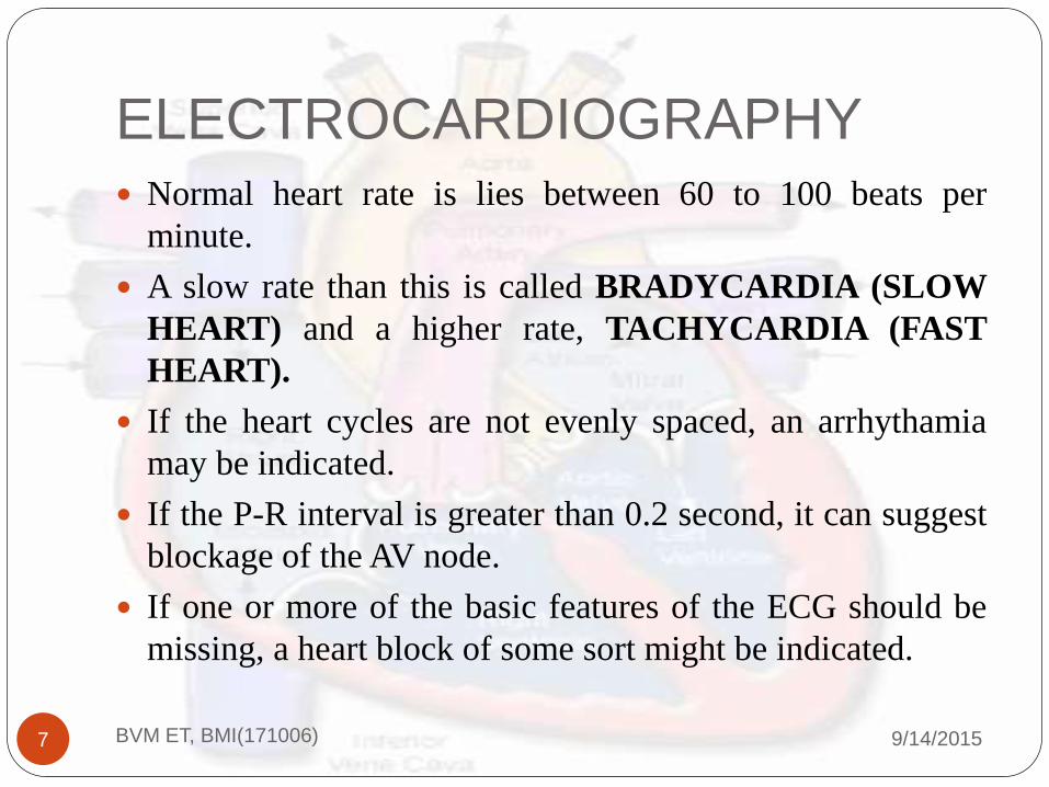
Normal heart rate is lies between 60 to 100 beats per
minute.
A slow rate than this is called BRADYCARDIA (SLOW
HEART) and a higher rate, TACHYCARDIA (FAST
HEART).
If the heart cycles are not evenly spaced, an arrhythamia
may be indicated.
If the P-R interval is greater than 0.2 second, it can suggest
blockage of the AV node.
If one or more of the basic features of the ECG should be
missing, a heart block of some sort might be indicated.

ELECTROCARDIOGRAPHY
9/14/2015BVM ET, BMI(171006)8
When we use electronic amplifiers to measure bioelectricpotentials, interference problem occur due to groundthrough their power supply.
To reduce this, differential amplifier are used.
A differential amplifier can be considered as two amplifierswith separate inputs but with common output terminal,which deceivers the sum of the two amplifier outputvoltages.
The ratio of the differential gain to the common mode gainis called the common-mode rejection ratio of thedifferential amplifier, which in modern amplifiers can be ashigh as 1000000:1.

ELECTROCARDIOGRAPHY
9/14/2015BVM ET, BMI(171006)9
Fig.6.2.1: Differential amplifier with common out put

ELECTROCARDIOGRAPHY
9/14/2015BVM ET, BMI(171006)10
Fig.6.2.2:Differential amplifier used to amplify the bioelectric signals

ELECTROCARDIOGRAPHY
9/14/2015BVM ET, BMI(171006)11
Electrodes and Leads:
To record an electrocardiogram, a number of electrodes,
usually five, are affixed to the body of the patient. The
electrodes are connected to the ECG machine by the same
number of electrical wires known as leads.
The electrode applied to the right leg of the patient, for
example, is called RL lead.
To avoid ambiguity between electrodes and the measuring
techniques, we use lead term for the particular group of
electrodes taken for the measurements.

ELECTROCARDIOGRAPHY
9/14/2015BVM ET, BMI(171006)12
For individual lead wire, as well as the physical connection
to the body of the patient, the term electrode will be used.
When ECG recorded by using certain placement of the
electrode, may be some aspects of the waveform missed.
To avoid this problem, usually 12 different leads techniques
are used so that no important detail of the waveform is
missed.
Placement of electrodes and names and configurations of
the leads have become standardized and are used the same
way throughout the world.
ELECTROCARDIOGRAPHY
9/14/2015BVM ET, BMI(171006)13

ELECTROCARDIOGRAPHY
9/14/2015BVM ET, BMI(171006)14
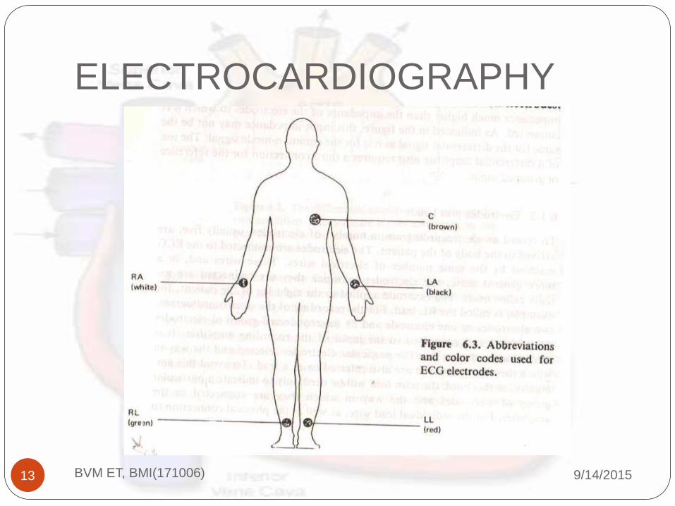
Electrodes: The placement of the electrodes, as well as
the color code use to identify each electrode, is shown
in Figure 6.3.
Mr. Einthoven had found better results using the
electrodes placements at specific locations.
As a ground reference, we can use right leg.(It can be
anywhere but it became convention to use right leg).
Chest or precordial electrodes will be introduced later.

ELECTROCARDIOGRAPHY
9/14/2015BVM ET, BMI(171006)15
Leads: The placement of the electrodes as shown in
figure 6.3.
Because the input of the ECG recorder has only two
terminals, a selection must be made among the
available active electrodes.
The 12 standard leads used most frequently are shown
in upcoming figure 6.4.
The three bipolar limp lead selections first introduced
by Einthoven.

ELECTROCARDIOGRAPHY
9/14/2015BVM ET, BMI(171006)16
LEAD CONNECTIONS
LEAD-I Left Arm(LA) and Right Arm (RA)
LEAD-II Left Leg (LL) and Right Arm (RA)
LEAD-III Left leg (LL) and Left Arm (LA)
• These three leads are called bipolar because for
each lead the ECG is recorded from two electrodes
and third electrode is not connected.
•In each of these lead positions, the QRS of a
normal heart is such that the R wave is positive.

ELECTROCARDIOGRAPHY
9/14/2015BVM ET, BMI(171006)17
Figure.6.4.1. Three lead techniques for measurement of ECG
ELECTROCARDIOGRAPHY
9/14/2015BVM ET, BMI(171006)18
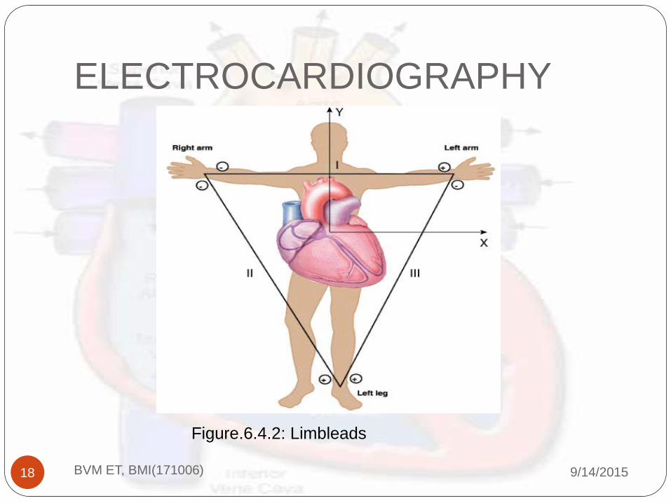
Figure.6.4.2: Limbleads

ELECTROCARDIOGRAPHY
9/14/2015BVM ET, BMI(171006)19
Einthoven made few assumption related to the cardiacactivity and ECG.
He said that at any given instant of the cardiac cycle,the frontal plane representation of the electrical axis ofthe heart is two dimensional vector.
The ECG measured from any three limb leadtechniques is time variant single dimensionalcomponent of that vector.
He also said that the heart is near the center of anequilateral triangle, the apexes of which are the rightand left shoulders and the crotch.

ELECTROCARDIOGRAPHY
9/14/2015BVM ET, BMI(171006)20

ELECTROCARDIOGRAPHY
9/14/2015BVM ET, BMI(171006)21
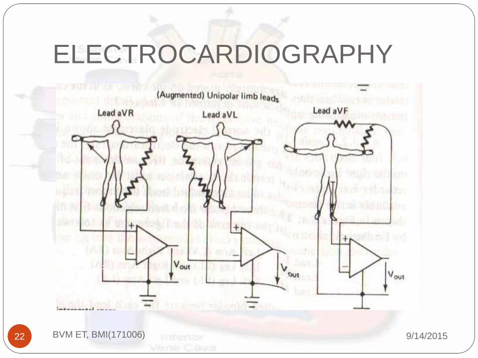
The other leads are known as unipolar type, which wasintroduced by Wilson in 1994.
For unipolar leads, the ECG is recorded between singleexploratory electrode and the central terminal, whichhas a potential corresponding to the center of the body.Three active limb electrodes together through resistorsof equal size.
In augmented unipolar limb leads, the electrode usedas an exploratory electrode is not used for the centralterminal. These leads are aVR, aVL and aVF.(seeupcoming figure)
ELECTROCARDIOGRAPHY
9/14/2015BVM ET, BMI(171006)22

ELECTROCARDIOGRAPHY
9/14/2015BVM ET, BMI(171006)23
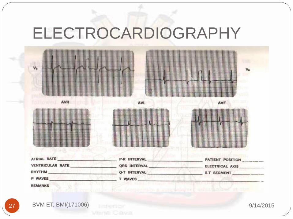
The remaining leads are unipolar chest leads.
These chest positions are called the precordial unipolar
leads and are designated V1 through V6.
All three active limb electrodes are used to obtain the
central terminal, while a separate chest electrode is
used as an exploratory electrode.
We will see the connection type of the precordial
unipolar leads also we will see the various ECG wave
forms for the particular patient by using different leads
techniques in upcoming diagrams.

ELECTROCARDIOGRAPHY
9/14/2015BVM ET, BMI(171006)24

ELECTROCARDIOGRAPHY
9/14/2015BVM ET, BMI(171006)25

ELECTROCARDIOGRAPHY
9/14/2015BVM ET, BMI(171006)26
ELECTROCARDIOGRAPHY
9/14/2015BVM ET, BMI(171006)27
ELECTROCARDIOGRAPHY
9/14/2015BVM ET, BMI(171006)28
Some special modified leads are also used for the ECG
measurements. The most widely used modification for
ongoing ECG monitoring is modified chest lead I
(MCL1) also called the marriott lead, named after its
inventor.
For this lead technique, the placement of the electrodes
on the body is different.
Recordings obtained in this techniques are very useful
in differentiating left ventricular ectopic rhythms from
aberrant right ventricular or super ventricular rhythms.
ELECTROCARDIOGRAPHY
9/14/2015BVM ET, BMI(171006)29

ELECTROCARDIOGRAPHY
9/14/2015BVM ET, BMI(171006)30

ELECTROCARDIOGRAPHY
9/14/2015BVM ET, BMI(171006)31
Fig.6.9: Single channel ECG
Recorder

ELECTROCARDIOGRAPHY
9/14/2015BVM ET, BMI(171006)32
Fig.6.10: Three channel ECG Recorder.

ELECTROCARDIOGRAPHY
9/14/2015BVM ET, BMI(171006)33
The other ECG Recorders are:
Vector Electrocardiograph (vectorcardiograph)
Electrocardiograph system for stress testing
Electrocardiograph for computer processing
Continuous ECG recording (Holter Recording)

MEASUREMENT OF BLOOD
PRESSURE
9/14/2015BVM ET, BMI(171006)34
Blood Pressure is considered a good indicator of thestatus of the cardiovascular system.
Measurement of BP saved many life by indicating theHypertension.
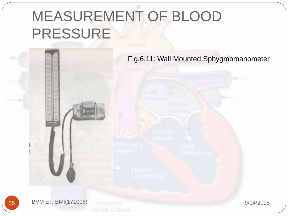
In clinical measurement its usually measured usingindirect measuring instrument calledsphygmomanometer (greek word sphygmos meaningpulse).
The demerit of this system are:
We cant get the continuous reading and the detail waveforms of the BP also cant me taken.
MEASUREMENT OF BLOOD
PRESSURE
9/14/2015BVM ET, BMI(171006)35
Fig.6.11: Wall Mounted Sphygmomanometer

MEASUREMENT OF BLOOD
PRESSURE
9/14/2015BVM ET, BMI(171006)36
Basically two types of Blood Pressure measurements
are in use.
Indirect Measurements
Direct Measurements.
Here, we are going to discuss First Indirect
Method(Noninvasive), than some automatic methods
of measuring BP and after that we are going to discuss
Direct measurement(Invasive ) techniques.

MEASUREMENT OF BLOOD
PRESSURE
9/14/2015BVM ET, BMI(171006)37
Indirect Measurements:
Well known method is using sphygmomanometer and the
stethoscope.
The sphygmomanometer is consists of an inflatable
pressure cuff and mercury or aneroid manometer to
measure the pressure in the cuff.
The cuff consists of a rubber bladder inside and inelastic
fabric covering that can wrapped around the upper arm.
This meter is works on the principle that when the cuff is
placed on the upper arm and inflated, arterial blood can….

MEASUREMENT OF BLOOD
PRESSURE
9/14/2015BVM ET, BMI(171006)38
Flow past the cuff only when the arterial pressure
exceeds the pressure in the cuff.
When the cuff is inflated to a pressure that only
partially occludes the brachial artery, turbulence is
generated in the blood as it spurts through the tiny
arterial opening during each systole.
The sound generated by this turbulence (Korotkoff
sound) can be heard through a stethoscope placed over
the artery downstream from the cuff.

MEASUREMENT OF BLOOD
PRESSURE
9/14/2015BVM ET, BMI(171006)39
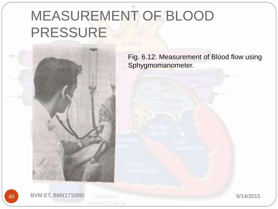
Use of stethoscope while we measure the blood pressure.
When the cuff pressure reaches above the systole level, we
can not hear any sound in stethoscope.
Once the valve release the air, cuff pressure reduces
continuously, at one point the when the blood spurt from
the vessel, we can here one sound and that measure is know
as a systolic pressure.
Now we continuously decreases the pressure of the cuff, up
to certain limit we can able to here the sound, but when the
sound disappear that pressure noted down as a diastolic
blood pressure.
MEASUREMENT OF BLOOD
PRESSURE
9/14/2015BVM ET, BMI(171006)40
Fig. 6.12: Measurement of Blood flow using
Sphygmomanometer.

MEASUREMENT OF BLOOD
PRESSURE
9/14/2015BVM ET, BMI(171006)41
This familiar method of locating the systolic and
diastolic pressure values by listening to the korotkoff
sound is called the auscultatory method of
sphygmomanometery.
An alternative method is known as the palpatory
method, by which the physician identifies the flow of
the blood in the artery by feeling the pulse of the
patient downstream from the cuff.
Diastolic pressure is more difficult to measure using
this technique.

MEASUREMENT OF BLOOD
PRESSURE
9/14/2015BVM ET, BMI(171006)42
Automated Indirect Methods.
The method of measurement is same as the ausculatory
indirect method which is done manually but here, the
pressure transducer is connected to cuff and for the
sound recording microphone is placed beneath the
cuff(Over the artery).
The pressure cuff is automatically inflated to about 220
mm Hg and allowed to deflate slowly.
This instrument is still semiautomatic because the
recording must be interpreted by the observer.

MEASUREMENT OF BLOOD
PRESSURE
9/14/2015BVM ET, BMI(171006)43
False indications are there due to motion artifacts.
The fully automated machine are recording the firstand last sound and also preserve the display indicationof both the pressure value.
This type of machine are very much user-friendly andavailable in market.
While doing some activity, many of the automaticmeasuring instruments shows errors in readings.
So, some advance machine are invented. HereProgrammed electrosphygmomanometer PE-300 willbe discussed.

MEASUREMENT OF BLOOD
PRESSURE
9/14/2015BVM ET, BMI(171006)44

MEASUREMENT OF BLOOD
PRESSURE
9/14/2015BVM ET, BMI(171006)45
In PE-300, we can simultaneously measure the soundof blood or pulse and pressure value.
This can be done repeatedly by adjusting the timeintervals.
There are sound sensors, pressure sensors, controllingunit for pump and valve with comparator circuit isused as shown in the diagram.
We can use one more advanced Pressure measuringunit for indirect measurement which gives a MAP(mean arterial pressure) which can also be very usefulinformation related to blood pressure.

MEASUREMENT OF BLOOD
PRESSURE
9/14/2015BVM ET, BMI(171006)46

MEASUREMENT OF BLOOD
PRESSURE
9/14/2015BVM ET, BMI(171006)47
The value of MAP can be found out by using the
simple equation.
Most of the electrical monitor provides both diagnostic
systolic/diastolic waveforms information and added
option of a single value MAP indication.
Some instruments are using large gauge for easy
reading.( See the diagram 6.15)
1( )
3MAP systolic diastolic diastolic

MEASUREMENT OF BLOOD
PRESSURE
9/14/2015BVM ET, BMI(171006)48
MEASUREMENT OF BLOOD
PRESSURE
9/14/2015BVM ET, BMI(171006)49
Inflation and Deflation of Cuff can be done manually,
so this instrument is called semiautomatic electronic
sphygmomanometer.
In Dinamap the MAP can be measure using
oscillometric method.
See the diagram of the Dinamap in figure 6.16.
Some advanced ambulatory automatic blood pressure
monitor are also very useful which uses the halter
method for the monitoring.

MEASUREMENT OF BLOOD
PRESSURE
9/14/2015BVM ET, BMI(171006)50

MEASUREMENT OF BLOOD
PRESSURE
9/14/2015BVM ET, BMI(171006)51
Using this type of the blood pressure monitor we
can able to record around 26 hours of blood
pressure values in magnetic tape. The controlling
unit is always taking care of time interval and
inflation deflation of the cuff.
The recording can be read with in 12 minutes by
using specific scanner and abnormal reading of
pressure can be automatically given by the
machine.
The advantage of this unit is that, it can also be
used for the ECG measurements.
We can see the diagram in upcoming figure. 6.17

MEASUREMENT OF BLOOD
PRESSURE
9/14/2015BVM ET, BMI(171006)52

MEASUREMENT OF BLOOD
PRESSURE
9/14/2015BVM ET, BMI(171006)53
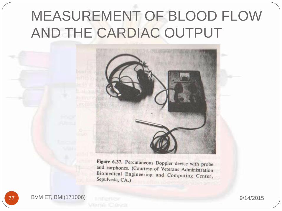
Another approach utilizes ultrasound to measure thepulsatile motion of the brachial artery wall.
High frequency sound energy is transmitted into thepatients arm and is reflected back from the arterialwalls.
By means of Doppler effect, the movement of thearterial walls can be detected as they snap open andclosed with each pulsation of blood.
The advantage of this type of instrument is that resultscloser to direct instruments can be obtain. Its good touse this instruments for the patient under shock.

MEASUREMENT OF BLOOD
PRESSURE
9/14/2015BVM ET, BMI(171006)54
Direct measurements.
First experiment done in1728 by Hales by inserting
glass tube into the artery of horse.
That continuous measurement is taken and is called
kymograph.
Recently the strain gage used for the measurement and
kymograph where piezo sensors are used.
There are specifically three methods for Direct
measurements.

MEASUREMENT OF BLOOD
PRESSURE
9/14/2015BVM ET, BMI(171006)55
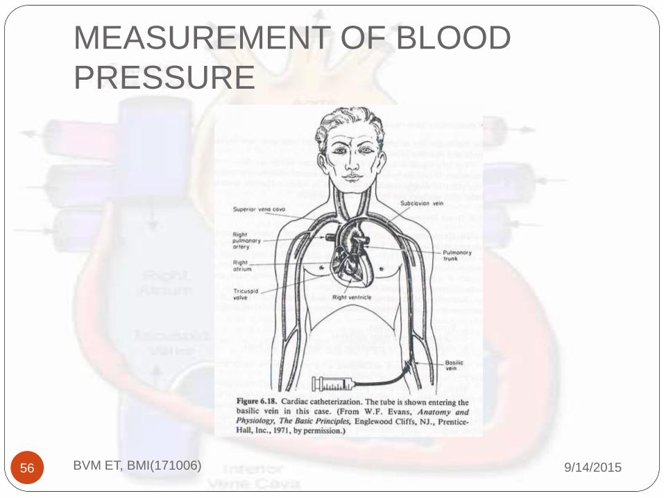
1. Percutaneous insertion
2.Catheterization (Vessel cutdown)
3. Implantation of a transducer in a vessel or in theheart.
For first two methods we can see the diagram number6.18.
In that one before percutaneous insertion, a localanesthetic injected near the site of invasion.
Catheter is fed through the hollow needle.
The blood pressure sensed directly by attaching atransducer to the tube.
MEASUREMENT OF BLOOD
PRESSURE
9/14/2015BVM ET, BMI(171006)56

MEASUREMENT OF BLOOD
PRESSURE
9/14/2015BVM ET, BMI(171006)57
Catheter is used in many measurements.
Using catheterization we can do measurement by
using two methods.
1. After insertion of saline solution we can check
the pressure outside the body using the
transducer.
Or we can attach a transducer at the tip of the
catheter and get the direct measurements.
Implantation techniques involves major surgery
so that method is normally used for experimental
purpose only.

MEASUREMENT OF BLOOD
PRESSURE
9/14/2015BVM ET, BMI(171006)58
Specific direct measurement techniques.
Basically two types.
1. By the clinical method
2.By the electrical principle involved.
There are different types of blood pressure
measuring transducers are used for various
methods.
Here we are going to see the diagrams of the
different transducers.

MEASUREMENT OF BLOOD
PRESSURE
9/14/2015BVM ET, BMI(171006)59

MEASUREMENT OF BLOOD
PRESSURE
9/14/2015BVM ET, BMI(171006)60

MEASUREMENT OF BLOOD
PRESSURE
9/14/2015BVM ET, BMI(171006)61
MEASUREMENT OF BLOOD
PRESSURE
9/14/2015BVM ET, BMI(171006)62

MEASUREMENT OF BLOOD
PRESSURE
9/14/2015BVM ET, BMI(171006)63

MEASUREMENT OF BLOOD
PRESSURE
9/14/2015BVM ET, BMI(171006)64

MEASUREMENT OF BLOOD
PRESSURE
9/14/2015BVM ET, BMI(171006)65

MEASUREMENT OF BLOOD
PRESSURE
9/14/2015BVM ET, BMI(171006)66

MEASUREMENT OF BLOOD FLOW
AND THE CARDIAC OUTPUT
9/14/2015BVM ET, BMI(171006)67
All the organs required an adequate amount of blood.
So, by measuring the flow rate of the blood we can
diagnosis the diseases.
We can not use the industrial principles to measure the
flow rate of liquid because here we could not cut the
Wessel every time.
So some basic principles on which the measurements
are to be done are described.
MEASUREMENT OF BLOOD FLOW
AND THE CARDIAC OUTPUT
9/14/2015BVM ET, BMI(171006)68
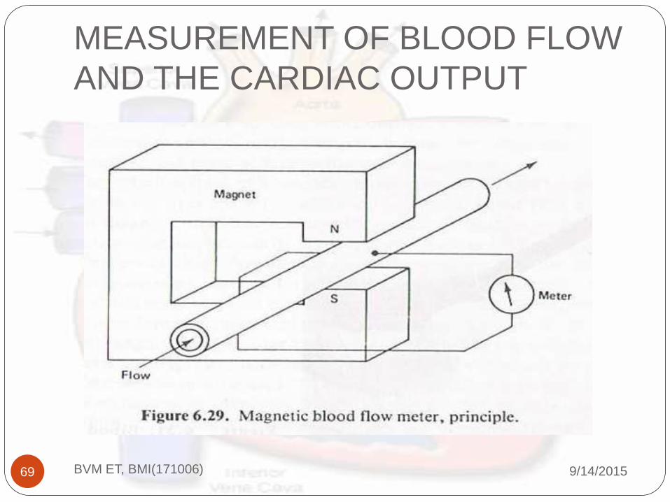
Electromagnetic Induction
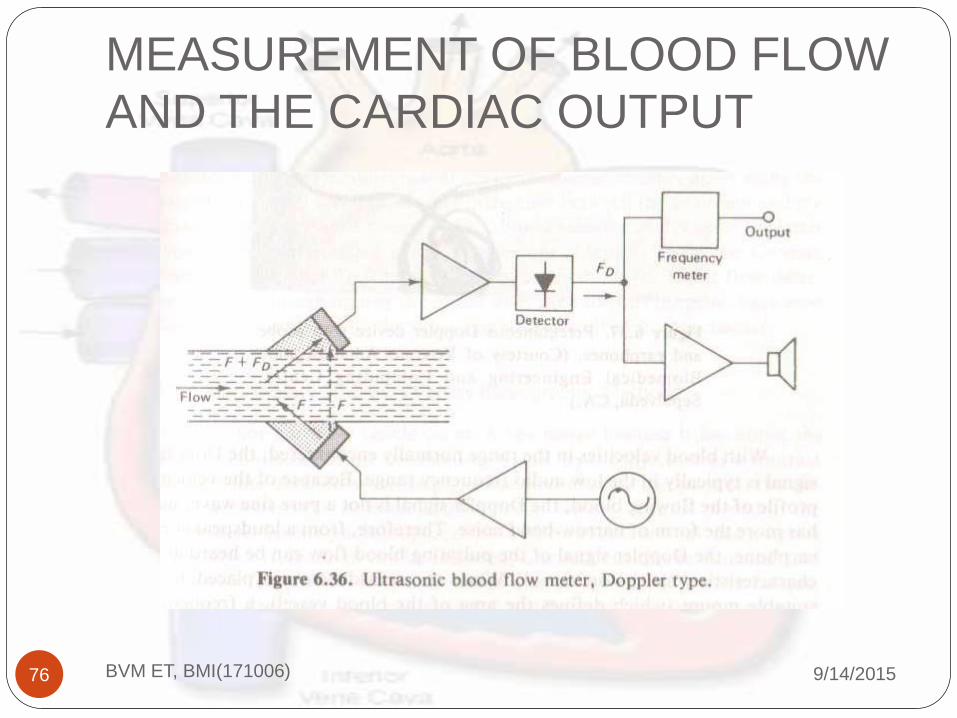
Ultrasound transmission or reflection
Thermal Convection
Radiographic Principles
Indicator dilution
MEASUREMENT OF BLOOD FLOW
AND THE CARDIAC OUTPUT
9/14/2015BVM ET, BMI(171006)69

MEASUREMENT OF BLOOD FLOW
AND THE CARDIAC OUTPUT
9/14/2015BVM ET, BMI(171006)70

MEASUREMENT OF BLOOD FLOW
AND THE CARDIAC OUTPUT
9/14/2015BVM ET, BMI(171006)71

MEASUREMENT OF BLOOD FLOW
AND THE CARDIAC OUTPUT
9/14/2015BVM ET, BMI(171006)72

MEASUREMENT OF BLOOD FLOW
AND THE CARDIAC OUTPUT
9/14/2015BVM ET, BMI(171006)73
MEASUREMENT OF BLOOD FLOW
AND THE CARDIAC OUTPUT
9/14/2015BVM ET, BMI(171006)74

MEASUREMENT OF BLOOD FLOW
AND THE CARDIAC OUTPUT
9/14/2015BVM ET, BMI(171006)75
MEASUREMENT OF BLOOD FLOW
AND THE CARDIAC OUTPUT
9/14/2015BVM ET, BMI(171006)76
MEASUREMENT OF BLOOD FLOW
AND THE CARDIAC OUTPUT
9/14/2015BVM ET, BMI(171006)77

MEASUREMENT OF BLOOD FLOW
AND THE CARDIAC OUTPUT
9/14/2015BVM ET, BMI(171006)78

MEASUREMENT OF BLOOD FLOW
AND THE CARDIAC OUTPUT
9/14/2015BVM ET, BMI(171006)79
MEASUREMENT OF BLOOD FLOW
AND THE CARDIAC OUTPUT
9/14/2015BVM ET, BMI(171006)80
PLATHYSMOGRAPHY
9/14/2015BVM ET, BMI(171006)81
Instruments measuring volume changes or providing
outputs that can be related to them are called
‘Plethysmographs’, and measurement of these volume
changes, or phenomena related thereto, is called
plethysmography.
The upcoming diagrams shows a various techniques
for plethysmogrphy.

PLATHYSMOGRAPHY
9/14/2015BVM ET, BMI(171006)82

PLATHYSMOGRAPHY
9/14/2015BVM ET, BMI(171006)83
PLATHYSMOGRAPHY
9/14/2015BVM ET, BMI(171006)84

PLATHYSMOGRAPHY
9/14/2015BVM ET, BMI(171006)85

PLATHYSMOGRAPHY
9/14/2015BVM ET, BMI(171006)86

PLATHYSMOGRAPHY
9/14/2015BVM ET, BMI(171006)87

MEASUREMENT OF HEART
SOUND
9/14/2015BVM ET, BMI(171006)88
The sound technology by which we can diagnosis thedisease related to heart its known asphonocardiography.
Initially the method was by putting ears on the chest ofthe patient.
Then after the stethoscopes are invented.
Now the electronics stethoscopes are also in use.
We can record the frequency spectrum using thephonocardiogram.
The upcoming waveform shows one of the output ofthe phonocardiogram machine.

MEASUREMENT OF HEART
SOUND
9/14/2015BVM ET, BMI(171006)89

QUESTIONS?
9/14/2015BVM ET, BMI(171006)90
?

REFERENCE
9/14/2015BVM ET, BMI(171006)91
Book: “Biomedical instrumentation and
measurements “ ,by L. Cromwell, F .Weibell, and
E. Pfeiffer. PHI publication 2nd Edition.

9/14/2015BVM ET, BMI(171006)92
THANK YOU