gt ds
TRANSCRIPT

GESTATIONAL TROPHOBLASTIC DISEASE

INTRODUCTION
•GTD forms a spectrum of interrelated tumors which arise in the epithelia of the trophoblast.
• Trophoblastic tumors are either benign, potentially malignant or malignant tumors and they show various grades of differentiation from recognizable chorionic villi structure to highly virulent anaplastic masses of cells.

The malignant GTD’s whether locally invasive or metastatic, even with wide
spread dissemination can be cured completely, “Cure rate approaches
100% in most GTD’s

The tumors spectrum consist of:
•Hydatiform mole (molar pregnancy)
•Invasive mole
•Placental site trophoblastic tumor (PSTT)
•Choriocarcinoma

•The tumors elaborate a unique and characteristic tumor marker, “human chorionic gonadotropin (hCG)”
•Biological activity and clinical behavior of the tumor are indication used to prognosticate the patient

•Most of the GTD’s are non-cancerous (80%).
•The GTD’s arise from Trophoblastic tissues which form the placenta and fetal membranes. The tissues continue to grow but the fetus doesn’t develop

EPIDEMIOLOGY
•Estimates of the incidence of GTD’s vary dramatically in different regions of the world.
•They have been a striking geographical distribution of GTD’s across the world.
• In Europe and North America are rare

• More common in Middle East and Asia, Malaysia, Singapore, Hong Kong, Indonesia, Philippines and China.
• The incidence is also relatively high in Central Africa
•But they are found all around the world.

•Hydatiform mole (Molar pregnancy) is the most common GTD encountered in clinical practice.
• It occurs in 1 out of 125 deliveries in Mexico, 1 out of 1500 deliveries in US. The incidence also seems to be higher among women who are under 20 and over 40

•Invasive mole is reported in 10 -15% of patients who have had a primary molar pregnancy.
•PSTT is a rare tumor that arises from the placental site.

Choriocarcinoma are rare. Reported in 2 – 5% of all cases of GTD’s. The
incidence in the US is about 1: 40,000 pregnancies but reported to be higher
in Asia

PATHOLOGY

HYDATIFORM MOLE (MOLAR PREGNANCY)
Is an abnormal pregnancy characterized grossly by multiple
grape vesicles filling and distending the uterus

Molar pregnancy

•They are neoplasia of the trophoblast which involves both the epithelial layers (cytotrophoblast and syncytiotrophoblast) in different proportions.
•The disease is usually benign and non-metastatic in nature

•Molar pregnancies are categorized as partial or complete in the basis of gross morphology, histopathology and karyotype.
•Most of the moles have been found to be female.

COMPLETE MOLE• Lack identifiable embryonic or fetal tissues and the
chorionic villi exhibit generalized hydatiform swelling and diffuse trophoblastic hyperplasia
The molar chromosomes are entirely of paternal origin.

COMPLETE MOLE
• Empty ovum + 23X sperm → 23X →duplicate → 46XX
• Empty ovum + 23X and 23X → 46XX
• Empty ovum + 23X and 23Y → 46XY

•The cysts vary in size from a pin-head to a cherry. No fetal tissue can be identified.
•The molar chromosomes are entirely of paternal origin.

PARTIAL MOLES• 23X ovum +23X sperm = 69XXX
23X
• 23X ovum + 23Y sperm =69XYY
23Y sperm
• 23X ovum + 23X sperm =69XXY
23Y

PARTIAL MOLE
•Hydatiform change in the placenta may take place without the death of the fetus (rare phenomenon).
•Generally partial moles have a triploid karyotype (69 x’somes), the extra haploid set of chromosomes from the father

Partial mole

PARTIAL MOLE• In twin pregnancy one of the conceptus may be a
partial mole and the other being a normal conceptus. The normal conceptus develops normally to maturity.
• The disease usually arises in very early pregnancy so that the fetus remains as only as a rudimentary structure

PARTIAL MOLE
•Fetuses which survive exhibit a stigma of triploidy like IUGR and congenital malformations like hydrocephalus.
•Occasionally normal birth have however been reported in partial moles.

RISK FACTORS• Maternal age
• Previous molar pregnancy
• Parity
• Nutritional deficiencies of carotene, folate and proteins.
• Paternity
• ?Contraceptive use
• Life style-smoking and alcohol

CLINICAL FEATURES
Symptoms of complete mole are at first those of early pregnancy but
with development of a mole

The general reactions to pregnancy are exaggerated;
•Excessive nausea and vomiting•Wt loss and ill feeling
-Pre-eclampsia in the first trimester in about 30% of cases

•Recurrent vaginal bleeding associated with brown discharge in more than 50% of cases.
•Passing vesicles per vaginum

The physical signs
• Uterus too large for gestation age in 50% of cases.
• Uterus is dough and doesn’t contract• Fetal part cant be appreciated

• Fetal heart beets cant be heard• No fetal movements• Bilateral ovarian enlargement (Theca
Lutein cysts) in 25-50% of cases.
-Thyrotoxicosis

PARTIAL MOLES
•Clinical features not so drastic
•Vaginal bleeding is the symptom that is usually seen.
•The rest of the symptoms are only seen in 4% of cases

PARTIAL V/S COMPLETE MOLE
FEATURES COMPLETE PARTIAL
Fetal/embryonic tissue Absent present
Hydatidiform swelling of chorionic villi Diffuse Focal
Trophpblastic hyperplasia Diffuse Focal
Trophoblastic stromal inclussions Absent Present
Karyotype 46XX,46XY triploid
Fetal Rbcs Absent Present
B-hCG level in serum High/>50000 slight?/<50000
Classical symptoms Common Rare
Risk for development of persistent GTD 20-30% <5%

DIAGNOSIS OF MOLAR PREGNANCY
• Clinical features
• USS • The diffuse hydropic swelling of the chorionic villi produces a characteristic snow
storm appearance on USS throughout the uterine cavity.
• No gestation sac can be identified
• In partial moles; Focal cystic spaces are seen in the placental tissue and transverse diameter of the gestation sacs is increased.
• B-hCG levels in the blood: Markedly elevated

Snow storm appearance on USS

TREATMENT
If the mole has expelled itself, manage as incomplete
abortion

• If not expelled;• I/V oxytocin in a drip• cervical dilatation• Evacuate the uterus by suction curate at any
gestation age• Then do a gentle sharp curate after removing the
products by suction•Rh negative women are given ant D antibody
injection.

HYSTERECTOMY
May be done with the mole insitu in women who have completed their families or just
wish the operationAlso if bleeding is uncontrollable.

POST MOLAR PROPHYLAXIS
•Controversial issue
•Complete mole
•Patients are given either Actinomycin-D or Methotrexate

POST MOLAR PROPHYLAXIS
•Criteria being;•Age >35yrs•Previous molar pregnancy•Trophoblastic hyperplasia•Anticipated persistent GTD

FURTHER MANAGEMENT
Material removed at evacuation are always to
be examine microscopically

FOLLOW UP
•Clinical symptoms and signs of persistent GTD should be asked at every visit•Persistent vaginal bleeding post evacuation•Cough•Headache• Jaundice and epigastric pain

FOLLOW UP•B-hCG levels taken weekly till 3 consecutive normal
values then monthly for at least a year.
•Normal levels are expected 9-14 weeks post evacuation.
•Plateauing or rising B-hCG levels calls for attention (possibility of persistent molar pregnancy or malignancy transformation).

MALIGNANT GTD’s

PERSISTENT/MALIGNANT GTD
• INVASIVE MOLE
• PLACENTAL SITE TROPHOBLASTIC TUMOR
• CHORIOCARCINOMA

INVASIVE MOLE
• Is a hydatiform mole that is locally invasive
• It invades the myometrium or adjacent structures
• It may totally penetrate the myometrium and be associated with uterine rupture and haemoperitoneum

INVASIVE MOLE• Microscopic findings are the same those of
hydatidiform mole.
• Develop in up to 15% of patients who have had molar pregnancy though it may infrequently follow any other type of gestation.
• The mole consists of cyto and syncytiotrophoblastic cells

INVASIVE MOLE
•Syncytiotrophoblast produced large amounts of B-hCG and therefore patients present with persistent elevation of B-hCG
•The patient presents with persistent vaginal bleeding

PLACENTAL SITE TROPHBLASTIC TUMOR
• Is a very rare tumor
•Derived from the intermediate trophoblast with minimal or absent syncytiotrophoblastic tissue.
•Histologically, local invasion occurs into the myometrium and lymphatics

PLACENTAL SITE TROPHBLASTIC TUMOR
•Vascular invasion is a rare phenomenon•It may follow any type of gestation

PLACENTAL SITE TROPHBLASTIC TUMOR
•It produces low levels of B-hCG but relatively high amounts of human placental lactogen compared to Choriocarcinoma

PLACENTAL SITE TROPHBLASTIC TUMOR
• It shows a range of behavior from benign with capacity to regress spontaneously to a highly
malignant form which can be highly resistant to chemotherapy and therefore hysterectomy is
recommended route of treatment

CHORIOCARCINOMA
• Is a rare trophoblastic tumor
• Geographical distribution similar to that of Hydatidiform mole.
• Pure epithelial tumor composed of cyto and syncytiotrophoblastic cells

CHORIOCARCINOMA
• It may follow any type of gestation
• In 50% of cases it is preceded by a hydatidiform mole,25% follow an abortion or ectopic pregnancy, and 25% following normal pregnancy
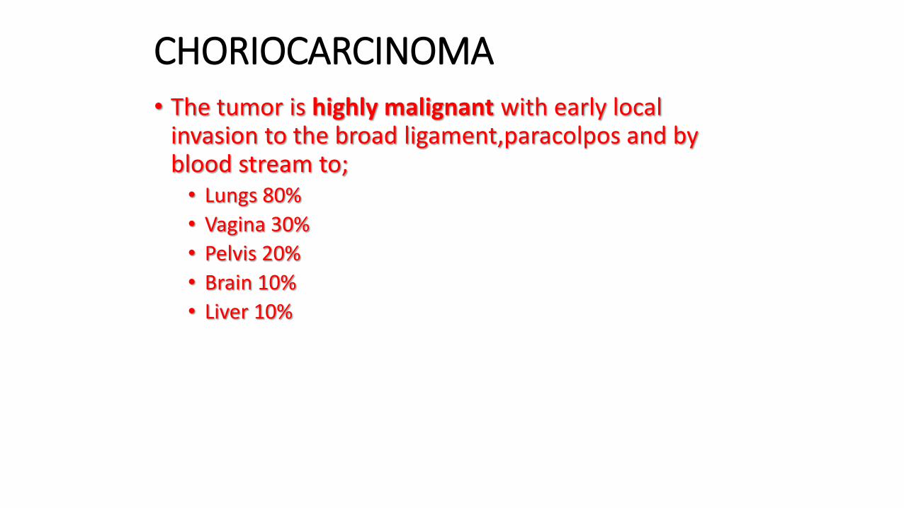
CHORIOCARCINOMA• The tumor is highly malignant with early local
invasion to the broad ligament,paracolpos and by blood stream to;• Lungs 80%
• Vagina 30%
• Pelvis 20%
• Brain 10%
• Liver 10%

PATHOLOGY
•Primary growth is usually in the uterine wall though it may be in the cervix, vagina, tubes or broad ligamentfollowing an ectopic pregnant.
• The tumor is soft, necrotic and hemorrhagic so it appears plum-colored to the naked eye

PATHOLOGY
• On section the tumor shows cyto and syncytiotrophoblastic tissues in varying proportions, actively proliferating and assuming bizarre forms.
• There is also mononucleated and multinucleated giant cells

PATHOLOGY
• Chorionic villi are characterically absent
• Choriocarcinoma is functional secreting large quantities of B-hCG
• B-hcG causes luteinization of the ovaries.
• Choriocarcinoma also secretes some amounts of human placental lactogen

CLINICAL FEATURES
• Irregular haemorrhage-characteristically intermittent but alarming
•Metastatic features• Dyspnoea• Haemothorax• Haemoptysis
-Neurological signs of headache, visual disturbance, focal deficits

CLINICAL FEATURES• CXR reveals cannon balls or snow storm appearance,
pleural effusion etc
• Hepatic metastasis• Epigastric pain or right upper quadrant pain
• Acute emergency massive haemopertonial haemorrhage.
• Jaundice

‘Cannon ball’ x ray appearance

CLINICAL FEATURES• Vaginal metastasis
• Often enlarged uterus
• Palpable ovarian cysts

DIAGNOSIS
• Clinical features
• High levels of B-hCG in serum
• Diagnostic curate

METASTATIC WORK UP
• Abdominal/pelvic ultrasound
• CT scan
• MRI
• Lumbar puncture to demonstrate B-hCG in the CSF

• CLASSIFICATION OF MALIGNANT GTDs

CLASSIFICATION OF PERSISTENT/MALIGNANT GTDs
• NATIONAL CANCER INSTITUTE (USA)
• WHO
• FIGO SYSTEM (STAGING

NATIONAL CANCER INSTITUTE• Ultilized in the US to determine if the patient is in the
poor or good prognosis to respond well to single-agent chemotherapy

NATIONAL CANCER INSTITUTE
• Non metastatic disease; no evidence of disease outside the uterus.
• Metastatic diseases; evidence of disease outside the uterus

Metastatic diseases
• Good prognosis• Short duration (<4months)
• Serum B-hCG <40,000mIU/ml
• No metastasis to brain or liver
• No significant prior chemotherapy

Metastatic diseases
• Poor prognosis
• Long duration (>4months)
• Serum B-hCG >40,000mIU/ml
• Metastasis to brain or liver
• Unsuccessful prior chemotherapy
• GTN following a term pregnancy

RIVISED FIGO STAGING
I. Disease confined to the uterus
II. Disease extending outside the uterus but confined to the genital structures(adenexa,vagina,broadligament)
III. Disease extending to the lungs with or without a known genital tract involvement
IV. Disease at other metastatic sites

RIVISED FIGO STAGING
• A. No risk factor
• B.One risk factor
• C.Two risk factor

RIVISED FIGO STAGING
RISK FACTORS
• 1. B-hCG >100,000mIU/ml
• 2. Duration from termination of antecedent pregnancy to diagnosis>6months

WHO PROGNOSTIC SCORING
parameter 0 1 2 3
Age <39 >39
Antecedent pregnancy Mole Abortion Term preg
Interval in months <4 4-6 7-12 >12
Pretreatment B-hCG 1000 1000-10000 10000-
100000>100000
ABO (F/M) A*O,A*O B,AB
Largest tumor(cm) 3-5 >5
Sites of mets Spleen,kidne
y
GIT,liver Brain
Number of mets 1-4 4-8 >8
Prior chemotherapy failed single >2

WHO PROGNOSTIC SCORING
• Interval=time between termination of antecedent pregnancy and initiation of chemotherapy.
• Score <5=low risk,5-7=medium risk,>7=high risk

TREATMENT OF MALIGNANT GTD
• CHEMOTHERAPY

PRE-TREATMENT WORK UP;
• FBP
• RFT
• LFT
• Bone marrow
• B-hCG levels

Non metastatic and low risk/good prognosis patients
• Methotraxate i/m .30-60mg/m2 once a week*
• Methotraxate i/v or i/m 0.4mg/kg/day for 5days. Repeated every 14days.
• Methotraxate i/m 1mg/kg/day on day 1, 3,5,7,9 and folinic acid 0.1mg/kg i/m on day 2,4,6,8 repeated every 15-18days

Non metastatic and low risk/good prognosis patients
• Dactinomycin 1.25mg/m2 every 14days
• Dactinomycin 10-12ug/kg/day i/v for 5days repeat every 14days

FOLLOW-UP
• Follow B-hcG weekly. Switch to alternative drug if levels rise 10 fold or more or if plateau or new metastasis.
• Obtain weekly CBC. Hold chemotherapy if WBC<3000(Neut <1500), platelets<100000

FOLLOW_UP• Obtain weekly RFT/LFT. Hold chemotherapy when
elevation of BUN,Cr,AST,ALT Bilirubin or significant side effects severe stomatitis,GIT ulceration of severe anaemia or febrile course

FOLLOW_UP
• Oral contraceptives taken at least one yr after remission.
• Chemotherapy cont for one or two causes after negative B-hCG.
• Physical exam

*Follow up B hCG program
• B-hCG wkly until 3 consecutive negative titres, monthly for 12months, then every 2months for 1additional yr.

Metastatic and poor prognosis
• MAC regimen

EMA/CO
• Etoposide, methotraxate, actinomycinD, cyclophosphamyde and vincristine

EMA/CO REGIMEN
• Day 1 –etoposide i/v100mg/m2 slowly infused,actinomycynD i/v0.5mg bolus,MTX i/v100mg/m2bolus then infuse 200mg/m2 over 12hrs

EMA/CO REGIMEN
• Day 2 Etoposide 100mg/m2 i/v slow over30minute, actinomycinD 0.5,gi/v bolus,folinic acid

EMA/CO REGIMEN
• Day 8 cyclophosphamide 600mg/m2 i/v infusion, vincristine 1mg/m2 i/v bolus
*EMA/CO is repeated every 2weeks.

SURGERY
• Stage I cases
• Future fertility not desired
• Resistant to chemotherapy
• PSTT normally resistant to chemotherapy

MODALITIES OF SURGERY
• Hysterectomy
• Thoracotomy
• Hepatic resection or hepatic artery embolization
• Craniotomy

RADIATION THERAPY• Cerebral mets
• Liver mets

SUBSEQUENT PREGNANCIES
• No increased risk of complications like preterm labor, anormalies or still birth.
• Pregnancies should be closely followed up with B-hCG, USS

SUBSEQUENT PREGNANCIES
• After delivery placenta sent to pathologists and B-hCG followed up at 6weeks postpartum.
• Most pregnancies go uneventfully.

Thanks

REFERENCES
• Current diagnosis and treatment
• Jeffcoate’s principles of gynaecology.
• www.emedicine.com