growth of nasal and laryngeal airways in children
TRANSCRIPT

Growth of Nasal and Laryngeal Airways in Children:Implications in Breathing and Inhaled Aerosol Dynamics
Jinxiang Xi PhD, Xiuhua Si PhD, Yue Zhou PhD, JongWon Kim PhD, and Ariel Berlinski MD
BACKGROUND: The human respiratory airway undergoes dramatic growth during infancy andchildhood, which induces substantial variability in air flow pattern and particle deposition. How-ever, deposition studies have typically focused on adult subjects, the results of which cannot bereadily extrapolated to children. We developed models to quantify the growth of human nasal-laryngeal airways at early ages, and to evaluate the impact of that growth on breathing resistanceand aerosol deposition. METHODS: Four image-based nasal-laryngeal models were developedfrom 4 children, ages 10 days, 7 months, 3 years, and 5 years, and were compared to a nasal-laryngeal model of a 53-year-old adult. The airway dimensions were quantified in terms of differentparameters (volume, cross-section area, and hydraulic diameter) and of different anatomies (nose,pharynx, and larynx). Breathing resistance and aerosol deposition were computed using a high-fidelity fluid-particle transport model, and were validated against the measurements made with the3-dimensional models fabricated from the same airway computed tomography images. RESULTS:Significant differences in nasal morphology were observed among the 5 subjects, in both morphol-ogy and dimension. The turbinate region appeared to experience the most noticeable growth duringthe first 5 years of life. The nasal airway volume ratios of the 10-day, 7-month, 3-year, and5-year-old subjects were 6.4%, 18.8%, 24.2%, and 40.3% that of the adult, respectively. Remark-able inter-group variability was observed in air flow, pressure drop, deposition fraction, andparticle accumulation. The computational fluid dynamics predicted pressure drops and depositionfractions were in close agreement with in vitro measurements. CONCLUSIONS: Age effects aresignificant in both breathing resistance and micrometer particle deposition. The image/computa-tional-fluid-dynamics coupled method provides an efficient and effective approach in understand-ing patient-specific air flows and particle deposition, which have important implications in pediatricinhalation drug delivery and respiratory disorder diagnosis. Key words: nasal morphology; child-adult discrepancy; infants; breathing resistance; aerosol deposition; pediatric drug delivery. [RespirCare 2014;59(2):263–273. © 2014 Daedalus Enterprises]
Introduction
Considerable structural changes occur in human respi-ratory tracts during early ages, which lead to vast varia-
tions in breathing disorders that are specific to children.Compared to adults, infants and children are more suscep-
Drs Xi and Kim are affiliated with the Department of Mechanical andBiomedical Engineering, Central Michigan University, Mount Pleasant,Michigan. Dr Si is affiliated with the Department of Engineering, CalvinCollege, Grand Rapids, Michigan. Dr Zhou is affiliated with the Aerosoland Respiratory Dosimetry Program, Lovelace Respiratory Research In-stitute, Albuquerque, New Mexico. Dr Berlinski is affiliated with theDepartment of Pediatrics, University of Arkansas for Medical Science,Little Rock, Arkansas.
Dr Berlinski has disclosed relationships with Johnson & Johnson, MPEXPharmaceutical, Gilead, Philips, Genentech, Vertex, Abvie, and
S&T Technologies. None of their products are discussed in this paper.The other authors have disclosed no conflicts of interest.
Dr Xi presented a version of this paper at the OPEN FORUM of theAARC Congress 2012, held November 10–13, 2012, in New Orleans,Louisiana; for this research he was awarded the 2012 Monaghan-TrudellFellowship for Aerosol Technique Development from the American Re-spiratory Care Foundation.
Correspondence: Jinxiang Xi PhD, Department of Mechanical and Bio-medical Engineering, Central Michigan University, 1200 South FranklinStreet, Mount Pleasant MI 48858. E-mail: [email protected].
DOI: 10.4187/respcare.02568
RESPIRATORY CARE • FEBRUARY 2014 VOL 59 NO 2 263

tible to respiratory distresses, due to their immature de-fense mechanism. Respiratory disease remains a leadingcause of childhood morbidity in the United States and otherdeveloped countries, and is a leading cause of childhooddeaths worldwide. Only by understanding the develop-mental respiratory system can we formulate effective pro-tocols to treat an impaired physiology in pediatric patients.
A number of in vivo studies have been conducted tounderstand the development of respiratory morphology ofearly ages. Summaries of such studies can be found inrecent reviews by Bassham et al1 and Ghuman et al.2 How-ever, these studies mainly focused on the lower respiratorytract. Very few studies have considered the developmentof upper airway morphology.3 Clinical techniques for eval-uating nasal pathology include visual analog scales,4 rhi-nostereometry,5 video-endoscopy,6 rhinomanometry,7 andacoustic rhinometry.8 A combination of the above tech-niques is usually employed in practice, based on the spe-cific performance of each technique.9-12 Compared withclinical studies, computational fluid dynamics predictionshave the advantage of providing detailed information onair flow and aerosol deposition, such as regional dosage,which are more relevant to health outcome than the aver-age deposition. However, to our knowledge, very fewcomputational fluid dynamics studies have been reportedon the nasal morphology of children; one exception isXi et al,13 who examined the upper airway structure andassociated air flow dynamics in a 5-year-old child. Thegeneral neglect of child and infant airways in previousstudies may largely be attributed to limited accessibility ofpediatric medical images to computational fluid dynamicsresearchers, as well as the complexities involved in con-structing physiologically realistic models of nasal passages.
The objective of this study was to characterize thegrowth of the upper airway with models created from im-ages from 4 children, ages 10 days, 7 months, 3 years, and5 years, using a coupled image/computational fluid dy-namics approach. Our specific aims were: to develop an-atomically accurate airway models of children, based oncomputed tomography and magnetic resonance imagingimages; to quantify the airway dimensions; to numericallyand experimentally determine the nasal-laryngeal resis-tance; and to numerically determine the inhaled particledeposition. The results of this study may improve ourunderstanding of developmental respiratory morphologyand its effects on children’s health responses to environ-mental exposures and inhaled therapies.
Methods
Nasal-Laryngeal Airway Models
One example was used to illustrate the computer methodto develop respiratory airway models based on computedtomography and magnetic resonance images. Figure 1
shows the procedures of translating a 2-dimensional mag-netic resonance image (of a healthy 5-year-old boy, weight21 kg, height 109 cm)13 into a 3-dimensional model. Theimage tracings (see Fig. 1A) contained 128 slices, each1.5 mm apart, that spanned from the nostrils to the uppertrachea. The multi-slice tracings were segmented in med-ical image processing (Mimics, Materialise, Leuven, Bel-gium), based on the contrast between soft tissue and in-tranasal air, to isolate the nasal-laryngeal airway, whichwas further converted into a set of cross-sectional contoursthat define the airway. Based on these contours, an internalnasal surface geometry was constructed in engineering-design software (ANSYS Workbench, CAE Associates,Middlebury, Connecticut). The airway model can be eitherprinted with 3-dimensional prototyping techniques for ex-perimental purposes (see Fig. 1B) or discretized into acomputational mesh for numerical analysis (see Fig. 1C).Due to the high complexity of the model geometry, anunstructured tetrahedral mesh was created with high-resolution pentahedral elements in the near-wall region(see Fig. 1C). The respiratory geometry retained in thisexample extended from the nostrils to the upper trachea. Inparticular, anatomical details such as the epiglottal foldand laryngeal sinuses were retained (see Fig. 1A). Theresulting model was intended to accurately represent themorphology of the upper airway, with only minor surfacesmoothing.
Study Design
Age effects on airway morphology, air flow, pressuredrop, and inhaled particle deposition were assessed withnasal-laryngeal models created from computed tomogra-phy scans of 4 children (a 10-day-old female, a 7-month-old female, a 3-year-old female, and a 5-year-old male),seen at Arkansas Children Hospital, and one adult (a 53-
QUICK LOOK
Current knowledge
Aerosol deposition in the respiratory tract is affected byvarious well studied anatomic, equipment, and breath-ing-pattern variables, but most of the data have beenfrom adults.
What this paper contributes to our knowledge
Anatomic differences associated with age affect totaland regional aerosol deposition. There were importantdifferences in airway anatomy, air-flow dynamics, andaerosol deposition between subjects ages 10 days,7 months, 3 years, 5 years, and 53 years.
GROWTH OF NASAL AND LARYNGEAL AIRWAYS IN CHILDREN
264 RESPIRATORY CARE • FEBRUARY 2014 VOL 59 NO 2

year-old male). No health information was disclosed, and theuse of the images was approved by the institutional reviewboard of the University of Arkansas for Medical Science.
Steady inhalations were assumed for all simulations,with a wide spectrum of breathing conditions (1–45 L/min). Comparison of the 5 models under equivalent phys-ical activities (quiet breathing) were conducted, based onpublished respiratory parameters.14-16 Table 1 shows theliterature-based, quiet-breathing respiratory parameters. In-terestingly, the tidal volumes in all 5 models intimatelycorrelated with the nasal-laryngeal airway volumes. Asshown in Tables 1 and 2, relative to the adult model, thetidal volume ratios for the 10-day-old, 7-month-old, 3-year-old, and 5-year-old were, respectively, 4.4%, 17.4%, 26.0%,and 35.4%, while the airway volume ratios were respec-tively 6.4%, 18.8%, 24.2%, and 40.3%. Based on the nos-tril areas in Table 3, the inlet velocities were similar be-tween the 5 models under quiet breathing conditions. Initialparticle velocities were assumed to be the same as thelocal fluid velocity. The airway surface was assumed to besmooth and rigid, with a no-slip (flow velocity at the wall[uwall] � 0) condition.
Pressure Measurement
We measured the pressure drops through the nasal-laryngeal models with a manometer (Magnehelic Gage,Dwyer Instrument, Michigan City, Indiana). The modelnostrils were open to room air, and a vacuum was con-nected to the manometer at the model outlet. We testedconstant inhalation flows between 1 and 48 L/min, byadjusting the valve on the vacuum line. Flow was mea-sured at the model outlet, with a flow meter (4140, TSI,Shoreview, Minnesota) positioned upstream of the flowvalve.
Numerical Method
The low Reynolds number k-� model was selected tosimulate the air flow dynamics, based on its ability toaccurately predict pressure drop, velocity profiles, and shearstress for multi-regime flows.13,17-19 The low Reynoldsnumber k-� model applies to both laminar and turbulentflow regimes. Deposition fraction was calculated as theratio of the amount of particles deposited on the airway
Fig. 1. Nasal-laryngeal airway model development. A: 3-dimensional rendering of computed tomography images of a 5-year-old male for(B) in vitro measurement and (C) numerical analysis. The airway was divided into regions: nasal vestibule and valve region, nasal turbinate,nasopharynx, pharynx, and larynx. The computational mesh is composed of approximately 1.8 million unstructured tetrahedral elementsand a fine near-wall pentahedral grid.
Table 1. Respiratory Parameters Under Quiet Breathing Conditions
10-Day-OldFemale
7-Month-OldFemale
3-Year-OldFemale
5-Year-OldMale
53-Year-OldMale
Breathing frequency, breaths/min 44 25 22 21 12Inspiratory-expiratory ratio 1:3 1:2 1:2 1:2 1:2Inhalation time, s 0.34 0.8 0.9 0.95 1.67Tidal volume, mL 22 87 130 177 500Percent of adult tidal volume 4.4 17.4 26.0 35.4 100Flow, L/min 3.8 6.5 8.6 11.2 18.0Inlet velocity, m/s 1.91 1.84 1.66 1.84 1.48
(From data in References 14–16.)
GROWTH OF NASAL AND LARYNGEAL AIRWAYS IN CHILDREN
RESPIRATORY CARE • FEBRUARY 2014 VOL 59 NO 2 265

surface to the amount of particles entering the nostrils. Thetransport and deposition of inhaled particles are simulatedwith a well tested discrete Lagrangian tracking model,enhanced with near-wall treatment. The inhaled particleswere assumed to be dilute and to have no influence on thecontinuous phase (ie, one-way coupled particle motion). Inour previous studies the Lagrangian tracking model, en-hanced with user-defined routines, was shown to providea close match to experimental deposition data in upperrespiratory airways for both sub-micrometer18 and micro-meter particles.17
To establish grid-independent results, convergencesensitivity analysis was conducted, following the methodsof Xi et al.17 The final grids for reporting flow field con-sisted of 1.6–2.0 million cells, with a thin 5-layer penta-hedral grid in the near-wall region and a cell height of0.05 mm in the first layer (see Fig. 1C). The final numberof particles tracked was 60,000; increasing the number oftracked particles did not alter the deposition fractions.
Results
Airway Morphological and Dimensional Variations
There were substantial morphological and dimensionaldifferences between the 5 nasal-laryngeal airway models(Figs. 2 and 3). Younger subjects have smaller nostrils, ashorter turbinate region, a narrower nasopharynx, and anarrower pharynx-larynx. The nostril shape is more circu-lar at birth, becomes more oval during infancy and child-hood, and becomes wedge-shaped in adulthood.20 Of par-ticular interest is the turbinate region, which seemed to beundeveloped in both the 10-day-old model and the 7-month-old model, and was much simpler in morphology than inour older children and adult models. The inferior meatus ismissing in the 10-day-old. The inferior and middle me-atuses apparently become both larger and more complexwith age (see Fig. 2).
Table 2. Nasal Airway Volume
Airway Volume (cm3)
10-Day-OldFemale
7-Month-OldFemale
3-Year-OldFemale
5-Year-OldMale
53-Year-OldMale
Vestibule and valve 0.79 1.25 1.41 3.37 5.50Turbinate region 1.57 2.83 5.83 11.03 12.63Nasopharynx 0.48 1.74 1.07 3.95 16.33Pharynx 0.31 3.19 3.10 2.64 13.89Larynx 0.36 1.32 1.92 1.22 6.70Total 3.51 10.33 13.32 22.21 55.05Percent of adult value 6.4 18.8 24.2 40.3 100
Table 3. Nasal Airway Surface Area
Airway Surface Area (cm2)
10-Day-OldFemale
7-Month-OldFemale
3-Year-OldFemale
5-Year-OldMale
53-Year-OldMale
Vestibule and valve 7.45 9.75 18.97 23.74 35.58Turbinate region 21.09 35.63 54.71 107.34 112.59Nasopharynx 3.72 9.27 9.79 15.27 40.93Pharynx 2.96 13.71 13.92 14.59 45.10Larynx 2.87 8.37 9.46 7.20 12.81Total 38.09 76.73 106.85 168.14 256.01Ratio, % 22.7 30.0 41.7 65.7 100Inlets
Right nostril 0.166 0.280 0.434 0.492 1.013Left nostril 0.166 0.315 0.431 0.437 1.013Total 0.332 0.595 0.865 0.929 2.026Ratio, % 16.4 29.4 42.7 45.9 100
OutletsTrachea 0.178 0.506 0.671 0.832 1.487Ratio, % 12.0 34.0 45.1 56.0 100
GROWTH OF NASAL AND LARYNGEAL AIRWAYS IN CHILDREN
266 RESPIRATORY CARE • FEBRUARY 2014 VOL 59 NO 2
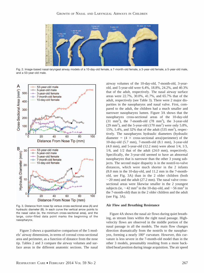
Figure 3 shows a quantitative comparison of the 5 mod-els’ airway dimensions, in terms of coronal cross-sectionalarea and perimeter, as a function of distance from the nosetip. Tables 2 and 3 compare the airway volumes and sur-face areas in the different anatomic sections. The nasal
airway volumes of the 10-day-old, 7-month-old, 3-year-old, and 5-year-old were 6.4%, 18.8%, 24.2%, and 40.3%that of the adult, respectively. The nasal airway surfaceareas were 22.7%, 30.0%, 41.7%, and 65.7% that of theadult, respectively (see Table 3). There were 2 major dis-parities in the nasopharynx and nasal valve. First, com-pared to the adult, the children had a much smaller andnarrower nasopharynx lumen. Figure 3A shows that thenasopharynx cross-sectional areas of the 10-day-old(31 mm2), the 7-month-old (78 mm2), the 3-year-old(29 mm2), and the 5-year-old (170 mm2) were only 5.8%,15%, 5.4%, and 32% that of the adult (535 mm2), respec-tively. The nasopharynx hydraulic diameters (hydraulicdiameter � (4 � cross-sectional area)/perimeter) of the10-day-old (5.7 mm), 7-month-old (8.1 mm), 3-year-old(4.0 mm), and 5-year-old (12.2 mm) were about 1/4, 1/3,1/6, and 1/2 that of the adult (24.0 mm), respectively.Specifically, the 3-year-old seemed to have an abnormalnasopharynx that is narrower than the other 3 young sub-jects. The second major disparity is in the nostril-to-valvedistances, which were much shorter in the 2 infants(8.0 mm in the 10-day-old, and 11.2 mm in the 7-month-old, see Fig. 3A) than in the 2 older children (both�20 mm) and the adult (27.2 mm). The nasal valve cross-sectional areas were likewise smaller in the 2 youngestsubjects (ie, �42 mm2 in the 10-day-old, and �56 mm2 inthe 7-month-old) than in the 2 older children and the adult(see Fig. 3A).
Air Flow and Breathing Resistance
Figure 4A shows the nasal air flows during quiet breath-ing, as stream lines within the right nasal passage. High-velocity flows are observed in the middle portion of thenasal passage in all the models. The main flow changesdirection dramatically from the nostrils to the nasophar-ynx, forming a nearly 180° curvature. However, this cur-vature is less severe in the 7-month-old model than in theother 3 models, presumably resulting from a more back-tilted head position during image acquisition. The air speed
Fig. 2. Image-based nasal-laryngeal airway models of a 10-day-old female, a 7-month-old female, a 3-year-old female, a 5-year-old male,and a 53-year-old male.
Fig. 3. Distance from nose tip versus cross-sectional area (A) andhydraulic diameter (B). In each curve the vertical arrow points tothe nasal valve (ie, the minimum cross-sectional area), and thelarge, color-filled data point marks the beginning of thenasopharynx.
GROWTH OF NASAL AND LARYNGEAL AIRWAYS IN CHILDREN
RESPIRATORY CARE • FEBRUARY 2014 VOL 59 NO 2 267

was higher in the nasopharynx of the 3-year-old, due to thesevere nasopharynx constriction. No recirculation zone wasobserved in the nasopharynx of the 3 young subjects, dueto a much smaller airway diameter in this region, whichdiffers from the adult nasopharynx, in which flow recir-culation is obvious (see Fig. 4A).
Air flow dynamics within the nasal passages are furthervisualized in Figure 4B as snapshots of particle locationsat selected instants. Two thousand 1.0-�m particles werereleased at the right nostril, and particle positions wererecorded at designated time points. Due to their smallinertia, 1.0-�m particles are assumed to closely followthe air flow. Faster transport and deeper penetration ofaerosols are apparent in the medial passages, while slow-moving particles are found near the airway walls. Be-cause of the dramatic airway bend from the nostrils to thenasopharynx, a high-concentration of particles constantlyadjust their directions, following the mean stream-line cur-vature of inhaled air flow. The seemingly random particledistributions in the pharynx indicate enhanced turbulentmixing in this region. Due to the smaller nasopharynx-larynx regions in children, particles are transported fasterand reach the glottal aperture faster than in the adult. Thisdifference was more pronounced in the 3-year-oldmodel, which had an abnormally narrow naso- and velo-pharynx.
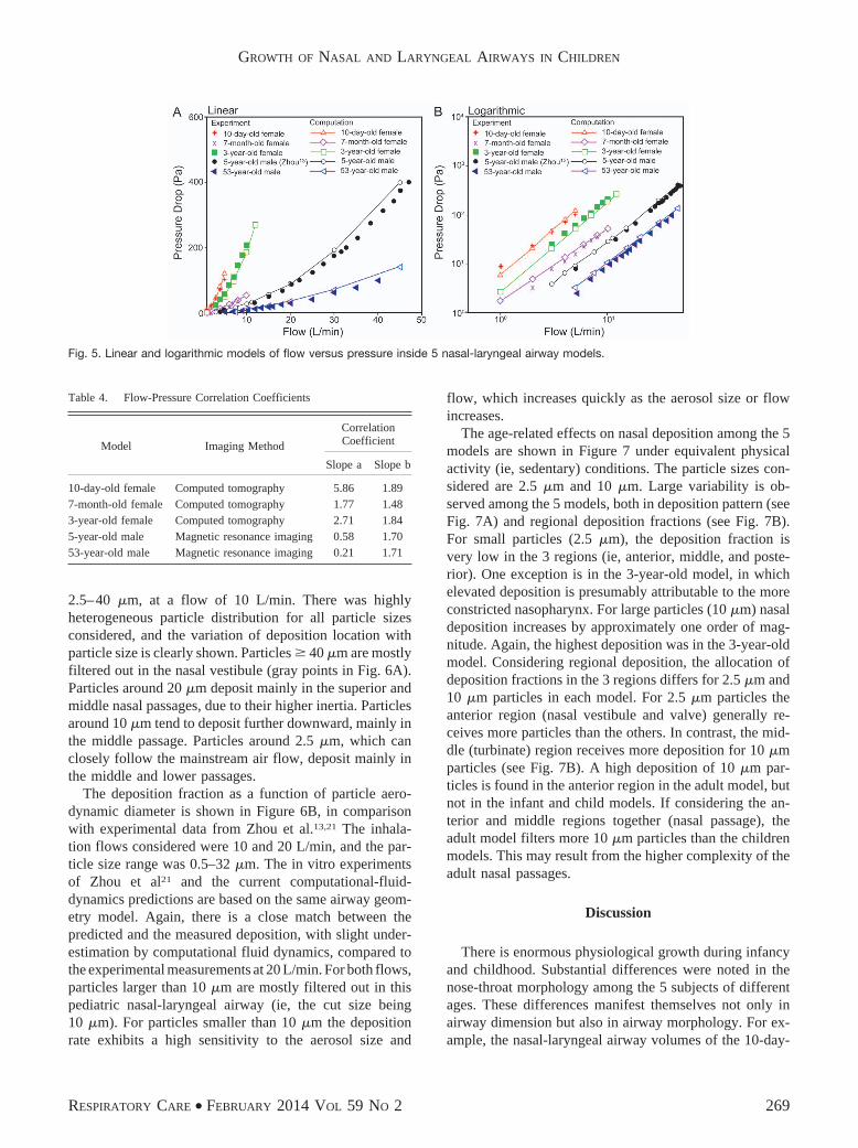
The comparison of measured and computational-fluid-dynamics-predicted pressure drop (�P) for the 5 models isshown in Figure 5, for the flow range 1–48 L/min. All themodels are in close agreement. The in vitro experimentsand the computational-fluid-dynamics-predictions wereconducted in the same airway geometry models, so directcomparison is possible. In general, the pressure drop (ie,breathing resistance) decreased as age increased (seeFig. 5). For a given flow, the infant and children modelshad much higher breathing resistance than the adultmodel. However, it is also not surprising that the 3-year-old model had a much higher pressure drop than the7-month-old model, considering the abnormally con-stricted nasopharynx in the 3-year-old model and the lesssevere airway curvature in the 7-month-old model. Theflow-pressure relationships can be expressed as a powerfunction (�P � a�Qb), which can be plotted as straightlines on a log-log scale with a slope of b, as shownin the logarithmic graph in Figure 5. The best-fitted coef-ficients a and b for the different model ages are listed inTable 4.
Particle Deposition
Particle deposition in the nasal-laryngeal airway of the5-year-old is displayed in Figure 6A, for the particle range
Fig. 4. A: Inhaled-gas stream lines inside the right nasal-laryngeal airways during quiet breathing. B: Flow pattern visualized with mass-lessfluid aerosol particles at various time points.
GROWTH OF NASAL AND LARYNGEAL AIRWAYS IN CHILDREN
268 RESPIRATORY CARE • FEBRUARY 2014 VOL 59 NO 2
2.5–40 �m, at a flow of 10 L/min. There was highlyheterogeneous particle distribution for all particle sizesconsidered, and the variation of deposition location withparticle size is clearly shown. Particles � 40 �m are mostlyfiltered out in the nasal vestibule (gray points in Fig. 6A).Particles around 20 �m deposit mainly in the superior andmiddle nasal passages, due to their higher inertia. Particlesaround 10 �m tend to deposit further downward, mainly inthe middle passage. Particles around 2.5 �m, which canclosely follow the mainstream air flow, deposit mainly inthe middle and lower passages.
The deposition fraction as a function of particle aero-dynamic diameter is shown in Figure 6B, in comparisonwith experimental data from Zhou et al.13,21 The inhala-tion flows considered were 10 and 20 L/min, and the par-ticle size range was 0.5–32 �m. The in vitro experimentsof Zhou et al21 and the current computational-fluid-dynamics predictions are based on the same airway geom-etry model. Again, there is a close match between thepredicted and the measured deposition, with slight under-estimation by computational fluid dynamics, compared tothe experimental measurements at 20 L/min. For both flows,particles larger than 10 �m are mostly filtered out in thispediatric nasal-laryngeal airway (ie, the cut size being10 �m). For particles smaller than 10 �m the depositionrate exhibits a high sensitivity to the aerosol size and
flow, which increases quickly as the aerosol size or flowincreases.
The age-related effects on nasal deposition among the 5models are shown in Figure 7 under equivalent physicalactivity (ie, sedentary) conditions. The particle sizes con-sidered are 2.5 �m and 10 �m. Large variability is ob-served among the 5 models, both in deposition pattern (seeFig. 7A) and regional deposition fractions (see Fig. 7B).For small particles (2.5 �m), the deposition fraction isvery low in the 3 regions (ie, anterior, middle, and poste-rior). One exception is in the 3-year-old model, in whichelevated deposition is presumably attributable to the moreconstricted nasopharynx. For large particles (10 �m) nasaldeposition increases by approximately one order of mag-nitude. Again, the highest deposition was in the 3-year-oldmodel. Considering regional deposition, the allocation ofdeposition fractions in the 3 regions differs for 2.5 �m and10 �m particles in each model. For 2.5 �m particles theanterior region (nasal vestibule and valve) generally re-ceives more particles than the others. In contrast, the mid-dle (turbinate) region receives more deposition for 10 �mparticles (see Fig. 7B). A high deposition of 10 �m par-ticles is found in the anterior region in the adult model, butnot in the infant and child models. If considering the an-terior and middle regions together (nasal passage), theadult model filters more 10 �m particles than the childrenmodels. This may result from the higher complexity of theadult nasal passages.
Discussion
There is enormous physiological growth during infancyand childhood. Substantial differences were noted in thenose-throat morphology among the 5 subjects of differentages. These differences manifest themselves not only inairway dimension but also in airway morphology. For ex-ample, the nasal-laryngeal airway volumes of the 10-day-
Fig. 5. Linear and logarithmic models of flow versus pressure inside 5 nasal-laryngeal airway models.
Table 4. Flow-Pressure Correlation Coefficients
Model Imaging Method
CorrelationCoefficient
Slope a Slope b
10-day-old female Computed tomography 5.86 1.897-month-old female Computed tomography 1.77 1.483-year-old female Computed tomography 2.71 1.845-year-old male Magnetic resonance imaging 0.58 1.7053-year-old male Magnetic resonance imaging 0.21 1.71
GROWTH OF NASAL AND LARYNGEAL AIRWAYS IN CHILDREN
RESPIRATORY CARE • FEBRUARY 2014 VOL 59 NO 2 269

old, 7-month-old, 3-year-old, and 5-year-old were 6.4%,18.8%, 24.2%, and 40.3% that of the 53-year-old, respec-tively. The 4 young subjects have smaller nostrils, a shorterturbinate region, and a narrower nasopharynx. The resultsof this study suggest that the nasal valve and vestibuleregion mature between the ages 3 and 5. This is supportedby the much shorter nostril-valve distance and much smallercross-sectional area of the nasal valve in the 2 infants,compared to the 2 older children, as well as the proximityof the these 2 parameters between the adult and the 2 olderchildren (see Fig. 3A). Specifically, the nostril-valve dis-
tances are 8.0–11.2 mm for the 2 infants, versus 19.0–20.8 mm for the 2 children and 27.2 mm for the adult. Thenasal valve areas are 42–56 mm2 for the infants, versus120–180 mm2 for the children and adult.
Our models suggest that the turbinate region experi-ences fast growth from birth to the age of 5, as indicatedby the remarkable volume increase of this region: 1.57 cm3
in the 10-day-old, 2.83 cm3 in the 7-month-old, 5.83 cm3
in the 3-year-old, 11.03 cm3 in the 5-year-old, and 12.63 cm3
in the 53-year-old (see Table 2). However, a lack of sim-ilarity in shape between the 5-year-old and the adult may
Fig. 6. A: Particle deposition in the nasal-laryngeal airway of the 5-year-old model. There was heterogeneous deposition distribution on theairway surface. B: There was a close match between the measured and computed deposition fractions for both 10 L/min and 20 L/min.
Fig. 7. Surface deposition (A) and sub-region deposition (B) fractions in 5 nasal-laryngeal airway models during quiet breathing. The modelsurfaces are 50% translucent.
GROWTH OF NASAL AND LARYNGEAL AIRWAYS IN CHILDREN
270 RESPIRATORY CARE • FEBRUARY 2014 VOL 59 NO 2

still indicate an undeveloped turbinate region at the age of5. It is apparent that the nasopharynx grows the least, com-pared to other parts of the respiratory anatomy, during thefirst 5 years of life: the nasopharynx volume was 0.48 cm3
in the 10-day-old, 1.74 cm3 in the 7-month-old, 1.07 cm3
in the 3-year-old, 3.95 cm3 in the 5-year-old, and 16.33 cm3
in the 53-year-old (see Table 2). Further studies on moreage groups are necessary to quantify the developmentalrespiratory morphology and its effects on breathing andaerosol filtration.
A power function (�P � a�Qb) has been used to cor-relate the breathing frequency and airway pressure dropin previous studies.22,23 The flow-pressure relation showshigh sensitivity to age in this study, as indicated by thelarge variation of “a” for different age groups (5.86–0.21from 10-D-F to 53-Y-M, Table 4). For an equivalent phys-ical activity, the inhalation rates are different for the dif-ferent ages, due to their particular respiratory variablessuch as frequency, inspiratory-expiratory ratio, and tidalvolume. Based on the selected flows (3.8 L/min for the10-day-old, 6.5 L/min for the 7-month-old, 9.5 L/min forthe 3-year-old, 11.2 L/min for the 5-year-old, and 18 L/min for the 53-year-old), the nasal breathing resistance ishighest for the 10-day-old model, and decreases with agein normal subjects. Furthermore, this decrease appears mostdramatic in the first year (10-day-old vs 7-month-old modelherein), and the rate of decrease becomes gradually smallerbeyond this age. Interestingly, a similar observation inbreathing frequencies was reported by Fleming et al,15
who studied 3,381 children and found a constant decline inbreathing frequency from birth to adolescence, with thesteepest fall in infants under 2 years of age.
The flow-pressure relationship also shows high sensi-tivity to airway abnormalities, as suggested by the abruptincrease in magnitude of slope b in the 3-year-old, whohad a more constricted nasopharynx (see Table 4). In thiscase, the subject may expect increased respiratory effortwhen awake and obstructive sleep apnea symptoms duringsleep. The results of this study indicate that computationalmodeling could be a supplemental tool to study breathing-related disorders. By providing detailed air flow informa-tion that is not readily measured by conventional diagnosistechniques,24 additional clues may be unveiled that areclinically relevant to breathing disorders such as snoringand sleep apnea.
Deposition patterns of inhaled aerosols are not uniformin human upper airways; there is heterogeneous depositionin the different anatomic regions.17,25,26 The regional de-position allocations we observed varied substantiallyamong the different age models. Considering that tissuesreceiving higher depositions of toxicants are more vulner-able to injury, such results might help to elucidate thediverse etiology and symptoms of respiratory disorders indifferent subjects or age groups. The deposition in the 3
regions considered (vestibule-valve, turbinate, and naso-pharynx) was remarkably different among the 5 models,indicating a different level of risks upon the region ofinterest, even when exposed to the same environment. Thehigher turbinate deposition in the 3-year-old model maysuggest a higher chance of nasal inflammation and exac-erbate existing symptoms of difficult breathing. On theother hand, outcomes from inhalation therapies requiredrugs to be delivered to the targeted tissues at a sufficientdose. The heterogeneity in regional deposition among thedifferent ages implies that existing adult deposition resultsmight not guarantee an accurate dosimetry planning forinfants and children.
Limitations
Limitations of this study include the assumptions ofsteady flow, quiet breathing only, rigid airway walls,idealized particles, and a limited number of samples perage. Other studies have highlighted the physical impor-tance of tidal breathing,27 effects of physical activities,27
airway wall motion,28 and nasal valve collapse.29 Envi-ronmental aerosols are mostly non-spherical,30 inter-acting among themselves,31 and undergo size changes dueto hygroscopic effects32 or coagulation.33 Moreover, eachmodel in this study was based on images of one subject,which does not account for inter-subject variability whichcan be substantial.34,35 Another limitation is the typicalsupine position of the subjects during data acquisition,which is different from sedentary breathing. Images ac-quired at the end of the inhalation may not reflect varia-tions in airway geometry during a full breathing cycle.Therefore, future studies are needed that should be orientedtoward improving physical realism and including a broaderpopulation group. Our knowledge of nasal deposition iscurrently lacking in subpopulations such as pediatrics, ge-riatrics, and patients with respiratory diseases. Due to phys-iological development, aging, or disease states, the airwaymorphology can be remarkably different from that of ahealthy adult. Concentrating on these specific subpopula-tions will help to clarify inter-group and inter-individualvariability and will allow for the design of more efficientpharmaceutical formulations and drug delivery protocolsfor different age groups. In addition, further methodologydevelopments and deposition measurements are requiredto provide robust estimates of airway depositions of eitherairborne contaminants or inhaled pharmacologic particles.
Conclusions
Substantial variability exists in airway morphology, airflow dynamics, and aerosol deposition among subjects ofdifferent ages. Specific observations include:
GROWTH OF NASAL AND LARYNGEAL AIRWAYS IN CHILDREN
RESPIRATORY CARE • FEBRUARY 2014 VOL 59 NO 2 271

• The nasal airway volumes of the 10-day-old, 7-month-old, 3-year-old, and 5-year-old were 6.4%, 18.8%, 24.2%,and 40.3% that of the 53-year-old adult, respectively,and the nasal airway surface areas were 22.7%, 30.0%,41.7%, and 65.7% that of the adult, respectively.
• The airway pressure drop is sensitive to age and airwayabnormalities. Flow-pressure correlations have been pro-posed for different age groups based on a wide range ofbreathing conditions.
• Age effects are large in both total and regional aerosoldeposition, and should be considered in future environ-mental health assessment and inhaled drug delivery.
• Satisfactory agreements between computational-fluid-dynamics predictions and in vitro experiments wereobtained in pressure drop and particle deposition, in-dicating that the image/computational-fluid-dynamicscoupled method is a practical tool in diagnosing respi-ratory disorders and developing effective inhalationdevices.
REFERENCES
1. Bassham BS, Kane I, MacKeil-White K, Fischer J, Arnold D,Whatley V, et al. Difficult airways, difficult physiology and difficulttechnology: respiratory treatment of the special needs child. ClinPediatr Emerg Med 2012;13(2):81-90.
2. Ghuman AK, Newth CJL, Khemani RG. Respiratory support in chil-dren. Paediatr Child Health 2011;21(4):163-169.
3. Brodsky L. Chapter 35 - Structure and Development of the UpperRespiratory System in Infants and Children. Pediatric Critical Care,4th edition. Saint Louis: Mosby; 2011;485-489.
4. Ernstgard L, Bottai M. Visual analogue scales: how can we interpretthem in experimental studies of irritation in the eyes, nose, throat andairways? J Appl Toxicol 2012;32(10):777-782.
5. Ellegard E. Practical aspects on rhinostereometry. Rhinology 2002;40(3):115-117.
6. Keck T, Leiacker R, Kuhnemann S, Lindemann J, Rozsasi A,Wantia N. Video-endoscopy and digital image analysis of the nasalvalve area. Eur Arch Otorhinolaryngol 2006;263(7):675-679.
7. Demirbas D, Cingi C, Cakli H, Kaya E. Use of rhinomanometryin common rhinologic disorders. Exp Rev Med Dev 2011;8(6):769-777.
8. Wheeler SM, Corey JP. Evaluation of upper airway obstruction - anENT perspective. Pulm Pharmacol Ther 2008;21(3):433-441.
9. Kesavanathan J, Swift DL, Fitzgerald TK, Permutt T, Bascom R.Evaluation of acoustic rhinometry and posterior rhinomanometry astools for inhalation challenge studies. J Toxicol Environ Health 1996;48(3):295-307.
10. Pirila T, Nuutinen J. Acoustic rhinometry, rhinomanometry and theamount of nasal secretion in the clinical monitoring of the nasalprovocation test. Clin Exp Allergy 1998;28(4):468-477.
11. Porter MJ, Williamson IG, Kerridge DH, Maw AR. A comparison ofthe sensitivity of manometric rhinometry, acoustic rhinometry, rhi-nomanometry and nasal peak flow to detect the decongestant effectof xylometazoline. Clin Otolaryngol 1996;21(3):218-221.
12. Pirila T, Tikanto J. Acoustic rhinometry and rhinomanometry in the
preoperative screening of septal surgery patients. Am J Rhinol Al-lergy 2009;23(6):605-609.
13. Xi J, Si X, Kim JW, Berlinski A. Simulation of airflow and aerosoldeposition in the nasal cavity of a 5-year-old child. J Aerosol Sci2011;42(3):156-173.
14. Gagliardi L, Rusconi F, Castagneto M, Porta GLN, Razon S, Pelle-gatta A. Respiratory rate and body mass in the first three years oflife. Arch Dis Child 1997;76(2):151-154.
15. Fleming S, Thompson M, Stevens R, Heneghan C, Pluddemann A,Maconochie I, et al. Normal ranges of heart rate and respiratory ratein children from birth to 18 years of age: a systematic review ofobservational studies. Lancet 2011;377(9770):1011-1018.
16. Rusconi F, Castagneto M, Porta N, Gagliardi L, Leo G, Pellegatta A,et al. Reference values for respiratory rate in the first 3 years of life.Pediatrics 1994;94(3):350-355.
17. Xi J, Longest PW. Transport and deposition of micro-aerosols inrealistic and simplified models of the oral airway. Ann Biomed Eng2007;35(4):560-581.
18. Xi J, Longest PW. Effects of oral airway geometry characteristics onthe diffusional deposition of inhaled nanoparticles. J Biomech Eng2008;130:011008.
19. Xi J, Longest PW, Martonen TB. Effects of the laryngeal jet onnano- and microparticle transport and deposition in an approximatemodel of the upper tracheobronchial airways. J Appl Physiol 2008;104:1761-1777.
20. Xi J, Longest PW. Numerical predictions of submicrometer aero-sol deposition in the nasal cavity using a novel drift flux approach.Int J Heat Mass Transfer 2008;51(23-24):5562-5577.
21. Zhou Y, Xi J, Simpson J, Irshad H, Cheng YS. Aerosol deposition ina nasopharyngolaryngeal replica of a 5-year-old child. Aerosol SciTechnol 2013;47:275-282.
22. Garlick SR, Gehring JM, Wheatley JR, Amis TC. Nasal airflowresistance and the flow resistive work of nasal breathing duringexercise: effects of a nasal dilator strip. Am J Respir Crit Care Med1999;159(3):A417-A417.
23. Wheatley JR, Amis TC, Engel LA. Nasal and oral airway pressure-flow relationships. J Appl Physiol 1991;71(6):2317-2324.
24. Flemons WW, Buysse D, Redline S, Pack A, Strohl K, Wheatley J,et al. Sleep-related breathing disorders in adults: recommendationsfor syndrome definition and measurement techniques in clinical re-search. Sleep 1999;22(5):667-689.
25. Si X, Xi J, Kim JW, Zhou Y, Zhong H. Modeling of release positionand ventilation effects on olfactory aerosol drug delivery. RespirPhysiol Neurobiol 2013;186:22-32.
26. Xi J, Longest PW. Evaluation of a drift flux model for simulatingsubmicrometer aerosol dynamics in human upper tracheobronchialairways. Annals of Biomed Eng 2008;36(10):1714-1734.
27. Haussermann S, Bailey AG, Bailey MR, Etherington G, YoungmanM. The influence of breathing patterns on particle deposition in anasal replicate cast. J Aerosol Sci 2002;33(6):923-933.
28. Fodil R, Brugel-Ribere L, Croce C, Sbirlea-Apiou G, Larger C,Papon JF, et al. Inspiratory flow in the nose: a model coupling flowand vasoerectile tissue distensibility. J Appl Physiol 2005;98(1):288-295.
29. Bridger GP, Proctor DF. Maximum nasal inspiratory flow and nasalresistance. Ann Otol Rhinol Laryngol 1970;79(3):481-488.
30. Tian L, Ahmadi G, Wang ZC, Hopke PK. Transport and depositionof ellipsoidal fibers in low Reynolds number flows. J Aerosol Sci2012;45:1-18.
31. Nasr H, Ahmadi G, McLaughlin JB. A DNS study of effectsof particle-particle collisions and two-way coupling on particledeposition and phasic fluctuations. J Fluid Mech 2009;640:507-536.
GROWTH OF NASAL AND LARYNGEAL AIRWAYS IN CHILDREN
272 RESPIRATORY CARE • FEBRUARY 2014 VOL 59 NO 2

32. Longest PW, Xi JX. Condensational growth may contribute to theenhanced deposition of cigarette smoke particles in the upperrespiratory tract. Aerosol Sci Technol 2008;42(8):579-602.
33. Okuyama K, Kousaka Y, Hayashi K. Change in size distribution ofultrafine aerosol particles undergoing Brownian coagulation. J Col-loid Interface Sci 1984;101(1):98-109.
34. Storey-Bishoff J, Noga M, Finlay WH. Deposition of micrometer-sized aerosol particles in infant nasal airway replicas. J Aerosol Sci2008;39(12):1055-1065.
35. Garcia GJM, Tewksbury EW, Wong BA, Kimbell JS. Interindividualvariability in nasal filtration as a function of nasal cavity geometry.J Aerosol Med Pulm Drug Deliv 2009;22(2):139-155.
GROWTH OF NASAL AND LARYNGEAL AIRWAYS IN CHILDREN
RESPIRATORY CARE • FEBRUARY 2014 VOL 59 NO 2 273