growth of eyebrow after alpha-interferon administration
TRANSCRIPT
50 Letters and Correspondence
Fig. 1. Erythematous rash on the face and neck.
tosensitivity with thrombocytopenia has not been reported. The mechanism by which photosensitivity and thrombocytopenia are produced is unknown. The possibility that the fall in platelet count represented an idiopathic thrombocytopenia cannot be dismissed [5], however, the temporal relation- ahip between the initiation of the drug and the onset of a skin rash and thrombocytopenia suggests more than a chance association.
SHOJI TANIGUCHI TOSHIO HAMADA
Department of Dermatology, Osaka City University Medical School, Osaka, Japan
REFERENCES
I . 2.
3.
4.
5.
Hollister LE: Tricyclic antidepressant5. N Engl J Med 299:1106-1109. 1978. Basler RSW, Goetz CS: Synergism of minocycline and amitriptyline in cutaneous hyperpigmentation. J Am Acad Dermatol 12377, 1985. Ziircher K, Krebs A: Psychotherapeutic drugs. In “Cutaneous Dmg Reactions.” Ed 2. Basel: Karger, 1992, pp 178-199. Herschthal D, Robinson MJ: Blister3 of the skin in coma induced by amitriptyline and clorazepate dipotasaium: Report of a case with underlying sweat gland necrosis. Arch Dermatol 115:499, 1979. Nixon DD: Thrombocytopenia following doxepin treatment. JAMA 220:418, 1972.
1933. Radiographic examination revealed punched-out lesions of the skull, tumor ofthe sternum, fracture of the ribs and throacic veretebrae. Hemato- logical examination revealed: Hb 6.3 g/dl, platelet 8.3 X 10⁴/pl, WBC 4,300/~1. Biochemistry revealed: total protein 14.7 g/dl, albumin 2.8 g/dl, IgA 10,980 mgldl, K. Bone marrow aspiration revealed 100% of myeloma cells. The patient was in stage 111. He was treated with three courses of VAD (vincristine, adriamycin, and dexamethasone) chemotherapy with simultaneous administration of interferon alpha-2b, 300 MU/ddy. Radiation therapy was performed for the sternum tumor. Bone pain decreased, IgA level decreased to 1,360 mg/dl, and bone marrow plasma cells decreased to 7.6%. The patient was discharged and daily administration of a-IFN was continued. The patient was hospitalized again in September 1994 since IgA level increased to 5,000 mg/dl and bone marrow aspiration revealed 27.6% myeloma cells. Three more courses of VAD chemotherapy were administered and a-IFN was continued thereafter.
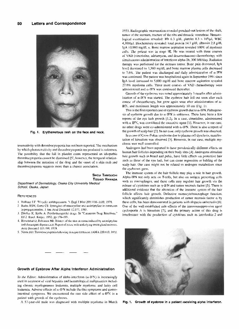
Growth of the eyebrows was noted approximately 3 months after admin- istation of a-IFN was started. The eyebrow hair fell out soon after each course of chemotherapy, but grew again soon after administration of a- IFN, and maximum length was approximately 10 cm (Fig. 1).
This is the first reported case of eyebrow growth due to a-IFN. Pathogene- sis of eyebrow growth due to a-IFN is unknown. There have been a few reports of the eye lash growth [1,2]. In a case, cimetidine, administered with a-IFN, was considered the causative agent [l]. However, in our case, no other drugs were co-administered with a-IFN. There is also a report of the growth of scalp hair 121. In our case, only eyebrow growth was observed.
In a case of Crow-Fukas syndrome due to plasma cell dysclasia, manifes- tation of hirsutism was observed [3]. However, in our case, multiple my- eloma was well controlled.
Androgens had been reported to have paradoxically different effects on human hair follicles depending on their body sites [4]. Androgens stimulate hair growth such as beard and pubis, have little effects on protective hair such as those of the eye lash, but can cause regression or balding of the scalp hair. Our case might not be related to androgen metabolism since the eyebrows grew.
The immune system of the hair follicle may play a role in hair growth. Alpha-IFN not only acts on T-cells, but also on antigen presenting cells such as macrophages, and these cells may regulate hair growth via the release of cytokines such as y I F N and tumor necrosis factor IS]. There is additional evidence that the alteration of the immune system of the hair follicle affects hair growth. Defective monocyte/macrophage function, which significantly diminishes production of tumor necrosis factor a by these cells, has been demonstrated in patients with alopecia universalis 161. One of the well-established side effects of the immunosuppressive agent cyclosporin A is hirsutism [7], and the primary action of this drug is interference with the production of cytokines such as interleukin-2 and
Growth of Eyebrow After Alpha Interferon Administration
To the Editor: Administration of alpha-interferon (a-IFN) is increasingly used in treatment of viral hepatitis and hematological malignancies includ- ing chronic myelogeneous leukemia, multiple myeloma, and hairy cell leukemia. Adverse effects of a-IFN include flu-like symptoms and gastro- intestinal symptoms. We encountered the rare side effect of a-IFN in a patient with growth of the eyebrows.
A 57-year-old male was diagnosed with multiple myeloma in March Fig. 1. Growth of eyebrow in a patient receiving alpha interferon.
y-IFN. The rare effect ofa-IFN on the growth of the eyebrow in the present patient might be due to the altered immune function of the follicle.
KOUICHI ARIYOSHI KENJI SHINOHARA
Xu RUIRONG Division of Hematology, Department of Medicine, Yamaguchi Prefecture Central Hospital, Hofu, Japan
REFERENCES
I . Mughal TI, Robinson WA, Thomas MR, Speigel R: Role ofrecombinant interferon alpha and cimetidine in patients with advanced malignant melanoma. J Cancer Res Clin Oncol 114:IO8-109, 1988.
2. Mughal TI, Thomas MR, Robinson WA: Role of recombinant alpha-interferon in the treatment of advanced cutaneous malignant melanoma. Oncology 48365- 368, 1991.
3. Case Records of the Massachusetts General Hospital (Case 10-1987). N Engl J Med 316:606418, 1987.
4. Randall VA, Thomton MJ, Hamada K, Messenger AG: Mechanism of androgen action in cultured dermal papilla cells derived from human hair follicles with varying responses to androgens in vivo. J Invest Dermatol 98:86S-91S, 1992.
5. Conlon KC, Urba WJ, Smith JW, Steis RG, Longo DL, Clark Jw: Exacerbation of symptoms of autoimmune disease in patients receiving alpha-interferon therapy. Cancer 65:2237-2242, 1990.
6. Skoutelis A, Freinkel RK, Kaufman DS, Leibovich SJ: Angiogenic activity is defective in monocytes from patients with alopecia universalis. J Invest DermatOl 95: 139-143, 1990.
7. Harper 11, Kendra JR, Desa IS, Staughton RCD, Barrett AJ, Hobbs JR: Dermdtoiogi- cal aspects of the use of cyclosporin A for prophylaxis of graft-verses-host disease. Br J Dermatol 110:469474, 1984.
Adult T-cell Leukemia Diagnosed After 22 Years
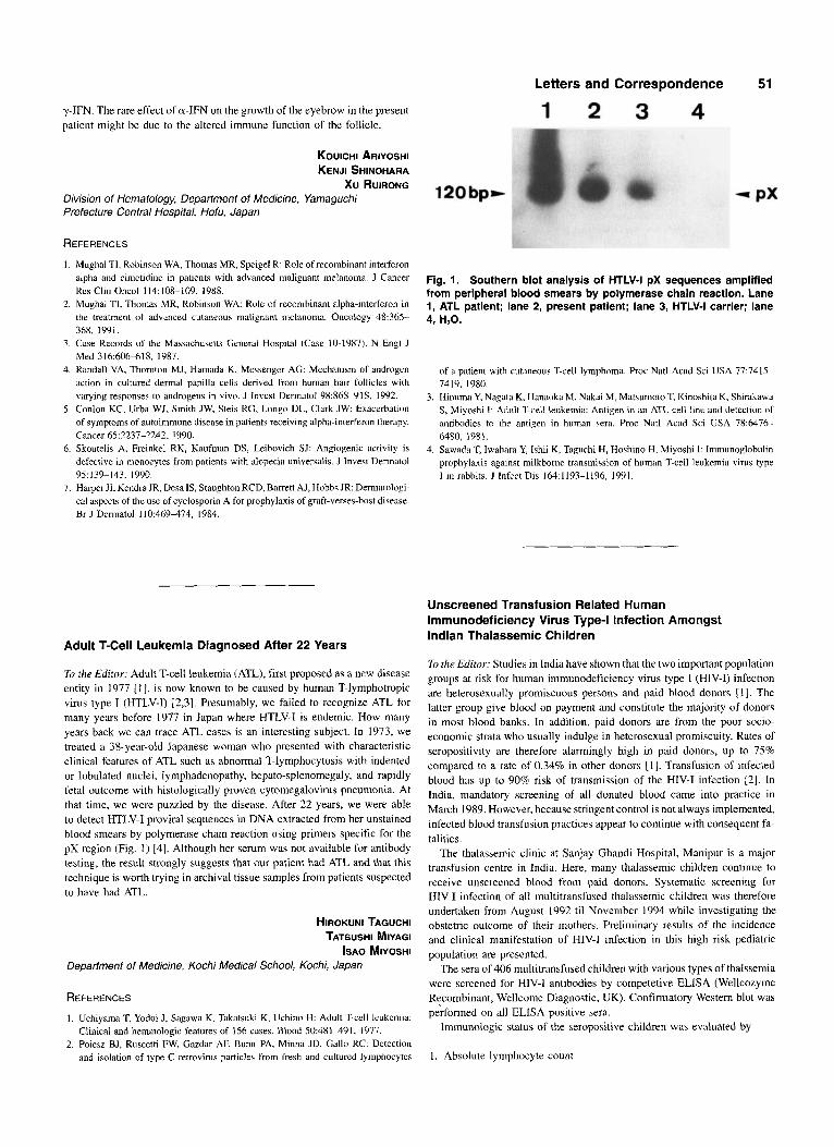
T i the Editor: Adult T-cell leukemia (ATL), first proposed as a new disease entity in 1977 [ I ] , is now known to be caused by human T-lymphotropic virus type I (HTLV-I) [2,3]. Presumably, we failed to recognize ATL for many years before 1977 in Japan where HTLV-I is endemic. How many years back we can trace ATL cases is an interesting subject. In 1973, we treated a 38-year-old Japanese woman who presented with characteristic clinical features of ATL such as abnormal T-lymphocytosis with indented or lobulated nuclei, lymphadenopathy, hepato-splenomegaly, and rapidly fetal outcome with histologically proven cytomegalovirus pneumonia. At that time, we were puzzled by the disease. After 22 years, we were able to detect HTLV-I proviral sequences in DNA extracted from her unstained blood smears by polymerase chain reaction using primers specific for the pX region (Fig. I ) 141. Although her serum was not available for antibody testing, the result strongly suggests that our patient had ATL and that this technique is worth trying in archival tissue samples from patients suspected to have had ATL.
HIROKUNI TAGUCHI TATSUSHI MIYAGI
ISAO MIYOSHI Department of Medicine, Kochi Medical School, Kochi, Japan
REFERENCES 1. Uchiyama T, Yodoi J, Sagawa K, Takatsuki K, Uchino H: Adult T-cell leukemia:
Clinical and hematologic features of 156 cases. Blood 50:481491, 1977. 2. PoiesL BJ, Ruscetti FW, Gazdar AF, Bunn PA, Minna JD, Gallo RC: Detection
and isolation of type C retrovirus particles from fresh and cultured lymphocytes
120bpw
Letters and Correspondence 51
1 2 3 4
Fig. 1. Southern blot analysis of HTLV-I pX sequences amplified from peripheral blood smears by polymerase chain reaction. Lane I, ATL patient; lane 2, present patient; lane 3, HTLV-I carrier; lane 4, H,O.
of a patient with cutaneous T-cell lymphoma. Proc Natl Acad Sci USA 77:7415-
Hinuma Y, Nagata K, Hanaoka M, Nakai M, Matsumoto T, Kinoshita K, Shirakawa S , Miyoshi I: Adult T-cell leukemia: Antigen in an ATL cell line and detection of antibodies to the antigen in human sera. Proc Natl Acad Sci USA 78:6476- 6480, 1981. Sawada T, Iwahara Y, lshii K, Taguchi H, Hoshino H, Miyoshi I: Immunoglobulin prophylaxis against milkborne transmission of human T-cell leukemia virus type I in rabbita. J Infect Dis 164:1193-1196, 1991.
7419, 1980.
Unscreened Transfusion Related Human Immunodeficiency Virus Type-I Infection Amongst Indian Thalassemic Children
Ti the Editor: Studies in India have shown that the two important population groups at risk for human immunodeficiency virus type I (HIV-I) infection are heterosexually promiscuous persons and paid blood donors [ I ] . The latter group give blood on payment and constitute the majority of donors in most blood banks. In addition, paid donors are from the poor socio- economic strata who usually indulge in heterosexual promiscuity. Rates of seropositivity are therefore alarmingly high in paid donors, up to 75% compared to a rate of 0.34% in other donors 111. Transfusion of infected blood has up to 90% risk of transmission of the HIV-I infection [2]. In India, mandatory screening of all donated blood came into practice in March 1989. However, because stringent control is not always implemented, infected blood transfusion practices appear to continue with consequent fa- talities.
The thalassemic clinic at Sanjay Ghandi Hospital, Manipur is a major transfusion centre in India. Here, many thalassemic children continue to receive unscreened blood from paid donors. Systematic screening for HIV-I infection of all multitransfused thalassemic children was therefore undertaken from August 1992 ti1 November 1994 while investigating the obstetric outcome of their mothers. Preliminary results of the incidence and clinical manifestation of HIV-I infection in this high risk pediatric population are presented.
The sera of 406 multitransfused children with various types of thalssemia were screened for HIV-I antibodies by competetive ELISA (Wellcozyme RFcombinant, Wellcome Diagnostic, UK). Confirmatory Western blot was performed on all ELISA positive sera.
Immunologic status of the seropositive children was evaluated by
1. Absolute lymphocyte count