growth and muscle cellularity of diploid and triploid … possible deficiencies in nutrients’...
TRANSCRIPT

Journal of Applied Ichthyology Accepted manuscript: 25.12.2014 DOI: 10.1111/jai.12792
Growth and muscle cellularity of diploid and triploid Atlantic cod
(Gadus morhua Linnaeus, 1758) larvae
Cecilia Campos Vargas*1, Stefano Peruzzi2, Ørjan Hagen1
1 Faculty of Biosciences and Aquaculture, University of Nordland, Bodø, Norway
2 Faculty of Biosciences, Fisheries and Economics, University of Tromsø, Norway
Correspondence: Cecilia C Vargas, University of Nordland, Universitetsalleen 11, 8049, Norway
E mail: [email protected]
Running title: Muscle growth of triploid Atlantic cod larvae
Key words: Gadus morhua, Atlantic cod, triploidy, growth, muscle, hyperplasia, hypertrophy.
Summary
The aim of this study was to compare somatic growth and muscle fibre
development in diploid and triploid siblings of Atlantic cod (Gadus morhua
Linnaeus, 1758) during the larval stage. Newly hatched larvae were
transferred into 200 L tanks, three tanks per ploidy group (70 larvae L-1,
continuous light, gradually increasing seawater temperature 7-11 °C and flow
rates 50-117 Lh-1). Larvae were fed rotifers from 2-22 days post hatch (dph),
Artemia 19-31 dph and weaned onto a microparticulate diet from 26 dph until
the end of the experiment. Measurements of growth (dry weight, standard
length) and muscle cellullarity were taken at intervals between 1 and 44 dph.
Ploidy groups showed a similar performance throughout the trial, although a
marked stagnation in growth was observed for triploids during the weaning
from Artemia onto dry feed. Overall, diploid and triploid cod larvae showed a
similar development in muscle fibre growth pattern during the experimental
period. For both groups, the total number of fast muscle fibres showed a 10-
fold increase (from 384 to 3462), whereas the diameter of fast fibre increased
from 8.9 to 13.3 µm (mean number from all treatments). Thus, a temporary

Journal of Applied Ichthyology Accepted manuscript: 25.12.2014 DOI: 10.1111/jai.12792
but significant effect of triploidy on fast muscle fibre growth pattern was
observed in 19 dph larvae in terms of fibre size and number, with triploids
showing larger mean fast fibre diameter (11.62 ± 0.63 vs. 10.05 ± 0.34) and a
lower number of fibres with diameter < 5 µm than their diploid siblings. Thus,
this was found to be related to larvae size and to the differences in total fast
fibre cross sectional areas rather than to ploidy status. Overall, our results
suggest possible deficiencies in nutrients’ digestion and absorption of triploid
cod larvae particularly during the transitional period from live food to inert
diets.
1. Introduction
During early stages, rapid growth is directed to organs associated to food
intake and swimming, enabling fish larva to catch prey items and avoid
predators (Galloway et al., 1999; Osse and VandenBoogaart, 1997). As for
adult fish, the axial musculature of fish larvae represents the largest and
fastest growing organ (Alami-Durante et al., 1997) and can constitute up to
40 % of the total body mass during early stages (Galloway et al., 1999).
In fish, muscle development through hyperplasia can be divided into three
phases: embryonic, stratified and mosaic hyperplasia. During embryonic
myogenesis, the adaxial and posterior cells generate respectively the
superficial and deep muscle cells (Devoto et al., 1996; Rescan, 2005 and
2008), components of the primary myotome (Stellabotte and Devoto, 2007).
Although during early stages both fibre types have an aerobic metabolism,
they will then differentiate into slow (red) aerobic and fast (white) anaerobic
fibres around metamorphosis (Johnston, 1999). From that stage onwards,

Journal of Applied Ichthyology Accepted manuscript: 25.12.2014 DOI: 10.1111/jai.12792
growth of the primary myotome will occur by stratified hyperplasia. During
this phase, fibres from the dermomyotome (external cells) move from the
outer to the inner surface of slow fibres to position in discrete germinal zones
situated mainly at the dorsal and ventral regions of the myotome (Hollway et
al., 2007; Stellabotte and Devoto, 2007). Finally, mosaic hyperplasia which is
characterized by the formation of new fast fibres between the existing fibres
will give rise to an assortment of fibre sizes. The onset of mosaic hyperplasia
varies between species, occurring late in fast-growing species capable of
reaching large ultimate size or being greatly reduced (not occurring) in slow
growing species with small ultimate size (Johnston, 1999).
Skeletal muscle in fish shows high phenotypic plasticity to environmental
clues like temperature, swimming activity and food availability (Johnston,
2006). For some fish species, myotomal muscle growth might also be
affected by ploidy status. For instance, is known that triploidy may be
associated with greater cell size in various tissues as reported in zebrafish
Danio rerio (Hamilton, 1822), various salmonids and marine species, and
such effects may extend to muscle fibres (see Benfey, 1999; Feindel et al.,
2011; Piferrer et al., 2009) However, comparison of muscle growth between
diploid and triploid fish remains scarce and mostly limited to juvenile and
adult stages. In Atlantic salmon, Johnston et al. (1999) found that the
embryonic phase of myogenesis was relatively little affected by ploidy status
in all-female (AF) populations, whereas diploids displayed ~30% more white
muscle fibres than triploids in a normal sex-ratio (NSR) population at first
feeding. This suggests that differences in muscle fibre growth between these
ploidies might be related to the stage of development. The number, diameter

Journal of Applied Ichthyology Accepted manuscript: 25.12.2014 DOI: 10.1111/jai.12792
and size distribution of fast fibres are directly related to growth (Johnston,
1999). Thus, improved knowledge of possible effects of triploidization on
muscle growth and cellularity may contribute to elucidate the variable and
contradictory growth performance of triploids versus diploids.
The aim of this study was therefore to investigate the effect of triploidization
on muscle fibre growth dynamics in Atlantic cod larvae reared under similar
conditions.
2. Material and methods
All husbandry procedures and experimental protocols were conducted in
accordance to the guidelines set by the National Animal Research Authority
(Forsøksdyrutvalget, Norway).
Fish husbandry and treatment
Gametes origin and fertilization
Gametes were obtained by stripping 6 females and 3 males of the 2nd
generation (2008 year class, 3 years old; 4-5 kg body weight) selected
Atlantic cod broodstock reared at Cod Juveniles AS – hatchery. Egg
fertilization and induction of triploidy were conducted in November 2012.
Male and female gametes were pooled in two separated containers prior to
fertilization. Sperm and eggs were gently mixed (2 ml sperm . 300 ml eggs-1
or ca. 300.000 eggs) followed by addition of 6 °C seawater and gametes left
undisturbed for 15 minutes in a temperature-controlled room at 6 °C to allow
completion of the fertilization process before being rinsed thoroughly with 6
°C seawater. Two-third of the total volume of eggs was used for triploid

Journal of Applied Ichthyology Accepted manuscript: 25.12.2014 DOI: 10.1111/jai.12792
production, while the remaining egg volume served as untreated diploid
control.
Triploidy induction
Triploid fish were produced by a hydrostatic pressure treatment applied to
180 minute degrees eggs (30 min post-fertilization at 6 °C). For this purpose,
fertilized eggs and 6 °C seawater were poured into the chamber of a
pressure device (TRC-HPC™ Pressure machine, TRC Hydraulics Inc. New
Brunswick, Canada). The hydrostatic pressure was then quickly elevated
manually until it reached 8500 psi and kept constant for 5 min, following the
protocol of Trippel et al. (2008). Post treatment the eggs were gently poured
into a 280 L incubator. Diploid and triploid eggs were incubated in separate
incubators (two per ploidy group) at 6.0 °C with a flow rate of 3 – 5 L min-1
until hatching and the eggs were treated with a surface disinfectant (Pyceze,
Novartis Ltd., Litlington, Near Royston, UK; 0.8 ml L-1 seawater for 6 min).
Seawater at the incubation room was taken from an inlet at a depth of 50 m,
filtered through a Bernoulli filter to remove particles over 20 μm, skimmed,
aerated and cooled with seawater controlled by a thermostat.
Larval rearing
Newly hatched larvae were transferred into 200 L water volume, green,
slightly cone-bottomed tanks (triplicates per ploidy group) with a density of 70
larvae L-1. To estimate the number of larvae to be transferred into the tanks,
samples of 2-liters volume were taken from each incubator (2 per ploidy
group) after lowering the water level to concentrate the larvae and counting
Journal of Applied Ichthyology Accepted manuscript: 25.12.2014 DOI: 10.1111/jai.12792
the larvae contained in a 100 ml graduated cylinder. Water flow rate was kept
constant at 0.6 L min-1 until 29 (dph). During the co-feeding (Artemia-dry
feed) and dry feed periods water flow rate was increased to 1.1 and 2 L min-
1, respectively. Seawater temperature was gradually increased from 6 to 10
°C ± 0.3 °C (1 - 10 dph) and kept constant thereafter until the end of the
experiment (44 dph) by a heating system with a thermostatic device.
Continuous light conditions (600 lux, measured at the water surface) was
applied to each tank. Larvae were fed on rotifers (Brachionus plicatilis,
Cayman strain), short-term enriched with OriGreen (Skretting AS, France)
from 2 to 22 dph, added to the tanks in 4 meals of 8000 individuals L-1 every
six hours. A solution of Neptune (60 ppm tank-1; Skretting AS, France) was
added to the tanks as green water technique simultaneously with the rotifers.
Artemia enriched with OriGold (Skretting AS, France) was offered to the
larvae at a rate of 3000 individuals L-1, 3 meals during the co-feeding period
with rotifers (19-22 dph) then at 6-h intervals from 23 to 31 dph. The weaning
onto Gemma Micro 150-300 (Skretting AS, France) was from 26 dph until the
end of the experiment. Dead larvae were removed as a part of the daily
maintenance.
Analytical methods
Larvae were randomly sampled from each tank and anaesthetized with
Tricaine methanesulfonate - Finquel vet. (Western Chemical Inc., WA, USA)
directly after sampling. Thereafter, larvae were treated as described for the
respective analytical methods.
Triploidy assessment

Journal of Applied Ichthyology Accepted manuscript: 25.12.2014 DOI: 10.1111/jai.12792
2-day old larvae from control and treated groups were anaesthetized, rinsed
with distilled water, individually placed into 1.5ml Eppendorf tubes, and deep
frozen (-80 °C) until analysis. Larvae were then prepared for propidium iodide
(PI) flow cytometric analysis as described by Peruzzi et al. (2007). The DNA
content was measured using a FACScan (Becton Dickinson, San Jose, CA,
USA) flow cytometer, based on 20.000 nuclei counts per sample. Ploidy was
assessed by calculating the ratio of the mean fluorescence intensity of triploid
to diploid and fish were considered triploid when the ratio was 1.5 ± 0.1. The
flow-cytometry data were analyzed using the software CyFlow v. 1.2.1
(©Pertthu Thero and CyFlow Ltd).
Growth
For dry weight (DW), standard length (SL) and myotome height (MH)
measurements, n= 90 larvae from each ploidy group were randomly sampled
from the incubators (1 dph) immediately prior to being placed into their
respective tanks. Thereafter n= 30 larvae per tank were sampled at 8, 18, 29,
36 and 44 dph. Larvae were rinsed in distilled water, photographed with a
Stereo light microscope equipped with a Olympus Colour - View IIIu camera
(Olympus, Soft Imaging Solutions, GmbH, Oslo, Norway) before being
transferred to tin pre-weighed capsules (SÄNTIS Analytical AG,
Landhausstrasse 1, CH–9053 Teufen, Switzerland), and dried at 60 °C for a
minimum of 24 hours (depending on larval size). Dry weight (DW) was
measured to the nearest 0.1 µg on a microbalance (Mettler Toledo UMX2,
Columbus, OH, USA) and the standard length (SL) was measured from the
tip of the snout to the end of the notochord by image analysis (Cell P

Journal of Applied Ichthyology Accepted manuscript: 25.12.2014 DOI: 10.1111/jai.12792
software Olympus Soft Imaging Solutions, GmbH, Oslo, Norway). The daily
weight increase (mg, % day-¹) of cod larvae was calculated according to
Ricker (1958) as: %DWI = (expg – 1)100; where g is the growth coefficient =
(ln W1 – ln W0) (t1 – t0)-1 and W=weight and t=time at sampling.
Muscle cellularity
For muscle cellularity analysis n= 5 larvae per tank were sampled at 1, 8, 19,
29, 36 and 44 dph, rinsed in distilled water and the total length (TL) was
recorded. Larvae were sectioned transversely to the body axis at post-anal
level, and the anterior part were mounted using cryomatrix (Anatomycal
pathology, Bergmann AS, Oslo) in aluminum capsules (SÄNTIS Analytical
AG , Landhausstrasse 1, CH–9053 Teufen, Switzerland) and snap frozen (60
sec) in isopentane cooled to its near freezing point (-159 °C) in liquid
nitrogen. Histological sections were cut at -20 °C in a cryostat (Microm HM
550, MICROM International GmbH) to obtain 2 μm thick histological sections,
stained in Harris haemotoxylin (Merck KGaA, Darmstadt, Germany) after
dehydration and mounted using glycerol gelatin (Sigma-Aldrich, USA) on
poly-L-lysine treated slides. Sections were analyzed with a light microscope
(Axioscop 2 mot plus; Carl Zeiss INC., Germany) equipped with a camera.
The area and diameter of 200-600 fibres (dependent on size) from the left
epaxial (dorsal) and hypaxial (ventral) side of the steak of white muscle
sections were calculated for each fish using the software Axio Vision (Rel.
4.2, Carl Zeiss INC., Germany). The total fibre number was calculated as
[106 x total cross-sectional area of fast muscle (mm2) x number of analyzed
fibres] [total area of analyzed fibres (µm)]-1. The fibre density (number of
Journal of Applied Ichthyology Accepted manuscript: 25.12.2014 DOI: 10.1111/jai.12792
fibres per unit area (mm2)) was calculated as [106 x the number of fibres
measured] [total area of analyzed fibres (µm)]-1.
Statistics
When necessary, data were logarithmically (log 10) transformed while data in
percentage were arcsine transformed to normalize distributions. All
transformed data were tested for normality of distribution (Shapiro Wilk’s test)
and homogeneity of variance (Levene’s test). Normally distributed data were
compared using a one-way ANOVA. When differences between means were
found (P < 0.05), post-hoc analyses were conducted using paired
comparisons (Tukey’s HSD) for homogeneous data and a 2-t (assuming non
equal variances) for non-homogeneous data. Non parametric testing
(Kruskal-Wallis, Moods Median Test) was used for non-normally distributed
data. ANCOVA was used to analyze data of fibre muscle diameter, number
and density with ploidy as factor and total cross-sectional area (TCA) and TL
as covariates. Non-parametric statistical techniques were used to fit
smoothed probability density functions (pdfs) to the measured fast fibre
diameters using a kernel function (Johnston et al., 1999). The average value
of the smoothing coefficient h varied between 0.117 and 0.141. A pdfs of the
fast fibre diameter of one average sized larva of each ploidy group was
plotted to show the development during the experiment (1-44 dph). Minitab
version 16 (Minitab Statistical software Inc., US) was used for testing of data.
Data are presented as mean ± SEM (N = number of samples).
Journal of Applied Ichthyology Accepted manuscript: 25.12.2014 DOI: 10.1111/jai.12792
3. Results
Growth characteristics
DW (mean ± SEM) at 1 dph were 0.12 ± 0.01 mg larva-1 for diploids (2n) and
0.13 ± 0.0 mg larva-1 for triploids (3n), and increased to 1.28 ± 0.09 and 1.3 ±
0.09 respectively at the end of the experiment (44 dph). No significant
differences in DW between the ploidy groups were observed throughout the
experiment (Fig. 1A). The DWI (%) did not differ between the two groups
during the trial. The highest and lowest growth rate were observed during the
rotifer - Artemia co-feeding period (2n: 9.9 ± 0.7 %, 3n: 10.6 ± 1.3 %) and
during the Artemia-dry feed co-feeding period (2n: 2.9 ± 2.1 % vs. 3n: 0.3 ±
2.4 %) respectively.
At the beginning of the experiment, SL and MH increased from 4.47 ± 0.01
mm and 0.25 ± 0.00 mm at 1 dph to 10.33 ± 0.12 mm and 0.98 ± 0.03 mm at
44 dph for diploids respectively. In comparison, the triploid group showed an
increase from 4.54 ± 0.02 mm and 0.25 ± 0.00 mm to 10.56 ± 0.13 mm and
1.03 ± 0.03 mm in SL and MH respectively. No significant differences in SL
and MH between the two ploidy groups were observed throughout the
experiment (Fig. 1B-C). However, triploids showed stagnation in SL and MH
during the rotifers - Artemia co-feeding period with 8.97 ± 0.41mm and 0.69 ±
0.0 mm at 29 dph) and 9.27 ± 0.22 mm and 0.71 ± 0.03 mm at 36 dph
respectively (Fig. 1B-C).
Muscle growth patterns
The total cross sectional area of fast muscle fibre increased from 0.023 mm2
and 0.024 mm2 at 1dph to 0.68 ± 0.08 mm2 and 0.68 ± 0.05 mm2 at 44 dph

Journal of Applied Ichthyology Accepted manuscript: 25.12.2014 DOI: 10.1111/jai.12792
for diploids and triploids respectively (Fig. 2). This corresponds to a 24 fold-
increase for the 2n group and a 22 fold-increase for the 3n group,
respectively. Although no significant differences were found between diploids
and triploids at any sample date, the latter experienced a decrease in TCA
during the Artemia – dry feed co-feeding period (29 dph: 0.29 ± 0.05 mm2
and 36 dph: 0.21 ± 0.02 mm2) (Fig. 2).
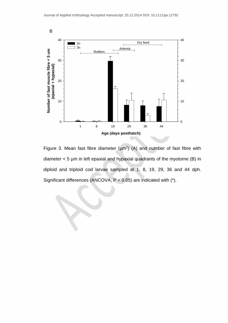
The mean fast fibre diameter at 1 dph increased from 8.87 ± 0.43 µm and
9.01 ± 0.26 µm for diploids and triploids respectively, to 13.24 ± 0.58 µm and
13.38 ± 0.48 µm at the end of the trial (44 dph) (Fig. 3A). Significant
differences in fast fibre diameter between the two ploidy groups were found
at 19 dph only. At this stage, triploids had larger fast fibres than diploids
(11.62 ± 0.63 µm vs. 10.05 ± 0.34 µm) and fewer fast fibres of a diameter < 5
µm (Fig. 3B). However, the differences in fast fibre diameter were related to
TCA (P < 0.05), rather than larval length (P > 0.05), while ploidy showed a
close to significant effect on fibre diameter (P = 0.06), (ANCOVA analysis).
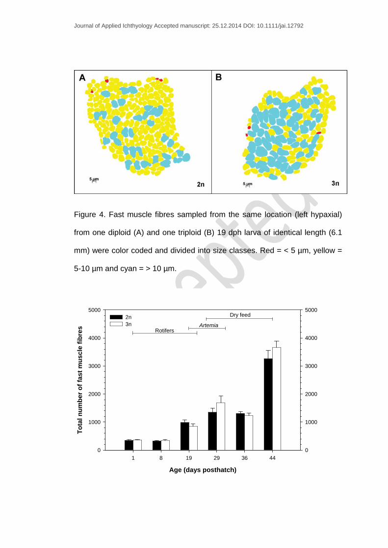
Fast muscle fibre measurements from the same location (ventral “hypaxia”
left side) of one diploid and one triploid 19 dph larva (a representative larva
from each group, 6.1 mm) were color coded and divided into size classes to
illustrate that the triploid larvae had fewer small fibres compared to diploid
larvae (Fig. 4A-B).
The total number of fast fibre in 1 dph larvae was 387 ± 36 for diploids and
380 ± 22 for triploids, increasing to 3262 ± 296 (diploids) and 3661 ± 232
(triploids) at 44 dph (Fig. 5). This represented an 8.4 fold increase for the
diploid and a 9 fold increase for the triploid group. No differences were found
Journal of Applied Ichthyology Accepted manuscript: 25.12.2014 DOI: 10.1111/jai.12792
in the mean total number of fast fibre between the two ploidy groups at any
sampling date (Fig. 5).
Smooth distributions were fitted to number of measurements (depending on
the larval stage) of fast fibre diameter per fish using a kernel function and the
corresponding probability density functions (pdfs) plotted. Comparisons of the
distribution of fast fibre diameter showed that for 1 dph larvae, the pdf peak
corresponded to a muscle fibre diameter of approx. 8 µm in both ploidy
groups (Fig. 6A-B). In older larvae, there was a tendency towards an
increase in fibre diameter and the peak pdfs shifted towards the right (Fig.
6A-B). However, an exception was observed in 19 dph larvae with diploids
showing a peak for 5 µm (Fig. 6A) and triploids for 8 µm fibres (Fig. 6B).
4. Discussion
Growth
During the live food period, triploids grew better than diploids whereas the
opposite trend was observed during weaning onto micro particulate diets,
although no significant. Similar growth patterns have recently been reported
by Opstad et al. (2013), showing that triploid cod are more affected by the
shift from live food to dry feed. The authors related such low performance to
possible behavioral differences as reported for other species. In Atlantic
salmon, triploid fry named “non-feeding fry” were shown to disperse slowly
through the water column with more difficulties accepting food compared to
their diploid siblings (Cotter et al., 2002). This assumption is supported by
recent findings showing that triploid salmon have smaller olfactory bulbs and

Journal of Applied Ichthyology Accepted manuscript: 25.12.2014 DOI: 10.1111/jai.12792
larger cerebella and telencephalon than diploids (Fraser et al., 2012). These
organs are implicated in functions such as foraging ability, aggression and
spatial cognition. If such morphological brain alterations caused by
triploidization are confirmed in triploid Atlantic cod too this might explain the
stagnant growth observed in our study during the transition between Artemia
and dry feed.
Contradictory and variable performance results have been published for
triploids of several species such as Atlantic salmon (McGeachy et al., 1995;
Cotter et al., 2002; Taylor et al., 2011; Taylor et al., 2013), rainbow trout,
(Cotter et al., 2002; Taylor et al., 2011; Taylor et al., 2013), Atlantic cod
(Opstad et al., 2013), turbot (Cal et al., 2006) and European sea bass
(Radaelli et al., 2010) when compared to their diploid siblings. The
differences have been related to factors such as unfavorable culture
conditions (Maxime, 2008; Piferrer et al., 2009; Tiwary et al., 2004), gamete
quality (Taylor et al., 2011), stage of the life cycle (Wagner et al., 2006) and
family and/or strain effects (e.g. Blanc et al., 2001; Johnson et al., 2004;
Taylor et al., 2013). However, in our study, the marked growth stagnation
displayed by triploids during the weaning period, may also indicate
deficiencies in nutrients’ digestion and/or absorption. For instance,
morphological differences in the guts of diploid and triploid siblings have
recently been reported for juvenile Atlantic cod (Vargas et al., 2014) and
Atlantic salmon (Peruzzi et al., 2014). This has been hypothesized to play a
role in determining the digestive efficiency and growth of fish that differ in
ploidy status. Overall, this highlights the need for further research on the

Journal of Applied Ichthyology Accepted manuscript: 25.12.2014 DOI: 10.1111/jai.12792
nutritional requirements of triploids and the optimization of culture protocols
specific for these fish.
Muscle cellullarity
In the present study, the somatic growth of diploids and triploids mirrored the
two groups’ muscle growth dynamics. As larvae grew, an increase in
diameter and number of fast fibre was observed in the two ploidies.
Differences in muscle cellullarity between diploids and triploids were found
only at 19 dph (end of the rotifer period, Fig. 3A-B), with larger mean fast
fibre diameter and lower numbers of fibres with diameter < 5 µm in triploids
than in diploids. This temporal difference might indicate that the recruitment
of new fibres was higher in the diploid group, whereas the triploid group
showed greater hypertrophy at this developmental stage. However, these
differences were attributed to the TCA of the larvae (ANCOVA, P < 0.05)
rather than a ploidy effect. A marked decrease in the number of fibres < 5 um
following the switch from Artemia to dry feed (29-36 dph) was observed in the
triploid group (Fig. 3B) without any increase in the total number of fast fibres
(Fig. 5), indicating a halt in hyperplasia during the temporary growth
stagnation (DWI % of 2.9% and 0.31% for diploids and triploids respectively).
Our observation of reduced hyperplasia related to somatic growth is in
accordance with early findings in first-feeding diploid cod larvae (Galloway et
al., 1999). Galloway et al. (1999) found that larvae with high somatic growth
showed a higher hyperplasia contribution to axial fast muscle growth
compared to slower-growing larvae.
Journal of Applied Ichthyology Accepted manuscript: 25.12.2014 DOI: 10.1111/jai.12792
The total mean number ( ̴ 350) and mean diameter (9 µm) of fast fibres
reported in the present study, regardless of ploidy, differ from previous
observations in newly hatched cod larvae showing lower numbers of fibres ( ̴
250) and with smaller diameter ( ̴ 6 µm) (Galloway et al., 1999; Johnston and
Andersen, 2008). Such discrepancy might be related to genetic differences
between the larvae used in our experiment (issued from F2 generation
selected broodstock) and those employed in previous studies.
The use of probability density curves provides a clearer picture of the
differences and similarities between diploid and triploid fibre size
distributions. The diploid and triploid larvae selected from each sampling
point with the exception of 19 dph (Fig. 6A-B) showed similar size
distributions. During endogenous feeding and the first days of exogenous
feeding (1 – 8 dph), muscle growth hypertrophy was the main growth
mechanism. From 1 to 8 dph (Fig. 5), the number of fast muscle fibres
remained constant while the mean fast fibre diameter increased from 9 to
11.6 µm (Fig. 3A). In contrast, a distinct stratified hyperplasia was observed
in the two ploidy groups at 19 dph as denoted by a left-shift in the distribution
of muscle fibre diameter compared to the other sampling points (Fig. 6A-B).
New fibres were recruited on the periphery of the myotomal epaxial and
hypaxial apices while fibres undergoing hypertrophy were located in the
center of the myotomal muscle (Fig. 4). An increase in the number of muscle
fibres < 5 µm compared to earlier stages contributed to the substantial
somatic growth observed at 19 and 29 dph, indicating that hyperplasia was
the major mechanism of muscle growth during that period. The shift from
Journal of Applied Ichthyology Accepted manuscript: 25.12.2014 DOI: 10.1111/jai.12792
hypertrophy to stratified hyperplasia and the proliferative zones of newly
recruited fibres observed in this study are in accordance with previous
findings reported for the same species by Galloway et al. (1999) and
Johnston and Andersen (2008).
Considering that the differences found at 19 dph were not related to ploidy
status, our results suggest that triploidization has no effect of on muscle
growth in cod during early larval stages. This is in line with work on early
stages of Atlantic salmon where no clear differences were found between
diploids and triploids in diameter, number and size distribution of fast fibres
during the freshwater period (Johnston et al., 1999). However, a possible
effect of triploidization on muscle growth might depend of developmental
stage as reported for species such as Atlantic salmon and rainbow trout
(Suresh and Sheehan 1998; Johnston et al., 1999). In seawater stages,
diploid Atlantic salmon showed significantly greater fibre recruitment than
their triploid counterparts (Johnston et al., 1999). Suresh and Sheehan
(1998) found differences in fibre size distribution between diploid and triploid
rainbow trout of 100 – 300 mm length. In the latter work, diploids showed a
higher proportion of fibres < 20 µm whereas triploids had higher proportion of
larger fibres (> 20 µm), indicating a reduced hyperplasia within triploids.
However, in both cases, the mean diameter of fast fibres did not differ
between ploidies.
The lack of triploidy effect on muscle cellullarity between diploids and triploids
at larval stages found in our study may be explained by the rapid growth
Journal of Applied Ichthyology Accepted manuscript: 25.12.2014 DOI: 10.1111/jai.12792
experienced during larval stages. This growth pattern is characteristic for
many fish species including Atlantic cod to cope with activities linked to active
foraging and predators’ avoidance. For example, Alami-Durante et al. (1997)
reported a daily muscle mass increase of 28 % during the period 4-18 dph in
the common carp Cyprinus carpio L. Considering that the growth potential
depends on several aspects, such as environmental conditions and nutrition
(Valente et al., 2013), these factors may therefore hind any potential effect of
triploidization on muscle cellullarity during early stages. Nevertheless, the
trial lasted 44 days and any potential differences in myotomal growth pattern
could occur after this rearing period.
In conclusion, muscle growth and cellullarity of Atlantic cod during early
stages was not affected by triploidization. However, the markedly stagnant
growth observed in triploids during the transition from Artemia to dry feed
might suggest a possible deficiency in digestion and absorption capacity of
triploids compared to diploids at that developmental stage. Larval growth is
affected by numerous environmental and nutritional factors, thus any
potential impact of triploidization on muscle characteristics might have been
overshadowed. Therefore, further research needs to be addressed to
develop larval rearing protocols adapted to triploids and to investigate in
more details factors influencing early growth performance.
5. Acknowledgements
We thank Cod Juveniles AS for providing access to broodstock and the
technical staff of Mørkvedbukta Research Station, University of Nordland for
Journal of Applied Ichthyology Accepted manuscript: 25.12.2014 DOI: 10.1111/jai.12792
their technical support in the experiment. This research was supported by the
Marine Larval Platform – University of Nordland and the program for Marine
Biotechnology in Northern Norway – Project AF0048.
6. References
Alami-Durante, H.; Fauconneau, B.; Rouel, M.; Escaffre, A.M.; Bergot, P.
1997: Growth and multiplication of white skeletal muscle fibres in carp
larvae in relation to somatic growth rate. J. Fish Biol. 50(6), 1285-
1302.
Blanc, J.M.; Poisson, H.; Vallée, F. 2001: Covariation between diploid and
triploid progenies from common breeders in rainbow trout,
Oncorhynchus mykiss (Walbaum). Aquac. Res. 32, 507-516.Benfey,
T.J. 1999: The Physiology and Behavior of Triploid Fishes. Rev. Fish.
Sci. 7(1), 39-67.
Benfey, T.J. 2011: The physiology of triploid fish. In: Encyclopedia of fish
physiology: from genome to environment. Ed: Farrell AP. Academic
Press, San Diego, pp 2009–2015.
Cal, R.M.; Vidal, S.; Gómez, C.; Álvarez-Blázquez, B.; Martínez, P. and
Piferrer, F. 2006: Growth and gonadal development in diploid and
triploid turbot (Scophthalmus maximus). Aquaculture. 251(1), 99-108.
Cotter, D.; O'Donovan, V.; Drumm, A.; Roche, N.; Ling, E.N. and Wilkins,
N.P. 2002: Comparison of freshwater and marine performances of all-
female diploid and triploid Atlantic salmon (Salmo salar L.). Aquac.
Res. 33(1), 43-53.
Journal of Applied Ichthyology Accepted manuscript: 25.12.2014 DOI: 10.1111/jai.12792
Devoto, S.H.; Melancon, E.; Eisen, J.S. and Westerfield, M. 1996:
Identification of separate slow and fast muscle precursor cells in vivo,
prior to somite formation. Development. 122(11), 3371-80.
Feindel, N.J.; Benfey, T.J. and Trippel, E.A. 2011: Gonadal development of
triploid Atlantic Cod Gadus morhua. J. Fish Biol. 78(7), 1900-1912.
Fraser, T.W.K.; Fjelldal, P.G.; Skjæraasen, J.E.; Hansen, T. and Mayer, I.
2012: Triploidy alters brain morphology in pre-smolt Atlantic salmon
Salmo salar: possible implications for behaviour. J. Fish Biol. 81(7),
2199-2212.
Galloway, T.F.; Kjorsvik, E. and Kryvi, H. 1999: Muscle growth and
development in Atlantic cod larvae (Gadus morhua L.) related to
different somatic growth rates. J. Exp. Biol. 202(15), 2111-2120.
Hollway, G.E.; Bryson-Richardson, R.J.; Berger, S.; Cole, N.J.; Hall, T.E. and
Currie, P.D. 2007: Whole-Somite Rotation Generates Muscle
Progenitor Cell Compartments in the Developing Zebrafish Embryo.
Dev. Cell. 12(2), 207-219.
Johnson, R.M.; Shrimpton, J.M.; Heath, J.W. and Heath, D.D. 2004: Family,
induction methodology and interaction effects on the performance of
diploid and triploid chinook salmon (Oncorhynchus tshawytscha).
Aquaculture. 234, 123-142.
Johnston, I.A. 1999: Muscle development and growth: potential implications
for flesh quality in fish. Aquaculture. 177(1–4), 99-115.
Johnston, I.A. 2006: Environment and plasticity of myogenesis in teleost fish.
J. Exp. Biol. 209(12), 2249-2264.

Journal of Applied Ichthyology Accepted manuscript: 25.12.2014 DOI: 10.1111/jai.12792
Johnston, I.A. and Andersen, Ø. 2008: Number of muscle fibres in adult
Atlantic cod varies with temperature during embryonic development
and pantophysin (PanI) genotype. Aquat. Biol. 4(2), 167-173.
Johnston, I.A.; Strugnell, G.; McCracken, M.L. and Johnstone, R. 1999:
Muscle growth and development in normal-sex-ratio and all-female
diploid and triploid Atlantic salmon. J. Exp. Biol. 202(15), 1991-2016.
Maxime, V. 2008: The physiology of triploid fish: current knowledge and
comparisons with diploid fish. Fish Fish. 9(1), 67-78.
McGeachy, S.A.; Benfey, T.J. and Friars, G.W. 1995: Freshwater
performance of triploid Atlantic salmon (Salmo salar) in New
Brunswick aquaculture. Aquaculture. 137(1–4), 333-341.
Opstad, I.; Fjelldal, P.G.; Karlsen, Ø.; Thorsen, A.; Hansen, T.J. and
Taranger, G.L. 2013: The effect of triploidization of Atlantic cod
(Gadus morhua L.) on survival, growth and deformities during early life
stages. Aquaculture. 388–391(0), 54-59.
Osse, J.W. and VandenBoogaart, J.G.M. 1997: Priorities during early growth
of fish larvae. Eur. J. Morphol. 35(1), 52-53.
Peruzzi, S., Hagen, Ø. & Jobling, M. (2014) Gut morphology of diploid and
triploid Atlantic salmon (Salmo salar L.). Aquacult. Int. 1-4. In press.
DOI: 10.1007/s10499-014-9867-2.
Peruzzi, S.; Kettunen, A.; Primicerio, R.; Kaurić, G. 2007: Thermal shock
induction of triploidy in Atlantic cod (Gadus morhua L.). Aquac. Res.
38, 926-932.
Piferrer, F.; Beaumont, A.; Falguiere, J.-C.; Flajshans, M.; Haffray, P. and
Colombo, L. 2009: Polyploid fish and shellfish: Production, biology and
Journal of Applied Ichthyology Accepted manuscript: 25.12.2014 DOI: 10.1111/jai.12792
applications to aquaculture for performance improvement and genetic
containment. Aquaculture. 293(3-4), 125-156.
Radaelli, G.; Poltronieri, C.; Simontacchi, C.; Negrato, E.; Pascoll, F.;
Libertini, A.; Bertotto, D. 2010: Immunohistochemical localization of
IGF-I, IGF-II and MSTN proteins during development of triploid sea
bass (Dicentrarchus labrax). Eur. J. Histochem. 54, 74-80.
Rescan, P.Y. 2005: Muscle growth patterns and regulation during fish
ontogeny. Gen. Comp. Endocr. 142(1-2), 111-116.
Rescan, P.Y. 2008: New Insights Into Skeletal Muscle Development and
Growth in Teleost Fishes. J. Exp. Zool. Part B - Molecular and
Developmental Evolution. 310B(7), 541-548.
Stellabotte, F. and Devoto, S.H. 2007: The teleost dermomyotome. Dev. Dyn.
236(9), 2432-2443.
Suresh, A.V.; Sheehan, R.J. 1998: Muscle fibre growth dynamics in diploid
and triploid rainbow trout. J. Fish Biol. 52, 570-587.
Taylor, J.F.; Preston, A.C.; Guy, D. and Migaud, H. 2011: Ploidy effects on
hatchery survival, deformities, and performance in Atlantic salmon
(Salmo salar). Aquaculture. 315(1–2), 61-68.
Taylor, J.F.; Sambraus, F.; Mota-Velasco, J.; Guy, D.R.; Hamilton, A.;
Hunter, D.; Corrigan, D. and Migaud, H. 2013: Ploidy and family
effects on Atlantic salmon (Salmo salar) growth, deformity and harvest
quality during a full commercial production cycle. Aquaculture. 410–
411(0), 41-50.
Tiwary, B.K.; Kirubagaran, R.; Ray, A.K. 2004: The biology of triploid fish.
Rev. Fish Biol. Fisher. 14, 391-402.

Journal of Applied Ichthyology Accepted manuscript: 25.12.2014 DOI: 10.1111/jai.12792
Trippel, E.A.; Benfey, T.J.; Neil, S.R.E.; Cross, N.; Blanchard, M.J.; Powell,
F. 2008: Effects of continuous light and triploidy on growth and sexual
maturation in Atlantic cod, Gadus morhua. Cybium. 32, 136-138.
Valente, L.M.P.; Moutou, K.A.; Conceição, L.E.C.; Engrola, S.; Fernandes,
J.M.O. and Johnston, I.A. 2013: What determines growth potential and
juvenile quality of farmed fish species? Rev. Aquacult. 5, S168-S193.
Vargas, C.C.; Hagen, Ø.; Solberg, C.; Jobling, M. and Peruzzi, S. 2014:
Growth and gut morphology of diploid and triploid juvenile Atlantic cod
(Gadus morhua). Aquac. Res. n/a-n/a. DOI: 10.1111/are.12603
Wagner, E.J.; Arndt, R.E.; Routledge, M.D.; Latremouille, D.; Mellenthin, R.F.
2006: Comparison of hatchery performance, agonistic behavior, and
poststocking survival between diploid and triploid rainbow trout of
three different Utah strains. N. Am. J. Aquacult. 68, 63-73.
Journal of Applied Ichthyology Accepted manuscript: 25.12.2014 DOI: 10.1111/jai.12792
Figures and legends:
Age (days posthatch)
1 8 19 29 36 44
Dry
wei
ght (
mg
larv
a-1)
0,0
0,2
0,4
0,6
0,8
1,0
1,2
1,4
1,6
0,0
0,2
0,4
0,6
0,8
1,0
1,2
1,4
1,6
2n3n
Dry feed
ArtemiaRotifers
A
Age (days posthatch)
1 8 19 29 36 44
Stan
dard
leng
th (m
m)
0
4
5
6
7
8
9
10
11
12
0
4
5
6
7
8
9
10
11
12
2n3n
Dry feed
ArtemiaRotifers
B
Journal of Applied Ichthyology Accepted manuscript: 25.12.2014 DOI: 10.1111/jai.12792
Age (days posthatch)
1 8 19 29 36 44
Myo
tom
e he
ight
(mm
)
0,0
0,2
0,4
0,6
0,8
1,0
1,2
0,0
0,2
0,4
0,6
0,8
1,0
1,2
2n3n
Dry feed
ArtemiaRotifers
C
Figure 1. Mean ± SEM values of dry weight (mg larva-1) (A), standard length
(mm) (B) and myotome height (mm) (C) of diploid and triploid cod larvae
sampled between 1 – 44 dph.

Journal of Applied Ichthyology Accepted manuscript: 25.12.2014 DOI: 10.1111/jai.12792
Age (days posthatch)
1 8 19 29 36 44
Tota
l cro
ss s
ectio
nal a
rea
(mm
2 )
0,0
0,2
0,4
0,6
0,8
0,0
0,2
0,4
0,6
0,82n3n
Dry feed
ArtemiaRotifers
Figure 2. Mean ± SEM total cross sectional area (mm2) of the myotome of
diploid and triploid cod larvae sampled at 1, 8, 19, 29, 36 and 44 dph.
Age (days posthatch)
1 8 19 29 36 44
Fast
mus
cle
fibre
dia
met
er (u
m)
0
5
6
7
8
9
10
11
12
13
14
15
16
0
5
6
7
8
9
10
11
12
13
14
15
162n3n
Dry feed
ArtemiaRotifers
A
*
Journal of Applied Ichthyology Accepted manuscript: 25.12.2014 DOI: 10.1111/jai.12792
Age (days posthatch)
1 8 19 29 36 44
Num
ber o
f fas
t mus
cle
fibre
< 5
um
(epa
xial
+ h
ypax
ial)
0
10
20
30
40
0
10
20
30
402n3n
Dry feed
ArtemiaRotifers
B
Figure 3. Mean fast fibre diameter (µm2) (A) and number of fast fibre with
diameter < 5 µm in left epaxial and hypaxial quadrants of the myotome (B) in
diploid and triploid cod larvae sampled at 1, 8, 19, 29, 36 and 44 dph.
Significant differences (ANCOVA, P < 0.05) are indicated with (*).
Journal of Applied Ichthyology Accepted manuscript: 25.12.2014 DOI: 10.1111/jai.12792
Figure 4. Fast muscle fibres sampled from the same location (left hypaxial)
from one diploid (A) and one triploid (B) 19 dph larva of identical length (6.1
mm) were color coded and divided into size classes. Red = < 5 µm, yellow =
5-10 µm and cyan = > 10 µm.
Age (days posthatch)
1 8 19 29 36 44
Tota
l num
ber o
f fas
t mus
cle
fibre
s
0
1000
2000
3000
4000
5000
0
1000
2000
3000
4000
50002n3n
Dry feed
ArtemiaRotifers
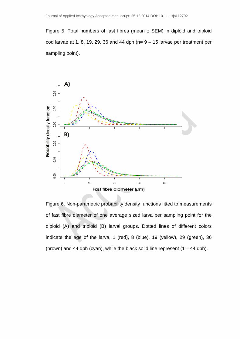
Journal of Applied Ichthyology Accepted manuscript: 25.12.2014 DOI: 10.1111/jai.12792
Figure 5. Total numbers of fast fibres (mean ± SEM) in diploid and triploid
cod larvae at 1, 8, 19, 29, 36 and 44 dph (n= 9 – 15 larvae per treatment per
sampling point).
Figure 6. Non-parametric probability density functions fitted to measurements
of fast fibre diameter of one average sized larva per sampling point for the
diploid (A) and triploid (B) larval groups. Dotted lines of different colors
indicate the age of the larva, 1 (red), 8 (blue), 19 (yellow), 29 (green), 36
(brown) and 44 dph (cyan), while the black solid line represent (1 – 44 dph).