greatly elevated urea excretion after air exposure appears...
TRANSCRIPT

893
Greatly Elevated Urea Excretion after Air Exposure Appears to Be
Carrier Mediated in the Slender Lungfish (Protopterus dolloi)
Chris M. Wood1,2,*Patrick J. Walsh2
Shit F. Chew3
Yuen K. Ip4
1Department of Biology, McMaster University, Hamilton,Ontario L8S 4K1, Canada; 2National Institute ofEnvironmental Health Sciences Marine and FreshwaterBiomedical Sciences Center, Rosenstiel School of Marine andAtmospheric Science, University of Miami, Miami, Florida33149; 3Natural Sciences, National Institute of Education,Nanyang Technological University, 1 Nanyang Walk,Singapore 637616, Republic of Singapore; 4Department ofBiological Sciences, National University of Singapore, 10Kent Ridge Road, Singapore 117543, Republic of Singapore
Accepted 4/22/2005; Electronically Published 9/21/2005
ABSTRACT
Under aquatic conditions, Protopterus dolloi is ammoniotelic,excreting only small amounts of urea-N. However, upon returnto water after 30 d estivation in air, the lungfish excretes onlysmall amounts of ammonia-N but massive amounts of urea-N. A similar pattern is seen after 21–30 d of terrestrialization,a treatment in which the lungfish is air exposed but kept moistthroughout. After both treatments, the time course of urea-Nexcretion is biphasic with an immediate increase, then a fall,and finally a second larger increase that peaks at about 12 hand may be prolonged for several days thereafter. Urea-N ex-cretion rates during the second peak reach 2,000–6,000 mmolN kg�1 h�1, two to three orders of magnitude greater than ratesin most fish and comparable only to rates in species known toemploy UT-A type facilitated diffusion urea transporters. Di-vided chamber studies and measurements of the clearance ratesof [3H]-PEG-4000 (a glomerular filtration and paracellular dif-fusion marker) and two structural analogs of urea ([14C]-acet-amide and [14C]-thiourea) were performed to characterize thetwo peaks of urea-N excretion. The smaller first peak was al-most equally partitioned between the head (including internaland external gills) and the body compartment (including uri-
* Corresponding author; e-mail: [email protected].
Physiological and Biochemical Zoology 78(6):893–907. 2005. � 2005 by TheUniversity of Chicago. All rights reserved. 1522-2152/2005/7806-4157$15.00
nary opening), was accompanied by only a modest increase in[14C]-acetamide clearance equal to that in [14C]-thiourea clear-ance, and could be accounted for by a large but short-lastingincrease in [3H]-PEG-4000 clearance (to about fivefold the ter-restrial rate). The delayed, much larger second peak in urea-Nexcretion represented an elevated efflux into both compart-ments but occurred mainly (72%) via the body rather than thehead region. This second peak was accompanied by a substan-tial increase in [14C]-acetamide clearance but only a modestfurther rise in [14C]-thiourea clearance. The acetamide to thio-urea permeability ratio was typical of UT-A type transportersin other fish. [3H]-PEG-4000 clearance was stable at this timeat about double the terrestrial rate, and excretion rates of ureaand its analogs were many fold greater than could be accountedfor by [3H]-PEG-4000 clearance. We conclude that the firstpeak may be explained by elevated urinary excretion and para-cellular diffusion across the gills upon resubmergence, whilethe second peak is attributable to a delayed and prolongedactivation of a UT-A type facilitated diffusion mechanism, pri-marily in the skin and perhaps also in branchial epithelia.
Introduction
Traditionally, the N-waste molecule urea was thought to moveby simple diffusion through biological membranes, but it isnow clear that a variety of carrier-mediated urea transportmechanisms are present in vertebrate cell membranes and playimportant roles in the physiological regulation of urea move-ments (reviewed by Sands et al. 1997; Smith and Rousselet2001; Bagnasco 2003; Sands 2003). The best characterized ofthese are members of the UT-A family (bidirectional facilitateddiffusion type transporters), which were first discovered in themammalian kidney (You et al. 1993; Smith et al. 1995) andlater in amphibian bladder (Couriaud et al. 1999). The cDNA’sfor homologous members of this same family (reviewed byWalsh and Smith 2001) have now been cloned in part or infull from several ureotelic fish, including the elasmobranchsSqualus acanthias (Smith and Wright 1999), Triakis scyllia (Hy-odo et al. 2004), and Raja erinacea (Morgan et al. 2003a), thefacultatively ureotelic teleost Opsanus beta (Walsh et al. 2000),and the obligatory ureotelic teleost Alcolapia grahami (Walshet al. 2001a). Furthermore, there is now direct or indirect evi-dence for similar transporters even in obligatory ammoniotelicteleosts such as Anguillla japonica (full-length cDNA; Mistry et

894 C. M. Wood, P. J. Walsh, S. F. Chew, and Y. K. Ip
al. 2001), Porichthys notatus (Walsh et al. 2001b; McDonald etal. 2002), and Oncorhynchus mykiss (Pilley and Wright 2000;McDonald et al. 2003; McDonald and Wood 2004). In elas-mobranchs, these appear to occur mainly in the kidney and,in teleosts, mainly in the gills.
Active urea transport mechanisms, at least some of whichappear to be Na� coupled, also occur, though they have notyet been identified at the molecular level. Physiological evidencefor active urea transport has been reported in mammalian kid-ney (reviewed by Sands 2003), amphibian kidney (Schmidt-Nielsen and Shrauger 1963) and skin (Garcia-Romeu et al.1981; Rapoport et al. 1988; Lacoste et al. 1991), elasmobranchkidney (S. acanthias; Schmidt-Nielsen et al. 1972; Morgan etal. 2003b) and gill (Part et al. 1998; Fines et al. 2001), andteleost kidney (O. mykiss: McDonald and Wood 1998, 2003; O.beta: McDonald et al. 2000; P. notatus: McDonald et al. 2002).
In fish, our ability to differentiate these various transportersand our understanding of their physiology is fairly preliminary,but they are clearly involved in the regulation of urea excretion.The branchial UT-A transporter (tUT; Walsh et al. 2000) ofthe gulf toadfish (O. beta) has been best studied; tUT appearsto be activated periodically to cause pulses of urea excretionthrough the gills (reviewed by Walsh and Smith 2001; Woodet al. 2003), a phenomenon first detected by fine timescaleanalysis of urea appearance in the external water (Wood et al.1995). A particularly useful tool in demonstrating the carriermediation of this phenomenon was the use of previously in-jected urea analogs ([14C]-acetamide, [14C]-thiourea), whichwere “pulsed” simultaneously to urea at different rates (Woodet al. 1998; McDonald et al. 2000). Indeed, the differentialability of various urea transporters to handle different analogsof urea has proven to be diagnostic of the presence and cate-gorization of urea transporters in fish (Schmidt-Nielsen andRabinowitz 1964; Wood et al. 1998; McDonald et al. 2000, 2002,2003; Pilley and Wright 2000; Walsh et al. 2001a).
With this background in mind, we set out to investigate ureaexcretion in the slender lungfish, Protopterus dolloi, with a par-ticular focus on possible carrier mediation of urea transport.Members of the class Dipnoi (the lungfishes) occupy a pivotalposition on the pathway to tetrapod evolution and terrestriality,and the African genus Protopterus appears to be the most ter-restrial of the class. These animals have both internal and ex-ternal gills but are obligatory air breathers via a primitive lung,and they are capable of prolonged estivation when water avail-ability becomes limiting (reviewed by Fishman et al. 1986).They are ammoniotelic when in water but rely on ureogenesisthrough the ornithine-urea cycle when air exposed (Smith 1930;Janssens 1964; Forster and Goldstein 1966; Janssens and Cohen1966, 1968; Chew et al. 2003, 2004). Urea is accumulated in-ternally, and after return to water, urea excretion is known tobe greatly elevated for several days (“in spectacular quantities”:Smith 1930; see also Janssens 1964; Chew et al. 2003). However,the temporal pattern (i.e., continuous or pulsatile), sites (i.e.,
gills, kidney, or skin), and mechanisms of urea excretion (sim-ple diffusion vs. some type of carrier mediation) are unknown.
Protopterus dolloi was selected as a particularly useful modelbecause it can be induced to estivate simply by removal ofwater from an otherwise empty aquarium. Under these con-ditions, it forms its own cocoon without the complicating pres-ence of mud (Chew et al. 2004). In Zaire, it digs burrows inthe raftlike substrate of floating swamps as a breeding nest andmay use this same burrow for estivation during the dry season,without formation of a mud cocoon (Brien et al. 1959). Wealso found that it tolerates prolonged exposure to air in thecontinued presence of a thin film of water (“terrestrialization”;Chew et al. 2003), a less extreme condition that can be con-sidered the precursor to true estivation. In this study, we ex-ploited these treatments in combination with fine timescalerecording of urea (and ammonia) excretion after return toaquatic conditions. Divided chamber and renal function studies([3H]-polyethylene glycol-4000 clearance) were performed tolook for sites of urea excretion, and urea analog studies ([14C]-acetamide, [14C]-thiourea clearances) were carried out to lookfor evidence of possible carrier mediation.
Material and Methods
Experimental Animals
Protopterus dolloi with a mean weight of about 40 g (range 15–95 g) were collected in Nigeria and Zaire and air shipped to acommercial dealer in Singapore. After purchase, they were theneither housed at the University of Singapore, where some ofthe experiments were performed, or else further air shipped toMcMaster University in Canada, where other experiments werecarried out. Holding and experimental conditions were essen-tially identical at the two locations. At both laboratories, theywere held at 25�–27�C under a 12L : 12D photoperiod for sev-eral weeks before experimentation. During the holding period,lungfish were kept individually in small plastic aquaria con-taining approximately 2 L of dechlorinated tap water that wasnot aerated but supplemented with seawater to a salinity of 2ppt, pH about 7.1–7.2. Approximate water composition wasNa� 30, Cl� 36, Mg2� 3.1, and Ca2� 2.6 mmol L�1, with titrationalkalinity (to ) 0.64 mmol L�1. Since freshwater pondspH p 4.0evaporate during the dry season before estivation of the lungfish(Brien et al. 1959; Poll 1961), it is likely that similar elevationsin ionic strength occur in nature. Water of this compositionwas used in all experiments. The lungfish were fed frozen blood-worms every second day, and the water was changed the dayafter feeding. These conditions were found to keep the animalsvery healthy, with the slightly saline water being important inpreventing fungal infections. The animals were not fed for 48h before the start of experiments.
Urea Excretion in Lungfish 895
Experimental Series
Experiments examining N-waste excretion after estivation (se-ries 1), after terrestrialization (series 2), and in lungfish individed chambers after various treatments (series 3) and 3[H]-PEG-4000 clearance after terrestrialization (series 4) were per-formed at the University of Singapore. Experiments examiningthe clearances of 14[C]-acetamide and 14[C]-thiourea after ter-restrialization (series 5), plasma urea-N and ammonia-N con-centrations after terrestrialization (series 6), and some addi-tional divided chamber studies (series 3) were performed atMcMaster University.
Series 1: Return to Aquatic Conditions after Estivation
Lungfish were induced to estivate in their aquaria as describedby Chew et al. (2004). All but 10 mL of water was removed,which was allowed to dry up over the following 3 or 4 d. Bythis point, the lungfish had curled up into the typical estivationposture, became motionless, and formed dry brown mucuscocoons. They were kept minimally hydrated thereafter byspraying with 1–2 mL water every 6 d until day 30. The aquariawere placed in 24-h darkness throughout the estivation period,and no food was given.
On day 30, the fish ( ) were returned to water. TheN p 7aquaria were moved into lighted conditions and quickly flushedwith mL of water to remove any accumulated N-waste;2 # 500then 1 L of water was added, together with an airstone formixing, and -mL samples withdrawn for initial N-analyses2 # 5(ammonia-N and urea-N). Within 5–10 min, the lungfish be-came active, breaking the cocoon and struggling to the surfaceto breathe. Any pieces of cocoon still adhering to the lungfishwere manually removed to help this process. After 1 h, thewater was sampled again for N-analyses and then replaced witha volume equal to #25 the animal’s mass. Sequential watersamples were taken at 2-h intervals up to 15 h postestivation,then again at 21 h. The water was renewed, and sampling at2-h intervals continued until 37 h postestivation, then again at46 h, followed by water renewal. A final flux measurementcovered the period from 49.5 to 68 h postestivation.
Series 2: Return to Aquatic Conditions after Terrestrialization
This treatment started identically to estivation, with all but 10mL of water being removed at the start, when the aquaria weretransferred to constant darkness and feeding was suspended.However, rather than being allowed to dry up, the water wasreplenished by daily spraying so as to maintain the volume atabout 10 mL. Under these conditions, the lungfish’s skin re-mained wet, and the animal lay in a thin film of water con-tacting its ventral surface. In general, these lungfish also secretedsome cocoon material, though it tended to be incomplete orbroken, probably disturbed by infrequent movements.
On day 21, the aquaria of these lungfish ( ) were rinsedN p 6with mL of water, and then exactly 20 mL of water2 # 500was added. Of this, mL were immediately removed for2 # 5initial N analyses. After 12 h, the remaining approximately 10mL (into which N-flux had occurred) were removed for finalN-analyses and the exact volume recorded. The procedure wasrepeated for another 12-h flux determination. The purpose wasto monitor rates of ammonia-N and urea-N excretion into thewater film under terrestrial conditions, before the return toaquatic conditions. On day 22, after two 12-h “terrestrial” fluxperiods, the animals were returned to aquatic conditions byreplacing the water film with a volume equal to #25 the an-imal’s mass, together with an air stone for mixing. The aquariawere also moved into lighted conditions at this time. Watersamples ( mL) were taken at 1-h intervals until 12 h and2 # 5then again at 24 h. The N-excretion rates in series 4 (see “Series4: [3H]-PEG-4000 Clearance Rates during and after Terrestrial-ization”) were virtually identical to those of these series 2 fish,so the data were combined ( ).N p 13
To check for possible effects of starvation or disturbance, acontrol group ( ) was also transferred to constant dark-N p 7ness and starved for 21 d but kept under aquatic conditions.On day 21, the aquaria were flushed with mL of water,2 # 500the volume was then set to #25 the animal’s mass, and two12-h flux measurements of N-excretion were made. On day 22,the water was then renewed, and the animals were transferredto lighted conditions, with water samples taken at 1-h intervalsuntil 12 h and then again at 24 h.
Series 3: Divided Chamber Studies
In order to localize the sites of urea-N and ammonia-N ex-cretion, the divided chamber approach pioneered by Smith(1929) was employed to separate excretion through the headregion (containing the internal and external gills and com-prising about 15% of the body mass) from excretion throughthe body (containing the bulk of the skin and the urinaryopening and comprising about 85% of the body mass). Mea-surements were made in control lungfish, which had been keptthroughout under aquatic conditions ( ), and in terres-N p 10trialized lungfish at approximately 0–3 ( ) and 12–15 hN p 5( ) after return to aquatic conditions. [3H]-PEG-4000N p 9clearance rates through the head versus body regions were alsomeasured in lungfish ( ) kept under aquatic conditionsN p 7throughout.
Lungfish were placed for 1–2 h (precise time recorded) inan apparatus such that the head region was separated from theremainder of the body using a latex dental dam to make awater-tight seal. Water samples for N-analyses or [3H]-PEG-4000 cpm were drawn at the start and end of the period. Therear chamber containing most of the body was a water-filledgraduated cylinder, which was placed horizontally in a plastictray tilted so as to have a reservoir of aerated water at one

896 C. M. Wood, P. J. Walsh, S. F. Chew, and Y. K. Ip
corner into which the lungfish’s head was submerged. The lung-fish was able to easily raise its head to breathe air periodically.The volumes of the head and body reservoirs were typicallyabout 200 mL each and were measured precisely at the end ofthe experiment.
Series 4: [3H]-PEG-4000 Clearance Rates during and afterTerrestrialization
The purpose of this experiment was to monitor possiblechanges in kidney function and paracellular diffusion associatedwith the time course of changes in urea-N excretion that hadbeen observed in series 2. The animals were too small to befitted with urinary catheters to measure urine flow rate, soinstead the clearance of [3H]-polyethylene glycol-4000 (PEG-4000) was used to monitor glomerular filtration rate (GFR).PEG-4000 is a well-established extracellular space, paracellulardiffusion, and GFR marker in fish (Beyenbach and Kirschner1976; Erickson and Gingerich 1986; Curtis and Wood 1991;Munger et al. 1991; Wood et al. 1998). Experimental lungfish( ) were treated identically to those in series 2, with theN p 7exception that the animals received an injection of 50 mCi kg�1
of [3H]-PEG-4000 (NEN-Dupont; specific mCiactivity p 2,000g�1) in 5 mL kg�1 saline (140 mmol L�1 NaCl) into the caudalhaemal arch approximately 4 h before the terrestrial flux mea-surements. Additional water samples (5 mL) were taken forscintillation counting to monitor [3H]-PEG-4000 appearanceinto the water at each sample time, and at 24 h, a blood sample(200 mL) to monitor [3H]-PEG-4000 cpm remaining in theextracellular fluid was drawn by caudal puncture. For injectionsand blood sampling, a #23 needle attached to a 1-mL syringewas used, and to prevent clotting, the needle and syringe wereprerinsed with a 5,000 i.u. mL�1 lithium heparin solution(Sigma) made up in 140 mmol L�1 NaCl. [3H]-PEG-4000 clear-ance rates were also measured in a control group ( ) keptN p 5under aquatic conditions throughout.
Series 5: [14C]-Acetamide and [14C]-Thiourea Clearance Ratesduring and after Terrestrialization
The goals of this experiment were to test whether changes inthe clearance rates of two structural analogs of urea corre-sponded with the time course of changes in urea-N excretionthat had been observed in series 2 and to look for differencesin their clearance rates diagnostic of carrier-mediated transport.The protocol was the same as in series 4, except that the periodof terrestrialization was 30 d, the initial injection was 10 mCikg�1 of either [14C]-acetamide ( ) or [14C]-thioureaN p 13( ; NEN-Dupont; specific activity 58.0 mCi mmol�1),N p 14and water sampling (for N-analyses and [14C]-analog counting)continued at 2-h intervals until 16 h after return to aquaticconditions, with final water and blood samples (as above) takenat 24 h.
Series 6: Plasma Urea-N and Ammonia-N Concentrationsduring and after Terrestrialization
Lungfish were put through the same protocol of terrestriali-zation and returned to aquatic conditions as in series 5, withblood samples (as above, only one per animal) being drawn at0 ( ), 8 ( ), 12 ( ), or 24 h ( ) after returnN p 7 N p 7 N p 7 N p 8to aquatic conditions to establish the time course of changesin plasma urea-N and ammonia-N concentrations. Blood sam-ples were also drawn from control lungfish ( ) kept un-N p 12der aquatic conditions. Additional blood plasma samples( ) taken in series 5 at 24 h were also analyzed, yieldingN p 27very similar data, for a total of for the 24-h point.N p 35Plasma was immediately separated by centrifugation and storedin liquid nitrogen before analysis.
Analytical Methods and Calculations
The diacetyl monoxime method of Rahmatullah and Boyde(1980) and the indophenol blue method of Ivancic and De-gobbis (1984) were used to measure the concentrations of urea-N and ammonia-N, respectively, in water, using freshly pre-pared urea and NH4Cl standards made up in the test water.The diacetyl monoxime method was also used for plasma urea-N determinations, while plasma ammonia-N was measured bythe enzymatic method of Kun and Kearney (1974). Water sam-ples (5 mL) and plasma samples (25 uL diluted to 5 mL in thetest water) were added either to 13 mL BCS fluor (Amersham)for [3H]-PEG-4000 determination in a Wallac 1414 liquid scin-tillation counter or to 10 mL ACS fluor (Amersham) for [14C]-acetamide and [14C]-thiourea analysis in an LKB Rackbeta 1217Counter, using onboard programs for quench correction.
Flux rates of urea-N and ammonia-N (mmol N kg�1 h�1) werecalculated from changes in concentration (mmol N L�1), factoredby the known lungfish mass (kg), volume (L), and time (h). Tocalculate clearance rates of [3H]-PEG-4000, we assumed that thismarker was distributed at plasma concentration in a space equiv-alent to 25% of the lungfish’s mass, that is, a volume chosen toapproximate the extracellular space (Holmes and Donaldson1969). Similarly, we assumed that the urea analogs [14C]-acet-amide and [14C]-thiourea were distributed at plasma concentra-tion in a space equivalent to 75% of the lungfish’s mass, that is,a volume chosen to approximate the body water space (Holmesand Donaldson 1969). Knowing the final measured plasma con-centration in the blood sample taken at the end of the experi-ment, and by keeping track of the total amount excreted in eachflux period, we calculated the amount present in the animal atthe beginning and end of each flux period and therefore theaverage plasma concentration of the radiolabeled compound dur-ing the flux period in question. Clearance rate (mL kg�1 h�1)was then calculated as the amount excreted during the flux period(cpm), factored by the lungfish mass, time (h), and estimatedplasma concentration (cpm mL�1).

Urea Excretion in Lungfish 897
Figure 1. Rates of (A) ammonia-N excretion and (B) urea-N excretion in Protopterus dolloi of series 1 during return to aquatic conditions after30 d of estivation. Note the biphasic pattern and sustained elevation of urea-N excretion relative to the monophasic pattern of ammonia-Nexcretion. In A, only the means at 0–1 and 1–3 h postestivation are significantly different ( ) from the reference aquatic rate of ammonia-P ≤ 0.05N excretion. In B, means at all times are significantly different ( ) from the reference aquatic rate of urea-N excretion. In addition, twoP ≤ 0.05peaks are well defined by the following significant differences ( ) relative to the trough at 7–9 h postestivation: means at 0–1, 1–3, andP ≤ 0.053–5 h (i.e., first peak) and all means at 9 through 29 h (i.e., second peak). SEM ( ).Means � 1 N p 7
Statistical Analyses
Data have been expressed as SEM (N), where Nmeans � 1represents the number of lungfish sampled. A Student’s pairedt-test (two-tailed) was employed for single comparisons withina treatment, and a Student’s unpaired t-test (two-tailed) wasemployed for comparisons between treatments at the sametime. The Bonferroni correction was used when more than twotreatments were compared. A one-way ANOVA followed by
Dunnett’s test was used for multiple comparisons against asingle reference value within a treatment. A significance levelof was used throughout.P ≤ 0.05
Results
As a point of reference, lungfish kept under normal aquaticconditions and fasted for 48 h before measurement are stronglyammoniotelic, with ammonia-N excretion rates of 220.1 �
898 C. M. Wood, P. J. Walsh, S. F. Chew, and Y. K. Ip
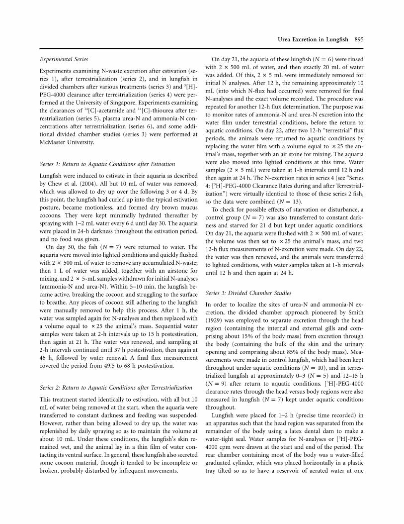
Figure 2. Rates of (A) ammonia-N excretion and (B) urea-N excretion in Protopterus dolloi of series 2 on day 21 of terrestrialization and duringreturn to aquatic conditions thereafter. Note the biphasic pattern and sustained elevation of urea-N excretion relative to the monophasic patternof ammonia-N excretion during return to aquatic conditions, qualitatively similar to the patterns seen after estivation (Fig. 1). Asterisks indicatemeans significantly different ( ) from the terrestrial rate on day 21. In B, two separate peaks of urea-N excretion are well defined byP ≤ 0.05the following significant differences ( ) relative to the trough at 5–6 h postestivation: means at 0–1 and 1–2 h (i.e., first peak) and allP ≤ 0.05means at 7 through 24 h (i.e., second peak). Furthermore, all means at 9 through 24 h are significantly higher than the initial peak.
SEM ( ).Means � 1 N p 13
mmol N kg�1 h�1 and urea-N excretion rates of31.2 31.9 �
mmol N kg�1 h�1 ( ; reported in Wood et al. 2005).10.8 N p 7
Series 1: Return to Aquatic Conditions after Estivation
In the first hour after lungfish were returned to water following30 d of estivation, ammonia-N excretion was around 2,500mmol N kg�1 h�1, but by 3–5 h it had decreased to about 140
mmol N kg�1 h�1, a value that was not significantly differentfrom the reference aquatic rate ( mmol N kg�1 h�1)220.1 � 31.2and that remained unchanged until the end of the measure-ments at 68 h (Fig. 1A). In contrast, urea-N excretion rateswere far higher and exhibited a clear biphasic pattern (Fig. 1B).The initial rate in the first hour was about 5,000 mmol N kg�1
h�1, but this decreased progressively to about 750 mmol N kg�1
h�1 at 7–9 h before increasing to a second, even higher peak

Urea Excretion in Lungfish 899
Figure 3. Rates of (A) ammonia-N excretion and (B) urea-N excretion in a control group of Protopterus dolloi of series 2 kept under aquaticconditions throughout and otherwise treated identically (same starvation and handling) as the experimental animals of the same series shownin Figure 2. Asterisks indicate means significantly different ( ) from the aquatic rate on day 21. SEM ( ).P ≤ 0.05 Means � 1 N p 7
of around 6,300 mmol N kg�1 h�1 at 11–13 h. This second surgewas prolonged, slowly falling to about 600 mmol N kg�1 h�1 at31–33 h and remaining significantly elevated at this level relativeto the reference aquatic rate right through 49–68 h.
Series 2: Return to Aquatic Conditions after Terrestrialization
For this treatment, a comparably handled and starved controlgroup held under aquatic conditions throughout was available
(Fig. 3). Qualitatively similar patterns were seen after terres-trialization (Fig. 2) as after estivation (Fig. 1). Interestingly,measured rates of ammonia-N (about 160 mmol N kg�1 h�1)and urea-N excretion (65 mmol N kg�1 h�1) into the 10-mLwater film on day 21 of terrestrialization (Fig. 2) were com-parable to reference aquatic rates (above) and also not signif-icantly different from the rates on day 21 in the aquatic controlgroup (Fig. 3).

900 C. M. Wood, P. J. Walsh, S. F. Chew, and Y. K. Ip
Figure 4. Partitioning of ammonia-N excretion and urea-N excretionbetween the anterior (i.e., head) and posterior (i.e., body) compart-ments in Protopterus dolloi of series 3 placed in divided chambers under(A) control aquatic conditions ( ), (B) the first 3 h after returnN p 10to aquatic conditions following terrestrialization ( ), and (C) 12–N p 513 h after return to aquatic conditions following terrestrialization( ). Note the different scales in each panel, the similar increasesN p 9in urea-N excretion in the two compartments in B, and the muchlarger increase in the posterior compartment in C. Asterisks indicatemeans significantly different ( ) from the aquatic rates in A.P ≤ 0.05Crosses indicate means significantly different ( ) from the cor-P ≤ 0.05responding rates in the anterior compartment. SEM.Means � 1
In the first hour after return to aquatic conditions on day 22,ammonia-N excretion was elevated to around 3,000 mmol N kg�1
h�1, but by 3–4 h it had dropped to about 200 mmol N kg�1 h�1,which was sustained through 12–24 h when measurements wereended (Fig. 2A). This rate was not significantly different fromthe pretransfer terrestrial rate on day 21, the reference aquaticrate (above), or the simultaneous rate in the control group (Fig.3A), despite a tendency for elevation at 12–24 h (Fig. 2A). Atleast part of the initial burst of ammonia-N excretion after trans-fer was attributable to disturbance, since the rate also increasedsignificantly in the comparably handled control group (to about1,150 mmol N kg�1 h�1 in the first hour; Fig. 3A).
Urea-N excretion during reimmersion after terrestrializationagain exhibited a biphasic trend (Fig. 2B) very different fromthat in the control group, where there were no significantchanges over time (Fig. 3B). Urea-N excretion reached about1,000 mmol N kg�1 h�1 in the first 2 h after return to aquaticconditions, subsequently fell to around 400 mmol N kg�1 h�1
at 2–7 h, then rose again to about 3,800 mmol N kg�1 h�1 at10–12 h, remaining strongly elevated through 12–24 h.
Terrestrialization (which caused no mortalities) was an easierand less stressful experimental treatment than estivation, inwhich several lungfish died apparently because the initial rateof dehydration was too high. Since terrestrialization revealedthe same biphasic pattern in urea-N excretion (Figs. 1B, 2B)and monophasic pattern in ammonia-N excretion as estivation(Figs. 1A, 2A), this treatment was used in subsequent mech-anistic studies.
Series 3: Divided Chamber Studies
Under control aquatic conditions ( ), both ammonia-NN p 10( anterior) and urea-N excretion rates (49% � 6% 41% � 4%anterior) were partitioned almost equally between the anteriorcompartment, which contained the internal and external gills,and the posterior compartment, which contained the bulk ofthe skin and the urinary opening (Fig. 4A).
During the first 3 h after return to aquatic conditions fol-lowing terrestrialization ( ), the partitioning did notN p 5change significantly (ammonia-N anterior, urea-57% � 12%N anterior; Fig. 4B). Notably, the absolute urea-N52% � 9%excretion rates into both compartments were significantly el-evated at this time, in accord with the first peak in urea-Nexcretion seen in the unencumbered animals of series 2 (Fig.2B). The increases in ammonia-N excretion rates in the fishconfined in the divided chamber were rather variable at thistime. They were significant overall, in accord with the patternin Figure 2A, though not in either compartment alone.
At 12–13 h of resubmergence following terrestrialization( ), the partitioning changed significantly for both NN p 9products (ammonia-N anterior, urea-N28% � 9% 28% �
anterior; Fig. 4C). Notably, absolute ammonia-N excretion5%rates were no longer elevated, but urea-N excretion rates weremassively elevated at this time in both compartments, in accordwith the second peak in urea-N excretion alone seen in theunencumbered animals of series 2 (Fig. 2B). The absolute in-crease was much greater in the posterior chamber (Fig. 4C),so the bulk of urea-N excretion at this time clearly occurredvia the body compartment rather than via the gills.
Series 4: [3H]-PEG-4000 Clearance Rates during and afterTerrestrialization
In view of this much greater contribution of the body com-partment to the second peak of urea-N excretion, [3H]-PEG-

Urea Excretion in Lungfish 901
Figure 5. Rate of [3H]-PEG-4000 clearance in Protopterus dolloi of series 4 on day 21 of terrestrialization and during return to aquatic conditionsthereafter. Asterisks indicate means significantly different ( ) from the terrestrial rate on day 21. SEM ( ).P ≤ 0.05 Means � 1 N p 7
4000 clearance studies were carried out to see whether eitherpeak was associated with a change in renal function and urinaryexcretion. The rate of [3H]-PEG-4000 clearance on day 21 ofterrestrialization (Fig. 5) was mL kg�1 h�1 ( ),3.68 � 0.55 N p 5significantly lower than the rate in a control group (6.93 �
mL kg�1 h�1, ) that had been kept under aquatic0.52 N p 5conditions throughout. In the first 2 h after return to aquaticconditions, the rate increased fivefold to about 17.5 mL kg�1 h�1,but thereafter it immediately declined to around 7 mL kg�1 h�1
and remained stable at this value through 24 h (Fig. 5). This wassignificantly higher than the terrestrial rate throughout. However,this sustained [3H]-PEG-4000 clearance rate of about 7 mL kg�1
h�1 was almost identical to the control aquatic rate (6.93 �
mL kg�1 h�1), so the latter was quickly reestablished.Notably,0.52there was no increase in [3H]-PEG-4000 clearance associated withthe second, larger peak in urea-N excretion (cf. Fig. 2B).
The partitioning of [3H]-PEG-4000 clearance between headand body was measured only in lungfish under control aquaticconditions ( ), where mL kg�1 h�1 (32%)N p 7 2.60 � 0.64occurred into the anterior compartment and mL5.64 � 1.34kg�1 h�1 (68%) into the posterior compartment.
Series 5: [14C]-Acetamide and [14C]-Thiourea Clearance Ratesduring and after Terrestrialization
In these experiments performed at McMaster University (Fig.6A), absolute magnitudes of postterrestrialization urea-N fluxes
were lower than those performed at the University of Singapore(cf. Fig. 2B), but the temporal pattern of urea-N excretion wasvery similar, with a second, larger surge starting at 6–8 h, peak-ing at 12–16 h, and remaining strongly elevated through 16–24 h after return to aquatic conditions. The pattern of am-monia-N excretion (significantly elevated only at 0–2 h, datanot shown) was also similar.
The rates of [14C]-acetamide (Fig. 6B) and [14C]-thioureaclearance (Fig. 6C) under terrestrial conditions were both about4 mL kg�1 h�1 and not significantly different from each otheror from the clearance rate of [3H]-PEG-4000 measured underthese conditions in series 4 (cf. Fig. 5). Upon return to aquaticconditions, the clearance rates of both analogs were comparablyelevated to about 20 mL kg�1 h�1 at 0–2 h (Fig. 6B, 6C), againvery similar to the increase in [3H]-PEG-4000 clearance mea-sured at this time. However, thereafter they both remainedsignificantly elevated through 24 h, unlike [3H]-PEG-4000clearance. Notably, [14C]-acetamide clearance exhibited a sec-ond, more pronounced peak reaching about 90 mL kg�1 h�1
by 16–24 h (Fig. 6B). This was approximately coincident withbut slightly delayed behind the second peak in urea-N excretion(Fig. 6A). In contrast, the [14C]-thiourea clearance rate in-creased gradually to only around 35 mL kg�1 h�1 at this time(Fig. 6C). [14C]-acetamide clearance rate was significantlygreater than [14C]-thiourea clearance rate at 12–14 h and 16–24 h after return to aquatic conditions.

902 C. M. Wood, P. J. Walsh, S. F. Chew, and Y. K. Ip
Figure 6. Rates of (A) urea-N excretion ( ), (B) [14C]-acetamideN p 27clearance ( ), and (C) [14C]-thiourea clearance ( ) inN p 13 N p 14Protopterus dolloi of series 5 on day 30 of terrestrialization and duringreturn to aquatic conditions thereafter. Asterisks indicate means sig-nificantly different ( ) from the corresponding rate under ter-P ≤ 0.05restrial conditions. Crosses indicate [14C]-thiourea clearance rates sig-nificantly different ( ) from the clearance rates ofP ≤ 0.05[14C]-acetamide at the same time. SEM.Means � 1
Series 6: Plasma Urea-N and Ammonia-N Concentrationsduring and after Terrestrialization
In these experiments at McMaster University, plasma urea-Nconcentration had increased about threefold after 30 d of ter-
restrialization relative to levels under control aquatic conditions(Table 1). Despite the marked activation of urea-N excretionduring the first 24 h of return to aquatic conditions (e.g., Figs.2B, 6A), plasma concentrations remained essentially unchangedat this significantly elevated level at 8, 12, and 24 h. In contrast,plasma ammonia-N concentrations, while variable, remainedextremely low (about 0.2% of plasma urea-N levels) andshowed no significant variation among sample times (Table 1).
Discussion
Our study confirms the “spectacular” increases in rates of urea-N excretion after estivation that were first reported by Smith(1930) in the congener Protopterus aethiopicus (see also Janssens1964). Smith (1930) estivated his P. aethiopicus for a muchlonger period (11 yr); his “example” data for daily urea-Nexcretion rates ranged from 3,000 to 16,000 mmol N kg�1 h�1
in individual fish on the first and second days after resub-mergence, gradually declining over the following week. Janssens(1964) recorded about 1,700 mmol N kg�1 h�1 in the first 2 dof resubmergence following an unstated estivation period. Inthis study on Protopterus dolloi estivated for only 30 d, ratesreached 5,000–6,000 mmol N kg�1 h�1 (Fig. 1B), while animalsresubmerged after 21–30 d of terrestrialization exhibited ratesof 2,000–4,000 mmol N kg�1 h�1 (Figs. 2B, 6A). Chew et al.(2003) reported about 500 mmol N kg�1 h�1 after only 6 d ofterrestrialization in P. dolloi. Overall, these urea-N excretionrates are about two to three orders of magnitude higher thanin most fish and are comparable only to rates in two teleoststhat are known to express UT-A type facilitated diffusion typetransporters in their gills (Walsh and Smith 2001). These arethe obligatory ureotelic Lake Magadi tilapia Alcolapia grahami,which excretes urea-N through the gills at comparable rates allthe time (Randall et al. 1989; Wood et al. 1994; Walsh et al.2001a), and the facultatively ureotelic gulf toadfish Opsanusbeta, which achieves these rates only when the branchial UT-A transporters are activated for a short period once or twiceper day (Wood et al. 1995, 2003; Walsh et al. 2000). Contraryto popular belief, urea is a dipole and not freely diffusiblethough biological membranes (see Wood 1993; Walsh andSmith 2001), so the magnitude alone of these postestivationfluxes argues for the activation of some type of carrier-mediatedmechanism to facilitate urea-N excretion at this time in thelungfish.
While it is generally agreed that ammonia-N production isturned down or off during prolonged air exposure in lungfish(note the absence of plasma ammonia-N buildup in Table 1),it has been controversial whether urea-N production is actuallyupregulated or whether urea-N simply accumulates because itcannot be excreted (e.g., Smith 1930; Janssens and Cohen 1968;Chew et al. 2003, 2004). The present results support the viewthat upregulated urea-N production occurs, in accord withmeasured increases of OUC enzyme activities (Chew et al.

Urea Excretion in Lungfish 903
Table 1: Concentrations of plasma urea-N (mmol L�1) and plasmaammonia-N (mmol L�1) in the blood plasma of Protopterus dolloi
Treatment NPlasma Urea-N(mmol L�1)
Plasma Ammonia-N(mmol L�1)
Control aquatic 12 17.7 � 5.1 95.8 � 16.8Terrestrialization 7 51.0 � 10.3* 88.3 � 34.5Resubmergence:
8 h 7 57.4 � 10.7* 53.1 � 13.612 h 7 58.8 � 5.3* 67.5 � 25.624 h 35 58.7 � 9.2* 122.0 � 29.7
Note. Concentrations under control aquatic conditions, after 30 d of terrestrialization, and
at various times after return to control aquatic conditions (resubmergence). SEMMeans � 1
(N).
* relative to control aquatic value.P ≤ 0.05
2003). Thus, protein metabolism may not be reduced duringestivation as much as commonly believed. For example, fol-lowing 30 d estivation, summated urea-N excretion into thewater over the first 68 h of resubmergence (at which timeexcretion still remained elevated; Fig. 1B) amounted to about125 d of urea-N production at the control aquatic rate or about16 d of total-N production. Similar conclusions can be drawnfrom the terrestrialization experiments, although the situationwas complicated there by some continued N-excretion into thewater film (Figs. 2B, 6A). In these experiments, urea-N accu-mulated in the lungfish even though it continued to be excretedinto the water film at close to control rates.
Factors such as nutrition, hydration levels, length of air ex-posure, and internal sequestration of urea-N may explain someof the variability between studies. Indeed, in this study, lungfishat McMaster University exhibited a smaller absolute urea-Nexcretion in the first 24 h of resubmergence than did animalsat the University of Singapore (cf. Figs. 2B, 6A). The reasonfor this difference is not known, but it is noteworthy that theMcMaster fish did not reduce their plasma urea-N levels duringthe resubmergence period (Table 1), despite the excretion ofabout 25,000 mmol kg�1 urea-N, suggesting that some of theurea-N was sequestered internally and/or that urea-N produc-tion rate was actually further increased during the first day ofreturn to aquatic conditions. Measurements of metabolic rate,organ-specific urea-N distribution, and OUC activities duringestivation, terrestrialization, and resubmergence should proveinstructive in the future.
This study is the first to examine the detailed temporal pat-tern of urea-N excretion after resubmergence in lungfish; earlierstudies measured only daily rates (Smith 1930; Janssens 1964;Chew et al. 2003). Clearly, the pattern is biphasic with an im-mediate increase, then a fall, and finally a second larger increasethat peaks at about 12 h and may be prolonged for several daysthereafter (Figs. 1B, 2B, 6A). This contrasts with the pattern ofammonia-N excretion, which shows only the immediate in-
crease (Figs. 1A, 2A) at least partially attributable to distur-bance, as demonstrated by the control experiment (Fig. 3A).Recently, we have found that some of this ammonia-N washesout from the cocoon, whereas virtually no urea-N is attributableto this source (R. Smith, M. Kajimura, A. Ip, and C. M. Wood,unpublished data). Thus, both peaks of urea-N appearancerepresent direct excretion by the animal, but their character-istics differ. The first peak is smaller (Figs. 1B, 2B, 6A), almostequally partitioned between the head (anterior compartment)and the body (posterior compartment) in divided chamberstudies (Fig. 4B), and, most importantly, is accompanied byonly a modest increase in [14C]-acetamide clearance equal tothat in [14C]-thiourea clearance (Figs. 6B, 6C). The second peakis larger (Fig. 1B, 2B, 6A), occurs largely via the body ratherthan the head region (Fig. 4B), and, most importantly, is ac-companied by a substantial increase in [14C]-acetamide clear-ance but only a modest further increase in [14C]-thiourea clear-ance (Fig. 6B, 6C).
In this regard, we have calculated the relative permeabilitiesof the lungfish to urea, acetamide, and thiourea at the timesof the first and second peaks (setting urea permeability duringthe second peak arbitrarily to 100) and compared the resultswith data recently reported for UT-A type transporters in fish(Table 2). Clearly, during the first peak, the permeabilities tothe three structural analogs are similar, whereas during thesecond peak, acetamide is transported much more effectivelythan thiourea. This pattern during the second peak fits wellwith the characteristics of other facilitated diffusion UT-Atransporters in fish. Furthermore, it differs from the charac-teristics of apparent “active” urea transporters in the kidneysof amphibians (Schmidt-Nielsen and Shrauger 1963) and tel-eost fish (McDonald et al. 2000; McDonald et al. 2002), wherethiourea is transported more effectively than acetamide.
The first immediate peak in urea-N excretion may not in-volve carrier mediation, given the similar apparent permeabil-ities of urea and its analogs at this time (Table 2). Coincident

904 C. M. Wood, P. J. Walsh, S. F. Chew, and Y. K. Ip
Table 2: Relative permeabilities of Protopterus dolloi to urea, acetamide, and thiourea
Species Urea Acetamide Thiourea Reference
Lungfish (P. dolloi):Second peak 100 96 36 This studyFirst peak 28 23 21 This study
Magadi tilapia (Alcolapia grahami) 100 … 18 Walsh et al. 2001aGulf toadfish (Opsanus beta) 100 43 16 McDonald et al. 2000Midshipman (Porichthys notatus) 100 74 55 McDonald et al. 2002Rainbow trout (Oncorhynchus mykiss) 100 48 22 McDonald and Wood 2003
Note. Permeabilities during the first and second peaks of urea-N excretion when previously terrestrialized lungfish were returned
to aquatic conditions (calculated from experiments of Fig. 6). The data are compared with those reported for UT-A type transport
systems in other fish. In each case, the permeability to urea has been set to 100 (for the second peak in lungfish).
with this event, [3H]-PEG-4000 clearance increased markedly(Fig. 5) and indeed was sufficient to account for the increasesin rates of urea-N excretion and [14C]-acetamide and [14C]-thiourea clearances at this time (Fig. 6). This suggests thatglomerular filtration and urine production, and therefore renalexcretion rates, increased. However, the first peak was parti-tioned almost equally between anterior and posterior com-partments, suggesting that branchial excretion also increased(Fig. 4B). In this regard, it must be noted that 32% of [3H]-PEG-4000 clearance occurred into the anterior compartment,at least under control aquatic conditions. This is not surprising,because a comparable portion of [3H]-PEG-4000 clearance alsooccurs across the gills of freshwater teleosts via paracellulardiffusion (Curtis and Wood 1991; Scott et al. 2004). Blood flowto internal and external gills would likely have increased at thetime of resubmergence (Burggren and Johansen 1986), so someof the increased flux of urea and its analogs may have occurredvia passive paracellular diffusion through the branchialepithelia.
During the second peak, the major portion of urea-N wasexcreted from the body (Fig. 4C), but the involvement of thekidney or indeed of any [3H]-PEG-4000 clearance route ap-peared to be minimal. The [3H]-PEG-4000 clearance rate re-mained unchanged at this time (Fig. 5). If we assume that urinemight have the same urea-N concentration as blood plasma(Table 1), then urine flow rate would have to have been at leastsixfold greater than [3H]-PEG-4000 clearance for the kidney toaccount for all the urea-N excretion via the body compartmentat this time (Fig. 4C), which seems most unlikely since [3H]-PEG-4000 clearance actually overestimates GFR (see above).Similarly, urine flow rate would have to have been about 12-fold and fourfold higher than [3H]-PEG-4000 clearance to ac-count for [14C]-acetamide and [14C]-thiourea clearances, re-spectively, which again seems unlikely. Smith (1930) andJanssens (1964) concurred that the role of the kidney is minimalin postestivation excretion of urea-N, although they reportedonly anecdotal evidence in this regard. However, it still remainspossible that urea and acetamide could be secreted into the
urine in copious amounts at this time rather than being washedout by an unreasonably high urine flow rate. Clearly, in futurestudies, it will be very useful to obtain actual measurements ofurine urea concentrations to evaluate this possibility.
We therefore speculate that activation of a UT-A type facil-itated diffusion transporter in the skin accounts for the secondpeak, the bulk of urea-N excretion, as well as the differentiallyelevated clearances of [14C]-acetamide and [14C]-thiourea at thistime. Certainly, mitochondria-rich ionocytes are abundant inthe skin of the congeneric Protopterus annectens (Sturla et al.2001), so the skin is probably a transport epithelium. To ourknowledge, urea transporters have not been reported in theskin of other fish, but they are certainly present in the skin ofamphibians (Garcia-Romeu et al. 1981; Rapoport et al. 1988;Lacoste et al. 1991). We also speculate that the delay before thesecond peak occurs may have some adaptive value in the wild,since it may prevent the animal from responding to transitoryrainfall events that are insufficient to restore aquatic conditions.
Such a transporter would presumably be expressed in theskin of the head also and perhaps also in the small externaland internal gills, thereby accounting for the smaller but stillsignificant elevation of urea-N excretion into the anterior com-partment during the second peak (Fig. 4C). In this regard, wehave recently cloned a 500-bp cDNA fragment from the internalgill that has high homology to other UT-As in fish and am-phibians (F. Galvez, P. J. Walsh, A. Ip, and C. M. Wood, un-published data). In addition to further physiological studies(e.g., tests for bidirectionality [Wood et al. 1998] and inhibitorprofiles [Pilley and Wright 2000; Walsh et al. 2001a]), a criticalnext step is an expression study to examine UT-A tissue-specificdistribution and to determine whether mRNA and/or proteinlevels change coincident with the second peak in which itsinvolvement is suspected. In the gulf toadfish (Opsanus beta),pulsatile activation of the branchial UT-A transporter occurswithout a change in mRNA levels (Walsh et al. 2000; Walshand Smith 2001), so posttranslational modification of trans-porter function must also be considered.

Urea Excretion in Lungfish 905
Acknowledgments
This work was supported by grants to C.M.W. from the NaturalSciences and Engineering Research Council (Canada) DiscoveryProgram, to P.J.W. from the National Science Foundation (IBN-0090355) and the National Institute of Environmental HealthSciences (ES 11005 and ES 05705), and to Y.K.I. from theNational University of Singapore (Overseas Attachment Pro-gram) for the visit of P.J.W. and C.M.W. to his laboratory inDecember 2002. We thank Wong Wai Peng for excellent tech-nical support at the University of Singapore; Tammie Morgan,Victoria Kjoss, Monika Patel, Mike Wilkie, and Chris Gloverfor help at McMaster University; all the students in the Ip labfor cheerful assistance; and two anonymous reviewers whoseconstructive comments improved the manuscript. C.M.W. issupported by the Canada Research Chair Program.
Literature Cited
Bagnasco S.M. 2003. Gene structure of urea transporters. AmJ Physiol 284:F3–F10.
Beyenbach K.W. and L.B. Kirschner. 1976. The unreliability ofmammalian glomerular markers in teleostean renal studies.J Exp Biol 64:369–378.
Brien P., M. Poll, and J. Boiullon. 1959. Ethologie de la repro-duction de Protopterus dolloi. Ann Mus R Congo Belge 71:3–21.
Burggren W.W. and K. Johansen. 1986. Circulation and res-piration in lungfishes (Dipnoi). J Morphol 1(suppl.):217–236.
Chew S.F., N.K.Y. Chan, A.M. Loong, K.C. Hiong, W.L. Tam,and Y.K. Ip. 2004. Nitrogen metabolism in the African lung-fish (Protopterus dolloi) aestivating in a mucus cocoon onland. J Exp Biol 207:777–786.
Chew S.F., T.F. Ong, L. Ho, W.L. Tam, A.M. Loong, K.C. Hiong,W.P. Wong, and Y.K. Ip. 2003. Urea synthesis in the Africanlungfish (Protopterus dolloi): hepatic carbamoyl phosphatesynthetase III and glutamine synthetase are upregulated by6 days of aerial exposure. J Exp Biol 206:3615–3624.
Couriaud C., C. Leroy, M. Simon, C. Silberstein, P. Bailly, P.Ripoche, and G. Rousselet. 1999. Molecular and functionalcharacterization of an amphibian urea transporter. BiochimBiophys Acta 1421:347–352.
Curtis B.J. and C.M. Wood. 1991. The function of the urinarybladder in vivo in the freshwater rainbow trout. J Exp Biol155:567–583.
Erickson D.A. and W.H. Gingerich. 1986. Effect of injectedrotenone on the production and composition of urine fromthe rainbow trout. Aquat Toxicol 9:263–274.
Fines G.A., J.S. Ballantyne, and P.A. Wright. 2001. Active ureatransport and an unusual basolateral membrane compositionin the gills of a marine elasmobranch. Am J Physiol 280:R16–R24.
Fishman A.P., A.I. Pack, R.G. Delaney, and R.J. Galante. 1986.Estivation in Protopterus. J Morphol 1(suppl.):237–248.
Forster R.P. and L. Goldstein. 1966. Urea synthesis in the lung-fish: relative importance of purine and ornithine cycle path-ways. Science 153:1650–1653.
Garcia-Romeu F., A. Masoni, and J. Isaia. 1981. Active ureatransport through isolated skins of frog and toad. Am J Phys-iol 241:R114–R123.
Holmes W.N. and E.M. Donaldson. 1969. The body compart-ments and the distribution of electrolytes. Pp. 1–89 in W.S.Hoar and D.J. Randall, eds. Fish Physiology. Vol. 1. AcademicPress, New York.
Hyodo S., F. Katoh, T. Keneko, and Y. Takei. 2004. A facilitativeurea transporter is localized in the renal collecting tubule ofthe dogfish Triakis scyllia. J Exp Biol 207:347–356.
Ivancic I. and D. Degobbis. 1984. An optimal manual procedurefor ammonia analysis in natural waters by the indophenolblue method. Water Res 18:1143–1147.
Janssens P.A. 1964. The metabolism of the aestivating Africanlungfish. Comp Biochem Physiol 11:105–117.
Janssens P.A. and P.P. Cohen. 1966. Ornithine-urea cycle en-zymes in the African lungfish, Protopterus aethiopicus. Science152:358–359.
———. 1968. Biosynthesis of urea in the estivating Africanlungfish and in Xenopus laevis under conditions of watershortage. Comp Biochem Physiol 24:887–898.
Kun E. and E.B. Kearney. 1974. Ammonia. Pp. 1802–1806 inH.U. Bergmeyer and K. Gawehn, eds. Methods of EnzymaticAnalysis. Academic Press, New York.
Lacoste I., S. Dunel-Erb, B.J. Harvey, P. Laurent, and J.M. Eh-renfeld. 1991. Active urea transport independent of H� andNa� transport in frog skin epithelium. Am J Physiol 261:R898–R906.
McDonald M.D., M. Grosell, C.M. Wood, and P.J. Walsh. 2003.Branchial and renal handling of urea in the gulf toadfish,Opsanus beta: the effect of exogenous urea loading. CompBiochem Physiol 134A:763–776.
McDonald M.D., P.J. Walsh, and C.M. Wood. 2002. Branchialand renal excretion of urea and urea analogues in the plainfinmidshipman, Porichthys notatus. J Comp Physiol 172:699–712.
McDonald M.D. and C.M. Wood. 1998. Reabsorption of ureaby the kidney of the freshwater rainbow trout. Fish PhysiolBiochem 18:375–386.
———. 2003. Differential handling of urea and its analoguessuggests carrier-mediated urea excretion in the freshwaterrainbow trout. Physiol Biochem Zool 76:791–802.
———. 2004. Evidence for facilitated diffusion of urea acrossgill basolateral membranes of the rainbow trout (Oncorhyn-chus mykiss). Biochim Biophys Acta 1663:89–96.
McDonald M.D., C.M. Wood, Y.X. Wang, and P.J. Walsh. 2000.Differential branchial and renal handling of urea, acetamide,

906 C. M. Wood, P. J. Walsh, S. F. Chew, and Y. K. Ip
and thiourea in the gulf toadfish, Opsanus beta: evidence fortwo transporters. J Exp Biol 203:1027–1037.
Mistry A.C., S. Honda, T. Hirata, A. Kato, and S. Hirose. 2001.Eel urea transporter is localized to chloride cells and is sa-linity dependent. Am J Physiol 281:R1594–R1604.
Morgan R.L., J.S. Ballantyne, and P.A. Wright. 2003a. Regu-lation of a renal urea transporter with reduced salinity in amarine elasmobranch, Raja erinacea. J Exp Biol 206:3285–3292.
Morgan R.L., P.A. Wright, and J.S. Ballantyne. 2003b. Ureatransport in kidney brush-border membrane vesicles froman elasmobranch, Raja erinacea. J Exp Biol 206:3293–3302.
Munger R.S., S.D. Reid, and C.M. Wood. 1991. Extracellularfluid volume measurements in rainbow trout and their effectson intracellular pH and ion calculations. Fish PhysiolBiochem 9:313–323.
Part P., P.A. Wright, and C.M. Wood. 1998. Urea and waterpermeability in dogfish gills (Squalus acanthias). CompBiochem Physiol 119A:117–123.
Pilley C.M. and P.A. Wright. 2000. The mechanisms of ureatransport by early life stages of rainbow trout (Oncorhynchusmykiss). J Exp Biol 203:3199–3207.
Poll M. 1961. Revision systematic et raciation geographique desProtopteridae de l’Afrique centrale. Ann Mus R Congo Belge103:3–50.
Rahmatullah M. and T.R. Boyde. 1980. Improvements in thedetermination of urea using diacetyl monoxime: methodswith and without deproteinization. Clin Chim Acta 107:3–9.
Randall D.J., C.M. Wood, S.F. Perry, H.L. Bergman, G.M.O.Maloiy, T.P. Mommsen, and P.A. Wright. 1989. Urea excre-tion as a strategy for survival in a fish living in a very alkalineenvironment. Nature 337:165–166.
Rapoport J., A. Abuful, C. Chaimovitz, Z. Noeh, and R.M.Hays. 1988. Active urea transport by the skin of Bufo viridis:amiloride- and phloretin-sensitive transport sites. Am J Phys-iol 255:F429–F433.
Sands J.M. 2003. Molecular mechanisms of urea transport. JMembr Biol 191:149–163.
Sands J.M., R.T. Timmer, and R.B. Gunn. 1997. Urea trans-porters in kidney and erythrocytes. Am J Physiol 273:F321–F339.
Schmidt-Nielsen B. and L. Rabinowitz. 1964. Methylurea andacetamide: active reabsorption by elasmobranch kidney tu-bules. Science 146:1587–1588.
Schmidt-Nielsen B. and C.R. Shrauger. 1963. Handling of ureaand related compounds by the renal tubules of the frog. AmJ Physiol 205:483–488.
Schmidt-Nielsen B., B. Truniger, and L. Rabinowitz. 1972. So-dium-linked urea transport by the renal tubule of the spinydogfish, Squalus acanthias. Comp Biochem Physiol 42A:13–25.
Scott G.R., J.T. Rogers, J.G. Richards, C.M. Wood, and P.M.
Schulte. 2004. Intraspecific divergence of ionoregulatoryphysiology in the euryhaline teleost Fundulus heteroclitus:possible mechanisms of freshwater adaptation. J Exp Biol207:3399–3410.
Smith C.P., W.S. Lee, S. Martial, M.A. Knepper, G. You, J.M.Sands, and M.A. Hediger. 1995. Cloning and regulation ofexpression of the rat kidney urea transporter (rUT2). J ClinInvesting 96:1556–1563.
Smith C.P. and G. Rousselet. 2001. Facilitative urea transport-ers. J Membr Biol 183:1–14.
Smith C.P. and P.A. Wright. 1999. Molecular characterizationof an elasmobranch urea transporter. Am J Physiol 276:R622–R626.
Smith H.W. 1929. The excretion of ammonia and urea by thegills of fishes. J Biol Chem 81:727–742.
———. 1930. Metabolism of the lungfish, Protopterus aethio-picus. J Biol Chem 88:97–130.
Sturla M., M.A. Masini, P. Prato, and C. Grattarola. 2001. Mi-tochondria-rich cells in gills and skin of an African lungfish,Protopterus annectens. Cell Tissue Res 303:351–358.
Walsh P.J., M. Grosell, G.G. Goss, H.L. Bergman, A.N. Bergman,P. Wilson, P. Laurent, et al. 2001a. Physiological and molec-ular characterization of urea transport by the gills of the LakeMagadi tilapia (Alcolapia grahami). J Exp Biol 204:509–520.
Walsh P.J., M. Heitz, C.E. Campbell, G.J. Cooper, M. Medina,Y.X. Wang, G.G. Goss, V. Vincek, C.M. Wood, and C.P.Smith. 2000. Molecular identification of a urea transporterin gill of the ureotelic gulf toadfish (Opsanus beta). J ExpBiol 203:2357–2364.
Walsh P.J. and C.P. Smith. 2001. Urea transport. Pp. 279–307in P.A. Wright and P.M. Anderson, eds. Fish Physiology. Vol.20. Nitrogen Excretion. Academic Press, San Diego, CA.
Walsh P.J., Y.X. Wang, C.E. Campbell, G. De Boeck, and C.M.Wood. 2001b. Patterns of nitrogenous waste excretion andgill urea transporter mRNA expression in several species ofmarine fish. Mar Biol 139:839–844.
Wood C.M. 1993. Ammonia and urea metabolism and excre-tion. Pp. 379–425 in D.H. Evans, ed. The Physiology ofFishes. CRC, Baton Rouge, LA.
Wood C.M., H.L. Bergman, P. Laurent, J.N. Maina, A. Narahara,and P.J. Walsh. 1994. Urea production, acid-base regulationand their interactions in the Lake Magadi tilapia, a uniqueteleost adapted to a highly alkaline environment. J Exp Biol189:13–36.
Wood C.M., K.G. Gilmour, S.F. Perry, P. Part, P. Laurent, andP.J. Walsh. 1998. Pulsatile urea excretion in gulf toadfish(Opsanus beta): evidence for activation of a specific facilitateddiffusion transport system. J Exp Biol 201:805–817.
Wood C.M., T.E. Hopkins, C. Hogstrand, and P.J. Walsh. 1995.Pulsatile urea excretion in the ureagenic toadfish, Opsanusbeta: an analysis of rates and routes. J Exp Biol 198:1729–1741.
Wood C.M., M.D. McDonald, L. Sundin, P. Laurent, and P.J.
Urea Excretion in Lungfish 907
Walsh. 2003. Pulsatile urea excretion in the gulf toadfish:mechanisms and controls. Comp Biochem Physiol 136B:667–684.
Wood C.M., P.J. Walsh, S.F. Chew, and Y.K. Ip. 2005. Ammoniatolerance in the slender lungfish (Protopterus dolloi): the im-
portance of environmental acidification. Can J Zool 83:507–517.
You G., C.P. Smith, Y. Kanai, W.S. Lee, M. Stelzner, and M.A.Hediger. 1993. Cloning and characterization of the vaso-pressin regulated urea transporter. Nature 365:844–847.