got a bug with
TRANSCRIPT

This material was developed by and produced for the Members of the California Perinatal Quality Care Collaborative. Reproduction for commercial purposes is prohibited. Utilization and copying of the materials to improve the care of newborns is encouraged with proper citation of source.
March 2007
Got a bug with
osocomial nfection?
Neonatal Hospital-Acquired Infection Prevention
Susan Bowles, MSN, RNC, Janet Pettit, RN, NNP, MSN, Nick Mickas, MD, Courtney Nisbet, RN, MS,
Teresa Proctor MSN, RN, David Wirtschafter, MD
on behalf of the Perinatal Quality Improvement Panel (PQIP), California Perinatal Quality Care Collaborative (CPQCC)
Version Date: March 19, 2007
CALIFORNIA PERINATAL QUALITY CARE COLLABORATIVE

This material was developed by and produced for the Members of the California Perinatal Quality Care Collaborative. Reproduction for commercial purposes is prohibited. Utilization and copying of the materials to improve the care of newborns is encouraged with proper citation of source.
Staff: Courtney Nisbet, RN, MS CPQCC Quality Coordinator Barbara Murphy, RN, MSN CPQCC Program Director Grace Villarin Duenas, MPH CPQCC Program Manager Cele Quaintance, RN, MS Physicians and Nurses: Shabbir Ahmad, DVM, MS, Ph.D. Chief, Epidemiology and Evaluation Section Maternal, Child and Adolescent Health/Office of Family Planning Branch Department of Health Services, Sacramento Richard Bell, MD North Bay Medical Center, Fairfield Mary Campbell Bliss, RN, CNS, CLC Sutter Women and Children Services Sacramento D. Lisa Bollman, RN, MSN, CPHQ Community Perinatal Network, Whittier Kathy Chance, MD Medical Consultant DHS, Children's Medical Services Branch Program Standards and Quality Assurance Section, Sacramento David J. Durand MD Children’s Hospital Oakland, Oakland Neil Finer, MD UCSD Medical Center Division of Neonatology, San Diego
This material was developed by and produced for the Members of the California Perinatal Quality Care Collaborative. Reproduction for commercial purposes is prohibited. Utilization and copying of the materials to improve the care of newborns is encouraged with proper citation of source.
Mary Goldberg, RN Nurse Consultant III Program and Policy Section Maternal, Child and Adolescent Health/Office of Family Planning Branch Department of Health Services, Sacramento Jeff Gould, MD, MPH Director, Perinatal Epidemiology and Health Outcomes Research Unit Stanford University, Palo Alto Balaji Govindaswami, MD, MPH Director Neonatal Outreach Cedars Sinai Medical Center Los Angeles Kim Gregory, MD, MPH OB/GYN - Cedars-Sinai Medical Center Los Angeles Sandy King Perinatal Outreach Education Program Long Beach Memorial Medical Center, Long Beach Lisa Korst, MD, PhD Children’s Hospital, Los Angeles Elliott Main, MD California Pacific Medical Center, San Francisco Frank L. Mannino, M.D. Professor of Pediatrics Director, Infant Special Care Center UCSD Medical Center, San Diego Anita Mitchell, M.D. Chief, Programs and Policy Section Maternal, Child and Adolescent Health/Office of Family Planning Branch Department of Health Services, Sacramento Guadalupe Padilla-Robb, MD Miller Children’s Hospital At Long Beach Memorial, Long Beach Janet Pettit, RN, MSN, NNP Doctors Medical Center, Modesto

This material was developed by and produced for the Members of the California Perinatal Quality Care Collaborative. Reproduction for commercial purposes is prohibited. Utilization and copying of the materials to improve the care of newborns is encouraged with proper citation of source.
Richard Powers, MD Medical Director, NICU Good Samaritan Hospital, San Jose Asha Puri, MD Associate Clinical Director, NICU Clinical Professor at UCLA Cedars Sinai Medical Center Virender Rehan, MD Assistant Professor of Pediatrics Pediatrics, Torrance William Rhine, MD Stanford University, Department of Neonatology, Palo Alto Charles F. Simmons, MD Director of Neonatology Cedars-Sinai Medical Center Division of Neonatology, Los Angeles Susann J. Steinberg, M.D., ABPM, Chief Maternal Child Adolescent Health/ Office of Family Planning Branch, Sacramento Richard E. Topel, MD NICU, Kaiser Permanente San Francisco Nadarasa Visveshwara, MD Neonatology - Valley Children’s Hospital David Wirtschafter, MD-Chair Kaiser Foundation Hospital, Los Angeles Paul Wozniak, MD Neonatology - Children’s Hospital and Health Center, San Diego, CA Paul Zlotnik, MD Neonatology - Children’s Hospital and Health Center, San Diego
3-8-07 CPQCC
Neonatal Hospital-Acquired Infection Prevention Toolkit
1. Introduction a. Toolkit Introductory Letter from David Wirtschafter, MD, Chair of Perinatal
Quality Improvement Panel b. CPQCC Narrative Summary c. How to Use Toolkit
2. Vascular Access Devices 3. Hand Hygiene
4. Diagnosis
5. Benchmarking
6. Analyzing your practices
7. Implementation
8. FOCUS/PDCA
9. References
10. Appendices
11. Frequently Asked Questions

March 19, 2007 Dear NICU Director or CPQCC Member:
The California Perinatal Quality Care Collaborative (CPQCC) is dedicated to improving quality of perinatal health care throughout the State. The enclosed document is our eleventh CPQCC Quality Improvement Toolkit, titled “Neonatal Hospital-Acquired Infection Prevention”. This toolkit is designed to aid you and your colleague’s management in the prevention of hospital-acquired infections. It consists, as all CPQCC Toolkits, with sections describing the practice’s SUMMARY, BENCHMARKING, ANALYZING YOUR PRACTICES, IMPLEMENTATION AND FOCUS/PDCA.
The first ten CPQCC Toolkits, addressing antenatal steroid administration, improving initial lung function: surfactant and other means, nosocomial infection prevention, postnatal steroid administration, nutritional support of the very low birth weight infant Parts I & II, early onset sepsis prevention, severe hyperbilirubinemia prevention, perinatal HIV prevention and delivery room management of the VLBW infant are available through our website: www.cpqcc.org. This toolkit continues the effort to stimulate self-analysis as the basis for quality improvement efforts, by bringing together all of the essential elements of quality improvement: awareness of authoritative opinion, self-examination of one's own processes and results, and ready access to easily used means to enable change. We hope that you will have the opportunity to review the Toolkit during the next few months, and you are able to implement the activities described within. Please do not hesitate to contact us with comments and/or questions. Best Regards, David Wirtschafter, M.D. Barbara Murphy, RN, MSN Chair, Perinatal Quality Improvement Panel Project Director, CPQCC Courtney Nisbet, RN, MS Quality Improvement Specialist, CPQCC

CPQCC Perinatal Quality Improvement Panel Summary
The California Perinatal Quality Care Collaborative’s (CPQCC) objectives remain to improve the quality and outcomes of perinatal health care in California by: 1) allowing for the timely analysis of perinatal care, outcomes and resource utilization based upon a uniform statewide database; 2) providing mechanisms for benchmarking and continuous quality improvement activities; and 3) serving as a model for other states.
The Perinatal Quality Improvement Panel’s (PQIP) strategy includes aiding development of high-quality and reliable data, development of risk-adjustment methods and reports that inform and organize work and subsequently support the perinatal providers in their work of improving perinatal outcomes and effectiveness. The goals of PQIP are to develop an interactive perinatal-neonatal community in California, foster benchmark performance at all perinatal-neonatal units, and make change attractive. The interactive community involves the data sharing for benchmarking. The data is compiled within the VON/CPQCC Annual Reports and special-purpose datasets. Quality improvement activities are identified through the data and practice sharing opportunities result from these activities. Thus far, CPQCC has hosted QI workshops and webcasts to assist member hospitals in obtaining information on new practices.
The Perinatal Quality Improvement Panel (PQIP)’s first quality improvement effort, the Antenatal Steroid (ANS) Toolkit, has received excellent feedback from member hospitals. The Regional Perinatal Programs of California have been critical to the implementation of the Toolkit in their regions, and have suggested that PQIP refine the kits to be user-friendlier.
The development of PQIP’s second improvement topic, Prevention and Treatment of Chronic Lung Disease, has proven more challenging, due to complexity of CLD and conflicting evidence for improvement strategies.
A prototype toolkit focusing on Surfactant Administration was developed and distributed to selected hospitals in July 1999. A revised version, titled Improving Initial Lung Function: Surfactant and Other Means was distributed June 2000.
The third Toolkit, titled, Nosocomial Infection Prevention was re-released in November 2002 with major revisions. The revisions include updated CDC/HICPAC Guidelines and a new Appendices section with policies and procedures, photographs, and various QI Tools.
Postnatal Steroid Administration, the fourth CPQCC quality improvement Toolkit, is directed toward decreasing the complications associated with treating chronic lung disease among very low birth weight infants. It is designed as an all-inclusive

CPQCC Perinatal Quality Improvement Panel Summary
package that promotes best practice at the hospital level, based upon hospital-specific data.
Our fifth quality improvement toolkit Nutritional Support of the Very Low Birth Weight Infant: Part I. This first part of a two section Toolkit is designed to provide background information regarding the importance of nutrition and human milk in the VLBW infant population, and to optimize human milk production and utilization.
The sixth QI toolkit, Early Onset Group B Streptococcus Prevention, is designed to aid you and your colleagues understanding and successful implementation of the CDC’s Recommendations for the Prevention of Perinatal GBS Disease.
Nutritional Support of the Very Low Birth Weight Infant: Part II, our seventh Toolkit, is a second part of a two section Toolkit designed to provide information on practices to optimize parenteral nutrition and the numerous transitions of enteral feedings, from their introduction through discharge.
Our eighth Quality Improvement Toolkit, Severe Hyperbilirubinemia Prevention (SHP), is designed to aid you and your colleagues understanding and successful implementation of the American Academy of Pediatrics (AAP) Subcommittee’s “Management of hyperbilirubinemia in the newborn infant 35 or more weeks gestation” Clinical Practice Guideline.
Our ninth Toolkit, Perinatal HIV Prevention, reviews and assists you in the understanding and successful implementation of prenatal and peripartum strategies for HIV prevention and management.
Our tenth Quality Improvement Toolkit, titled Delivery Room Management of the Very Low Birth Weight Infant, is to aid you and your colleagues’ management of the VLBW infant in the delivery room setting.
Our eleventh quality improvement toolkit Care and Management of the Late Preterm Infant addresses the dimensions of Care Planning, Nutritional Support and Managing the Risk for Sepsis and Respiratory Compromise.
Finally, our twelfth toolkit titled “Neonatal Hospital-Acquired Infection Prevention” is geared to units anticipating responding to SB 739, California’s recently signed legislation mandating hospital reporting of infections.

Revised 3/8/08
How to use the CPQCC “Neonatal Hospital-Acquired Infection Prevention”
Toolkit Left Hand Column Right Hand Column EVIDENCE-BASED GUIDELINES NEONATAL PERSEPCTIVES,
PRACTICES AND PRIORITIES
4. Begin QI at your Center! Validate your center’s reported rates of hospital-acquired infection by filling out the Problem
Identification Worksheets (PIW) Determine if the completed PIW’s matches the reported quarterly or yearly rates If not, utilize the FOCUS-PDCA Process to improve your data collection and reporting process.
3. Review your Center’s Data CPQCC member hospitals should look over their data in the VON’s annual NICU Quality Management Report for year 2001. See Sections 5, 7, & 8.
5. Continue the Improvement Process Identify Process to be improved Do the improvement, data collection and analysis Check and study the results
1. Read through the information first on the left hand side of the chart. Material on the left-hand side of the table represents available and authoritative Evidence-Based Guidelines of leading health-care organizations. On occasion, these guidelines may reflect more of an orientation to challenges in providing care to adults. For this reason, CPQCC has provided information and statements on the right-hand column to reflect Neonatal Perspectives, Practices and Priorities.
2. Read through the information on the right hand side of the chart. Where there are currently no neonatal perspectives, no additional information is noted. Where we have found relevant communications in the literature, we have noted one or more relevant quotations from the communications. Where the relevant communications have suggested a need for a formal statement about the item’s priority, then the right-hand column will contain a CPQCCs Perinatal Quality Improvement Panel (PQIP) statement (with references) on that particular topic.

3/8/08 Version 3 1
California Perinatal Quality Care Collaborative
Quality Improvement Toolkit
NEONATAL HOSPITAL-ACQUIRED INFECTION PREVENTION: NEONATAL PERSPECTIVES, PRACTICES AND PRIORITIES
KEY ASPECT OF CARE #2:
PREVENTING HOSPITAL-ACQUIRED INFECTIONS ASSOCIATED WITH THE USE OF VASCULAR ACCESS DEVICES
CPQCC has organized this information as follows: material on the left-hand side of the table represents available and authoritative Evidence-Based Guidelines of leading health-care organizations. On occasion, these guidelines may reflect more of an orientation to challenges in providing care to adults. For this reason, CPQCC has provided information and statements on the right-hand column to reflect Neonatal Perspectives, Practices and Priorities. Where there are currently no neonatal perspectives, no additional information is noted. Where we have found relevant communications in the literature, we have noted one or more relevant quotations from the communication’s Abstract (provided in full in the Appendices). Where the relevant communications have suggested a need for a formal statement about the items’ priority, then the right-hand column will contain CPQCC’s Perinatal Quality Improvement Panel (PQIP) “statement” (with references) on that particular topic. Where the relevant communications have suggested data of potential interest to Toolkit users, then the right-hand column will contain a CPQCC’s Perinatal Quality Improvement Panel (PQIP) “comment” about the item.
Evidence-Based Guideline Neonatal Perspectives, Practices & Priorities Guideline Statement: “Therefore, by several analyses, the cost of CVC-associated BSI is substantial, both in terms of morbidity and in terms of financial resources expended. The data are compelling that a major effort is warranted to implement strategies to reduce the incidence of these infections if we are to improve patient outcome and reduce healthcare costs. This effort must be multidisciplinary, involving healthcare professionals who insert and maintain intravascular catheters, healthcare managers who allocate resources, and patients who are capable of assisting in the care of their catheters.” Source: HICPAC Guidelines for
CPQCC: Priority should be given to those recommendations categorized as IA (strongly recommended for implementation and strongly supported by well-defined experimental, clinical, or epidemiological studies). Explanation of categorical ratings is at the conclusion of this document. PQIP STATEMENT: There are increasing numbers of neonatal reports, both published and anecdotal, to indicate CABSIs can be reduced after implementing a “bundle” (variously defined) of multiple interventions. Because these interventions have not been conducted as

3/8/08 Version 3 2
the Prevention of Intravascular Catheter-Related Infections (2002)
randomized trials of single interventions, it is difficult to discern which intervention(s) are critical, although when used in various combinations, they do appear to be effective as evidenced by the following reports. There was an observed reduction in the incidence of coagulase-negative staphylococcus bacteremia from 24.6% in 1997 to 16.4% in 2000 among six NICUs that participated in a collaborative quality improvement effort focused on NI reduction. The “bundle” variously adopted by these NICUs included: a) standardizing diagnostic criteria; b) hand hygiene augmentation; c) line management; d) use of “closed” vascular systems; e) earlier enteral feeding. (Kilbride 2003a, Kilbride 2003b). Sustained reductions in nosocomial infection rates in a NICU have been observed for 3 years following an intensive intervention program focusing on education and awareness of infection rates, establishing common improvement goals, training in hand and environment care and implementing a specialty nursing team for central venous and arterial catheter care. (Schelonka 2005) Changes in handwashing solutions and hand hygiene education, standardization in vascular device insertion using specialized packs, change in skin antiseptic solution to chlorhexidiine solutions, mandatory removal or replacement of the PIV after 48 hours and removal once enteral intake was >120 ml/kg/day were components of the strategy that led to a significant reduction in BSI in this prospective study. (Andersen 2005) Use of a closed medication delivery system, limiting the number of times the PICC can be accessed, standardizing PICC dressing changes comprised the strategies that led to a statistically significant reduction in BSI in infants with PICCs. (Aly 2005)
3/8/08 Version 3 3
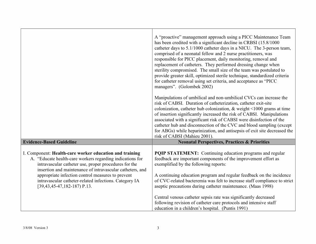
A “proactive” management approach using a PICC Maintenance Team has been credited with a significant decline in CRBSI (15.8/1000 catheter days to 5.1/1000 catheter days in a NICU. The 3-person team, comprised of a neonatal fellow and 2 nurse practitioners, was responsible for PICC placement, daily monitoring, removal and replacement of catheters. They performed dressing change when sterility compromised. The small size of the team was postulated to provide greater skill, optimized sterile technique, standardized criteria for catheter removal using set criteria, and acceptance as “PICC managers”. (Golombek 2002) Manipulations of umbilical and non-umbilical CVCs can increase the risk of CABSI. Duration of catheterization, catheter exit-site colonization, catheter hub colonization, & weight <1000 grams at time of insertion significantly increased the risk of CABSI. Manipulations associated with a significant risk of CABSI were disinfection of the catheter hub and disconnection of the CVC and blood sampling (except for ABGs) while heparinization, and antisepsis of exit site decreased the risk of CABSI (Mahieu 2001).
Evidence-Based Guideline Neonatal Perspectives, Practices & Priorities I. Component: Health-care worker education and training
A. “Educate health-care workers regarding indications for intravascular catheter use, proper procedures for the insertion and maintenance of intravascular catheters, and appropriate infection control measures to prevent intravascular catheter-related infections. Category IA [39,43,45-47,182-187) P.13.
PQIP STATEMENT: Continuing education programs and regular feedback are important components of the improvement effort as exemplified by the following reports: A continuing education program and regular feedback on the incidence of CVC-related bacteremia was felt to increase staff compliance to strict aseptic precautions during catheter maintenance. (Maas 1998) Central venous catheter sepsis rate was significantly decreased following revision of catheter care protocols and intensive staff education in a children’s hospital. (Puntis 1991)
3/8/08 Version 3 4
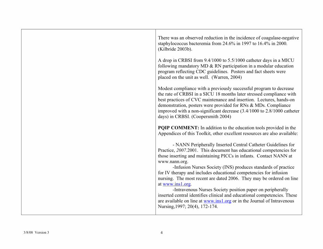
There was an observed reduction in the incidence of coagulase-negative staphylococcus bacteremia from 24.6% in 1997 to 16.4% in 2000. (Kilbride 2003b). A drop in CRBSI from 9.4/1000 to 5.5/1000 catheter days in a MICU following mandatory MD & RN participation in a modular education program reflecting CDC guidelines. Posters and fact sheets were placed on the unit as well. (Warren, 2004) Modest compliance with a previously successful program to decrease the rate of CRBSI in a SICU 18 months later stressed compliance with best practices of CVC maintenance and insertion. Lectures, hands-on demonstration, posters were provided for RNs & MDs. Compliance improved with a non-significant decrease (3.4/1000 to 2.8/1000 catheter days) in CRBSI. (Coopersmith 2004) PQIP COMMENT: In addition to the education tools provided in the Appendices of this Toolkit, other excellent resources are also available:
- NANN Peripherally Inserted Central Catheter Guidelines for Practice, 2007.2001. This document has educational competencies for those inserting and maintaining PICCs in infants. Contact NANN at www.nann.org. -Infusion Nurses Society (INS) produces standards of practice for IV therapy and includes educational competencies for infusion nursing. The most recent are dated 2006. They may be ordered on line at www.ins1.org. -Intravenous Nurses Society position paper on peripherally inserted central identifies clinical and educational competencies. These are available on line at www.ins1.org or in the Journal of Intravenous Nursing,1997; 20(4), 172-174.

3/8/08 Version 3 5
PQIP Educational and Implementation Tools: (See Appendices) -Sample Implementation Aids, e.g. staff education modules, skills laboratory modules, Policy and Procedures, competencies, assessment tools from CPQCC member units.
B. “Assess knowledge of and adherence to guidelines
periodically for all persons who insert and manage intravascular catheters. Category IA [39,43,46,182,188) P. 13
PQIP Appendix: Sample Vascular Set-Up Monitor Tool This tool facilitates concurrent assurance of desired nursing practices within a NICU related to the use of vascular access devices. PQIP COMMENT: Assessment of clinical practice is imperative and one of the initial steps in reduction of nosocomial sepsis.
C. “Ensure appropriate nursing staff levels in ICUs to
minimize the incidence of CRBSI.. Category IB [48,189,190].” P.13
California Childrens Services and Guidelines for Perinatal Care have staffing guidelines for NICUs. Additionally, California law has set minimum staffing ratios. Oslo, Norway university NICU report on how understaffing and overcrowding was associated with methicillin-resistant Staphylococcus outbreak. (Anderson, 2002)
II. Component: Surveillance (General recommendations for all intravascular catheters in adults and pediatric patients.)
A. "Monitor the catheter sites visually or by palpation through the intact dressing on a regular basis depending on the clinical situation of individual patients. If patients have tenderness at the insertion site, fever without obvious source, or other manifestations suggesting local or BSI, the dressing should be removed to allow thorough examination of the site. Category IB [1, 191-193].” P.13
PQIP STATEMENT: Each NICU should implement a regularly scheduled, standardized process to daily assess line discontinuation as well as every shift assessment for signs of infection and dressing integrity. B. PQIP COMMENT: Neonatal patients are unable to directly report signs of discomfort, however their providers can and should regularly assess them for signs of discomfort or pain, e.g. guarding, withdrawal, decreased limb movement, etc.

3/8/08 Version 3 6
B. “Encourage patients to report to their healthcare provider any changes in their catheter site or any new discomfort. Category II.” P. 13
C. “Record the operator, date and time of catheter insertion and removal, and dressing changes on a standardized form. Category II.” P.13
D. “Do not routinely culture catheter tips. Category IA” [8,194,195].” P.13
The National Healthcare Safety Network (NHSN) formerly known as the National Nosocomial Infections Surveillance or NNIS, provides data from participating hospitals about the incidence of central line associated infection. Current data is available for 2004 (NNIS System 2004), but future reports will include data for the year 2006 and will be published in the American Journal of Infection Control and posted on the CDC’s website (www.cdc.gov/ncidod/dhqp/nnis_pubs.html) in late Spring 2007. A “proactive” management approach using a PICC Maintenance Team has been credited with a significant decline in CRBSI (15.8/1000 catheter days to 5.1/1000 catheter days in a NICU. The 3-person team, comprised of a neonatal fellow and 2 nurse practitioners, was responsible for PICC placement, daily monitoring, removal and replacement of catheters. They performed dressing change when sterility compromised. The small size of the team was postulated to provide greater skill, optimized sterile technique, standardized criteria for catheter removal using set criteria, and acceptance as “PICC managers”. (Golombek, et al, 2002)

3/8/08 Version 3 7
Evidence-Based Guideline Neonatal Perspectives, Practices & Priorities (Central venous catheters, including PICC, hemodialysis, and pulmonary artery catheters, in adult and pediatric patients) E. “Conduct surveillance in ICUs and other patient populations to determine CRBSI rates, monitor trends in those rates, and to assist in identifying lapses in infection control practices. Category IA [3, 12,16,247-250].” P.16
First national point-prevalence survey NICU NI events demonstrates both their high rates and significant burden and the need for effective prevention measures. (Sohn 2001) Improving survival beyond postnatal day 2 (associated with increasing device use) increases total NI rates, even though device-specific NI rates per day are unchanging. (Zafar 2001).
F. “Express ICU data as the number of catheter-associated BSIs per 1,000 catheter-days for both adults and children and stratify by birth weight categories for neonatal ICUs to facilitate comparisons with national data in comparable patient populations and health-care settings. Category IB [3, 12,16,247-250].” P.16
Include in Appendix their actual form PQIP COMMENT: There are variable rates of catheter associated infections reported. Some reports combine all central lines (i.e. umbilical, PICC, tunneled) into one category, while others report infection per catheter type. The rates of infection vary depending on type of catheter. Note, the CDC National Healthcare Safety Network (NHSN) has promulgated new definitions and reporting formats for hospitals to begin using January 1, 2007. They specifically call for the segregation of umbilical catheter line days and event statistics from other central catheters line days and event statistics The new reporting definitions and formats can be found in Appendices _______. They can also be found on the CDC’s website: (http://www.cdc.gov/ncidod/dhqp/nhsn_members.html) A cohort study including 19,507 infants admitted to 17 Canadian NICUs assessed the incidence of nosocomial blood stream infection (one or more positive blood cultures obtained after 48 h of life in clinically symptomatic infant). Twenty-two percent of infants received CVCs. Incidence of BSI was 2.9/1000 noncatheter days, 7.2/1000 umbilical venous catheter days, 13.1/1000 percutaneous catheter days, and 12.1/1000 BroviacTM catheter days. Risk adjusted rates (EGA, sex, SGA, 5 min Apgar, outborn status, and SNAP-II score) were 2.5 for umbilical

3/8/08 Version 3 8
venous catheters, 4.6 for percutaneous catheters and 4.3 for BroviacTM catheters. (Chien, 2002)
G. “Investigate events leading to , unexpected, life-threatening or fatal outcomes. This includes any process variation for which a recurrence would carry a significant chance of a serious adverse outcome. Category IC [13].” P.16
Examples include: Use of lipid emulsions in very low birthweight infants is associated with an increased risk of coagulase-negative staphylococcal bacteremia. (Avila-Figueroa 1998) Changing from one brand of mechanical valve injection port to another was credited with an increase in CRBSI (1.55 to 2.79/1000 catheter days) in neonates over a 9-month period. Return to the original product was followed with a return in the CRBSI rate to the earlier rate. (Maragakis 2006) (Class III) PQIP STATEMENT: Successful promotion and advancement of enteral feeds decreases the duration of parenteral feeding and thus the opportunity for complications such as line-associated nosocomial infection. (Unger 1986, McClure 2000; Kennedy 2002; Tyson 2002, Anderson 2005). (CPQCC’s Nutritional Support of the VLBW Infant Part I and II toolkits are available online at: http://www.cpqcc.org/qualityimprovement.htm ) No specific recommendation is made when to implement feedings or about the rate of advancement pending further evidence. (CPQCC’s Nutritional Support of the VLBW Infant Part I and II Toolkits are available online at: http://www.cpqcc.org/qualityimprovement.htm)
Evidence-Based Guideline Neonatal Perspectives, Practices & Priorities III. Component: Hand hygiene
A. “Observe proper hand hygiene procedures either by washing hands with conventional antiseptic-containing soap and water or with waterless alcohol-based gels or foams. Observe hand hygiene before and after palpating
PQIP STATEMENT: As the following neonatal reports indicate, a comprehensive program for hand hygiene that emphasizes the use of waterless alcohol based gels is superior in attaining staff compliance. Any program requires continuing observation and feedback. Jewelry, such as rings, should not be worn by healthcare workers in the NICU.

3/8/08 Version 3 9
catheter insertion sites, as well as before and after inserting, replacing, accessing, repairing, or dressing an intravascular catheter. Palpation of the insertion site should not be performed after the application of antiseptic, unless aseptic technique is maintained. Category IA [43,70,196-200].” P.13
However this STATEMENT is based on data indicating only that hand contamination with potential pathogens is significantly more likely rather than upon evidence indicating actual increases in laboratory confirmed bloodstream infections. A clinical trial using a self-selected convenience sample and crossover design in 2 NICUs conducted over 2 years tested use of an antiseptic hand wash and alcohol sanitizer. No significant difference in HAI or mean microbial counts on the nurses’ hands was noted. The alcohol sanitizer was credited with improved skin condition and quality of hand hygiene and increased frequency of use over the antiseptic hand wash, however, the frequency of hand hygiene remained low. A need for systems-level interventions to increase quality of hand hygiene practices was needed. (Larson, 2005). Hand hygiene practices of 88 nurses in 6 NICUs showed a significantly shorter duration of hand hygiene with alcohol hand rub (6.26 sec) vs 12.24 sec with hand washing. There wasn’t a relationship between knowledge and hand decontamination technique. Hand decontamination at the beginning of a 12-hour shift was significantly longer, more thorough, with hands dried more effectively than at the end of the shift (no difference with an 8-hour shift). Knowledge of infection control practices was poor (56.3%-73.3%). Hand washing technique was significantly better with senior nurses, but not with alcohol gel. (Chedleigh 2005) An evidence-based hand hygiene policy, supported by an intensive education program, resulted in a significant increase in compliance and a significant decrease in false-positive coagulase-negative staphyloccal blood and CSF culture rates. (Sharek 2002) Physician adherence to hand hygiene was observed in 163 MDs in a variety of specialty areas. Compliance averaged 57% and varied among

3/8/08 Version 3 10
medical specialties. Adherence was higher when hand-rub solutions were easily accessible and when physicians valued hand hygiene, awareness of being observed and considered themselves role models. High workload, activities associated with a high risk for cross-transmission, and certain technical medical specialties (surgery, anesthesiology, emergency medicine and intensive care) were risk factors for no-compliance. (Pittet 2004) Compliance with hand hygiene using alcohol hand rub was significantly higher with nurses and physicians when they had been notified they would be observed than when they were covertly observed. There was no significant difference with other healthcare workers.(Eckmanns 2006) In a study of surgical ICU RNs ring wearing was associated with 10-fold higher median skin organism counts; contamination with Staphylococcus aureus, gram-negative bacilli, or Candida species; and a stepwise increased risk of contamination with any transient organism as the number of rings worn increased (odds ratio [OR] for 1 ring worn, 2.6; OR for >1 ring worn, 4.6). Ring wearing increased the frequency of hand contamination with potential nosocomial pathogens. Use of an alcohol-based hand rub resulted in significantly less frequent hand contamination. (Trick 2003) Laboratory personnel were observed for hand hygiene practices and found to 100% compliant while working within the lab. Compliance with the no jewelry policy (rings and watches) was poor initially with improvement after being provided with feedback about performance. Cultures taken from the skin under the ring or watch showed greater densities of commensal flora and pathogenic microorganisms. (Alp 2006) PQIP COMMENT: Physician neck ties (Dixon 2000, Ditchburn 2006) have been shown as NI vectors as have white coats (Wong 1991); their status in the NICU is

3/8/08 Version 3 11
again being debated.
B. "Use of gloves does not obviate the need for hand
hygiene. Category IA [43,198,199].” P. 13
PQIP COMMENT: Disinfection of hands before gloving is significantly more efficacious than hand hygiene alone or donning of gloves without prior hand hygiene.
In a prospective multi-centre study involving 1132 peripheral venous catheters in three hospitals, the relationship between various measures of hand hygiene before insertion of peripheral venous catheters and the frequency of infectious complications', such as local reddening, swelling, pain, purulence and fever of unknown origin, were analyzed. In comparison with simple hand washing, disinfection of hands before the insertion or wearing of gloves resulted in significantly fewer complications (relative risk 0.59 and 0.66, respectively). Normal hand washing was no better than no hand hygiene (relative risk 1.13), with regard to reduction of complications. This underlines the necessity of employing more effective measures of hand hygiene. (Hirschmann 2001) (Class II)
IV. Component: Aseptic technique during catheter insertion and care
A. “Maintain aseptic technique for the insertion and care of intravascular catheters. Category IA [22,71,201,201].” P.13
B. “Wear clean or sterile gloves when inserting an intravascular catheter as required by the Occupational Safety and Health Administration Bloodborne Pathogens Standard. Category IC. Wearing clean gloves rather than sterile gloves is acceptable for the insertion of peripheral intravascular catheters if the access site is not touched after the application of skin antiseptics. . Sterile gloves should be worn for the insertion of arterial and central
8 of 47 CVCs (17%) were found to have evidence of bacterial contamination prior to their insertion into the vein in a randomized prospective study in pediatric patients. Catheters were either opened normally or injected with normal saline through the wrapping prior to opening. Injection into the wrapper did not decrease the incidence of infection. None of the contaminated catheters was associated with a BSI during the first 90 days of dwell. (Hall 2005)

3/8/08 Version 3 12
catheters. Category IA [201,203].” P.14 C. “Wear clean or sterile gloves when changing the dressing
on intravascular catheters. Category IC.” P.14 V. Component: Catheter insertion
A. “Do not routinely use arterial or venous cutdown procedures as a method to insert catheters. Category IA [204-206].” P.14
PQIP STATEMENT: Each clinician should be limited to two attempts to achieve vascular access (INS, 2006), and each NICU should establish a reasonable limit to the number of practitioners allowed to attempt access to prevent trauma and an increase risk of infection. Document the total number of attempts to achieve access. The algorithm in Appendix ___ addresses a process for limiting the number of PIV attempts by evaluating the infant’s vascular access device needs. The risk of primary bacteremia increases when neonates required greater than 5 attempts to place a peripheral IV within a 48-hour period. (Grant 1997) A randomized, control trial was conducted to determine whether percutaneously inserted central venous catheters (PICC) and peripheral intravenous catheters (PIV) in infants with very low birth weight (VLBW). There was no difference in the incidence of sepsis, number of courses of antibiotics, or total duration of IV use between the 2 groups. The number of insertion attempts required for total IV therapy was significantly lower in the PICC group than in the PIV group (P=.002). PICC lines reduced the number of painful IV procedures in VLBW infants without additional morbidity. (Janes 2000). Insertion of a PICC carries a significantly lower risk of bacteremia (3/1138 catheter days) in infants <1000 grams than use of multiple PIVs (12/1114 catheter days (p<0.03). Infants were matched for birth weight, gestational age and gender, and CRIB scores in this prospective study. (Liossis 2003)
3/8/08 Version 3 13
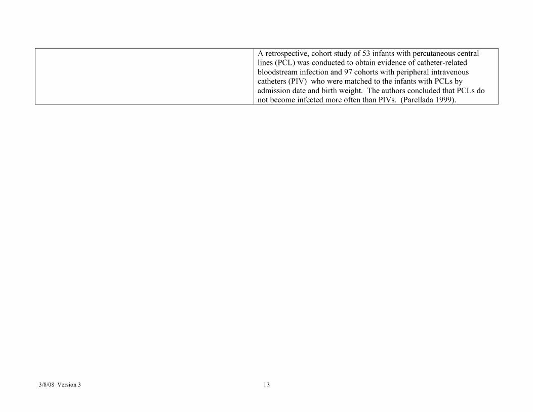
A retrospective, cohort study of 53 infants with percutaneous central lines (PCL) was conducted to obtain evidence of catheter-related bloodstream infection and 97 cohorts with peripheral intravenous catheters (PIV) who were matched to the infants with PCLs by admission date and birth weight. The authors concluded that PCLs do not become infected more often than PIVs. (Parellada 1999).

3/8/08 Version 3 14
Evidence-Based Guideline Neonatal Perspectives, Practices & Priorities B. Maximal sterile barrier precautions during catheter insertion (central venous catheters)
1. “Use aseptic technique including the use of a cap, mask, sterile gown, sterile gloves, and a large sterile sheet, for the insertion of CVCs (including PICCs) or guidewire exchange. Category IA [22,71].” P. 17
PQIP STATEMENT: Use of maximal barrier precautions for insertion of central catheters is recommended. Reports of fewer infectious complications among those whose catheters were placed in the Operating Room (non-randomized cohort studies) emphasizes the need to ensure near operating room like conditions (maximal barrier precautions and adequate antisepsis) wherever catheters are inserted. PICCs inserted in the operating room have fewer infectious complications than those inserted on the ward or in the outpatient clinic. (Hirschmann 2001). A similar observation was reported by Chowdhary in a retrospective review of 125 PICs placed in neonatal surgical patients. (Chowhary 2001)
VI. Component: Catheter site care
A. Cutaneous antisepsis 1. “Disinfect clean skin with an appropriate
antiseptic before catheter insertion and during dressing changes. Although a 2% chlorhexidine-based preparation is preferred, tincture of iodine, an iodophor, or 70% alcohol could be used. Category IA [73,75,207,208].” P.14
2. No recommendation can be made for the use of
chlorhexidine in infants aged < 2 months. Unresolved issue.” P.14
PQIP STATEMENT: There are no data that show any antiseptic agent to be superior to chlorhexidine gluconate (CHG) for skin antisepsis. Many CHG containing products exist on the market in both aqueous and alcoholic formulations and in a variety of strengths, contributing to the complexity of “best” newborn skin antisepsis. Taking into consideration the issues of efficacy and the potential of local irritation and systemic absorption, CHG or PI are the skin disinfectants recommended by PQIP as outlined below. Chlorhexidine Gluconate (CHG) Alcoholic-based:
• Apply over 30 seconds using side to side motion • Allow to dry over 30 seconds
Chlorhexidine Gluconate (CHG) Aqueous:
• Apply over 30 seconds • Remove with sterile water or saline following the procedure
(aqueous CHG will not dry due to its soapy consistency)

3/8/08 Version 3 15
(Malathi et al 1993, Lund et al 2001). Povidone iodine (PI):
• Apply over 30 seconds and allow to dry • Remove with sterile water or saline following the procedure
“After topical applications of chlorhexidine, some percutaneous absorption occurs, particularly in preterm newborns, but only at trace levels.” Studies to date have used a variety of concentrations for multiple interventions. Tens of thousands of neonates have received chlorhexidine for umbilical cord care, bathing and maternal vaginal lavage prior to birth without reported adverse effects. (Mullany, 2006). Povidone iodine containing solutions are commonly used for skin antisepsis prior to invasive procedures. Current practice is to remove the solution at the conclusion of the procedure. Caution should be exercised with use, particularly in very immature and sick infants who require repeated applications over large areas. (Linder, 1997). Four of 36 (11%) infants < 1000 grams exposed to 2% aqueous chlorhexidine developed severe skin irritation (all had erythema and one progressed to breakdown with exudates). The study used 2% chlorhexidine for all central & arterial catheters and PIVs for infants <1000 grams and<14 days and 1% chlorhexidine in ethanol for all other IVs. (Anderson 2005) Eight studies investigated in this meta analysis involving a total of 4143 catheters met the inclusion criteria. All studies were conducted in a hospital setting, (ICUs or hospital wards) and various catheter types were used. The summary risk ratio for catheter-related bloodstream infection was 0.49 (95% CI, 0.28 to 0.88) in patients whose catheter sites were disinfected with chlorhexidine gluconate instead of povidone-iodine. Among patients with a central vascular catheter, chlorhexidine

3/8/08 Version 3 16
gluconate reduced the risk for catheter-related bloodstream infection by 49% (risk ratio, 0.51 [CI, 0.27 to 0.97)]). Subset analyses of aqueous and nonaqueous solutions showed similar effect sizes, but only the subset analysis of the 5 studies that used alcoholic solution produced a statistically significant reduction in CRBSI. The lack of significant difference may be a result of inadequate statistical power. (Chaiyakunapruk 2002) (Class I) 0.5% chlorhexidine gluconate in 70% isopropyl alcohol is more efficacious than 10% povidone iodine for the prevention of peripheral intravenous catheter colonization in neonates. (Garland 1995) PQIP COMMENT: Alcohol applied topically may damage some polyurethane catheters when applied at the time of a dressing change. Check with the catheter manufacturer’s recommendations for compatibility.
3. “Allow the antiseptic to remain on the insertion site and to air dry before catheter insertion. Allow povidone iodine to remain on the skin for at least 2 minutes, or longer if it is not yet dry before insertion. Category IB [73,75,207,208].” P.14
3/8/08 Version 3 17
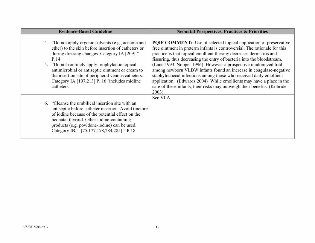
Evidence-Based Guideline Neonatal Perspectives, Practices & Priorities 4. “Do not apply organic solvents (e.g., acetone and
ether) to the skin before insertion of catheters or during dressing changes. Category IA [209].” P.14
5. “Do not routinely apply prophylactic topical antimicrobial or antiseptic ointment or cream to the insertion site of peripheral venous catheters. Category IA [107,213] P. 16 (includes midline catheters
PQIP COMMENT: Use of selected topical application of preservative-free ointment in preterm infants is controversial. The rationale for this practice is that topical emollient therapy decreases dermatitis and fissuring, thus decreasing the entry of bacteria into the bloodstream. (Lane 1993, Nopper 1996) However a prospective randomized trial among newborn VLBW infants found an increase in coagulase-negative staphylococcal infections among those who received daily emollient application. (Edwards 2004) While emollients may have a place in the care of these infants, their risks may outweigh their benefits. (Kilbride 2003).
6. “Cleanse the umbilical insertion site with an
antiseptic before catheter insertion. Avoid tincture of iodine because of the potential effect on the neonatal thyroid. Other iodine-containing products (e.g. povidone-iodine) can be used. Category IB.” [75,177,178,284,285].” P.18
See VI.A

3/8/08 Version 3 18
Evidence-Based Guideline Neonatal Perspectives, Practices & Priorities VII. Component: Catheter-site dressing regimens a-f apply to all intravascular catheters in adult and pediatric patients A. “Use either sterile gauze or sterile, transparent, semipermeable dressing to cover the catheter site. Category IA [146,210-212].”P.14
B. “If the patient is diaphoretic, or if the site is bleeding or oozing, a gauze dressing is preferable to a transparent, semi-permeable dressing. Category II [146,210-212].”P.14 C. “Replace catheter-site dressing if the dressing becomes damp, loosened, or visibly soiled. Category IB [146,210].” P.14
D. “Change dressings at least weekly for adult and adolescent patients; depending on the circumstances of the individual patient. Category II” [211].” P.14
PQIP STATEMENT: Dressings covering vascular devices should be evaluated by the bedside nurse as part of the shift assessment to ensure that they are intact and the site is not soiled. Routine changing of transparent, semi-permeable polyurethane dressings is not supported in the neonatal literature. Perform dressing change using sterile technique. (See Appendix ___). Changing the dressing on the central venous catheter in neonates only when the integrity of the dressing was compromised did not significantly increase the rate of nosocomial infection. (Zenk 1993) Routine changing of dressings may lead to loss of skin integrity and potentially increase the rate of sepsis. (Lund 1997)
E. “Do not use topical antibiotic ointment or creams on insertion sites (except when using dialysis catheters) because of their potential to promote fungal infections and antimicrobial resistance. Category IA [107,213].” P.14 F. “Do not submerge the catheter under water. Showering should be permitted if precautions can be taken to reduce the likelihood of introducing organisms into the catheter (e.g.,if the and connecting device are protected with an impermeable cover during the shower. Category II [214,215].” P.14

3/8/08 Version 3 19
Evidence-Based Guideline Neonatal Perspectives, Practices & Priorities g- m apply to central venous catheters in adult and pediatric patients
G. “Replace catheter-site dressing when it becomes damp, loosened, or soiled or when inspection of the site is necessary. Category IA [65,146,211].” P.17 H. “Replace dressings used on short-term CVC sites every 2 days for gauze dressings and every 7 days for transparent dressings, except in those pediatric patients in which the risk for dislodging the catheter outweighs the benefit of changing the dressing. Category IB [211].” P. 17
PQIP STATEMENT: See VII. A The weekly dressing change for infants with PICCs utilized two people ,one wearing sterile gown, cap and mask and the assistant a mask. They cleaned the site with povidone-iodine for two minutes and redressed with a transparent dressing, as part of a multidimensional strategy that showed a statistically significant decrease in CRBSI (Aly 2005) Changing the dressing on the central venous catheter in neonates only when the integrity of the dressing was compromised did not significantly increase the rate of nosocomial infection. (Zenk 1993)
I. “Replace dressings used on tunneled or implanted CVC sites no more than once per week, until the insertion site has healed. Category IB [211].P. 17
J. “Tunneled CVC sites that are well healed may not require dressings. Category II.” P.14
K. “No recommendation can be made for the use of chlorhexidine sponge dressings to reduce the incidence of infection. Unresolved issue.” P.18

3/8/08 Version 3 20
Evidence-Based Guideline Neonatal Perspectives, Practices & Priorities L. “Do not use chlorhexidine sponge dressings in neonates aged <7 days or of gestational age < 26 weeks. Category II [181].” P.18
PQIP STATEMENT: There are insufficient data to recommend the use of chlorhexidine sponge dressings in neonates at this time. PQIP COMMENT: The Garland trial described below did not compare the common practice of no routine dressing change to the use of the chlorhexidine-impregnated dressing. Additionally, alcohol served as the skin prep agent during catheter insertion and dressing changes for the infants receiving the chlorhexidine dressing, which is contrary to established practice. A multicenter randomized clinical trial determined that a novel chlorhexidine-impregnated dressing on the CVC sites, replaced weekly after cleaning the skin using 70% alcohol, was as effective as cutaneous disinfection with 10% PI and redressing the site every 3 to 7 days for preventing CRBSI and BSI without a source in critically ill neonates requiring prolonged central venous access. The risk of local contact dermatitis under the chlorhexidine dressing limits its use in low birth weight infants who require prolonged central access during the first 2 weeks of life. (Garland 2000) This prospective, randomized, controlled study was conducted with patients 0-18 years of age who were admitted to a pediatric cardiac intensive care unit and required a CVC for >48 hours. Patients were randomized to receive a transparent polyurethane insertion site dressing (control group) or a chlorhexidine gluconate-impregnated sponge (Biopatch®) dressing covered by a transparent polyurethane dressing (study group). CVC colonization occurred in 21 control patients (29%) and 11 (14.8%) study patients (P = 0.0446) Bloodstream infection occurred in 3 patients (4.2%) in the control group and 4 patients (5.4%) in the study group. Local redness was noted in 1 control patient and 4 study group patients. (Levy 2005)

3/8/08 Version 3 21
M. “No recommendation can be made for the use of sutureless securement devices. Unresolved issue” P. 18
N. “Ensure that catheter-site care is compatible with the catheter material.
Category IB [109,110].” P.18
PQIP COMMENT: Mupirocin ointment may adversely affect the integrity of some polyurethane catheters. In addition, alcohol used to clean catheter exit site may also damage some polyurethane catheters. Check the catheter manufacturer’s recommendations for compatibility.
O. "Do not use topical antibiotic ointment or creams on umbilical catheter insertion sites because of the potential to promote fungal infections and antimicrobial resistance. Category IA [107,213].” P.18 P. “Use a sterile sleeve for all pulmonary artery catheters
Category IB” (148). P. 18

3/8/08 Version 3 22
Evidence-Based Guideline Neonatal Perspectives, Practices & Priorities VIII. Component: Selection of intravascular catheters
A. “Select the catheter, insertion technique, and insertion site with the lowest risk for complications (infectious and noninfectious) for the anticipated type and duration of IV therapy. Category IA [22,55,59,216-218].” P.14
There are conflicting reports on the risk of infection and optimal time for removal for vascular access devices in the neonate. (Cronin 1990; Landers 1991) See Component V for catheter insertion content. Retrospective data analysis from two NICUs revealed NI (positive blood culture after the 3rd postnatal day) in 10.4% infants. Infected infants, in contrast to non-infected, had a significantly (P < 0.001) greater number of multiple catheters (2.3 vs 1.4) had lower birth weights (1.2 vs 2.1) kg), were younger (28 vs 33 weeks) and had lower 1 and 5 minute Apgar scores (4.3 and 6.7 vs 5.5 and 7.4). The most common organism was coagulase negative Staphylococcus. In a subset population as analyses revealed, longer duration of UA use was associated with higher infection rates [13.6% with UA use for > 8 days vs 1.3% for < 7 days (P < 0.0001)]. PC use had a lower rate of sepsis than CV use (5.1% vs 15.2%; P<0.05). Use of intravascular catheters should be balanced between the need for vascular access and the risk for sepsis. (Bhandari 1997). Infants <1250 grams were randomly assigned to a long-term UVC (up to 28 days) or short-term (7-10 days) followed by a PICC. Time to infection did not differ between groups. Infection occurred in 13% (7.4/1000 catheter days) in the short-term group and 20% (11.5/1000 catheter days) in the long-term group (NS). Seven infections in the short-term group were in UVCs and 18 in the long-term group. The remainder of infections were in PICCs. Long-term use of UVCs did not increase infection compared with short-term use of a UVC followed by placement of a PICC. Although the study had limited power, the authors suggest that it is may be reasonable to extend beyond 14 days the current,CDC recommendation to limit UVC use to 14 days. (Butler-O”Hara 2006)

3/8/08 Version 3 23
A retrospective review of 79 surgical newborns with tunneled, cuffed CVCs (such as tunneled central venous catheter, as described by Dr. John Broviac) who subsequently developed sepsis (+ blood culture) identified 19 cases of proven sepsis (9.9/1000 catheter days) and 8 (1.9/1000 catheter days). Sepsis occurred in 12 infants with intestinal surgical procedures, 11 of who had stomas. Lower gestational age, more than 1 operation, younger when first stoma created all contributed significantly to the risk of sepsis. (Klein 2003) A retrospective review of PICCs inserted into 112 surgical term and preterm neonates (49 inserted in the OR at the beginning or end of surgery and 34 in the ICU) to assess rate of complications (sepsis, occlusion & dislodgement) based on place on insertion. Twenty-four PICCs were confirmed infected (blood and tip cultures) with the predominance being due to Staphylococcus epidermidis. Mean complication-free line survival was 22 days with a maximum of 56 days. Twenty-six PICCs survived beyond 28 days. Complications (sepsis, occlusion and dislodgement) were significantly lower when catheters were placed in the OR, presumably because the infants were more affectively sedated and subjected to more adequate antiseptic and barrier measures. The authors concluded that there was no convincing evidence to recommend limiting the catheter dwell to 28 days. (Chowdhary 2001)(Class
Selection of peripheral venous including midline
catheter: b-c, h
B. “Select catheters based on the intended purpose and
duration of use, known complications (e.g., phlebitis and infiltration), and experience of individual catheter operators Category IB [67,68,244].” P.16
Use of peripheral catheters composed of Vialon® (a type of polyurethane) demonstrated a significant decrease in infiltration over Teflon® catheters in neonates in a randomized trial. (Stanley 1992)
C. “Avoid the use of steel needles for the administration of
A randomized, controlled study in neonates demonstrated that Teflon® catheters remain functional three times longer than steel needles with no

3/8/08 Version 3 24
fluids and medication that might cause tissue necrosis if extravasation occurs. Category IA [67,68].” P. 16
apparent increase in complications. Steel needles remained in place for a significantly shorter period of time and were associated with a 100% rate of infiltration. (Batton 1982).
D. “Use a midline catheter or PICC when the duration of
IV therapy will likely exceed 6 days. Category IB [244].” P.16
PQIP STATEMENT: There are now many retrospective series of neonatal patients which support the CDC recommendation about when to insert a PICC rather than a PIV. However, the one RCT on point found only a significant reduction in painful sticks, but no difference in sepsis rates between the PIV and PICC groups. Decision making should be individualized given the unresolved data to this date. A randomized, control trial was conducted to determine whether percutaneously inserted central venous catheters (PICC) and peripheral intravenous catheters (PIV) in infants with very low birth weight (VLBW). There was no difference in the incidence of sepsis , number of courses of antibiotics, or total duration of IV use between the 2 groups. The number of insertion attempts required for total IV therapy was significantly lower in the PICC group than in the PIV group (P=.002). PICC lines reduced the number of painful IV procedures in VLBW infants without additional morbidity. (Janes 2000). Insertion of a PICC carries a significantly lower risk of bacteremia (3/1138 catheter days) in infants <1000 grams than use of multiple PIVs (12/1114 catheter days (p<0.03). Infants were matched for birth weight, gestational age and gender, and CRIB scores in this prospective study. (Liossis 2003) A retrospective, cohort study of 53 infants with percutaneous central lines (PCL) was conducted to obtain evidence of catheter-related bloodstream infection and 97 cohorts with peripheral intravenous catheters (PIV) who were matched to the infants with PCLs by admission date and birth weight. The authors concluded that PCLs do not become infected more often than PIVs. (Parellada 1999).

3/8/08 Version 3 25
Evidence-Based Guideline Neonatal Perspectives, Practices & Priorities
D. continued. “Use a midline catheter or PICC when the duration of IV therapy will likely exceed 6 days. Category IB [244].” P.16
1,130 midline catheters were inserted in 858 patients ranging in age at insertion from 1 to 249 days, 360-8,000 gm in weight, and 23-42 weeks gestational age at birth. Overall mean catheter dwell time was 8.7 days. Elective removal represented 43 percent of all removals. Incidence of positive blood culture was 3.5 percent (0.41/1,000 catheter days), with the risk significantly higher if a central line was also in place. Other complications leading to catheter removal include 22% infiltration, 11% leaking, 17% occlusion, 4% dislodgement, 2% phlebitis, and 0.2% malposition. (Leick-Rude 2006) Midline catheters were placed in infants requiring intravenous therapy for >3 days, but nor requiring long-term vascular access and in new admissions if a short peripheral iv could not be placed after three attempts. A total of 143 midline catheters were placed in premature and term infants (25 – 40 weeks) weighing 540 – 4010 grams. Of the 135 catheters with data available, the mean indwelling time was 10 days (range 1 – 80 days). Forty-nine of the catheters survived to the conclusion of therapy. Reported complications included: leaking or edema at insertion site 34%, dislodged or clotted catheter 17%, and catheter-related infection 0%. (Wyckoff 1999)
Nine infants of 25 to 34 weeks gestation (675 to 1710 grams) were enrolled in the study to compare dwell time and reason for removal of midline catheters with respective data for peripheral intravenous catheters. Average dwell time for midline catheters was 9 days and peripheral IVs 3.1 days. There were no episodes of suspected or confirmed sepsis or major complications with either type of catheter. (Lesser 1999).

3/8/08 Version 3 26
Evidence-Based Guideline Neonatal Perspectives, Practices & Priorities E. “Use a CVC with the minimum number of ports or
lumens essential for the management of the patient. Category IB [251-254].” P.16
Three studies qualified for inclusion in this review (Khilnani 1991; Loisel 1996; Soupre 1998-see below). The use of multiple lumen umbilical venous cathethers ( ML-UVCs) in comparison to single lumen (SL)UVCs in neonates is associated with decrease in the usage of PIVs in first week of life, but an increase in catheter malfunctions. As the quality of included randomized studies is poor and the estimates of clinically important complications are imprecise, no firm recommendations can be made regarding the choice of UVC. (Kabra 2005). A meta-analysis of 15 published studies concluded that multilumen central venous catheters may be associated with a slightly higher risk of infection when compared with single-lumen catheters; however, this relationship diminishes when only high-quality studies that control for patient differences are considered. The slight increase in infectious risk when using multilumen catheters is likely offset by their improved convenience, thereby justifying the continued use of multilumen vascular catheters.(Dezfulian 2003) Infants randomized to receive a single or double lumen umbilical venous catheter experienced significantly fewer venipunctures and peripheral intravenous lines placed during their first two weeks of life. The incidence of sepsis or other complications was not higher in the group having umbilical catheters over the group with the peripheral IVs. (Loisel 1996). Double lumen umbilical venous catheters are well tolerated for short-term use, decrease the need for additional venous catheters in critically ill neonates, and may not significantly increase the risk of mechanical complications when compared with single lumen umbilical venous catheters. (Khilnani 1991).

3/8/08 Version 3 27
Use of double-lumen umbilical venous catheters entails no greater risk than use of single-lumen umbilical venous catheters and may reduce iatrogenic stress associated with the starting of peripheral intravenous lines. (Ramachandran 1994)
F. “Designate one port exclusively for hyperalimentation if
a multilumen catheter is used to administer parenteral nutrition. Category II [266].” P.17
G. “ Use totally implantable access devices for patients who require long-term, intermittent vascular access. For patients requiring frequent or continuous access, a PICC or tunneled CVC is preferable. Category II [256,257]” P. 17
H. Use of antimicrobial or antiseptic-impregnated CVCs
1. “No Recommendation can be made for the use of impregnated catheters in children. Unresolved issue.” P.17
PQIP COMMENT: There are, as yet, no data on the use of these catheters in neonates.

3/8/08 Version 3 28
Evidence-Based Guideline Neonatal Perspectives, Practices & Priorities I. “Designate trained personnel for the insertion and maintenance of intravascular catheters. Category IA [46,47,210,242]. P. 15
See INS Standards of Practice. Refer to Component 14 for use of designated personnel for placement of central venous catheters. See Component II
J. “Designate personnel who have been trained and exhibit competency to supervise trainees who perform catheter insertion. Category IA [39,43,46,182,187,188 P.17 (central venous catheters)
See INS Standards of Practice and NANN PICC Guidelines See Component II
IX. Component: Selection of catheter insertion site A. In pediatric patients, the hand, the dorsum of the foot, or
the scalp can be used as the catheter insertion site. Category II P. 16 (refers to peripheral catheters)
B. “Weigh the risk and benefits of placing a device at a recommended site to reduce infectious complications against the risk for mechanical complications (e.g., pneumothorax, subclavian artery puncture, subclavian vein laceration, subclavian vein stenosis, hemothorax, thrombosis, air embolism, and catheter misplacement). Category IA [22,55,59,218]” P. 17 (refers to CVCs)
C. “No recommendation can be made for a preferred site of insertion to minimize infection risk for a nontunneled CVC. Unresolved issue [61-63]” P. 17
X. Component: Replacement/removal of intravascular catheters
A. “Promptly remove any intravascular catheter that is no longer essential. Category IA [219,220].” P.14
There are conflicting reports on the risk of infection and optimal time for removal for vascular access devices in the neonate. (Cronin 1990; Landers 1991) As part of a multifactor intervention study showing a significant reduction in nosocomial BSI, removal of each PIV was required at 48 hours. (Anderson 2005).

3/8/08 Version 3 29
As part of a multidimensional project to decrease CRBSI, Golombek et al, (2002) removed PICCs after 6 weeks of therapy and showed a statistically significant reduction in CRBSI. (Golombek 2002)
B. “Do not routinely replace central venous or arterial
catheters solely for the purposes of reducing the incidence of infection. Category IB [134,135,221].” P. 14
C. “Do not routinely replace central venous catheters or PICCs as a method to prevent catheter-related infections. Category IB [119, 121, 122].” P.14
D. “Do not routinely replace midline catheters as a means to reduce the risk for infection. Category IB [131].” P. 16
E. “Do not routinely replace CVCs, PICCs, hemodialysis
catheters, or pulmonary artery catheters to prevent catheter-related infections. Category IB [132,134,135].” P.17
F. “Do not remove CVCs or PICCs on the basis of fever alone. Use clinical judgement regarding the appropriateness of removing the catheter if infections evidenced elsewhere or if a noninfectious cause of fever is suspected. Category II [224.264].” P.17
PQIP COMMENT: The species of micro-organism, the degree and scope of systemic inflammatory response and co-existing morbidities should all be part of the decision-making as to whether to retain or remove any and all catheters. In general, infections with Staphylococcal Aureus, Enterococcus, fungi, and most gram-negative organisms require immediate catheter removal. Infections with coagulase-negative staphylococcus may be successfully treated with the catheter still in place, unless the blood culture is positive three or more days. The species of bacterium should be an essential component of the decision to remove the catheter. Bacteremic infants experienced fewer infection-related complications (osteomyelitis, vital organ abscess, positive echocardiogram, positive lumbar puncture, death) when the

3/8/08 Version 3 30
central catheter was removed promptly (within 24 hours of species identification on blood culture) rather than attempt to treat through the catheter when the blood culture was positive for S aureus or a non-enteric Gram-negative rod. A blood culture for Coagulase negative staph may merit catheter retention. After 3 positive blood cultures for CoNS, the morbidity increases significantly. Treatment was successful and without complications in 34% of infants with enteric Gram-negative rods and in 42% of infants infected with Enterococcus when the CVC was retained. (Benjamin 2001). Failure to remove CVC as soon as candidemia was detected (within 3 days after the first positive blood culture) was associated with a significantly increased mortality in C albicans candidemia and prolonged duration of candidemia regardless of Candida species. (Karlowicz 2000). “…antibiotic therapy for suspected central venous line infection should always be administered through the central line.” (Craft 2001). Retention of CVCs was unlikely when bacteremia lasted >1 day OR when severe thrombocytopenia was present. (Nazemi 2003) Early CVC removal (<3days following a positive blood culture for CONS) did not increase the complication rate when compared to late removal (>3 days) in a retrospective cohort study. There was a 13% incidence of CONS >3 days in the early removal group vs a 43% incidence in the late removal group. Vancomycin was not started until the blood culture became positive.(Karlowitz 2002)

3/8/08 Version 3 31
Evidence-Based Guideline Neonatal Perspectives, Practices & Priorities G. “…Leave peripheral venous catheters in place in children until IV therapy is completed, unless complications (e.g. phlebitis and infiltration) occur. Category IB [174,175,222,223].”P.14 & 20. This includes midline catheters.
Anderson (2005) removed the PIV @48 hours as part of a multidimensional strategy, which significantly decreased CRBSI.
H. " When adherence to aseptic technique cannot be ensured (i.e., when catheters are inserted during a medical emergency),replace all catheters as soon as possible and after no longer than 48 hours. Category II [22,71,210,201]. P.14
Anderson (2005) removed the PIV @ 48 hours as part of a multidimensional strategy, which significantly decreased CRBSI..
I. “Do not routinely replace peripheral arterial catheters to prevent catheter-related infections. Category II [132,147,221,274] p. 18
J. “Remove and do not replace umbilical artery catheters if any signs of CRBSI, vascular insufficiency, or thrombosis are present. Category II [283].” P.18
PQIP COMMENT: There are several clinical series reporting successful thrombolytic treatment of umbilical catheter and aortic thromboses (usually without concomitant infections) (Erdstrom 2000, Harman 2001, Cheah 2001). However the meta-analysis of existing studies concludes that: “Clinically symptomatic thromboses are infrequent but serious complications in infants undergoing intensive care Most are related to central vascular catheters. Symptomatic thrombosis may cause severe morbidity due to irreversible organ damage and also loss of limbs...There is a need for adequately powered muticentre trials to determine the safety and efficacy of thrombolytic therapy for major thromboses in the neonate.” (John 2006)
K. “Remove and do not replace umbilical venous catheters if any signs of CRBSI or thrombosis are present [283]. Category II” P.18
PQIP COMMENT: Regarding management of thromboses, see X. J.
L. “Replace umbilical venous catheters only if the catheter malfunctions. Category II” P. 18
An occluded lumen of a multilumen device warrants removal of the catheter unless opening with an agent identified in X..O is successful.

3/8/08 Version 3 32
M. “Remove umbilical catheters as soon as possible when no longer needed or when any sign of vascular insufficiency to the lower extremities is observed. Optimally, umbilical artery catheters should not be left in place > 5 days. Category II [283,289].” P.18
PQIP Comment: Regarding management of thromboses, see X.J.
N. "Umbilical venous catheters should be removed as soon as possible when no longer needed but can be used up to 14 days if managed aseptically. Category II [290,291].” P.18
See Component VIII.A PQIP COMMENT: Additional data may lead to revision of the current CDC recommendation suggesting a limit on the duration of an umbilical venous catheter. Infants <1250 grams were randomly assigned to a long-term UVC (up to 28 days) or short-term (7-10 days) followed by a PICC. Time to infection did not differ between groups. Infection occurred in 13% (7.4/1000 catheter days) in the short-term group and 20% (11.5/1000 catheter days) in the long-term group (NS). Seven infections in the short-term group were in UVCs and 18 in the long-term group. The remainder of infections were in PICCs. Long-term use of UVCs did not increase infection compared with short-term use of a UVC followed by placement of a PICC. Although the study had limited power, the authors suggest that it is may be reasonable to extend beyond 14 days the current,CDC recommendation to limit UVC use to 14 days. (Butler-O”Hara 2006)
3/8/08 Version 3 33
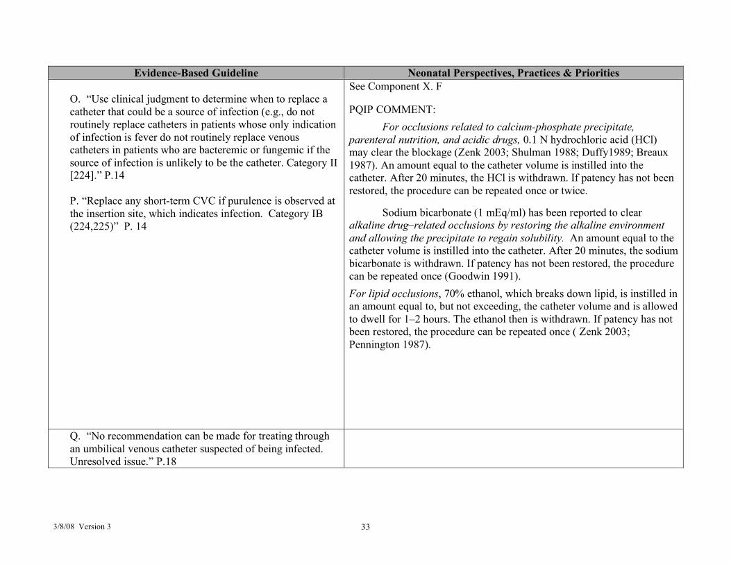
Evidence-Based Guideline Neonatal Perspectives, Practices & Priorities O. “Use clinical judgment to determine when to replace a catheter that could be a source of infection (e.g., do not routinely replace catheters in patients whose only indication of infection is fever do not routinely replace venous catheters in patients who are bacteremic or fungemic if the source of infection is unlikely to be the catheter. Category II [224].” P.14 P. “Replace any short-term CVC if purulence is observed at the insertion site, which indicates infection. Category IB (224,225)” P. 14
See Component X. F PQIP COMMENT:
For occlusions related to calcium-phosphate precipitate, parenteral nutrition, and acidic drugs, 0.1 N hydrochloric acid (HCl) may clear the blockage (Zenk 2003; Shulman 1988; Duffy1989; Breaux 1987). An amount equal to the catheter volume is instilled into the catheter. After 20 minutes, the HCl is withdrawn. If patency has not been restored, the procedure can be repeated once or twice.
Sodium bicarbonate (1 mEq/ml) has been reported to clear alkaline drug–related occlusions by restoring the alkaline environment and allowing the precipitate to regain solubility. An amount equal to the catheter volume is instilled into the catheter. After 20 minutes, the sodium bicarbonate is withdrawn. If patency has not been restored, the procedure can be repeated once (Goodwin 1991). For lipid occlusions, 70% ethanol, which breaks down lipid, is instilled in an amount equal to, but not exceeding, the catheter volume and is allowed to dwell for 1–2 hours. The ethanol then is withdrawn. If patency has not been restored, the procedure can be repeated once ( Zenk 2003; Pennington 1987).
Q. “No recommendation can be made for treating through an umbilical venous catheter suspected of being infected. Unresolved issue.” P.18

3/8/08 Version 3 34
Evidence-Based Guideline Neonatal Perspectives, Practices & Priorities
R. “Replace all CVCs if the patient is hemodynamically unstable and CRBSI is suspected. Category II [224,225].” P.15
S. Guidewire exchange (central venous catheters)
1. “Do not use guidewire exchanges routinely for non-tunneled catheters to prevent infection. Category IB [135,265].” P. 17
2. “Use a guidewire exchange to replace a malfunctioning nontunneled catheter if there is no evidence of infection is present Category IB [135,265].” P. 17
3. "Use a new set of sterile gloves before handling the new catheter when guidewire exchanges are performed. Category II [22,71].” P.17
T. Guidewire exchange (all catheters) “Do not use guidewire techniques to replace catheters in patients suspected of having catheter-related infection. Category IB [134,135].” P.15
A guidewire is a device placed into the vasculature over which a vascular catheter is placed. A stylet is a device placed within the lumen of a vascular catheter to facilitate placement. PQIP COMMENT: There are anecdotal reports in neonates of exchanging central venous catheters over guidewires, as well as initially placing them over guidewires. (Valk 1995; Stephenson 1993) The incidence of serious complications, especially infectious complications, was low with percutaneous placement using the Seldinger (guidewire) technique. (Valk 1995) Improved success without an increase in insertion-related complications was demonstrated during placement of peripherally inserted central venous catheters using a guidewire, which was kept in the peripheral circulation. (Stephenson 1993)
U. “Evaluate the catheter insertion site daily, by palpation through the dressing to discern tenderness and by inspection if a transparent dressing is in use. Gauze and opaque dressings should not be removed if the patient has no clinical signs of infection. If the patient has local tenderness or other signs of possible CRBSI, an opaque dressing should be removed and the site inspected visually. Category II.” P.16 (peripheral venous catheters, including midline catheters, in adult and pediatric patients)

3/8/08 Version 3 35
Evidence-Based Guideline Neonatal Perspectives, Practices & Priorities V. “Remove peripheral venous catheters if the patient develops signs of phlebitis (i.e., warmth, tenderness, erythema, and palpable venous cord), infection, or a malfunctioning catheter. Category IB [66].” P.16 W. “In pediatric patients, leave peripheral venous catheters
in place until IV therapy is completed, unless a complication (e.g., phlebitis and infiltration) occurs. Category IB (174,175,222,223)” P. 16
XI. Component: Replacement of administration sets*, needleless systems, and parenteral fluids (* Administration sets include the area from the spike of tubing entering the fluid container to the hub of the vascular access device. However, a short extension tube might be connected to the catheter and might be considered a portion of the catheter to facilitate aseptic technique when changing administration sets.” P.15)
PQIP STATEMENT: Entry into any vascular catheter should be minimized and blood sample collection batched whenever possible. Follow the process outlined in Section XII. The potential for contamination is minimized by standardizing the design of the unit’s closed systems for infusion and the set-up of component lines. Appendix ___ describes a standard procedure for line management during entry of a line for replacement of parenteral solutions or drawing blood.
A. “Replace administration sets, including secondary sets
and add-on devices, no more frequently than at 72-hour intervals, unless catheter-related infection is suspected or documented. Category IA” [149-151]. P. 15
As part of a multidimensional change process that significantly decreased the rate of CRBSI, tubing and fluid changes on PICCs were performed upon catheter insertion and daily. Entry into the CVC was limited to once every 24-hours. Two nurses performed the tubing change, with one wearing a mask and sterile gloves and the other a mask. A closed medication delivery system is part of the tubing set-up and is also changed every 24-hours. (Aly 2004) Microbial contamination rate of infusate in the intravenous tubing of newborns receiving lipid therapy was compared by changing tubing interval at 72 hours versus 24 hours. The rate of blood cultures ordered was higher in the 72-versus the 24-hour group and a higher proportion of infants randomized to the 72-hour group died although the excess deaths could not clearly be attributed to bacteremia.” (Matlow 1999).

3/8/08 Version 3 36
Educational Tools (Appendix)
Kaiser Method of tubing set-up: 1. Standardize tubing set-up for all lines and present on a poster
board). 2. Maintain a closed system for all lines. 3. Stopcocks should not be left open and should be covered with
injection ports. 4. Teach consistent method to access all injection ports. 5. FBI (Fight Bacterial Infection) approach (Kilbride 2003a)
Sample policies and photos from NICUs demonstrating tubing set-up and care and maintenance of vascular devices.

3/8/08 Version 3 37
Evidence-Based Guideline Neonatal Perspectives, Practices & Priorities B. “Replace tubing used to administer blood, blood
products, or lipid emulsions (those combined with amino acids and glucose in a 3-in-1 admixture or infused separately) within 24 hours of initiating the infusion. Category IB [158,226-229].” P.15 If the solution contains only dextrose and amino acids, the administration set does not need to be replaced more frequently than every 72 hours. Category II [226]” P. 15
C. “Replace tubing used to administer propofol infusions every 6 or 12 hours, depending on its use, per the manufacturer’s recommendation. Category IA [230]. P. 15
D. Needleless intravascular devices
1. “Change the needleless components at least as often as the administration set. Category II [160-162,164-167].” P.15
2. “Change caps no more frequently than every 72 hours or according to manufacturers’ recommendations. Category II [160,162,165,166].” P.15
3. “Ensure that all components of the system are compatible to minimize leaks and breaks in the system. . Category II [163].” P.15
4. “Minimize contamination risk by wiping the access port with an appropriate antiseptic and accessing the port only with sterile devices. Category IB [162,163,165].” P.15
It is important to ensure the compatibility of components of the infusion pump tubing, add on devices (i.e. T connectors or Y connectors, and the injection ports). Some plastics will crack with exposure to lipids over time. See XII for cleaning injection ports
E. Parenteral fluids
Removal of the VAD when the infant was tolerating >120 ml/kg/day of enteral nutrition was part of a multifactorial strategy resulting in a

3/8/08 Version 3 38
1. “Complete the infusion of lipid-containing
colutions (e.g., 3-in-1 solutions) within 24 hours of hanging the solution. Category IB [156-158,226,229]” P. 15
2. “Complete the infusion of lipid emulsions alone within 12 hours of hanging the emulsion. If volume considerations require more time, the infusion should be completed with 24 hours. Category IB [156-158].” P. 15
statistically significant reduction in BSIs. (Anderson 2005) There is evidence of an association between duration of IV lipid use and bacteremia, and the group believed that along with the practices observed at benchmark sites, this evidence was strong enough to recommend limiting the use of IV lipids (Kilbride 2003).
3. “Complete infusions of blood or other blood
products within 4 hours of hanging the blood. Category II [231-234].” P. 15
4. “No recommendation can be made for the hang
time of other parenteral fluids. Unresolved issue.” P. 15
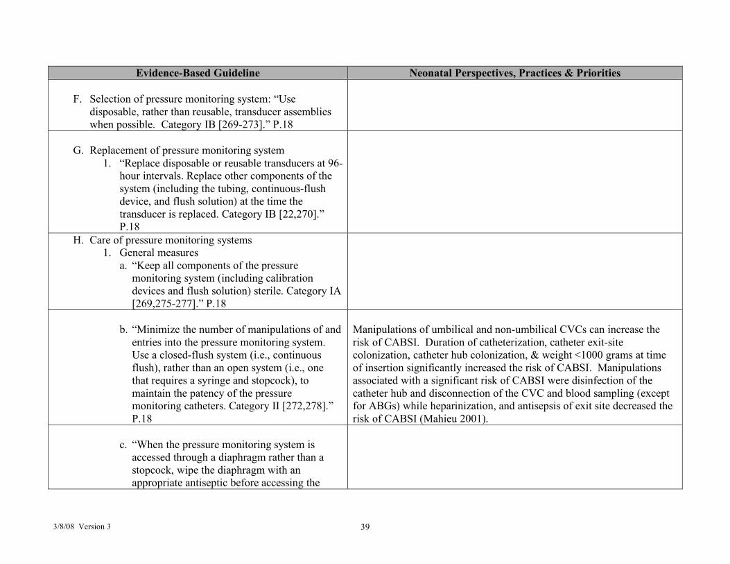
3/8/08 Version 3 39
Evidence-Based Guideline Neonatal Perspectives, Practices & Priorities F. Selection of pressure monitoring system: “Use
disposable, rather than reusable, transducer assemblies when possible. Category IB [269-273].” P.18
G. Replacement of pressure monitoring system
1. “Replace disposable or reusable transducers at 96-hour intervals. Replace other components of the system (including the tubing, continuous-flush device, and flush solution) at the time the transducer is replaced. Category IB [22,270].” P.18
H. Care of pressure monitoring systems 1. General measures
a. “Keep all components of the pressure monitoring system (including calibration devices and flush solution) sterile. Category IA [269,275-277].” P.18
b. “Minimize the number of manipulations of and
entries into the pressure monitoring system. Use a closed-flush system (i.e., continuous flush), rather than an open system (i.e., one that requires a syringe and stopcock), to maintain the patency of the pressure monitoring catheters. Category II [272,278].” P.18
Manipulations of umbilical and non-umbilical CVCs can increase the risk of CABSI. Duration of catheterization, catheter exit-site colonization, catheter hub colonization, & weight <1000 grams at time of insertion significantly increased the risk of CABSI. Manipulations associated with a significant risk of CABSI were disinfection of the catheter hub and disconnection of the CVC and blood sampling (except for ABGs) while heparinization, and antisepsis of exit site decreased the risk of CABSI (Mahieu 2001).
c. “When the pressure monitoring system is
accessed through a diaphragm rather than a stopcock, wipe the diaphragm with an appropriate antiseptic before accessing the

3/8/08 Version 3 40
system. Category IA [272].” P.18

3/8/08 Version 3 41
Evidence-Based Guideline Neonatal Perspectives, Practices & Priorities d. “Do not administer dextrose-containing
solutions or parenteral nutrition fluids through the pressure monitoring circuit. Category IA [272,279,280].” P.18
Dextrose containing solutions are often infused through the pressure transducer set-up in the NICU because of the nature of the line set-up and the necessity to provide dextrose to the neonate.
2. “Sterilization or disinfection of pressure monitoring systems
a. “Use disposable transducers. Category IB [272,279,280].” P.18
b.“Sterilize reusable transducers according to the manufacturers’ instructions if the use of disposable transducers is not feasible. Category IA [272,279-282].” P. 18
XII. Component: IV- injection ports
A. “Clean injection ports with 70% alcohol or an iodophor before accessing the system. Category IA [164,235,236].” P. 15
PQIP STATEMENT: The type and brand of the injection port (mechanical valve and split septum) and the type and method of disinfecting the injection port may influence the rate of CRBSI. W hen entering the vascular access device through an injection port, prep the injection port with alcohol or CHG, using sufficient friction and allow adequate time for the surface to dry. Repeat this process prior to disconnecting infusion tubing components as well. Hand antisepsis and use of a sterile field under the connections are important. Contamination rates following prepping of mechanical valve style injection ports (Posiflow®) with 0.5% chlorhexidine in 70% isopropyl alcohol was 31%,, with isopropyl alcohol 69%, and 42% with povidone iodine following patient use for 72 hours. There was no significant difference in the internal colonization rates after clinical use of any of the agents. (Casey 2003) A randomized prospective study comparing use of a mechanical valve style injection port (Clave®) and a conventional open system in adults

3/8/08 Version 3 42
showed a significant decrease in catheter tip colonization, hub colonization, skin colonization. The incidence of CRBSI was lower, but not statistically significant. The injection port and open system were disinfected with 2% chlorhexidine. The mechanical valve was changed every 7 days and the open system every 3 days. (Bouza 2003) Changing from one brand of mechanical valve injection port to another was credited with an increase in CRBSI (1.55 to 2.79/1000 catheter days) in neonates over a 9-month period. Return to the original product was followed with a return in the CRBSI rate to the earlier rate. While the increase in CRBSI in the NICU was not statistically significant, the rate in the other ICUs in this one facility was. (Maragakis 2006) (Class III) Use of an antiseptic cap containing 2% chlorhexidine in 70% isopropyl alcohol to cover the injection port contaminated with Enterococcus faecalis resulted in 1 (1.6%) transmission of microorganisms when compared to a vigorous 3-5 second swab with 70% isopropyl alcohol in a simulation study (p=0.001) (Menyhay 2006)
B. “Cap all stopcocks when not in use. Category IB [235].” P.15
PQIP STATEMENT: The rational for closed systems stems from the observation that significant numbers of catheter-associated infections were preceded by or coincided with contamination of the hub. Closed systems are recommended for all vascular entry points (inclusive of peripheral IV lines, PICCs, umbilical venous and arterial lines) as they are a practical way to achieve fewer catheter-associated infections, not withstanding the anecdotal reports of NICUs having achieved low rates of infection without arterial lines being closed. See Appendix ___ The catheter hub may be a major portal of entry for microorganisms causing sepsis in a NICU. 54% of episodes of CRS were preceded by or coincided with contamination of the hub. (Salzman 1993)

3/8/08 Version 3 43
Use of a hub containing an antiseptic chamber (3% iodinated alcohol) on CVCs in adults has been shown to significantly decrease the incidence of hub colonization, and CRBSI associated. (Leon 2003) Stopcocks covered with standard leur injection ports showed a significantly higher microbial contamination rate compared with those covered with a mechanical valve injection port. (Casey 2003) A randomized prospective study comparing use of a mechanical valve style injection port (Clave®) and a conventional open system in adults showed a significant decrease in catheter tip colonization, hub colonization, skin colonization. The incidence of CRBSI was lower, but not statistically significant. The injection port and open system were disinfected with 2% chlorhexidine. The mechanical valve was changed every 7 days and the open system every 3 days. (Bouza 2003) A closed medication delivery system is part of the tubing set-up and is also changed every 24-hours. (Aly 2004) Use of a disinfectable mechanical valve style of injection port resulted in a statistically significant decrease in CRBSI when compared to a capped stopcock in adults with CVCs in a randomized, prospective study. (Yebenes 2004)

3/8/08 Version 3 44
Evidence-Based Guideline Neonatal Perspectives, Practices & Priorities XIII. Component: Preparation and quality control of IV admixtures
A. “Admix all routine parenteral fluids in the pharmacy in a laminar-flow hood using aseptic technique. Category IB [237,238].” P.15
B. “Do not use any container of parenteral fluid with visible turbidity, leaks, cracks, particulate matter, or if the manufacturer’s expiration date has passed. Category IB [237].” P.15
C. "Use single-dose vials for parenteral additives or medications when possible. Category II [237,239].” P.15
D. “Do not combine the leftover content of single-use vials for later use. Category IA [237,239].” P. 15
E. “If multidose vials are used 1. “Refrigerate multidose vials after they are
opened if recommended by the manufacturer. Category II.” P15
2. “Cleanse the access diaphragm of multidose vials with 70% alcohol before inserting a device into the vial. Category IA [236].” P.15
3. “Use a sterile device to access a multidose vial and avoid touch contamination of the device before penetrating the access diaphragm. Category IA [235,240].” P.15
4. “Discard multidose vial if sterility is compromised. Category IA [235,240].” P.15
PQIP STATEMENT: Single use containers are recommended. Medications and solutions should be prepared in a Pharmacy by pharmacy personnel, except during emergent situations. (Find relevant references) Multidose vials (MDV) contain preservatives against most bacteria, but they are bacteriostatic not bacteroicidal, and are not effective antiviral agents. Studies show contamination rates of 0-27%. (Longfield 1984). Variation in user technique and faulty aseptic techniques, improper vial and syringe labeling and maintenance of proper temperature for storage pose risks for the patient. (ISMP 2005, Mattner 2004). A median contamination rate of 22% (range, 7% to 44%) was observed for syringes prepared from 10-mL ampules by intensive care unit nurses, compared with only 1% for the syringes prepared from ampules by technicians (p <.001). In >75% of all contaminated syringes, Gram-positive cocci were identified. At least 12% of all prepared syringes proved to be contaminated with staphylococci species. The contamination rate of syringes prepared from vials was much lower: 2% in the intensive care unit and 0% at the department of clinical pharmacy. In the intensive care unit, standard procedures for preparing syringes for intravenous administration of drugs lack vigorous aseptic precautions, leading to a high contamination rate of the infusate. (van Grafthorst 2002) As a result of the sentinel events arising from infections and in response to the identified root causes, health care organizations have implemented various risk reduction strategies, one of which is switching to the use of single-use IV flush vials. (JCAHO 2003)

3/8/08 Version 3 45
Evidence-Based Guideline Neonatal Perspectives, Practices & Priorities
F. Add low doses of heparin (0.25-1.0 u/ml to the fluid infused through umbilical arterial catheters. Category IB [286,288]”. P. 18
PQIP STATEMENT: Low doses of heparin (0.25-1.0 units/ml) in umbilical artery infusates are widely used in current practice to maximize catheter patency. In addition, heparin is used to keep PICCs patent (current references given below). One trial (Kamala 2002) of 66 neonates of adequate methodology met the eligibility criteria. There was no statistically significant differences in the incidence of thrombosis (RR 0.79, 95% CI 0.35, 1.79), occlusion (RR 0.63, 95% CI 0.22), catheter related sepsis (RR 0.89, 95% CI 0.06, 13.57), extension of intraventricular hemorrhage (RR 0.87, 95% CI 0.25, 3.03), mortality (RR 0.59, 95% CI 0.18, 1.90) or duration of catheter patency (WMD 1.50 days, 95% CI -1.35 days, 4.35 days) between heparin and no heparin groups. “Heparinization of fluids administered via umbilical arterial catheter appears to decrease the incidence of catheter occlusion, so prolonging the life of the catheter. The effective concentration may be as low as 0.25 units/ml. Until further data are available it appears to be reasonable to use the lowest effective concentration of heparin to maintain catheter patency. (Barrington 2000-Cochrane review)
Forty-seven infants were randomized to receive either heparin at a dose of 1 unit/m: added to the infusate and no heparin in the solution used to flush the catheter, or no heparin in the infusate and 1 unit/ml added to the flush solution. Infants were stratified by birth weight. The group with heparin in the infusate received 80 to 220 U/kg/d of heparin, with a mean of 120 U/kg/d. The group with heparin in the flush received 0.8 to 9.8 U/kg/d with a mean of 3.9 U/kg/d. There were fewer episodes of catheter occlusion in the catheters infused with a heparinized solution. (Bosque1986)

3/8/08 Version 3 46
“A randomized controlled study found the infusion of heparin (1U/ml) to double the duration of patency of intravenous catheters in premature infants and to reduce significantly the incidence of phlebitis. No complications related to the administration of heparin were noted. Heparinization of intravenous alimentation solutions should therefore be considered in premature infants as a means of reducing the workload and incidence of complications associated with peripheral lines. (Alpan 1984) Heparin used to maintain patency of intravascular catheters may be linked to infection with Candida albicans (Stephenson, 2001). PQIP COMMENT: No further clinical reports have appeared about this letter to the editor. PQIP COMMENT: Heparin is also used to prolong patency of PICC lines One trial (Kamala) of 66 neonates of adequate methodology met the eligibility criteria. There was no statistically significant differences in the incidence of thrombosis (RR 0.79, 95% CI 0.35, 1.79), occlusion (RR 0.63, 95% CI 0.22), catheter related sepsis (RR 0.89, 95% CI 0.06, 13.57), extension of intraventricular hemorrhage (RR 0.87, 95% CI 0.25, 3.03), mortality (RR 0.59, 95% CI 0.18, 1.90) or duration of catheter patency (WMD 1.50 days, 95% CI -1.35 days, 4.35 days) between heparin and no heparin groups. Shah et al performed a multicenter, randomized, controlled trial of heparin infusion (0.5 U/kg per hour) versus placebo for peripherally inserted central venous catheters in neonates.: Heparin infusion prolonged the duration of peripherally inserted central venous catheter usability, which permitted a higher percentage of neonates to complete therapy without increasing adverse effects. (Shah 2007)

3/8/08 Version 3 47
Evidence-Based Guideline Neonatal Perspectives, Practices & Priorities
XIV. Component: In-line filters “Do not use filters routinely for infection-control purposes. Category IA [80,241].” P.15
PQIP COMMENT: In the NICU, in-line filters are often used for other purposes, i.e. air elimination. Use of an inline filter on umbilical and peripherally inserted central catheters in sick premature infants (27-36 weeks) led to a decrease in the incidence of thrombi, sepsis and NEC in the study group in this randomized study, but it did not reach statistical significance. (van Lingen 2004))
XV. Component: IV-therapy personnel “Designate trained personnel for the insertion and maintenance of intravascular catheters. Category IA [46,47,210,242].” P.15
Limiting the number of caregivers who insert and maintain PICCs, use of a uniform policy, developing standardized protocols for line maintenance, and teaching line maintenance to the bedside nurse contributed to the improvement in infection rate. (Rourke 1998) See Component II
XVI. Component: Prophylactic antimicrobials
A. “Do not administer intranasal or systemic antimicrobial prophylaxis routinely before insertion or during use of an intravascular catheter to prevent catheter colonization or BSI. Category IA [97,98,108,243].” P.16
Routine prophylaxis with vancomycin should not be undertaken at present.” (Craft 2000).
B. Antibiotic lock solutions (central vascular catheters)
“Do not routinely use antibiotic lock solutions as a means of preventing CR-BSI. Use prophylactic antibiotic lock solution only in special circumstances (e.g., in treating a patient with a long-term catheter or port who has a history of multiple CRBSIs despite optimal aseptic technique). Category II [115,116,267,268].” P.17
Prophylactic use of a vancomycin-heparin lock solution markedly reduced the incidence of CRBSI in high-risk neonates with long-term central catheters and did not promote vancomycin resistance but was associated with asymptomatic hypoglycemia. (Garland 2005)
Seven prospective, randomized controlled trials comparing a vancomycin-heparin lock or flush solution with heparin alone for prevention of BSI associated with long-term central venous IVDs were identified. ; 5 studies were conducted among patients with cancer, 1 among a critically ill neonatal population, and 1 among patients with
3/8/08 Version 3 48
cancer or who required parenteral nutrition.. The summary risk ratio with a vancomycin heparin-lock solution for IVD-associated BSI was 0.49 (95% confidence interval [CI], 0.26-0.95; P = .03). Results of the test for heterogeneity were statistically significant; however, when a single study was removed from the analysis, heterogeneity was no longer present. Use of vancomycin as a true lock solution--instilling it for a defined period, rather than simply flushing it directly through the device--conferred a much greater benefit, with a risk ratio of 0.34 (95% CI, 0.12-0.98; P = .04). The 2 studies that performed prospective surveillance cultures to identify colonization or infection by vancomycin-resistant organisms did not find an increased risk. Use of a vancomycin lock solution in high-risk patient populations being treated with long-term central IVDs reduces the risk of BSI. The use of an anti-infective lock solution warrants consideration for patients who require central access but who are at high risk of BSI, such as patients with malignancy or low-birthweight neonates. (Safdar 2006).

3/8/08 Version 3 49
CDC/HICPAC System for Categorizing Recommendations
“These recommendations are designed to reduce the infectious complications associated with intravascular catheter use.
Recommendations should be considered in the context of the institution’s experience with catheter-related infections, experience with other adverse catheter-related complications (e.g. thrombosis, hemorrhage, pneumothorax), and availability of personnel skilled in the placement of intravascular devices.”
Each recommendation is categorized on the basis of existing scientific data, theoretical rationale, applicability, and economic impact. Category IA. Strongly recommended for implementation and strongly supported by well-designed experimental, clinical, or
epidemiologic studies. Category IB. Strongly recommended for implementation and supported by some experimental, clinical, or epidemiologic studies
and a strong theoretical rationale. Category IC. Required state or federal regulation, rules or standard. Category II. Suggested for implementation and supported by suggestive clinical or epidemiologic studies or a theoretical rationale.
Unresolved issue. Represents an unresolved issue for which evidence is insufficient or no consensus regarding efficacy exist

2/26/07 Version 1 1
California Perinatal Quality Care Collaborative
Quality Improvement Toolkit
NEONATAL HOSPITAL-ACQUIRED INFECTION PREVENTION: NEONATAL PERSPECTIVES, PRACTICES AND PRIORITIES
KEY ASPECT OF CARE #1: HAND HYGIENE
CPQCC has organized this information as follows: material on the left-hand side of the table represents available and authoritative Evidence-Based Guidelines of leading health-care organizations. On occasion, these guidelines may reflect more of an orientation to challenges in providing care to adults. For this reason, CPQCC has provided information and statements on the right-hand column to reflect Neonatal Perspectives, Practices and Priorities. Where there are currently no neonatal perspectives, no additional information is noted. Where we have found relevant communications in the literature, we have noted one or more relevant quotations from the communication’s Abstract (provided in full in the Appendices). Where the relevant communications have suggested a need for a formal statement about the items’ priority, then the right-hand column will contain CPQCC’s Perinatal Quality Improvement Panel (PQIP) statement (with references) on that particular topic.
Evidence-Based Guideline Neonatal Perspectives, Practices & Priorities Guideline Statement: “There is convincing evidence that hand antisepsis can reduce transmission of healthcare-acquired microorganisms. Alcohol-based handrubs reduce bacterial counts on the hands of personnel more effectively than plain or antimicrobial soaps, can be made more accessible than sinks and other handwashing facilities, and require less time to use and cause less skin irritation and dryness than washing hands with soap and water. Long-term multimodal, multidisciplinary programs that address individual and institutional barriers are necessary to achieve enduring improvements in hand hygiene adherence.” Source: CDC/HICPAC. (2002) Guideline for hand hygiene in healthcare settings. P. 1-56.
CPQCC: Priority should be given to those recommendations categorized as IA (strongly recommended for implementation and strongly supported by well-defined experimental, clinical, or epidemiological studies). Explanation of categorical ratings is at the conclusion of this document. The CDC/HICPAC draft guideline for hand hygiene in healthcare settings can be viewed on the following website: http://www.cpqcc.org/qualityimprovement.htm During routine patient care handrubbing with an

2/26/07 Version 1 2
alcohol based solution is significantly more efficient in reducing hand contamination than handwashing with antiseptic soap. (Girou 2002) PERINATAL QUALITY IMPROVEMENT PANEL STATEMENT: Patients in the NICU are both immune compromised and at high risk due to multiple indwelling devices. Healthcare workers should perform hand hygiene before and after contact with NICU patients and their immediate environment. An initial wash using an agent with both detergent and antimicrobial properties should be performed to remove soil and transient microflora and chemically eliminate resident microflora. Subsequent decontamination with a waterless alcohol product is sufficient when the hands are not visibly soiled. (Larson et al. 2000; AHCPR, p 2)
1. I. Component: Indications for handwashing and hand antisepsis A. “When hands are visibly dirty or contaminated with proteinaceous
material or are visibly soiled with blood or other body fluids, wash hands with either a non-antimicrobial soap and water or an antimicrobial soap and water. (IA)” p. 32
B. “If hands are not visibly soiled, use an alcohol-based hand rub for routinely decontaminating hands in all other clinical situations described in items I C-J (IA). Alternatively, wash hands with an antimicrobial soap and water in all clinical situations described in items 1C-J (IB). ” p. 32
C. “Decontaminate hands before having direct contact with patients. (IB)”. p. 32
D. “Decontaminate hands before donning sterile gloves when inserting a central intravascular catheter (IB).” p. 32
Physician adherence to hand hygiene was observed in 163 MDs in a variety of specialty areas. Compliance averaged 57% and varied among medical specialties. Adherence was higher when hand-rub solutions were easily accessible and when physicians valued hand hygiene, awareness of being observed and considered themselves role models. High workload, activities associated with a high risk for cross-transmission, and certain technical medical specialties (surgery, anesthesiology, emergency medicine and intensive care) were risk factors for no-compliance. (Pittet et al, 2004)

2/26/07 Version 1 3
E. “Decontaminate hands before inserting indwelling urinary catheters, peripheral vascular catheters, or other invasive devices that do not require a surgical procedure (IB)”. p. 32
F. “Decontaminate hands after contact with a patient’s intact skin (e.g., when taking a pulse or blood pressure, and lifting a patient) (IB). p. 32
G. “Decontaminate hands after contact with body fluids or excretions, mucous membranes, nonintact skin, and wound dressings if hands are not visibly soiled” (IA). p. 32
H. “Decontaminate hands if moving from a contaminated-body site to a clean-body site during patient care (II)”. p. 32
I. “Decontaminate hands after contact with inanimate objects (including medical equipment) in the immediate vicinity of the patient (II)”. p. 32
J. “Decontaminate hands after removing gloves (IB)”. p. 32 K. “Before eating and after using a restroom, wash hands with a non-
antimicrobial soap and water or with an antimicrobial soap and water (IB). p. 32
L. “Antimicrobial-impregnated wipes (i.e., towelettes) may be considered as an alternative to washing hands with non-antimicrobial soap and water. Because they are not as effective as alcohol-based hand rubs or washing hands with an antimicrobial soap and water for reducing bacterial counts on the hands of HCWs, they are not a substitute for using an alcohol-based hand rub or antimicrobial soap. (IB)” p. 32
M. “Wash hands with non-antimicrobial soap and water or with anitmicrobial soap and water if exposure to action of washing and rinsing hands under such circumstances is recommended because alcohols, chlorhexidine, iodophors, and other antiseptic agents have poor activity against spores. (II)”. p. 32
N. No recommendation can be made regarding the routine use of non-alcohol-based hand rubs for hand hygiene in health-care settings. Unresolved issue. p. 32
Hand hygiene – “A general term that applies to either handwashing, antiseptic

2/26/07 Version 1 4
handwash or antiseptic handrub, or surgical hand antisepsis.” Hand antisepsis – “Refers to either antiseptic handwash or antiseptic handrub” Decontaminate hands – “Reducing bacterial counts on hands by performing antiseptic handrub or antiseptic handwash.”
2/26/07 Version 1 5
II. Component: Hand Hygiene Technique
A. “When decontaminating hands with a waterless antiseptic agent such as an alcohol-based hand rub, apply product to palm of one hand and rub hands together covering all surfaces of hands and fingers, until hands are dry. (IB). Follow the manufacturer’s recommendations regarding the volume of product to use”. p. 32
B. “When washing hands with soap and water, wet hands first with water, apply an amount of product recommended by the manufacturer to hands, and rub hands together vigorously for at least 15 seconds, covering all surfaces of the hands and fingers. Rinse hands with water and dry thoroughly with a disposable towel. Use towel to turn off the faucet. (1B). Avoid using hot water, because repeated exposure to hot water may increase the risk of dermatitis (IB). ” p. 32
C. Liquid, bar, leaflet or powdered forms of plain soap are acceptable when washing hands with a non-microbial soap and water. When bar soap is used soap racks that facilitate drainage and small bars of soap should be used (II). p. 32
D. Multiple-use cloth towels of the hanging or roll type are not recommended for use in health-care settings (II). p. 32
CDC, OPRP-General Information on Hand Hygiene (http://w w w.cdc.gov/nceh/vsp/cruiselines/hand_hygiene_general.htm)
II.Hand washing is defined as the vigorous, brief rubbing together of all surfaces of lathered hands, followed by rinsing under a stream of water. Handwashing suspends microorganisms and mechanically removes them by rinsing with water. The fundamental principle of hand washing is removal, not killing.
III. The amount of time spent washing hands is important to reduce the transmission of pathogens to other food, water, other people and inanimate objects (fomites), such as door
In a prospective multi-centre study involving 1132 peripheral venous catheters in three hospitals, the relationship between various measures of hand hygiene before insertion of peripheral venous catheters and the frequency of infectious complications', such as local reddening, swelling, pain, purulence and fever of unknown origin, were analyzed. In comparison with simple hand washing, disinfection of hands before the insertion or wearing of gloves resulted in significantly fewer complications (relative risk 0.59 and 0.66, respectively). Normal hand washing was no better than no hand hygiene (relative risk 1.13), with regard to reduction of complications. This underlines the necessity of employing more effective measures of hand hygiene. (Hirschmann 2001) (Class II)
Educational Tools – -Videotape of correct handwashing technique. -Use fluorescent solution applied to hands and observe coverage of agent under fluorescent light to demonstrate to healthcare workers (HCW) their individual technique of applying cleaning agent to hands. Next, have HCW remove solution by “washing” under running water. Apply fluorescent light again to hands to demonstrate efficacy of washing technique. Monitor time HCW spends during the procedure and compare to recommendations. (Ordering information Nasco, Modesto, CA (209) 545-1600 www.enasco.com or school supply store). - A module at www.engenderhealth.org/ip/handwash

2/26/07 Version 1 6
knobs, hand railings and other frequently touched surfaces. Proper hand hygiene involves the use of soap and warm, running water, rubbing hands vigorously for at least 20 seconds. The use of a nail brush is not necessary or desired, but close attention should be paid to the nail areas, as well as the area between the fingers.
Wet hands have been known to transfer pathogens much more readily than dry hands or hands not washed at all. The residual moisture determines the level of bacterial and viral transfer following hand washing
provides an overview of handwashing and explains why good practices are an essential part of infection prevention. In only eight steps, this short but concise presentation summarizes good handwashing behavior. The site is available in English and Spanish and is easy to navigate. It includes useful information, a knowledge test, an overview of three kinds of handwashing used in the clinical setting, four case studies, and a recipe for making an alcohol handrub solution. Additional modules, complete with tests, are available on topics, such as gloving, aseptic technique. Additional educational tools can be obtained at: The CDC www.cdc.gov/handhygiene The Hand Hygiene Resource Center: www.handhygiene.org also see Appendix _____
III. Component: Surgical Hand Antisepsis A. “Remove rings, watches, and bracelets before beginning the surgical
hand scrub. (II)” p. 32 B. “Remove debris from underneath fingernails using a nail cleaner under
running water (II).” p. 32 C. “Surgical hand antisepsis using either an antimicrobial soap or an
alcohol-based hand rub with persistent activity is recommended before donning sterile gloves when performing surgical procedures (IB)”. p. 33
D. “When performing surgical hand antisepsis using an antimicrobial soap, scrub hands and forearms for the length of time recommended by

2/26/07 Version 1 7
the manufacturer, usually 2-6 minutes. Long scrub times (e.g., 10 minutes) are not necessary (IB)”. p. 33
E. When using an alcohol-based surgical hand-scrub product with persistent activity, follow the manufacturer’s instructions. Before applying the alcohol solution prewash hands and forearms with a non-antimicrobial soap and dry hands and forearms completely. After application of the alcohol-based product as recommended, allow hands and forearms to dry thoroughly before donning sterile gloves (IB).” p. 33

2/26/07 Version 1 8
IV. Component: Selection of hand hygiene agents
A. “Provide personnel with efficacious hand hygiene products that have low irritancy potential, particularly when used multiple times per shift (IB), This recommendation applies to products used for hand antisepsis before and after patient care in clinical areas and to products used for surgical hand antisepsis by surgical personnel.”
PERINATAL QUALITY IMPROVEMENT PANEL STATEMENT: For decontamination, when hands are not visibly soiled, alcohol-based waterless products are emerging as the best solution. Wide variation in product types exists, with different concentrations of alcohol and different emollients added for better tolerance. Collaboration with the entire NICU staff in selecting agents is critical to achieving compliance. Most products leave a sticky residue after serial uses, making periodic rinsing of the hands with water necessary No significant difference in HAI or mean microbial counts on the hands of NICU nurses’ was noted when use of an antiseptic hand wash and alcohol sanitizer were compared. The alcohol sanitizer was credited with improved skin condition and quality of hand hygiene and increased frequency of use over the antiseptic hand wash, the frequency of hand hygiene remained low. A need for systems-level of interventions to increase quality of hand hygiene practices was needed. (Larson, et al, 2005) For the initial wash and when hands are visibly soiled, CHG (chlorhexidine gluconate), Triclosan, iodophors and PCMX (Para-chloro-meta-xyenol) have similar spectra of efficacy against common transient and resident microflora. The first two, CHG and Triclosan, have residual effect, giving these agents an advantage over the other two.
2/26/07 Version 1 9
(Webster, 1992) PQIP COMMENT: Two reports (Gordin 2005, Boyce 2006) have not found any increase in drug resistant bacteria (specifically Clostridia sp) after 3 years of alcohol gel use in their hospitals.
B. “To maximize acceptance of hand-hygiene products by HCWs, solicit input from these employees regarding the feel, fragrance, and skin tolerance of any products under consideration. The cost of hand-hygiene products should not be the primary factor influencing product selection (IB).” p. 33
C. When selecting non-antimicrobial soaps, antimicrobial soaps, or alcohol-based hand rubs, solicit information from manufacturers regarding any known interactions between products used to clean hands, skin care products, and the types of gloves used in the institution (II).”
D. Before making purchasing decisions, evaluate the dispenser systems of various product manufacturers or distributors to ensure that dispensers function adequately and deliver an appropriate volume of product (II).” p. 33
E. Do not add soap to a partially empty soap dispenser. This practice of “topping off” dispensers can lead to bacterial contamination of soap (IA).” p. 33
“To put expenditures for hand hygiene products into perspective, healthcare facilities should consider comparing their budget for hand hygiene products to estimated excess hospital costs associated with healthcare-acquired infections.” (HICPAC, 2000)
2/26/07 Version 1 10
V. Component: Skin Care
A. “Provide HCWs with hand lotions or creams in order to minimize the occurrence of contact dermatitis associated with hand antisepsis or handwashing. (IA)” p. 33
Severe hand dermatitis may have contributed to an outbreak of S. marcescens in a PICU, which was unexplained by epidemiological or microbiologic studies. Interviews suggested that during the outbreak period, handwashing frequency among HCWs (healthcare workers) might have been reduced because of severe hand dermatitis. (Manning, et al., 2001) 4% chlorhexidine gluconate causes more dermatitis than 2% chlorhexidine gluconate, and both products have equal efficacy. (Larson & Laughon 1987)
B. “Solicit information from manufacturers regarding any effects that hand lotions, creams, or alcohol-based hand antiseptics may have on the persistent effects of antimicrobial soaps being used in the institution”. (IB) p. 33
VI. Component: Other Aspects of Hand Hygiene A. “Do not wear artificial fingernails or extenders when having direct
contact with patients at high risk (e.g., those in intensive-care units or operating rooms) (IA).” p. 33
B. “Keep natural nails tips less than 1/4 inch long. (II)” p. 33 C. “Wear gloves when contact with blood or other potentially infectious
materials, mucous membranes, and non-intact skin could occur. (IC)” p. 33
D. “Remove gloves after caring for a patient. Do not wear the same pair of gloves for the care of more than one patient, and do not wash gloves between uses with different patients. (IB)” p. 33
E. “Change gloves during patient care if moving from a contaminated body sight to a clean body site. (II) p. 33
F. “No recommendation can be made regarding wearing rings in health-care settings. (Unresolved issue)” p. 33
PQIP STATEMENT: A comprehensive program for hand hygiene that emphasizes the use of waterless alcohol based gels is superior in attaining staff compliance. Any program requires continuing observation and feedback. Jewelry, such as rings, should not be worn by healthcare workers in the NICU. However this STATEMENT is based on data indicating only that hand contamination with potential pathogens is significantly more likely rather than upon evidence indicating actual increases in laboratory confirmed bloodstream infections.

2/26/07 Version 1 11
Gram-negative organisms have been cultured in greater quantity from artificial nails than natural nails of healthcare workers. Two separate outbreaks of Pseudomonas in NICU’s have been reported, both showing an association with long and artificial nails. Eliminating long and artificial nails should be considered in NICU’s, especially when outbreaks of Pseudomonas or other gram negatives have been experienced. (Moolenaar 2000; Foca 2000) Laboratory personnel were observed for hand hygiene practices and found to be 100% compliant while working within the lab. Compliance with the no jewelry policy (rings and watches) was poor initially with improvement following feedback. Cultures taken from the skin under the ring or watch showed greater densities of commensal flora and pathogenic microorganisms. (Alp et al, 2006) In a study of surgical ICU RNs ring wearing was associated with 10-fold higher median skin organism counts; contamination with Staphylococcus aureus, gram-negative bacilli, or Candida species; and a stepwise increased risk of contamination with any transient organism as the number of rings worn increased (odds ratio [OR] for 1 ring worn, 2.6; OR for >1 ring worn, 4.6). Ring wearing increased the frequency of hand contamination with potential nosocomial pathogens. Use of an alcohol-based hand rub resulted in significantly less frequent hand contamination. (Trick 2003) PQIP COMMENT:

2/26/07 Version 1 12
Physician neck ties (Dixon 2000, Ditchburn 2006) have been shown as NI vectors as have white coats (Wong 1991); their status in the NICU is again being debated. One recent report ( Panhotra 2005) documents the carriage rate on patient charts of potential pathogens, again emphasizing the need to practice good hand hygiene contacts both before and after each patient contact. Another recent report (Johnson 2005) describes the success of a applying a bundle of hygiene techniques, one of which specifically addressed improved cleaning of shared ward equipment. When viewed in their totality, these reports emphasize that anything that comes into contact with the patient should be considered a potential vector for the transmission of pathogens.
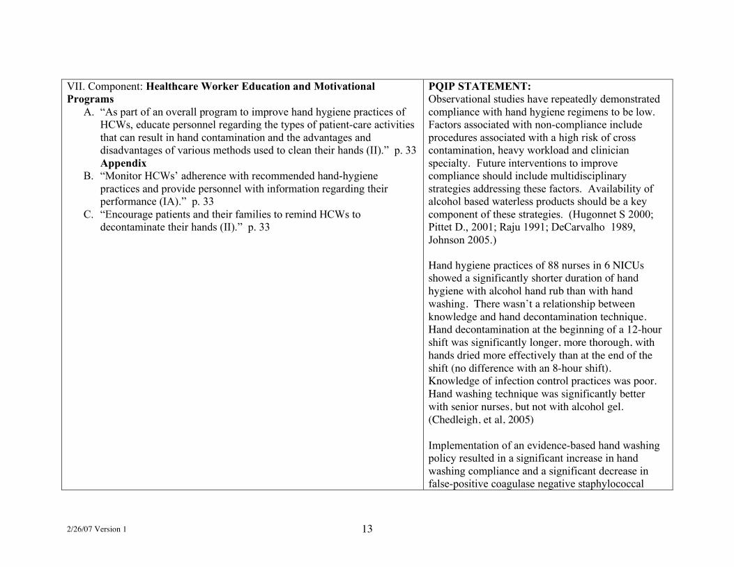
2/26/07 Version 1 13
VII. Component: Healthcare Worker Education and Motivational Programs
A. “As part of an overall program to improve hand hygiene practices of HCWs, educate personnel regarding the types of patient-care activities that can result in hand contamination and the advantages and disadvantages of various methods used to clean their hands (II).” p. 33 Appendix
B. “Monitor HCWs’ adherence with recommended hand-hygiene practices and provide personnel with information regarding their performance (IA).” p. 33
C. “Encourage patients and their families to remind HCWs to decontaminate their hands (II).” p. 33
A.
PQIP STATEMENT: Observational studies have repeatedly demonstrated compliance with hand hygiene regimens to be low. Factors associated with non-compliance include procedures associated with a high risk of cross contamination, heavy workload and clinician specialty. Future interventions to improve compliance should include multidisciplinary strategies addressing these factors. Availability of alcohol based waterless products should be a key component of these strategies. (Hugonnet S 2000; Pittet D., 2001; Raju 1991; DeCarvalho 1989, Johnson 2005.) Hand hygiene practices of 88 nurses in 6 NICUs showed a significantly shorter duration of hand hygiene with alcohol hand rub than with hand washing. There wasn’t a relationship between knowledge and hand decontamination technique. Hand decontamination at the beginning of a 12-hour shift was significantly longer, more thorough, with hands dried more effectively than at the end of the shift (no difference with an 8-hour shift). Knowledge of infection control practices was poor. Hand washing technique was significantly better with senior nurses, but not with alcohol gel. (Chedleigh, et al, 2005) Implementation of an evidence-based hand washing policy resulted in a significant increase in hand washing compliance and a significant decrease in false-positive coagulase negative staphylococcal

2/26/07 Version 1 14
blood and CSF culture rates. (Sharek 2002) Ready access to alcohol based waterless disinfectants and sinks in the patient care area are critical. The design of sinks and surrounding space should be optimized. Identifies effectiveness of hand hygiene and other measures in controlling Enterobacter outbreaks in the NICU. (Hervas 2001 and Fern 2001) To assist the Infection Control community in up-dating educational materials dealing with hand hygiene practices, the Hospital of Saint Raphael has launched a new informational Internet Web site entitled HAND HYGIENE RESOURCE CENTER at www.handhygiene.org. The Web site contains the following: * Link to the new Hand Hygiene Guideline at CDC * PowerPoint presentations (downloadable) that can be used for educating healthcare personnel about hand hygiene. Recommendations are consistent with those in the new guideline * One slide presentation is geared toward clinical personnel (MDs, RNs, etc.) * Another slide presentation is more appropriate for personnel working in other departments such as dietary or transport services * Annotated bibliography of articles dealing with hand hygiene (contains Direct links to PubMed abstracts for each article) * Frequently asked questions section

2/26/07 Version 1 15
* Glossary of terms dealing with hand hygiene * Links to other useful hand hygiene web sites

2/26/07 Version 1 16
VIII. Component: Administrative Measures
A. “Make improved hand hygiene adherence an institutional priority, and provide appropriate administrative support and financial resources. (IB)” p. 33
B. “Implement a multidisciplinary program designed to improve adherence of health personnel to recommended hand-hygiene practices. (IB)” p.33
C. “As part of a multidisciplinary program to improve hand hygiene adherence, provide HCWs with a readily accessible waterless antiseptic agent such as an alcohol-based handrub product. (IA)” p. 33
D. “To improve hand-hygiene adherence among personnel who work in areas in which high workloads and high intensity of patient care are anticipated, make an alcohol-based hand rub available at the entrance to the patient’s room or at the bedside, in other convenient locations, and in individual pocket-sized containers to be carried by HCWs (IA).” p. 33
E. Store supplies of alcohol-based hand rubs in cabinets or areas approved for flammable materials (IC).” p. 33
On March 25, 2005, the Centers for Medicare and Medicaid Services (CMS) issued an interim final rule adopting an amendment by the NFPA allowing for the installation of alcohol hand-rub dispensers in egress corridors. (Note: Local or state fire code requirements may differ from the national codes; therefore, facilities are strongly urged to determine requirements for their particular locale.) For additional information see: www.cdc.gov/handhygiene/firesafety/cmsRuling.htm or the Federal Register (could put notice in appendix): a257.g.akamaitech.net/7/257/2422/01jan20051800/edocket.access.gpo.gov/2005/pdf/05-5919.pdf Healthcare organizations are also encouraged to install dispensers in patient rooms, treatment rooms, suites and other appropriate locations. Healthcare facilities should work with local fire marshals to ensure that these installations are consistent with local fire codes. (Federal Register 2005).
�Three Safety Tips When Using Alcohol-based Hand-Rubs
1. When using alcohol-based hand-rubs, CDC recommends that:
Healthcare personnel rub their hands until the alcohol has evaporated (i.e., hands are dry).
2. Alcohol-based hand-rubs be stored away from

2/26/07 Version 1 17
high temperatures or flames, in accordance with CDC and National Fire Protection Agency recommendations. 3. Supplies of alcohol-based hand-rubs be stored in cabinets or areas approved for flammable materials. (Alcohol-Based Hand-Rubs and Fire Safety:�CDC Update. September 15, 2003; www.cdc.gov/handhygiene/firesafety/default.htm)

2/26/07 Version 1 18
IX. Component: Performance Indicators
“The following performance indicators are recommended for measuring improvements in HCWs’ hand-hygiene adherence:”
A. “Periodically monitor and record adherence as the number of hand hygiene episodes performed by personnel/number of hand hygiene opportunities, by ward or by service. Provide feedback to personnel regarding their performance.” p. 34
B. “Monitor the volume of alcohol-based hand rub (or detergent used for handwashing or hand antisepsis) used per 1000 patient-days”. p. 34
C. “Monitor adherence to policies dealing with wearing of artificial nails.” p. 34
D. “When outbreaks of infection occur, assess the adequacy of healthcare-worker hand hygiene.” p. 33
Compliance with hand hygiene using alcohol hand rub was significantly higher with nurses and physicians when they had been notified they would be observed than when they were covertly observed. There was no significant difference with other healthcare workers. (Eckmanns et al, 2006) A clinical trial using a self-selected convenience sample and crossover design in 2 NICUs conducted over 2 years tested use of an antiseptic hand wash and alcohol sanitizer. No significant difference in HAI or mean microbial counts on the nurses’ hands was noted. The alcohol sanitizer was credited with improved skin condition and quality of hand hygiene and increased frequency of use over the antiseptic hand wash, the frequency of hand hygiene remained low. A need for systems-level of interventions to increase quality of hand hygiene practices was needed. (Larson, et al, 2005) Observation guides: University of Geneva Hospitals; www.hopisafe.ch, and Lucile Packard Children’s Hospital at Stanford; Hand Hygiene Educational Tool. (Both located in the Appendices Section) Two fatal neonatal Klebsiella oxytoca infections associated with greater resistance to disinfectants resulting from improper handling of cleaning utensils. (Gebel 2002)
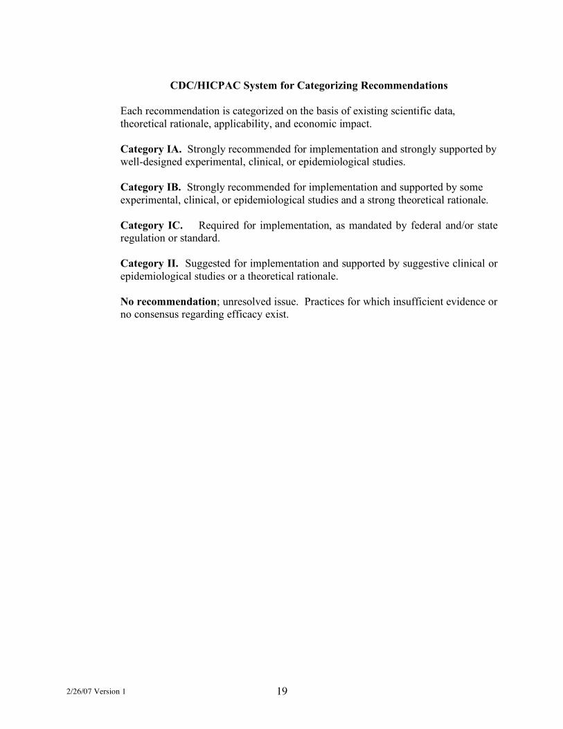
2/26/07 Version 1 19
CDC/HICPAC System for Categorizing Recommendations
Each recommendation is categorized on the basis of existing scientific data, theoretical rationale, applicability, and economic impact. Category IA. Strongly recommended for implementation and strongly supported by well-designed experimental, clinical, or epidemiological studies. Category IB. Strongly recommended for implementation and supported by some experimental, clinical, or epidemiological studies and a strong theoretical rationale. Category IC. Required for implementation, as mandated by federal and/or state regulation or standard. Category II. Suggested for implementation and supported by suggestive clinical or epidemiological studies or a theoretical rationale. No recommendation; unresolved issue. Practices for which insufficient evidence or no consensus regarding efficacy exist.
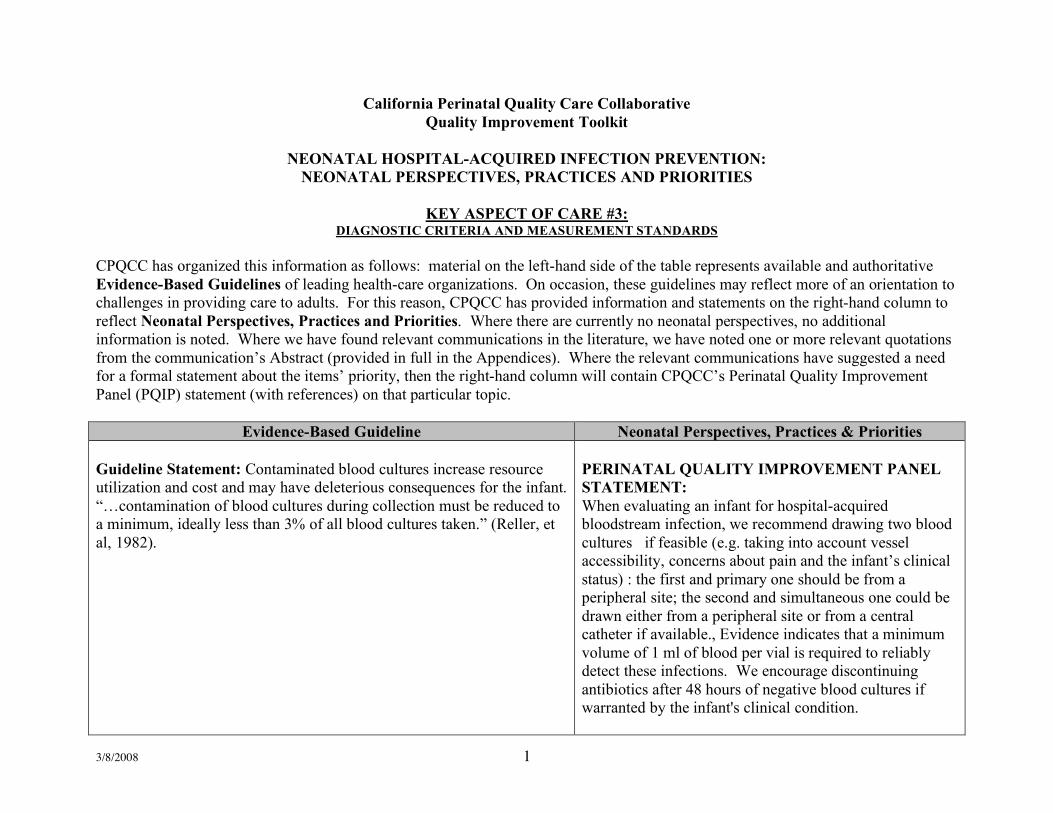
3/8/2008 1
California Perinatal Quality Care Collaborative
Quality Improvement Toolkit
NEONATAL HOSPITAL-ACQUIRED INFECTION PREVENTION: NEONATAL PERSPECTIVES, PRACTICES AND PRIORITIES
KEY ASPECT OF CARE #3:
DIAGNOSTIC CRITERIA AND MEASUREMENT STANDARDS CPQCC has organized this information as follows: material on the left-hand side of the table represents available and authoritative Evidence-Based Guidelines of leading health-care organizations. On occasion, these guidelines may reflect more of an orientation to challenges in providing care to adults. For this reason, CPQCC has provided information and statements on the right-hand column to reflect Neonatal Perspectives, Practices and Priorities. Where there are currently no neonatal perspectives, no additional information is noted. Where we have found relevant communications in the literature, we have noted one or more relevant quotations from the communication’s Abstract (provided in full in the Appendices). Where the relevant communications have suggested a need for a formal statement about the items’ priority, then the right-hand column will contain CPQCC’s Perinatal Quality Improvement Panel (PQIP) statement (with references) on that particular topic.
Evidence-Based Guideline Neonatal Perspectives, Practices & Priorities Guideline Statement: Contaminated blood cultures increase resource utilization and cost and may have deleterious consequences for the infant. “…contamination of blood cultures during collection must be reduced to a minimum, ideally less than 3% of all blood cultures taken.” (Reller, et al, 1982).
PERINATAL QUALITY IMPROVEMENT PANEL STATEMENT: When evaluating an infant for hospital-acquired bloodstream infection, we recommend drawing two blood cultures if feasible (e.g. taking into account vessel accessibility, concerns about pain and the infant’s clinical status) : the first and primary one should be from a peripheral site; the second and simultaneous one could be drawn either from a peripheral site or from a central catheter if available., Evidence indicates that a minimum volume of 1 ml of blood per vial is required to reliably detect these infections. We encourage discontinuing antibiotics after 48 hours of negative blood cultures if warranted by the infant's clinical condition.

3/8/2008 2
Note: Why at least one peripheral blood culture? The CDC criteria for a laboratory-confirmed catheter-related blood stream infection (LC CRBSI) requires only one positive blood culture for “recognized pathogens.” Thus, one BC is sufficient to establish their diagnosis. However, two positive blood cultures are necessary to establish the LC CRBSI diagnosis for common contaminants (e.g. CONS) if clinical criteria are not going to be used in addition to establish the diagnosis. This is the basis for PQIP’s recommendation that clinicians consider the optional second blood culture in assessing potential line related infections. Clinicians may prefer to rely solely on collaborating clinical signs and laboratory findings to establish the LC CRBSI diagnosis, which, if present along with but one positive BC, are sufficient to diagnose LC CR BSI.

3/8/2008 3
Evidence-Based Guideline Neonatal Perspectives, Practices & Priorities
Two vs One Blood Culture: Two peripherally obtained blood cultures do not have a higher yield in pathogen detection when at least 1 ml of blood was obtained. All neonates had the same organism with a similar sensitivity pattern in the paired cultures. Skin prep consisted of at least a 10 second cleaning with 10% povidone iodine and allowed to dry at least 30 seconds. (Sarkar 2006) 96 culture bottles were inoculated with either 0.25, 0.5, 1.0, or 4.0 ml of two seeded blood concentrations. Blood culture bottles were incubated in the BacT/Alert device for 5 days, and time to positivity was noted when applicable. There was a statistically significant difference with time to positivity and inocula volume (p<0.01), but the difference was not clinically important. Conclusion – If one of two viable colony-forming units are in the blood inoculated into culture media, the BacT/Alert system will detect growth rapidly. Because there appears to be a sizable subset of neonates who are at risk of sepsis with a colony count less than 4 CFU/ml, then a 0.5 ml inoculum of blood into the culture media is inadequate for sensitive and timely detection of bacteremia. One to two millilters of blood should increase microorganism recovery in the face of low-colony-count sepsis. (Schelonka 1996). Significance of Paired Coagulase Negative Staphylococci Blood Cultures: Of 13 paired blood culture isolates obtained simultaneously from septic neonates, 12 pairs were identical species with identical genotypes, The authors concluded that simultaneously obtained positive blood cultures usually represent true infection. (Huang 2006) Duration of Culture Observation: The length of time taken
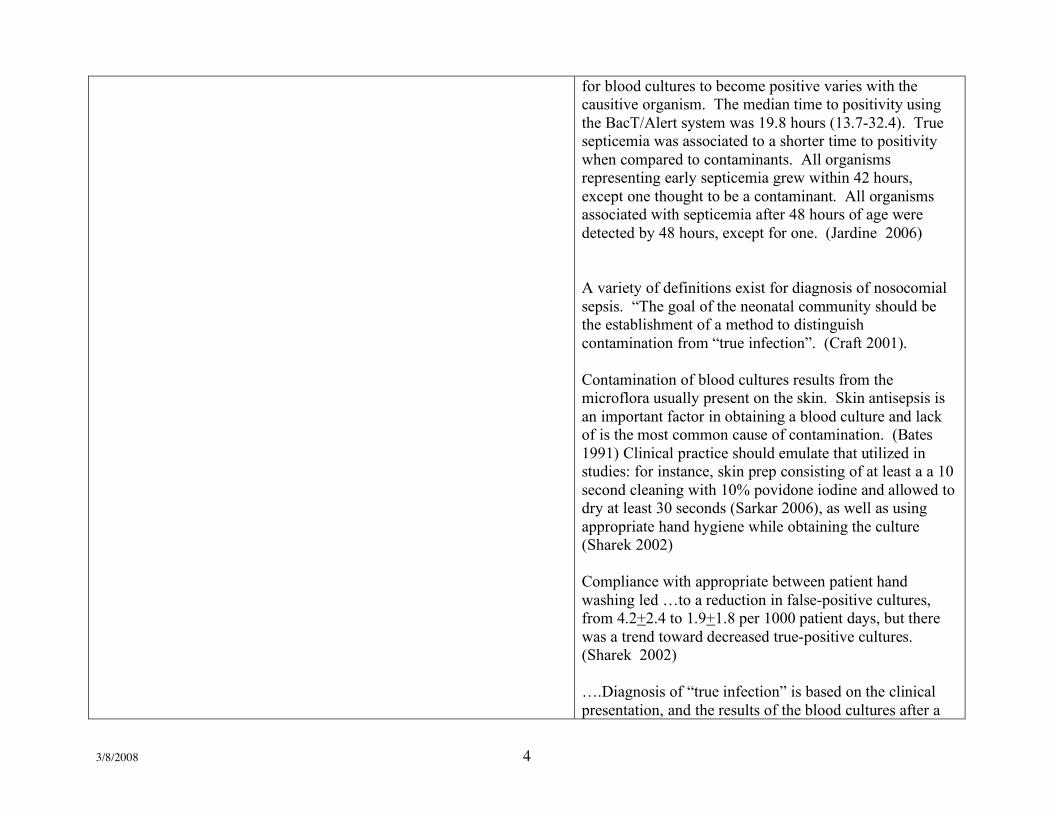
3/8/2008 4
for blood cultures to become positive varies with the causitive organism. The median time to positivity using the BacT/Alert system was 19.8 hours (13.7-32.4). True septicemia was associated to a shorter time to positivity when compared to contaminants. All organisms representing early septicemia grew within 42 hours, except one thought to be a contaminant. All organisms associated with septicemia after 48 hours of age were detected by 48 hours, except for one. (Jardine 2006) A variety of definitions exist for diagnosis of nosocomial sepsis. “The goal of the neonatal community should be the establishment of a method to distinguish contamination from “true infection”. (Craft 2001). Contamination of blood cultures results from the microflora usually present on the skin. Skin antisepsis is an important factor in obtaining a blood culture and lack of is the most common cause of contamination. (Bates 1991) Clinical practice should emulate that utilized in studies: for instance, skin prep consisting of at least a a 10 second cleaning with 10% povidone iodine and allowed to dry at least 30 seconds (Sarkar 2006), as well as using appropriate hand hygiene while obtaining the culture (Sharek 2002) Compliance with appropriate between patient hand washing led …to a reduction in false-positive cultures, from 4.2+2.4 to 1.9+1.8 per 1000 patient days, but there was a trend toward decreased true-positive cultures. (Sharek 2002) ….Diagnosis of “true infection” is based on the clinical presentation, and the results of the blood cultures after a
3/8/2008 5
48-hour incubation period (Craft 2001) …..Ancillary tests may be useful in the infant who remains ill despite negative blood cultures, and in whom other sources of infection are being considered (i.e., osteomyelitis, necrotizing enterocolities) (Craft 2001). Measures of neutrophil CD-64, combined with IL-6 and C-reactive protein, proved to be very sensitive markers for diagnosing late-onset NI in VLBWs, but routine use awaits availability of cytometric technology and additional clinical trials. (Ng 2002)

3/8/2008 6
Evidence-Based Guideline Neonatal Perspectives, Practices & Priorities
GUIDELINES FOR DIAGNOSING NOSOCOMIAL INFECTION
I. Vermont Oxford Network/CPQCC Coding Instructions for Declaring an Event a Nosocomial Infection after day 3 ITEM 32: Sepsis and/or Meningitis Late (after day 3) Note 1: The date of birth counts as day 1 regardless of the time of birth. For an infant born at11:59 PM on September 1, day 3 will be September 3. Note 2: If a bacterial pathogen and a coagulase negative staph are recovered during the same sepsis workup performed after day 3, check only "Bacterial Pathogen" for that episode. If a bacterial pathogen is recovered during one episode of sepsis after day 3, and coagulase negative staphylococcus is recovered during another episode of sepsis after day 3 (associated with the three clinical criteria listed below) check both "Bacterial Pathogen" and "Coag Neg Staph".
A. Bacterial Pathogen
Check “Yes” if a bacterial pathogen from the list in Appendix III is recovered from a blood and/or cerebrospinal fluid culture obtained after day 3 of life. Check “No” if a bacterial pathogen from the list in Appendix III is not recovered from a blood and/or cerebrospinal fluid culture obtained after day 3 of life. Check “Not Applicable” if infant died or was discharged on day 1, 2 or 3 and the infant was not readmitted on or before day 28.
3/8/2008 7
Evidence-Based Guideline Neonatal Perspectives, Practices & Priorities
B. Coag Negative Staph Check “Yes” if the infant has all 3 of the following: 1. Coagulase Negative Staphylococcus recovered from a
blood culture obtained from either a central line, or peripheral blood sample and/or is recovered from cerebrospinal fluid obtained by lumbar puncture, ventricular tap or ventricular drain AND
2. signs of generalized infection (such as apnea, temperature instability, feeding intolerance, worsening respiratory distress or hemodynamic instability) AND
3. treatment with 5 or more days of intravenous antibiotics after the above cultures were obtained.
Check “No” if any or all of the above are not true. Check “Not Applicable” if infant died or was discharged on day 1, 2 or 3 and the infant was not readmitted on or before day 28.
C. Fungal
Check “Yes” if a fungus was recovered from a blood culture obtained from either a central line or peripheral blood sample after day 3 of life. Check “No” if a fungus was not recovered from a blood culture obtained from either a central line or peripheral blood sample after day 3 of life. Check "Not Applicable" if infant died or was discharged on day 1, 2 or 3 and infant was not readmitted on or before day 28.

3/8/2008 8
Evidence-Based Guideline Neonatal Perspectives, Practices & Priorities II. NICHD Neonatal Research Network ….one positive blood culture drawn after 72 hours of age, in the presence of signs of infection. Stoll et al. J Pediatr 1996;129:63-71
III. National Nosocomial Infections Surveillance System [270]. Surveillance definitions for intravascular catheter associated infections. Emori TG, et al. Am J Infect Control 1991 Feb;19(1): 19-35. Revised by the Centers for Disease Control as a website publication by their National Health Safety Network (NHSN) as the Patient Safety Component Protocol, May 16, 2006 (http://www.dhs.ca.gov/pcfh/cms/nqi/pdf/CDCPSProtocols.pdf) and further amended in the CDC NHSN User Manual (for data collection), October 23, 2006) (http://www.dhs.ca.gov/pcfh/cms/nqi/pdf/CDCPSProtocols.pdf
Bloodstream infections, the most frequent nosocomial infections in all birth weight groups, should be a major focus of surveillance and prevention efforts in HRNs. For bloodstream infections, stratification of surveillance data by maternal acquisition will help focus prevention efforts for group B streptococci outside the HRN. Within the nursery, bloodstream infection surveillance should focus on umbilical or central intravenous catheter use, a major risk factor for infection. Gaynes, et al. Pediatrics 1996 Sep; 98(3 Pt 1):357-61. First national point-prevalence survey NICU NI events demonstrates both their high rates and significant burden and the need for effective prevention measures. (Sohn 2001) Improving neonatal survival beyond postnatal day two (associated with increased duration of device use) increases total NI events in aggregate, even when device-specific NI rates (events per day) are unchanged. (Zafar 2001) Concludes that daily blood cultures are not useful in neonatal ECMO, but that tracheal aspirates may be useful for patients on ECMO greater than 5 days. (Elerian 2001)

3/8/2008 9
A. Laboratory-confirmed bloodstream infection
Must meet at least one of the following criteria: Criterion 1: Patient has a recognized pathogen cultured from one or more blood cultures, and the pathogen cultured from the blood is not related to an infection at another site.

3/8/2008 10
Evidence-Based Guideline Neonatal Perspectives, Practices & Priorities Criterion 2: Patient has at least one of the following signs or symptoms: fever (>38°C), chills, or hypotension, and at least one of the following: a. Common skin contaminant (e.g., diptheroids, Bacillus species,
Propionibacterium species, coagulase-negative staphylococci, or micrococci) cultured from two or more blood cultures drawn on separate occasions.
b. Common skin contaminant (e.g., diptheroids, Bacillus species, Propionibacterium species, coagulase-negative staphylococci, or micrococci) cultured from at least one blood culture from a patient with an intravenous line, and the physician institutes appropriate antimicrobial therapy.
Criterion 3: Patient <1 year of age has at least one of the following signs or symptoms: fever (>38°C), hypothermia (<37°), apnea, or bradycardia, and at least one of the following: a. Common skin contaminant (e.g., diptheroids, Bacillus species,
Propionibacterium species, coagulase-negative staphylococci, or micrococci) cultured from two or more blood cultures drawn on separate occasions.
b. Common skin contaminant (e.g., diptheroids, Bacillus species, Propionibacterium species, coagulase-negative staphylococci, or micrococci) cultured from at least one blood culture from a patient with an intravenous line, and the physician institutes appropriate antimicrobial therapy.

3/8/2008 11
Evidence-Based Guideline Neonatal Perspectives, Practices & Priorities B. Clinical sepsis
Must meet at least one of the following criteria:
Criterion 1: Patient has at least one of the following clinical signs with no other recognized cause: fever (>38°C), hypotension (systolic pressure <90 mm Hg), or oliguria (<20 mL/hr), and blood culture not done or no organisms or antigen detected in blood and no apparent infection at another site, and physician institutes treatment for sepsis.
C. Catheter associated bloodstream infection Central Line is defined by the following: 1. Vascular access device that terminates at or close to the heart
or one of the great vessels. An umbilical artery or vein catheter is considered a central line.
2. Bloodstream infection is considered to be associated with a central line if the line was in use during the 48-hour period before development of the bloodstream infection. If the time interval between onset of infection and device use is longer than 48 hours, there must be compelling evidence that the infection is related to the central line.

3/8/2008 12
Evidence-Based Guideline Neonatal Perspectives, Practices & Priorities D. Arterial or venous infection
Included are arteriovenous grafts, shunt, fistula, or intravenous cannulation. Must meet at least one of the following criteria:
Criterion 1: Patient has organisms cultured from arteries or veins removed during a surgical operation and blood culture not done or no organisms cultured from blood. Criterion 2: Patient has evidence of arterial or venous infection seen during a surgical operation or histopathologic examination. Criterion 3: Patient has at least one of the following signs or symptoms with no other recognized cause: fever (>38°C), pain, erythema, or heat at involved vascular site and >15 CFUs cultured from an intravascular cannula tip using a semiquantitative culture method and blood culture not done or no organisms cultured from blood. Criterion 4: Patient has purulent drainage at the involved vascular site and blood culture not done or no organisms cultured from blood.
E. Ventilator-Associated Pneumonia
Definition: positive tracheal culture, clinical and laboratory signs of BSI, and routine radiology reports consistent with VAP. (Cordero 2002)
PQIP COMMENT: Important study on how current CDC definitions for ventilator-associated pneumonia are not specific for the newborn population and calls for their reformulation. (Cordero 2002)

3/8/2008 13
Evidence-Based Guideline Neonatal Perspectives, Practices & Priorities IV. CDC/HICPAC 2002
A. Example of clinical definition for Catheter-related Bloodstream Infection: Bacteremia/fungemia in a patient with an intravascular catheter with at least one positive blood culture and with clinical manifestations of infections (i.e., fever, chills, and/or hypotension) and no apparent source for the bloodstream infection except the catheter or any of the following:
1. A positive semiquantitative (>15 CFU/catheter segment) or quantitative (>103 CFU/catheter segment catheter) culture whereby the same organism (species and antibiogram) is isolated from the catheter segment and peripheral blood.
2. Simultaneous quantitative blood cultures with a >5:1 ration (central venous catheter (CVC) vs. peripheral)
3. Differential time period of CVC culture vs. peripheral blood culture positivity of >2 hours.
Classification of Coagulase-negative Staph positive blood cultures: Bacteremia, or “true infection”: Simultaneous positive blood cultures for the same organism through the central line and a peripheral site. Line colonization: A positive blood culture through the central line, with a simultaneous negative peripheral blood culture….This applies to an infant who appears stable at the time of the culture result, without other clinical and laboratory evidence to suggest ongoing infection, such as continuing thrombocytopenia or neutropenia. Contaminated blood culture: A positive peripheral blood culture…and a simultaneous negative central line blood culture. This applies to an infant who appears stable at the time of the culture result, without other clinical and laboratory evidence to suggest ongoing infection, such as continuing thrombocytopenia or

3/8/2008 14
neutropenia. (Craft 2001)

3/8/2008 15
PROCEDURES FOR DIAGNOSING NOSOCOMIAL INFECTION V. Procedure for obtaining blood culture
A. Sampling site “Blood for culture should not be drawn through an indwelling intravenous or intraarterial catheter unless it cannot be obtained by venipuncture.” (Reller 1982)
B. Peripheral puncture
1. Vigorously cleanse the venipuncture site with 2. Starting at the center of the site, swab concentrically with
povidone iodine and with a back and forth motion when using chlorhexidine. Prep for 30 seconds.
3. Allow the site to dry 4. Do not touch the venipuncture site after preparation and
prior to phlebotomy 5. Perform venipuncture and withdraw sample. If vein is
missed, a new needle should be used for each venipuncture attempt.
6. Cleanse top of blood culture bottle with alcohol prior to injecting blood sample (or per manufacturer’s recommendations)..
7. Place sample into blood culture bottle.
C. Catheter sample
1. Prep connection (injection port, extension set, etc.) closest to catheter with alcohol and rub with friction. 1. Disconnect connection from catheter 2. Withdraw a minimum of 1 ml of blood and place in blood
culture bottle 3. Flush catheter with saline solution 4. Change injection port and extension tubing and reattach
catheter to infusion tubing.
….Blood cultures should always contain 1 ml of blood and be drawn from both the central line, if present, and a peripheral site. (Craft 2001) Eight neonates were diagnosed with a positive blood culture for Paenibacillus macerans. Environmental contamination of the rubber stoppers in blood culture bottles was confirmed. This pseudobacteremia outbreak highlights the importance of adhering to well-established methods for blood culture collection and ongoing infection control surveillance. (Noskin 2001) Education – Staff competency and minimizing the number of personnel obtaining blood specimens for culture is linked to fewer false positive blood cultures. (Reller 1982).

3/8/2008 16
5. Cleanse top of blood culture bottle with alcohol prior to injecting blood sample (or per manufacturer’s recommendations)..
7. Place sample into blood culture bottle.

3/8/2008 17
Evidence-Based Guideline Neonatal Perspectives, Practices & Priorities VI. Procedure for skin prep (Reller, et.al., 1982)
A. “After palpation, the venipuncture site should be cleansed with
70% isopropyl or ethyl alcohol and then swabbed concentrically starting at the center with 1 to 2% tincture of iodine or 10% povidone-iodine solution (1% available iodine).”
B. “Individually packaged pledgets or swabsticks of iodine or iodophor are both preferable and convenient.”
C. “For maximal effectiveness, the disinfectant should be allowed to dry before blood is aspirated.”
-Alcoholic chlorhexidine solution preferred (bactericidal effect of CHG on gram-positive cocci is dramatically improved by the addition of alcohol and is superior to that of aqueous povidone iodine in reducing contamination of blood cultures). Mimoz, et al, 1999. VII. Culture bottle preparation (Reller, et.al., 1982) “Diaphragm tops or stoppers of culture bottles or collection tubes must also be disinfected with alcohol or an iodine preparation and allowed to dry.”
VII. Volume of blood to obtain (Reller, et.al., 1982) “Although cultures of less than 1 ml of blood will detect bacteremia in infants when the concentrations of microorganisms are sufficiently high, samples of >1 ml show a greater yield even in children.”
PQIP STATEMENT: There are no data that show an antiseptic agent to be superior to chlorhexidine gluconate (CHG) for skin antisepsis. Many CHG containing products exist on the market in both aqueous and alcoholic formulations and in a variety of strengths, contributing to the complexity of “best” newborn skin antisepsis. Taking into consideration the issues of efficacy and the potential of local irritation and systemic absorption, CHG or PI are the skin disinfectants recommended by PQIP as outlined below. Chlorhexidine Gluconate (CHG) Alcoholic-based:
• Apply over 30 seconds using side to side motion • Allow to dry over 30 seconds
Chlorhexidine Gluconate (CHG) Aqueous:
• Apply over 30 seconds • Remove with sterile water or saline following the
procedure (aqueous CHG will not dry due to its soapy consistency) (Malathi et al 1993, Lund et al 2001).
Povidone iodine (PI):
• Apply over 30 seconds and allow to dry • Remove with sterile water or saline following the
procedure “After topical applications of chlorhexidine, some percutaneous absorption occurs, particularly in preterm newborns, but only at trace levels.” Absorption appears to be reduced when an aqueous solution is applied. There is no evidence of sustained toxicity with CHG remaining on

3/8/2008 18
the skin Studies to date have used a variety of concentrations for multiple interventions. Tens of thousands of neonates have received chlorhexidine for umbilical cord care, bathing and maternal vaginal lavage prior to birth without reported adverse effects. (Mullany 2006) Povidone iodine containing solutions are commonly used for skin antisepsis prior to invasive procedures. Current practice is to remove the solution at the conclusion of the procedure. Caution should be exercised with use, particularly in very immature and sick infants who require repeated applications over large areas. (Linder, 1997). Despite concerns about skin reactions with CHG, and thyroid suppression with povidone-iodine (PI) these agents were recommended in the AWHONN /NANN evidence based guideline for skin care as the recommended skin disinfectants (Lund 2001). The decision was based on the fact that CHG and PI were felt to represent the least risk of local and systemic toxicity. (for full text of this review, see Skin Disinfection in the Neonate by Powers 2002 in the Appendix) 96 culture bottles were inoculated with either 0.25, 0.5, 1.0, or 4.0 ml of two seeded blood concentrations. Blood culture bottles were incubated in the BacT/Alert device for 5 days, and time to positivity was noted when applicable. There was a statistically significant difference with time to positivity and inocula volume (p<0.01), but the difference was not clinically important. Conclusion – If one of two viable colony-forming units are in the blood inoculated into culture media, the BacT/Alert system will detect growth rapidly. Because there appears to be a sizable subset of neonates who are at risk of sepsis with a colony

3/8/2008 19
count less than 4 CFU/ml, then a 0.5 ml inoculum of blood into the culture media is inadequate for sensitive and timely detection of bacteremia. One to two millilters of blood should increase microorganism recovery in the fact of low-colony-count sepsis. Schelonka, et al, 1996. Two vs One Blood Culture: Two peripherally obtained blood cultures do not have a higher yield in pathogen detection when at least 1 ml of blood was obtained. All neonates had the same organism with a similar sensitivity pattern in the paired cultures. Skin prep consisted of at least a 10 second cleaning with 10% povidone iodine and allowed to dry at least 30 seconds. (Sarkar et al, 2006)

3/8/2008 20

Neonatal Hospital-Acquired Infection Prevention Toolkit 5. Benchmarking
1
A. Benchmarking Process and Location of your Center’s Reports
CPQCC centers submit standardized data forms for very low birth weight infants to the CPQCC Data Center where they are reviewed for errors and omissions. These forms contain information on nearly 50 variables. CPQCC Data Center submits data to Vermont Oxford Network (VON) for analysis. CPQCC/VON aggregates data and computes indicators that reflect clinical procedures and outcomes. Each center receives its respective set of indicators, as well as the national and state median and interquartile range for each indicator in the CPQCC quarterly report. Indicators are displayed in graphs to facilitate comparisons. Readers should consult the tables/figures provided in your Center’s most recent VON Annual Quality Management report for specific reports, such as:
• Listings of those patients reported as having infections (useful for data quality improvement activities);
• Tabular and graphic presentations of infection data (including mean and interquartile ranges)
o Stratified by birthweight (Figures 13,14,15,16) o Stratified by gestational age (Figures 39,40,41,42) o Stratified by similar Type of Care Level, i.e. NICU Levels A, B and C
• Risk-Adjusted infection rates (Expected-Observed) shown by: o Individual year o Last 3 year’s data combined
• Graphical displays of co-morbidities o Infection, NEC and GI perforation
National VON Trends for “any late infection”:

Neonatal Hospital-Acquired Infection Prevention Toolkit 5. Benchmarking
2
B. Benchmarking with California (CPQCC) and VON Centers
1. CPQCC maintains infection reports on-line (cpqcc.org) that indicate many of these same parameters, but with just California NICUs as the reference group. In addition, a different risk-adjustment model is available for looking at infection rates; it differs in that it was formed only from California infants and it includes a slightly different set of variables than used by VON. Go to www.cpqcc.org and click reports to find your NICU’s reports.
2. Another meaningful way to view one’s progress in reducing hospital-acquired infections
is to gauge your own unit’s performance against an achievable benchmark of careTM (ABC). The ABC concept was developed by Kiefe and associates (Kiefe CI et al Identifying achievable benchmarks of care: concepts and methodology. Int J Qual Health Care. 1998 Oct;10(5):443-7) as a means for practitioners to understand how well one could do in taking care of patients in the same neighborhood. Calculation of the ABC for late infection prevention in California (our neighborhood of interest!) proceeds as follows:
• Rank order providers (NICUs) in ascending order of their “any late infection”
rate; • Beginning with the best performing provider, add sequentially in ascending order
until this subset of providers represents at least 10% of all patients (infants reported from their NICU to CPQCC/VON for a particular year;
• Calculate benchmark based on subset as follows: o total number of patients having an hospital-acquired infection divided by
the total number of patients in the subset (either 10% -or, as we did also- 20% of the total number of newborns reported in the year.
• Additional issues: o For NICUs whose number of patients is small (~ < 60/year), apply a
Bayesian estimator technique. We completed this operation for 2003 CPQCC dataset. You will note that we have
graphed the results (either best 10% or best 20% distinguished from the rest of the sample) versus the unit’s annual admission volume (VLBWs only). The point of this is to demonstrate that the size of the unit does not distinguish who can achieve rates of infection in the lowest 10% range. We would suggest that the Achievable Benchmark of Care presentation can be used to improve everyone’s perception of what can be achieved in preventing hospital-acquired infections in this fragile population.
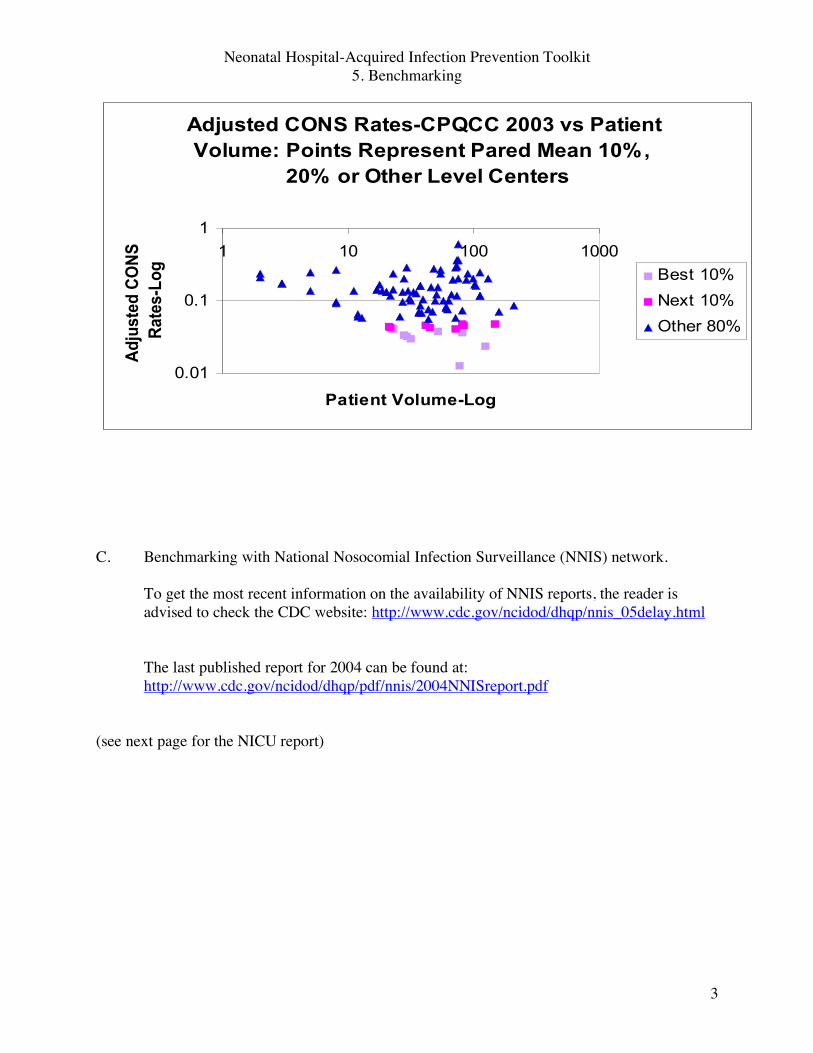
Neonatal Hospital-Acquired Infection Prevention Toolkit 5. Benchmarking
3
C. Benchmarking with National Nosocomial Infection Surveillance (NNIS) network.
To get the most recent information on the availability of NNIS reports, the reader is advised to check the CDC website: http://www.cdc.gov/ncidod/dhqp/nnis_05delay.html The last published report for 2004 can be found at: http://www.cdc.gov/ncidod/dhqp/pdf/nnis/2004NNISreport.pdf
(see next page for the NICU report)
Adjusted CONS Rates-CPQCC 2003 vs Patient
Volume: Points Represent Pared Mean 10%,
20% or Other Level Centers
0.01
0.1
1
1 10 100 1000
Patient Volume-Log
Ad
juste
d C
ON
S
Rate
s-L
og
Best 10%
Next 10%
Other 80%

The following Tables indicate the NICU data published in AJIC December 2005:

Neonatal Hospital-Acquired Infection Prevention Toolkit 5. Benchmarking
5
D. NNIS to become NATIONAL HEALTH SAFETY NETWORK (NHSN) The following announcement is on the NNIS website: NNIS Report will be replaced with new NHSN Report November 29, 2006 The next report from the Centers for Disease Control and Prevention (CDC) containing comparative healthcare-associated infection rates for hospitals will be comprised of data from the National Healthcare Safety Network (NHSN) for the year 2006. The new report will be published in the American Journal of Infection Control and posted on this website in late spring of 2007. It will contain device-associated infection rates and device utilization ratios for various types of intensive care units and other patient care areas. It will not contain surgical site infection (SSI) rates or antimicrobial use and resistance (AUR) rates, as the amount of data for these calculations will be insufficient to produce stable estimates. Those who wish to compare SSI and AUR rates to the national aggregates may do so using the data in the 2004 NNIS Report. The web address for NHSN is: http://www.cdc.gov/ncidod/hip/NNIS/members/members.htm However more information is actually available at: http://www.cdc.gov/ncidod/dhqp/index.html

CPQCC Neonatal Hospital-Acquired Infection Prevention Toolkit
6. Analyzing Your Practices: Hospital-Acquired Infection Prevention 1. Hand Hygiene: 1.A. Observe patient contacts for completeness of hand hygiene
using the suggested Hand Hygiene Observation Tool ( Problem Identification Worksheet # 1 that follows on the next page-please modify as per your own unit’s special needs).
Quality Assessment Exercises: Hospital-Acquired Infection (HAI) Prevention There are three components related to HAI Prevention, each of which is separately addressed by an exercise below.
1. Hand hygiene: observe patient contacts for completeness of hand hygiene; understand the factors associated with successful hand hygiene practices by assessing your unit’s Hand Hygiene Practices Inventory (Fishbone).
2. Line management: understand your current line set-up practices by diagramming your line set-up for a central venous line AND for an umbilical vessel; observe instances of your line entry technique when connecting a new TPN solution; observe instances of your line entry technique when drawing a blood sample from an umbilical line, understand the factors associated with successfully reducing catheter-related blood stream infections by assessing your unit’s Vascular Access Device Practices Inventory (Fishbone).
3. Diagnosis and Trending of Catheter-Related Blood Stream Infections (CRBSI): understand your current practices for obtaining and assessing blood culture and clinical data relevant to diagnosing CRBSI events; understand the factors associated with diagnosing CRBSI events in accord with CDC recommendations; understand your current practices for trending line days.

2
Problem Identification Worksheet # 1
Hand Hygiene Observation Tool
Date of Observation __________ Time Observed _____ - _____ Person Observed RN, RT, NNP, MD, Surgeon, OT/PT, lab, x-rayetc.
Opportunity Assessed A. Before patient care B. During patient care C. After patient care
Adequacy of Cleaning A. Adequate (10-15 sec) B. Inadequate (<10-15 sec) C. Noncompliant (not done)
Potential Break in Compliance 1=Initial 2 min scrub 2=Using phone 3=Using beeper 4=Diaper change 5=Chart use 6=Computer Use 7=Scale use 8=One touch 9=Use of supplies 10=Touch glasses 11=Touch face 12=Touch hair 13=Other
Method Discipline of Person Observed
Opportunity Assessed Hand
Wash Gel
Adequacy of Hand Hygiene
Break in Compliance if
Observed
1 2 3 4 5 6 7 8 9 10 11 12 13 14 15 16 17 18 19 20 21 22 23 24

3
1.B Understand the factors associated with successful hand hygiene practices by assessing your unit’s Hand Hygiene Practices using the Hand Hygiene Inventory (Fishbone-Problem Identification Worksheet # 2) shown on the next page.
Instructions: On the Hand Hygiene Fishbone diagram, label each item
using the following code: 1. Procedure in place & no perceived problems with this item
2. Procedure in place and working on improvement 3. Procedure needed 4. Procedure viewed as not being needed

4
Problem Identification Worksheet # 2

5
2. Line Set-up, Management and Entry:
2.A. Understand your current line set-up and line entry practices.
1) Diagram a typical line set-up for a PICC or BroviacTM: Show every
connector and tube from the skin entry point back to the parenteral fluid bottle/bag distributed by your pharmacy. Better yet: Take a picture of the set-up, preferably while a nurse is injecting a medication into the line.
2) Does your set-up have the essentials of a closed system: the central line catheter is tipped with an injection site device, so that when fluids are changed, re-connecting does not require breaking into the line?
Yes/ No (please circle)
3). Diagram a typical line set-up for an umbilical arterial or venous line: Show
every connector and tube from the umbilicus back to the parenteral fluid bottle/bag distributed by your pharmacy. Better yet: Take a picture of the set-up and also one while a nurse is taking a blood sample.
4) Does your UAC/UVC set-up have the essentials of a closed system: umbilical catheter tipped with a Three-Way Stopcock, whose Top Port (Injection Site) is tipped by a needleless injection site device through which needleless entries can be made for withdrawing samples and injecting solutions, medications, etc. or a manufactured closed arterial system device? Yes/ No (please circle)

6
2.B. Understanding your current line entry practice: connecting a new TPN
Observe and describe your line entry technique when connecting a new TPN solution. Items to note: number of nurses involved, sterile field, hand hygiene, and materials used to sterilize the entry point, duration of sterilization maneuver(s). For best results, we recommend that you record at least three different nurses; this form could be used as the basis of a competency step in a skills lab. Here is a checklist of items to observe (or if different, list your own set of steps):
Problem Identification Worksheet # 3 TPN CHANGE OBSERVATIONS
RN # 1
YES NO COMMENTS
1. Hands antisepsis before IV line manipulations? 2. Created sterile field (sterile gauze under
connection sites)?
3. Clean connections before disconnecting with alcohol or chlorhexidine?
4. Cleaned injection ports when connecting new tubing with alcohol or chlorhexidine?
5. Used friction when cleaning. (For best results, actually count the number of wiping strokes)
Count:____
RN # 2
YES NO COMMENTS
1. Hands antisepsis before IV line manipulations? 2. Created sterile field (sterile gauze under
connection sites)?
3. Clean connections before disconnecting with alcohol or chlorhexidine?
4. Cleaned injection ports when connecting new tubing with alcohol or chlorhexidine?
5. Used friction when cleaning. (For best results, actually count the number of wiping strokes)
Count:____
RN # 3
YES NO COMMENTS
1. Hands antisepsis before IV line manipulations? 2. Created sterile field (sterile gauze under
connection sites)?
3. Clean connections before disconnecting with alcohol or chlorhexidine?
4. Cleaned injection ports when connecting new tubing with alcohol or chlorhexidine?
5. Used friction when cleaning. (For best results, actually count the number of wiping strokes)
Count:____

7
2.C. Understanding your current line entry practice: withdrawing blood from an umbilical line Observe instances of your line entry technique when drawing a blood sample from an umbilical line. Items to note: number of nurses/therapists/physicians involved, sterile field, hand hygiene before entry, materials used to sterilize the entry point, duration of sterilization maneuver(s), how the line is broken into and with what. This form could be used as the basis of a competency step in a skills lab. If you do not have a closed system or have a different set of steps, just list the steps you use in order.
Problem Identification Worksheet # 4 BLOOD DRAWING OBSERVATIONS RN # 1
YES NO COMMENTS
1. Hands antisepsis and application of gloves before IV line manipulations?
2. Created sterile field (sterile gauze under connection sites)?
3. Cleaned injection ports with alcohol or chlorhexidine?
4. Used friction when cleaning. (For best results, actually count the number of wiping strokes)
Count:____
5. Capped syringe containing blood used to clear the catheter (blood that will be reinfused)?
6. Removal of gloves and hand antisepsis at conclusion of procedure?
RN # 2
YES NO COMMENTS
1. Hands antisepsis and application of gloves before IV line manipulations?
2. Created sterile field (sterile gauze under connection sites)?
3. Cleaned injection ports with alcohol or chlorhexidine?
4. Used friction when cleaning. (For best results, actually count the number of wiping strokes)
Count:____
5. Capped syringe containing blood used to clear the catheter (blood that will be reinfused)?
6. Removal of gloves and hand antisepsis at conclusion of procedure?
RN # 3
YES NO COMMENTS
1. Hands antisepsis and application of gloves before IV line manipulations?
2. Created sterile field (sterile gauze under connection sites)?
3. Cleaned injection ports with alcohol or chlorhexidine?
4. Used friction when cleaning. (For best results, actually count the number of wiping strokes)
Count:____
5. Capped syringe containing blood used to clear the catheter (blood that will be reinfused)?
6. Removal of gloves and hand antisepsis at conclusion of procedure?

8
2.D. Understand the factors associated with successful line management
and line entry practices by assessing your unit’s Vascular Access Device Practices using the VAD Inventory (Fishbone-Problem Identification Worksheet # 5) shown on the next page.
Instructions: On the Hand Hygiene Fishbone-Problem Identification
Worksheet # 5 diagram, label each item using the following code: 1. Procedure in place & no perceived problems with this item
2. Procedure in place and working on improvement 3. Procedure needed 4. Procedure viewed as not being needed

9
Problem Identification Worksheet # 5

CPQCC Neonatal Hospital-Acquired Infection Prevention Toolkit
3. Diagnosis and Trending of Catheter-Related Blood Stream Infections (CRBSI)
Problem Identification Worksheet # 6 3.A. Identification of Catheter Associated Bloodstream Infections (CABSI)
1). Blood culture practices when infection is suspected: Specify your usual practices with a check mark a. ___ obtain one peripheral BC b. ___ obtain two peripheral BC c. ___ obtain one BC through the line d. ___ obtain one BC through the line and one peripheral BC e. ___ other: specify:________________________________ f. ___ usually obtain 0.5ml blood per sample g. ___ usually obtain 1.0 ml blood per sample h. ___obtain a different volume (specify ______ ml)
2). Criteria used to label an event as a CABSI: Specify whether your usual practice would to be to label these circumstances as CABSI; indicate your answer as T(rue) or F(alse)
a. ___if even only one BC grows a recognized pathogen and you judge the infection is not related to another site (“and signs and symptoms and positive laboratory tests not related to an infection at another site”)
b. ___if only one BC grows a common skin contaminant* and you judge that the infection is not related to another site;
c. ___if two BCs grow a common skin contaminant* on separate occasions and you judge that the infection is not related to another site
d. ___if one BC grows a common skin contaminant* and you judge that the infection is not related to another site, then you would apply which of the following additional criteria to establish the diagnosis of a CABSI: specify all that apply
1) ___fever (> 38 Co), chills, OR hypotension
2) ___ age < 1 year
3) ___ elevated temperature ( >38 Co) or hypothermia (temperature < 37 Co) or apnea or bradycardia (at least one)
4) ___a second peripheral BC is growing the same common skin contaminant
5) ___patient has an intravascular device in place
6) ___physician instituted appropriate antimicrobial therapy

11
# Common skin contaminants defined: e.g., diphtheroids, Bacillus sp., Propionibacterium sp., coagulase-negative staphylococci, or micrococci
3.B. Understand the factors associated with diagnosis and trending of Hospital-Associated Infections by assessing your unit’s practices by using the Diagnosis and Trending Inventory (Fishbone-Problem Identification Worksheet # 7) shown on the next page.
Instructions: On the Diagnosis and Trending Fishbone-Problem
Identification Worksheet # 7 diagram, label each item using the following code:
1. Procedure in place & no perceived problems with this item 2. Procedure in place and working on improvement
3. Procedure needed 4. Procedure viewed as not being needed

12
Problem Identification Worksheet # 7
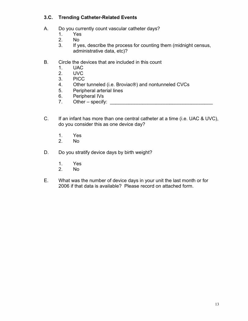
13
3.C. Trending Catheter-Related Events A. Do you currently count vascular catheter days? 1. Yes 2. No 3. If yes, describe the process for counting them (midnight census,
administrative data, etc)? B. Circle the devices that are included in this count 1. UAC 2. UVC 3. PICC 4. Other tunneled (i.e. Broviac®) and nontunneled CVCs 5. Peripheral arterial lines 6. Peripheral IVs 7. Other – specify: ______________________________________
C. If an infant has more than one central catheter at a time (i.e. UAC & UVC), do you consider this as one device day?
1. Yes 2. No
D. Do you stratify device days by birth weight?
1. Yes 2. No
E. What was the number of device days in your unit the last month or for 2006 if that data is available? Please record on attached form.
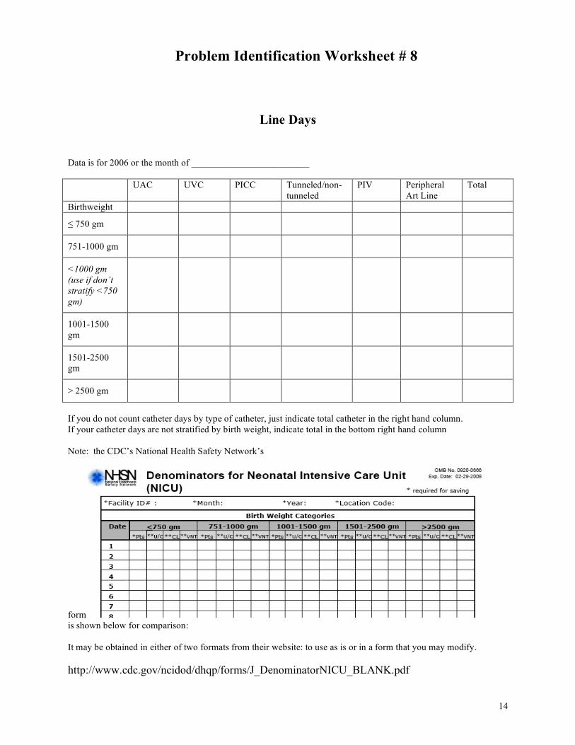
14
Problem Identification Worksheet # 8
Line Days
Data is for 2006 or the month of _________________________ UAC UVC PICC Tunneled/non-
tunneled PIV Peripheral
Art Line Total
Birthweight
≤ 750 gm
751-1000 gm
<1000 gm (use if don’t stratify <750 gm)
1001-1500 gm
1501-2500 gm
> 2500 gm
If you do not count catheter days by type of catheter, just indicate total catheter in the right hand column. If your catheter days are not stratified by birth weight, indicate total in the bottom right hand column Note: the CDC’s National Health Safety Network’s
form is shown below for comparison: It may be obtained in either of two formats from their website: to use as is or in a form that you may modify. http://www.cdc.gov/ncidod/dhqp/forms/J_DenominatorNICU_BLANK.pdf
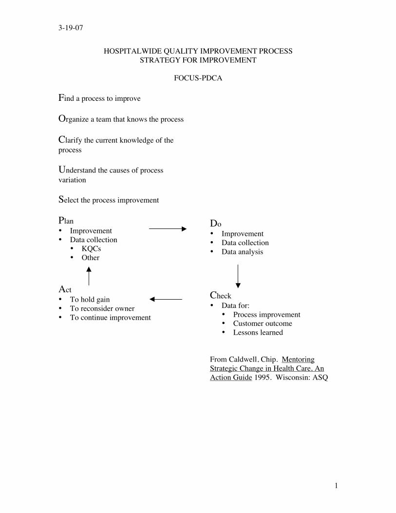
3-19-07
1
HOSPITALWIDE QUALITY IMPROVEMENT PROCESS STRATEGY FOR IMPROVEMENT
FOCUS-PDCA
Find a process to improve Organize a team that knows the process Clarify the current knowledge of the process Understand the causes of process variation Select the process improvement Plan • Improvement • Data collection
• KQCs • Other
Act • To hold gain • To reconsider owner • To continue improvement
Do • Improvement • Data collection • Data analysis Check • Data for:
• Process improvement • Customer outcome • Lessons learned
From Caldwell, Chip. Mentoring Strategic Change in Health Care, An Action Guide 1995. Wisconsin: ASQ

3-19-07
2
FIND a Process to Improve Find a process to improve by answering the questions below based upon your data and data analysis. Possible answers to the questions are provided, followed by corresponding processes with improvement potential in bold. Note that questions may arise from your data that are not presented here. You are encouraged to think critically about your data, to probe for further information if necessary, and to find a unique “process to improve” if it is not described below. Use information from the completed Problem Identification Worksheets in the previous section to find a process that constitutes an “opportunity for improvement”. If your analysis indicates problems related to these tasks, then you may wish to identify one or more of these tasks as a “process to improve.” Identifying Processes For Improvement: Question: Using your NI Prevention problem identification worksheets including the Hand Hygiene Observation Tool (Problem Identification Worksheet #1) and the Fishbone (Problem Identification Worksheet #2) that address the entire set of integrated processes for ensuring appropriate Hand Hygiene, Acceptable Line Care and accurate and timely Diagnosis and Trending, decide on one of these three problem areas to concentrate your initial efforts. Answer: We decided that our greatest need is to improve Hand Hygiene compliance. • Process to be improved: Your center’s hand hygiene compliance. Question: What is the compliance rate for appropriate hand hygiene among the following specialties at your center: (Use your Problem Identification Worksheet # 1 data to answer this question.)
o Bedside nurses o Pediatric residents and neonatal fellows o NNP’s o Neonatologists o Surgeons
Answer: The compliance rate varied widely with the most variance being with the non-NICU staff.
• Process to be improved: Re-implement more effective hand hygiene training and reinforcement processes, re-educate and re-motivate the staff and involve every specialty group in the auditing process.
Question: Using the Hand Hygiene Fishbone (Problem Identification Worksheet #2), identify priority areas (at least two) that interest you as possible areas for improvement.

3-19-07
3
Answer: We found, after we utilized the fishbone document, that we need to focus on our policies/procedures related to decontamination of hands before donning gloves and prohibiting the wearing of artificial nails and having chipped nail polish. • Process to be improved: Improvement and possible development of
polices/procedures related to hand hygiene. Question: What are your procedures at your center for entering central lines and either connecting fluids for infusion or drawing blood specimens (Use your Problem Identification Worksheets # 3 and # 4) to help inform your answer to this question) and Vascular Access Device Fishbone (Problem Identification Worksheet # 5) to help answer this question.
o Connecting the TPN solution o Injecting a medication o Drawing blood from an umbilical catheter
Answer: We found that we need to work on developing protocols relating to drawing blood from an umbilical catheter.
• Process to be improved: Develop and implement policies/protocols that decrease the risk of introducing a catheter-related blood stream infection when drawing blood from an umbilical catheter.
Question: How does your Unit’s practice compare to those recommended by CPQCC Panel and the CDC on collecting blood specimens for diagnosing bloodstream infections? Identify specifics upon which you agree and disagree. Answer: We typically obtain only one BC through the line.
• Process to be improved: Gain consensus among our physicians on how to utilize blood cultures to diagnose catheter-related blood stream infections.
Question: Does your UAC/UVC set-up have the essentials of a closed system?
Answer: No, our closed system is missing an a three-way stopcock and the injection port is missing a needleless injection site device.
• Process to be improved: Review current closed systems and develop a consensus on products. Develop policies/procedures on closed systems.

3-19-07
4
Question: Does your Unit trend catheter days (Problem Identification Worksheet # 8) and catheter-associated blood stream infections in accord with the CPQCC and CDC recommendations? Answer: No. We have no consistent way to track these events in a timely manner.
• Process to be improved: Develop a process with nursing to ensure daily collection of line days sorted by birth weight and line location (central or umbilical)-our denominator data. Develop a process to collaborate with our Infection Control Nurse that ensures weekly review of all positive blood and CSF cultures, logging of ID events as to type of event, e.g. hospital-acquired, and ensure daily postings in the NICU of the number days since the last catheter-related blood stream infection.
Question: Does your unit evaluate every potential catheter-related blood stream infection using the criteria suggested by the CDC and CPQCC (Use Problem Identification Worksheet # 6 to solicit the point by point data on each of the criteria utilized in classified infections in accordance with the CDC schemata)? Answer: We found much variability between individual practitioners on their knowledge of these criteria and how they were been invoked (or not invoked) in classifying infectious events.
• Process to be improved: Develop an education and consensus conference(s) to familiarize all those in diagnosing and classifying these events. Develop a process in which all infectious events are, on a scheduled basis, reviewed by one clinician and the Infection Control Practitioner associated with the NICU, in order to assure regular and consistent review and classification of all events.
Question: Use the Diagnosis/Trending Fishbone (Problem Identification Worksheet # 7) to describe the list of practices you wish to implement in your Unit and select one or two of them as your priority tasks. Answer: We found that there is inadequate documentation when the specimen is being drawn.
• Process to be improved: Work with your unit’s Nursing Council (or like-minded groups) to improve the documentation practices relating to specimen collection.

ORGANIZE a Team that Knows the Process Once a process has been targeted for improvement, the next step is to identify individuals who have “ownership” of the process, have insights into the process, and/or play key roles in the process. Their participation in efforts to improve the process is critical. When identifying participants, consider their current role, position and perspective within the hospital, and their appropriate role and position within the quality improvement effort. There is no set formula for team composition. Depending on the process to be improved, a team might consist of one or two members or could require a larger, multidisciplinary group. The team will also depend on the factors unique to each hospital, such as the schedules and priorities of staff. The following table provides examples of teams constructed to address the processes identified in the previous section. These examples are meant to be illustrative, not exhaustive, and should be adapted to the resources and constraints of your hospital. Once appropriate team members have been identified, their participation should be secured. Individuals identified as potential team members may not be entirely enthusiastic about participating. Clearly stated hospital/department commitment to improving the process will encourage individual participation. Inclusion of well-reputed and/or neutral parties in teams will also serve to encourage those who might feel threatened or challenged by the activity.

3-19-07
6
Table I. Processes to be improved and corresponding example teams Process to Be Improved Example of Team members Neonatal care providers will understand Hand Hygiene compliance, acceptable line care and diagnosis/trending challenges, concepts and techniques.
Neonatologists, Fellows, Residents, NICU Nurses, Quality Improvement Specialists, Case Managers, Pharmacists, Respiratory Care Practitioners, Data Managers.
Re-implement more effective hand hygiene training and reinforcement processes, re-educate and re-motivate the staff and involve every specialty group in the auditing process.
Neonatologists, Fellows, Residents, NICU Nurses, Pharmacists, Respiratory Care Practitioners, ancillary personnel and all medical specialties who take care of NICU pts
Improve (or develop) policies/procedures addressing all aspects of hand hygiene practices
Neonatologists, NICU Leadership, NICU Nurses, Respiratory Care Practitioners, Quality Improvement Specialists.
Develop and implement policies/protocols that decrease the risk of introducing a catheter-related blood stream infection when drawing blood from an umbilical catheter.
NICU leadership, NICU Nurses, Respiratory Care Practitioners, MDs, QI Specialists.
Gain consensus among our physicians on how to utilize blood cultures to diagnose catheter-related blood stream infections.
Neonatologists, NICU Leadership, NICU Nurses, Respiratory Care Practitioners, Quality Improvement Specialists.
Develop a process with nursing to ensure daily collection of line days sorted by birth weight and line location (central or umbilical)-our denominator data. Develop a process to collaborate with our Infection Control Nurse that ensures weekly review of all positive blood and CSF cultures, logging of ID events as to type of event, e.g. hospital-acquired, and ensure daily postings in the NICU of number days since the last catheter-related blood stream infection.
Neonatologists, NICU Leadership, NICU Nurses, Respiratory Care Practitioners, Quality Improvement Specialists, Infection Control
Work with your unit’s Nursing Council (or like-minded groups) to improve the documentation practices relating to specimen collection.
Neonatologists, NICU Leadership, NICU Nurses, Respiratory Care Practitioners, Quality Improvement Specialists
Develop policies/procedures on closed systems. Review current closed systems and develop a consensus on products.
Neonatologists, NICU Leadership, NICU Nurses, Respiratory Care Practitioners, Quality Improvement

3-19-07
7
Specialists
3-19-07
8
CLARIFY the Current Knowledge of the Process Once a team has been constructed, several issues should be presented to the team members. These are: • The Recommendations/Rationale for the prevention of nosocomial infection in the neonate.
The Documents included in Section I, II and II of the Toolkit will provide a basis for discussion. Other background material, such as documents describing relevant internal hospital policy, should be made available to team members.
• Update on the current practices in your center. This information is presented in the
Benchmarking Section of this Toolkit. Also, information in your center as developed in Section 5 (Benchmarking Section) and by patient data in Section 6 (Analyzing your Practice Section).
• The method by which your hospital identified a process to be improved, and the evidence
that the process needs improvement. Provide team members with copies of the Data and Data Analysis section (with patient identifying information removed to ensure confidentiality if necessary) and the results that it provided. Demonstrate to the Team how the data gave rise to the “process to be improved.”
In clarifying current knowledge, consider calling upon team members, other hospital staff and outside sources with appropriate expertise to assist. The mechanism chosen for clarifying knowledge will depend on the process to be improved, the team and its needs, and the resources available. You may wish to distribute materials in advance and then provide a venue for discussion and exchange, such as a team meeting. Alternately, it may be effective to review information together or to re-package the information. For example, summaries can be sent around via email.

3-19-07
9
UNDERSTAND the Causes of Process Variation Process variation should be discussed following the previous activity, Clarification of current knowledge of the process. The key points related to process variation are: • The possible range of process variation. Thus, one could address why there are differences
in responding to apparently equally challenging clinical circumstances. • The acceptable range of process variation. Identify how much variation appears to be
random (“common cause”) and how much is derived from a specific process (“special cause”).
• The apparent range of process variation within your hospital. It is helpful to consider the reasons for process variation in your Center.
• The extent to which process variation is justified.

3-19-07
10
SELECT the Process Improvement Restate the Process to be improved as an actual improvement project. This should be a team activity, with attention given to the methods used to select the process improvement activities. The following chart provides an example of an improvement activity for each previously identified process challenge. Table II. Process to be Improved and Examples of Improvement Process Improvement Activity Examples of Improvement Activity Neonatal care providers will understand Hand Hygiene compliance, acceptable line care and diagnosis/trending challenges, concepts and techniques.
Staff presentations, lectures, poster boards, skills lab, patient education information sheets
Re-implement more effective hand hygiene training and reinforcement processes, re-educate and re-motivate the staff and involve every specialty group in the auditing process.
Staff presentations, lectures, poster boards, skills lab, involvement of other disciplines is key
Improve (or develop) policies/procedures addressing all aspects of hand hygiene practices
Leadership meetings to review and/or develop policies and procedures related to hand hygiene, Staff presentations, lectures, poster boards, skills lab, involvement of other disciplines is key
Develop and implement policies/protocols that decrease the risk of introducing a catheter-related blood stream infection when drawing blood from an umbilical catheter.
Leadership meetings to review and/or develop policies and procedures on blood draws technique. Staff presentations, lectures, poster boards, skills lab, involvement of other disciplines is key
Gain consensus among our physicians on how to utilize blood cultures to diagnose catheter-related blood stream infections.
Leadership meetings with physicians and nurses to brainstorm on diagnosis techniques.
Develop a process with nursing to ensure daily collection of line days sorted by birth weight and line location (central or umbilical)-our denominator data. Develop a process to collaborate with our Infection Control Nurse that ensures weekly review of all positive blood and CSF cultures, logging of ID events as to
Leadership meetings with nursing and Infection Control to review data collection procedures and possibly develop a process if needed.

3-19-07
11
type of event, e.g. hospital-acquired, and ensure daily postings in the NICU of number days since the last catheter-related blood stream infection. Work with your unit’s Nursing Council (or like minded groups) to improve the documentation practices relating to specimen collection.
Leadership meetings to review and/or develop policies and procedures on documentation.
Develop policies/procedures on closed systems. Review current closed systems and develop a consensus on products.
New product review along with leadership meetings to review and/or develop policies and procedures on new products in the NICU.
Plan the Improvement and Continued Data Collection This stage involves visualizing how the specified improvement will be made. The first column of the table below re-states “examples of improvements” from the previous table. The second column provides key steps towards realizing the specified improvement. Note that only the key steps are described. You may want to go into more detail, outlining intermediate steps. Be sure to include target dates for completing each step. Use the Implementation Worksheet at the end of this section to document the proposed improvement, key steps planned towards realizing the improvement, and actual steps taken (see next heading, Do the Improvement). Entries into the Implementation Worksheet are to be made on a regular basis, thereby charting the progress of quality improvement efforts. Table III. Improvements and Key Steps Towards Making Improvements Example of Improvement Examples of key steps towards
realizing improvement Staff presentations, lectures, poster boards, skills lab, patient education information sheets
Identify speakers, materials, audiovisual aids and implement post presentation testing.
Staff presentations, lectures, poster boards, skills lab, involvement of other disciplines is key
Identify speakers, materials, audiovisual aids and implement post presentation testing.
Leadership meetings to review and/or develop policies and procedures related to hand
Development and education of new policies and procedures
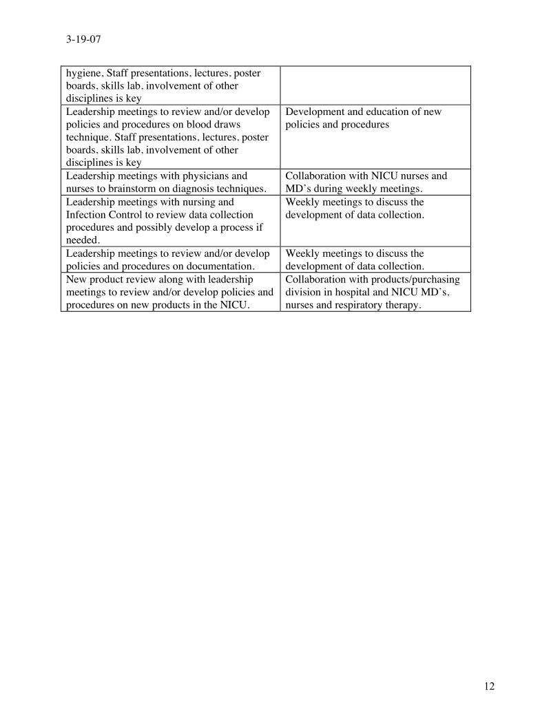
3-19-07
12
hygiene, Staff presentations, lectures, poster boards, skills lab, involvement of other disciplines is key Leadership meetings to review and/or develop policies and procedures on blood draws technique. Staff presentations, lectures, poster boards, skills lab, involvement of other disciplines is key
Development and education of new policies and procedures
Leadership meetings with physicians and nurses to brainstorm on diagnosis techniques.
Collaboration with NICU nurses and MD’s during weekly meetings.
Leadership meetings with nursing and Infection Control to review data collection procedures and possibly develop a process if needed.
Weekly meetings to discuss the development of data collection.
Leadership meetings to review and/or develop policies and procedures on documentation.
Weekly meetings to discuss the development of data collection.
New product review along with leadership meetings to review and/or develop policies and procedures on new products in the NICU.
Collaboration with products/purchasing division in hospital and NICU MD’s, nurses and respiratory therapy.

3-19-07
13
Do the Improvement, Data Collection, and Analysis The planning is complete, and the next step is implementation. Fundamental to CQI is timely feedback on the progress and success of improvement efforts. Thus, implementation is accompanied by ongoing data collection, analysis and planning. Important activities during this stage are to: • Revisit lessons learned from the Data Analysis. Regularly collect data using the PIW
worksheet forms to chart progress towards achieving the stated improvement, to determine whether the strategy in place is working, and to identify new or evolving areas for improvement.
• Describe planned steps towards realizing the proposed improvement. This should be repeated
at regular intervals. Based on changes and/or new information from the Data form the Team may decide to adjust the Proposed Improvement and to update, revise and refine the plan (see the next two headings, Check and Study the Results and Act to Hold the Gain and to Continue to Improve the Process).
• Document actual steps taken to date. Note what has been accomplished including team
meetings, agreements reached, new procedures and so forth. Use the Implementation Worksheet or similar tool to keep track of progress.
Depending on the improvement and on the resources at hand, the team may decide to update the Implementation Worksheet weekly, monthly or quarterly.

3-19-07
14
Check and Study the Results The completed Implementation Worksheet serves as a one-page results summary. This summary should be copied and distributed to team members. When discussing and analyzing results, the team should consider: • Changes over time in process identified for improvement. • What caused the change? That is, which elements of the plan were effective? • If no change over time was observed, why not? Which elements of the plan were not
effective or were ineffectively carried out? • Changes over time in other processes. Have the steps taken towards improving a process
helped or hindered other processes? • The lessons learned for each component of the FOCUS PDCA activity. Reflect on what was
done well, and what can be improved. Write down the most important lessons learned.

3-19-07
15
Act to Hold the Gain... Now that improvements have been made, how will the Team ensure that new systems and behaviors become permanent? One way to accomplish this is to continue to complete the Data form and Implementation Worksheet. Though worksheets should be completed regularly, the Team may decide that for monitoring/maintenance purposes worksheets can be filled out less frequently. and To Continue to Improve the Process Using lessons learned from the previous section, revise your plan to improve the process at hand. Activities that proved useful may be enhanced, while others that were less useful may be de-emphasized. The Team should work together, coming to a consensus if possible, to make revisions. These revisions should be documented in the Implementation Worksheet, under Key Planned Steps.

CPQCC Nosocomial Infection Prevention Toolkit
Appendices Table of Contents 1) VAD
a) Policies and Procedures i) Procedure for Broviac or Central Line Catheter Care ii) Umbilical Arterial Catheter Procedure and Protocol iii) Photograph of a Needleless closed system access to umbilical artery line iv) Peripherally and midline inserted central catheter protocol v) Umbilical venous Catheter Procedure and Protocol vi) Standardized procedure insertion of peripherally inserted central catheter (PICC) vii) Catheter dressing changes viii) Care of the Infant with a percutaneous catheter
b) Photographs i) Needleless hardware connections used to implement “closed systems” for PICC,
radial artery, and umbilical artery and umbilical venous lines ii) Blood sampling from an umbilical catheter iii) Closed System
c) QI Tools i) IV Line compliance audit ii) Hub care competency audit tool iii) Vascular Access Algorithm

Nosocomial Infection Prevention Toolkit Appendices
1). c. iii
GOAL Each baby will be stuck no more than 4 times for a PIV/blood draw.
IV Team RN notified after one person has attempted PIV (1-2 attempts only) and been
unsuccessful to develop IV plan.
IV Team Nurse assesses: -Venous status -IV history -Length of therapy remaining -Type of therapy ordered
IV Team Nurse develops plan: -Consult with NP/MD as needed. -Assign RN with best skill to attempt PIV.
Considerations for central access: Weight <1500 grams Needing >6 days of therapy Limited # of veins TPN Poor feeding NEC IDMs requiring > D10W Infection Fat babies with few visible veins
Considerations for blood sampling/access: -1+ml volume needed -Sampling history -Available vessels for sampling -Unsuccessful venipuncture attempt
PIV Blood Sampling
IV Team RN notified after one person has been unsuccessful
(limit 2 attempts)
IV Team Nurse assesses: -Amount of blood needed -Type of test (i.e. coags, K) -Quality of pulses -Quality of veins
IV Team Nurse develops plan: -Consult with RCP about art stick -Utilize transilluminator -Consult NP/MD PRN
Vascular Access Algorithm

CPQCC Nosocomial Infection Prevention Toolkit
Appendices Table of Contents
2) Hand Hygiene
a) Policies and Procedures i) Example #1 ii) Example #2 iii) Example #3
b) QI Tools i) Hand hygiene observation tool ii) Audit tool iii) Clean Touch educational pamphlet iv) CDC Strategies for Successful promotion of hand hygiene in hospitals chart v) CDC Elements of healthcare worker educational and motivational programs vi) Problem Identification Worksheets vii) Hand Hygiene Improvement Cycle Examples
3) Diagnosis a) Policies and Procedures
i) Blood culture obtainment procedure