golden-angle radial sparse parallel mri: combination of ... · full paper golden-angle radial...
TRANSCRIPT
FULL PAPER
Golden-Angle Radial Sparse Parallel MRI: Combinationof Compressed Sensing, Parallel Imaging, andGolden-Angle Radial Sampling for Fast and FlexibleDynamic Volumetric MRI
Li Feng,1,2* Robert Grimm,3 Kai Tobias Block,1 Hersh Chandarana,1 Sungheon Kim,1,2
Jian Xu,4 Leon Axel,1,2 Daniel K. Sodickson,1,2 and Ricardo Otazo1,2
Purpose: To develop a fast and flexible free-breathing dynamicvolumetric MRI technique, iterative Golden-angle RAdial Sparse
Parallel MRI (iGRASP), that combines compressed sensing,parallel imaging, and golden-angle radial sampling.
Methods: Radial k-space data are acquired continuouslyusing the golden-angle scheme and sorted into time series bygrouping an arbitrary number of consecutive spokes into tem-
poral frames. An iterative reconstruction procedure is then per-formed on the undersampled time series where joint multicoilsparsity is enforced by applying a total-variation constraint
along the temporal dimension. Required coil-sensitivity profilesare obtained from the time-averaged data.
Results: iGRASP achieved higher acceleration capability thaneither parallel imaging or coil-by-coil compressed sensingalone. It enabled dynamic volumetric imaging with high spatial
and temporal resolution for various clinical applications,including free-breathing dynamic contrast-enhanced imaging
in the abdomen of both adult and pediatric patients, and inthe breast and neck of adult patients.Conclusion: The high performance and flexibility provided by
iGRASP can improve clinical studies that require robustness tomotion and simultaneous high spatial and temporal resolution.
Magn Reson Med 000:000–000, 2013. VC 2013 WileyPeriodicals, Inc.
Key words: compressed sensing; parallel imaging; radial sam-pling; golden-angle; joint sparsity; dynamic imaging
INTRODUCTION
Dynamic MRI requires rapid data acquisition to providean appropriate combination of spatial resolution, tempo-ral resolution, and volumetric coverage for clinical stud-
ies. For example, rapid imaging speed is needed for
dynamic contrast-enhanced (DCE) examinations, in
which fast signal-intensity changes must be monitored
during the passage of the contrast agent (1,2). A variety
of fast MRI techniques have been developed to accelerate
the data acquisition. Parallel imaging (PI) techniques,
such as SMASH (3), SENSE (4), and GRAPPA (5), use
spatial information from multiple receiver coils with dif-
ferent sensitivity patterns to reconstruct images from
undersampled multicoil data. Temporal PI techniques,
such as TSENSE (6) or TGRAPPA (7), remove the need
to acquire extra coil reference data, by combining differ-
ent temporal frames acquired with shifted undersam-
pling patterns. However, the acceleration in PI is limited
by SNR constraints and restrictions in the coil design,
which can result in a poorly conditioned inverse prob-
lem for high accelerations. The presence of extensive
spatial and temporal correlations can be also exploited
to accelerate the data acquisition (8). The k-t acceleration
methods, such as k-t BLAST/k-t SENSE (9), k-t GRAPPA
(10), and SPEAR (11), are based on the fact that the rep-
resentation of dynamic images in the combined spatial
(x) and temporal Fourier (f) domain is typically sparse,
which reduces the signal aliasing in x-f space due to reg-
ular k-t undersampling and makes higher accelerations
feasible. Other techniques, such as keyhole imaging (12)
or TRICKS (13), aim to accelerate data acquisition and
increase temporal resolution by sharing portions of the
k-space data.Compressed sensing (CS) (14–16) is another strategy to
accelerate data acquisition in dynamic MRI. CS methodsexploit spatial and temporal correlations by using irregu-lar undersampling schemes to create incoherent aliasingartifacts and using a nonlinear reconstruction to enforcesparsity in a suitable transform domain (17–24). Incoher-ent aliasing artifacts are often created using Cartesian k-space trajectories with random undersampling patterns(16). However, the incoherence achievable in this way isrelatively low, which limits the performance of CS.Radial k-space trajectories (25,26) are an interesting alter-native due to the inherent presence of incoherent alias-ing in multiple dimensions (25), even for regular(nonrandom) undersampling. Moreover, radial trajecto-ries are less sensitive to motion, which improves captur-ing dynamic information (26,27). When acquiring radialdata according to the golden-angle ordering scheme (28),
1Bernard and Irene Schwartz Center for Biomedical Imaging, New York Uni-versity School of Medicine New York, New York, USA.2Sackler Institute of Graduate Biomedical Sciences, New York UniversitySchool of Medicine New York, New York, USA.3Pattern Recognition Lab, University of Erlangen-Nuremberg, Erlangen,Germany.4Siemens Medical Solutions Inc, New York, New York, USA.
Grant sponsor: National Institutes of Health; Grant number: R01 EB000447.
*Correspondence to: Li Feng, M.S., Bernard and Irene Schwartz Center forBiomedical Imaging, Department of Radiology, New York University Schoolof Medicine, 660 First Avenue, New York, NY 10016. E-mail:[email protected]
Received 5 February 2013; revised 12 September 2013; accepted 12September 2013
DOI 10.1002/mrm.24980Published online 00 Month 2013 in Wiley Online Library (wileyonlinelibrary.com).
Magnetic Resonance in Medicine 00:00–00 (2013)
VC 2013 Wiley Periodicals, Inc. 1
where the angle of the radial lines is increased continu-ously by 111.25�, a rather uniform coverage of k-spacewith high temporal incoherence is obtained for any arbi-trary number of consecutive lines. This enables dynamicimaging studies using continuous data acquisition andretrospective reconstruction of image series with arbi-trary temporal resolution by grouping different numbersof consecutive radial lines into temporal frames (29,30).Higher accelerations can be achieved by combining CSand PI using the idea of joint multicoil sparsity, as previ-ously demonstrated for accelerated dynamic MRI in Car-tesian k-space trajectories with the k-t SPARSE-SENSEtechnique (21–24).
In this work, the idea of k-t SPARSE-SENSE isextended to volumetric golden-angle radial acquisitionsand demonstrated for various clinical dynamic imagingapplications, including free-breathing liver DCE MRI,pediatric body MRI, breast and neck imaging. The per-formance of the extended approach, entitled iterativeGolden-angle RAdial Sparse Parallel MRI (iGRASP), iscompared with coil-by-coil CS and PI-onlyreconstructions.
METHODS
Golden-Angle Radial Sampling
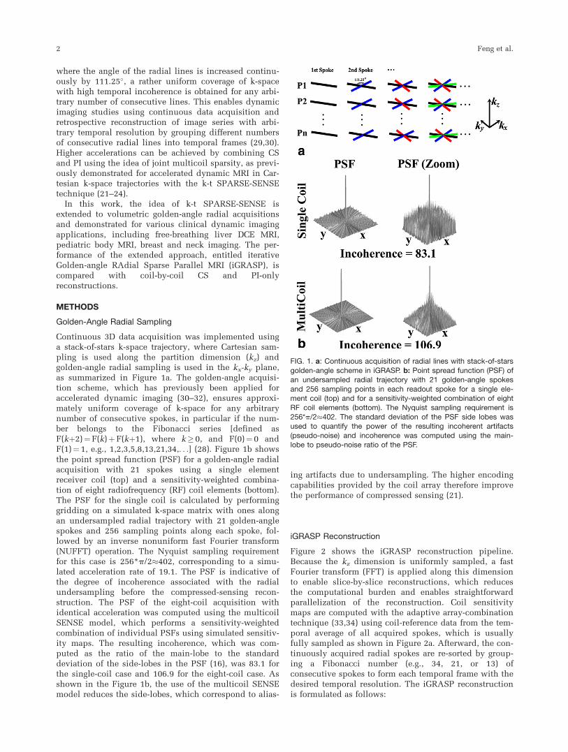
Continuous 3D data acquisition was implemented usinga stack-of-stars k-space trajectory, where Cartesian sam-pling is used along the partition dimension (kz) andgolden-angle radial sampling is used in the kx-ky plane,as summarized in Figure 1a. The golden-angle acquisi-tion scheme, which has previously been applied foraccelerated dynamic imaging (30–32), ensures approxi-mately uniform coverage of k-space for any arbitrarynumber of consecutive spokes, in particular if the num-ber belongs to the Fibonacci series [defined asF(kþ2)¼F(k)þF(kþ1), where k� 0, and F(0)¼ 0 andF(1)¼ 1, e.g., 1,2,3,5,8,13,21,34,. . .] (28). Figure 1b showsthe point spread function (PSF) for a golden-angle radialacquisition with 21 spokes using a single elementreceiver coil (top) and a sensitivity-weighted combina-tion of eight radiofrequency (RF) coil elements (bottom).The PSF for the single coil is calculated by performinggridding on a simulated k-space matrix with ones alongan undersampled radial trajectory with 21 golden-anglespokes and 256 sampling points along each spoke, fol-lowed by an inverse nonuniform fast Fourier transform(NUFFT) operation. The Nyquist sampling requirementfor this case is 256*p/2�402, corresponding to a simu-lated acceleration rate of 19.1. The PSF is indicative ofthe degree of incoherence associated with the radialundersampling before the compressed-sensing recon-struction. The PSF of the eight-coil acquisition withidentical acceleration was computed using the multicoilSENSE model, which performs a sensitivity-weightedcombination of individual PSFs using simulated sensitiv-ity maps. The resulting incoherence, which was com-puted as the ratio of the main-lobe to the standarddeviation of the side-lobes in the PSF (16), was 83.1 forthe single-coil case and 106.9 for the eight-coil case. Asshown in the Figure 1b, the use of the multicoil SENSEmodel reduces the side-lobes, which correspond to alias-
ing artifacts due to undersampling. The higher encodingcapabilities provided by the coil array therefore improvethe performance of compressed sensing (21).
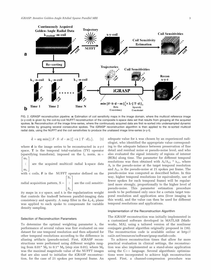
iGRASP Reconstruction
Figure 2 shows the iGRASP reconstruction pipeline.Because the kz dimension is uniformly sampled, a fastFourier transform (FFT) is applied along this dimensionto enable slice-by-slice reconstructions, which reducesthe computational burden and enables straightforwardparallelization of the reconstruction. Coil sensitivitymaps are computed with the adaptive array-combinationtechnique (33,34) using coil-reference data from the tem-poral average of all acquired spokes, which is usuallyfully sampled as shown in Figure 2a. Afterward, the con-tinuously acquired radial spokes are re-sorted by group-ing a Fibonacci number (e.g., 34, 21, or 13) ofconsecutive spokes to form each temporal frame with thedesired temporal resolution. The iGRASP reconstructionis formulated as follows:
FIG. 1. a: Continuous acquisition of radial lines with stack-of-stars
golden-angle scheme in iGRASP. b: Point spread function (PSF) ofan undersampled radial trajectory with 21 golden-angle spokes
and 256 sampling points in each readout spoke for a single ele-ment coil (top) and for a sensitivity-weighted combination of eightRF coil elements (bottom). The Nyquist sampling requirement is
256*p/2�402. The standard deviation of the PSF side lobes wasused to quantify the power of the resulting incoherent artifacts
(pseudo-noise) and incoherence was computed using the main-lobe to pseudo-noise ratio of the PSF.
2 Feng et al.
d̂ ¼ arg min k F � S � d �m k22 þl k T � dk1
� �; [1]
where d is the image series to be reconstructed in x-y-tspace, T is the temporal total-variation (TV) operator(sparsifying transform), imposed on the l1 norm, m ¼
m1
�
mc
2664
3775 are the acquired multicoil radial k-space data
with c coils, F is the NUFFT operator defined on the
radial acquisition pattern, S ¼
S1
�
Sc
2664
3775 are the coil sensitiv-
ity maps in x-y space, and l is the regularization weightthat controls the tradeoff between parallel-imaging dataconsistency and sparsity. A ramp filter in the kx-ky planewas applied to each spoke to compensate for variabledensity sampling.
Selection of Reconstruction Parameters
To determine the optimal weighting parameter l, theperformance of several values was first evaluated on onedataset for one temporal resolution and then adjusted forother temporal resolutions according to the difference inaliasing artifacts (pseudo-noise). First, iGRASP recon-structions were performed using different weights rang-ing from 0.01* M0 to 0.1* M0 (step size 0.01), where M0
was the maximal magnitude value of the NUFFT imagesthat are also used to initialize the iGRASP reconstruc-tion, for the case of 21 spokes per temporal frame. An
adequate value for l was chosen by an experienced radi-ologist, who identified the appropriate value correspond-ing to the adequate balance between preservation of finedetail and residual noise or pseudo-noise level, and whoalso evaluated the signal intensity of regions of interest(ROIs) along time. The parameter for different temporalresolutions was then obtained with At/A21 * l21, whereAt is the pseudo-noise at the target temporal resolutionand A21 is the pseudo-noise at 21 spokes per frame. Thepseudo-noise was computed as described before. In thisway, higher temporal resolutions (or equivalently, use offewer spokes for each temporal frame) will be regular-ized more strongly, proportionally to the higher level ofpseudo-noise. This parameter estimation procedureneeds to be performed only once for a certain target tem-poral resolution and application area (liver imaging inthis work), and the value can then be used for differenttemporal resolutions and applications.
Implementation of the Reconstruction Algorithm
The iGRASP reconstruction was initially implemented ina customized software developed in MATLAB (Math-works, MA), using a tailored version of the nonlinearconjugate gradient algorithm originally proposed in (16).The reconstruction code is available online at http://cai2r.net/resources/software/grasp-matlab-code.
To achieve reconstruction times that allow for morepractical evaluation in clinical settings, the reconstruc-tion was also implemented as a stand-alone applicationusing the Cþþ language. Several algorithmic optimiza-tions were incorporated to achieve high reconstructionspeed. First, a channel-compression procedure was
FIG. 2. iGRASP reconstruction pipeline. a: Estimation of coil sensitivity maps in the image domain, where the multicoil reference image
(x-y-coil) is given by the coil-by-coil NUFFT reconstruction of the composite k-space data set that results from grouping all the acquiredspokes. b: Reconstruction of the image time-series, where the continuously acquired data are first re-sorted into undersampled dynamic
time series by grouping several consecutive spokes. The iGRASP reconstruction algorithm is then applied to the re-sorted multicoilradial data, using the NUFFT and the coil sensitivities to produce the unaliased image time-series (x-y-t).
iGRASP: Iterative Golden-Angle RAdial Sparse Parallel MRI 3

applied to reduce the amount of k-space data, whichcombined the receiver channels into eigenmodes basedon a principal component analysis and discarded higher-order modes such that 95% of the signal power was pre-served (35). Second, the reconstruction was parallelizedacross slices using the OpenMP framework (36), yieldingan almost linear reduction of the reconstruction timewith the number of processor cores. The NUFFT wasimplemented by means of convolution with a Kaiser-Bessel kernel. Interpolation coefficients were precalcu-lated and shared across threads. Corner rounding wasapplied to allow for differentiation of the TV l1 norm.Minimization of the cost function was achieved with a Cimplementation of the limited-memory Broyden-Fletcher-Goldfarb-Shanno (L-BFGS) algorithm (37).
Representative Imaging Applications
iGRASP dynamic imaging was clinically implementedand evaluated for a variety of representative imagingapplications, as described in the following subsections.Human imaging was approved by institutional reviewboard and was Health Insurance Portability andAccountability Act (HIPAA) compliant. Written informedconsent was obtained from all the subjects before theimaging.
Dynamic Contrast-Enhanced Liver Imaging
DCE liver MRI was performed in six healthy volunteers(age, 34.5 6 5.2 years) and seven patients (age, 51 6 8.4years) in axial orientation during free breathing usingwhole-body 3 Tesla (T) or 1.5T scanners (MAGNETOMVerio / Avanto, Siemens AG, Erlangen, Germany) with acombination of body-matrix and spine coil elementswith 12 channels in total. Data acquisition was initiatedsimultaneously with intravenous injection of 10 mL ofgadopentate dimeglumine (Gd-DTPA) (Magnevist, BayerHealthcare, Leverkusen) followed by a 20-mL salineflush, both injected at a rate of 2 mL/s. A radial stack-of-stars three-dimensional (3D) Fast Low Angle SHot(FLASH) pulse sequence with golden-angle ordering wasemployed for the data acquisitions. Two-fold readoutoversampling was applied to avoid spurious aliasingalong the spokes. All partitions corresponding to oneradial angle were acquired sequentially before moving tothe next angle. The ordering scheme along kz wasswitched between linear (from kz¼�kzmax/2 tokz¼þkzmax/2) and centric out (starting at kz¼0) depend-
ing on the number of slices, as done in most of the mod-ern 3D gradient echo (GRE) sequences. Frequency-selective fat suppression was used and 60 initial calibra-tion lines were acquired to correct system-dependent gra-dient-delay errors as described by Block and Uecker (38).Relevant imaging parameters are listed in Table 1.
Dynamic Contrast-Enhanced Pediatric Body Imaging
Abdominal DCE MRI was performed in five pediatricpatients (age 4.8 6 4.1 years) in axial orientation on a1.5T scanner (MAGNETOM Avanto, Siemens AG) usinga body/spine coil array with 12 elements. Acquisitionswere performed during free-breathing because thepatients were sedated during the exam. The imaging andcontrast-injection protocols were comparable to the liverexample described above. Relevant parameters are listedin Table 1.
Dynamic Contrast-Enhanced Breast Imaging
Free-breathing unilateral breast DCE MRI was performedin six patients (age, 55.3 6 6.7 years) in sagittal orienta-tion before MRI-guided biopsy using the radial 3DFLASH protocol on a 3T scanner (MAGNETOM Trio,Siemens AG), equipped with a seven-element breast-coilarray (InVivo Corporation, Gainesville, FL). A singledose of Gd-DTPA with concentration of 0.1 mM/kg bodyweight was injected at 3 mL/s into an antecubital vein.Relevant imaging parameters are listed in Table 1.
Dynamic Contrast-Enhanced Neck Imaging
DCE MRI of the neck was performed in 10 patients (age,66.2 6 19.9 years) in axial orientation using the radial 3DFLASH protocol on a 1.5T scanner (MAGNETOMAvanto, Siemens AG), using a head/spine coil with 15elements. The contrast-injection protocol was identicalto the liver example. Relevant imaging parameters arelisted in Table 1.
Image Reconstruction
Iterative SENSE, coil-by-coil CS, and iGRASP reconstruc-tions were performed on all the datasets using 21 spokesfor each temporal frame. The reconstructed in-planematrix size was 256 � 256 or 384 � 384, depending onthe number of readout samples. The achieved temporalresolution was approximately 3 s/volume for the liverapplication, 5 s/volume for the pediatric application,
Table 1Representative Imaging Parameters Of Dynamic Volumetric MRI In Different Applications
DCE Liver DCE Pediatrics DCE Breast DCE Neck
#Sampling in Each Readout (2�) 512�768 512 512 512#Partitions 29�40 48 35 69#Spokes in Each Partition 600�800 800 2280 800
Slice Thickness (mm) 3 3 2 2FOV (mm2) 370�370 250�250 270�270 256�256
TR/TE (ms) 3.83/1.71 4.24/2.07 3.6/1.47 4.57/2.06Flip Angle (Degree) 12 12 12 12Acquisition Time (s) 90 193 331 283
4 Feng et al.
3 s/volume for breast imaging and 7 s/volume for neckimaging. Compared with the Nyquist sampling rate, thereconstructions correspond to an acceleration rate of 19.1to 28.7.
The iterative SENSE reconstruction was performed usingthe iGRASP implementation with a regularization weightof l¼ 0. The reference CS reconstruction was performedseparately for each coil element, followed by sensitivity-weighted combination. The regularization parameter wasselected only once, as described for iGRASP.
To demonstrate the flexibility of iGRASP, reconstruc-tions were also performed with different temporal resolu-tions for one of the pediatric datasets (13 and 34 spokes,corresponding to 3 and 8 s/volume).
Image reconstruction was performed using the Cþþimplementation on a Linux server equipped with four IntelXeon E5520 quad core CPUs at 2.27 GHz and 96 GB ofRAM. The reconstruction time ranged from 30 to 45 min fora complete 3D data set, depending on the size of datasets.
Image Analysis and Statistics
To evaluate the image quality and temporal fidelityachieved with iGRASP, one representative partition wasselected from each reconstructed dataset for image qual-ity assessment. Images were compared between iGRASPversus iterative SENSE, iGRASP versus coil-by-coil CS,and for temporal fidelity assessment between iGRASPversus NUFFT.
Image Quality Assessment
A total of 39 liver datasets (13 iterative SENSE, 13 coil-by-coil CS and 13 iGRASP), 15 pediatrics datasets (5 iterativeSENSE, 5 coil-by-coil CS and 5 iGRASP), 18 breast data-sets (6 iterative SENSE, 6 coil-by-coil CS and 6 iGRASP),and 30 neck datasets (10 iterative SENSE, 10 coil-by-coilCS and 10 iGRASP) were pooled and randomized forblinded qualitative evaluation by three radiologists withexpertise on abdominal imaging, breast imaging and neu-roimaging, respectively. The score levels for all the imagequality assessments were: 1¼nondiagnostic, 2¼poor,3¼ adequate, 4¼ good, and 5¼ excellent.
The reported scores in each reconstruction categoryfrom all four applications were pooled together to repre-sent mean 6 standard deviation. Wilcoxon signed-ranksum test was chosen to compare the scores betweeniGRASP versus iterative SENSE and iGRASP versus coil-by-coil CS (n¼34), using Excel (Microsoft, Redmond,WA) where P< 0.05 was considered to be statisticallysignificant difference.
Temporal Fidelity Assessment
For each of the iGRASP datasets, a ROI was manuallydrawn to evaluate the signal-intensity time courses. Theupslope was computed using a linear fit of the curvepoints chosen between 10% and 90% of the relative peakenhancement, which usually corresponded to the firstpass of contrast agent. The corresponding NUFFT data setwas evaluated using the same ROI, and the upslope wascalculated using the same length of curve points as refer-ence. The analysis was performed on all iGRASP datasets
(n¼ 34) and the corresponding NUFFT results. Theupslope of NUFFT and iGRASP reconstructions werepooled separately and the relative accuracy was evaluatedby performing linear correlation and Intraclass correlation(ICC) in Excel (Microsoft, Redmond, WA).
RESULTS
Selection of Reconstruction Parameters
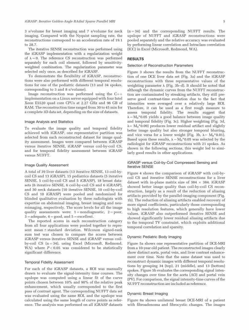
Figure 3 shows the results from the NUFFT reconstruc-tion of one DCE liver data set (Fig. 3a) and the iGRASPreconstructions with three representative values of theweighting parameter l (Fig. 3b–d). It should be noted thatalthough the dynamic curves from the NUFFT reconstruc-tion are contaminated by streaking artifacts, they still pre-serve good contrast-time evolution due to the fact thatintensities were averaged over a relatively large ROI.Therefore, it can be used as a first rough measure toassess temporal fidelity. The results suggest thatl¼M0*0.05 yields a good balance between image qualityand temporal fidelity (Fig. 3c). Higher weighting (Fig. 3d,l¼ M0*0.09) produces lower residual artifact and slightlybetter image quality but also stronger temporal blurring,and vice versa for a lower weight (Fig. 3b, l¼ M0*0.01).Based upon these results, l¼M0*0.05 was selected by theradiologist for iGRASP reconstructions with 21 spokes. Asshown in the following sections, this weight led to simi-larly good results in other applications.
iGRASP versus Coil-by-Coil Compressed Sensing andIterative SENSE
Figure 4 shows the comparison of iGRASP with coil-by-coil CS and iterative SENSE reconstructions for a liverdataset with in-plane matrix size of 384 � 384. iGRASPshowed better image quality than coil-by-coil CS recon-struction, largely as a result of the reduction of aliasingartifacts provided by the parallel-imaging component (Fig.1b). The reduction of aliasing artifacts enabled recovery ofmore signal coefficients, particularly those correspondingto high resolution features, which generally have lowervalues. iGRASP also outperformed iterative SENSE andshowed significantly lower residual aliasing artifacts dueto the temporal TV constraint, which exploits additionaltemporal correlation and sparsity.
Dynamic Pediatric Body Imaging
Figure 5a shows one representative partition of DCE-MRIfrom a 10-year old patient. The reconstructed images clearlyshow distinct aorta, portal vein, and liver contrast enhance-ment over time. Note that the same dataset was used toreconstruct dynamic images with different temporal resolu-tions by grouping 34 (top), 21 (middle), and 13 (bottom)spokes. Figure 5b evaluates the corresponding signal inten-sity changes over time for the aorta (AO) and portal vein(PV). For comparison, the signal intensity-time curves of theNUFFT reconstruction are included as reference.
Dynamic Breast Imaging
Figure 6a shows unilateral breast DCE-MRI of a patientwith fibroadenoma and fibrocystic changes. The images
iGRASP: Iterative Golden-Angle RAdial Sparse Parallel MRI 5

reconstructed with iGRASP show appropriate contrastenhancement over time in the normal breast tissue andin a suspicious breast lesion indicated by the whitearrow. iGRASP also provided good image quality anddepiction of relevant morphological features, such asfibroglandular tissue, skin layer, and the suspiciouslesion. Figure 6b shows the corresponding signal inten-sity changes over time of the breast lesion, heart cavity,vessel and breast tissue (white arrows and ROI). TheiGRASP reconstruction did not introduce significantnotable temporal blurring.
Dynamic Neck Imaging
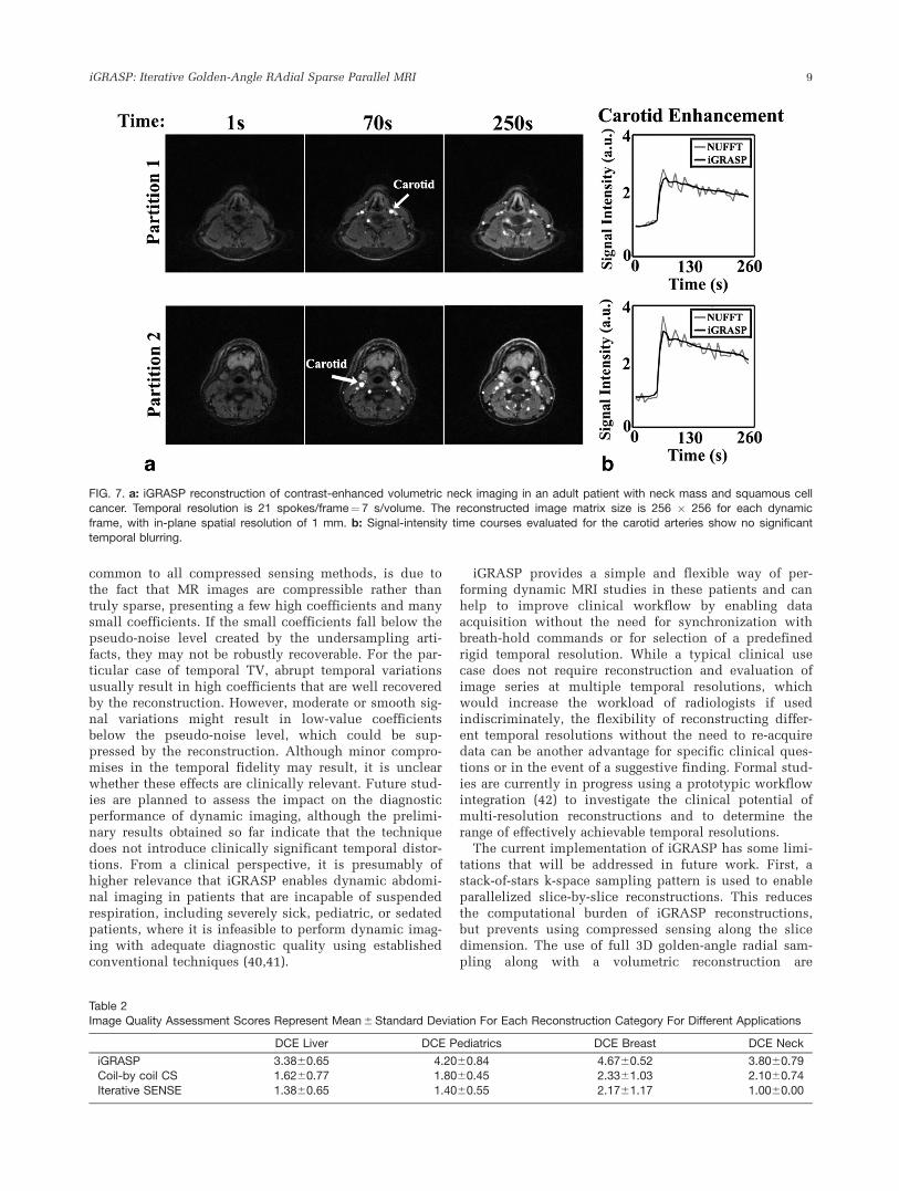
Figure 7 shows representative images of two partitionsfrom a patient with neck mass and squamous cell cancer,together with the corresponding signal-intensity changesfor the carotid artery (white arrows). The reconstruction
shows good image quality in different phases and similarcontrast enhancement to the NUFFT curves.
Image Quality Comparison
Table 2 summarizes the mean scores and standard devia-tions for different reconstruction strategies in each appli-cation. iGRASP yielded significantly better scores(P< 0.05) when compared with both iterative SENSE andcoil-by-coil CS reconstructions. The score of iGRASPwas above 3.0 in all applications, suggesting that goodimage quality can be achieved with the proposed acceler-ation rate and temporal resolution.
Temporal Fidelity Comparison
For the upslope calculated from the data pairs (n¼ 34,iGRASP versus NUFFT), the linear regression coefficient
FIG. 3. Reconstruction of one representative partition from the contrast-enhanced volumetric liver dataset acquired with golden-angleradial sampling scheme using NUFFT (a) and iGRASP with three different weighting parameters (b–d) by grouping 21 consecutive
spokes in each temporal frame. Results with l¼M0*0.05 achieved an appropriate compromise between image quality and temporalfidelity. This value was therefore chosen for iGRASP reconstruction with temporal resolutions of 21 spokes per frame. The weighting
parameter was adjusted for different temporal resolutions according to the level of incoherent aliasing artifacts or pseudo-noise in thePSF. M0 was the maximal magnitude value of the NUFFT images that were also used to initialize the iGRASP reconstruction.
6 Feng et al.

was 0.99, the linear fitting slope was 0.98, and ICC was0.99, indicating strong agreement between the upslopesobtained from iGRASP and NUFFT. This result suggeststhat iGRASP does not introduce significant temporalblurring.
DISCUSSION
This work introduces a robust approach for rapiddynamic volumetric MRI named iGRASP, which isapplicable for a broad spectrum of clinical applications.Even though individual components of the method havebeen proposed before, the synergistic combination of CS,PI, and golden-angle radial sampling results in a tech-nique that is particularly well-suited to obtain high spa-tial resolution, high temporal resolution, and largevolumetric coverage at the same time. iGRASP achievedsignificantly better performance than either PI or CSalone and demonstrated high value for clinical studiesthat require robustness to patient motion and simultane-ous high spatial and temporal resolution. iGRASP can bealso used in other applications such as cardiac cineimaging (39).
The motion robustness can be mainly attributed to theuse of radial k-space sampling. Radial sampling is well-known for being less susceptible than Cartesian samplingdue to (a) lower sensitivity to motion-induced phase shiftsand (b) signal averaging at the center of k-space. Moreover,it is well-suited for CS because radial undersampling cre-ates incoherent low-intensity streaking artifacts. The
golden-angle ordering scheme additionally introducestemporal incoherence of the k-space acquisition.
In radial acquisitions, the image contrast correspondsto the average over the acquisition window because alllines cover k-space center. In this regard, radial samplingintroduces a certain amount of temporal blurring, whichmanifests as slightly lower vessel-tissue contrast com-pared with Cartesian acquisitions that use a narrow timewindow for the acquisition of the k-space center. How-ever, as opposed to other radial approaches that use abroad temporal view-sharing filter to extract differenttemporal phases without streaking artifacts (28), iGRASPenforces data fidelity only within a relatively small tem-poral window (e.g., 21 spokes), which enables to pre-serve high temporal sharpness.
iGRASP reconstruction removes streaking artifacts inthe undersampled time-series of images at the expense ofsuppressing small coefficients in the temporal TVdomain, which can compromise temporal fidelity forhigh acceleration factors because rapidly oscillatingintensity changes may be dampened in this case whilethe temporal onset of sharp intensity changes remainsunaffected due to the use of the l1 norm. However,unlike reconstruction approaches that use TV constrainsin the spatial domain, iGRASP does not lead to spatialimage blurring or synthetic appearance. In cases wherethere is motion between temporal frames, temporal blur-ring artifacts might under certain circumstances appearas spatial blurring artifacts, but these artifacts originatein the temporal dimension. This penalty, which is
FIG. 4. Comparison of iGRASP (top) reconstruction with coil-by-coil CS (middle) and iterative SENSE (bottom) reconstructions in theliver dataset with the same acceleration rate and temporal resolution of 21 spokes/frame¼3 s/volume. iGRASP showed superior imagequality compared with both coil-by-coil CS and iterative SENSE reconstructions.
iGRASP: Iterative Golden-Angle RAdial Sparse Parallel MRI 7
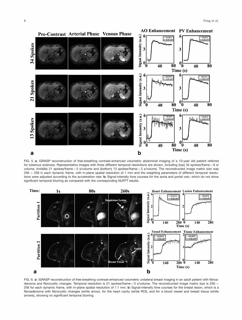
FIG. 5. a: iGRASP reconstruction of free-breathing contrast-enhanced volumetric abdominal imaging of a 10-year old patient referredfor tuberous sclerosis. Representative images with three different temporal resolutions are shown, including (top) 34 spokes/frame¼8 s/volume, (middle) 21 spokes/frame¼5 s/volume and (bottom) 13 spokes/frame¼3 s/volume. The reconstructed image matrix size was
256 � 256 in each dynamic frame, with in-plane spatial resolution of 1 mm and the weighting parameters of different temporal resolu-tions were adjusted according to the acceleration rate. b: Signal-intensity time courses for the aorta and portal vein, which do not show
significant temporal blurring as compared with the corresponding NUFFT results.
FIG. 6. a: iGRASP reconstruction of free-breathing contrast-enhanced volumetric unilateral breast imaging in an adult patient with fibroa-denoma and fibrocystic changes. Temporal resolution is 21 spokes/frame¼3 s/volume. The reconstructed image matrix size is 256 �256 for each dynamic frame, with in-plane spatial resolution of 1.1 mm. b: Signal-intensity time courses for the breast lesion, which is afibroadenoma with fibrocystic changes (white arrow), for the heart cavity (white ROI), and for a blood vessel and breast tissue (white
arrows), showing no significant temporal blurring.
8 Feng et al.
common to all compressed sensing methods, is due tothe fact that MR images are compressible rather thantruly sparse, presenting a few high coefficients and manysmall coefficients. If the small coefficients fall below thepseudo-noise level created by the undersampling arti-facts, they may not be robustly recoverable. For the par-ticular case of temporal TV, abrupt temporal variationsusually result in high coefficients that are well recoveredby the reconstruction. However, moderate or smooth sig-nal variations might result in low-value coefficientsbelow the pseudo-noise level, which could be sup-pressed by the reconstruction. Although minor compro-mises in the temporal fidelity may result, it is unclearwhether these effects are clinically relevant. Future stud-ies are planned to assess the impact on the diagnosticperformance of dynamic imaging, although the prelimi-nary results obtained so far indicate that the techniquedoes not introduce clinically significant temporal distor-tions. From a clinical perspective, it is presumably ofhigher relevance that iGRASP enables dynamic abdomi-nal imaging in patients that are incapable of suspendedrespiration, including severely sick, pediatric, or sedatedpatients, where it is infeasible to perform dynamic imag-ing with adequate diagnostic quality using establishedconventional techniques (40,41).
iGRASP provides a simple and flexible way of per-forming dynamic MRI studies in these patients and canhelp to improve clinical workflow by enabling dataacquisition without the need for synchronization withbreath-hold commands or for selection of a predefinedrigid temporal resolution. While a typical clinical usecase does not require reconstruction and evaluation ofimage series at multiple temporal resolutions, whichwould increase the workload of radiologists if usedindiscriminately, the flexibility of reconstructing differ-ent temporal resolutions without the need to re-acquiredata can be another advantage for specific clinical ques-tions or in the event of a suggestive finding. Formal stud-ies are currently in progress using a prototypic workflowintegration (42) to investigate the clinical potential ofmulti-resolution reconstructions and to determine therange of effectively achievable temporal resolutions.
The current implementation of iGRASP has some limi-tations that will be addressed in future work. First, astack-of-stars k-space sampling pattern is used to enableparallelized slice-by-slice reconstructions. This reducesthe computational burden of iGRASP reconstructions,but prevents using compressed sensing along the slicedimension. The use of full 3D golden-angle radial sam-pling along with a volumetric reconstruction are
FIG. 7. a: iGRASP reconstruction of contrast-enhanced volumetric neck imaging in an adult patient with neck mass and squamous cellcancer. Temporal resolution is 21 spokes/frame¼7 s/volume. The reconstructed image matrix size is 256 � 256 for each dynamic
frame, with in-plane spatial resolution of 1 mm. b: Signal-intensity time courses evaluated for the carotid arteries show no significanttemporal blurring.
Table 2Image Quality Assessment Scores Represent Mean 6 Standard Deviation For Each Reconstruction Category For Different Applications
DCE Liver DCE Pediatrics DCE Breast DCE Neck
iGRASP 3.3860.65 4.2060.84 4.6760.52 3.8060.79Coil-by coil CS 1.6260.77 1.8060.45 2.3361.03 2.1060.74
Iterative SENSE 1.3860.65 1.4060.55 2.1761.17 1.0060.00
iGRASP: Iterative Golden-Angle RAdial Sparse Parallel MRI 9

expected to further increase the performance, at theexpense of higher computational demand. Second,although temporal TV has been used before for differentdynamic MRI reconstructions and was shown to be bet-ter in some specific applications (19), it may be not opti-mal to use it as the only sparsifying transform for allcases and applications. Other advanced temporal sparsi-fying transforms, such as dictionary learning, might bealso useful to increase temporal fidelity for high under-sampling factors. Third, the current work did not use rig-orous mathematical criteria to select the weightingparameter l, which controls the tradeoff betweenremoval of streaking aliasing artifacts and temporal fidel-ity. The empirical rule to make l proportional to thepseudo-noise level in the PSF produced reasonable per-formance for different undersampling factors. The samel was also used in different applications for a given tem-poral resolution, which suggests that the reconstructioncan be automated without intervention. However, evalu-ation on a larger set of patients comparing with standardclinical techniques is required to test the robustness ofthis new approach. Finally, because it is impossible inpractice to acquire a fully-sampled volumetric DCE data-set with the target spatial and temporal resolution, thecurrent study used NUFFT reconstructions as temporalreference. While NUFFT reconstructions provide timecurves without artificial temporal blurring effects, theycan be affected by strong streaking artifacts at high accel-erations that limit their value for assessing the ground-truth signal evolution. A comprehensive analysis of thetemporal fidelity achieved with iGRASP using numericalsimulations and dynamic phantom scans is currently inprogress.
CONCLUSIONS
The combination of compressed sensing, parallel imag-ing, and golden-angle radial sampling used in iGRASPenables rapid dynamic volumetric MRI studies with highspatial resolution, temporal resolution, and motionrobustness. Because of the continuous data acquisitionand the flexibility to reconstruct images retrospectivelyat different temporal resolutions, dynamic imaging withiGRASP can be integrated easily into clinical workflow(42). iGRASP can be used for a wide range of clinicalapplications and demonstrated particular value forexaminations of patients that are unable to suspendrespiration.
ACKNOWLEDGMENTS
The authors thank Linda Moy and Girish Fatterpekar fortheir help with image scoring on the breast and neckdata sets, respectively.
REFERENCES
1. Padhani AR. Dynamic contrast-enhanced MRI in clinical oncology:
current status and future directions. J Magn Reson Imaging 2002;16:
407–422.
2. Xu B, Spincemaille P, Chen G, Agrawal M, Nguyen TD, Prince MR,
Wang Y. Fast 3D contrast enhanced MRI of the liver using temporal
resolution acceleration with constrained evolution reconstruction.
Magn Reson Med 2013;69:370–381.
3. Sodickson DK, Manning WJ. Simultaneous acquisition of spatial har-
monics (SMASH): fast imaging with radiofrequency coil arrays. Magn
Reson Med 1997;38:591–603.
4. Pruessmann KP, Weiger M, Scheidegger MB, Boesiger P. SENSE: sen-
sitivity encoding for fast MRI. Magn Reson Med 1999;42:952–962.
5. Griswold MA, Jakob PM, Heidemann RM, Nittka M, Jellus V, Wang J,
Kiefer B, Haase A. Generalized autocalibrating partially parallel
acquisitions (GRAPPA). Magn Reson Med 2002;47:1202–1210.
6. Kellman P, Epstein FH, McVeigh ER. Adaptive sensitivity encoding
incorporating temporal filtering (TSENSE). Magn Reson Med 2001;45:
846–852.
7. Breuer FA, Kellman P, Griswold MA, Jakob PM. Dynamic autocali-
brated parallel imaging using temporal GRAPPA (TGRAPPA). Magn
Reson Med 2005;53:981–985.
8. Tsao J, Kozerke S. MRI temporal acceleration techniques. J Magn
Reson Imaging 2012;36:543–560.
9. Tsao J, Boesiger P, Pruessmann KP. k-t BLAST and k-t SENSE:
dynamic MRI with high frame rate exploiting spatiotemporal correla-
tions. Magn Reson Med 2003;50:1031–1042.
10. Huang F, Akao J, Vijayakumar S, Duensing GR, Limkeman M. k-t
GRAPPA: a k-space implementation for dynamic MRI with high
reduction factor. Magn Reson Med 2005;54:1172–1184.
11. Xu D, King KF, Liang ZP. Improving k-t SENSE by adaptive regulari-
zation. Magn Reson Med 2007;57:918–930.
12. van Vaals JJ, Brummer ME, Dixon WT, Tuithof HH, Engels H, Nelson
RC, Gerety BM, Chezmar JL, den Boer JA. "Keyhole" method for
accelerating imaging of contrast agent uptake. J Magn Reson Imaging
1993;3:671–675.
13. Jones RA, Haraldseth O, Muller TB, Rinck PA, Oksendal AN. K-space
substitution: a novel dynamic imaging technique. Magn Reson Med
1993;29:830–834.
14. Candes E, Romberg JT. Robust uncertainty principles: exact signal
reconstruction from highly incomplete frequency information. IEEE
Trans Inf Theory 2006;52:489–509.
15. Donoho D. Compressed sensing. IEEE Trans Inf Theory 2006;52:
1289–1306.
16. Lustig M, Donoho D, Pauly JM. Sparse MRI: the application of com-
pressed sensing for rapid MR imaging. Magn Reson Med 2007;58:
1182–1195.
17. Lustig M, Santos J, Donoho D, Pauly J. k-t SPARSE: high frame rate
dynamic MRI exploiting spatio-temporal sparsity. In Proceedings of
the 14th Annual Meeting of ISMRM, Seattle, Washington, USA, 2006.
p. 2420.
18. Gamper U, Boesiger P, Kozerke S. Compressed sensing in dynamic
MRI. Magn Reson Med 2008;59:365–373.
19. Adluru G, Awate SP, Tasdizen T, Whitaker RT, Dibella EV. Tempo-
rally constrained reconstruction of dynamic cardiac perfusion MRI.
Magn Reson Med 2007;57:1027–1036.
20. Jung H, Sung K, Nayak KS, Kim EY, Ye JC. k-t FOCUSS: a general
compressed sensing framework for high resolution dynamic MRI.
Magn Reson Med 2009;61:103–116.
21. Otazo R, Kim D, Axel L, Sodickson DK. Combination of compressed
sensing and parallel imaging for highly accelerated first-pass cardiac
perfusion MRI. Magn Reson Med 2010;64:767–776.
22. Feng L, Otazo R, Jung H, Jensen JH, Ye JC, Sodickson DK, Kim D.
Accelerated cardiac T2 mapping using breath-hold multiecho fast
spin-echo pulse sequence with k-t FOCUSS. Magn Reson Med 2011;
65:1661–1669.
23. Kim D, Dyvorne HA, Otazo R, Feng L, Sodickson DK, Lee VS. Accel-
erated phase-contrast cine MRI using k-t SPARSE-SENSE. Magn
Reson Med 2012;67:1054–1064.
24. Feng L, Srichai MB, Lim RP, Harrison A, King W, Adluru G, Dibella
EV, Sodickson DK, Otazo R, Kim D. Highly accelerated real-time car-
diac cine MRI using k-t SPARSE-SENSE. Magn Reson Med 2013;70:
64–74.
25. Block KT, Uecker M, Frahm J. Undersampled radial MRI with multi-
ple coils. Iterative image reconstruction using a total variation con-
straint. Magn Reson Med 2007;57:1086–1098.
26. Glover GH, Pauly JM. Projection reconstruction techniques for reduc-
tion of motion effects in MRI. Magn Reson Med 1992;28:275–289.
27. Gai N, Axel L. Correction of motion artifacts in linogram and projec-
tion reconstruction MRI using geometry and consistency constraints.
Med Phys 1996;23:251–262.
10 Feng et al.
28. Winkelmann S, Schaeffter T, Koehler T, Eggers H, Doessel O. An
optimal radial profile order based on the Golden Ratio for time-
resolved MRI. IEEE Trans Med Imaging 2007;26:68–76.
29. Chan RW, Ramsay EA, Cheung EY, Plewes DB. The influence of
radial undersampling schemes on compressed sensing reconstruction
in breast MRI. Magn Reson Med 2012;67:363–377.
30. Usman M, Atkinson D, Odille F, Kolbitsch C, Vaillant G, Schaeffter
T, Batchelor P, Prieto C. Motion corrected compressed sensing for
free-breathing dynamic cardiac MRI. Magn Reson Med 2012. doi:
10.1002/mrm.24463.
31. Hansen MS, Sorensen TS, Arai AE, Kellman P. Retrospective
reconstruction of high temporal resolution cine images from real-time
MRI using iterative motion correction. Magn Reson Med 2012;68:
741–750.
32. Prieto C, Uribe S, Razavi R, Atkinson D, Schaeffter T. 3D
undersampled golden-radial phase encoding for DCE-MRA using
inherently regularized iterative SENSE. Magn Reson Med 2010;64:
514–526.
33. Walsh DO, Gmitro AF, Marcellin MW. Adaptive reconstruction of
phased array MR imagery. Magn Reson Med 2000;43:682–690.
34. Griswold MA WD, Heidemann RM, Haase A, Jakob PM. The use of
an adaptive reconstruction for array coil sensitivity mapping and
intensity normalization. In Proceedings of the 10th Annual Meeting
of ISMRM, Honolulu, Hawaii, USA, 2002. Abstract 2410.
35. Buehrer M, Pruessmann KP, Boesiger P, Kozerke S. Array compres-
sion for MRI with large coil arrays. Magn Reson Med 2007;57:1131–
1139.
36. Dagum L, Open MP. An industry standard API for shared-memory
programming. IEEE Comput Sci Eng 1998;5:46–55.
37. Nocedal J. Updating quasi-Newton matrices with limited storage.
Math Comput 1980;35:773–782.
38. Block KT, Uecker M. Simple method for adaptive gradient-delay com-
pensation in radial MRI. In Proceedings of the 19th Annual Meeting
of ISMRM, Montreal, Canada, 2011. p. 2816.
39. Feng L, Xu J, Axel L, Sodickson DK, Otazo R. High spatial and tem-
poral resolution 2D real time and 3D whole-heart cardiac cine MRI
using compressed sensing and parallel imaging with golden angle
radial trajectory. In Proceedings of the 20th Annual Meeting of
ISMRM, Melbourne, Australia, 2012. p. 225.
40. Chandarana H, Block TK, Rosenkrantz AB, Lim RP, Kim D, Mossa DJ,
Babb JS, Kiefer B, Lee VS. Free-breathing radial 3D fat-suppressed
T1-weighted gradient echo sequence: a viable alternative for contrast-
enhanced liver imaging in patients unable to suspend respiration.
Invest Radiol 2011;46:648–653.
41. Chandarana H, Feng L, Block KT, Rosenkrantz AB, Lim P, Babb J,
Sodickson DK, Otazo R. Free-breathing contrast-enhanced multiphase
MRI of the liver using a combination of compressed sensing, parallel
imaging, and golden-angle radial sampling. Invest Radiol 2013;48:10–16.
42. Block KT, Grimm R, Feng L, Otazo R, Chandarana H, Bruno M,
Sodickson DK. Bringing compressed sensing to clinical reality: proto-
typic setup for evaluation in routine applications. In Proceedings of the
21st Annual Meeting of ISMRM, Salt Lake City, Utah, USA 2013. p. 3809.
iGRASP: Iterative Golden-Angle RAdial Sparse Parallel MRI 11