gneuromuscular (gnm) approach (part 2) - wordpress.com · by clayton a. chan, dds gneuromuscular...
TRANSCRIPT
by Clayton A. Chan, DDS
Gneuromuscular (GNM)ApproAch (pArt 2)
Seeing the bigger picture – Recognizing the signs and symptoms
Using any form of manual manipulation of the mandible, even if it is repeatable in a posterior superior
or anterior superior position, does not necessarily mean that such techniques are scientifically sound or relevant to establishing a physiologically stable occlusion.
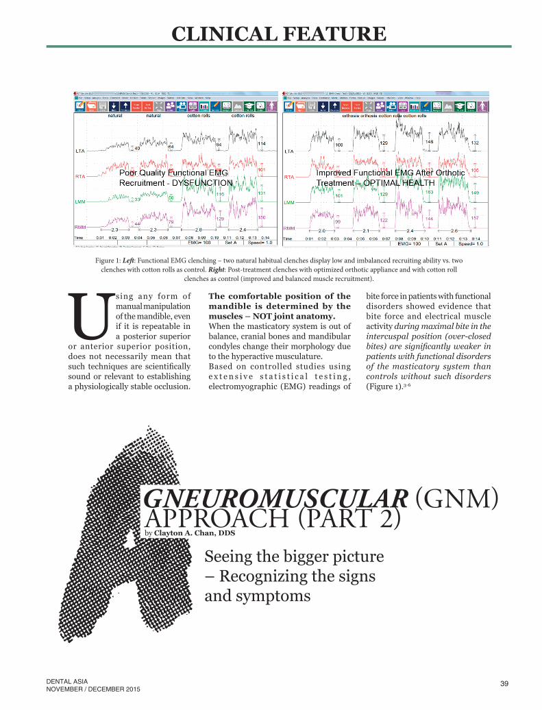
Figure 1: Left: Functional EMG clenching – two natural habitual clenches display low and imbalanced recruiting ability vs. two clenches with cotton rolls as control. Right: Post-treatment clenches with optimized orthotic appliance and with cotton roll
clenches as control (improved and balanced muscle recruitment).
The comfortable position of the mandible is determined by the muscles – NOT joint anatomy.When the masticatory system is out of balance, cranial bones and mandibular condyles change their morphology due to the hyperactive musculature.Based on controlled studies using e x t e n s i v e s t a t i s t i c a l t e s t i n g , electromyographic (EMG) readings of
bite force in patients with functional disorders showed evidence that bite force and electrical muscle activity during maximal bite in the intercuspal position (over-closed bites) are significantly weaker in patients with functional disorders of the masticatory system than controls without such disorders (Figure 1).3-6
39
CLINICAL FEATURE
DENTAL ASIANOVEMBER / DECEMBER 2015

Figure 3: CMS or jaw tracking measures mandibular position, including fine and gross functional movements relative to centric occlusion. It is used in combination with low-frequency TENS to observe any difference in an isotonic mandibular closing path compared to the voluntary mandibular closing path. CMS (K7 kinesiograph) is the dental profession’s “gold standard” in measuring mandibular function, occlusal evaluation and establishing optimum occlusion.
Figure 2: The lower anatomical GNM orthotic is an orthopedic device based on objective measurements determining an optimal physiologic mandibular to maxillary relationship in six dimensions using low-frequency transcutaneous electrical nerve stimulation (TENS) and computerized mandibular scanning. This is worn 24/7 to enhance optimal function and resting modes, allowing freedom of entry and exit to a terminal contact position.
TEChNoloGy DENTISTS INCorPoraTE INTo aN aDvaNCED DENTal PraCTICE:
Low Frequency TENS: J5 Myomonitor
Mandibular Jaw Tracking: Computerized Mandibular Scanning (CMS)
Electromyography: EMG
Lower anatomical orthosis: Conservative and reversibleUse of a full-coverage lower anatomical orthosis has been found to be effective in establishing the temporomandibular joints (TMJ) as well as the masticatory muscles in a physiologic state with positive reduction of temporomandibular disorder (TMD) symptoms (Figure 2).2,7,8 It is used as a conservative, diagnostic, non-invasive and reversible treatment approach to validate whether functional occlusal and joint stability can be achieved. Physiologic improvement must be demonstrated with certainty before more definitive, comprehensive treatment is rendered. A well-designed orthotic using both the gnathic and neuromuscular (GNM) principles of proper occlusal balance and disclusion in an optimal position has shown improved patient health, restoring balance to overall posture.9
Low-frequency TENS (J5 Myomonitor, Myotronics, Inc., Kent, WA, USA) has been found to be scientifically helpful to dentists to relax hypertonic muscles 45 to 60 minutes prior to recording a bite registration. Computerized mandibular scanning (CMS) or jaw tracking has also been used to measure and record mandibular positioning and functional movements in conjunction with TENS to objectively locate an individual’s physiologic maxillomandibular relationship when recording a bite registration for the lower anatomical GNM orthosis (Figures 3 and 4).1,9
Craniomandibular positioning is critical to any case requiring changes in the anteroposterior (AP), frontal/lateral and vertical dimensions. Without knowing specifically how to relate the upper and lower jaws together physiologically, the treating clinician and laboratory can only depend on educated guesses and assumptions at best.Today, the clinician can determine a physiologic mandibular position without manipulating the patient’s jaw. Manipulating the jaw adds a potential for human error in diagnosis and treatment.Using the techniques described in this article, a mandibular position can be easily obtained with the greatest articulator ever designed – the human jaw. Instead of instructing the dental lab to open a bite arbitrarily by increasing the incisal pin, the clinician guides the
40 DENTAL ASIANOVEMBER / DECEMBER 2015
CLINICAL FEATURE

Figure 4: Jaw tracking (K7 kineseograph with J5 TENS, Myotronics, Kent, Wa, USa) allows the clinician to accurately and precisely record a maxillomandibular relationship from six dimensions. This technology is able to assist the dentist in locating an isotonic maxillomandibular jaw registration accurately (within 0.1mm) in six dimensions (aP, lateral, vertical, pitch, yaw and roll) free of any occlusal interferences in an upright postural position.
Figure 5: Left: CMS (jaw track, Scan 5), showing sagittal and frontal habitual closing paths to Co (centric occlusion) and physiologic myo-trajectory closing path. The black myo-target indicates where the bite registration was recorded in three dimensional spaces. Right (Split screen): Left side displays mandibular position in vertical, anteroposterior (aP) and frontal modes relative to Co over time. Right side displays final habitual path of closure coincident with myo-trajectory path of closure (Scan 4/5). Green lines indicate frontal/lateral positions of the mandible before and after physiologic occlusal treatment.
patient to accurately determine the position with the use of low-frequency, involuntary TENS and CMS/jaw tracking instrumentation to visualize the mandibular position (Figure 5). Obtaining an optimal physiologic mandibular position is possible without manipulation as ascertained by modern science and technology.
Objective recording instrumentation for dentistry today has
clearly removed doubts by providing scientific evidence that allows interested clinicians to confirm the state
of muscles, joints and mandibular position as well
as occlusion and the state of structures related to the
stomatognathic system.An understanding of
how to optimally apply these occlusal principles, techniques and protocols
in a balanced way is what Gneuromuscular (GNM)
dentistry (occlusion) is all about.
ConclusionModern medical and dental science has progressed beyond the traditional concepts. Gneuromuscular (GNM) dentistry focuses on how the gnathic (G) and neuromuscular (NM) principles are applied clinically in occlusal management disciplines at the highest levels, recognizing the various musculoskeletal occlusal signs and symptoms. The goal of GNM is to physiologically address how teeth, muscles and joints optimally move and rest in health. Rather than assume that a certain habitual occlusion is healthy, the dentist begins to recognize how occlusal signs could be indicators of possible
underlying dysfunctions and identifies what is “unhealthy” and correlates these problems to misalignments within the oral cavity and beyond.Clinically, low frequency TENS can assist the dentist in capturing a more physiologic bite registration. CMS instrumentation is used to scientifically and objectively align the mandible with TENS with precision. Past occlusal and joint concepts have proven insufficient to predictably address muscular issues, thereby distracting dentists from seeing how muscular problems can impact the diagnosis and treatment planning of cases that may require more than routine habitual occlusal care.
41DENTAL ASIANOVEMBER / DECEMBER 2015
CLINICAL FEATURE

Clayton A. Chan, DDS is a general dentist, clinician, teacher/mentor and educator to hundreds of dentist around the world. He is considered an authority on dental occlusion, TMD and its applications in clinical dentistry. With his training as a dental laboratory technician, along with his clinical experience, he has combined both gnathologic and neuromuscular perspectives now noted as a gneuromuscular (GNM) approach, which is now widely recognized as a logical and systematic approach to occlusal challenges amongst advanced clinicians.
Dr. Chan’s conservative approach to treatment has attracted patients and dentists from all over North America and outside the U.S. who have sought his teachings on the treatment of TMD, craniomandibular myofascial pain and orthopedic problems. His unique approach to Phase I (stabilization) and Phase II (completion) applying these GNM principles is a hallmark in comprehensive care.Dr. Chan is the founder and director of Occlusion Connections™ (www.occlusionconnections.com), a post-graduate training program for GPs, specialists and laboratory technicians in Las Vegas, NV (USA).
About the AuthorThe GNM occlusal approach uses scientifically-based technology that records and verifies the clinician’s observations beyond mere subjective assessment. Symptoms presented by patients in the everyday clinical practice can be recorded and quantified accurately to determine the muscles’ state of health and the TMJ’s condition.The GNM approach is a comprehensive clinical approach that addresses the cause and source of musculoskeletal occlusal signs and symptoms and why they present in the dental practice. It provides protocols and techniques for dentists to implement effective restorative, orthodontic and TMD treatment for their patients. Masticatory stability is established using a lower anatomical orthosis, which is the key to conservative and reversible occlusal and muscle problems. DA
The first of this two-part series was featured in the SepOct15 issue of Dental Asia.
References1. Cooper B. Neuromuscular dentistry: The next millenium. Scientific rationale for biomedical instrumentation, anthology volume v, International College of
Craniomandibular orthopedics, Seattle, Wa, 1999.2. Cooper and Kleinberg: Establishment of a temporomandibular physiological state with neuromuscular orthosis treatment affects reduction of TMD
symptoms in 313 patients. J. Cranio. Practice, april 2008, vol. 26, No. 2, pp104-117.3. Thomas Nr. The effect of fatigue and TEN on the EMG mean power frequency. In: Bergamini M. ed. Pathophysiology of head and neck
musculoskeletal disorders. Frontiers of oral Pathology. Basil, Switzerland: Karger, 1990;7:162-170.4. helkimo E, Carlsson GE, Carmeli y. Bite force in patients with functional disturbances of the masticatory system. J. oral rehab,
vol. 2, 1975;397-406.5. Sheikholeslam a, Moller E, lous I. Postural and maximal activity in elevators of mandible before and after treatment
of functional disorders. Scand J Dent res, 1982;90:37-46.6. randow K, Carlsson K, Edlund J, oberg T. The effect of an occlusal interference on the masticatory system.
an experimental investigation. odontol revy, 1976;27:245-256.7. Cooper B. 1997 classic study on 1182 TMD patients: The role of bioelectronic instrumentation
in the documentation and management of temporomandibular disorders. oral Surg oral Pathol oral Med oral radiol Endod, Mosby-yearbook, Inc., 1997;83(1)91-100.
8. Chan, Ca: Treating craniomandibular dysfunctional patients implementing gnathological or neuromuscular concepts. International College of Craniomandibular orthopedics (ICCMo) anthology vI, 2003.
9. K7 Kineseograph and J5 TENS, Myotronics K7 occlusal Evaluation System, Kent, Wa, USa.
42 DENTAL ASIANOVEMBER / DECEMBER 2015
CLINICAL FEATURE