global trends in proteome remodeling of the outer membrane
TRANSCRIPT

Global Trends in Proteome Remodeling of the OuterMembrane Modulate Antimicrobial Permeability in Klebsiellapneumoniae
Andrea Rocker,a Jake A. Lacey,b Matthew J. Belousoff,a Jonathan J. Wilksch,a,c Richard A. Strugnell,c Mark R. Davies,c
Trevor Lithgowa
aInfection and Immunity Program, Biomedicine Discovery Institute and Department of Microbiology, Monash University, Melbourne, AustraliabDoherty Department, at the Peter Doherty Institute for Infection and Immunity, The University of Melbourne and the Royal Melbourne Hospital, Melbourne, AustraliacDepartment of Microbiology and Immunology, at the Peter Doherty Institute for Infection and Immunity, The University of Melbourne and the Royal MelbourneHospital, Melbourne, Australia
ABSTRACT In Gram-negative bacteria, the permeability of the outer membranegoverns rates of antibiotic uptake and thus the efficacy of antimicrobial treatment.Hydrophilic drugs like �-lactam antibiotics depend on diffusion through pore-forming outer membrane proteins to reach their intracellular targets. In this study,we investigated the distribution of porin genes in more than 2,700 Klebsiella isolatesand found a widespread loss of OmpK35 functionality, particularly in thosestrains isolated from clinical environments. Using a defined set of outer-membrane-remodeled mutants, the major porin OmpK35 was shown to be largelyresponsible for �-lactam permeation. Sequence similarity network analysis character-ized the porin protein subfamilies and led to discovery of a new porin family mem-ber, OmpK38. Structure-based comparisons of OmpK35, OmpK36, OmpK37, OmpK38,and PhoE showed near-identical pore frameworks but defining differences in the se-quence characteristics of the extracellular loops. Antibiotic sensitivity profiles of iso-genic Klebsiella pneumoniae strains, each expressing a different porin as its dominantpore, revealed striking differences in the antibiotic permeability characteristics ofeach channel in a physiological context. Since K. pneumoniae is a nosocomial patho-gen with high rates of antimicrobial resistance and concurrent mortality, these ex-periments elucidate the role of porins in conferring specific drug-resistant pheno-types in a global context, informing future research to combat antimicrobialresistance in K. pneumoniae.
IMPORTANCE Klebsiella pneumoniae is a pathogen of humans with high rates ofmortality and a recognized global rise in incidence of carbapenem-resistant K. pneu-moniae (CRKP). The outer membrane of K. pneumoniae forms a permeability barrierthat modulates the ability of antibiotics to reach their intracellular target. OmpK35,OmpK36, OmpK37, OmpK38, PhoE, and OmpK26 are porins in the outer membraneof K. pneumoniae, demonstrated here to have a causative relationship to drug resis-tance phenotypes in a physiological context. The data highlight that currently trialedcombination treatments with a carbapenem and �-lactamase inhibitors could be ef-fective on porin-deficient K. pneumoniae. Together with structural data, the resultsreveal the role of outer membrane proteome remodeling in antimicrobial resistanceof K. pneumoniae and point to the role of extracellular loops, not channel parame-ters, in drug permeation. This significant finding warrants care in the developmentof phage therapies for K. pneumoniae infections, given the way porin expression willbe modulated to confer phage-resistant—and collateral drug-resistant—phenotypesin K. pneumoniae.
Citation Rocker A, Lacey JA, Belousoff MJ,Wilksch JJ, Strugnell RA, Davies MR, Lithgow T.2020. Global trends in proteome remodeling ofthe outer membrane modulate antimicrobialpermeability in Klebsiella pneumoniae. mBio11:e00603-20. https://doi.org/10.1128/mBio.00603-20.
Editor Julian E. Davies, University of BritishColumbia
Copyright © 2020 Rocker et al. This is an open-access article distributed under the terms ofthe Creative Commons Attribution 4.0International license.
Address correspondence to Trevor Lithgow,[email protected].
Received 12 March 2020Accepted 16 March 2020Published
RESEARCH ARTICLEMolecular Biology and Physiology
crossm
March/April 2020 Volume 11 Issue 2 e00603-20 ® mbio.asm.org 1
14 April 2020
on May 4, 2020 by guest
http://mbio.asm
.org/D
ownloaded from

KEYWORDS antimicrobial resistance, porin, OmpK37, beta-barrel, carbapenem,carbapenems, porins
Klebsiella pneumoniae is the causative agent of invasive and blood-borne infectionsand, as a prime example of carbapenem-resistant Enterobacteriaceae (CRE), it is
regarded by the Centers for Disease Control and Prevention as an “urgent” threat tohuman health. These and related Gram-negative bacteria are prevalent in the environ-ment and play an important role in soil ecosystems (1). However, in just a few decadesK. pneumoniae has evolved from this innocuous existence to become a common andsignificant nosocomial pathogen (2). Initially associated only with the chronically unwelland immunocompromised individuals, Klebsiella’s proficiency at horizontal gene trans-fer has seen the rapid evolution of hypervirulent K. pneumoniae strains that infect evenimmunosufficient people (3, 4). High antibiotic selection pressure in hospitals and otherenvironments precipitated the emergence of plasmid-mediated resistance, and Kleb-siella now harbors antimicrobial resistance (AMR) phenotypes ranging from carbap-enem resistance to colistin resistance, qualifying it as extremely drug resistant (5, 6).Until now, the most successful treatment regime for Klebsiella infections relied onantibiotics of the �-lactam type, particularly carbapenems. However, more and moreKlebsiella strains are being identified with a growing diversity of �-lactamases, includingthe carbapenemases (7–9); carbapenem-resistant K. pneumoniae (CRKP) was first iden-tified in China in 2007, and just 6 years later, carbapenem resistance was found in 13%of K. pneumoniae isolated from hospital patients across the country (10, 11).
Carbapenem resistance in Klebsiella has been observed in isolates confirmed to becarbapenemase negative (7, 12–17). A mutation in either of the genes ompK35 andompK36 was also identified in these strains, leading to the suggestion that resistance iscaused by the diminished import of carbapenem across the outer membrane, togetherwith upregulation of �-lactamases such as AmpC cephalosporinases or extended-spectrum �-lactamases (ESBLs) (7, 12–17). The porins OmpK35 and OmpK36 belong tothe Porin_1 (PF00267) group of bacterial outer membrane proteins. Both OmpK35 andOmpK36 form trimers composed of 16-stranded �-barrels integrated into the outermembrane, and the crystal structures of two of these proteins showed polar residueslining the internal pores (18). Structural and biophysical data agree that polar moleculesof less than 600 Da in size would permeate the channels formed by OmpK35 andOmpK36 with limited selectivity (18–21). As most �-lactams are between 300 and550 Da, they are believed to enter the periplasm via passive diffusion through theseporins (22). In Escherichia coli, the homologous proteins OmpC and OmpF are desig-nated “major porins” in the sense that they represent approximately half of the proteinmass contributed by all �-barrel proteins in the outer membrane; they are so abundantacross the outer membrane surface as to form large diffusion-limited arrays of poresthat provide excellent permeability to small solutes (23–27).
As prominent surface molecules of K. pneumoniae, porins are involved in environ-mental sensing and nutrient acquisition. Correspondingly, loss of porin function maygenerate fitness costs due to impaired nutrient uptake (28–30) and to increased ratesof phagocytosis and bacterial clearing (28, 31, 32) and correlates with decreasedvirulence as determined in mouse models of infection (28, 30, 31, 33). An increasedabundance of OmpK26 or LamB has been observed in some clinical isolates of K.pneumoniae lacking the major porins (34–37), leading to suggestions that these poresmight defray the fitness costs in terms of providing nutrient acquisition pores that donot also allow entry of antibiotics. Recently, similar suggestions were made regardinga third alternative porin in K. pneumoniae, OmpK37, which was suggested to possess anarrower channel to explain the observed lower diffusion rates of substrate molecules(38). Despite these interesting propositions, there remains limited knowledge of theantibiotic uptake through these various alternative porins, with their importance inantimicrobial resistance inferred indirectly from expression levels in clinical strains
Rocker et al. ®
March/April 2020 Volume 11 Issue 2 e00603-20 mbio.asm.org 2
on May 4, 2020 by guest
http://mbio.asm
.org/D
ownloaded from

(34–37). Furthermore, there is a disagreement in the literature as to whether OmpK35or OmpK36 is more important for clinical antibiotic resistance (17, 21, 28, 30, 39–41).
We sought to address three questions in this study. First, while loss-of-functionmutations in ompK35 and ompK36 correlate with multidrug resistance, are they directlycausative? Second, how prevalent are loss-of-function mutations in ompK35 andompK36 around the world? Third, to what extent can remodeling of the outer mem-brane proteome impact antimicrobial resistance phenotypes? We analyzed all publiclyavailable K. pneumoniae genome sequences and catalogued nonsense and missensemutations in the ompK35 and ompK36 genes, with population analysis suggesting thatindependent mutations have frequently occurred. Sequence similarity network analysisof this porin family revealed the existence of a new porin family member, OmpK38. Tosystematically address whether loss-of-function mutations in the major porin genes arecausative for drug resistance, and to explore the idea that upregulation of other outermembrane pores may impact fitness or antibiotic sensitivity, we created an ΔompK35ΔompK36 strain isogenic to a known clinical isolate. A series of strains were thengenerated in which either OmpK35, OmpK36, OmpK37, OmpK38, OmpK26, PhoE, orLamB was upregulated to be the major porin in the strain. Drug sensitivity, includingsensitivity to carbapenems, was directly impacted by the identity of the porin ex-pressed. This study provides evidence to highlight the consequences and causality ofouter membrane proteome remodeling for AMR phenotypes in K. pneumoniae.
RESULTSGlobal analysis of Klebsiella reveals major porin mutations. To assess the
prevalence of loss-of-function mutations in the genes encoding OmpK35 or OmpK36,a global analysis was conducted on 2,706 publicly available genome data sets ofKlebsiella (see Table S1 in the supplemental material). For OmpK35, premature stopcodons were detected in 772 (29%) gene products (Table S1). These terminatingmutations are found throughout the phylogeny but were enriched in the dominantlineage of K. pneumoniae CC258 and strains from a recent survey of a major hospital inPakistan (42) (Fig. 1A). In contrast, mutations in ompK36 were much less frequentlyobserved (3.7%) and were randomly distributed across the global Klebsiella phylogeny(Fig. 1A). Similarly rare and random distributions of inactivating mutations were foundin the other porin-encoding genes (Fig. S1).
Specific tonB alleles are used in multilocus sequence typing of K. pneumoniae, andthe clonal tonB79-containing subgroup CC258 has a premature stop codon in ompK35(Fig. 1A). This group includes the K. pneumoniae ST258 and ST512 lineages character-ized by the KPC carbapenemase. All of these strains possess the same mutation inompK35, with a premature stop codon in place of amino acid position 88. Recentanalysis suggested that ST258 has become one of the most successful multidrug-resistant bacterial pathogens in health care settings throughout the world (43). Aninsertion in loop 3 of OmpK36, previously proposed to contribute to the antimicrobialresistance of ST258 (30), is found in only 16% of these strains.
Within the global data set, mutations leading to a loss of OmpK35 were identifiedin 27.0% of isolates associated with infection, and in a further 13.6% of isolates fromhuman carriage, pointing at an important association of ompK35 inactivation withhealth care settings (Table S1). Furthermore, approximately 50% of OmpK36-deficientstrains had an additional mutation in ompK35 (49 of 99 strains), corroborating theglobal significance of genetic ompK35 inactivation.
One such strain is Klebsiella FK688, recently isolated as the causative agent of a fatal,carbapenem-resistant sepsis (5). This isolate has an insertion in the 5= end of ompK35that would change the promoter region and polypeptide sequence in the presumptivesignal peptide, as well as a premature stop codon that was identified in the sequencedata for ompK36 (accession no. SRR11108934). The same study identified K. pneumoniaestrain FK1934, which is phylogenetically distinct (Fig. 1A) yet also shows a lack of majorporin expression (Fig. 1B) (accession no. SRR11108933). AJ218 is an isolate of K.pneumoniae from a human urinary tract infection (44, 45), with intact ompK35 and
Porin Channels Modulating Antimicrobial Resistance ®
March/April 2020 Volume 11 Issue 2 e00603-20 mbio.asm.org 3
on May 4, 2020 by guest
http://mbio.asm
.org/D
ownloaded from
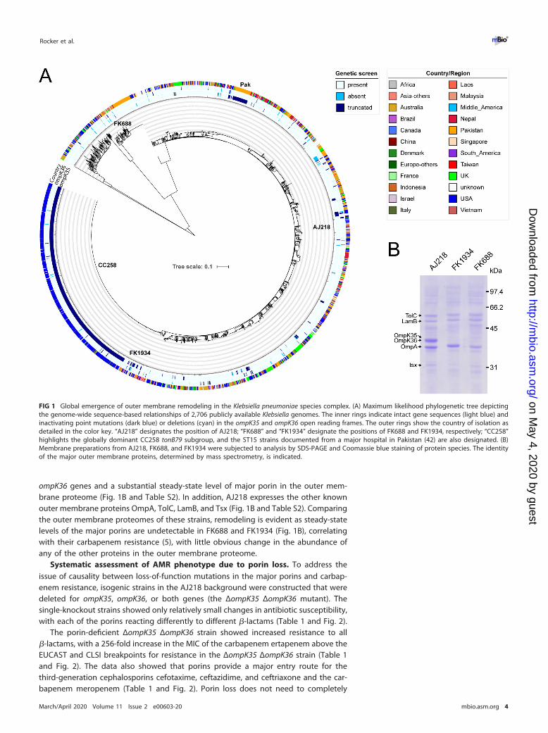
ompK36 genes and a substantial steady-state level of major porin in the outer mem-brane proteome (Fig. 1B and Table S2). In addition, AJ218 expresses the other knownouter membrane proteins OmpA, TolC, LamB, and Tsx (Fig. 1B and Table S2). Comparingthe outer membrane proteomes of these strains, remodeling is evident as steady-statelevels of the major porins are undetectable in FK688 and FK1934 (Fig. 1B), correlatingwith their carbapenem resistance (5), with little obvious change in the abundance ofany of the other proteins in the outer membrane proteome.
Systematic assessment of AMR phenotype due to porin loss. To address theissue of causality between loss-of-function mutations in the major porins and carbap-enem resistance, isogenic strains in the AJ218 background were constructed that weredeleted for ompK35, ompK36, or both genes (the ΔompK35 ΔompK36 mutant). Thesingle-knockout strains showed only relatively small changes in antibiotic susceptibility,with each of the porins reacting differently to different �-lactams (Table 1 and Fig. 2).
The porin-deficient ΔompK35 ΔompK36 strain showed increased resistance to all�-lactams, with a 256-fold increase in the MIC of the carbapenem ertapenem above theEUCAST and CLSI breakpoints for resistance in the ΔompK35 ΔompK36 strain (Table 1and Fig. 2). The data also showed that porins provide a major entry route for thethird-generation cephalosporins cefotaxime, ceftazidime, and ceftriaxone and the car-bapenem meropenem (Table 1 and Fig. 2). Porin loss does not need to completely
FIG 1 Global emergence of outer membrane remodeling in the Klebsiella pneumoniae species complex. (A) Maximum likelihood phylogenetic tree depictingthe genome-wide sequence-based relationships of 2,706 publicly available Klebsiella genomes. The inner rings indicate intact gene sequences (light blue) andinactivating point mutations (dark blue) or deletions (cyan) in the ompK35 and ompK36 open reading frames. The outer rings show the country of isolation asdetailed in the color key. “AJ218” designates the position of AJ218; “FK688” and “FK1934” designate the positions of FK688 and FK1934, respectively; “CC258”highlights the globally dominant CC258 tonB79 subgroup, and the ST15 strains documented from a major hospital in Pakistan (42) are also designated. (B)Membrane preparations from AJ218, FK688, and FK1934 were subjected to analysis by SDS-PAGE and Coomassie blue staining of protein species. The identityof the major outer membrane proteins, determined by mass spectrometry, is indicated.
Rocker et al. ®
March/April 2020 Volume 11 Issue 2 e00603-20 mbio.asm.org 4
on May 4, 2020 by guest
http://mbio.asm
.org/D
ownloaded from

prevent carbapenem permeation but simply to reduce permeation rates to a pointwhere very low rates of hydrolysis by other �-lactamases can control carbapenem levelsin the periplasm. Typically, K. pneumoniae strains have a chromosomal gene encodinga �-lactamase called SHV-1 that is responsible for such intrinsic �-lactam resistance.
SHV-1-like �-lactamases (Bush-Jacoby group 2b) possess low-level activity againstfirst-generation cephalosporins like cefazolin (46, 47). AJ218 encodes two variants ofSHV-1, called SHV-44 and SHV-27: a single amino acid substitution in SHV-1 (R205L) givesrise to SHV-44 and does not greatly change the activity of the enzyme (48). Conversely,the single amino acid substitution (G152D) in the SHV-27 variant leads to increasedactivity in hydrolyzing cefotaxime (49). MICs of cefazolin increased from 8 �g/ml in theparent strain 128 times to 1,024 �g/ml in the ΔompK35 ΔompK36 strain (Table 1 andFig. 2). Thus, loss of the major porins is both necessary and sufficient to make theΔompK35 ΔompK36 strain highly resistant to this class of �-lactams. To further testthe proposition that the SHV1-like �-lactamases are important in contributing to theobserved carbapenem resistance, we made use of �-lactamase inhibitors tazobactamand avibactam (50, 51). Treatment with 4 �g/ml avibactam or 64 �g/ml tazobactamdecreased the MIC for AJ218 of cefazolin 16- or 32-fold to 1 or 0.5 �g/ml (Table 2),respectively, indicating that SHV �-lactamases are contributing to the resistance in thisstrain. Conversely, addition of the �-lactamase inhibitors to ertapenem led to only a 2-to 4-fold decrease in the MIC for the ΔompK35 ΔompK36 strain. Taken together, thedata indicate that �-lactamases play a minor role in making K. pneumoniae clinicallyresistant to ertapenem, whereas outer membrane remodeling exerts the major effect.
OmpK38 is a new member of the general bacterial porin family. In the proteinclassification system organized by Pfam, OmpK35 and OmpK36 belong to the Porin_1(PF00267) group of bacterial outer membrane proteins together with the anion-
TABLE 1 MIC assessments of porin knockout strains
Drug
MIC (�g/ml)
AJ218�ompK35mutant
�ompK36mutant
�K35 �K36mutant Breakpointa
Ampicillin 4,096 4,096 8,192 8,192 �32Cefazolin 8 8 64 1,024 �8Ceftazidime 0.5 4 1 4 �16Ceftriaxone 0.125 0.125 0.5 1 �4Cefotaxime 0.0625 0.125 0.5 0.5 �4Imipenem 0.25 0.5 0.25 0.5 �4Meropenem 0.03125 0.03125 0.03125 0.5 �4Ertapenem 0.015625 0.03125 0.0625 4 �2aClinical breakpoints as given in reference 97.
FIG 2 Effects of porin loss on antimicrobial resistance. Graphical representation of fold changes in theMICs between AJ218 and the porin-deficient isogenic derivative AJ218 ΔompK35 ΔompK36 depicted inTable 1. Red denotes a further increase in resistance that remains above the clinical breakpoint, whileblue denotes an increase converting the strain from clinically sensitive to resistant.
Porin Channels Modulating Antimicrobial Resistance ®
March/April 2020 Volume 11 Issue 2 e00603-20 mbio.asm.org 5
on May 4, 2020 by guest
http://mbio.asm
.org/D
ownloaded from

selective porin PhoE, which adopt a conserved protein structure as shown by X-raycrystallography (18, 20, 52). A fourth porin in Klebsiella, OmpK37, is also a member ofthis family. Sequence-based clustering of PF00267 for all proteins from the genusKlebsiella revealed that OmpK35 and PhoE porins cluster into clearly distinct subfamilies(Fig. 3A). In contrast, OmpK36 porins show sequence similarities to the OmpK37subfamily, which therefore cluster together. Surprisingly, this analysis demonstrated afurther separation of what were annotated as OmpK37 homologs into two separatesubgroups (Fig. 3A, depicted in light and dark blue). Sequence analysis of the model K.pneumoniae AJ218 genome revealed the presence of two ompK37-like genes, each witha defined gene synteny conserved in other Klebsiella genome contexts (Fig. 3B). BothompK37-like genes were identified in nearly all 2,706 strains in the phylogeny (97.8%and 98.3%, respectively) (Table S1 and Fig. S1). We designate the novel porin familyOmpK38, suggesting that gene synteny should be used as the basis for distinguishingthese genes in genome sequencing projects.
Structure-based comparisons of Klebsiella porins. We solved the structure ofOmpK37 by X-ray crystallography, providing a structural map at 2.6-Å resolution tobetter understand the relationship of OmpK37 and OmpK38 to the major porins(Table 3 and Fig. 3C). Like the other porins, OmpK37 assembles into a trimer, with eachmonomeric unit composed of 16 �-strands that cross the outer membrane to form a�-barrel (Fig. 3C). All sequence variation seen within and between OmpK36 andOmpK37 trimers maps to the extracellular loops of the �-barrels and residues lining theupper reaches of the pore (Fig. 3D). The influence of these loops on the permeation ofsmall molecules is unknown. Analysis of the structure also illustrates an importantfeature of the �-barrel porins in terms of the solute channels that they form: no clearpath can be seen through the pore, due to the channel being particularly constrainedin the central region of the protein (Fig. 3D). This impediment is dominated by theinward folding of extracellular loop 3 into the �-barrel lumen, to form a “constrictionzone” (Fig. 3E). This feature has been shown to impact the relative diffusion rates ofsolutes through a porin (20, 53, 54). In this loop 3, OmpK37 carries a bulky residue(Tyr118) not found in OmpK36, a residue that was previously speculated to lead to adecreased pore diameter (38). However, the crystal structure of OmpK37 dismisses thisconjecture as the tyrosine side chain is oriented toward the pore exit such that it doesnot contribute to the constriction zone (Fig. 3E). All other residues of the constrictionzone in OmpK37 (Arg37, Arg75, Arg126, Lys16, Asp106, and Glu110) are conserved andadopt the same conformation as in OmpK35 and OmpK36.
To complete the comparative analysis of pore properties, we calculated an in silicomodel of OmpK38 based on the published high-resolution structure of OmpK36 (PDBID 5O79). The modeled OmpK38 closely resembles the structure of OmpK36 in the�-barrel domain, with variations observed only in the external loops. This is in agree-ment with the observation that the sequence similarity is high for residues located atthe periplasmic surface, while residues in the extracellular loops are more variable(Fig. 3F).
TABLE 2 MIC assessments with �-lactamase inhibitor combinations
Drug
MIC (�g/ml)
AJ218 plus empty vector AJ218 �K35 �K36 plus empty vector
No inhibitor Tazobactama Avibactamb No inhibitor Tazobactama Avibactamb
Ampicillin 8,192 4 4 8,192 1,024 32Cefazolin 8 0.5 1 1,024 256 16Ceftazidime 0.5 0.125 0.25 4 1 1Ceftriaxone 0.0625 0.03125 0.0625 0.5 0.5 0.5Cefotaxime 0.0625 0.03125 0.0625 0.5 0.5 0.5Imipenem 0.5 0.25 0.25 0.5 0.25 0.5Meropenem 0.03125 0.015625 0.03125 0.5 0.5 0.0625Ertapenem 0.015625 0.008 0.015625 4 2 1aTazobactam was used at a final concentration of 64 �g/ml.bAvibactam was used at a final concentration of 4 �g/ml.
Rocker et al. ®
March/April 2020 Volume 11 Issue 2 e00603-20 mbio.asm.org 6
on May 4, 2020 by guest
http://mbio.asm
.org/D
ownloaded from

For �-lactams, the pore characteristics of the major entry route across the outermembrane will determine how readily an effective concentration of drug can equili-brate in the periplasm, in order to inhibit its target. The structural frameworks of variousporins are largely superimposable: the diameter of the entry and exit pore sizes areessentially the same, with crystal structures showing key differences in the length ofloops 3, 4, 5, and 6, with the loop 3 differences changing aspects of the constrictionzone geometry (18, 20, 52). Comparing the pore trajectories and sizes of the deter-mined crystal structures (Fig. 4A) revealed that OmpK35 porins show a slightly largerconstriction zone radius, while OmpK36 porins are slightly more constricted, as previ-ously proposed (20). PhoE porins have a constriction zone radius comparable to
FIG 3 OmpK37 is a member of the general bacterial porin family. (A) Sequence similarity networksgraphically depict the homology between all Porin_1 (PF00267) sequences within the genus Klebsiella(NCBI taxID 570) available in UniProt (release 2019_02). Each dot represents the sequence of a proteinspecies. Dark lines between sequences represent similar sequences, and lighter lines represent lesssimilar sequences with a minimal alignment score of 138. The analysis shows distinct subclasses forOmpK35 (red) and PhoE (purple) homologues, as well as the close relationships between OmpK36(green), OmpK37 (dark blue), and OmpK38 (light blue) subclasses. Brown dots depict individual homol-ogous gene transfers of porins from Proteus/Morganella species into Klebsiella strains, while the orangedots represent a putative porin subfamily found in a subset of Klebsiella variicola genomes. (B) Genesynteny definitions for the genes encoding OmpK37 and OmpK38. (C) The structure of OmpK37determined by X-ray crystallography (PDB ID 6V78), showing the typical trimeric arrangement ofmonomers. Each monomer adopts a �-barrel fold and is colored a shade of blue. The position of theprotein within the outer membrane (OM) and the extracellular and periplasmic sides are indicated. (D)Sequence similarity of OmpK37 and OmpK36 from AJ218, ranging from identical (dark blue) to medium(blue and light blue) and low (gray), mapped onto the surface representation of OmpK37 viewed fromthe extracellular (left) or periplasmic (right) side of the outer membrane. White patches representdeletions/insertions in the loop regions. (E) Closeup view of the constriction zone (light blue), formingthe narrowest point within the pore and contributing to substrate selection by size and charge filtering.The infolded loop 3 is highlighted in light blue. The side chain of Tyr118 (red) is pointing toward theextracellular pore entrance. (F) Sequence similarity of OmpK37 and OmpK38 from AJ218, colored as inpanel C.
Porin Channels Modulating Antimicrobial Resistance ®
March/April 2020 Volume 11 Issue 2 e00603-20 mbio.asm.org 7
on May 4, 2020 by guest
http://mbio.asm
.org/D
ownloaded from

OmpK36, while OmpK37 is in between the OmpK35 and OmpK36 pores (Table 4). Thepore trajectory and constriction zone radius of the OmpK38 model more closely matchOmpK36 than OmpK37 (Fig. 4A and Table 4).
The effect of outer membrane proteome remodeling on �-lactam permeation.To determine whether these structural differences are meaningful for antimicrobialsensitivity, the porin-deficient ΔompK35 ΔompK36 strain was engineered to expresseach of the other porins under the control of a heterologous promoter, to increase theirexpression level to become “major” porins (Fig. 4B). In rich growth media, the cross-complemented strains grow at rates equivalent to the isogenic AJ218 strain (Fig. S2),with doubling times and final cell densities of the individual strains comparable over 24h under conditions equivalent to those used for MIC experiments.
High-level expression of OmpK35 was found to sensitize Klebsiella to both penicillinsand cephalosporins, indicating that OmpK35 provides the major influx pathway forthese compounds (Table 5 and Fig. 4C). The MIC values are even lower in this strainthan in the wild type, with the OmpK35 expression levels 7.7-fold higher uponinduction with anhydrotetracycline (ATc) than in the wild type (Fig. S3). In contrast withthe expression of OmpK35, MIC values of the OmpK36-expressing strain are restored forthe carbapenems (meropenem and ertapenem) but not for the cephalosporins (Ta-ble 5). This indicates that while carbapenems diffuse through either OmpK35 orOmpK36 equally well, cephalosporins favor the channel in OmpK35.
Comparing the effects of OmpK37 and OmpK38 to the related porin OmpK36 wasrevealing. OmpK37 has a selectively lower permeability to ertapenem, ceftriaxone, andcefotaxime than OmpK36. While OmpK38 matches the permeation properties ofOmpK37 nearly exactly, it is characterized by a higher permeability to ertapenem,reaching MIC values similar to strains expressing OmpK36. The structural similaritiessuggest that it is not the pore size, but perhaps the physicochemical properties of thepore surface (20), that would impact these selective differences in drug sensitivity.
TABLE 3 Data collection and refinement statistics
Statistic Value for OmpK37 (6V78)
Data collectionSpace group P21212Cell dimension
a, b, c (Å) 109.52, 138.51, 91.70�, �, � (°) 90, 90, 90
Resolution (Å) 50–2.6 (2.7–2.6)Rmeas 19.3 (176.4)I/�I 11.11 (1.73)Completeness (%) 99.2 (98.8)Redundancy 7.4 (7.5)CC1/2 99.7 (69.2)
RefinementResolution (Å) 49.3–2.6No. of reflections 43,308Rwork/Rfree 25.7/32.3No. of atoms
Protein 8,382 (trimer)Water 73
B-factorProtein 56.3Water 47.2
RMSa deviationsBond lengths (Å) 0.008Bond angles (°) 1.08
Ramachandran statistics (%)Preferred 95.9Allowed 4.1Outliers 0
aRMS, root mean square.
Rocker et al. ®
March/April 2020 Volume 11 Issue 2 e00603-20 mbio.asm.org 8
on May 4, 2020 by guest
http://mbio.asm
.org/D
ownloaded from

PhoE, OmpK26, and LamB are considered substrate-specific porins. This expectationwas evidenced in the strain expressing LamB as its dominant pore: there was no changein the MIC values, indicating that LamB acts as a substrate (maltose and maltodextrin)-specific porin that does not allow significant permeation by �-lactam antibiotics
FIG 4 Effects of alternative porin expression in the Klebsiella outer membrane. (A) Pore radius analysis of variousstructurally determined porins (OmpK35 [5O77], red; OmpK36 [5O79], dark green; OmpK37 [6V78], dark blue; OmpF[2OMF], orange; OmpC [2J1N], light green; PhoE [1PHO], purple) and the in silico model of OmpK38 (light blue) ascalculated by HOLE (94). All porins display a similar pore trajectory with only minor variations of the pore radius along theentire axis of the pore. At the constriction zone, the narrowest point along the pore, OmpF and OmpK35 show a slightlylarger pore radius than OmpC/OmpK36, while OmpK37, OmpK38, and PhoE are comparable to the latter. (B) Membranepreparations from wild-type AJ218 (wt AJ218) and AJ218 ΔompK35 ΔompK36 (ΔΔ) strains expressing the indicated porinsfrom an anhydrotetracycline-inducible promoter. The extracts were subjected to SDS-PAGE and Coomassie blue staining.(C) Graphical representation of the drug resistance data in Table 5. Log2 fold change of the MIC values of the indicatedporin-expressing strains are given compared to wild-type MIC levels.
TABLE 4 Constriction zone radii (Å) of porin structures
Porin (PDB_ID) Species
Radius (Å) determined by:
BetaCavity MOLEonline HOLE
OmpK37 (6V78) K. pneumoniae 3.42 3.39 3.26OmpC (2J1N) E. coli 3.05 3.05 2.84OmpK36 (5O79) K. pneumoniae 3.34 3.22 3.17OmpF (2OMF) E. coli 3.73 3.67 3.56OmpK35 (5O77) K. pneumoniae 3.97 3.77 3.78OmpK38 (in silico) K. pneumoniae 3.20 3.16PhoE (1PHO) E. coli 3.30 3.26 3.13
Porin Channels Modulating Antimicrobial Resistance ®
March/April 2020 Volume 11 Issue 2 e00603-20 mbio.asm.org 9
on May 4, 2020 by guest
http://mbio.asm
.org/D
ownloaded from

(Table 5 and Fig. 4C). The converse was true for PhoE, which enables the influx of most�-lactams as readily as OmpK36, and for OmpK26, which provides permeability to alltested cephalosporins (Table 5 and Fig. 4C).
Do the various pore types play a role in restricting the permeability for otherdrugs? Sensitivity to the �-lactamase inhibitors avibactam and tazobactam was alsoassessed. Both inhibitors possess an intrinsic low-level antimicrobial activity (55, 56),which leads to bacterial killing in MIC experiments. Differences in inhibitor uptake wereobserved (Table 6), including that upregulation of OmpK26 or OmpK35 sensitized theΔompK35 ΔompK36 strain to tazobactam but not to avibactam, while OmpK36,OmpK37, and PhoE seem to provide preferential entry routes for avibactam.
Imipenem is a relatively hydrophobic zwitterionic carbapenem with a neutral netcharge and the smallest �-lactam tested (molecular weight, 299 Da). It has beensuggested that the combination of imipenem-relebactam with an aminoglycoside maybe a promising approach for isolates with reduced susceptibility to imipenem-relebactam (57). To address the drug resistance to non-�-lactam drugs, the ΔompK35ΔompK36 strain and the porin-complemented strains were subjected to further MICtesting.
Only 2-fold differences in MIC values between the wild-type and porin-deficientstrains were observed for gentamicin, neomycin, tobramycin, and spectinomycin (Ta-ble S3), indicating that diffusion of aminoglycosides through porins is of minor impor-tance in Klebsiella. Quinolones are thought to be able to diffuse both through the outermembrane layer and through the porin channels (58, 59). However, only 2-fold differ-ences in MIC values between the wild-type and porin-deficient strain are observed forthe fluoroquinolone ciprofloxacin and for the more hydrophobic nalidixic acid. Likethese other hydrophobic compounds, the large (734-Da) macrolide erythromycin hasbeen suggested to permeate the outer membrane via the lipid bilayer (60, 61). Thistheory is supported by the limited (i.e., 2-fold) change in antibiotic sensitivity (Table S3).A sensitization to antibiotics is seen in the strain expressing OmpK35 as the major porin,indicating that OmpK35 is permeable to aminoglycosides, quinolones, and macrolides.Relative MIC values revealed that the expression of other porins has only minor effects,while LamB has no effect on modulating the drug sensitivity of the porin-deficientstrain. The antimicrobial peptide polymyxin B binds to the lipopolysaccharide (LPS)
TABLE 5 MIC assessments of porin-expressing strains
Drug
MIC (�g/ml) for:
AJ218,emptyvector
AJ218 �K35 �K36 plus:
Emptyvector OmpK35 OmpK36 OmpK37 OmpK38 OmpK26 PhoE LamB
Ampicillin �2,048 �2,048 512 �2,048 �2,048 �2,048 �2,048 �2,048 �2,048Carbenicillin �2,048 �2,048 1,024 �2,048 �2,048 �2,048 1,024 �2,048 �2,048Cefazolin 4 512 2 16 16 16 32 4 1,024Ceftazidime 0.25 2 0.03125 1 1 1 1 1 2Ceftriaxone 0.03125 0.5 0.002 0.125 0.5 0.25 0.0625 0.0625 0.5Cefotaxime 0.03125 0.5 0.004 0.0625 0.25 0.25 0.0625 0.25 0.5Imipenem 0.25 0.5 0.25 0.25 0.25 0.25 0.25 0.25 0.25Meropenem 0.03125 0.25 0.03125 0.03125 0.03125 0.03125 0.25 0.03125 0.5Ertapenem 0.015625 2 0.015625 0.015625 0.0625 0.015625 0.5 0.015625 4
TABLE 6 MIC assessments for �-lactamase inhibitors
Drug
MIC (�g/ml) for:
AJ218,emptyvector
AJ218 �K35 �K36 plus:
Emptyvector OmpK35 OmpK36 OmpK37 OmpK38 OmpK26 PhoE LamB
Tazobactam 256 512 128 256 512 512 64 512 512Avibactam �256 �256 256 128 128 64 �256 128 256
Rocker et al. ®
March/April 2020 Volume 11 Issue 2 e00603-20 mbio.asm.org 10
on May 4, 2020 by guest
http://mbio.asm
.org/D
ownloaded from

layer to exert its antimicrobial activity (62). As expected, no difference in MIC wasobserved for the tested strains. The data confirm that mutations inducing porin loss willnot greatly contribute to resistance phenotypes against antibiotics other than�-lactams.
DISCUSSIONThe prevalence of outer membrane proteome remodeling in response to drug
treatment. A major finding of this study comes from comparing carbapenem suscep-tibility profiles for AJ218 and the isogenic ΔompK35 ΔompK36 strain. This indicated thatporin loss alone can confer carbapenem resistance in Klebsiella. Given the preponder-ance of ompK35 mutations globally (Fig. 1A), the apparent ease with which additionalompK36 mutations can be tolerated in these strains, and the presence of chromosomalSHV �-lactamases in the core genome of K. pneumoniae, this carbapenemase-negativeand ESBL-negative CRKP phenotype too will be global.
The experiments provide a further significant finding. In combination treatment,tazobactam addition is less effective against the ΔompK35 ΔompK36 strain, whileavibactam shows superior effectiveness. Tazobactam is itself a �-lactam compound of300 Da in size (63), and it appears to be dependent on porins for uptake into theperiplasm. While of similar overall size (265 Da), avibactam is not a �-lactam but abridged diazabicyclo[3.2.1]octanone (51, 64), and our data showed that its permeationinto the periplasm of the ΔompK35 ΔompK36 strain is more effective than for tazobac-tam, i.e., that it likely can cross the outer membrane via channels other than OmpK35and OmpK36 (Table 6). Recent successful combination treatments with meropenemand the �-lactamase inhibitor vaborbactam (65) might also be effective on porin-deficient K. pneumoniae, given that vaborbactam is a compound based on a cyclicboronic acid pharmacophore (66, 67) and, at 267 Da in size, it may access the periplasmvia porins other than OmpK35 and OmpK36.
Our current knowledge on regulation of porin gene expression in K. pneumoniae isminimal. Studies support the hypothesis that OmpK35 is thus the main contributor toantibiotic uptake in Klebsiella (40, 41), consistent with the observation that mutationsin ompK35 are more prevalent globally in parallel with the rise in �-lactamase resistance(Fig. 1A), and that OmpK35 is dispensable for growth and virulence in the human niche(30). In E. coli, expression of the OmpK35 and OmpK36 homologs, OmpF and OmpC,respectively, is regulated so as to have one or the other (but not both) expressed athigh levels (68). This switching between expression of the major porins is regulated bysmall RNAs which can be dysregulated in the presence of �-lactam drugs (69). Whilelittle is known about the regulation of expression of ompK35, ompK36, ompK37, andompK38 in K. pneumoniae, a study in Klebsiella aerogenes shows that overexpression ofthe small RNAs MicF and MicC can suppress expression of omp35 and omp36, respec-tively (70). Another study reported that ompK37 expression is induced in response to aloss of the major porins (38). Our results with upregulation of different major porins(Table 5 and Fig. 4C) give credence to the hypothesis that changes in gene expressionlevels induced during growth in human tissues including serum would modulate drugsensitivity. We suggest further studies are warranted to measure the expression levelsof porins in clinical isolates grown in human serum, to determine which of the ompK35,ompK36, ompK37, or ompK38 genes would be most active under those conditions.
The different �-barrel pores and features of outer membrane proteome re-modeling. LamB is known as a transporter of maltodextrins and efficiently transportssugar polymers. Overexpression of this porin in knockout strains does not change theobtained MIC values, indicating that the substrate specificity of LamB is too narrow forit to efficiently transport �-lactam antibiotics. Conversely, the �-lactam MICs of PhoE-expressing strains are very similar to the parental AJ218 with the exception of cefta-zidime and cefotaxime. This indicates that the PhoE channel is largely equivalent to themajor porins for the influx of most �-lactam antibiotics. OmpK26 has been reported tobe overexpressed in porin-deficient hospital isolates selected by carbapenem treatment(34, 36, 37). In our study, the strain expressing OmpK26 as its major porin is relatively
Porin Channels Modulating Antimicrobial Resistance ®
March/April 2020 Volume 11 Issue 2 e00603-20 mbio.asm.org 11
on May 4, 2020 by guest
http://mbio.asm
.org/D
ownloaded from

impermeable to carbapenem, which is consistent with the observation that prolongedcarbapenem treatment might select for increased ompK26 expression (34, 36, 37).Importantly, AJ218 expressing OmpK26 as its major porin showed MIC values similar toa porin-expressing wild-type strain for the tested third-generation cephalosporins.While there is no clear structure-based rationale for these observations, the MIC datasuggest that OmpK26-expressing strains should be treated with cephalosporins.
OmpK37 and OmpK38 are homologous to OmpK36, showing �70% identity at theamino acid sequence level. In a previous report, OmpK37 was speculated to have anarrower pore, based on a lower rate of sugar influx and increased antimicrobialresistance (38). Our results show a lower permeability to some antibiotics (ertapenem,cefotaxime, and ceftazidime), but the structure of OmpK37 reported here demonstratesthat the pore diameters do not differ and could not determine drug specificity. Tomodel porin permeability, Acosta-Gutiérrez et al. (20) highlighted the importance of thesize and charge of the constriction zone for drug permeation between differentGram-negative major porins. While these properties may be important for the differ-ences between some of the Klebsiella porins observed in this study, the residues withinthe constriction zone of OmpK36, OmpK37, and OmpK38 are largely identical. Sincedifferences in drug uptake were observed between strains expressing these relatedporins, we suggest that variation in the external loops acts as a crucial determinant ofantibiotic permeability. It remains unclear what the selection pressure is for thisvariation. One possibility is host factors involved in driving antigenic variation forprotection against antibodies and/or complement, as OmpK36 has been shown to betargeted by components of the complement pathway (71–73). Another possibility, andnot mutually exclusive, is that selection for variation in OmpK36, OmpK37, and OmpK38is being driven by microbial factors, such as bacteriophages or colicins (74, 75).Bacteriophages such as GH-K3 that use OmpK36 as a receptor would produce theselective pressure for the evolution of phage-resistant ompK36 mutants (74), whichwould select for a collateral drug resistance phenotype given the MIC data in our study.Given prospects for phage therapy to treat CRE infections, the evolutionary drivers onouter membrane protein remodeling in K. pneumoniae and other CRE pathogens needfurther attention.
MATERIALS AND METHODSChemicals and reagents. Ampicillin, carbenicillin, and tetracycline were purchased from Astral
Scientific. Avibactam was purchased from Selleck Chemicals. All other antibiotics, including tazobactam,were purchased from Sigma-Aldrich in the highest possible grade.
Comparative genomics. To examine the distribution of porin proteins in Klebsiella, a database of2,706 publicly available genome sequences was constructed (42, 76) (see Table S1 in the supplementalmaterial). All genomes that required assembly were assembled using Skesa v2.3.0 (77), and chromosomalsequence types were determined for each genome assembly using the genotyping toolkit Kleborate(https://github.com/katholt/Kleborate), which is aligned to the BIGSdb-Kp multilocus sequence typing(MLST) scheme (78). For visual purposes, a single nucleotide polymorphism (SNP) midpoint-rootedmaximum-likelihood phylogenetic tree was generated. Genomes were mapped to the K. pneumoniaereference strain NTUH-K2044 (GenBank accession no. AP006725.1 [79]) using minimap2, and SNPs werecalled using snippy v4.3.9 (https://github.com/tseemann/snippy). Phylogenetic inference was performedusing IQ-TREE v1.6.10 (80) using the GTR�F�G4 model based on 4,285 parsimonious SNPs and 1,000ultrafast bootstraps (81). Porin variants were identified in the 2,706 draft genome assemblies by a BLASTNscreening tool (82) applying the cutoffs 80% identity and 90% reference length. Hits were translated intoprotein sequence for identification of putative premature stop codons. Images were made usingInteractive Tree Of Life (iTOL) v4 (83).
Whole-genome sequences were generated for FK688 and FK1934 using Illumina short-read genomesequencing on the Illumina NextSeq 500 platform with 150-bp paired-end reads. Libraries were gener-ated using the Illumina Nextera XT DNA sample preparation kit.
Protein sequences belonging to Pfam family Porin_1 (PF00267) and the genus Klebsiella (NCBI taxID570) were downloaded from UniProt (release 2019_02). Sequence similarities were calculated usingEFI-EST (84), performing an all-by-all BLAST search and clustering based on a minimal pairwise alignmentscore of 138. Sequence similarity networks were visualized using Cytoscape 3.7.1 (85).
Bacterial strains and cultures. An overview of the strains and plasmids used in this study is givenin Table S4. Knockout strains were constructed using the “gene gorging” technique (45, 86), describedin detail in Text S1. The cloning of plasmids used for protein expression in E. coli or K. pneumoniae isdescribed in Text S1.
Rocker et al. ®
March/April 2020 Volume 11 Issue 2 e00603-20 mbio.asm.org 12
on May 4, 2020 by guest
http://mbio.asm
.org/D
ownloaded from
Structure determination and modeling. OmpK37 was overexpressed and purified from E. coli C41cells as described in Text S1. Initial crystallization conditions were screened at the Monash MolecularCrystallization Facility using commercially available screens. Following optimization, crystals of OmpK37were grown using a sitting drop vapor diffusion setup with a reservoir solution of 125 mM SPG buffer(2:7:7 succinic acid-sodium hydrogen phosphate-glycine) (pH 4.5) and 20% (wt/vol) polyethylene glycol1,500 and directly flash-frozen in liquid nitrogen. Diffraction data were collected at 100 K at the Australiansynchrotron and processed with the XDS software package (87) in the space group P21212 to 2.6-Åresolution. Five percent of the reflections were randomly selected for calculation of Rfree and inherited toall data sets. Initial phases were obtained by molecular replacement using Phaser for MR (88) and thepublished structure of the OmpK36 porin (1OSM) trimmed of its external loops as a search model. Theinitial model was improved in iterative cycles of manual building in Coot and refinement using Phenix(89, 90). Final structure validation was performed using MolProbity (91).
Structure prediction of an OmpK38 model was performed using the mini.rosetta threading protocolas implemented in the Rosetta software package, using OmpK36 (5O79) as a reference structure. This wasfollowed by an energy minimization using the relax protocol in Rosetta.
Pore geometries of OmpK37 (6V78) and published structures of OmpC (2J1N), OmpK36 (5O79), OmpF(2OMF), OmpK35 (5O77), and PhoE (1PHO) were analyzed using BetaCavity (92), MOLEonline (93), andHOLE (94). Figures were prepared using PyMOL (95).
Outer membrane protein analysis. Overnight cultures of strains harboring porin expression plas-mids were diluted 1:50 in cation-adjusted Mueller-Hinton Broth containing chloramphenicol (CaMHB-Cm) and grown at 37°C, 200 rpm, until an optical density at 600 nm (OD600) of 0.5 was reached.Subsequently, cultures were diluted to an OD600 of 0.0005 in CaMHB-Cm, induced with the appropriateamount of ATc (empty � 400 ng/ml, K35 � 150 ng/ml, K36 � 200 ng/ml, K37 � 10 ng/ml, K26 � 100 ng/ml, PhoE � 5 ng/ml, LamB � 400 ng/ml), and grown at 37°C, 200 rpm, for 4.5 or 20 h. At these timepoints, 14 ml (4.5 h) or 2 ml (20 h) of culture was harvested by centrifugation (4,500 � g, 4°C, 10 min) andresuspended in 1.8 ml of buffer A (50 mM Tris, 150 mM NaCl, 5 mM EDTA, pH 7.5). Cells were broken bysonication on ice (5 � 10 s, amplitude 2, duty 100%) and centrifuged at 2,000 � g to remove cell debris.Sodium lauroyl sarcosinate was added to the supernatant to a final concentration of 0.5% (wt/vol), andthe mixture was incubated for 30 min on ice before centrifugation at 25,000 � g at 4°C for 30 min. Thepellet containing the outer membranes was resuspended in 200 �l of buffer B (25% [wt/vol] sucrose,50 mM Tris, 5 mM EDTA, pH 7.5) and loaded onto an SDS-polyacrylamide gel containing 11% (wt/vol)37.5:1 acrylamide-bisacrylamide, 0.375 M Tris (pH 8.8), 0.2% (wt/vol) sodium dodecyl sulfate, and 0.5 mMEDTA in the separating gel and 4% (wt/vol) 37.5:1 acrylamide-bisacrylamide, 0.25 M Tris (pH 6.8), 0.1%(wt/vol) sodium dodecyl sulfate, and 0.5 mM EDTA in the stacking gel.
MIC determination. MICs were determined and interpreted using the broth microdilution methodoutlined by the Clinical and Laboratory Standards Institute (96).
Data availability. The X-ray structure of OmpK37 has been deposited in the PDB with the accessioncode 6V78. Genome sequence data for Klebsiella quasipneumoniae subsp. similipneumoniae (FK688) andKlebsiella pneumoniae (FK1934) have been deposited at the NCBI under the BioProject ID PRJNA607402with Sequence Read Archive codes SRR11108934 (FK688) and SRR11108933 (FK1934).
SUPPLEMENTAL MATERIALSupplemental material is available online only.TEXT S1, DOCX file, 0.03 MB.FIG S1, PDF file, 0.6 MB.FIG S2, PDF file, 0.2 MB.FIG S3, DOCX file, 2.2 MB.TABLE S1, XLSX file, 0.6 MB.TABLE S2, DOCX file, 0.01 MB.TABLE S3, DOCX file, 0.02 MB.TABLE S4, DOCX file, 0.02 MB.
ACKNOWLEDGMENTSWe thank the staff of the Monash Molecular Crystallization Facility and the Austra-
lian Synchrotron for help with X-ray structure determination. Diffraction data have beencollected on the MX1 and MX2 beamlines at the Australian Synchrotron, Clayton,Australia. Mass spectrometry was performed by the Monash Proteomics and Metabo-lomics Facility. We thank L. McIntyre, R. Grinter, I. D. Hay, L. Perlaza-Jiménez, and V. V. L.Torres for advice and assistance. We thank R. S. Bamert and C. Stubenrauch for criticalcomments on the manuscript.
The research was supported by program grant 1092262 from the National Healthand Medical Research Council of Australia.
Porin Channels Modulating Antimicrobial Resistance ®
March/April 2020 Volume 11 Issue 2 e00603-20 mbio.asm.org 13
on May 4, 2020 by guest
http://mbio.asm
.org/D
ownloaded from

REFERENCES1. Bagley ST. 1985. Habitat association of Klebsiella species. Infect Control
6:52–58. https://doi.org/10.1017/s0195941700062603.2. Paczosa MK, Mecsas J. 2016. Klebsiella pneumoniae: going on the offense
with a strong defense. Microbiol Mol Biol Rev 80:629 – 661. https://doi.org/10.1128/MMBR.00078-15.
3. Holt KE, Wertheim H, Zadoks RN, Baker S, Whitehouse CA, Dance D,Jenney A, Connor TR, Hsu LY, Severin J, Brisse S, Cao H, Wilksch J, GorrieC, Schultz MB, Edwards DJ, Nguyen KV, Nguyen TV, Dao TT, Mensink M,Minh VL, Nhu NT, Schultsz C, Kuntaman K, Newton PN, Moore CE,Strugnell RA, Thomson NR. 2015. Genomic analysis of diversity, popula-tion structure, virulence, and antimicrobial resistance in Klebsiella pneu-moniae, an urgent threat to public health. Proc Natl Acad Sci U S A112:E3574 –E3581. https://doi.org/10.1073/pnas.1501049112.
4. Sellick JA, Russo TA. 2018. Getting hypervirulent Klebsiella pneumoniaeon the radar screen. Curr Opin Infect Dis 31:341–346. https://doi.org/10.1097/QCO.0000000000000464.
5. Bi W, Liu H, Dunstan RA, Li B, Torres VVL, Cao J, Chen L, Wilksch JJ,Strugnell RA, Lithgow T, Zhou T. 2017. Extensively drug-resistant Kleb-siella pneumoniae causing nosocomial bloodstream infections in China:molecular investigation of antibiotic resistance determinants, informingtherapy, and clinical outcomes. Front Microbiol 8:1230. https://doi.org/10.3389/fmicb.2017.01230.
6. Navon-Venezia S, Kondratyeva K, Carattoli A. 2017. Klebsiella pneumoniae: amajor worldwide source and shuttle for antibiotic resistance. FEMS Micro-biol Rev 41:252–275. https://doi.org/10.1093/femsre/fux013.
7. Logan LK, Weinstein RA. 2017. The epidemiology of carbapenem-resistant Enterobacteriaceae: the impact and evolution of a global men-ace. J Infect Dis 215:S28 –S36. https://doi.org/10.1093/infdis/jiw282.
8. Gupta N, Limbago BM, Patel JB, Kallen AJ. 2011. Carbapenem-resistantEnterobacteriaceae: epidemiology and prevention. Clin Infect Dis 53:60 – 67. https://doi.org/10.1093/cid/cir202.
9. Nordmann P, Poirel L. 2014. The difficult-to-control spread of carbapen-emase producers among Enterobacteriaceae worldwide. Clin MicrobiolInfect 20:821– 830. https://doi.org/10.1111/1469-0691.12719.
10. Hu FP, Guo Y, Zhu DM, Wang F, Jiang XF, Xu YC, Zhang XJ, Zhang CX, JiP, Xie Y, Kang M, Wang CQ, Wang AM, Xu YH, Shen JL, Sun ZY, Chen ZJ,Ni YX, Sun JY, Chu YZ, Tian SF, Hu ZD, Li J, Yu YS, Lin J, Shan B, Du Y, HanY, Guo S, Wei LH, Wu L, Zhang H, Kong J, Hu YJ, Ai XM, Zhuo C, Su DH,Yang Q, Jia B, Huang W. 2016. Resistance trends among clinical isolatesin China reported from CHINET surveillance of bacterial resistance,2005–2014. Clin Microbiol Infect 22(Suppl 1):S9 –S14. https://doi.org/10.1016/j.cmi.2016.01.001.
11. Wei ZQ, Du XX, Yu YS, Shen P, Chen YG, Li LJ. 2007. Plasmid-mediatedKPC-2 in a Klebsiella pneumoniae isolate from China. Antimicrob AgentsChemother 51:763–765. https://doi.org/10.1128/AAC.01053-06.
12. Bradford PA, Urban C, Mariano N, Projan SJ, Rahal JJ, Bush K. 1997.Imipenem resistance in Klebsiella pneumoniae is associated with thecombination of ACT-1, a plasmid-mediated AmpC beta-lactamase, andthe foss of an outer membrane protein. Antimicrob Agents Chemother41:563–569. https://doi.org/10.1128/AAC.41.3.563.
13. Cao VT, Arlet G, Ericsson BM, Tammelin A, Courvalin P, Lambert T. 2000.Emergence of imipenem resistance in Klebsiella pneumoniae owing tocombination of plasmid-mediated CMY-4 and permeability alteration. JAntimicrob Chemother 46:895–900. https://doi.org/10.1093/jac/46.6.895.
14. Hamzaoui Z, Ocampo-Sosa A, Fernandez Martinez M, Landolsi S, FerjaniS, Maamar E, Saidani M, Slim A, Martinez-Martinez L, Boutiba-Ben Bou-baker I. 2018. Role of association of OmpK35 and OmpK36 alteration andblaESBL and/or blaAmpC genes in conferring carbapenem resistanceamong non-carbapenemase-producing Klebsiella pneumoniae. Int J An-timicrob Agents 52:898 –905. https://doi.org/10.1016/j.ijantimicag.2018.03.020.
15. Jacoby GA. 2009. AmpC beta-lactamases. Clin Microbiol Rev 22:161–182.https://doi.org/10.1128/CMR.00036-08.
16. MacKenzie FM, Forbes KJ, Dorai-John T, Amyes SG, Gould IM. 1997.Emergence of a carbapenem-resistant Klebsiella pneumoniae. Lancet350:783. https://doi.org/10.1016/s0140-6736(05)62567-6.
17. Martínez-Martínez L, Pascual A, Hernández-Allés S, Alvarez-Díaz D,Suárez AI, Tran J, Benedí VJ, Jacoby GA. 1999. Roles of beta-lactamasesand porins in activities of carbapenems and cephalosporins againstKlebsiella pneumoniae. Antimicrob Agents Chemother 43:1669 –1673.https://doi.org/10.1128/AAC.43.7.1669.
18. Dutzler R, Rummel G, Albertí S, Hernández-Allés S, Phale P, RosenbuschJ, Benedí V, Schirmer T. 1999. Crystal structure and functional charac-terization of OmpK36, the osmoporin of Klebsiella pneumoniae. Structure7:425– 434. https://doi.org/10.1016/s0969-2126(99)80055-0.
19. Nikaido H. 1994. Porins and specific diffusion channels in bacterial outermembranes. J Biol Chem 269:3905–3908.
20. Acosta-Gutierrez S, Ferrara L, Pathania M, Masi M, Wang J, Bodrenko I,Zahn M, Winterhalter M, Stavenger RA, Pages JM, Naismith JH, van denBerg B, Page MGP, Ceccarelli M. 2018. Getting drugs into gram-negativebacteria: rational rules for permeation through general porins. ACSInfect Dis 4:1487–1498. https://doi.org/10.1021/acsinfecdis.8b00108.
21. Sugawara E, Kojima S, Nikaido H. 2016. Klebsiella pneumoniae majorporins OmpK35 and OmpK36 allow more efficient diffusion of beta-lactams than their Escherichia coli homologs OmpF and OmpC. J Bacte-riol 198:3200 –3208. https://doi.org/10.1128/JB.00590-16.
22. Nikaido H, Pages JM. 2012. Broad-specificity efflux pumps and their rolein multidrug resistance of Gram-negative bacteria. FEMS Microbiol Rev36:340 –363. https://doi.org/10.1111/j.1574-6976.2011.00290.x.
23. Lugtenberg B, Peters R, Bernheimer H, Berendsen W. 1976. Influence ofcultural conditions and mutations on the composition of the outermembrane proteins of Escherichia coli. Mol Gen Genet 147:251–262.https://doi.org/10.1007/bf00582876.
24. Lugtenberg B, Van Alphen L. 1983. Molecular architecture and function-ing of the outer membrane of Escherichia coli and other gram-negativebacteria. Biochim Biophys Acta 737:51–115. https://doi.org/10.1016/0304-4157(83)90014-x.
25. Shimizu K. 2013. Metabolic regulation of a bacterial cell system withemphasis on Escherichia coli metabolism. ISRN Biochem 2013:645983.https://doi.org/10.1155/2013/645983.
26. Spector J, Zakharov S, Lill Y, Sharma O, Cramer WA, Ritchie K. 2010.Mobility of BtuB and OmpF in the Escherichia coli outer membrane:implications for dynamic formation of a translocon complex. Biophys J99:3880 –3886. https://doi.org/10.1016/j.bpj.2010.10.029.
27. Rassam P, Copeland NA, Birkholz O, Toth C, Chavent M, Duncan AL, CrossSJ, Housden NG, Kaminska R, Seger U, Quinn DM, Garrod TJ, Sansom MS,Piehler J, Baumann CG, Kleanthous C. 2015. Supramolecular assembliesunderpin turnover of outer membrane proteins in bacteria. Nature523:333–336. https://doi.org/10.1038/nature14461.
28. Tsai YK, Fung CP, Lin JC, Chen JH, Chang FY, Chen TL, Siu LK. 2011. Klebsiellapneumoniae outer membrane porins OmpK35 and OmpK36 play roles inboth antimicrobial resistance and virulence. Antimicrob Agents Chemother55:1485–1493. https://doi.org/10.1128/AAC.01275-10.
29. Phan K, Ferenci T. 2017. The fitness costs and trade-off shapes associatedwith the exclusion of nine antibiotics by OmpF porin channels. ISME J11:1472–1482. https://doi.org/10.1038/ismej.2016.202.
30. Fajardo-Lubian A, Ben Zakour NL, Agyekum A, Qi Q, Iredell JR. 2019. Hostadaptation and convergent evolution increases antibiotic resistancewithout loss of virulence in a major human pathogen. PLoS Pathog15:e1007218. https://doi.org/10.1371/journal.ppat.1007218.
31. Chen JH, Siu LK, Fung CP, Lin JC, Yeh KM, Chen TL, Tsai YK, Chang FY.2010. Contribution of outer membrane protein K36 to antimicrobialresistance and virulence in Klebsiella pneumoniae. J Antimicrob Che-mother 65:986 –990. https://doi.org/10.1093/jac/dkq056.
32. March C, Cano V, Moranta D, Llobet E, Perez-Gutierrez C, Tomas JM,Suarez T, Garmendia J, Bengoechea JA. 2013. Role of bacterial surfacestructures on the interaction of Klebsiella pneumoniae with phagocytes.PLoS One 8:e56847. https://doi.org/10.1371/journal.pone.0056847.
33. Wong JLC, Romano M, Kerry LE, Kwong HS, Low WW, Brett SJ, ClementsA, Beis K, Frankel G. 2019. OmpK36-mediated carbapenem resistanceattenuates ST258 Klebsiella pneumoniae in vivo. Nat Commun 10:3957.https://doi.org/10.1038/s41467-019-11756-y.
34. García-Sureda L, Doménech-Sánchez A, Barbier M, Juan C, Gascó J,Albertí S. 2011. OmpK26, a novel porin associated with carbapenemresistance in Klebsiella pneumoniae. Antimicrob Agents Chemother 55:4742– 4747. https://doi.org/10.1128/AAC.00309-11.
35. García-Sureda L, Juan C, Doménech-Sánchez A, Albertí S. 2011. Role ofKlebsiella pneumoniae LamB porin in antimicrobial resistance. Antimi-crob Agents Chemother 55:1803–1805. https://doi.org/10.1128/AAC.01441-10.
36. Wang XD, Cai JC, Zhou HW, Zhang R, Chen GX. 2009. Reduced suscep-tibility to carbapenems in Klebsiella pneumoniae clinical isolates associ-
Rocker et al. ®
March/April 2020 Volume 11 Issue 2 e00603-20 mbio.asm.org 14
on May 4, 2020 by guest
http://mbio.asm
.org/D
ownloaded from

ated with plasmid-mediated beta-lactamase production and OmpK36porin deficiency. J Med Microbiol 58:1196 –1202. https://doi.org/10.1099/jmm.0.008094-0.
37. Ruiz E, Ocampo-Sosa AA, Rezusta A, Revillo MJ, Román E, Torres C,Martínez-Martínez L. 2012. Acquisition of carbapenem resistance inmultiresistant Klebsiella pneumoniae strains harbouring blaCTX-M-15,qnrS1 and aac(6’)-Ib-cr genes. J Med Microbiol 61:672– 677. https://doi.org/10.1099/jmm.0.038083-0.
38. Domenech-Sanchez A, Hernandez-Alles S, Martinez-Martinez L, BenediVJ, Alberti S. 1999. Identification and characterization of a new poringene of Klebsiella pneumoniae: its role in beta-lactam antibiotic resis-tance. J Bacteriol 181:2726 –2732. https://doi.org/10.1128/JB.181.9.2726-2732.1999.
39. Pulzova L, Navratilova L, Comor L. 2017. Alterations in outer membranepermeability favor drug-resistant phenotype of Klebsiella pneumoniae.Microb Drug Resist 23:413– 420. https://doi.org/10.1089/mdr.2016.0017.
40. Hernandez-Alles S, Alberti S, Alvarez D, Domenech-Sanchez A, Martinez-Martinez L, Gil J, Tomas JM, Benedi VJ. 1999. Porin expression in clinicalisolates of Klebsiella pneumoniae. Microbiology 145:673– 679. https://doi.org/10.1099/13500872-145-3-673.
41. Doménech-Sánchez A, Martínez-Martínez L, Hernández-Allés S, del Car-men Conejo M, Pascual A, Tomás JM, Albertí S, Benedí VJ. 2003. Role ofKlebsiella pneumoniae OmpK35 porin in antimicrobial resistance. Anti-microb Agents Chemother 47:3332–3335. https://doi.org/10.1128/aac.47.10.3332-3335.2003.
42. Ejaz H, Wang N, Wilksch JJ, Page AJ, Cao H, Gujaran S, Keane JA, LithgowT, Ul-Haq I, Dougan G, Strugnell RA, Heinz E. 2017. Phylogenetic analysisof Klebsiella pneumoniae from hospitalized children, Pakistan. EmergInfect Dis 23:1872–1875. https://doi.org/10.3201/eid2311.170833.
43. Bowers JR, Kitchel B, Driebe EM, MacCannell DR, Roe C, Lemmer D, deMan T, Rasheed JK, Engelthaler DM, Keim P, Limbago BM. 2015. Genomicanalysis of the emergence and rapid global dissemination of the clonalgroup 258 Klebsiella pneumoniae pandemic. PLoS One 10:e0133727.https://doi.org/10.1371/journal.pone.0133727.
44. Jenney AW, Clements A, Farn JL, Wijburg OL, McGlinchey A, SpelmanDW, Pitt TL, Kaufmann ME, Liolios L, Moloney MB, Wesselingh SL,Strugnell RA. 2006. Seroepidemiology of Klebsiella pneumoniae in anAustralian tertiary hospital and its implications for vaccine development.J Clin Microbiol 44:102–107. https://doi.org/10.1128/JCM.44.1.102-107.2006.
45. Wilksch JJ, Yang J, Clements A, Gabbe JL, Short KR, Cao H, Cavaliere R,James CE, Whitchurch CB, Schembri MA, Chuah ML, Liang ZX, WijburgOL, Jenney AW, Lithgow T, Strugnell RA. 2011. MrkH, a novel c-di-GMP-dependent transcriptional activator, controls Klebsiella pneumoniae bio-film formation by regulating type 3 fimbriae expression. PLoS Pathog7:e1002204. https://doi.org/10.1371/journal.ppat.1002204.
46. Bush K, Fisher JF. 2011. Epidemiological expansion, structural studies,and clinical challenges of new beta-lactamases from gram-negativebacteria. Annu Rev Microbiol 65:455– 478. https://doi.org/10.1146/annurev-micro-090110-102911.
47. Bush K, Jacoby GA. 2010. Updated functional classification of beta-lactamases. Antimicrob Agents Chemother 54:969 –976. https://doi.org/10.1128/AAC.01009-09.
48. Arpin C, Dubois V, Coulange L, Andre C, Fischer I, Noury P, Grobost F,Brochet JP, Jullin J, Dutilh B, Larribet G, Lagrange I, Quentin C. 2003.Extended-spectrum beta-lactamase-producing Enterobacteriaceae in com-munity and private health care centers. Antimicrob Agents Chemother47:3506–3514. https://doi.org/10.1128/aac.47.11.3506-3514.2003.
49. Corkill JE, Cuevas LE, Gurgel RQ, Greensill J, Hart CA. 2001. SHV-27, anovel cefotaxime-hydrolysing beta-lactamase, identified in Klebsiellapneumoniae isolates from a Brazilian hospital. J Antimicrob Chemother47:463– 465. https://doi.org/10.1093/jac/47.4.463.
50. Aronoff SC, Jacobs MR, Johenning S, Yamabe S. 1984. Comparativeactivities of the beta-lactamase inhibitors YTR 830, sodium clavulanate,and sulbactam combined with amoxicillin or ampicillin. AntimicrobAgents Chemother 26:580 –582. https://doi.org/10.1128/aac.26.4.580.
51. Bonnefoy A, Dupuis-Hamelin C, Steier V, Delachaume C, Seys C, StachyraT, Fairley M, Guitton M, Lampilas M. 2004. In vitro activity of AVE1330A,an innovative broad-spectrum non-beta-lactam beta-lactamase inhibi-tor. J Antimicrob Chemother 54:410 – 417. https://doi.org/10.1093/jac/dkh358.
52. Cowan SW, Schirmer T, Rummel G, Steiert M, Ghosh R, Pauptit RA,Jansonius JN, Rosenbusch JP. 1992. Crystal structures explain functional
properties of two E. coli porins. Nature 358:727–733. https://doi.org/10.1038/358727a0.
53. Schulz GE. 2002. The structure of bacterial outer membrane proteins.Biochim Biophys Acta 1565:308 –317. https://doi.org/10.1016/s0005-2736(02)00577-1.
54. Acosta-Gutierrez S, Scorciapino MA, Bodrenko I, Ceccarelli M. 2015.Filtering with electric field: the case of E. coli porins. J Phys Chem Lett6:1807–1812. https://doi.org/10.1021/acs.jpclett.5b00612.
55. Asli A, Brouillette E, Krause KM, Nichols WW, Malouin F. 2016. Distinctivebinding of avibactam to penicillin-binding proteins of Gram-negativeand Gram-positive bacteria. Antimicrob Agents Chemother 60:752–756.https://doi.org/10.1128/AAC.02102-15.
56. Moosdeen F, Williams JD, Yamabe S. 1988. Antibacterial characteristicsof YTR 830, a sulfone beta-lactamase inhibitor, compared with those ofclavulanic acid and sulbactam. Antimicrob Agents Chemother 32:925–927. https://doi.org/10.1128/aac.32.6.925.
57. Balabanian G, Rose M, Manning N, Landman D, Quale J. 2018. Effect ofporins and blaKPC expression on activity of imipenem with relebactamin Klebsiella pneumoniae: can antibiotic combinations overcome resis-tance? Microb Drug Resist 24:877– 881. https://doi.org/10.1089/mdr.2018.0065.
58. Chapman JS, Georgopapadakou NH. 1988. Routes of quinolone perme-ation in Escherichia coli. Antimicrob Agents Chemother 32:438 – 442.https://doi.org/10.1128/aac.32.4.438.
59. Weingart H, Petrescu M, Winterhalter M. 2008. Biophysical characteriza-tion of in- and efflux in Gram-negative bacteria. Curr Drug Targets9:789 –796. https://doi.org/10.2174/138945008785747752.
60. Farmer S, Li ZS, Hancock RE. 1992. Influence of outer membrane muta-tions on susceptibility of Escherichia coli to the dibasic macrolide azi-thromycin. J Antimicrob Chemother 29:27–33. https://doi.org/10.1093/jac/29.1.27.
61. Nikaido H. 2003. Molecular basis of bacterial outer membrane permea-bility revisited. Microbiol Mol Biol Rev 67:593– 656. https://doi.org/10.1128/mmbr.67.4.593-656.2003.
62. Trimble MJ, Mlynárcik P, Kolár M, Hancock REW. 2016. Polymyxin: alter-native mechanisms of action and resistance. Cold Spring Harb PerspectMed 6:a025288. https://doi.org/10.1101/cshperspect.a025288.
63. Drawz SM, Papp-Wallace KM, Bonomo RA. 2014. New beta-lactamaseinhibitors: a therapeutic renaissance in an MDR world. Antimicrob AgentsChemother 58:1835–1846. https://doi.org/10.1128/AAC.00826-13.
64. Coleman K. 2011. Diazabicyclooctanes (DBOs): a potent new class ofnon-beta-lactam beta-lactamase inhibitors. Curr Opin Microbiol 14:550 –555. https://doi.org/10.1016/j.mib.2011.07.026.
65. Wilson WR, Kline EG, Jones CE, Morder KT, Mettus RT, Doi Y, Nguyen MH,Clancy CJ, Shields RK. 2019. Effects of KPC variant and porin genotype onthe in vitro activity of meropenem-vaborbactam against carbapenem-resistant Enterobacteriaceae. Antimicrob Agents Chemother 63:e02048-18.https://doi.org/10.1128/AAC.02048-18.
66. Lomovskaya O, Sun D, Rubio-Aparicio D, Nelson K, Tsivkovski R, GriffithDC, Dudley MN. 2017. Vaborbactam: spectrum of beta-lactamase inhi-bition and impact of resistance mechanisms on activity in Enterobacte-riaceae. Antimicrob Agents Chemother 61:e01443-17. https://doi.org/10.1128/AAC.01443-17.
67. Hecker SJ, Reddy KR, Totrov M, Hirst GC, Lomovskaya O, Griffith DC, KingP, Tsivkovski R, Sun D, Sabet M, Tarazi Z, Clifton MC, Atkins K, RaymondA, Potts KT, Abendroth J, Boyer SH, Loutit JS, Morgan EE, Durso S, DudleyMN. 2015. Discovery of a cyclic boronic acid beta-lactamase inhibitor(RPX7009) with utility vs class A serine carbapenemases. J Med Chem58:3682–3692. https://doi.org/10.1021/acs.jmedchem.5b00127.
68. Dam S, Pages JM, Masi M. 2018. Stress responses, outer membranepermeability control and antimicrobial resistance in Enterobacteriaceae.Microbiology 164:260 –267. https://doi.org/10.1099/mic.0.000613.
69. Dam S, Pages JM, Masi M. 2017. Dual regulation of the small RNAMicC and the quiescent porin OmpN in response to antibiotic stressin Escherichia coli. Antibiotics (Basel) 6:E33. https://doi.org/10.3390/antibiotics6040033.
70. Hao M, Ye M, Shen Z, Hu F, Yang Y, Wu S, Xu X, Zhu S, Qin X, Wang M.2018. Porin deficiency in carbapenem-resistant Enterobacter aerogenesstrains. Microb Drug Resist 24:1277–1283. https://doi.org/10.1089/mdr.2017.0379.
71. Albertí S, Marqués G, Hernández-Allés S, Rubires X, Tomás JM, Vivanco F,Benedí VJ. 1996. Interaction between complement subcomponent C1qand the Klebsiella pneumoniae porin OmpK36. Infect Immun 64:4719 – 4725. https://doi.org/10.1128/IAI.64.11.4719-4725.1996.
Porin Channels Modulating Antimicrobial Resistance ®
March/April 2020 Volume 11 Issue 2 e00603-20 mbio.asm.org 15
on May 4, 2020 by guest
http://mbio.asm
.org/D
ownloaded from

72. Doorduijn DJ, Rooijakkers SH, van Schaik W, Bardoel BW. 2016. Comple-ment resistance mechanisms of Klebsiella pneumoniae. Immunobiology221:1102–1109. https://doi.org/10.1016/j.imbio.2016.06.014.
73. Kojouharova MS, Tsacheva IG, Tchorbadjieva MI, Reid KB, Kishore U.2003. Localization of ligand-binding sites on human C1q globular headregion using recombinant globular head fragments and single-chainantibodies. Biochim Biophys Acta 1652:64 –74. https://doi.org/10.1016/j.bbapap.2003.08.003.
74. Cai R, Wu M, Zhang H, Zhang Y, Cheng M, Guo Z, Ji Y, Xi H, Wang X, XueY, Sun C, Feng X, Lei L, Tong Y, Liu X, Han W, Gu J. 2018. A smooth-type,phage-resistant Klebsiella pneumoniae mutant strain reveals that OmpCis indispensable for infection by phage GH-K3. Appl Environ Microbiol84:e01585-18. https://doi.org/10.1128/AEM.01585-18.
75. Evans LJ, Cooper A, Lakey JH. 1996. Direct measurement of the associ-ation of a protein with a family of membrane receptors. J Mol Biol255:559 –563. https://doi.org/10.1006/jmbi.1996.0047.
76. Lam MMC, Wyres KL, Judd LM, Wick RR, Jenney A, Brisse S, Holt KE. 2018.Tracking key virulence loci encoding aerobactin and salmochelin sidero-phore synthesis in Klebsiella pneumoniae. Genome Med 10:77. https://doi.org/10.1186/s13073-018-0587-5.
77. Souvorov A, Agarwala R, Lipman DJ. 2018. SKESA: strategic k-mer ex-tension for scrupulous assemblies. Genome Biol 19:153. https://doi.org/10.1186/s13059-018-1540-z.
78. Diancourt L, Passet V, Verhoef J, Grimont PA, Brisse S. 2005. Multilocussequence typing of Klebsiella pneumoniae nosocomial isolates. J ClinMicrobiol 43:4178 – 4182. https://doi.org/10.1128/JCM.43.8.4178-4182.2005.
79. Wu KM, Li LH, Yan JJ, Tsao N, Liao TL, Tsai HC, Fung CP, Chen HJ, Liu YM,Wang JT, Fang CT, Chang SC, Shu HY, Liu TT, Chen YT, Shiau YR,Lauderdale TL, Su IJ, Kirby R, Tsai SF. 2009. Genome sequencing andcomparative analysis of Klebsiella pneumoniae NTUH-K2044, a straincausing liver abscess and meningitis. J Bacteriol 191:4492– 4501. https://doi.org/10.1128/JB.00315-09.
80. Nguyen LT, Schmidt HA, von Haeseler A, Minh BQ. 2015. IQ-TREE: a fastand effective stochastic algorithm for estimating maximum-likelihoodphylogenies. Mol Biol Evol 32:268 –274. https://doi.org/10.1093/molbev/msu300.
81. Hoang DT, Chernomor O, von Haeseler A, Minh BQ, Vinh LS. 2018.UFBoot2: improving the ultrafast bootstrap approximation. Mol Biol Evol35:518 –522. https://doi.org/10.1093/molbev/msx281.
82. Davies MR, McIntyre L, Mutreja A, Lacey JA, Lees JA, Towers RJ, DucheneS, Smeesters PR, Frost HR, Price DJ, Holden MTG, David S, Giffard PM,Worthing KA, Seale AC, Berkley JA, Harris SR, Rivera-Hernandez T, Berk-ing O, Cork AJ, Torres R, Lithgow T, Strugnell RA, Bergmann R, Nitsche-Schmitz P, Chhatwal GS, Bentley SD, Fraser JD, Moreland NJ, CarapetisJR, Steer AC, Parkhill J, Saul A, Williamson DA, Currie BJ, Tong SYC,Dougan G, Walker MJ. 2019. Atlas of group A streptococcal vaccinecandidates compiled using large-scale comparative genomics. NatGenet 51:1035–1043. https://doi.org/10.1038/s41588-019-0417-8.
83. Letunic I, Bork P. 2019. Interactive Tree Of Life (iTOL) v4: recent updatesand new developments. Nucleic Acids Res 47:W256 –W259. https://doi.org/10.1093/nar/gkz239.
84. Gerlt JA, Bouvier JT, Davidson DB, Imker HJ, Sadkhin B, Slater DR, WhalenKL. 2015. Enzyme function initiative-enzyme similarity tool (EFI-EST): aweb tool for generating protein sequence similarity networks. BiochimBiophys Acta 1854:1019 –1037. https://doi.org/10.1016/j.bbapap.2015.04.015.
85. Shannon P, Markiel A, Ozier O, Baliga NS, Wang JT, Ramage D, Amin N,Schwikowski B, Ideker T. 2003. Cytoscape: a software environment forintegrated models of biomolecular interaction networks. Genome Res13:2498 –2504. https://doi.org/10.1101/gr.1239303.
86. Herring CD, Glasner JD, Blattner FR. 2003. Gene replacement withoutselection: regulated suppression of amber mutations in Escherichia coli.Gene 311:153–163. https://doi.org/10.1016/s0378-1119(03)00585-7.
87. Kabsch W. 2010. XDS. Acta Crystallogr D Biol Crystallogr 66:125–132.https://doi.org/10.1107/S0907444909047337.
88. McCoy AJ, Grosse-Kunstleve RW, Adams PD, Winn MD, Storoni LC, ReadRJ. 2007. Phaser crystallographic software. J Appl Crystallogr 40:658 – 674. https://doi.org/10.1107/S0021889807021206.
89. Adams PD, Afonine PV, Bunkoczi G, Chen VB, Davis IW, Echols N, HeaddJJ, Hung LW, Kapral GJ, Grosse-Kunstleve RW, McCoy AJ, Moriarty NW,Oeffner R, Read RJ, Richardson DC, Richardson JS, Terwilliger TC, ZwartPH. 2010. PHENIX: a comprehensive Python-based system for macromo-lecular structure solution. Acta Crystallogr D Biol Crystallogr 66:213–221.https://doi.org/10.1107/S0907444909052925.
90. Emsley P, Lohkamp B, Scott WG, Cowtan K. 2010. Features and devel-opment of Coot. Acta Crystallogr D Biol Crystallogr 66:486 –501. https://doi.org/10.1107/S0907444910007493.
91. Chen VB, Arendall WB, Headd JJ, Keedy DA, Immormino RM, KapralGJ, Murray LW, Richardson JS, Richardson DC. 2010. MolProbity:all-atom structure validation for macromolecular crystallography.Acta Crystallogr D Biol Crystallogr 66:12–21. https://doi.org/10.1107/S0907444909042073.
92. Kim JK, Cho Y, Lee M, Laskowski RA, Ryu SE, Sugihara K, Kim DS. 2015.BetaCavityWeb: a webserver for molecular voids and channels. NucleicAcids Res 43:W413–W418. https://doi.org/10.1093/nar/gkv360.
93. Pravda L, Sehnal D, Tousek D, Navratilova V, Bazgier V, Berka K, Svobo-dova Varekova R, Koca J, Otyepka M. 2018. MOLEonline: a web-basedtool for analyzing channels, tunnels and pores (2018 update). NucleicAcids Res 46:W368 –W373. https://doi.org/10.1093/nar/gky309.
94. Smart OS, Neduvelil JG, Wang X, Wallace BA, Sansom MS. 1996. HOLE: aprogram for the analysis of the pore dimensions of ion channel struc-tural models. J Mol Graph 14:354 –360. https://doi.org/10.1016/S0263-7855(97)00009-X.
95. Schrödinger LLC. 2018. The PyMOL molecular graphics system, version2.2. Schrödinger LLC. New York, NY.
96. CLSI. 2012. Methods for Dilution Antimicrobial Susceptibility Tests forBacteria That Grow Aerobically; approved standard, 9th ed. CLSI docu-ment M07-A9. Clinical and Laboratory Standards Institute, Wayne, PA.
97. CLSI. 2017. Performance standards for antimicrobial susceptibility test-ing, 27th ed. CLSI supplement M100. Clinical and Laboratory StandardsInstitute, Wayne, PA.
Rocker et al. ®
March/April 2020 Volume 11 Issue 2 e00603-20 mbio.asm.org 16
on May 4, 2020 by guest
http://mbio.asm
.org/D
ownloaded from