gliwice, november 16-17, 2007 …gsn.io.gliwice.pl/materials/gsn_2007.pdfgliwice, november 16-17,...
TRANSCRIPT

XIth Gliwice Scientific Meetings 2007
Gliwice, November 16-17, 2007 http://gsn.io.gliwice.pl/

2

3
Organizers:
Maria Skłodowska-Curie Memorial Cancer Center and Institute of Oncology, Branch in Gliwice
Association for the Support of Cancer Research
The Silesian University of Technology Honorary Patronage:
Janusz Moszyński, the Marshal of Silesian Voivodship
Wojciech Zieliński, Rector, Silesian Technical University
Committee for Human Genetics and Molecular Pathology, Polish Academy of Sciences
Polish Academy of Sciences, Branch in Katowice
Polish Committee Against Cancer
Polish Network of Molecular and Cellular Biology
Network of Centers of Excellence BioMedTech Silesia
Zygmunt Frankiewicz, the Mayor of Gliwice Sponsored by:
Ryszard Wilk, Vice-Rector for Education, Silesian Technical University
Committee for Human Genetics and Molecular Pathology, Polish Academy of Sciences
Polish Network of Molecular and Cellular Biology
Network of Centers of Excellence BioMedTech Silesia
Zygmunt Frankiewicz, the Mayor of Gliwice
Polish Academy of Sciences, Branch in Katowice

4
Scientific Committee: Prof. Joanna Rzeszowska – President Prof. Wojciech Cholewa Prof. Mieczysław Chorąży Prof. Marek Los Prof. Andrzej Świerniak Prof. Piotr Widłak Prof. Ryszard Wilk Organizing Committee: Jacek Rogoliński Alicja Bebel-Baron Iwona Domińczyk Sylwia Drzewiecka Agnieszka Gdowicz-Kłosok Jakub Hanus Robert Herok Magdalena Kalinowska-Herok Joanna Łanuszewska Halina Miazgowicz Wojciech Pigłowski Lucyna Ponge Aleksander Sochanik Katarzyna Szołtysek Anna Walaszczyk
5
Gliwice Scientific Meetings 2007
Gliwice, November 16-17, 2007
Molecular Biology and Bioinformatics in Cancer Diagnostics and Therapy
Friday, November 16, 2007
9:00 – Opening Session I: Modeling and Synthetic Approaches in Drug Discovery
Part A 9:15 -10:30
Chairman: Jarosław Polański
Johann Gasteiger (Computer-Chemie-Centre, Erlangen-Nurnberg, Germany) Explorations into Biochemical Pathways
Beata Walczak (University of Silesia, Katowice, Poland) A start-to-end approach to the analysis of proteomic data
Jaroslaw Polanski (University of Silesia, Katowice, Poland) The design and discovery of HIV integrase inhibitors
Alicja Ratuszna (University of Silesia, Katowice, Poland) The new photosensitizers and their application in photodynamic therapy
Coffee break 10:30 – 10:45
Part B 10:45 – 13:00
Chairman: Wiesław Szeja
Timothy Madden (University of Texas MD Anderson Cancer Center, Houston, TX, USA) An approach to drug development in an academic environment
Waldemar Priebe (University of Texas, MD Anderson Cancer Center, Houston, TX, USA) Design and development of novel anticancer drugs for treatment of brain tumors
Bogdan Lesyng (Warsaw University, Warsaw, Poland) Oncogenic JAK/STAT signalling pathways and design of JAK-kinase inhibitors
Grzegorz Grynkiewicz (Pharmaceutical Research Institute, Warsaw, Poland) Sugars in new drug design – a lesson from natural products with anticancer activity
Wiesław Szeja (Silesian University of Technology, Gliwice, Poland) Glycoconjugates. Development of a new biologically active compounds
Lunch 13:00 -14:00
6
Session II: 14:00 – 16:45
Ion Channels in Biology and Medicine Chairman: Stanisław Przestalski
Mustafa B. A. Djamgoz (Imperial College London, London,UK) Voltage-gated Na+ channel upregulation and potentation of cellular behaviours in metastatic disease
Zbigniew Grzywna (Silesian University of Technology, Gliwice, Poland) Wound healing as a diffusional sorption process
Steven White (University of California, Irvine, CA, USA) Making Sense of Voltage Sensors
Jerzy Mozrzymas (Wrocław Medical University, Wroclaw, Poland) Pharmacokinetic description of GABAergic current modulation by benzodiazepines in neurons
Maria Mycielska (Imperial College London, London,UK) Metabolic pathways in human prostate cancer cells: Regulation by voltage-gated Na+ channel activity
Krzysztof Dołowy (Warsaw University of Life Sciences, Warsaw, Poland) Ion channels and genetic disorders the cystic fibrosis case
Coffee break 16:45 – 17:15 Session III: Biotechnologies
Part A 17:15-19:15
Genomics and Proteomics in Medicine Chairman: Piotr Widłak
Adam Godzik (Joint Center for Structural Genomics, La Jolla, CA, USA) Metagenomics provides new perspectives on human health
Daria Handkiewicz-Junak (MSC Cancer Center and Institute of Oncology, Gliwice, Poland) Molecular PET imaging and gene expression profiling of malignant tumors
Krzysztof Fujarewicz (Silesian University of Technology, Gliwice, Poland) Monte Carlo methods for genomic data analysis
Piotr Widłak (MSC Cancer Center and Institute of Oncology, Gliwice, Poland) Mass Spectrometry Analyses of the Serum Proteome - an Important Tool of Clinical Proteomics
Andrzej Polański, Joanna Polańska (Silesian University of Technology, Gliwice, Poland) Application of the Gaussian mixture model to proteomic MALDI-ToF mass spectra
Tomasz Bieńkowski (Applera Poland) Discovery and Validation of Biomarkers by Mass Spectrometry
Jacek Leluk (University of Zielona Góra, Zielona Góra, Poland) Theoretical background and utility of commonly used sequence multiple alignment tools
Get-together party 20:00 - ???

7
Saturday, November 17, 2007
Session III: Biotechnologies
Part B 9:00-10:30
Biomaterials for Regenerative Medicine Chairman: Aleksander L. Sieroń
David Hulmes (Institut de Biologie et Chimie des Protéines, CNRS/Université de Lyon, Lyon, France) Controlling the assembly of the cellular microenvironment and applications in tissue regeneration and repair
Aleksander L. Sieroń (Medical University of Silesia, Katowice, Poland) Genetic engineering of ECM compounds for regenerative medicine
Marek Kowalczuk (PAS, Centre of Polymer and Carbon Materials, Zabrze, Poland) Biopolyesters and their synthetic analogues of controlled structure at the molecular level for regenerative medicine
Anna Szydło (Medical University of Silesia, Katowice, Poland) Copolymers of synthetic biodegradable scaffold with collagen type I
Ksymena Urbanek (Medical University of Silesia, Katowice, Poland) Characterization of nonadherent rat bone marrow stem cells (VSEL cells)
Coffee break 10:30-10:45 Session IV: New Trends in Cancer Diagnostics and Therapy
Part A 10:45 – 13:30
Chairman: Barbara Tudek
Thoralf Christoffersen (University of Oslo, Norway) Mechanisms integrating signalling from G protein-coupled receptors and EGF receptors in normal and malignant gastrointestinal cells
Dagny Sandnes (University of Oslo, Norway) Role of different prostanoid receptors in the growth-stimulatory effects of prostaglandins
Barbara Tudek (Institute of Biochemistry and Biophysics, PAS, Warsaw, Poland) DNA repair in the pathology of lung and colon cancer
Carmel Mothersill (McMaster University, Hamilton, Canada) Harnessing low dose radiation responses for radiation protection
Colin Seymour (McMaster University, Hamilton, Canada) Natural products as radiation response modifiers
Raisa Smolyakova (N.N. Alexandrov Research Institute of Oncology and Medical Radiology, Minsk) Prognostic molecular biological markers in breast cancer
Lunch 13:30-14:30
Poster Session 14:30 – 15:30

8
Session IV: New Trends in Cancer Diagnostics and Therapy
Part B 15:30 – 17:30
Chairman: Marek Los
Walter Mier (Universitätsklinikum Heidelberg, Heidelberg, Germany) The potential of endoradiotherapy for the development of highly specific cytostatics
Dariusz Iżycki University of Medical Sciences at GreatPoland Cancer Center, Poznan, Poland Genetic antimelanoma vaccines – promises and obstacles
Marek Los (Manitoba Institute of Cell Biology, Winnipeg, Canada) Targeted cancer therapies using derivates of natural products - Brevinin-2R and Apoptin as examples
Maciej Giefing (Institute of Human Genetics, PAS, Poznan, Poland) Array-GCH based identification of oncogenes and tumor suppressor genes involved in laryngeal cancer
Maria Obolenskaya (Institute of Molecular Biology and Genetics, NAS, Kiev, Ukraine) Novel approach in the study of IFN alpha activity
Krzysztof Puszyński (Silesian University of Technology, Gliwice, Poland) Deterministic model of p53|Mdm2 signaling pathway
Closing Ceremony 17:30
9

10

11
Lecture abstracts

12

13
DISCOVERY AND VALIDATION OF BIOMARKERS BY MASS SPECTROMETRY Tomasz Bieńkowski Applera Poland
There is a critical lack of validated early biomarkers for most conditions and diseases. Early diagnosis does enable treatment of less severe disease states, the use of less invasive techniques and could potentially reduce the costs of healthcare systems.
Biomarker discovery and verification/validation are two distinct workflows. During the discovery phase, a relatively small number of samples with a high number of potential biomarker candidates are screened. The high-throughput provided by the iTRAQ™ reagent strategy together with 4800 MALDI TOF/TOF coverage allows for simultaneous analysis of such samples. Once biomarker candidates have been identified with initial statistical significance, these have to be validated. This validation workflow involves analyzing a large number of samples with a relatively small number of candidates to establish the biological significance of the biomarker candidates. Rather than switching to immunological techniques for this validation step, we suggest a mass spectrometry based approach. This orthogonal strategy is a novel targeted, high throughput quantitative multiplexed multiple reaction monitoring (MRM) approach. The approach relies on assay development using a combination of MRMs to target specific peptides identified in discovery, followed by MS/MS to confirm that the quantitative MRM signal results from the target peptide. This unique verification workflow could be done on QTRAP like instruments.
The explanation why coupling LC with MALDI TOF/TOF instrument is so useful and why QTRAP technology is needed to the verification step will be presented.
14
MECHANISMS INTEGRATING SIGNALLING FROM G PROTEIN-COUPLED RECEPTORS AND EGF RECEPTORS IN NORMAL AND MALIGNANT GASTROINTESTINAL CELLS Thoralf Christoffersen, Olav Dajani, Kristin Meisdalen, Monica Aasrum, Ingun H Tveteraas, Tormod Guren & Dagny Sandnes Department of Pharmacology, Faculty of Medicine, University of Oslo, Norway
We and others have found that several agonists of heptahelical (G protein-coupled) receptors (GPCRs) may both stimulate and inhibit cell proliferation, depending on the biological context. In many cells, notably glandular and lining epithelium of the gastrointestinal tract, GPCR agonists, including prostaglandins (PGs), enhance cell growth by acting in synergism with receptor tyrosine kinases (RTKs), particularly the epidermal growth factor (EGF) receptor (EGFR). This is part of normal epithelial regulation and may also be important for tumour growth. Insights into the underlying mechanisms are of biological interest and also may have therapeutic implications, particularly in terms of strategies for combination therapy.
We have found that in the hepatocarcinoma cell line MH1C1, prostaglandin E2 (PGE2) elicits EGFR phosphorylation, and induces activation of the ERK-type of MAP kinases that is completely blocked by EGFR tyrosine kinase inhibitors (gefitinib or AG1478), suggesting that PGE2 produces EGFR transactivation in these transformed cells. Preliminary results indicated that this effect is mediated by Src. Furthermore, in the colon cancer cell line HCT 116, stimulation with neurotensin (which is also a GPCR agonist) induced EGFR phosphorylation and EGFR-dependent activation of PI3 kinase. However, the data indicated that in these cells, neurotensin-induced activation of ERK, unlike PI3 kinase, did not involve EGFR transactivation.
In normal hepatocytes, where many GPCR agonists exert comitogenic effects, we found that neither early response gene expression nor activation of DNA synthesis in response to GPCR agonists required EGF receptor transactivation. Studies focusing particularly on the effect of PGE2, showed no evidence of EGFR phosphorylation, and PGE2-stimulated activation of the ERK1/2 was totally insensitive to inhibitors of the EGF receptor tyrosine kinase, suggesting that effects mediated via the PG receptor(s) do not involve EGF receptor transactivation. However, PGE2 pretreatment, through a pertussis toxin-sensitive mechanism, increased the magnitude of subsequent EGF-stimulated phosphorylation of Akt and markedly extended the duration of EGF-induced ERK phosphorylation and ERK kinase activity. Thus, in these cells, PGE2 does not act by transactivating the EGF receptor, but instead induces effects that are, at least in part, Gi protein-mediated and result in a synergistic modulation of mitogenic signaling downstream of the EGF receptor, including upregulation of the PI3K/Akt and the Ras/ERK pathways.
In conclusion, we have identified two entirely different patterns of interaction between signalling pathways from receptors of the GPCR and RTK families, where growth-promoting effects mediated via GPCRs may either occur via the EGFR or converge with signalling downstream of the EGFR.
15
VOLTAGE-GATED Na+ CHANNEL UPREGULATION AND POTENTIATION OF CELLULAR BEHAVIOURS IN METASTATIC DISEASE Mustafa B. A. Djamgoz Division of Cell & Molecular Biology, Neuroscience Solutions to Cancer Research Group, Imperial College London, South Kensington Campus, London SW7 2AZ, UK [email protected]
In an electro-physiological approach to understanding the pathophysiology of metastatic disease, we have found that strongly metastatic cells express voltage-gated Na+ channels (VGSCs) in neonatal splice forms. Most work has been done on breast cancer (BCa) and prostate cancer (PCa) where VGSC activity potentiates a range of metastatic cell behaviours, including “motility” (Fraser et al.,2003, 2005; Brackenbury et al.,2007). This presentation will highlight a number of aspects of cancer cell motility in relation to VGSC control, as follows: Types of motility. Cellular motility has been measured and quantified in a number of ways: a) ‘wound-heal’ assays [cf. early, lateral local motile activity]; (b) ‘transverse migration’ [cf. intra/extravasation]; and (c) ‘galvanotaxis’ [cf. (b) taking into account also local field or trans-cellular voltage gradients]. All three types of cellular motility were suppressed significantly by suppressing VGSC activity using the highly specific blocker, tetrodotoxin (TTX). In the case of MDA-MB-231 BCa cells, even ~30% reduction in VGSC (neonatal Nav1.5) activity by siRNA completely removed the VGSC-dependent enhancement of motility. Mechanisms upstream of VGSC expression. Our data are consistent with VGSC upregulation occurring when BCa and PCa becomes hormone-independent and switches to dependence on growth factors. Two major growth factors were found to be involved in PCa: nerve growth factor and epidermal growth factor (EGF). Although both potentiated motile activity, it was only EGF that signalled through VGSCs. Interestingly, in media containing a high level of insulin, VGSC activity suppressed motility, raising the possibility of cellular motility being under servo-like control. Mechanisms downstream of functional VGSC expression / activity. We know much less about these. On the whole, there are two sets of possibilities: a) protein-protein interactions and (b) enzyme activity stimulated by VGSC-mediated Na+ influx and/or subsequent changes in intracellular Ca2+ or pH. As an example of the latter, evidence will be presented for VGSC/Na+-dependent PKA activity. References: 1. Brackenbury WJ et al. (2007). The neonatal splice variant of Nav1.5 potentiates in vitro invasive behaviour of MDA-MB-231 human breast cancer cells. Breast Cancer Res Treat. 101 : 149-160. 2. Fraser SP et al. (2005). Voltage-gated sodium channel expression and potentiation of human breast cancer metastasis. Clin Cancer Res. 11 : 5381-5389. 3. Fraser SP et al. (2003). Contribution of functional voltage-gated Na+ channel expression to cell behaviours involved in the metastatic cascade in rat prostate cancer. I Lateral motility. J Cell Physiol 195: 479-487.

16
ION CHANNELS AND GENETIC DISORDERS THE CYSTIC FIBROSIS CASE Krzysztof Dołowy Department of Biophysics, Warsaw University of Life Sciences, Poland
The defect in single gene encoding chloride channel (CFTR) protein in epithelia causes the most common fatal disease – cystic fibrosis. The CFTR protein is localized at the apical cell surface and is activated by cAMP. The efflux of chloride ions and electrogenic sodium ion flow via paracellular way causes water osmotic transport across the epithelia. A characteristic feature of the disease is impaired chloride transport across several tissues including those of the airway epithelia. The altered thicker mucus of greater viscosity leads to recurrent episodes of infections, inflammation and destruction of affected organs. The origin of the most common mutation in CFTR gene occurred in Northern Europe approximately in VIIth century AD. Its spread is owed to the selective advantage conferred to heterozygous individuals against cholera and enterotoxic diarrhoeal diseases. There are three possible ways to cure cystic fibrosis: gene therapy, activation of defective CFTR channel or activation of other channel which will take over the CFTR function. None was proven to be effective yet.

17
MONTE CARLO METHODS FOR GENOMIC DATA ANALYSIS Krzysztof Fujarewicz Silesian University of Technology, Gliwice, Poland; [email protected]
DNA microarrays became a very popular method of gene expression analysis. They can yield information about expression levels for almost the whole genome. The data coming from DNA microarray experiments have the property that distinguishes them from data sets obtained using other biometric techniques. In the case of microarrays the number of genes (using the language of machine learning theory: features or attributes) is much greater than the number of microarrays (observations). This property causes several problems with applying classical methods of statistical analysis which may be applied improperly or results of these analyses may be over-interpreted. For example, a frequently committed mistake is to select discriminating genes based only on p-value. Moreover, most of classical statistical methods work under some assumptions which are not necessarily fulfilled by microarray data.
In this presentation we show results of applying several Monte Carlo methods such as: permutation tests and resampling methods. These methods, unlike classical statistical methods, require weaker assumptions of applicability. They need more computational effort but, nowadays, powerful computers are able to perform computations even on such huge data sets.
In the presentation we show our two recently published methods based on bootstrapping: Bootstrap Based Feature Ranking (BBFR) and Bootstrap Based Outlier Detection (BBOD).
We present the results of analysis for thyroid cancer data set and for Barrett’s Esophagus data set. This study was supported by the Ministry of Science and Higher Education, Poland, Grant No. PBZ-MNiI-2/1/2005.
18
EXPLORATIONS INTO BIOCHEMICAL PATHWAYS Johann Gasteiger1,2
1Computer-Chemie-Centrum University of Erlangen-Nuremberg, Naegelsbachstr. 25, 91052 Erlangen, Germany http://www2.chemie.uni-erlangen.de; 2Molecular Networks GmbH, Henkestr. 91, 91052 Erlangen, Germany http://www.molecular-networks.de
Living species have to survive in a hostile environment. In order to achieve this goal they have to run chemical reactions to produce energy for maintaining a desired temperature and they have to metabolize nutrients to convert them into metabolic building blocks and, eventually, biological macromolecules.
Biochemical processes in living organisms are often represented by complicated two-dimensional networks. Finding the desired information, and, in particular, perceiving relationships between individual reactions in such networks can be quite difficult. In order to assist in this endeavor, we have stored the contents of the poster "Biochemical Pathways" originally distributed by Boehringer Mannheim (now Roche) in a reaction database and have enriched it with additional information. The database contains 1,500 structures and 2,200 reactions. Small as this database is, it nevertheless stores information on the most important reactions, those that keep us alive.
Searches can now be performed for names, full structures and substructures, reaction partners, enzymes and coenzymes, organisms, reaction centers, etc. By using a standard structure format, other chemical databases and computer programs can be connected to this database. Furthermore, connection to bioinformatics databases can be made through enzyme names and enzyme codes. [1]
As an application, we have investigated the geometric and electronic requirements of enzyme reactions. Three-dimensional models were automatically built by the 3D structure generator CORINA for all molecules involved in biochemical pathways. This then allowed testing the transition state hypothesis, stating that the role of an enzyme is primarily to stabilize the transition state of a reaction. This hypothesis was tested with inhibitors of some enzyme reactions by superimposing them on the intermediates of enzyme reactions by GAMMA, a program based on a genetic algorithm. [2] This allowed us to establish the geometric requirements of those reactions. In order to investigate the electronic requirements of enzyme reactions, various physicochemical effects such as charge distribution as well as inductive, resonance, and polarizability effects were calculated for the atoms and bonds of the reaction center, i.e. those directly participating in the reaction. These values were then used to train a self-organizing (Kohonen) neural network, clustering these reactions. These clusters, by and large, correspond to the classification of enzymes by the EC code. However, sometimes differences are observed indicating deficiencies of the EC classification and pointing out that the physicochemical descriptors show finer details of enzyme reactions.
The individual reactions of the reaction database have been connected into a network of biochemical pathways. Methods have been developed to search in such a network over many reaction steps. This allows one to find all pathways between two compounds and, thus, pinpoint alternative pathways if one reaction step is blocked such as through deficiencies in an enzyme or through the down-regulation of a gene.
Thus, this database provides deeper insights into the mechanism of biochemical pathways and can also be used for making inferences on the metabolism of compounds. References: 1. M. Reitz, O. Sacher, A. Tarkhov, D. Truembach, J. Gasteiger, Org. Biomol. Chem. 2004, 2, 3226-3237. 2. S. Handschuh, M. Wagener, J. Gasteiger, J. Chem. Inf. Comput. Sci, 1998, 38, 220-232.

19
CHARACTERIZATION OF HOMOZYGOUS DELETIONS IN LARYNGEAL SQUAMOUS CELL CARCINOMA CELL LINES
Giefing Maciej1,2, Martin-Subero Jose Ignacio2, Kiwerska Katarzyna1, Jarmuż Małgorzata1, Grenman Reider3, Siebert Reiner2, Szyfter Krzysztof1
1Institute of Human Genetics, Polish Academy of Sciences, 60-479 Poznan, Poland; 2Institute of Human Genetics, University Hospital Schleswig-Holstein Campus Kiel, Christian- Albrechts University 24105 Kiel, Germany; 3Department of Otorhinolaryngology – Head and Neck Surgery and Department of Medical Biochemistry, Turku University Central Hospital and Turku University, P.O. Box 52, FIN-20521 Turku, Finland; 4Department of Otolaryngology, University of Medical Sciences, 60-355 Poznań, Poland
The majority of classical tumor suppressor genes like CDKN2A or RB1 were identified by the delineation of bi-allelic losses called homozygous deletions. To systematically identify homozygous deletions in laryngeal squamous cell carcinoma and to unravel novel putative tumor suppressor genes we screened three laryngeal squamous cell carcinoma cell lines (LSCC) using array Comparative Genomic Hybridization (array-CGH).
Out of 31 candidate regions for homozygous deletions identified by array-CGH, 5 were verified further by PCR. Among others, these homozygous deletions affected the tumor suppressor CDKN2A and the apoptosis-inducing STK17A gene. To assess the frequency of the identified deletions we investigated the affected sites in 9 additional LSCC cell lines. In 5 out of the 9 cell lines the CDKN2A gene was homozygously lost. Thus, CDKN2A was homozygously deleted in a total of 7 out of 12 cell lines. No other recurrent homozygous deletions were found.
In this study we showed homozygous deletions as a frequent mechanism of CDKN2A inactivation. Moreover, we identified several other genes, including the putative tumor suppressor STK17A, which may be inactivated by homozygous deletions and thus, potentially implicated in laryngeal squamous cell carcinoma development.
20
METAGENOMICS PROVIDES NEW PERSPECTIVES ON HUMAN HEALTH Adam Godzik Joint Center for Structural Genomics, La Jolla, CA 92037, USA
Metagenomics (Environmental Genomics) is based on applying modern genomic techniques (DNA sequencing and/or proteomics) to the study of genetic material from microbial communities directly in their environment. It discovered an unexpected genetic and metabolic diversity of the microbial life. In most studies, previously known microorganisms comprised less than 0.1% of what was seen. One of the most interesting and diverse environment studied by metagenomics is ... humans! Human gastrointestinal tract or skin turned out to be rich in previously unknown microbial species, forming complex communities that play an integral role in human health and disease. Billions of benign microbes that constitute human microbiome, help us to digest food, break down toxins and fight off disease-causing microbes and form complex network of interactions with our body. Many human diseases, from cancer, diabetes, inflammation to obesity, can be correlated with specific changes in human microbiome. We present preliminary analysis of human gut microbiome by DNA sequencing and proteomics, focusing on novel functions and pathways.
21
SUGARS IN NEW DRUG DESIGN – A LESSON FROM NATURAL PRODUCTS WITH ANTICANCER ACTIVITY Grzegorz Grynkiewicz Pharmaceutical Research Institute, Rydygiera 8, 01-793 Warsaw, Poland Approximately half of the existing drugs have been derived from (or inspired by) natural products, while recently obtained large synthetic combinatorial libraries fail to deliver experimentally validated new drug candidates. Secondary metabolites, successfully exploited in medicinal chemistry as pharmacological models or drug leads, frequently contain in their structure a glycosidic element, seemingly indispensable for their biological activity. At a dawn of glycobiology and glycomic era we learn to appreciate molecular recognition mechanisms of carbohydrates on a biopolymer level (which govern majority of the vital cell sociology phenomena), but we are still mystified by functions performed by a single monosaccharide moiety in a low molecular weight ligand. Examples of the structure – function relationship of glycons will be drawn from natural products’ pool as well as from synthetic anticancer drugs. Carbohydrate scaffolds, ADEPT constructs and other pro – drugs will be discussed. Based on our own experience with anthracycline anticancer antibiotics and new synthetic flavonoid glycosides, 2-deoxypyranosides are singled out as a class of reasonably available derivatives for useful modification of pharmacological properties of structurally complex and multifunctional drug lead compounds. An example of cell growth phase-selective switch in the mechanism of action upon isoflavone glycosylation illustrates this point. In conclusion, we postulate that glycodiversification of drug leads by application of glycal chemistry [1,2] offers new opportunities in drug design and discovery. References: 1. G. Grynkiewicz, W. Szeja, Synthesis of the sugar moieties, in: “Anthracyclines Chemistry and Biology”(ed. K. Krohn), Top. Curr. Chem., Springer-Verlag Berlin Heidelberg 2008 2. W. Priebe, I. Fokt, G. Grynkiewicz, Glycal derivatives, synthesis and chemical reactions, in: “Glycoscience; Chemistry and Chemical Biology”2nd ed., (eds. B. Fraser-Reid, K. Tatsuta, J. Thiem), Springer-Verlag Berlin Heidelberg New York 2008 The study supported by the Ministry of Science and Higher Education, Poland, Grant No. PBZ-MIN-014/P05/2004

22
WOUND HEALING AS A DIFFUSIONAL SORPTION PROCESS Zbigniew J. Grzywna, Krzysztof Małysiak, Monika Krasowska Section of Physics and Applied Mathematics, Faculty of Chemistry, Silesian University of Technology, 44-100 Gliwice, ks. M. Strzody 9, Poland
The dynamics of wound healing as a differential sorption process is considered. The different operators i.e. Smoluchowski, quasilinear parabolic, and hyperbolic are compared. The velocity of a front tissue in all cases is calculated and analysed. Each case presents a solid and convincing mechanism of a process in question. Comparison with a previous approach, namely through Fisher's equation, is also provided.

23
MOLECULAR PET IMAGING AND GENE EXPRESSION PROFILING OF MALIGNANT TUMORS Daria Handkiewicz-Junak MSC Memorial Cancer Centre and Institute of Oncology, Gliwice Branch, Poland
The characterization of human diseases by their underlying molecular and genomic aberrations has been the hallmark of molecular medicine. From this, molecular imaging has emerged as new discipline that aims to visually characterise normal and pathologic processes at the cellular and molecular levels of living organism. Advances in molecular imaging can provide earlier and more precise disease diagnosis, improve disease characterisation and assessment of therapeutic response.
Three different non-invasive, in vivo imaging technologies have developed: 1) MRI; 2) nuclear imaging (PET, SPECT); and 3) optical imaging of small animals. The main advantage of nuclear imaging is high intrinsic sensitivity (within nanomolar to fentomolar quantities of radiolabelled probes), unlimited depth penetration and relative ease of radiolabelling molecular probes.
Molecular imaging strategies are classified as direct and indirect. Direct molecular imaging is as characterised by direct and specific interaction of molecular probe with a target while in indirect imaging most often reporter-transgene technology is used, which couples a reporter gene with a complementary reporter probe. This second approach is wildly applied in monitoring of gene therapy.
Although tremendous advances have been made in molecular imaging there is still a gap between a quick development of functional genomic and efficiency of molecular imaging. Gene expression profiling has shown that doze to hundreds genes play a pivotal role in human cancers. The steady development of technologies of non-invasive detection and measurement gene expression may help to fill this gap.

24
CONTROLLING THE ASSEMBLY OF THE CELLULAR MICROENVIRONMENT AND APPLICATIONS IN TISSUE REGENERATION AND REPAIR David J.S. Hulmes Institut de Biologie et Chimie des Protéines, CNRS/Université de Lyon UMR5086, Lyon, France
It is becoming increasingly apparent that the structure and physical properties of the extracellular matrix making up the local cellular microenvironment have important roles in cell behaviour. On the other hand, cells control the assembly and remodelling of the microenvironment through production of structural proteins and extracellular enzymes. Thus cell-matrix interactions represent a two-way dialogue involving both structural cues and remote enzymatic control. We are interested in the molecular mechanisms that control matrix assembly and in particular the role of tolloid proteinases and associated proteins. Tolloid proteinases have several substrates including structural proteins, proenzymes, growth factors and their antagonists. We have recently identified a novel substrate-specific mechanism of tolloid proteinase regulation that provides a new target for controlling collagen assembly in fibrotic disorders (Moali et al., 2005; Blanc et al., 2007). The mechanisms that control the assembly of highly organised tissues such as corneal stroma, which consists of an orthogonal stack of keratocyte containing collagenous lamellae, are poorly understood. Both cellular control and liquid crystalline self-assembly mechanisms have been invoked. In order to recreate this organisation for applications in tissue engineering, we have developed a novel procedure for building a corneal stromal scaffold by carrying out fibril formation in the presence of strong magnetic fields. In culture, corneal keratocytes penetrate this scaffold and become aligned along the direction of the collagen fibrils (Torbet et al., 2007). This is the first step towards reconstructing human corneas with the required optical and mechanical properties for use as alternatives to corneal grafting. References: 1. Blanc G, Font B, Eichenberger D, Moreau C, Ricard-Blum S, Hulmes DJS, Moali C (2007) Insights into how CUB domains can exert specific functions while sharing a common fold: Conserved and specific features of the CUB1 domain contribute to the molecular basis of procollagen C-proteinase enhancer-1 activity. J Biol Chem 282: 16924-16933. 2. Moali C, Font B, Ruggiero F, Eichenberger D, Rousselle P, Francois V, Oldberg A, Bruckner-Tuderman L, Hulmes DJS (2005) Substrate-specific modulation of a multisubstrate proteinase. C-terminal processing of fibrillar procollagens is the only BMP-1-dependent activity to be enhanced by PCPE-1. J Biol Chem 280: 24188-24194. 3. Torbet J, Malbouyres M, Builles N, Justin V, Roulet M, Damour O, Oldberg A, Ruggiero F, Hulmes DJS (2007) Orthogonal scaffold of magnetically aligned collagen lamellae for corneal stroma reconstruction. Biomaterials 28: 4268-4276.

25
GENETIC ANTIMELANOMA VACCINES: PROMISES AND OBSTACLES Dariusz Iżycki University of Medical Sciences at GreatPoland Cancer Center, Poznan, Poland
Classical therapy of advanced melanoma is disappointing but important progress has been made in the understanding of melanoma immunology and biology. Malignant melanoma is an immunogenic tumor, and its spontaneous remissions are associated with activation of the immune system. Discovery of this fact induced a rapid progress in the field of immunology of this cancer and in the development of novel strategies of immunotherapy. Recent achievements in cancer immunology have helped clarify the role of the most important players in the development of host anti-melanoma immune response (including different T cell subsets and dendritic cells). Moreover, these research attempts have allowed for development of numerous new diagnostic tools to supervise immune responses in vaccinated patients (penta-and tertramers techniques, ELISPOT assays and T-cell receptor analysis). Melanoma vaccines aim to stimulate host immune responses against the patient's own tumor. A large number of immunotherapies of advanced melanoma patients have been already studied in small-scale phase I-II trials. Several approaches have demonstrated high rate of immune response which, unfortunately, did not become translated into clinical benefit of patients.
445 advanced melanoma patients were, or have been enrolled since 1995 into Phase I and Phase II studies of various Hyper IL-6 and GM-CSF gene-modified whole-cell melanoma vaccines that have been performed in the Department of Cancer Immunology at the University of Medical Sciences in Poznan, Poland. In the phase II trial a 60% response rate was achieved (CR – 15%, PR – 15%, SD – 25%). The DFS of patients immunized after surgical removal of metastases has been extended from 7 to 24 months. In this lecture the author, basing on his 12-year experience in clinical studies of melanoma vaccines, will discuss the clinical outcomes of antimelanoma immunotherapy, novel biological and molecular targeted therapies and vaccine trials’ methodology.

26
BIOPOLYESTERS AND THEIR SYNTHETIC ANALOGUES OF STRUCTURE CONTROLLED AT THE MOLECULAR LEVEL THAT ARE SUITABLE FOR REGENERATIVE MEDICINE Marek Kowalczuk Polish Academy of Sciences, Centre of Polymer and Carbon Materials, M. Sklodowskiej-Curie 34, 41-819 Zabrze, Poland
Aliphatic polyesters are the most representative examples of biodegradable polymeric materials. Among them polyhydroxyalkanoates (PHA) constitute natural polymers of the renewable origin. PHA and their synthetic analogues, as well as copolymers with alpha-amino acids, become increasingly attractive due to their biodegradability and biocompatibility, which are the key requirements for a material to be applied in regenerative medicine.
Structural characterization of biomaterials to be used in regenerative medicine as scaffolds for cartilage and soft tissue engineering is essential for the understanding of their properties and function as, e.g., an attractive tool for preparing growth factors’ delivery systems. Using mass spectrometry technique, molar masses and structural details of mass-selected macromolecular ions can be determined, thus elucidating the chemical nature of the polymer and its end groups [1-4].
Recent results pertaining to preparation and molecular-level characterization (ESI-MS technique in particular) of selected biodegradable and biocompatible polyesters, including synthetic analogues of biopolymers, will be presented. References: 1. G. Adamus, M. Kowalczuk, Rapid Commun., Mass Spectrom. 2000, 14, 195 2. A. H. Arkin, B. Hazer, G. Adamus, M. Kowalczuk, Z. Jedliński, R.W. Lenz Biomacromolecules, 2001, 2, 623 3. G. Adamus, M.S. Montaudo, G. Montaudo, M. Kowalczuk, Rapid Commun., Mass Spectrom. 2004, 18, 1436. 4. G. Adamus, P. Rizzarelli, M.S. Montaudo, M. Kowalczuk, G. Montaudo, Rapid Commun., Mass Spectrom. 2006, 20, 804

27
THEORETICAL BACKGROUND AND UTILITY OF COMMONLY USED SEQUENCE MULTIPLE ALIGNMENT TOOLS Jacek Leluk
Department of Molecular Biology, Faculty of Biological Sciences, University of Zielona Góra, Zielona Góra, Szafrana 1 (bld. A-8), Poland
The multiple sequence alignment is a fundamental step of comparative protein and genomic studies. It is an important intermediate data source leading to obtain consensus sequence defining the whole sequence family, explaining the variability pathways, locating the structurally and functionally significant regions, and many other results, not only limited to the primary structural level. It is obvious that value and reliability of these results strongly depend on correct adjustment of the aligned sequences. The related problem is an accurate location of gaps. Otherwise all subsequent results are doubtful.
There are a number of algorithms for accomplishing the alignment procedure, which are implemented in many programs. Most of them refer to stochastic matrices of the observed nucleotide/amino acid replacement frequency. A tremendous number of applications comply with the Markovian model of mutational amino acid replacement.
This work demonstrates that the approaches based on Markovian model and applying stochastic matrices such as PAM and BLOSUM are not suitable for interpreting the protein variability occurring in nature. They do not reflect the natural mechanism of molecular micro- and macroevolution. The methods of gap location and continuity establishment are also not justifiable for the homologous proteins. The proposed solution, based on genetic semihomology approach, takes into account both levels (nucleotide and amino acid) simultaneously, to reflect the natural evolutionary process (consisting of two components: mutational variability and natural selection). It applies the three-dimensional diagram of genetic relationships between amino acids instead of stochastic matrices of the replacement frequency. The problem of gap location and gap continuity is also discussed. Acknowledgement: This study was supported by The Centre of Excellence for Applications in Biomolecular Modelling and Bioinformatics - BioExplortorium.
28
AN APPROACH TO DRUG DEVELOPMENT IN AN ACADEMIC ENVIRONMENT Timothy Madden Director, Pharmaceutical Development Center, Department of Experimental Therapeutics, University of Texas M. D. Anderson Cancer Center, Houston, TX, USA
The PDC was initially proposed in 1998 as a unique program to facilitate and optimize anticancer drug development within The University of Texas M.D. Anderson Cancer Center (MDACC) from early stages of development through to FDA approval of Investigational New Drug applications (INDs). This included relevant aspects of production, testing and manufacturing of drug entities, as well as the clinical testing of such agents in relevant Phase I/II clinical trials. The PDC was built around institutional core strengths in pharmacy, chemistry, pharmacology, toxicology, veterinary medicine and other components of drug development in place within the institution. Now with a continual pipeline of potential therapeutics under evaluation, and several agents brought to early phases of clinical testing, the PDC has fulfilled its initial promise of being able to facilitate development of novel therapeutic entities for the benefit of our patients.
WP744/RTA 744, a novel anthracycline, which crosses the blood-brain barrier is one example of the strength of the PDC. This innovative new DNA-binding agent was designed for the treatment of glioblastoma and developed as a lead compound by Prof. Waldemar Priebe and subsequently followed by preclinical studies at the PDC prior to and following licensing by Reata Pharmaceuticals, Inc. of Dallas, Texas. Research performed at the PDC provided 85% of the IND content filed with the FDA. That IND was approved in December, 2005 and the initial Phase I trial, under the direction of Dr. Charles Conrad of the UTMDACC Department of Neuro-Oncology, was recently completed. This trial has provided the required information concerning the toxicity and appropriate dose of RTA 744 for Phase II trials. It has also provided 2 complete responses, 2 partial responses, and 5 minor responses. This degree of activity is unheard of in early clinical trials of drugs directed at the treatment of brain tumors. The FDA had recently granted orphan drug status for RTA 744, in large part due to data provided to Reata by the PDC. These data were generated through Sponsored Research agreements between the PDC and Reata. This type of activity clearly demonstrates that the PDC aids in the fulfillment of UTMDACC's mission to "Make Cancer History" while at the same time protecting and enriching the value of the institution’s intellectual property.

29
THE POTENTIAL OF ENDORADIOTHERAPY FOR THE DEVELOPMENT OF HIGHLY SPECIFIC CYTOSTATICS Walter Mier Department of Nuclear Medicine, University of Heidelberg, Germany
With the advances in genomics, molecular biology including gene vector technologies today’s molecular imaging modalities have strongly been improved. The major progress is based on peptide and antibody targeting vectors. When labeled with ß-emitting radioisotopes these agents are applicable for endoradiotherapy and exploit the targeting potential for highly specific therapeutic applications. This novel class of pharmaceuticals offers the potential to develop patient-specific therapies and might provide the means to go beyond the possibilities of current chemotherapy and radiation therapy. The clinical potential of a new radiopharmaceutical relies not only on the high receptor affinity and selectivity, high metabolic stability, low non-specific uptake and high specific accumulation but also on favorable blood clearance and excretion kinetics. The clearance kinetics and excretion routes of a radiopharmaceutical are crucially important for high target-to-nontarget ratios both for imaging and therapeutic applications. For targeted radionuclide therapy (endoradiotherapy) applied are radioactively labeled carrier molecules, such as monoclonal antibodies that possess high specificity for target antigens on the surface of tumor cells. In the first instance the radiopharmaceutical is labeled with a single photon- or positron-emitting isotope, e.g. 68Ga. This allows precise determination of the tumor uptake with PET. After substitution of the nuclide by a particle emitting isotope e.g. Y90 the identical pharmaceutical can be used for endoradiotherapy. Several of these drugs such as 90Y-rituximab (Zevalin), 131I-tositumomab (Bexxar) and the somatostatin receptor-binding 90Y-DOTATOC are nowadays successfully applied in oncological therapy. Future generations of endoradiopharmaceuticals will address yet unknown targets which might be identified by screening techniques such as ribosome and phage display peptide libraries.
30
HARNESSING LOW DOSE RADIATION RESPONSES FOR RADIATION PROTECTION Carmel Mothersill Medical Physics and Applied Radiation Sciences Dept, McMaster University, Hamilton, Ontario, Canada. Email [email protected]
The biological effects of low dose radiation exposure are the subject of intense research at present because of the recent upsurge in medical exposures due to advances in diagnostic imaging. It is estimated that the average dose to a North American has doubled in the last 10 years from approx 3mGy/year to more than 6mGy. Biological responses to low doses are now known to differ from high-dose responses. Among the most studied mechanisms are bystander effects, adaptive responses, genomic instability and low-dose hypersensitivity. All are induced at doses of concern in the environment and the clinic. Unlike high dose responses, which result from direct or indirect damage to DNA resulting from radiation-induced strand breaks, most low-dose responses are similar to stress or adaptive responses and are active defense mechanisms resulting in the induction of pathways which at the level of the organism and species are protective, although they can result in death of damaged cells. While the existence of such pathways is not disputed, their relevance in vivo is disputed and the exact underlying mechanisms are poorly understood. This paper will discuss recent advanced in low-dose radiation biology and link the phenomena to discoveries in the fields of stress biology and chemical ecology. In all these fields, stress caused by exposure to environmentally damaging agents, induces protective chemicals which signal to cells or individuals the need to induce protective mechanisms. In the field of radiation biology, such research is in its infancy but in chemical and stress biology fields, there is a huge literature including methods for identification of stress signal molecules and investigation of their modes of action at the physiological and pharmacological level. Harnessing of these signal molecules would not only benefit radiation protection in the environment and workplace but could provide a new generation of drugs for use in the protection of normal tissues from radiation damage during therapy or imaging.
Methods used in these investigations include medium-transfer-style bystander experiments designed to determine whether non-targeted mechanisms of radiation damage are involved in the mechanism. Also we use a reporter system which always responds to a bystander signal if one is present. This allows us to screen living tissue harvested from organisms for possible expression of bystander signals after exposure to radiation. The ability to use tissues harvested from organisms allows us to monitor the effects of various treatments given in vivo.
Results so far suggest that bystander signaling is highly conserved and appears to be a stress response mediated by signal molecules of the size and chemical properties associated with alarm signals in nature. It is mediated in our system by a calcium flux which probably enables cellular responses to be activated. The dose needed to activate the response is very low – approx 3mGy (Fig 1). Preliminary experiments have confirmed that serotonin, nicotine and glycine all modulate or simulate bystander signaling. Fig 2 a and b show sample data for serotonin.
The ability of these small simple molecules to alter radiation response by affecting non-targeted mechanisms means these substances might have a role to play in radiation protection and also in the clinic.
31
PHARMACOKINETIC DESCRIPTION OF GABAERGIC CURRENT MODULATION BY BENZODIAZEPINES IN NEURONS Jerzy W. Mozrzymas, Tomasz Wójtowicz, Michał Piast, Katarzyna Lebida, Paulina Wyrembek and Katarzyna Mercik Laboratory of Neuroscience, Department of Biophysics, Wrocław Medical University, Chałubińskiego 3, 50-368 Wroclaw, Poland
Benzodiazepines (BDZs) are known to increase the amplitude and duration of GABAergic inhibitory postsynaptic currents (IPSCs). Moreover, at low [GABA], BDZs strongly enhance GABAergic currents suggesting up regulation of agonist binding while their action on conformational transitions of bound receptors (gating) is a matter of debate. The purpose of the lecture is to present our study in which we have examined the impact of flurazepam and zolpidem on mIPSCs by investigating their effects on GABAAR binding and gating and by considering specific conditions of synaptic receptor activation. It is known that synaptically released agonist remains within the synaptic cleft for less than 1 ms indicating that postsynaptic receptors are activated in very dynamic conditions. To describe the GABAA receptor kinetics with resolution adequate to the time scale of synaptic transmission, the applications of exogenous GABA were performed using the ultrafast perfusion system (agonist application within 0.1 ms). Synaptic currents (IPSCs) and current responses to exogenous GABA were recorded using the patch-clamp technique. Flurazepam and zolpidem enhanced the amplitude and prolonged decay of mIPSCs. Both compounds strongly enhanced responses to low [GABA] but, surprisingly, decreased the currents evoked by saturating or half-saturating [GABA]. Analysis of current responses to ultrafast GABA applications indicated that these compounds enhanced binding and desensitization of GABAA receptors. Flurazepam and zolpidem markedly prolonged deactivation of responses to low [GABA] but had almost no effect on deactivation at saturating or half-saturating [GABA]. Moreover, at low [GABA], flurazepam enhanced desensitization-deactivation coupling but zolpidem did not. Recordings of responses to half-saturating [GABA] applications revealed that appropriate timing of agonist exposure was sufficient to reproduce either decrease or enhancement of currents by flurazepam or zolpidem. Recordings of currents mediated by recombinant ("synaptic") a1b2g2 receptors reproduced all major findings observed for neuronal GABAARs. We conclude that extremely brief agonist transient renders IPSCs particularly sensitive to up regulation of agonist binding by BDZs. Moreover, our data suggest that BDZ-induced prolongation of IPSCs may be due to an enhancement of current evoked by GABA spilling over from synapse. Supported by Wellcome Trust International Senior Research Fellowship in Biomedical Science (grant no. 070231/Z/03/Z).

32
METABOLIC PATHWAYS IN HUMAN PROSTATE CANCER CELLS: REGULATION BY VOLTAGE-GATED NA+ CHANNEL ACTIVITY Maria E. Mycielska and Mustafa B. A. Djamgoz Neuroscience Solutions to Cancer Research Group, Division of Cell and Molecular Biology, Imperial College London, South Kensington Campus, SW7 2AZ London, UK
Prostate is a unique organ that produces and releases large amounts of citrate. Up to 180 mM citrate can be found in prostatic fluid (Kavanagh, 1994). This is necessary for vitality and motility of sperm. Elevated citrate production in prostatic epithelial cells (PECs) is possible due to the unusual regulation of mitochondrial aconitase (mACNT) by Zn2+, testosterone and prolactin. Importantly, the amount of citrate in prostate cancer (PCa) drops to the levels found normally in blood (~ 200 μM) (Costello and Franklin, 2000). Citrate release in normal PECs occurs through a K+-dependent mechanism whilst PCa cells express an additional, Na+-dependent component designed primarily for citrate uptake (Mycielska and Djamgoz, 2004; Mycielska et al., 2005). Citrate is a metabolic substrate that can be used by mitochodria (as a Krebs’ cycle intermediate) or in cytoplasm for fatty acid (FA) synthesis. Increased FA synthesis is associated with cancer, in particular PCa, where fatty acid synthase (FAS) is considered to be a metabolic oncogene. We found that the expressions of FAS and cytosolic aconitase (cACNT), an enzyme involved in NADPH production necessary for FAS activity, were elevated in PCa cells (PC-3M) compared with PECs (PNT2-C2). Furthermore, preincubation in extracellular citrate resulted in significant enhancement of the metastatic cell behaviours (MCBs) of PC-3M but not PNT2-C2 cells (Mycielska et al., 2006) Strongly metastatic PCa (eg PC-3M) cells have been shown previously to express functional voltage-gated Na+ channels (VGSCs) (Grimes et al.,1995; Laniado et al.,1997; Diss et al.,2005). We have determined the effects of long-term (24 and 48 h) preincubation of PNT2-C2 and PC-3M cells in tetrodotoxin (TTX), a highly specific blocker of VGSCs, on citrate uptake and associated metabolic pathways. Treatment of PC-3M cells with TTX resulted in (1) significant decrease of Na+-dependent citrate uptake mechanism, (2) decrease of expression and activity of cACNT, and (3) decrease of MCBs (adhesion, motility and endocytic membrane activity); there was no effect on FAS expression. Similar treatment of PNT2-C2 cells with TTX had no effect on any of the parameters studied. It is concluded that the VGSCs activity has a significant role in the regulation of FA synthesis in PCa cells through control of (i) expression of citrate supply (Na+-dependent uptake) mechanism and (ii) cACNT activity necessary for FA synthesis. Both these effects would increase PCa cells’ metastatic potential and further support the notion that functional VGSC expression is an early event in PCa progression. References: 1. Costello L. & Franklin RB. (2000). Oncology. 59, 269-282. 2. Diss JK, Stewart D, Pani F, Foster CS, Walker MM, Patel A & Djamgoz MB. (2005) Prostate CancerProstatic Dis 8, 266-273 3. Grimes JA, Fraser SP, Stephens GJ, Downing JE, Laniado ME, Foster CS, Abel PD & Djamgoz MB. (1995). FEBS Lett. 369, 290-294. 4. Kavanagh JP. (1994). Prostate. 24, 139-142. 5. Laniado ME, Lalani EN, Fraser SP, Grimes JA, Bhangal G, Djamgoz MB & Abel PD. (1997). Am J Pathol. 150, 1213-1221 6. Mycielska ME & Djamgoz MBA. (2004). J Physiol. 559, 821-833. 7. Mycielska ME, Palmer CP, Brackenbury WJ & Djamgoz MBA. (2005a). J Physiol. 563, 393-408. 8. Mycielska ME, Broke-Smith TP, Palmer CP, Beckerman R, Nastos T, Erguler K & Djamgoz MBA. (2006). Int J Biochem Cell Biol. 38, 1766-1777.

33
TARGETED CANCER THERAPIES USING DERIVATES OF NATURAL PRODUCTS- -BREVININ-2R AND APOPTIN AS EXAMPLES. Soumya Panigrahi†¶, Saeid Ghavami¶, Iran Rashedi‡†, Sudharsana R. Ande‡†, Emilia Wiechec#, Subbareddy Maddika§, Thomas Klonisch||, Marek Los†,‡,||,£ †MICB, CancerCare Manitoba, ‡Dept. Biochem. & Med. Genetics, ||Dept. Human Anatomy & Cell Sci., ¶Dept. Physiology, Univ. Manitoba, Winnipeg, Canada; §Department of Therapeutic Radiology, Yale School of Medicine, USA; #Institute of Human Genetics, University of Aarhus, Aarhus, Denmark; £BioApplications Enterprises, Manitoba, Canada
Targeted cancer therapies have been the “holy grail” of onco-therapy ever since researchers and clinicians have started to understand principles governing oncogenesis. While a lot of hope (and research funds) have been invested in recent years towards pharmaco-genetics, immuno-therapy and gene therapy, screening programs, or sometimes pure luck, have revealed new promising substances which (semi-)selectively kill cancer cells. Here we communicate our progress on the characterization of cancer (semi-)selective properties of anuran defensin Brevinin-2R and a viral protein Apoptin.
Brevinin-2R is a novel non-hemolytic defensin that was isolated from the skin of the frog Rana ridibunda. It exhibits preferential cytotoxicity towards human and rodent malignant cells, as compared to primary cells including peripheral blood mononuclear cells, T-cells, and human lung fibroblasts. Jurkat and MCF-7 cells over-expressing Bcl2, and L929 and MCF-7 over-expressing a dominant-negative mutant of a pro-apoptotic BNIP3 (ΔTM-BNIP3) were largely resistant towards Brevinin-2R treatment. The decrease in mitochondrial membrane potential (ΔΨm), or total cellular ATP levels, and increased reactive oxygen species (ROS) production, but not caspase activation or the release of Apoptosis Inducing Factor (AIF) or endonuclease G (Endo G), were early indicators of Brevinin-2R triggered death. Brevinin-2R interacts with both early and late endosomes. Lysosomal membrane permeabilization inhibitors and inhibitors of cathepsin-B and cathepsin-L prevented Brevinin-2R-induced cell death. Autophagosomes have been detected upon Brevinin-2R treatment. Our results show that Brevinin-2R activates the lysosomal-mitochondrial death pathway, and involves autophagy-like cell death.
Apoptin is a 14 kDa viral protein and known to induce apoptosis in a wide range of transformed but not in primary cells. During the initial phase of our study an array-based analysis demonstrated that Apoptin interacts with the SH3 domain of Abl and the oncogenic fusion protein Bcr-Abl(p210). Immuno-precipitation assays revealed that Apoptin also strongly interacts with PI3-K and Akt, all known to be involved in the regulation of cell proliferation and other metabolic processes. Using deletion mutants, we have mapped the critical interaction domains within Apoptin and PI3-K. Our data indicate that apoptin “hijacks” cell proliferation attempts and re-directs them toward induction of cell death.
34
APPLICATION OF THE GAUSSIAN MIXTURE MODEL TO PROTEOMIC MALDI-TOF MASS SPECTRA A. Polański, J.Polańska Faculty of Automatic Control, Electronics and Computer Scence, Silesian University of Technology, 44-100 Gliwice, Poland
We present a methodology of analyzing matrix-assisted laser desorption ionization time of flight mass spectra (MALDI-ToF MS) based on the Gaussian mixture decomposition. Gaussian mixture model is fitted to the data by maximizing the likelihood function by using a version of the expectation maximization (EM) algorithm. We applied the method to the data from head and neck cancer patients and healthy volunteers. MALDI ToF MS spectra obtained from blood plasma samples were analyzed using the Gaussian mixture model. Differentiating components (cancer versus control) were searched by applying statistical tests to variables given by weights of Gaussian components. Computations led to the detection of 10-20 reliable differentiating components. The obtained differentiating components envelop regions on the mass-to-charge (m/z) axis where there are most significant differences between cancer and control samples spectra. Their m/z values can be further processed with the aim of drawing biological conclusions.
This study was supported by Ministry of Science and Higher Education, Poland, Grant No. PBZ-MNiI-2/1/2005.
35
THE DESIGN AND DISCOVERY OF HIV INTEGRASE INHIBITORS Jarosław Polański
Department of Organic Chemistry, Institute of Chemistry, University of Silesia, Katowice, Poland, [email protected]; http://uranos.cto.us.edu.pl/~zchorg/
Reverse transcriptase, protease and integrase are three enzymes encoded by the HIV virus. About 20 drugs for HIV therapy are currently available on the market. However, none of the approved drugs targets integrase. It is believed that such a drug can significantly improve the therapy. A number of HIV integrase inhibitors have been described in the literature. The so-called diketoacids (DKA), the compounds found by Merck among the large compound library collected from more than 250000 molecules, accomplished an important breakthrough. However, clinical assays of several DKAs including L-870810 were stopped recently due to their toxicity. Just recently Merck reported a novel DKA modification, namely MK-0518 that is now in phase III clinical trial.
We designed and synthesized a series of novel integrase inhibitors based on quinoline scaffold. The molecular factors limiting the activity of these compounds will be discussed. A series of similar compounds with antiproliferative activity have also been designed and synthesized. The structure-activity relationships for this series will also be presented.

36
DEVELOPMENT OF INTEGRATED APPROACH TO TARGET BRAIN TUMORS: MODULAR DESIGN OF BLOOD-BRAIN BARRIER (BBB) PENETRATING DNA BINDING AGENTS Waldemar Priebe The University of Texas M. D. Anderson Cancer Center, 1515 Holcombe Blvd. Houston, Texas 77030, USA
Malignant gliomas are devastating cancers, which due to infiltration and location are difficult to treat; they also cause significant mortality in young populations. Development of effective chemotherapeutic strategies has been limited in part by the inaccessibility of the CNS to pharmacological intervention. As a possible target, topoisomerase II (topo II) overexpression has been documented in human gliomas and correlated to poor survival, but no effective topo II poison capable of reaching the target tissue after systemic administration has yet been developed.
Immunohistochemical studies of primary and secondary glioblastomas and their astrocytic precursor tumors have demonstrated that similar to topo II, ATP-binding cassette (ABC) transporters like MRP1, LRP, and P-gp are overexpressed in glioblastomas. We hypothesized that the presence of ATP-binding cassette (ABC) transporters in the blood-brain barrier (BBB) might, in part, be responsible for limiting the CNS penetration of most anticancer drugs, while their presence in tumor tissue also confers resistance to wide range of drugs at a cellular level.
To identify new agents effective in vivo against glioblastomas, we have developed an innovative approach combining our modular design of DNA-binding agents allowing for the creation of unique libraries of DNA binders and potential topo II poisons. By systematically screening such libraries, we have identified highly apoptotic compounds that can circumvent Pgp and MRP1-mediated resistance mechanisms, suggesting that such compounds will be potent cytotoxins against gliomas, while possessing the ability to cross the BBB.
We prepared and screened a selected library of over 400 DNA binding agents against a panel of cells overexpressing P-gp and MRP1, identifying < 10 compounds possessing the necessary characteristics from which the compounds WP744 and WP769 were selected for more detailed evaluation. Both compounds are structurally related to the well-known anticancer drug doxorubicin (DOX), but they possess in vitro and in vivo properties that are very different from those of DOX.
WP744 and WP769 are significantly more apoptotic than DOX against both wild-type tumor cells and multidrug-resistant tumor cell lines with the MDR1 and MRP1 phenotypes. WP769 is also a significantly more potent topo II poison than either DOX or WP744. Both WP744 and WP769 cross the BBB, reaching CNS and tumor concentrations that exceed that of plasma. In vitro, both are effective at nanomolar concentrations in inhibiting growth of the glioma cell lines U87MG, D54MG, and U251MG.
Because of their unique biological characteristics (potent topo II poisons, ability to cross BB barrier, activity against multidrug resistant tumors), these agents are uniquely placed to become effective therapeutic agents for the treatment of GBM. WP744/RTA744 is currently in Phase I clinical studies in humans and observed activity include complete, partial, and minor responses and stable disease.
37
TITLE: DETERMINISTIC MODEL OF P53|MDM2 SIGNALING PATHWAY Krzysztof Puszyński1, Tomasz Lipniacki2 1Faculty of Automatic Control, Electronics and Computer Scence, Silesian University of Technology, Gliwice, Poland;
2Institute of Fundamental Technological Research, Polish Academy of Sciences, Swietokrzyska 21, 00-049 Warsaw, Poland.
P53 is a transcriptional factor which in healthy cells remains at low level under the control of its inhibitor, mdm2. It becomes activated (phosphorylated) in response to DNA damage. When activated and present in high concentration, it induces the transcription of numerous genes involved in cell cycle arrest and DNA repair. If the last fails, p53 final job is to trigger the cell-death program called apoptosis. In this work we propose a new two-feedback model based on positive and negative feedback loops introduced by Ciliberto et al. [1]. Using our model, we considered the role of the time delay in positive feedback loop, which was neglected by Ciliberto et al. We show that time delay, changes dynamics of the system and is crucial for inducing apoptosis when irreparable DNA damage occurs.
This study was supported by the Ministry of Science and Higher Education, Poland,
Grant No. PBZ-MNiI-2/1/2005.
38
THE NEW PHOTOSENSITIZERS AND THEIR APPLICATION IN PHOTODYNAMIC THERAPY Alicja Ratuszna A. Chełkowski Institute of Physics, University of Silesia, Katowice, Poland Photodynamic therapy (PDT) is an established modality for the treatment of solid tumors and other accessible lesions. Photosensitized reactions are dependent on the generation of reactive oxygen species, in particular singlet oxygen, which accounts for the damaging effects on biological macromolecules, such as membrane lipids and proteins. Therefore, compounds that have a good 1O2 yield are used as photosensitizers. Compared to current treatments (e.g., surgery, radiation therapy, and chemotherapy) PDT is also relatatively non-invasive, can be targeted accurately and repeated doses can be given without total-dose limitations associated with radiotherapy, and the healing process results in little or no scarring. Regardless of all these facts, PDT has not yet gained general clinical acceptance. There is still a need for isomerically pure photosensitizers. Those which are currently approved absorb light in the visible spectral regions below 700 nm, thus enabling access to deeper residing tumors. There should be also enhancement in efficiency of singlet oxygen generation which would allow reducing concentration of the photosensitizer necessary to treat tumors as well as increasing biodistribution selectivity of the photosensitizer. Our goal is to develop and synthesize novel photosensitizers based structurally on the porphyrin or chlorin ring, with much better physicochemical features and then test their feasibility in vitro and in vivo. So far, several compounds have been synthesized at the University of Silesia and characterized by HPLC, absorption, X-ray diffraction, IR and X-ray photoemission spectroscopy [1]. Some of them which seemed to be the most interesting as potential photosensitizers were selected for in vitro studies [2, 3]. The next step will involve therapeutic studies of tumor-bearing mice. In vitro, we assessed dark and phototoxicity by MTS and clonogenic survival assays. Intracellular biodistribution of each compound was studied by confocal microscopy. To detect the mode of cell death, besides specific fluorescent dyes used to differentiate dying cells on the basis of staining and morphological criteria flow cytometry analysis was performed. All the results are presented on the poster: Szurko et. al.: Chlorin as potential photosensitizer for photodynamic therapy (PDT). References: 1. G. Kramer-Marek, A. Szurko, C. Serpa, L. Arnaut, P. Kuś, J. Habdas & A. Ratuszna – Spectroscopic studies and singlet oxygen quantum yields of synthetic porphyrin derivatives – potential novel photodynamic therapy agents – Physica Medica XX, Supl. 1, 46-48 (2004) 2. G. Kramer – Marek, C. Serpa, A. Szurko, M. Wideł, A. Sochanik, M. Śnietura, P. Kuś, R. Nunes, L. G. Arnaut & A. Ratuszna – Spectroscopic properties and photodynamic effects of new lipophilic porphyrin derivatives: efficacy, localization and cell death pathways, - Journal of Photochemistry and Photobiology, B84, 1 - 14 (2006) 3. A. Szurko, G. Kramer - Marek, M. Wideł, A. Ratuszna, J. Habdas & P. Kuś – Photodynamic effects of two water soluble porphyrins evaluated on human malignant melanoma cells in vitro – Acta Biochimica Polonica 50, 1165-1174 (2003)
39
ROLE OF DIFFERENT PROSTANOID RECEPTORS IN THE GROWTH STIMULATORY EFFECT OF PROSTAGLANDINS Dagny Sandnes, Kristin Meisdalen, Olav Dajani & Thoralf Christoffersen Department of Pharmacology, Faculty of Medicine, University of Oslo, Norway
Prostaglandins are locally produced agents that mediate their effects through interaction with G protein-coupled receptors. Nonsteroidal anti-inflammatory agents, which prevent the generation of prostaglandins through their inhibition of cyclooxygenases, appear to prevent tumor development. Thus, there has been much interest in the effects of prostaglandins on tumor cell proliferation, apoptosis, migration, and angiogenesis. The majority of studies suggest that these effects on tumor cells are mediated through interaction with EP2 and EP4 receptors. Numerous mechanisms appear to be involved, including activation of β-catenin-stimulated gene transcription and transactivation of EGF (epidermal growth factor) receptors by various mechanisms.
In primary cultures of rat hepatocytes, prostaglandins exert a small stimulatory effect on DNA synthesis on their own, compared with the strong stimulation of DNA synthesis induced by mitogens, such as as EGF, and they act mainly by enhancing the growth stimulatory effect of mitogens. Therefore, they belong to the group of substances that are termed comitogens. Using different prostanoid receptor agonists and antagonists, we have examined the receptors and signaling mechanisms involved in the effects of prostaglandins in hepatocytes. Although hepatocytes express EP2 and EP4 receptors, we have found no evidence of their involvement in growth stimulation in these cells. The growth stimulatory effects of prostaglandins in the hepatocytes are mediated mainly by Gi-coupled EP3 receptors, with a minor contribution from Gq-coupled FP receptors.
40
ONCOGENIC JAK/STAT SIGNALLING PATHWAYS AND DESIGN OF JAK-KINASE INHIBITORS Piotr Setny1,2, Waldemar Priebe3, Bogdan Lesyng1
1CoE BioExploratorium, Faculty of Physics, University of Warsaw, Warsaw, Poland; 2ICM, University of Warsaw, Warsaw, Poland; 3The University of Texas, MD Anderson Cancer Center, Houston, TX, USA
Recently recognized oncogenic signalling pathways involve the Signal Transducer and Activator of Transcription (STAT) proteins. Seven members: STAT1 - STAT4, STAT6, as well as closely related STAT5a and STAT5b, are activated by Janus Kinases (JAKs). In diverse human cancers, such like: lymphomas, leukemias, melanomas, breast cancers, ovarian cancers, lung cancers, pancreatic cancers or prostate cancers, constitutive activation of STATs has been detected. For review see e.g. [1].
In our studies we concentrate on JAK2/STAT3 and JAK3/STAT5 signalling pathways. One hypothesizes that inhibition of JAK2 and/or JAK3 decreases activation of the STAT proteins, and in consequence inhibits the downstream signaling. We found that compounds which are based on a caffeic acid scaffold, and a number of their derivatives, are effective inhibitors of these pathways. Modeling of these inhibitors has been reported [2,3]. During this conference new results will be presented. Synthesis of the inhibitors is being optimized and is the subject of separate presentations by W. Priebe and W. Szeja. Experimental studies of the influence of these inhibitors on tumor cell lines along with some elements of the JAK/STAT kinetic model can be found in [4] and [5], respectively. References: 1. H. Yu and R. Jove, The Stats of Cancer – New Molecular Targets Come of Age, Nature Reviews, Cancer, 4, 97-105 (2004) 2. W. Priebe, I. Fokt, S. Szymanski, T. Madden, Jia-Ju Bao, B. Lesyng, Ch. A. Conrad, M. Kupferman, J. L. Abbruzzese, J. N. Myers, Design, Synthesis and Structure-Activity Relationships of Novel, Jak2/STAT3 Signaling Inhibitors, 97th Annual Meeting of the American Association for Cancer Research, April 1-5, 2006, Washington DC, Proceedings of the American Association for Cancer Research, LB 298, 2006 (http://www.abstractsonline.com/viewer/SearchResults.asp). 3. P.Setny, B.Lesyng, W.Priebe, Modeling of Possible Binding Modes of Caffeic Acid Derivatives to JAK2 Kinase, 5th International Conference: Inhibitors of Protein Kinases and Workshop Session: Novel Molecular Design and Simulation Methods, Warsaw, Poland, June 23rd–27th, 2007, in Acta Biochim. Polon. 64-65, Suppl.3, 2007 4. K. Swiatek-Machado, A. Adach, A. Ellert-Miklaszewska, W. Szeja, W. Priebe, B. Lesyng, and B. Kamińska, Synthesis and Evaluation of Anti-tumor Activity of Novel Inhibitors of JAK2/ STAT3 Pathway in Glioma Cells, 5th International Conference: Inhibitors of Protein Kinases and Workshop Session: Novel Molecular Design and Simulation Methods, Warsaw, Poland, June 23rd–27th, 2007, in Acta Biochim. Polon. 38-38, Suppl.3, 2007 5. M. Rybinski, A.Gambin, Details of the JAK–STAT Signaling Pathway Model, 5th International Conference: Inhibitors of Protein Kinases and Workshop Session: Novel Molecular Design and Simulation Methods, Warsaw, Poland, June 23rd–27th, 2007, in Acta Biochim. Polon. 63-64, Suppl.3, 2007 Acknowledgements: These studies are supported by Ministry of Science and Higher Education (PBZ-MIN 014/P05/2004) and CoE BioExploratorium, University of Warsaw.
41
NATURAL PRODUCTS AS RADIATION RESPONSE MODIFIERS Colin Seymour and Carmel Mothersill McMaster University, Canada, [email protected]
Protection of cells and organisms against low doses of radiation is a complex issue which must be considered at the level of cells, tissues and organisms. “Protection” at one level, for example, prevention of cell death, may be adverse at another level, if it allows a damaged cell to survive and form a malignant tumour. Conversely, death of a cell carrying damage can be protective for the organism if it eliminates a damaged cell. Thus, it is important to understand the mechanisms involved in protection against radiation damage at several hierarchical levels.
The use of natural products as radiation response modifiers is very attractive. Many of these compounds are readily available and their function and pharmacology is well understood. Some derive from venoms or natural defenses and are currently used in medicine, others include vitamins, antioxidants or cofactors, which are tried and tested nutritional supplements.
Radiation effects may be targeted or untargeted. Radiation may interact directly within a cell causing a direct DNA lesion or it may elicit a bystander response from the irradiated cell. A bystander effect is produced when the irradiated cell apparently exhibits no damage from the radiation, but passes on biochemical signals, which induce neighbouring cells to apoptose or undergo a number of other responses usually associated with irradiation such as mutation induction, transformation, induction of ROS responses etc. Effects induced in progeny of non-targeted cells in receipt of bystander signals include genetic instability, mini and microsatellite mutations and carcinogenesis. A key characteristic of these non-targeted effects is that they occur at very low acute doses (of the order of 5mGy) and saturate so that effective prevention requires an agent which can effectively shut off the mechanism. While the mechanism is not fully known, it is thought to involve signals from irradiated cells communicating via membrane receptors, to induce stress. There is evidence in vivo from bomb survivors of the persistence of these effects for 50 years. The instability consequent on the process can predispose to later carcinogenic insult. At low radiation doses (as might be predicted from a dirty bomb where widespread, disruptive low level contamination is a desired outcome) untargeted effects may predominate in terms of long-term major human health effects.
Our hypothesis is that chemicals derived from marine invertebrates will be useful in terms of modifying and negating any long term health consequences. Sessile benthic invertebrates including marine tunicates, cnidarians, and sponges in particular, have developed an array of structurally unique bioactive natural products, which have been demonstrated to afford the producing organism a competitive advantage in ecosystems such as tropical coral reefs, characterized by extreme resource limitations. In addition to limited resources, environmental pressures such as predation, fouling, competition for space and exposure to ultraviolet radiation drive the production of these chemicals. In addition to the variety of toxic compounds produced as defensive agents, organisms use highly coloured pigments to protect against the high levels of UV radiation in tropical coral reefs and pigments such as these are known radioprotectors in radioresistant bacteria .
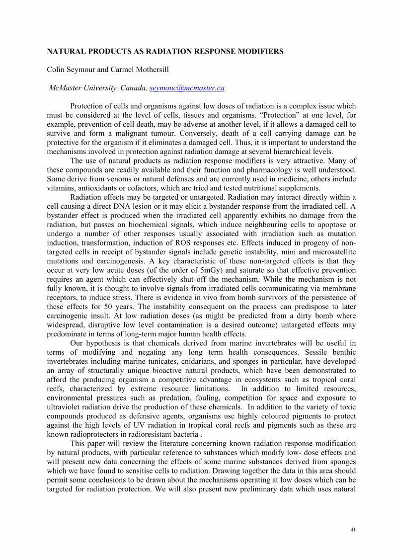
This paper will review the literature concerning known radiation response modification by natural products, with particular reference to substances which modify low- dose effects and will present new data concerning the effects of some marine substances derived from sponges which we have found to sensitise cells to radiation. Drawing together the data in this area should permit some conclusions to be drawn about the mechanisms operating at low doses which can be targeted for radiation protection. We will also present new preliminary data which uses natural
42
products derived from marine sponges. These products have been shown to have very active radiobiological activity. The structures are shown below
O
OH O
O
OH
OH
OH NH2
O
N
NH O
NH
NH OH
OO
OO
HOH
OO
OH
O O
O
O
H
H
H
H
H
HOH
H
OH
NH
NH
O
N
NH
NH2
Br
NH
OH
OHNH
O
Cl
NH2 NH
N
N
BrBrO
O
S
Discodermolide Lasonolide A
Stevensine Secobatzellin A
Plakortolide F Topsentin
Discorhabdin S
These compounds are radiation response modifiers acting via bystander mechanisms.
43
GENETIC ENGINEERING OF ECM COMPOUNDS FOR REGENERATIVE MEDICINE Aleksander L. Sieroń, Maciej Tarnowski, Anna Szydło Department of General and Molecular Biology and Genetics, Medical University of Silesia, Katowice, Poland, CoE for Study and Teaching of Molecular Biology of Matrix and Nanotechnology, CoE Network BioMedTech “Silesia”
Various human genetic disorders including mechanical damage leading to loss of tissue or function of the organ need novel treatments. One possibility is genetic engineering. Two approaches are presented, both related to collagen type I. Osteogenesis Imperfecta (OI) is a genetic disorder caused by defects in COL1A1 or COL1A2 genes affecting production of procollagen type I and quality of its fibrils. No cure for OI is available, except for orthopedic treatment and some prevention with orthopedic equipment. A gene or stem cell therapy or combination of both could be the solution for OI. Here, we present targeting col1a1 and col1a2 genes in rat mesenchymal stem cells with human homologous sequences. Five hybrid DNA constructs comprising isogenous sequences of rat and human collagen genes were introduced into the rat bone marrow stem cells. The G418 resistant clones were screened for human DNA. Over 90% of resistant clones incorporated human DNA and 2% have had targeted endogenous collagen loci. Increasing length of flanking sequences from 1 to 4kb increased 10-fold targeting loci genes. Also, DNA recombination strategies for defining domains critical for collagen stability as well as for interactions with ECM proteins, regulatory factors and cells are presented. Potential applications of results for tissue engineering, also, are discussed. Our results open new possibilities of fixing collagen gene mutations in patients, preferably using stem cells, expanding them and putting them back into the patient. Also, the enrichment of novel collagens with specific domains will create better material for regenerative medicine.

44
GLYCOCONJUGATES.DEVELOPMENT OF A NEW BIOLOGICALLY ACTIVE COMPOUNDS Wiesław Szeja1, Tadeusz Bieg1, Anna Kasprzycka1, Gabriela Pastuch1, Ilona Wandzik1, Jadwiga Zawisza1, Grzegorz Grynkiewicz2, Bogusław Szewczyk3
1Silesian University of Technology, 44-100 Gliwice, Poland; 2Pharmaceutical Research Institute, Rydygiera 8, 01-793 Warszawa, Poland; 3University of Gdańsk, Departament of Molecular Virusology, 80-822 Gdańsk, Poland
Carbohydrates are now recognized as playing a significant role in numerous physiological responses [1].The molecular diversity of carbohydrates offers a valuable tool for drug discovery[2].
Glycosyltransferases (GTS) are enzymes responsible for processing of biomacromolecules. They regulate many cellular functions and are therefore important targets in medicinal chemistry [3]. We report herein the synthesis of several analogues of uridine, which were designed to act as inhibitors, through binding to the active site of the enzyme in competition with natural donor substrates. In order to construct analogues of uridine diphospho sugars we have chosen to exploit glycal chemistry. First, uridine and glycals were selectively protected. Afterwards, addition of uridine derivative to a glycal, catalysed by triphenylphosphine hydrobromide, was performed [4]. In this way we have synthesized, in a totally stereoselective manner and in high yields, several uridine derivatives of 2-deoxy sugars.
In search for effective inhibitors of sugar-processing enzymes the heteroaryl thioglycosides and products of their oxidation are substrate analogs. We present a simple and efficient methodology for synthesizing glycosyl sulfoxides, inhibitors of GTS.
The effect of inhibitors on penetration and propagation of swine fever virus (SFV) will be presented. Even when the viability of SK6 cells was higher than 90%, low doses of this inhibitor (20 μg/ml), arrested the propagation of CSFV virus and the viral yield was decreased by over 80%. References: 1. Varki A., Glycobiology 3(1993)97;.Dwek R.A, Chem.Rev., 96(1996)683 2. Gruner S.A.W., Locardi E., Lohof E., Kessler H., Chem. Rev.,102(2002)491 3. Witczak Z.J., Nieforth K.A., Eds., Carbohydrates in Drug Design, Marcel Dekker Inc., New York, 1997. 4. Jung K.H,. Schmidt R.R., Glycosyltransferase Inhibitors in Carbohydrate-Based Drug Discovery, C.-H.Wong (Ed) Viley-VCH, vol.2(2003) 609. 5. Bolitt Ch., Mioskowski S., Lee G., and. Falck J. R.., J. Org. Chem., 55, 5812 (1990).

45
COPOLYMERS OF SYNTHETIC BIODEGRADABLE SCAFFOLD WITH COLLAGEN TYPE I Anna Szydlo1, Joanna Pogrzeba1, Maciej Tarnowski1, Piotr Kurcok2, Michal Kawalec2, Michal Sobota2, Sieron Aleksander L.1
1General and Molecular Biology and Genetics, Medical University of Silesia in Katowice, CoE for Study and Teaching of Molecular Biology of Matrix and Nanotechnology, CoE Network BioMedTech “Silesia”. Poland; 2Polish Academy of Sciences, Centre of Polymer and Carbon Materials, Zabrze, CoE Polymers 2000, CoE Network BioMedTech “Silesia”, Poland
New biodegradable polymeric materials are needed for artificial ECM-like scaffolds that mimic a three-dimensional environment in tissues. In this study we have focused on production of such scaffolds using procollagen type I and polyhydroxybutyrate (PHB). We have tested their feasibility for in vitro fibroblasts cultures. A higher number of cells were attached to plastic and PHB coat. After 4 hours the number of cells on plastic surface was 182 cells/mm2 and on PHB itself the number was 174 cells/mm2. After 8 hours fibroblasts attached to plastic numbered 192 cells/mm2 and on PHB itself ithe count was 171 cells/mm2. The number of cells attached to Procollagen I and Procollagen I coating PHB (Copolymer) was lower. On the other hand fibroblasts have spread much faster on the Copolymer and on Procollagen I. Already at the 1st hour about 23% of cells did spread on Copolymer and on Procollagen I itself, compared to only 1% spread on plastic and on PHB alone. After 2 hours of culture almost 80% of cells did spread on both Procollagen I and on Copolymer, whereas, only 20% of cells have spread on plastic and on PHB itself. Spreading on scaffolds was almost complete following 8 hours of culture, but not on plastic and PHB. PHB itself showed to be a poor substrate for cell spreading but not for cell attachment. Cell numbers spread on PHB itself were significantly lower than in cultures on plastic. Subsequently, we have created a three-dimensional structure of PHB. The method of electrospinning was used to form PHB-based fabric. The PHB fibers have created a 3D structure of the scaffold. Incubation of electrospun disc of PHB with procollagen type I, in the MES buffer, allows copolymer formation. Scaffolds prepared as described above were seeded with human fibroblasts. Attachment and spreading of fibroblasts was assessed by AlamarBlue® dye assay. Our results indicate that copolymers of collagen I with PHB are useful for building 3D scaffolds and could be successfully colonized with human fibroblasts. Such scaffolds could be also utilized as assay systems to test different compounds in preclinical trials, as well as to create prosthetics supporting cells in repairing damaged tissues and organs, e.g. skin grafts, tendons, etc.
Supporting grants: KBN2P05C02926, NN-1-015/05, RFSD Stipend Fund (EFS-2.6 ZPORR No. Z/2.24/II/2.6/17/04 RFSD to AS 9/2005).

46
NOVEL TECHNOLOGIES IN THE STUDY OF IFN ALPHA ACTIVITY Tokovenko B., Kuklin A., Perepelyuk M., Obolenskaya M.([email protected]) Institute of Molecular Biology and Genetics National Academy of Sciences of Ukraine Zabolotnogo str. 150, 03143 Kyiv, Ukraine
Background: Interferon alpha (IFNα) is used as main or adjuvant treatment in the therapy of viral infections and several types of cancer. However, very little is known about the physiological effects of IFNα in the absence of viral infection, and about the exact mechanisms of IFNα action. Although the list of IFNα direct and indirect targets encompasses more than 300 genes the IFNα gene regulatory network has not been defined.
The aim of the study was dual: a) to identify the genes of primary response to IFNα as potential targets for further investigation and the initial information link in gene regulatory network and b) to elucidate whether IFNα, a typical product of KC, and protein kinase R (PKR), classical IFNα target (both the components of innate immune response), are involved in liver regeneration.
Materials and methods: Identification of the genes of primary response to IFNα treatment was conducted using bioinformatics search in the promoters of all rat protein-coding genes (as identified by Ensembl) for the presence of evolutionary conserved interferon-stimulated response element (ISRE), the binding site of the interferon stimulated gene factor 3 (ISGF3).
The rats after 2/3 partial hepatectomy (PHE) and laparotomy (LAP) were used 1, 3, 6 and 12 h post-surgery to model correspondingly G0→S transition and acute phase response, the latter being a constituent part of the former. The gene expression was assessed in liver samples, isolated KCs and hepatocytes by RT-PCR, antiviral test, Western blot analysis and immunohistochemistry.
Results: Over 700 genes were identified to have evolutionary-conserved ISRE sites in promoters, and thus are potential primary-response genes to IFNα treatment.
PHE induces 2-fold increase of IFNα mRNA content and liver antiviral activity at 1h post surgery, decrease to the values lower than control at 3h and normalization during 6 - 12 h period. LAP induces half of IFNα mRNA content and the antiviral activity less than the detection limit. KCs in both models are responsible for IFNα expression.
The level of PKR mRNA increases 2 times at 1h after PHE, decreases at 3h and again increases to the 1h-level during 6 – 12h period after PHE. The PKR-antigen localizes in hepatocytes in the nuclei and cytoplasm with preferential localization in cytoplasm at 6h post PHE. LAP induces early decrease of PKR mRNA level with its subsequent up-regulation. Conclusions: The genes of potential primary response to IFN alpha constitute the basis for further elucidating the gene regulatory network induced by IFN alpha.
The changes in IFNα and PKR expression may be essential for liver G0 →S transition and acute phase response.

47
DNA REPAIR IN THE PATHOLOGY OF LUNG AND COLON CANCERS Barbara Tudek Institute of Biochemistry and Biophysics, Polish Academy of Sciences, Pawińskiego 5a, 02-106 Warsaw, Poland
Oxidative DNA damage and its repair are involved in pathogenesis of human cancers. The main pathway of oxidative DNA damage repair is base excision repair (BER). Functional studies performed on blood leukocytes have shown that some BER pathways may be decreased in cancer patients and this may be one of risk factors for the disease development. Decreased 8-oxoG excision rate was observed in lung as well as head and neck cancer patients. Lung cancer patients revealed also lower rate of 1, N6-ethenoadenine (εA) excision rate, DNA damage induced by lipid peroxidation (LPO). Repair of another LPO-induced modification, 3,N4-ethenocytosine (εC) was decreased only in individuals developing lung adenocarcinoma, histologically the type of cancer with etiology linked to inflammations. Activity of BER proteins is regulated by gene polymorphism, interaction of partner proteins and post-translational modifications. Polymorphism of DNA glycosylases may change their enzymatic activity. Some of these polymorphisms increase the risk of inflammation related cancers, e.g. colon, lung and other cancers. BER system efficiency may be also regulated by reactive oxygen species and diet, which stimulate transcription of some DNA glycosylases and the major human AP-endonuclease, APE1. The activity of repair enzymes may also be regulated by carcinogenic process. Stimulation of BER genes transcription or deregulation of repair proteins activity is frequently observed in cancer cells. Thus, modulation of BER proteins activity may be an important risk factor for cancer development, and may also contribute to cancer progression.

48
CHARACTERIZATION OF NONADHERENT RAT BONE MARROW STEM CELLS (VSEL CELLS) Ksymena Urbanek1, Halina Koryciak-Komarska1, Aleksander L. Sieroń1, Aleksandra Bryzek2, Magdalena Jackiewicz2, Piotr Czekaj2
1Department of General and Molecular Biology and Genetics, Medical University of Silesia, Katowice, Poland, CoE for Study and Teaching of Molecular Biology of Matrix and Nanotechnology, CoE Network BioMedTech “Silesia”; 2Department of Histology, Medical University of Silesia, Katowice, Poland
Stem cells can potentially offer treatment that many patients need. However, before using stem cells in the clinic, more studies are required. Of greatest potential and plasticity are embryonic cells, but it is unlikely that embryonic stem cell will be available at all for clinical use. Adult embryonic-like stem cells provide an alternative, which is more ethically acceptable and which could be soon available for transplantation purposes. Recent reports on cells with great plasticity, that were found in mice, claim that cells with marker pattern Sca-1+-lin--CD45- are a population that could be used for clinical applications. Cells of similar phenotype were also identified in human umbilical cord blood. They express antigens typical for embryonic cells: SSEA-1, Oct-4, Nanog, Rex-1. The cells may also be present in other tissues of adult individuals. However, little is understood so far about signals that induce stem cells to differentiate into particular cell types, nor how to get grafted cells to integrate effectively into tissues and organs. We postulate that these cells are present in bone marrow of adult rats and their differentiation depends on Notch signaling involving four Notch receptors encoded by genes Notch 1, 2, 3, and 4 and ligands, Delta 1 and 2, and Jagged 1 and 2. This pathway is one of the basic signaling pathways involved in the regulation of cell biology and metabolism in different physiological situations, both during embryogenesis and organogenesis as well as in later life. Disturbances of this pathway have been already linked to numerous pathological conditions, including cancer.
49
A START-TO-END APPROACH TO THE ANALYSIS OF PROTEOMIC DATA Beata Walczak Department of Chemometrics, Institute of Chemistry, University of Silesia, Katowice, Poland [email protected], www.chemometria.us.edu.pl
Comparative proteomics focuses on the differences in the protein pattern from normal and pathological (or treated) samples. When two-dimensional (2D) gel electrophoresis is used as an analytical tool for separating proteins, comparison of samples is based on the resulting digital images of the 2D gel electropherograms. Thus, an overall success of comparative proteomics critically depends on accuracy and reliability of the analysis of the gel images. The available software deals differently with the main steps of data analysis, such as images’ preprocessing, images’ warping, spot detection, etc. Different approaches cause a significant software-induced variance [1] and often lead to a significant amount of missing data in the resulting peaks’ table [2].
In the proposed start-to-end approach these drawbacks are eliminated [3]. Namely, the preprocessing step is optimized based on the classifier performance, analysis is performed on the pixels instead of spots level, multivariate methods are used for model construction, and the proteins differentiating the studied classes of objects are identified based on the embedded into the classifier feature selection method, validated based on the Monte Carlo technique. References: 1. A.W.Wheelock, A.R.Buckpitt, Software-induced variance in two-dimensional gel electrophoresis image analysis, Electrophoresis, 26 (2005) 4508-4520, 2. H.Grave et al.. Challenges related to analysis of protein spot volumes from two-dimensional gel electrophoresis as revealed by replicate gels, Journal of Proteome Research, 5 (2006) 3399-3410 3. M. Daszykowski, I. Stanimirova, A. Bodzon-Kulakowska, J. Silberring, G. Lubec, B. Walczak, The start-to-end processing of two-dimensional gel electrophoretic images, Journal of Chromatography A, 1158 (2007) 306-317
50
MAKING SENSE OF VOLTAGE SENSORS Stephen H. White Dept. of Physiology and Biophysics, University of California, Irvine, CA 92697-4560, USA
Voltage-gated ion channels open and close in response to changes in transmembrane potential due to the motion of their voltage-sensor (VS) domains. Long known exclusively as regulatory accessories for ion-channel pore domains, VS domains have now been identified in voltage-activated phosphatases, and more recently a novel VS protein that functions as a voltage-gated proton channel has been discovered. These findings and the crystallographic structure of the Kv1.2 potassium channel leave little doubt that the VS can exist as an independent transmembrane domain in lipid bilayers. Given the long-standing assumption that charged amino acid side chains cannot be exposed directly to hydrocarbon, how is it possible that VS proteins and their highly charged S4 segments can form stable transmembrane structures? The answer is that the lipid bilayer is far more complex—and interesting—than its usual lollypop cartoon suggests. Biological and physical evidence augmented with molecular dynamics simulations will be presented that reveal the extreme adaptability of phospholipids that arises from the privileged relationship between their phosphate groups and arginine residues. This adaptability makes possible the independent stability of VS domains. [Research supported by the National Institute of General Medical Sciences and the National Center for Research Resources.]

51
MASS SPECTROMETRY ANALYSES OF THE SERUM PROTEOME - - AN IMPORTANT TOOL OF CLINICAL PROTEOMICS Piotr Widlak Maria Sklodowska-Curie Memorial Cancer Center and Institute of Oncology, Branch in Gliwice; 15 Wybrzeze AK, 44-100 Gliwice, Poland
Analysis of the low-molecular-weight region of the blood proteome (using either serum or plasma samples) is an emerging method of clinical proteomics. Such analyses base on mass spectrometry (MS), on either relatively simple technologies like MALDI-TOF or more sophisticated combinations of multidimensional chromatography and MS. Although no single peptide is expected to be a reliable bio-marker in such analyses, multipeptide sets of markers selected in numerical tests have been already shown in a few studies to have prognostic and predictive value in cancer diagnostics.
We have initiated a large cohort study aimed to identify a set of polypeptide biomarkers for early detection and monitoring therapy of breast cancer patients. In additional study we analyze serum proteome of patients with head and neck cancer subjected to radiotherapy, aiming to identify biomarkers of radiosensitivity. Low-molecular-weight polypeptides (2-10 kD) are analyzed using MALDI-TOF MS after removal of albumin and other larger proteins from serum of cancer patients (in the course of therapy) and matched healthy controls. Specific polypeptide patterns identified through mathematical analyses are cross-correlated with clinical observations as well as results of routine biochemical and histopathological analyses in an attempt to select reliable biomarkers that could be applied in cancer diagnostics.

52
PROGNOSTIC MOLECULAR BIOLOGICAL MARKERS OF BREAST CANCER Iosif Zalutsky, Raisa Smolyakova, Alexander Mashevsky, Alexander Dubrovsky, Maria Budko N.N. Alexandrov Research Institute of Oncology and Medical Radiology, Minsk, Belarus
The principal prognostic factors of breast cancer (BC) are lymph node metastases, the tumour size and spread, its grade, the status of estrogen and progesterone receptors. At present, intensive search is being done in clinical oncology for molecular biological markers of cellular origin, which would allow prognosticating the response to chemo- and endocrine therapy, the disease course and long-term results of the treatment.
The molecular markers include oncogenes and proto-oncogenes, oncoproteins, different growth factors and their receptors, receptors of steroid and peptide hormones, suppressor genes and products of their expression, hormone-dependent proteins, proteases participating in metastasizing processes, integrins responsible for intercellular contacts. The determination of the tumour proliferative activity, of the hormonal status, growth factor receptors in BC patients is of vital importance for selection of the specific treatment regimen and prognosis of the disease course.
The objective of the study is to establish molecular biological markers in BC patients. The study material consisted of tumour tissue obtained from 60 BC patients during surgical intervention. All the BC patients had histological diagnoses. Infiltrative ductal carcinoma (45.1%) and infiltrative lobular carcinoma (37.1%) of differentiation grade 2 prevailed in this group of BC patients. The age of the patients included in the study varied from 31 to 85 years.
To define the expression of hormonal estrogen receptors (ER) and progesterone receptors (PR), Ki-67 proliferative protein, epidermal growth factor receptors (EGFR, Her-2/neu), p53 suppressor mutant protein, bcl-2 oncoprotein, immunohistochemical technique was used with DAKO Cytomation (LSAB, En Vision) kits.
The results of our investigations demonstrate that 23 (39%) BC patients had estrogens-positive tumours and 24 (42.11%) were progesterone-positive. It was established that ER was most frequently found in well-differentiated and moderately differentiated tumours while in high-grade BC the ER level was reduced. The PR analysis detected its hyperexpression in well- and moderately-differentiated tumours. The presence of steroid hormone receptors in BC is an indication of a relatively favourable prognosis and of potential sensitivity of the tumour to endocrine therapy. Her-2/neu, a protooncogene- coding receptor 2 of human epidermal growth factor, is a marker strongly influencing the antitumour treatment policy. Hyperexpression of Her-2/neu oncoprotein was found in the tumour tissues of 24.6% of the patients. Her-2/neu hyperexpression by the tumour is an independent marker of poor prognosis, high risk of recurrence and decreased survival. The evaluation of Her-2/neu is of special prognostic significance in the cases of small tumours. The expression of p53 suppressor mutant protein was detected in 57.1% of BC patients. Among them slight positive staining occurred in 37.5% of the cases, and strong positive – in 59.4%. Positive Ki-67 reaction was observed in 79.3% of tumour tissue specimens of BC patients. Among them, 41.4% of the total number demonstrated high proliferative activity. Ki-67 index inversely correlated with tumour differentiation grade (R= – 0.3024; p<0.05). High proliferative activity of the tumour in the BC patients under investigation was associated with p53 expression (R=0.5259; p<0.05). At the same time, inverse correlation with progesterone receptor expression was established (R= – 0.3129; p<0.05). Bcl-2 oncoprotein hyperexpression was registered in 77.7% of cases. Proliferative activity of the tumour inversely correlated with bcl-2 expression (R= – 0.3244; p<0.05). The evaluation of EGFR expression found 83.6% of the specimens with negative EGFR reaction. EGFR hyperexpression was diagnosed in 6.6% of the BC patients.
In conclusion: determination of the prognostic markers allows to obtain an objective molecular biological profile of the tumour and to shape the optimal antitumour treatment policy complying with individual indications of BC patients.

53
Poster abstracts

54

55
1. THE RESULTS OF CLINICOGENEALOGIC AND MOLECULAR GENETIC INVESTIGATIONS OF OVARIAN CANCER PATIENTS Natalia Antonenkova, Iosif Zalutsky, Galya Porubova, Elena Mokhon N.N Alexandrov Research Institute of Oncology and Medical Radiology, Minsk, Belarus
Objectives of the study: Establishing a cohort of probands with ovarian cancer (OC), whose relatives are subject to preventive examination aimed at timely treatment.
Materials and methods: OC patients aged 24 to 77 years, mean age 51.27±10.31. Clinicogenealogic analysis of the pedigrees of patients treated in the hospital of SI N.N. Alexandrov RIOMR identified 100 families with cancer-burdened familial history. The following mutations were determined using allele-specific PCR technique: BRCA1 (185delAG), BRCA1 (5382 insC), BRCA2 (6174delТ).
Results: The results are presented in Table 1.
Table 1. Types of mutations determined in tumour growth suppressor genes
BRCA1 (185delAG)
BRCA1 (5382 insC)
BRCA2 (6174delТ) Result
n % n % n % Negative 71 71.0 72 72.0 59 59.0 Positive in the homozygous state 10 10.0 12 12.0 18 18.0 Positive in the heterozygous state 19 19.0 16 16.0 23 23.0 Total 100 100 100 100 100 100
Thus, BRCA1 (185delAG) mutations were found in 29 (29%) patients of 100 BRCA1
(5382 insC) in 28 (28%), BRCA2 (6174delТ) in 31 (31%). The relatives of the carriers of germinal mutations predisposing to OC and breast cancer (BC) were invited for preventive examination.
Conclusions: 1. Activities are underway for early detection of individuals with hereditary predisposition to OC and BC, employing clinicogenealogic and molecular genetic methods of investigation. 2. The adoption of the techniques developed in clinical practice makes it possible to spare the resources needed for preventive examination of persons with high risk of inherited predisposition to OC and BC.
56
2. ANTITUMOR ACTIVITY OF MANNAN-METHOTREXATE CONJUGATE Renata Budzynska1, Dmitry Nevozhay2,3, Urszula Kanska1, Monika Jagiello1, Adam Opolski2,4, Joanna Wietrzyk2, Janusz Boratynski1,4, 1Laboratory of Biomedical Chemistry, 2Laboratory of Experimental Anticancer Therapy; 2Department of Experimental Oncology, Institute of Immunology and Experimental Therapy, Polish Academy of Sciences, Rudolfa Weigla 12, 53-114 Wroclaw, Poland; 3Kopvillem Institute of Medical Physics, Kirov St, 64, 690068 Vladivostok, Russia; 4J. Dlugosz Academy, Al. Armii Krajowej 13/15, 42-201 Czestochowa, Poland
Conjugation of anticancer drugs with different carriers has been extensively studied recently as a method of obtaining of improved drug forms. The conjugation often results in increased therapeutic effect, alteration of toxicity profile, and/or selective targeting of therapeutic agent to the tissue of interest. We have synthesized mannan-methotrexate conjugate using methotrexate anhydride and compared its antitumor properties both in vitro and in vivo with free methotrexate. Mannan-methotrexate conjugate showed significantly improved antitumor activity in the model of P388 mouse leukemia disseminated in the peritoneal cavity, following intraperitoneal administration of the chemotherapeutic. Conversely, the antitumor effects of free methotrexate and mannan-methotrexate conjugate were comparable when leukemia was implanted subcutaneously and chemotherapy agents were administered intravenously. These results suggest that mannan-methotrexate conjugate could be further investigated as the potential therapeutic agent for intraperitoneally disseminated tumors.

57
3. CELL ADHESION PROTEINS EXPRESSION IN PAPILLARY THYROID CANCER Mykola Chekan1, Barbara Nikiel2, Wojciech Wierzchowski3, Dariusz Lange2, Barbara Jarzab1, Jerzy Stachura3 1Dept. of Nuclear Medicine and Endocrine Oncology; 2Dept of Tumor Pathology, Maria Skłodowska-Curie Memorial Cancer Center and Institute of Oncology, Gliwice Branch, 44-101 Gliwice, ul. Wybrzeże Armii Krajowej 15, Poland; 3Department of Pathomorphology, Jagiellonian University, Krakow, Poland
Introduction: Many cell adhesion molecules are up-regulated in papillary thyroid cancer (PTC). In our previous microarray studies we have specified several of them (Jarzab et al. 2005), with strong diagnostic accuracy for galectin-3. The aim of the present study was to verify the differences in expression of a panel of cell adhesion proteins by tissue microarray approach.
Material and methods: Tissue microarray paraffin block with 101 spots (0.6 mm ) of PTC and surrounding normal thyroid tissue (ST) was prepared from 23 patients diagnosed with PTC and operated. EnVision (biotin-free), a highly-sensitive immunochemical technique was applied. 10 spots were not evaluated because of the connective tissue content, the remaining were scored for expression of galectin-3 (LGAL3), MUC1, cadherin P (CDH3), CD44v6 and carbohydrate antigen CA50. Also, the presence of endogenous biotin was visualised by LSAB+ method. Heterogeneity of reaction intensity was evaluated for all antigens.
Results. The significant difference between PTC and ST was noted for LGAL3 (100%, intensive reactions in PTC versus 17%, weak in ST ), CA50 (96.4% versus 37.9%), less for CD44v6 (100% versus 69.2%, weak) while CDH3 and MUC1 showed only weak or no difference. High amounts of endogenous biotin were revealed in PTC by labelled streptavidin-biotin without primary antibody.
Conclusions. Our tissue microarray study does not confirm differences, on the protein level, in expression of MUC1 and cadherin P genes, suggested by clear differences on RNA level, found using DNA microarrays. Simultaneously, our study indicates high expression of carbohydrate CA50 antigen, as well as high content of endogenous biotin in PTC cells, in addition to the well-known overexpression of galectin-3. Supported by Ministry of Science and Higher Education grant no 2P05A 0 22 30 Mykola Chekan was a fellow of a Fellowship Program totally supported by the National Cancer Institute – Office of International Affairs, NIH, Bethesda, MD, USA.
58
4. GPR40 & GPR120 RECEPTORS – POTENTIAL TARGETS FOR NEW TREATMENT OPTIONS IN GI TRACT Adam Cygankiewicz 1,2; Birgitte Holst 2; Wanda M. Krajewska 1 & Thue W. Schwartz 2,3 1Department of Cytobiochemistry, University of Lodz, Lodz, Poland; 2Laboratory for Molecular Pharmacology, The Panum Institute, University of Copenhagen, Copenhagen, Denmark 3 - 7TM Pharma A/S, Hørsholm, Denmark e-mail: [email protected]
7TM (7 trans-membrane) receptors constitute one of the greatest family in human genome. Many of them create new putative drug targets, hence understanding mechanisms underlying ligand-induced activation, constitutive activity and pharmacological features of these receptors may provide new insights into pathogenesis and novel treatment options for diseases like diabetes, obesity, hypertension and cancer.
Vast number of 7TM receptors (and among them GPR40 and GPR120) are expressed in the gastrointestinal tract and in the endocrine pancreas and are believed to be involved in the control of the secretion of, for example, insulin and glucagons-like peptide-1.
Free fatty acids are suggested as natural ligands for both studied receptors. Our investigations have demonstrated that these receptors are also activated in dose-dependent manner by rosiglitazone and other thiazolidinedione drugs as well as other compounds belonging to PPAR activators group.
In our studies we have also confirmed certain level of constitutive activity of both receptors. More in-depth knowledge about pharmacological features of these receptors may contribute to development of more efficient treatment for hyperglycemias. Alteration in constitutive signaling of 7TM receptors may be involved in other diseases as well.

59
5. PHOSPHOROTHIOATE ANALOGS OF NUCLEOTIDES ACCELERATE WOUND HEALING Edyta Gendaszewska-Darmach, Marek Kołodziejczyk, Alina Krystynowicz, Stanisław Bielecki, Maria Koziołkiewicz Institute of Technical Biochemistry, Faculty of Biotechnology and Food Sciences, Technical University of Łódź, Stefanowskiego 4/10, 90-924 Lodz, Poland
Biomaterials which are carbohydrate polymers (among them bacterial cellulose) have proved in recent years to be promising for many medical applications, in particular for wound healing and tissue regeneration. However, the existing biomaterials are not able to stimulate cell proliferation and migration - processes necessary to achieve formation of new blood vessels (angiogenesis).
Therefore, there exists the need to evaluate biomaterials with immobilized growth factors that are subsequently released into the wound bed over a period of 2-3 days and stimulate these processes. Our in vitro experiments have shown that phosphorothioate analogs of nucleotides (containing a sulfur atom instead of one of the oxygen atoms at the phosphate group) stimulated the proliferation of human umbilical vein endothelial cells (HUVECs). The highest stimulation of this process was caused by thymidine-5’-thiophosphate (TMPS) (Koziołkiewicz et al., 2001). This effect is associated with higher affinity of the nucleotide to specific P2 receptor and resistance of this compound to the action of nucleolytic enzymes. TMPS was immobilized to microbial cellulose produced at the Institute of Technical Biochemistry and such a wound dressing was tested in preliminary in vivo experiments. These experiments have shown the formation of new blood vessels network after application of the biomaterial and proved its wound-healing potential.
References: 1. Koziolkiewicz M, Gendaszewska E, Maszewska M, Stein CA and Stec WJ (2001) The mononucleotide-dependent, nonantisense mechanism of action of phosphodiester and phosphorothioate oligonucleotides depends upon the activity of an ecto-5’-nucleotidase. Blood 98:995-1002

60
6. ROLE OF DAMAGE SPECIFIC DNA POLYMERASES IN MUTAGENICITY OF LIPID PEROXIDATION-INDUCED DNA ADDUCTS Janowska B., Kurpios D., Komisarski M., Kuśmierek J. T., Tudek B. Department of Biological Chemistry, Institute of Biochemistry and Biophysics, Polish Academy of Sciences, Warsaw, Poland
Lipid peroxidation leads to the formation of a large family of alkenals, which form cyclic propano- or ethenoadducts to DNA bases bearing side chains of different length. One of the most ubiquitous derivative, trans-4-hydroxy-2-nonenal (HNE) reacts with all four DNA bases with different efficiency (G>C>A>T) forming DNA adducts, as well as DNA-DNA and DNA-protein cross-links. HNE-DNA adducts block DNA synthesis and, when present in bacteria in ssDNA, cause recombination. Removal of these lesions from dsDNA occurs via the Nucleotide Excision Repair (NER) pathway. An equally important mechanism is translesion DNA synthesis. In E.coli, dysfunction of DNA polymerase IV in uvrA mutant dramatically decreased the survival, but had no effect on mutation frequency in comparison to the wild type E.coli. In the strain lacking uvrABC exonuclease and DNA polymerase V, the survival was similar to that of uvrA single mutant, but no HNE-induced mutations were observed. In uvr- strains which possessed pol II alone or together with pol IV (uvrAdinBumuDC and uvrAumuDC respectively), mutation frequency was lower than in the wild type JM105. In uvrAdinpolB triple mutant, survival decreased dramatically, and mutation frequency increased substantially in comparison to the wild type strain. These results suggest that in E.coli, HNE adducts to DNA bases are processed by damage-specific DNA polymerases, of which DNA polymerase II and IV are able to bypass the adducts in an error-free manner. Lack of DNA pol IV results in the same sensitivity to HNE as the lack of nucleotide excision repair. DNA polymerase V is responsible for mutation induction on HNE-damaged templates. In the strains which possessed pol V, but not II and IV (JM dinBpolB, uvrdinBpolB) half of mutations were 54-nucleotide deletions of the phage polylinker region. Remaining 25% of mutations in uvrdinBpolB strain were double deletions of 54 and 93 (ΔM15) nucleotides resulting from recombination between the sequences of M13 and bacterial F’ lacZ genes. These data suggest that HNE-DNA adducts constitute a substantial hindrance for Pol V, which either bypasses these lesions in an error-prone manner or falls off to enable recombination.

61
7. AN EFFICIENT METHOD FOR THE SYNTHESIS OF 15N-LABELLED NUCLEOSIDES AND THEIR DERIVATIVES WITH THE USE OF BACTERIAL CULTURE B.Kalinowski, E.Zarakowska, D.Gackowski Department of Clinical Biochemistry, Collegium Medicum UMK, Bydgoszcz, Poland
Isotopically labelled nucleosides and their derivatives that show biological properties are important in many analytical investigations. Accordingly, there is an increasing demand for new and more effective methods of labelled nucleoside synthesis which can be analysed by nuclear magnetic spectroscopy or mass spectrometry.
In the presented study a method for synthesising 15N-labelled nucleosides with the help of E. coli cells was used. The bacteria were grown in the 15N-labelled culture medium (Silantes OD2). Nucleic acids were isolated from bacteria, digested with nuclease P1 and alkaline phosphatase to nucleosides. The hydrolysate was separated by high performance liquid chromatography using a C18 reverse-phase column (Phenomenex luna, size 10 mm x 250 mm). Nucleosides were separated with the help of gradient elution (1.5% acetic acid and methanol). The purified fractions of individual nucleosides were received. The isotopic purity of compounds determined by mass spectrometry was: 99.2% for deoxyadenosine (dA) and deoxythymidine (dT), 97% for deoxycytidine (dC) and deoxyguanosine (dG) and >98% for adenosine (A), uridine (U), guanosine (G) and cytidine (C). Next, labeled dG and G were chemically modified. In the system generating ˙OH radical (15N5)-7,8-dihydro-8-oxo-2’-deoxyguanosine ((15N5)-8-oxodG) and (15N5)-7,8-dihydro-8-oxo-guanosine ((15N5)-8-oxoG) were obtained. Also (15N4)-deoxyoxanosine ((15N4)-dOxo) (15N4)-deoxyxanthosine ((15N4)-dXao), (15N4)-oxanosine ((15N4)-Oxo), (15N4)-xanthosine ((15N4)-Xao) were obtained in the reaction with NOֹ radical.
The presented method is highly efficient. Collected nucleoside analogues have a high isotopic purity and can have potential use in investigations of processes taking place in living cells.

62
8. CLINICAL SIGNIFICANCE OF EWS/FLI1 AND EWS/ERG GENE FUSIONS EXPRESSED IN EWING'S FAMILY TUMORS L. Kisialeu1, N.V. Lipay2
1Department for Elder Children, 2Molecular Biology Department, Belarussian Center for Pediatric Oncology and Hematology, pos. Lesnoe, Minsk region, Belarus
Ewing’s sarcoma family of tumors (EFTs) which includes Ewing’s sarcoma and peripheral neuroectodermal tumors, has specific chromosomal translocations that result in the fusion of EWS gene and a gene encoding a member of the ETS family of transcription factors, thus forming different chimeric transcription factors, either EWS/FLI1 (90-95%) or EWS/ERG (5-10%). The latter ones may alter in its transactivating capacity. The existence of these alternative fusion genes may provide a basis for a clinical heterogeneity of EFTs.
So we investigated whether the two alternative gene fusion products, EWS/FLI1 and EWS/ERG, define different clinical outcome within EFTs. 50 patients were diagnosed between 1999-2006. Nested RT-PCR was performed to detect EWS/FLI1, EWS/ERG transcripts in RNA
extracted from samples according to L. Montanaro et al. RNA was isolated by phenol-chloroform extraction described by Chomczynski, Sacchi.
Molecular genetics analyses were performed for all 50 patients (84 samples). For all patients RT-PCR results were in accordance with histological analysis.
Event/Relapse-Free-Survival (EFS or RFS) was estimated by Kaplan and Meier and comparison of life-table outcome were performed with log-rank test. The median follow-up duration was calculated from the time of diagnosis for the patients who are event /relapse-free survivors.
EFT-specific transcripts were found in 40 of 50 patients (80%). For comparison we divided all positive patients into 2 groups according to the fusion gene:
Group 1– patients with EWS/ERG transcripts (10/40 or 25%) Group 2– patients with EWS/FLI1 transcripts (30/40 or 75%) The 5-year EFS in EWS/ERG-group was 37% (median follow-up 22 months), and was
not statistically decreased in comparison with EWS/FLI-group (55% with median follow-up 18 months).
40 patients were next divided in two groups according to high risk (24/40) or standard risk protocol (16/40) and EFS rates were estimated separately for patients in different risk groups with different fusion transcripts.
High risk: 18/24 patients had EWS/FLI1 transcripts (75%) and 6/24 patients had EWS/ERG transcripts (25%);
Standard risk: 12/16 patients had EWS/FLI1 transcripts (75%) and 4/16 had EWS/ERG transcripts (25%).
The 5-year EFS was 33% in EWS/ERG-positive patients with high risk (median follow-up 18 months) in comparison with 55% (median follow-up 18 months) in EWS/FLI-positive patients with high risk. This trend, though, was non-significant. Among patients with standard risk 4-year EFS was 38% (median follow-up 51 months) for EWS/ERG group and 67% (median follow-up 17 months) for EWS/FLI1 group. Again, this difference was non-significant.
There were no significant clinical differences observed between the two groups in event-free survival, or progression-free survival. At the given stage there are no significant proofs of influence of two alternative genes on a clinical outcome of pediatric patients with Ewing´s sarcoma family of tumors. This problem may, however, require further studies.

63
9. DETECTION OF THE SPECIFIC FUSION GENES WHICH IS CHARACTERIZED EWING'S FAMILY TUMORS FOR PEDIATRIC PATIENTS. L. Kisialeu1, N.V. Lipay2
1Department for Elder Children; 2Molecular Biology Department, Belarussian Center for Pediatric Oncology and Hematology, pos. Lesnoe, Minsk region, Belarus
Ewing’s sarcoma family of tumors (EFTs) are primitive malignant tumors of bone and soft tissues arising preferentially in children and young adults. EFTs show an extremely aggressive behavior and rapidly disseminates to bones, bone marrow, and lungs. Ewing’s tumors are characterized in at least 95% of cases by specific chromosomal rearrangments t(11;22)(q24;q12) and t(21;22)(q22;q12) which corresponding to specific fusion transcripts EWS/FLI1 and EWS/ERG, respectively.
The aim of our study was to evaluate the clinical significance of the EWS/FLI1, EWS/ERG fusion genes detection by RT-PCR method for diagnostic use in children.
Nested RT-PCR was performed to detect EWS/FLI1, EWS/ERG transcripts in RNA extracted from samples according to L. Montanaro et al. RNA was isolated by phenol-chloroform extraction, as described by Chomczynski and Sacchi.
During a 5-year period (2000-2005) we examined 77 samples from 52 patients: 30 samples– tumor fragments from primary sites; 1 sample–affected lymph node; 28 samples–bone marrow (BM) aspirates; 17 samples–peripheral blood (PB).
In case of 10 (19%) patients, only BM or PB samples were examined as the materials from primary sites biopsy was not available. All these samples were negative for EFT-specific transcripts.
For 32 (64%) patients histological analysis was in agreement with RT-PCR results (EFT-specific transcript was detected in at least one sample).
In case of 10 patients (19%) RT-PCR results influenced primary diagnosis: For 1 patient (1.9%) primary diagnosis, osteosarcoma, was substituted for Ewing’s tumor
(tumor fragments was EWS/FLI1 positive). For 1 patient (1.9%) primary diagnosis of EFT has been excluded as EFT-specific
transcripts were not detected in primary site biopsy. In this case the rabdomyosarcoma was diagnosed after secondary histological analysis.
For 2 patients (3.8%) with verified EFT, II stage was replaced by IV stage as the EFT-specific transcripts were detected in BM samples, though morphological analysis did not reveal tumor cells in BM.
For 6 patients (11.4%), in which histological analysis was insufficient, the presence of the EFT chimeric oncogenes in biological samples allows diagnosing EFT and to start therapy of patients in a grave condition immediately.
Overall, RT-PCR is a sensitive method for detection of the ET-specific transcripts and it is a relevant tool that allows gathering more accurate data concerning Ewing’s family of tumors. Thus, RT-PCR is the essential part of a diagnostic scheme, together with other diagnostic procedures.

64
10. DAMAGING EFFECT OF SCATTERED RADIATION UPON CELLS IRRADIATED AT DIFFERENT DEPTHS IN A WATER PHANTOM Maria Konopacka1, Jacek Rogolinski1, Aleksander Sochanik2, Krzysztof Ślosarek3 1Department of Experimental and Clinical Radiobiology; 2Department of Molecular Biology; 3Radiotherapy and Brachytherapy Treatment Planning Department; Maria Skłodowska-Curie Memorial Cancer Center and Institute of Oncology, Gliwice Branch,Poland
Radiation generated by linear electron accelerators used in cancer radiotherapy is non-monoenergetic. Energy broadening increases with penetration depth in the irradiated medium such as water. With greater penetration depth the number of interactions with the surroundings also increases. Part of radiation becomes scattered and the fraction of scattered radiation having decreased energy becomes larger at increased depth. A larger portion of scattered radiation, with energy lower than that of the incident beam, should change the biological response of irradiated cells.
The purpose of this study was to assess the damaging effect of scattered radiation, generated during penetration of medium, upon cultured cells placed inside a water phantom. Measurements were performed at different depths (3 – 20 cm) of a water phantom using BEAS-2B (human bronchial epithelial) cell line. Electron radiation (22 MeV) was employed, with radiation dose of 5Gy in build-up (3 cm) depth and two dose rates: 100 or 600 MU/min. Cytogenetic changes in cells irradiated at different depths were assessed and expressed as the frequency of cells with formed micronuclei as well as apoptotic cells. We compared the obtained results with expected data, i.e. those corresponding to doses received at each depth.
Our results show that, with increasing medium depth, the number of micronucleated as well as apoptotic cells was greater than that which should result from the corresponding dose received. This discrepancy between the observed and expected number of damaged cells becomes greater with increased medium depth and may be caused by the increased fraction of scattered radiation.
The observed relationship ought to be taken into consideration in both clinical practice and in treatment planning. This study was supported by MSC Memorial Cancer Center and Institute of Oncology, Branch in Gliwice, Internal Grant No. GW 0307

65
11. PERFORMANCE OF CYTOGENETIC TECHNIQUES IN IDENTIFYING DNA COPY NUMBER CHANGES Kostrzewska-Poczekaj M.1, Giefing M.1, Jarmuż M.1, Kujawski M.1, Martin-Subero J. I.3, Siebert R.3, Grenman R.4, Szyfter K.1,2 1Institute of Human Genetics, Polish Academy of Sciences, Poznan, Poland; 2Department of Otolaryngology, University of Medical Sciences, Poznan, Poland; 3Institute of Human Genetics, University Hospital Schleswig-Holstein Campus Kiel, Germany; 4Department of Otorhinolaryngology - Head and Neck Surgery and Department of Medical Biochemistry, Turku University Central Hospital and Turku University, Finland
The cell line UT-SCC-11, derived from squamous cell carcinoma of the larynx and established at the University of Turku, Finland was analyzed by banding (GTG, QFQ), comparative genomic hybridization (CGH) and the novel, high resolution array-CGH techniques. The aim of the study was to compare the performance of the three applied techniques (banding, CGH and array-CGH) in identifying DNA copy number changes (excluding translocation and inversion). Altogether, 21 chromosomal abnormalities have been detected by at least one of the used techniques. Separately, banding detected 12 abnormalities; CGH detected 7 abnormalities and array-CGH detected 18 abnormalities. The highest concordance was observed between banding and array CGH that detected 9/21 (43%) abnormalities. Banding technique presented higher sensitivity than CGH, but identified 2 additional DNA copy number changes that were not confirmed by any of the two other techniques, thus being probably false positives. In contrast, CGH delineated only 6 regions of analogy with array-CGH regions but with only one false positive.
In conclusion, the best performance was achieved by array-CGH which detected additional 8 aberrations of average size: 25 Mb (2.8-57 Mb) not detected by any of the two other techniques.
Chosen results from banding, CGH and array-CGH are shown as examples:
1. large 12.6 Mb deletion in 22q was detected by both banding and array-CGH besides classical CGH, but array-CGH detected a small additional 3.5 Mb amplification on chromosome 22, 2. small deletion ca. 4.8 Mb in chromosome 12p is not detectable by binding and classical CGH but is clearly detectable by array-CGH,
Due to low resolution of banding and CGH, array-CGH seems to be the most suitable
technique to delineate small deleted and amplified chromosomal regions.
66
12. VALIDATION OF POTENTIAL MOLECULAR MARKERS OF PAPILLARY THYROID CARCINOMA BY QUANTITATIVE REAL-TIME PCR Monika Kowal1, Aleksandra Kukulska1, Aleksandra Rusin2, Małgorzata Kowalska1, Ewa Chmielik3, Ewa Stobiecka3, Elżbieta Gubała1, Agnieszka Czarniecka4, Jan Włoch4, Tomasz Tyszkiewicz1, Barbara Jarząb1 1Department of Nuclear Medicine and Endocrine Oncology; 2Department of Tumor Biology; 3Department of Tumor Pathology; 4Oncology Surgery Clinic; Maria Skłodowska-Curie Memorial Cancer Center and Institute of Oncology, Gliwice Branch, 44-101 Gliwice, ul. Wybrzeże Armii Krajowej 15, Poland
Introduction: In our previous DNA microarray profiling studies we specified a multigene classifier of papillary thyroid carcinoma, which included some novel transcripts, not analyzed until now in the context of PTC biology. The aim of this study was to validate them by quantitative real-time PCR on an independent set of PTC samples. We included such new genes as EVA1, CDH3, KCJN2, LRP4, Q-PCT GALE (up-regulated) and HGD, TACSTD2 (down-regulated) and compared them to the well known PTC markers, which were confirmed by our microarray analysis (DPP4, FN1, KRT19, MET, RXRG, ADORA1, up-regulated, and TFF3, down-regulated).
Material and methods: Total RNA was isolated from 31 paired normal and PTC samples by Chomczynski-Sacchi method. 0.5 μg of RNA was used in the reverse transcription reaction. cDNA was further used in Q-PCR reaction. The expression was measured by Universal Probe Library LNA probes (Roche) and was normalized by the index obtained from 3 reference genes (UBE2D2, HADHA, EIF3S10) by GeNorm software.
Results: All analyzed genes, except TACSTD2, differentiated tumor and normal samples in a highly significant manner. When the diagnostic efficiency of these genes was assessed by ROC (relative operating characteristic) analysis, we found out that for overexpressed markers, the area under ROC curve (AUC) was higher than 0.9 for all of them and for down-regulated genes this criterion was met for TFF3 and HGD. We trained the multigene classifier on the obtained data and currently we are performing its validation using an independent set of samples.
Conclusions: Our results are a next step in validating PTC markers before the required verification can be made on routine diagnostic material obtained by fine needle biopsy of benign and malignant thyroid nodules. Study supported by the Ministry of Science and Higher Education, Poland, Grant No. PBZ-MNiI-2/1/2005

67
13. INFLAMMATION INCREASES OXIDATIVE DNA DAMAGE REPAIR AND STIMULATES PRENEOPLASTIC CHANGES IN COLONS OF NEWBORN RATS Paweł Kowalczyk1,5, Jolanta Jaworek2, Michalina Kot2, Beata Sokołowska3, Aleksandra Bieleń4 , Beata Janowska1, Jarosław M. Cieśla1, Grzegorz Szparecki1, Benita Sadoś1, Barbara Tudek1, 5
1Institute of Biochemistry and Biophysics, Polish Academy of Sciences, Warsaw, Poland; 2Department of Physiology, Jagiellonian University College of Medicine, Cracow, Poland; 3Department of Respiratory Research, Medical Research Center, Polish Academy of Sciences, Warsaw, Poland; 4Cancer Research UK, Clare Hall Laboratories, Blanche Lane, South Mimms, EN6 3PD, United Kingdom; 5Institute of Genetics and Biotechnology, Warsaw University, Warsaw, Poland Oxidative DNA damage may be a risk factor for development of various pathologies, including malignancy. We studied inflammation triggered modulation of repair activity in the intestines of three-week-old rats injected i.p. with E.coli or S.typhimurium lipoplysacharides (LPS) at doses of 1, 5 or 10 mg/kg. Subsequent formation in these animals of colonic preneoplastic lesions, aberrant crypt foci (ACF) was also investigated. Five days after LPS administration no differences were observed in repair rate of 1,N6-ethenoadenine (εA), 3,N4-ethenocytosine (εC) and 8-oxoguanine (8-oxoG) in intestines of these rats, as measured by the nicking assay. However, a significant increase in all three repair activities was found within one and two months after S. typhimurium LPS treatment. E. coli LPS significantly increased only the 8-oxoG repair. S. typhimurium LPS stimulated mRNA transcription of pro-inflammatory proteins, lipooxygenase-12 and cyclooxygenase-2, as well as some DNA repair enzymes like AP-endonuclease (Ape1) and εC-glycosylase (Tdg). mRNA level of DNA glycosylases excising εA (Mpg) and 8-oxoG (Ogg1) was also increased by LPS treatment, but only at the highest dose. Transcription of all enzymes increased for up to 30 days after LPS, and subsequently decreased, with the exception of Ape1, which remained elevated even two months after LPS administration. Thus, the repair efficiency of εA, εC and 8-oxoG depends on the availability of Ape1, which increases Ogg1 and Tdg turnover on damaged DNA, as well as presumably stimulates Mpg. One and two months after administration of E. coli or S. typhimurium LPS, the number of aberrant crypt foci in rat colons increased in a dose- and time-dependent manner. Thus, inflammation stimulates the repair capacity for εA, εC and 8-oxoG, but simultaneously triggers the appearance of preneoplastic changes in the colon. This may be due to increased oxidative stress and imbalance in DNA repair.

68
14. MATHEMATICAL DESCRIPTION OF WOUND HEALING ASSAY BY HYPERBOLIC DIFFUSION Monika Krasowska1, Krzysztof Małysiak1, I. Ozerlat2, M. Mycielska2, Zbigniew J. Grzywna1, Mustafa B.A. Djamgoz2 1Silesian University of Technology Faculty of Chemistry, Gliwice, Poland; 2Division of Cell & Molecular Biology, Imperial College, London, UK
Cell migration is a complex phenomenon that requires the coordination of numerous cellular processes. Investigation of a cell migration is of common interest for biologists as well as for clinicians. The wound healing assay is simple, inexpensive, and one of the earliest- developed methods to study directional cell migration in vitro. The basic steps involve creating a "wound" in a cellular monolayer, capturing the images at the beginning and at regular intervals during cells’ migration to close the wound. The above process was modeled in the presented study by symmetric sorption and described by the hyperbolic diffusion equation. We have compared this model with experimental data and show quite a good agreement.

69
15. SIMULTANEOUS DETECTION OF GENE MUTATIONS IN PEDIATRIC PATIENTS WITH ACUTE MYELOBLASTIC LEUKEMIA Anatoli Kustanovich, Maya Kryuko, Alexandr Mihas, Tatsyana Savitskaya BelarusianResearch Center for Pediatric Oncology and Hematology, P/O Lesnoe, Minsk region, 223040, Belarus Number of clinically relevant genetic abnormalities detected in acute leukemia increased recently as a result of screening using sequencing, microarrays etc. They include mutations of c-KIT, NPM1, CEBPa, FLT3-ITD, aberrant expression of WT1, BAALC. Modern treatment protocols are taking into account simultaneous detection of mutations, as they can influence treatment outcome. We report here preliminary results of mutational analysis of NPM1, CEBPa, FLT3-ITD and WT1. Sequencing of DNA samples for detection of mutations was performed using Genetic Analyzer 3130 (Applied Biosystems, USA). Presence of NPM1 gene mutations was evaluated in 15 patients. Mutation was detected in 1 patient (6.6% of observed cases). This frequency corresponds to that found in children (6.5%, Cazzaniga G. et al, 2005) and is lower comparing with one reported for adults (1/3 of AML, Suzuki T et al, 2005). Patient was 8-years-old, his blast cells did not express CD34 and most of them had normal karyotype – features that are typical for patients harboring NPM1 mutation in their blasts. Mutant allele had heterozygous insertion of TCTG nucleotides that corresponded to the most widespread type A mutation described for NPM1. This insertion results in disappearance of tryptophan residues (W) within the nucleolar localization signal domain that leads to retention of protein in cytoplasm (Chen W et al, 2006). Mutational analysis of CEBPa in this patient did not reveal any alterations, while FLT3 gene characterized by homozygous internal tandem duplication. WT1 gene also harbored mutation that included duplication of intron-exon junction before 6th exon of WT1 gene and tetranucleotide insertion in 6th exon. Analysis of 6 children revealed mutation of WT1 gene in another sample. It was difficult to deduce the right sequence using obtained data. That is why part of WT1 gene was cloned using TOPO TA Cloning kit (Invitrogen) and 10 clones were sequenced. Analysis showed (with help of Jude Fitzgibbon) that this patient was compound heterozygous with frameshift mutations in 7th exon on both alleles. Mutations in CEBPa and 12 exon of NPM1 gene were not detected in blast cells from this patient. This patient was characterized also by the presence of internal tandem duplication of FLT3 gene. Sequence of allele revealed 77 bp duplication localized in 14th exon of FLT3 gene, corresponding to juxtamembrane domain of FLT3 protein. In this study analysis of WT1, NPM1, FLT3 and CEBPa mutations was performed. Frequency of NPM1 mutations in children seems to be lower compared with adults. We did not detect any patient with CEBPa alteration. Mutation of WT1 was found in 33% of the analyzed patients while frequency of FLT3-ITD corresponded to that reported by other authors. Finding of multiple allele variants (>3) of gene can point out to the presence of several subclones of tumor cells. At least 2 patients who were studied for the presence of point mutations had 2-3 simultaneous aberrations that potentially can influence disease outcome and treatment response.

70
16. THE RESPONSE TO IONIZING RADIATION IN COLON CANCER CELLS DIFFERING IN P53 STATUS A. Lalik1, M. Skonieczna2, S. Student1 and J. Rzeszowska-Wolny1,3
1Institute of Automatic Control, Silesian University of Technology, Akademicka 16, 44-100 Gliwice, Poland; 2Institute of Computer Science, Silesian University of Technology, Akademicka 16, 44-100 Gliwice, Poland; 3Department of Experimental and Clinical Radiobiology, Maria Skłodowska-Curie Memorial Cancer Center and Institute of Oncology, Branch in Gliwice, Wybrzeże Armii Krajowej 15, 44-100 Gliwice, Poland
Colon cancer is the most common type of malignant disease in Europe. In the first decade of the 21st century 10% of all new cancer cases were cancers of the colon, and in more than half of them the p53 gene coding for a tumour suppressor was mutated. The inactivation of the TP53 protein correlated with aggressive tumor cell phenotypes and with uncontrolled cell proliferation. The protein TP53 is also one of the important factors in the cellular response to DNA damage [1-3].
The aim of our work was to study the possible influence of the presence of TP53 on the response of colon cells to ionizing radiation. Studies were performed on wild-type and p53 knocked-out HCT 116 cultured cells that originated from colon cancer. The cells were irradiated with 2 or 4 Gy and the DNA damage and repair induced by ionizing radiation were assessed by the micronucleus and comet assays. In the wild-type cells the level of p53 at different time points after irradiation was additionally analyzed by Western blotting.
The p53 wild type and knocked-out cell lines differed in the nuclear division index and in the level of binucleated cells in the control cultures. The DNA breaks induced by ionizing radiation and kinetics of their rejoining showed also significant differences between cell lines differing in p53 status. The p53 wild-type and knocked-out cells did not however differ in the frequency of micronucleated cells. Our results suggest that TP53 may influence directly the DNA strand break rejoining as well as the proliferation rate of irradiated cells. References: 1. Didkowska J., Wojciechowska U., Tarkowski W., Zatoński W.A. „Nowotwory złośliwe w Polsce w 2000 roku.” Centrum Onkologii, Warszawa 2003 2. Kaeser M. D., Pebernard S., Iggo R. D. (2004) Regulation of p53 Stability and Function in HCT116 Colon Cancer Cells. J. Biol. Chem., 279, 7598-7606 3. Hansen R, Oren M. (1997) p53: From inductive signal to cellular effect. Curr Opin Genet Dev, 7, 46-51. This study was supported by the Ministry of Science and Higher Education, Poland, Grant No. PBZ-MNiI 2/1/2005
71
17. RADIATION-INDUCED DNA DAMAGE AND REPAIR IN PERIPHERAL BLOOD LYMPHOCYTES FROM BREAST AND GYNECOLOGICAL CANCER PATIENTS AND THEIR CORRELATION WITH REACTIONS TO RADIOTHERAPY Lisowska, H.1, Lankoff, A.1, Węgierek, A.2, Wieczorek, A.2, Florek, A.2, Kuszewski T.2, Góźdź, S.2, Wójcik, A.1,3 1Department of Radiobiology and Immunology, Institute of Biology, Saint Cross Academy, Kielce, Poland; 2Holy Cross Cancer Center, Kielce, Poland; 3Department of Radiobiology and Health Protection, Institute of Nuclear Chemistry and Technology, Warsaw, Poland
Patients treated with identical radiotherapy schedules show a substantial variation in the degree of normal tissue reactions. The identification of radiosensitive patients before therapy would allow optimizing them. In addition, there are data suggesting that the sensitivity to ionising radiation of peripheral blood lymphocytes of cancer patients is higher than that of healthy donors. This effect is especially prominent when chromosomal aberrations induced in G2 phase of the cell cycle are analysed. The aim of our study was to investigate if the G2- aberration frequencies in lymphocytes of patients with breast and gynecological cancer are higher than in the case of healthy individuals and whether the in vitro radiosensitivity of lymphocytes derived from a blood sample predicts the effects after radiotherapy. Also, we tested if the kinetics of DNA repair in peripheral blood lymphocytes of breast cancer patients is different from healthy donors
Peripheral blood of 25 breast and 25 gynecological cancer patients was collected before the onset of radiotherapy, cultured and irradiated with Co-60 after 69 hours of culture time. Chromosome specimens were prepared from cells fixed at 72 hours of culture time. Colcemid was added for 2 hours before harvest. Lymphocytes of 20 healthy donors were cultured and irradiated in the same way like in the case of breast cancer patients. The kinetic of DNA repair was estimated by the alkaline comet assay after 0, 15, 30, 60 and 120 minutes post exposure to radiation.
The aberration frequencies in lymphocytes of breast cancer patients were on average higher than in the case of healthy donors. There was no difference between aberration frequencies with respect to the gynecological cancer patients. This result suggests, that the radiation sensitivity of lymphocytes of patients might be a marker of breast cancer predisposition, but not gynecological one.
The capacity of DNA repair in peripheral blood lymphocytes from healthy donors was better then in lymphocytes from breast cancer patients . No significant correlation was observed between the frequency of chromosome aberrations and capacity of DNA repair.
No marked correlation was observed between the aberration frequency of chromosome aberrations and kinetics of DNA repair as well as degree of normal tissue reaction. The study was supported by the Ministry of Science and Higher Education, Poland, Grant No. KBN 2 P05D 080 30.

72
18. BRCA1 MUTATION IN OVARIAN CANCER. MICROARRAY ANALYSIS Katarzyna Lisowska1, Magdalena Olbryt1, Volha Dudaladava1*, Michał Jarzab1, Jolanta Kupryjańczyk2 1Department of Tumor Biology, Maria Skłodowska-Curie Memorial Cancer Center and Institute of Oncology, Gliwice Branch, Poland; 2Department of Molecular Pathology, Maria Skłodowska-Curie Memorial Cancer Center and Institute of Oncology, Warsaw, Poland; * currently: Department of Medical Biology and Genetics, Grodno State Medical University, Grodno, Belarus
Rationale: Germline BRCA1 mutation is a major cause of hereditary ovarian cancer. It is not clear whether mutation-linked ovarian cancer has distinct clinical and pathological features. However it is apparent that mutation-bearing women develop ovarian cancer at younger age than non-carriers. It is also anticipated that BRCA1 mutation may influence tumor response to anticancer therapy based on DNA-damaging agents. Our aim was to compare gene expression profile in ovarian cancer from patients with BRCA1 mutation and in sporadic ovarian cancer. Additionally we analyzed gene expression profile in relation to other known molecular and clinical features: TP53 mutation and accumulation, tumor grade, stage, histology, response to chemotherapy, disease free and overall survival.
Materials and methods: tumor samples were obtained from 2. In total, we analyzed 99 ovarian cancer samples: 26 patients were BRCA1 mutation carriers, 73 were non-carriers. Expression profiling was done using Affymetrix HGU133 PLUS 2.0 chips. Data were analyzed using GeneSpring 7.3.1 software, Bioconductor package (www.Bioconductor.org) and Biocarta algorithm (www.Biocarta.com).
Results: Tumor samples from ovarian cancer patients with germline BRCA1 mutation have slight but statistically significant changes in gene expression profile as compared to tumors from non-carriers. This was proven in univariate and in multivariate analyses, as well as in the global test. Most significantly affected in tumors from BRCA1 mutation carriers were following signaling pathways (as defined by Biocarta): ATM pathway, “role of BRCA1, BRCA2 and ATR in cancer susceptibility” pathway and cyclin E destruction pathway. Significant changes were observed in gene expression profile also in relation to such features like TP53 mutation, tumor histology, grade, optimal cytoreduction and overall survival but not TP53 protein accumulation, stage and disease free survival.
Conclusion: Our study shows that ovarian cancer in BRCA1 mutation carriers is characterized by differentially regulated signaling pathways concerned with DNA repair and cell cycle regulation as compared to sporadic ovarian cancer. This study was partially supported by the Ministry of Science and Higher Education, Poland, Grant No. PBZ-KBN-0491P05/2003/56
73
19. MOLECULAR SUBTYPES OF OVARIAN CANCER? MICROARRAY ANALYSIS Katarzyna Lisowska1, Magdalena Olbryt1, Michał Jarzab1, Krzysztof Simek2, Jolanta Kupryjańczyk3 1Department of Tumor Biology, Maria Skłodowska-Curie Memorial Center and Institute of Oncology, Gliwice Branch, Poland; 2Department of Automatic Control, Silesian University of Technology, Gliwice, Poland; 3Department of Molecular Pathology, Maria Skłodowska-Curie Memorial Cancer Center and Institute of Oncology, Warsaw, Poland
Rationale: Ovarian cancer is difficult to diagnose in early stages due to the lack of evident symptoms, lack of reliable molecular diagnostic markers and imperfect visualization methods. Thus, most cases are diagnosed at an advanced stage. Therapy of choice is than surgery and chemotherapy based on platinum compounds and taksanes. Most cases respond well to the first line chemotherapy but later develop chemoresistance that is the cause of therapeutic failure. Recently several studies have indicated molecular subtypes of different cancers that may differ by clinical course and prognosis. Most significant were studies on breast cancer, one that revealed at least two molecular subtypes with different prognosis: basal-like and luminal-like, and second that identified 70-gene prognostic signature that may help to predict the patients risk of distant metastases. In order to detect possible molecular subtypes of ovarian cancer we analyzed gene expression profile in 99 tumor samples. Unsupervised methods were applied to searched for intrinsic sources of variability in this data set.
Material and methods: tumor samples were obtained from 3. Expression profiling was done using Affymetrix HGU133 PLUS 2.0 chips. Data were analyzed using Bioconductor package (www.Bioconductor.org) and Singular Value Decomposition algorithm developed by K. Simek.
Results: Principal Component Analysis did not show any evident subgroups within the analyzed sample set. In SVD analysis we found that first 5 modes (supergenes) were responsible for 28,5% of total variability. First mode (92 genes) alone covered over 8% of variability in the data set. Hierarchical clustering according to the expression of those genes revealed two main clusters. One of them contained mostly clearcell and endometrioid tumors while second cluster contained mostly serous and undifferentiated tumors. Thus, we concluded that first SVD mode represents mostly the differences between histological types of ovarian cancer. Second mode encompassed 216 genes and was not related to any obvious and known feature. Interestingly, when we performed SVD only on a subset of serous and undifferentiated tumors we found that 116 genes of the first mode in this analysis were all included within 216 gene set of second mode selected in the analysis of all tumor samples. Hierarchical clustering on the basis of expression of those 116 genes divided tumors into two distinct clusters that showed strikingly different gene expression profile. This division did not correlate with any known molecular or clinical feature. Analysis of ontology groups revealed that the genes of this mode code mostly for proteins engaged in cell adhesion and communication, response to external stimuli, development, metabolism and immune response.
Conclusion: Ovarian cancer samples of serous or undifferentiated histology segregate into two separate clusters characterized by distinct gene expression profile. Further studies are necessary to uncover the nature of these two subclasses and to describe their clinical features. This study was partially supported by the Ministry of Science and Higher Education, Poland, Grant No. 1/0-PBZ-MNiI-2/1/2005

74
20. ROBUST MULTIDIMENSIONAL QSAR MODELING Tomasz Magdziarz Department of Organic Chemistry, Institute of Chemistry, University of Silesia, Katowice, Poland, http://uranos.cto.us.edu.pl/~zchorg
It has become more evident nowadays that the primary objective of multidimensional methods of Quantitative Structure – Activity Relationships is the illustration of the molecular basis of the investigated activity rather than precise activity prediction. Nonetheless, the latter objective, as well as the former, cannot be gained without reliable mathematical model having high predictive ability. Due to general ambiguity of the interactions of chemical molecules in biological systems, modeling of such systems is prone to provide highly unstable noisy data. Origins of the data noise were classified as: the data, superimposition, molecular similarity, conformational, and molecular recognition noise. We also indicated possible robust solutions that can improve modeling and predictive ability of QSAR modeling. Robust approaches, especially self-organizing mapping (SOM), stochastic model validation (SMV) and modified uninformative variable elimination, coupled with partial least squares (UVE PLS), result in significant improvement of the efficiency of QSAR modeling.
75
21. MATHEMATICAL DESCRIPTION OF WOUND HEALING ASSAY BY QUASILINEAR PARABOLIC DIFFUSION. Krzysztof Małysiak1, I. Ozerlat2, M. Mycielska2, Zbigniew J. Grzywna1, Mustafa B.A. Djamgoz2 1Silesian University of Technology Faculty of Chemistry, Gliwice, Poland; 2Division of Cell & Molecular Biology, Imperial College, London, UK
Cell migration is a complex phenomenon that requires the coordination of a numerous cellular processes. It is often investigated by means of the wound healing assay that is based on the observation of scaring up of the scratched cellular monolayer. Wound healing has been given various theoretical descriptions, including the diffusional models with traveling waves. In this study we explored a diffusional model (proposed by Dale, 1995) of a wound healing in the chemoatractor concentration field coupled to the concentration field of cells. We looked for conditions under which the originally used set of coupled, quasilinear partial differential equations could be put in a simpler form that is much more suitable for the analysis and gives a new insight into the mechanism of this model. Finally, we examined the applicability of the model for the wound healing assay done using AT2 cancer cell line.
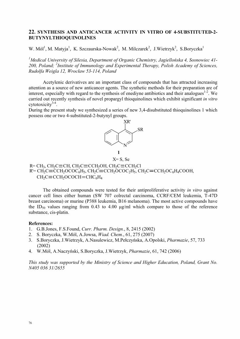
76
22. SYNTHESIS AND ANTICANCER ACTIVITY IN VITRO OF 4-SUBSTITUTED-2-BUTYNYLTHIOQUINOLINES W. Mól1, M. Matyja1, K. Szczaurska-Nowak2, M. Milczarek2, J.Wietrzyk2, S.Boryczka1
1Medical University of Silesia, Department of Organic Chemistry, Jagiellońska 4, Sosnowiec 41-200, Poland; 2Institute of Immunology and Experimental Therapy, Polish Academy of Sciences, Rudolfa Weigla 12, Wrocław 53-114, Poland
Acetylenic derivatives are an important class of compounds that has attracted increasing attention as a source of new anticancer agents. The synthetic methods for their preparation are of interest, especially with regard to the synthesis of enediyne antibiotics and their analogues1-2. We carried out recently synthesis of novel propargyl thioquinolines which exhibit significant in vitro cytotoxicity3-4. During the present study we synthesized a series of new 3,4-disubstituted thioquinolines 1 which possess one or two 4-substituted-2-butynyl groups.
N
SR
XR'
1
R= CH3, CH2C CH, CH2C CCH2OH, CH2C CCH2ClR'= CH2C CCH2OCOC6H5, CH2C CCH2OCOC2H5, CH2C CCH2OC6H4COOH,
X= S, Se
CH2C CCH2OCOCH CHC6H4
The obtained compounds were tested for their antiproliferative activity in vitro against cancer cell lines either human (SW 707 colrectal carcinoma, CCRF/CEM leukemia, T-47D breast carcinoma) or murine (P388 leukemia, B16 melanoma). The most active compounds have the ID50 values ranging from 0.43 to 4.00 μg/ml which compare to those of the reference substance, cis-platin.
References: 1. G.B.Jones, F.S.Found, Curr. Pharm. Design., 8, 2415 (2002) 2. S. Boryczka, W.Mól, A.Jowsa, Wiad. Chem., 61, 275 (2007) 3. S.Boryczka, J.Wietrzyk, A.Nasulewicz, M.Pełczyńska, A.Opolski, Pharmazie, 57, 733 (2002) 4. W.Mól, A.Naczyński, S.Boryczka, J.Wietrzyk, Pharmazie, 61, 742 (2006) This study was supported by the Ministry of Science and Higher Education, Poland, Grant No. N405 036 31/2655
77
23. THE ANALYSIS OF TRANSCRIPTOME OF MEDULLARY THYROID CARCINOMA: SEARCH FOR LINEAGE-SPECIFIC MARKERS Małgorzata Oczko-Wojciechowska1, Jan Włoch2, Jadwiga Żebracka1, Małgorzata Kowalska1, Zbigniew Wygoda1, Aleksandra Czarniecka2, Barbara Jarząb1 1Department of Nuclear Medicine and Endocrine Oncology; 2Oncology Surgery Clinic; Maria Skłodowska-Curie Memorial Cancer Center and Institute of Oncology, Gliwice Branch, 44-101 Gliwice, ul. Wybrzeże Armii Krajowej 15, Poland
Introduction: The analysis of medullary thyroid carcinoma (MTC) transcriptome by gene expression profiling encounters difficulties related to the lack of comparable normal tissue. Para-follicular C cells are embedded in thyroid parenchyma and are not easily accessible to RNA isolation in quantities sufficient for analysis. To omit this difficulty we present other, bioinformatical approach to specify genes characteristic for this histotype. We compare gene expression pattern of MTC to other types of thyroid tumors and normal samples to obtain transcripts specific only for MTC and discard overall cancer-related genes and genes characteristic for normal thyroid contamination. To verify the selected genes we apply quantitative real-time PCR analysis.
Material and methods: Microarray analysis was carried out by HG-U133A arrays (Affymetrix) in 40 MTC samples: 20 tumors and 20 corresponding normal tissue and 70 different thyroid tumor and normal samples. An independent set of 17 MTC with paired normal tissues and 17 PTC samples was used for QPCR validation.
Results. Up to now, 8 genes were analyzed: EEF1A2, GRP, NEFL1, SCG2, SCG3, SST, TFF1, TFF3. All analyzed transcripts were overexpressed in MTC samples, when compared to PTC and normal thyroid. The difference between PTC and MTC was statistically significant (p<0.01) for all analyzed transcripts. The significant differences in expression were also seen in comparison between MTC and corresponding normal thyroid tissue for all the genes, with the exception of TFF3.
Conclusion: We present a gene expression signature of medullary thyroid cancer, selected by the degree of differences in RNA level, not by previous knowledge on its origin, and useful for further analysis of MTC transcriptome. Supported by the Ministry of Science and Higher Education, Poland, Grant No. 2P05A 159 28

78
24. HYPOXIA-RESPONSIVE GENES IN B16(F10) MURINE MELANOMA CELLS IN VIVO – VALIDATION OF MICROARRAYS DATA Magdalena Olbryt1, Aleksandra Rusin1, Katarzyna Lisowska1, Tomasz Cichoń2, Tomasz Tyszkiewicz3, Zdzisław Krawczyk1 1Department of Tumor Biology; 2Department of Molecular Biology; 3Department of Nuclear Medicine and Oncological Endocrynology Maria Skłodowska-Curie Memorial Cancer Center and Institute of Oncology, Gliwice Branch, 44-101 Gliwice, ul. Wybrzeże Armii Krajowej 15, Poland
Rationale: Melanoma is the most aggressive skin cancer largely refractory to existing therapies. Lack of effective therapeutic strategies, as well as proper classification systems result in high mortality rate among melanoma patients. One of the most important features of tumor microenvironment is low oxygen tension within tumor mass. Hypoxia induces aggressive phenotype in tumor cells and contributes to cancer progression. It was observed that hypoxia may influence melanocyte transformation and melanoma progression in vivo. Thus, hypoxia-regulated genes seem to be potential therapeutic targets as well as prognostic/predictive markers in melanoma.
Aim: In our previous study we analyzed in vitro gene expression profile of B16(F10) murine melanoma cells cultured under hypoxic conditions and identified 454 hypoxia-responsive genes (Olbryt et al., 2006). The aim of this study was to investigate whether some genes from selected hypoxia signature are also regulated by hypoxia in experimental murine melanoma tumors.
Experimental design: Hypoxic areas in B16(F10) tumors were identified immunohistochemically with pimonidazole, an exogenous marker of hypoxia. Using laser-microdissection technique the hypoxic (perinecrotic regions) as well as normoxic ones (in the vicinity of blood vessels) were isolated from frozen, 10 μm thick tumor slices. Expression of the 15 selected genes was analyzed by semiquantitative RT-PCR.
Results: We confirmed differential expression of 12 genes between hypoxic and normoxic areas in murine melanoma tumors. Among these genes are some linked before to melanoma biology (Lgals3, Vcl, Mxi1, Nme1) as well as those not previously related to melanoma (e.g. Nppb, Selenbp1, Bnip3, Adm). Three genes that in the microarray study showed slightly changed expression (fold change < 2.5) did not reveal differential in vivo expression pattern in the studied material.
Conclusions: The results obtained herein confirmed differential expression of majority of the analyzed genes between hypoxic and normoxic areas in murine melanoma tumors. Thus, it seems that the molecular response to hypoxia under in vitro conditions may, to some extent, reflect that observed in vivo, at least for the same cell line. To validate genes, the expression of which in microarray study was changed less significantly (fold change < 2.5), more sensitive methods (real-time quantitative RT-PCR) may be required. The study was supported by the Ministry of Science and Higher Education, Poland, Grant No. 1/0-PBZ-MNiI-2/1/2005
79
25. STUDY ON THE MECHANISMS OF SELENIUM BIOTRANSFORMATION: SELENOPHOSPHATE SYNTHETASE Yuliya Preabrazhenskaya, Andriy Moiseenok Institute of Biochemistry and Pharmacology, Grodno, Belarus
Selenophosphate synthetase, a key enzyme of selenium metabolic pathway, provides biosynthesis of a new (the 21st) amino acid selenocysteine from selenium and L-serine making an active donor of Se ions, selenophosphate. Products of selenophosphate synthetase (SPS) genes are supposed to be responsible for incorporation of selenium in major selenium-containing proteins. At least two of these, glutathione peroxidase and Sep15, take part in cancerogenesis through support of redox potential balance in the living cell. Selenophosphate synthetase (SPS) activates selenium and transfers it to the other proteins in the form of selenophosphate. The first step catalyzed by SPS is a limiting step in selenium metabolic pathway. Human SPS2 can complement only a mutant product of SelD gene, a SPS from E. coli. Functionally, they differ in selenium binding as SPS2 cannot bind selenium the same as C17S mutant of SPS from E. coli. However, they may be identical in ATP-binding and ATPase activity. We produced 5 truncated mutants (from C-terminus) of SPS from E. coli using pET plasmid as a template and checked them for ATP-binding ability and Mn-ATP-binding. The binding stoichiometry of ATP to SPS and the role of Mn2+ in binding were determined using HPLC Agilent system and radioactive-labeled ATP. The role of selenium-containing compounds as an active form of selenium capable of increasing ATPase activity of SPS from E coli can be established. The interaction of selenium-containing substances with SPS may be one of the important mechanisms of activating enzymatic pathways of antioxidant defense, a way of active release of the microelement from depot or potential alternative (in the case of xenobiotic selenium derivatives) of accomplishing antioxidant effects.
80
26. GENE EXPRESSION INDUCED BY BRAF ONCOGENE MUTATION IN PAPILLARY THYROID CANCER Dagmara Rusinek, Małgorzata Wiench, Daria Handkiewicz-Junak, Małgorzata Oczko-Wojciechowska, Małgorzata Kowalska, Grzegorz Gala, Aleksandra Pfeiffer, Barbara Jarząb Department of Nuclear Medicine and Endocrine Oncology; Maria Skłodowska-Curie Memorial Cancer Center and Institute of Oncology, Gliwice Branch, 44-101 Gliwice, ul. Wybrzeże Armii Krajowej 15, Poland
Purpose: Giordano et al. (2005) first indicated distinct differences in gene expression profile of papillary thyroid carcinoma (PTC) related to the presence of BRAF mutations and this observation was confirmed in our own analysis. To validate the differences in gene expression related to the presence of BRAF mutation we used real time QPCR.
Methods: We performed a meta-analysis of joined sets of our 39 papillary thyroid carcinoma cases and 51 PTC cases analyzed by Giordano et al., based on Bayesian limma method. The verification of the selected genes was carried out on an independent group of PTCs by QPCR normalized to 3 control genes selected by GeNorm.
Results: So far, 3 genes selected from the microarray analysis were validated, PGF, a placental growth factor related to VEGF; PHLDA1, an evolutionarily conserved proline-histidine rich nuclear protein showing pleckstrin homology-like domain and TM7SF4, a transmembrane molecule preferentially expressed in dendritic cells. PGF and PHLDA1 exhibited a significantly reduced expression in PTCs harbouring BRAF V600E mutation (p<0.01) and TM7SF4 showed higher expression in these PTC cases (p<0.001).
Conclusions. The obtained results indicate different properties of BRAF-induced papillary thyroid cancers in comparison to other cases. The role of PGF is still unknown, the diminished expression of PHLDA1 may contribute to IGF-1 induced apoptosis while TM7SF4 may take part in antigen presentation by dendritic cells, thus, influence the immune response to PTC. Supported by the Ministry of Science and Higher Education, Poland, Grant No. N401 101 32/2138
81
27. BYSTANDER EFFECT IN A MODEL OF COCULTURED IRRADIATED AND NON-IRRADIATED CELLS. A. Szurko1,2, W. Przybyszewski1, K. Szołtysek1, Z. Maniakowski3, M. Wideł1
1Department of Experimental and Clinical Radiobiology, Maria-Skłodowska-Curie Memorial Cancer Center and Institute of Oncology, Gliwice Branch, Wybrzeże Armii Krajowej 15, 44-101 Gliwice; 2Department of Solid State Physics, August Chełkowski Memorial Insitute of Physics, University of Silesia in Katowice, ul. Uniwersytecka 4, 40-007 Katowice; 3Department of Medical Physics, Maria-Skłodowska-Curie Memorial Cancer Center and Institute of Oncology, Gliwice Branch, Wybrzeże Armii Krajowej 15, 44-101 Gliwice
There is an increasing body of data corroborating the observation that biological response to ionizing radiation affects, besides directly irradiated cells, also daughter cells (genetic instability) and adjacent non-irradiated cells (bystander effect). Cells present in solid tumors or remaining in close contact under cell culture conditions communicate in various ways. One of the most important is communication via gap-junctions, which makes possible passive transport of ions and small-molecular-weight moieties between cells. Another mechanism involves release of specific mediators into the surrounding milieu by the irradiated cells, which results in inhibited proliferation, increased number of mutations, as well as number of apoptotic cells and micronuclei among non-irradiated cells.
This report summarizes the results of study on the effect exerted by X-ray-irradiated (2 or 4 Gy, 6 MV, Clinac 600) murine Lewis lung carcinoma cells (LLC) upon non-irradiated cells of the same line. Especially attention-worthy are the results of experiments involving co-cultivated cells of both types, conducted in co-culture vessels with a filter enabling exchange of signal substances among cells but preventing mixing of the latter.
The ratio of apoptotic cells and cells with cytogenetic damage (micronuclei) was determined microscopically. Alterations of cell-cycle were estimated using flow cytometry. Expression of NF-κB transcription factor was assessed by Western-blot technique.
A significantly increased ratio of micronuclei was observed not only among directly irradiated cells but also among cells that were co-cultured. Also, the number of cells eliminated via apoptosis was significantly higher in both groups. No major cell cycle-affecting changes were noted following the lower irradiation dose. The observed cyclic fluctuations of NF-κB expression levels require further confirmation.

82
28. CHLORIN AS POTENTIAL PHOTOSENSITIZER FOR PHOTODYNAMIC THERAPY (PDT) Agnieszka Szurko* 1,2, Marzena Rams1, Aleksander Sochanik2, Alexeis Mikhailov2, Franz-Peter Montforts# 3, Agnieszka Kozielec3, Maria Widel2, Alicja Ratuszna1
1A. Chelkowski Institute of Physics, University of Silesia, Katowice, Poland, (*email: [email protected]); 2Maria Sklodowska-Curie Memorial Cancer Center and Institute of Oncology, Gliwice, Poland; 3Institute of Organic Chemistry, University of Bremen, Bremen, Germany, (#email: [email protected])
The crucial role in photodynamic tumor therapy is played by photosensitizers, which are dyes able to accumulate selectively in rapidly growing tumor cells. After activation by exposure to light of a specific wavelength they lead to generation of reactive singlet oxygen and radical species, which trigger a sequence of photochemical and photobiological processes that damage intracellular organelles and lead to cell death. Finding a suitable photosensitizers is important for improving the efficiency of PDT. One of the most promising photosensitizer candidates are chlorins. Chlorins have one pyrrolic double bond reduced, which is concomitant with bathochromic shift of the Q-bands with higher extinction coefficients than those in the corresponding parent porphyrins. Chlorins have been reported to have high singlet oxygen efficiency coupled with 10-fold stronger absorption than HpD or porphyrins in the therapeutic window; hence they are expected to be good photosensitizers.
Because the mode of transfer strongly influences subsequent localization of photosensitizer in cells and, consequently, determines photodynamic efficiency, we compared cell killing efficiency of chlorin delivered by liposome vehicles or dissolved in DMSO. The study was performed using Lewis lung carcinoma cells (LLC). Cellular distribution of chlorin was studied using confocal microscopy. Dark cytotoxicity and photodynamic efficiency of the explored chlorin were studied by MTS assay. We also studied the possible damaging effect of chlorin on DNA and on cytoskeleton using immunocytochemical staining of actin microfilaments followed by fluorescence microscopy.
Our preliminary results indicate that conjugation of chlorin with liposomes is an efficient means of transferring the sensitizer into the studied cells, leading to highly efficient photosensitization, whereas non-carrier delivery (DMSO) is rather useless in such experiments. Following PDT with the chlorin photosensitizer we also observed that cytoskeleton microfilaments become shorter and DNA undergoes intense defragmentation processes.
83
29. THE EFFECT OF COMBINED TREATMENT ON HUMAN PROMYELOCYTIC LEUKEMIA CELL LINE WITH NOVEL ANALOGS AND COMPLEXES OF GENISTEIN AND 1,24 (OH)2D3 (PRI-2191) Marta Świtalska1, Justyna Zielska1, Grzegorz Grynkiewicz2, Andrzej Kutner2, Joanna Wietrzyk1 1Ludwik Hirszfeld Institute of Immunology and Experimental Therapy, Department of Experimental Oncology, Rudolfa Weigla 12, 53-114 Wroclaw, Poland; 2Pharmaceutical Research Institute, L. Rydygiera 8, 01-793 Warsaw, Poland
Isoflavonoids play regulatory role in the expression of cytochrome P450 enzymes, and also up-regulate vitamin D3 receptor (VDR) on cancer cells. As a result, increased sensitivity of these cells is noted to 1,25-dihydroxyvitamin D3, a hormonally active form of vitamin D3. Isoflavonoids are also able to raise the serum level of active form of vitamin D3, due to their inhibitory activity on the CYP24, the enzyme involved in the degradation of 1,25-dihydroxyvitamin D3 and its precursor 25-OH-D3 to inactive compounds. Another enzyme, CYP27B1, involved in the synthesis of 1,25-dihydroxyvitamin D3 is stimulated by isoflavonoids. This may result in a similar increase effect of 1,25-dihydroxyvitamin D3 serum level. Therefore, combined treatment with isoflavonoids and 1,25-dihydroxyvitamin D3 might be effective in both prevention and in anticancer treatment.
In order to evaluate the effect of combined application of genistein analogs (IFG-027, IFG-043) and complexes (with schizophylan and xyloglucan) and new vitamin D analog PRI-2191: (24R)-1,24-dihydroxyvitamin D3), against the cells of human promyelocytic leukemia HL-60, antiproliferative activity and the effects on cell cycle were determined. The synergistic antiproliferative effect was observed for all analogs and complexes used after treatment of HL-60 cell line with PRI-2191. HL-60 cells treated with analogs of genistein and PRI-2191 cumulated in G0/G1 stage. The percentage of cells in G2/M and S phase decreased after combined treatment. This effect was significantly stronger than after treatment with PRI-2191 alone.
This study was supported by the Foundation for Development of Pharmaceutical Sciences, Poland, Grant No. 9/FB/2004

84
30. COTRASIF: CONSERVATION-AIDED TRANSCRIPTION FACTOR BINDING SITE FINDER Bogdan Tokovenko & Maria Obolenskaya Institute of Molecular Biology and Genetics, Kyiv, Ukraine; [email protected]
Summary: A new tool has been developed for identifying with increased specificity the putative transcription factor binding sites (TFBS) in eukaryotic gene promoters.
Rationale: Promoter analysis and TFBS identification are essential steps for the identification of gene regulatory networks. Low specificity of the TFBS prediction in eukaryotic gene promoters is a challenging task for modern bioinformatics. Based on our previous research, we observed better specificity of the TFBS search when comparing the promoters of gene orthologs of the evolutionarily close species (e.g. rat and mouse) for the presence of the target TFBS. If the putative TFBS is present in both promoters (with an optional constrain on the similar distance from the transcription start site), then it has higher probability of being biologically meaningful.
Results: We developed a web-accessible tool (conservation-aided transcription factor binding site finder, COTRASIF) for the conservation-aided TFBS search. It uses Ensembl genome databases, and is currently available for three organisms: human, mouse and rat. Promoters are defined as 800bp upstream from transcription start site, plus the 5’ UTR. For the initial TFBS search, classical position-weight matrix approach is used. Initial search results are further analyzed using the gene orthology information available in Ensembl for the selected pair of organisms. Results are presented as a list of genes with the positions of the putative found TFBS in the promoters of both organisms, selected for analysis. Further development plans include the addition of new organisms, and integration of the Gene Ontology functional gene analysis into the results page. Availability: http://biomed.org.ua/COTRASIF/ Supplementary information: http://biomed.org.ua/COTRASIF/help.html

85
31. IN VITRO STUDIES OF INDIVIDUAL RADIOSENSITIVITY RELATIONSHIPS IN CERVICAL CANCER PATIENTS BEFORE, DURING AND AFTER RADIOTHERAPY, A.Węgierek3, A. Lankoff1, H. Lisowska1, T. Kuszewski3, S.Góźdź3, A. Wójcik1,2
1Department of Radiobiology and Immunology, Institute of Biology, Saint Cross Academy, Kielce, Poland; 2Radiobiology and Health Protection Department, Institute of Chemistry and Nuclear Technology, Warsaw, Poland; 3Holy Cross Cancer Center, Kielce, Poland There has been considerable evidence that, even with analogous treatment, individuals differ widely in normal tissue response to radiation injury. It was suggested that this difference might result from a variation in the intrinsic cellular radiosensitivity. However, the mechanisms of such variability are not clear. The aim of the presented investigation was to examine: a) the correlation between the frequencies of spontaneous and radiation-induced micronuclei (MN) in vitro before radiotherapy b) the influence of radiotherapy on the frequencies of MN in vivo c) the correlation between the frequencies of radiation-induced MN in vitro before radiotherapy and the frequencies of radiation-induced MN in vitro during and after radiotherapy.
The study included 10 patients with cervical cancer. From each donor peripheral blood was collected before radiotherapy (0), 3 weeks after the first radiation fraction (3W) and immediately after completion of radiotherapy (TE). In addition, blood was re-irradiated in vitro with 2Gy. Differences in the individual radiosensitivity of peripheral blood lymphocytes were determined with the micronucleus assay. Lymphocyte cultures were set up by adding 0.5ml of blood to 4.5ml of RPMI 1640 medium supplemented with calf serum, phytohemaglutinin and antibiotics. Cytochalasin B was added 44 hours after the incubation. After subsequent 28 hours the cells were harvested and fixed according to the protocol of Fenech (2000).
Individual radiosensitivity of lymphocytes before radiotherapy. Part of the blood collected before the beginning radiotherapy was used to assess the intrinsic radiosensitivity of the patients. Our results show a wide variability in the frequencies of both spontaneous and radiation-induced micronuclei in vitro. The yield of spontaneous MN ranged from 9 to 52 MN/1000 BNC. The yield of radiation-induced MN in vitro ranged from 56 to 255 MN/ 1000BNC. Statistical analysis revealed that there was no significant correlation between MN frequencies in the compared groups (p = 0.159).
Influence of radiotherapy on the MN frequencies in vivo. The mid-treatment samples (3W) from the cervical cancer patients show a marked elevation in the MN frequency. However, a wide inter-individual variability is seen. The yield of radiotherapy-induced MN 3 weeks after the first radiation fraction ranged from 40 to 226 MN/1000 BNC. The samples collected from patients immediately upon completion of radiotherapy (TE) show an enhancement in the MN frequency, which is significantly higher than in spontaneous (0) as well as mid-treatment samples (3W), with the exception of three patients, where the MN frequency was lower as compared to the mid-treatment samples (3W). Statistical analysis revealed that there is no marked correlation between the frequency of spontaneous MN and the frequency of radiotherapy-induced MN neither 3 weeks after the first radiation fraction (p=0.193) nor immediately after completion of radiotherapy (p=0.427). Our results show a wide inter-individual variability in the frequency of radiation-induced MN in vitro during and after radiotherapy. The yield of radiation-induced MN in vitro in lymphocytes collected from patients 3 weeks after the first radiation fraction ranged from 123 to 448 MN/1000 BNC. The yield of radiation-induced MN in vitro in lymphocytes collected from patients upon completion of radiotherapy ranged from 64 to 452 MN/1000 BNC. Statistical analysis revealed that there is no significant correlation neither between the frequency of radiation-induced MN in vitro before radiotherapy and the frequencies of radiation-induced MN in vitro during radiotherapy (3W) (p=0.337) nor between the frequency of radiation induced MN in vitro before radiotherapy and the frequencies of radiation-induced MN in vitro upon completion of radiotherapy (p=0.328).

86
32. CHARACTERIZATION OF GENOMIC INSTABILITY IN EBV-INFECTED B LYMPHOCYTES Emilia Wiechec1, 2, Sandrine Lacoste2, Marie Henriksson3, George Klein3, Sabine Mai2 1Institute of Human Genetics, University of Aarhus, 8000 Aarhus C, Denmark; 2Manitoba Institute of Cell Biology, University of Manitoba, Winnipeg, Canada; 3Microbiology and Tumor Biology Centre, Karolinska Institute, Stockholm, Sweden
Background: Genomic instability is one of the earliest events found in most cancers. Epstein-Barr virus (EBV) infected human B cells display chromosomal aberrations (Kataoka H, 1997; Okubo M, 2001; Kamranvar SA, 2007). These EBV-associated chromosomal aberrations can be linked to telomere dysfunction and chromosomal missegregation.
Purpose: The overall aim of this study was to characterize the exact post-EBV infection period leading to genomic instability in B-lymphocytes and to determine whether such changes promote tumour development in mice.
Material and methods: The material for this study included human B-lymphocytes prior to and after EBV infection. Freshly, virally immortalized lymphoblastoid cell lines (LCLs) were followed over a six-month period post-infection, and cells were collected at specific time points. Telomeres were analyzed by fluorescent in situ hybridization (FISH) on both interphase nuclei (3D analysis) and metaphase plates (2D analysis) using the Telomere PNA FISH kit (DAKO). We then characterized the chromosomal abnormalities using spectral karyotyping (SKY).
Results: Our preliminary results suggest that EBV infected human B cells display telomere-driven genomic instability. The LCL samples display random chromosomal instability including aneuploidy, chromosomal breaks and fusions, translocations and sister chromatid fusions.
Work in progress: We will continue the analysis of primary human (non-EBV-infected) B cells to confirm or exclude tissue-culture-dependent formation of genomic instability that would have nothing to do with EBV-infection. Then, the EBV infected B-lymphocytes exhibiting a highest rate of genomic changes will be injected into mice to determine whether the observed instability is sufficient to promote tumour progression. Acknowledgement: This work was supported by grant from CIHR Strategic Training Program. The authors would like to thank Katalin Benedek and Mia Lowbeer for the preparation of cells.

87
33. COMBINATION OF NEW PRIMER DESIGN AND HIGH RESOLUTION MELTING FOR HIGH THROUGHPUT ANALYSIS OF METHYLATION IN CLINICAL SAMPLES Tomasz K Wojdacz1,2, Alexander Dobrovic2, Lise Lotte Hansen1 1Institute of Human Genetics, University of Aarhus, Denmark; 2Department of Molecular Pathology, Peter MacCallum Cancer Centre, Melbourne, Australia
There is an urgent need for straightforward and reliable methods to examine the extent of methylation changes in tumours. Current methods for single gene methylation determination have various limitations and pitfalls, and contradictory results can be obtained using different protocols [1]. Tumour DNA is a mixture of tumour and normal DNA with a wide range of tumour purity. The sensitivity for detection of methylated sequences should reflect this and should be sensitive to at least 5-10%. We have combined High Resolution Melting and novel primer design for amplification of bisulfite modified DNA regardless of its methylation status in studies of methylation markers. The standard guidelines for primer design to amplify locus of interest for post PCR methylation analyses (MIP, methylation independent primers) advise to avoid CpG dinucleotide in primer sequences or to replace Cs within CpGs by mismatched bases. We have shown that some CpGs in the primer sequence may be necessary; otherwise PCR bias towards unmethylated template may lead to underestimation of the degree of methylation. Moreover, by manipulating the annealing temperature of PCR amplification, we were able to control the efficiency of binding of our MIP primers to the methylated template and therefore reverse PCR bias towards the methylated allele [2]. The combination of the above primer design and a High Resolution Melting platform (HRM) allowed us unambiguously to detect methylated fractions of DNA in the samples containing as little as 0.1% methylated DNA [3]. Furthermore, Methylation Sensitive High Resolution Melting (MS-HRM) can be designed to estimate the methylation levels of screened material when HRM profiles of PCR product of unknown samples are compared with HRM profiles of PCR product derived from the standards with known methylated to unmethylated template ratio.
MS-HRM analysis can be performed in-tube, which allows for very rapid screening and avoidance of PCR product contamination issues in laboratories. In summary, in-tube methylation analysis with HRM methodology provides a fast and high-throughput tool that has a potential to be introduced into clinical practice. References: 1. Aggerholm A, Hokland P: DAP-kinase CpG island methylation in acute myeloid leukemia: methodology versus biology? Blood 2000, 95:2997-2999. 2. Wojdacz TK, Hansen LL: Reversal of PCR bias for improved sensitivity of the DNA methylation melting curve assay. Biotechniques 2006, 41(3):274, 276, 278. 3. Wojdacz TK, Dobrovic A: Methylation-sensitive high resolution melting (MS-HRM): a new approach for sensitive and high-throughput assessment of methylation. Nucleic acids research 2007, 35(6):e41.

88
34. MOLECULAR GENETIC DIAGNOSTICS OF THROMBOPHILIA IN CANCER PATIENTS Iosif Zalutsky, Alexander Mashevsky, Raisa Smolyakova, Elena Mokhon, Dmitri Kovalenko N.N. Alexandrov Research Institute of Oncology and Medical Radiology, Minsk, Belarus
The incidence of pulmonary embolism (PE) in cancer patients reaches 25–40%, the highest rates occurring in patients with lung cancer, large intestine cancer and female genital cancer. According to pathomorphology findings, venous thrombosis (VT) is detected in 40–60% of advanced malignant neoplasm cases, being asymptomatic in 70–80% of the patients and with no life-time diagnosis in 50% of them.
In the past decades, major accomplishments were made in decoding the mechanisms of VT development, which is associated with the introduction of new genetic and immunological methods of investigation and with discovery of hereditary forms of thrombophilia and its molecular markers. Diagnostically significant genetic factors of VT u PE risks are 1691G/A mutations in factor V gene (FV, Leiden) and 20210G/A mutations in factor II gene (FII), in methylenetetrahydrofolate reductase gene (MTHFR C-677-T).
The objective of the study is molecular genetic detection of mutations in the hemostasis system genes for diagnosis, prevention and treatment of thromboembolic morbidity in cancer patients.
We used in our studies findings of clinical, instrumental and molecular genetic examinations carried out in stage I–IV cancer patients treated at SI N.N. Alexandrov RIOMR.
All the patients underwent histological diagnosis (lung cancer, gastric cancer, esophageal cancer, colon cancer, cervical and corpus uteri cancer, bone and soft tissue sarcoma).
Genomic DNA was extracted from whole blood of cancer patients using Genomic DNA Purification Kit. Molecular genetic examinations of factor V (Leiden) genes, factor II and MTHFR genes were conducted with allele-specific polymerase chain reaction technique using reagent kits of PRONTO, Israel.
Analysis of the results of examinations performed in 346 cancer patients revealed mutations in the hemostasis system genes in 235 (67.9%) of them. Carrying mutation in factor V gene in the heterozygous state was diagnosed in 49 (14.2%) patients, in the homozygous state – in 21 (6.1%).
Prothrombin gene mutation resulting in thrombophilia is a genetic defect ranking second in clinical significance. In the course of the examinations the mutation in the heterozygous state was diagnosed in 53 (22.7%) cancer patients, in the homozygous state – in 22 (9.4%). One of the most frequent causes of developing mild and moderate hyperhomocysteinemia is hereditary deficit of 5, 10- MTHFR-folate-dependent enzyme catalyzing the process of remethylation.
Pleomorphism in MTHFR gene was detected in 174 cancer patients, with dominating heterozygous genotype in 132 (38.1%) patients and the homozygous type in 42 (12.1%) cases.
The comprehensive molecular genetic analysis of pleomorphisms associated with hemostasis dysfunction may be used for prognostication of VT recurrence and evaluation of PE development risk. G20210A mutation of prothrombin gene was found in combination with FV Leiden mutation in 8 cancer patients, combination of mutations in FV Leiden and MTHFR genes was diagnosed in 25 cases, prothrombin gene mutation associated with MTHFR mutations was noted in 28 patients. The development of severe pulmonary embolism in the postoperative period with unfavourable outcome in 11 cancer patients was associated with concurrent detection of combined mutation in the three hemostasis system genes analysed (factor V Leiden, factor II and MTHFR).
Thus, genetic defects of FV Leiden gene, G20210A of prothrombin and MTHFR genes were found in 67.9% of patients with malignant neoplasms. The high incidence of detecting genetic disorders in the hemostasis system by means of allele-specific polymerase chain reaction makes it possible to define the degree of PE development risk in cancer patients and to administer pathogenetically-oriented therapy.

89
Addendum

90

91
35. ANALYSIS OF CHANGES IN GENE EXPRESSION PROFILES INDUCED BY IONIZING RADIATION IN ME45 AND K562 CELLS 2K. Pawełek, 1R. Herok, 2Z. Grzywna 1,2J. Rzeszowska-Wolny 1Department of Experimental and Clinical Radiobiology, M. Sklodowska-Curie Memorial Cancer Centre and Institute of Oncology, Gliwice, Poland; 2Silesian University of Technology, Gliwice, Poland The levels of transcripts from all actually active genes (transcriptome, transcription profile) characterize the cell type and its physiological state. Microarray techniques allow determining transcription levels of thousands of genes in one experiment. In our studies transcriptome changes in cells exposed to ionizing radiation were studied by oligonucleotide microarray (Affymetrix) method. Transcription profiles in two human cancer cell lines, Me45 and K562, were determined in control cells and at 4 different time points (20 min, 12h, 24h, 36h) after irradiation with 4Gy. Affymetrix microarrays HU-133 allowed us assessing transcription levels of 22283 genes for each time point. A new approach based on calculating average speed of change of gene expression levels (Vex) was applied to the analysis and Vex distributions were approximated by normal distributions. We observed that in both cell lines the change of transcription level was the highest at first moments after irradiation (SD=225.43 for Me45 cell line and SD=281.9312 for K562 cell line for the 20-min time point). At the next time point (12 h) changes were much slower but they showed a tendency to increase 24 and 36h later. The dynamics of change were different in both cell lines. Analysis of processes that showed the highest change after irradiation revealed that genes coding for proteins of proteasome, oxidative phosforylation and ribosomes were characterized by the highest Vex. The expression levels of some genes belonging to these pathways were studied by Q-RT PCR method.

92
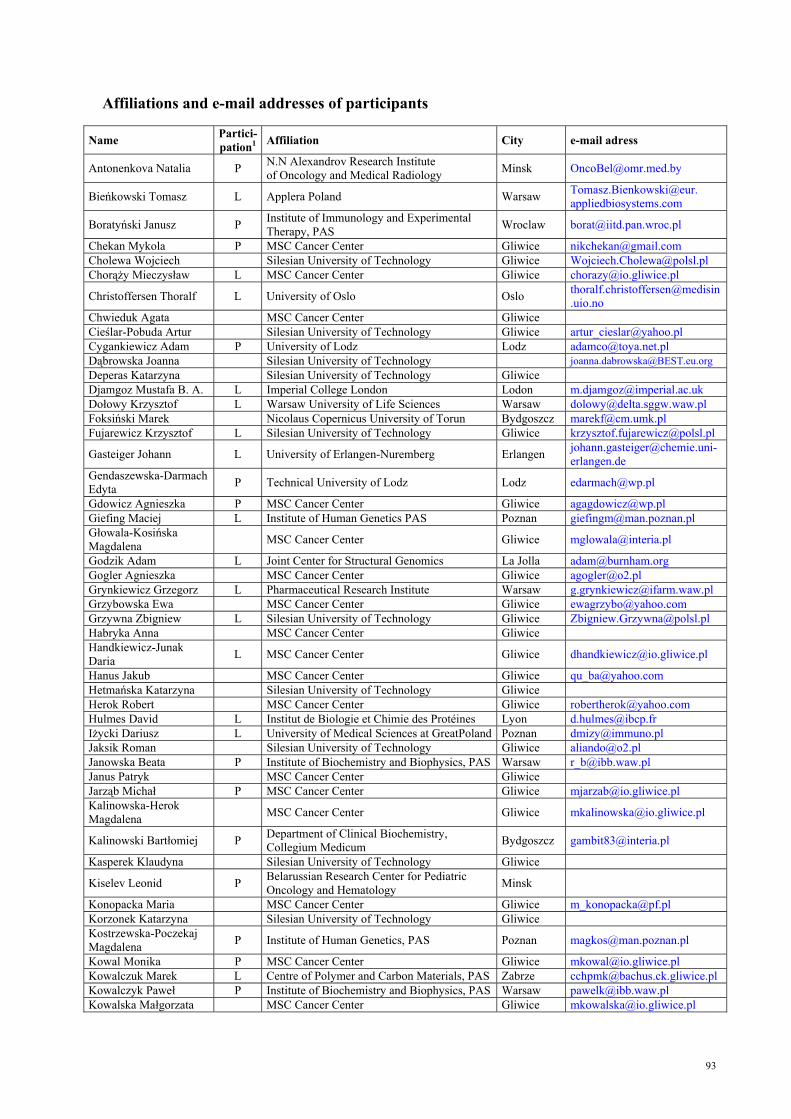
93
Affiliations and e-mail addresses of participants
Name Partici- pation1 Affiliation City e-mail adress
Antonenkova Natalia P N.N Alexandrov Research Institute of Oncology and Medical Radiology Minsk [email protected]
Bieńkowski Tomasz L Applera Poland Warsaw Tomasz.Bienkowski@eur. appliedbiosystems.com
Boratyński Janusz P Institute of Immunology and Experimental Therapy, PAS Wroclaw [email protected]
Chekan Mykola P MSC Cancer Center Gliwice [email protected] Cholewa Wojciech Silesian University of Technology Gliwice [email protected] Chorąży Mieczysław L MSC Cancer Center Gliwice [email protected]
Christoffersen Thoralf L University of Oslo Oslo [email protected]
Chwieduk Agata MSC Cancer Center Gliwice Cieślar-Pobuda Artur Silesian University of Technology Gliwice [email protected] Cygankiewicz Adam P University of Lodz Lodz [email protected] Dąbrowska Joanna Silesian University of Technology [email protected] Deperas Katarzyna Silesian University of Technology Gliwice Djamgoz Mustafa B. A. L Imperial College London Lodon [email protected] Dołowy Krzysztof L Warsaw University of Life Sciences Warsaw [email protected] Foksiński Marek Nicolaus Copernicus University of Torun Bydgoszcz [email protected] Fujarewicz Krzysztof L Silesian University of Technology Gliwice [email protected]
Gasteiger Johann L University of Erlangen-Nuremberg Erlangen [email protected]
Gendaszewska-Darmach Edyta P Technical University of Lodz Lodz [email protected]
Gdowicz Agnieszka P MSC Cancer Center Gliwice [email protected] Giefing Maciej L Institute of Human Genetics PAS Poznan [email protected] Głowala-Kosińska Magdalena MSC Cancer Center Gliwice [email protected]
Godzik Adam L Joint Center for Structural Genomics La Jolla [email protected] Gogler Agnieszka MSC Cancer Center Gliwice [email protected] Grynkiewicz Grzegorz L Pharmaceutical Research Institute Warsaw [email protected] Grzybowska Ewa MSC Cancer Center Gliwice [email protected] Grzywna Zbigniew L Silesian University of Technology Gliwice [email protected] Habryka Anna MSC Cancer Center Gliwice Handkiewicz-Junak Daria L MSC Cancer Center Gliwice [email protected]
Hanus Jakub MSC Cancer Center Gliwice [email protected] Hetmańska Katarzyna Silesian University of Technology Gliwice Herok Robert MSC Cancer Center Gliwice [email protected] Hulmes David L Institut de Biologie et Chimie des Protéines Lyon [email protected] Iżycki Dariusz L University of Medical Sciences at GreatPoland Poznan [email protected] Jaksik Roman Silesian University of Technology Gliwice [email protected] Janowska Beata P Institute of Biochemistry and Biophysics, PAS Warsaw [email protected] Janus Patryk MSC Cancer Center Gliwice Jarząb Michał P MSC Cancer Center Gliwice [email protected] Kalinowska-Herok Magdalena MSC Cancer Center Gliwice [email protected]
Kalinowski Bartłomiej P Department of Clinical Biochemistry, Collegium Medicum Bydgoszcz [email protected]
Kasperek Klaudyna Silesian University of Technology Gliwice
Kiselev Leonid P Belarussian Research Center for Pediatric Oncology and Hematology Minsk
Konopacka Maria MSC Cancer Center Gliwice [email protected] Korzonek Katarzyna Silesian University of Technology Gliwice Kostrzewska-Poczekaj Magdalena P Institute of Human Genetics, PAS Poznan [email protected]
Kowal Monika P MSC Cancer Center Gliwice [email protected] Kowalczuk Marek L Centre of Polymer and Carbon Materials, PAS Zabrze [email protected] Kowalczyk Paweł P Institute of Biochemistry and Biophysics, PAS Warsaw [email protected] Kowalska Małgorzata MSC Cancer Center Gliwice [email protected]
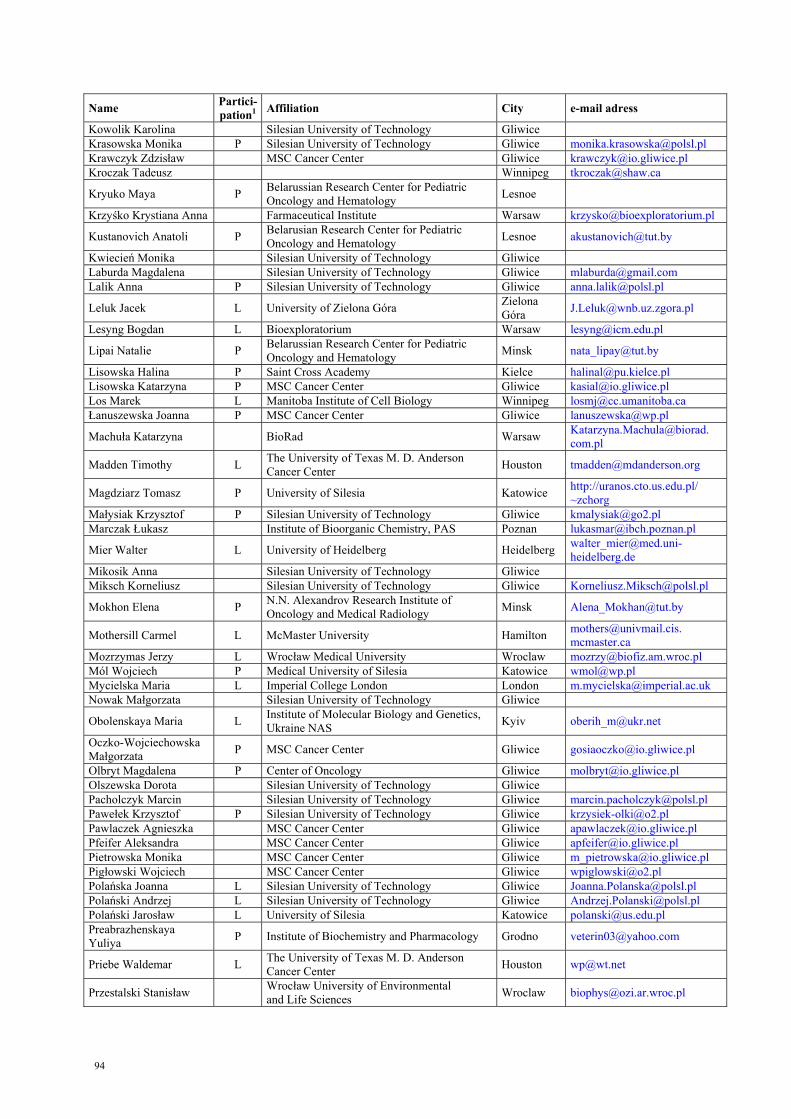
94
Name Partici- pation1 Affiliation City e-mail adress
Kowolik Karolina Silesian University of Technology Gliwice Krasowska Monika P Silesian University of Technology Gliwice [email protected] Krawczyk Zdzisław MSC Cancer Center Gliwice [email protected] Kroczak Tadeusz Winnipeg [email protected]
Kryuko Maya P Belarussian Research Center for Pediatric Oncology and Hematology Lesnoe
Krzyśko Krystiana Anna Farmaceutical Institute Warsaw [email protected]
Kustanovich Anatoli P Belarusian Research Center for Pediatric Oncology and Hematology Lesnoe [email protected]
Kwiecień Monika Silesian University of Technology Gliwice Laburda Magdalena Silesian University of Technology Gliwice [email protected] Lalik Anna P Silesian University of Technology Gliwice [email protected]
Leluk Jacek L University of Zielona Góra Zielona Góra [email protected]
Lesyng Bogdan L Bioexploratorium Warsaw [email protected]
Lipai Natalie P Belarussian Research Center for Pediatric Oncology and Hematology Minsk [email protected]
Lisowska Halina P Saint Cross Academy Kielce [email protected] Lisowska Katarzyna P MSC Cancer Center Gliwice [email protected] Los Marek L Manitoba Institute of Cell Biology Winnipeg [email protected] Łanuszewska Joanna P MSC Cancer Center Gliwice [email protected]
Machuła Katarzyna BioRad Warsaw Katarzyna.Machula@biorad. com.pl
Madden Timothy L The University of Texas M. D. Anderson Cancer Center Houston [email protected]
Magdziarz Tomasz P University of Silesia Katowice http://uranos.cto.us.edu.pl/ ~zchorg
Małysiak Krzysztof P Silesian University of Technology Gliwice [email protected] Marczak Łukasz Institute of Bioorganic Chemistry, PAS Poznan [email protected]
Mier Walter L University of Heidelberg Heidelberg [email protected]
Mikosik Anna Silesian University of Technology Gliwice Miksch Korneliusz Silesian University of Technology Gliwice [email protected]
Mokhon Elena P N.N. Alexandrov Research Institute of Oncology and Medical Radiology Minsk [email protected]
Mothersill Carmel L McMaster University Hamilton [email protected]. mcmaster.ca
Mozrzymas Jerzy L Wrocław Medical University Wroclaw [email protected] Mól Wojciech P Medical University of Silesia Katowice [email protected] Mycielska Maria L Imperial College London London [email protected] Nowak Małgorzata Silesian University of Technology Gliwice
Obolenskaya Maria L Institute of Molecular Biology and Genetics, Ukraine NAS Kyiv [email protected]
Oczko-Wojciechowska Małgorzata P MSC Cancer Center Gliwice [email protected]
Olbryt Magdalena P Center of Oncology Gliwice [email protected] Olszewska Dorota Silesian University of Technology Gliwice Pacholczyk Marcin Silesian University of Technology Gliwice [email protected] Pawełek Krzysztof P Silesian University of Technology Gliwice [email protected] Pawlaczek Agnieszka MSC Cancer Center Gliwice [email protected] Pfeifer Aleksandra MSC Cancer Center Gliwice [email protected] Pietrowska Monika MSC Cancer Center Gliwice [email protected] Pigłowski Wojciech MSC Cancer Center Gliwice [email protected] Polańska Joanna L Silesian University of Technology Gliwice [email protected] Polański Andrzej L Silesian University of Technology Gliwice [email protected] Polański Jarosław L University of Silesia Katowice [email protected] Preabrazhenskaya Yuliya P Institute of Biochemistry and Pharmacology Grodno [email protected]
Priebe Waldemar L The University of Texas M. D. Anderson Cancer Center Houston [email protected]
Przestalski Stanisław Wrocław University of Environmental and Life Sciences Wroclaw [email protected]

95
Name Partici- pation1 Affiliation City e-mail adress
Przybyszewski Waldemar P MSC Cancer Center Gliwice [email protected]
Puszyński Krzysztof L Silesian University of Technology Gliwice [email protected] Radlak Krystian Silesian University of Technology Gliwice Rams Marzena University of Silesia Katowice Ratuszna Alicja L University of Silesia Katowice [email protected] Rogoliński Jacek P MSC Cancer Center Gliwice [email protected] Rozwadowski Marek S. Linegal Chemicals Warsaw [email protected] Rusin Aleksandra MSC Cancer Center Gliwice [email protected] Rusinek Dagmara P MSC Cancer Center Gliwice [email protected] Rzeszowska –Wolny Joanna MSC Cancer Center Gliwice [email protected]
Sadoś Benita P Institute of Biochemistry and Biophysics, PAS Warsaw [email protected] Sandnes Dagny L University of Oslo Oslo [email protected] Seymour Colin L McMaster University Hamilton [email protected] Sieroń Aleksander L. L Medical University of Silesia Katowice [email protected]
Skonieczna Magdalena P Silesian University of Technology Gliwice [email protected]
Smoczyński Rafał Selvita sp.z o. o. Cracow [email protected]
Smolyakova Raisa L N.N. Alexandrov Research Institute of Oncology and Medical Radiology Minsk [email protected]
Sochanik Aleksander P MSC Cancer Center Gliwice [email protected] Student Sebastian P Silesian University of Technology Gliwice [email protected] Szala Stanisław MSC Cancer Center Gliwice [email protected] Szeja Wiesław L Silesian University of Technology Gliwice [email protected] Szelejewski Wiesław Pharmaceutical Institute Warsaw [email protected] Szołtysek Katarzyna P MSC Cancer Center Gliwice [email protected] Szurko Agnieszka P University of Silesia, MSC Cancer Center Katowice [email protected] Szydło Anna L Medical University of Silesia Katowice [email protected] Śmieja Jarosław Silesian University of Technology Gliwice [email protected] Świerniak Andrzej Silesian University of Technology Gliwice [email protected] Świerniak Michał MSC Cancer Center Gliwice [email protected]
Świtalska Marta P Institute of Immunology and Experimental Therapy, PAS Wroclaw [email protected]
Tokovenko Bohdan P Institute of Molecular Biology and Genetics, Ukraine NAS Kyiv [email protected]
Tomanek Marta Silesian University of Technology Gliwice Toporek Michał Silesian University of Technology Gliwice Tudek Barbara L Institute of Biochemistry and Biophysics, PAS Warsaw [email protected] Turoń Michał Silesian University of Technology Gliwice Tyszkiewicz Tomasz MSC Cancer Center Gliwice [email protected] Urbanek Ksymena L Medical University of Silesia Katowice [email protected] Urbaniak Monika Silesian University of Technology Gliwice Walaszczyk Anna MSC Cancer Center Gliwice [email protected] Walczak Beata L Institute of Chemistry, University of Silesia Katowice [email protected] Węgierek Aneta P Holy Cross Cancer Center Kielce [email protected] White Stephen L University of California Irvine [email protected] Wideł Maria P MSC Cancer Center Gliwice [email protected] Widlak Piotr L MSC Cancer Center Gliwice [email protected] Widłak Wiesława MSC Cancer Center Gliwice [email protected] Widziewicz Kamila Silesian University of Technology Gliwice Wiecheć Emilia P University of Aarhus Aarhus [email protected] Winczura Alicja Institute of Biochemistry and Biophysics, PAS Warsaw [email protected] Wilk Ryszard Silesian University of Technology Gliwice [email protected] Wojdacz Tomasz P University of Aarhus Aarhus [email protected] Wojtkiewicz Katarzyna Silesian University of Technology Gliwice Wolańska Katarzyna MSC Cancer Center Gliwice [email protected]
Zarakowska Ewelina P Department of Clinical Biochemistry, Collegium Medicum Bydgoszcz [email protected]
Żebracka Jadwiga MSC Cancer Center Gliwice [email protected]
1L-lecture; P-poster;

96

97
Index of Authors Aasrum M. 14 Ande S. R. 33 Antonenkova N. 55 Bieg T. 44 Bielecki S. 59 Bieleń A. 67 Bieńkowski T. 13 Boratynski J. 56 Boryczka S. 76 Bryzek A. 48 Budko M. 52 Budzynska R. 56 Chekan M. 57 Chmielik E. 66 Christoffersen T. 14, 39 Cichoń T. 78 Cieśla J. M. 67 Cygankiewicz A. 58 Czarniecka A. 66, 77 Czekaj P. 48 Dajani O. 14, 39 Djamgoz M. B. A. 15, 32, 68, 75 Dobrovic A. 87 Dołowy K. 16 Dubrovsky A. 52 Dudaladava V. 72 Florek A. 71 Fujarewicz K. 17 Gackowski D. 61 Gala G. 80 Gasteiger J. 18 Gendaszewska-Darmach E. 59 Ghavami S. 33 Giefing M. 19, 65 Godzik A. 20 Góźdź S. 71, 85 Grenman R. 19, 65 Grynkiewicz G. 21, 44, 83 Grzywna Z. J. 22, 68, 75, 89 Gubała E. 66 Guren T. 14 Handkiewicz-Junak D. 23, 80 Hansen L. L. 87 Henriksson M. 86 Herok R. 89 Holst B. 58 Hulmes D. J.S. 24 Iżycki D. 25 Jackiewicz M. 48 Jagiello M. 56 Janowska B. 60, 67 Jarmuż M. 19, 65 Jarzab B. 57, 66, 77, 80
Jarzab M. 72, 73 Jaworek J. 67 Kalinowski B. 61 Kanska U. 56 Kasprzycka A. 44 Kawalec M. 45 Kisialeu (Kiselev) L. 62, 63 Kiwerska K. 19 Klein G. Klonisch T. 33 Kołodziejczyk M. 59 Komisarski M. 60 Konopacka M. 64 Koryciak-Komarska H. 48 Kostrzewska-Poczekaj M. 65 Kot M. 67 Kovalenko D. 88 Kowal M. 66 Kowalczuk M. 26 Kowalczyk P. 67 Kowalska M. 66, 77, 80 Kozielec A. 82 Koziołkiewicz M. 59 Krajewska W. M. 58 Krasowska M. 22, 68 Krawczyk Z. 78 Krystynowicz A. 59 Kryuko M. 69 Kujawski M. 65 Kuklin A. 46 Kukulska A. 66 Kupryjańczyk J. 72, 73 Kurcok P. 45 Kurpios D. 60 Kuśmierek J. T. 60 Kustanovich A. 69 Kuszewski T. 71, 85 Kutner A. 83 Lacoste S. 86 Lalik A. 70 Lange D. 57 Lankoff A. 71, 85 Lebida K. 31 Leluk J. 27 Lesyng B. 40 Lipay (Lipai) N.V. 62, 63 Lipniacki T. 37 Lisowska H. 71, 85 Lisowska K. 72, 73, 78 Los M. 33 Madden T. 28 Maddika S. 33 Magdziarz T. 74
98
Mai S. 86 Małysiak K. 22, 68, 75 Maniakowski Z. 81 Martin-Subero J. I. 65, 19 Mashevsky A. 52, 88 Matyja M.76 Meisdalen K. 14, 39 Mercik K. 31 Mier W. 29 Mihas A. 69 Mikhailov A. 82 Milczarek M. 76 Moiseenok A. 79 Mokhon E. 55, 88 Mól W.76 Montforts F.-P. 82 Mothersill C. 30, 41 Mozrzymas J. W. 31 Mycielska M. 32, 68, 75 Nevozhay D. 56 Nikiel B. 57 Obolenskaya M. 46, 84 Oczko-Wojciechowska M. 77, 80 Olbryt M. 72, 73, 78 Opolski A. 56 Ozerlat I. 68, 75 Panigrahi S. 33 Pastuch G. 44 Pawełek K. 89 Perepelyuk M. 46 Pfeiffer A. 80 Piast M. 31 Pogrzeba J. 45 Polanska J. 34 Polanski A. 34 Polański J. 35 Porubova G. 55 Preabrazhenskaya Y. 79 Priebe W. 36, 40 Przybyszewski W. 81 Puszynski K. 37 Rams M. 82 Rashedi I. 33 Ratuszna A. 38, 82 Rogolinski J. 64 Rusin A. 66, 78 Rusinek D. 80 Rzeszowska-Wolny J. 70, 89 Sadoś B. 67 Sandnes D. 14, 39 Savitskaya T. 69 Schwartz T. W. 58 Setny P. 40 Seymour C. 41 Siebert R. 19, 65
Sieroń A. L. 43, 45, 48 Simek K. 73 Skonieczna M. 70 Ślosarek K. 64 Smolyakova R. 52, 88 Sobota M. 45 Sochanik A. 64, 82 Sokołowska B. 67 Stachura J. 57 Stobiecka E. 66 Student S. 70 Świtalska M. 83 Szczaurska-Nowak K. 76 Szeja W. 44 Szewczyk B. 44 Szołtysek K. 81 Szparecki G. 67 Szurko A. 81, 82 Szydło A. 43, 45 Szyfter K. 19, 65 Tarnowski M. 43, 45 Tokovenko B. 46, 84 Tudek B. 47, 60, 67 Tveteraas I. H. 14 Tyszkiewicz T. 66, 78 Urbanek K. 48 Walczak B. 49 Wandzik I. 44 Węgierek A. 71, 85 White S. H. 50 Wideł M. 81, 82 Widlak P. 51 Wiechec E. 33, 86 Wieczorek A. 71 Wiench M. 80 Wierzchowski W. 57 Wietrzyk J. 56, 76, 83 Włoch J. 66, 77 Wójcik A. 71, 85 Wojdacz T. K. 87 Wójtowicz T. 31 Wygoda Z. 77 Wyrembek P. 31 Zalutsky I. 52, 55, 88 Zarakowska E. 61 Zawisza J. 44 Żebracka J. 77 Zielska J. 83