glaucoma screening, diagnosis, and management of · pdf fileglaucoma screening, diagnosis, and...
TRANSCRIPT

GLAUCOMA
Screening, Diagnosis, and Management of Open Angle Glaucoma: An Evidence-Based Guideline for Canadian Optometrists
C A NA D I A N JO U R NA L o f O P T O M E T RY | R E V U E C A NA D I E N N E D ’O P T O M É T R I E
VO LU M E 7 9, S U P P L E M E N T 1 , 2 0 1 7

C A NA D I A N JO U R NA L o f O P T O M E T RY | R EV U E C A NA D I E N N E D ’O P T O M É T R I E VO L . 7 9 S U P P L E M E N T 1 , 2 0 1 72

CANADIAN JOURNAL of OPTOMETRY
REVUE CANADIENNE D’OPTOMÉTRIE
Vol 79, Supplement 1, 2017
Fall/Automne 2017
ISSN 0045-5075
The Canadian Journal of Optometry is the official
publication of the Canadian Association of
Optometrists (CAO) / La Revue canadienne
d’optométrie est la publication officielle de
l’Association canadienne des optométristes (ACO) :
234 Argyle Avenue, Ottawa ON, K2P 1B9. Phone 613
235-7924 / 888 263-4676, fax 613 235-2025, e-mail
[email protected], website www.opto.ca. Publications
Mail Registration No. 558206 / Envoi de publication
– Enregistrement no 558206.
The Canadian Journal of Optometry / La Revue
canadienne d’optométrie (USPS#0009-364) is
published four times per year. Address changes
should be sent to CAO, 234 Argyle Avenue, Ottawa,
ON K2P 1B9.
The CJO*RCO is the official publication of the CAO.
However, opinions and commentaries published in
the CJO*RCO are not necessarily either the official
opinion or policy of CAO unless specifically identified
as such. Because legislation varies from province to
province, CAO advises optometrists to consult with
their provincial licensing authority before following
any of the practice management advice offered in
CJO*RCO. The CJO*RCO welcomes new advertisers.
CAO reserves the right to accept or reject any
advertisement submitted for placement in the
CJO*RCO.
La CJO*RCO est la publication officielle de l’ACO.
Les avis et les commentaires publiés dans la CJO*RCO
ne représentent toutefois pas nécessairement la
position ou la politique officielle de l’ACO, à moins
qu’il en soit précisé ainsi. Étant donné que les lois
sont différentes d’une province à l’autre, l’ACO
conseille aux optométristes de vérifier avec l’organisme
provincial compétent qui les habilite avant de se con-
former aux conseils de la CJO*RCO sur la gestion de leurs
activités. La CJO*RCO est prête à accueillir de nouveaux
annonceurs. L’ACO se réserve le droit d’accepter ou de
refuser toute publicité dont on a demandé l’insertion
dans la CJO*RCO.
Editor- in - Chief / Éditeur en chef
Dr Ralph Chou
Academic Editors / Rédacteurs académiques
University of Waterloo, Dr B. Ralph Chou,
Université de Montréal, Dr Claude Giasson
Canadian Association of Optometrists/L’Association
canadienne des optométristes
Rhona Lahey, Director Marketing and Communications/
Directrice du marketing et des communications
Published by:
maracleinc.com
C A NA D I A N JO U R NA L o f O P T O M E T RY | R E V U E C A NA D I E N N E D ’O P T O M É T R I E
E S T. 1 9 3 9 VO LU M E 7 9, S U P P L E M E N T 1 , 2 0 1 7
5 MANAGING OPEN ANGLE GLAUCOMA
Screening, Diagnosis, and Management of Open Angle Glaucoma: An Evidence-Based Guideline for Canadian OptometristsS.MacIver OD, FAAO
D.MacDonald OD, FAAO
C.Lisa Prokopich OD, MSc
5 INTRODUCTION
7 “SCREENING” FOR POAG IN THE PRIMARY EYE CARE EXAMINATION
9 THE COMPREHENSIVE GLAUCOMA ASSESSMENT
26 ANCILLARY TESTING
41 MAKING A DIAGNOSIS
42 STAGING GLAUCOMATOUS DAMAGE
42 PROGRESSION ANALYSIS IN GLAUCOMA
46 MANAGEMENT
58 CONCLUSION
58 APPENDIX 1: GONIOSCOPY
61 APPENDIX 2 – TOPICAL GLAUCOMA MEDICATION REVIEW
63 REFERENCES
4 GUEST EDITORIAL
C. Lisa Prokopich, OD, MSc, FAAO
CONTENTS
CLINICAL RESEARCHC
C A NA D I A N JO U R NA L o f O P T O M E T RY | R EV U E C A NA D I E N N E D ’O P T O M É T R I E VO L . 7 9 S U P P L E M E N T 1 , 2 0 1 7 3

EDITORIALE
These guidelines follow on the heels of similar publications in the Canadian Journal of Optometry; all conceived to develop a framework for discussing topics central to the interest of contemporary Canadian optometry. While the vast and varied regions of this country, as well as other jurisdictions in North America and
beyond, may continue to differ in their particular legislation governing scope of practice, this evidence-based guideline was developed to address and de-mystify the challenges in the Screening, Diagnosis and Management of Glaucoma. Emphasis is placed on the general background evidence and specific clinical information required for critical thinking and problem solving in this complex disease, while providing a diagnostic and treatment paradigm that can be utilized in most patient encounters. For this guideline, primary open angle glaucoma is the focus; however, future issues will explore other aspects of this multifaceted group of primary and secondary diseases. l
C. Lisa Prokopich, OD, MSc, FAAOGuest Editorial
C A NA D I A N JO U R NA L o f O P T O M E T RY | R EV U E C A NA D I E N N E D ’O P T O M É T R I E VO L . 7 9 S U P P L E M E N T 1 , 2 0 1 74

Screening, Diagnosis, and Management of Open Angle Glaucoma: An Evidence-Based Guideline for Canadian Optometrists
Introduction
Glaucoma is the most common form of irreversible blindness in the world, and second only to cataract among all causes of blindness.1,2 There is still no universally agreed-upon definition of glaucoma, and as such, it remains a condition for which there are differing views on the classification of indi-viduals within the continuum of suspicion through diagnosis. Regardless, there appears to be consensus that glaucoma refers to a group of diseases that manifest as a characteristic progressive optic neuropathy and retinal ganglion cell loss that eventually leads to a permanent loss of visual field.3
Glaucoma is a major public health issue because individuals are typically as-ymptomatic until end stages of the disease when the associated vision loss is significant and irreversible. Studies have shown that the prevalence of unde-tected glaucoma is as high as 50% even in high income areas including North America and Australia, increasing to 90% in middle and low income areas such as Asia and Africa.4 This is at least in part a result of inadequate screening tools and strategies to detect this asymptomatic disease: without more individuals accessing routine eye examinations, glaucoma will continue to go undetected.
Vision loss from glaucoma imposes significant societal and economic bur-dens that increase with disease severity: the direct costs of vision loss from glaucoma exceed $300 million annually in Canada, and approach $2 billion across North America.5,6
S.MacIver OD, FAAOUniversity of Waterloo, School of Optometry and Vision Science
D.MacDonald OD, FAAOIlex Eye Associates
C.Lisa Prokopich OD, MScUniversity of Waterloo, School of Optometry and Vision Science
CLINICAL RESEARCH C
C A NA D I A N JO U R NA L o f O P T O M E T RY | R EV U E C A NA D I E N N E D ’O P T O M É T R I E VO L . 7 9 S U P P L E M E N T 1 , 2 0 1 7 5

CLINICAL RESEARCHC
REGIONAL DISTRIBUTION OF EYE CARE PROVIDERS AND INCREASING GLOBAL PREVALENCE OF GL AUCOMA It is difficult to examine global projections and trends regarding primary open angle glaucoma (POAG) because there is so much variation in the literature with respect to study design, examination methods and disease defini-tions. However, with the North American population growing and also aging, it is not surprising that the number of individuals with glaucoma is projected to continue to rise. Canada should be prepared for a significant increase in glaucoma burden by 2040.
Recent scope expansion within the profession of optometry in Canada has broadened optometry’s role in glaucoma man-agement and positioned the profession to help with the growing demands of eye health care. A 2015 study investigated the regional variation in distribution of optometrists and ophthalmologists in major cities across Canada.7 The investigators looked primarily at the census metropolitan areas (cities > 100 000 people) and census agglomerates (cities ≥ 50 000) and found that in these larger cities on average, for every 100 000 people there are 3.4 ophthalmologists and 16.5 optometrists. When considering subspecialties within ophthalmology, the ratios break down to 1.9 comprehensive ophthalmologists and 1.5 subspecialty ophthalmologists for every 100 000 people.7 The ideal ratio of optometrists needed to best address the in-creasing population and disease prevalence is unknown, but based on absolute numbers in these regions alone, optometry is well positioned to fill this need.
The purpose of this evidence-based guideline is to create a basic framework upon which Canadian optometrists can continue building their competence and confidence in managing primary open angle glaucoma.
PRIMARY OPEN ANGLE GL AUCOMA (POAG)The most basic classification of glaucoma is based upon examining the anterior chamber angle structure and classify-ing the disease as open or closed angle glaucoma. Both open and closed angle glaucoma can be further sub-classified into primary and secondary etiologies, with secondary glaucoma resulting from other ocular or systemic disease, trau-ma, or the use of certain drugs. POAG is the most common form of the disease in North America. By definition it is the development of glaucomatous optic neuropathy without any underlying cause.3,8 It is often a bilateral disease, but can be quite asymmetric. Elevated intraocular pressure (IOP) no longer has a place in the definition of glaucoma; in fact, it is estimated that up to 50% of individuals with POAG have IOPs less than 22mmHg at presentation.9,10 Despite this, intraocular pressure remains the single most important modifiable risk factor for glaucoma and a significant predictor of progression to vision loss.11-13 Indeed, the higher the IOP, the more likely the development of optic nerve damage.9 Conversely, lowering IOP can significantly delay the onset of glaucoma and reduce the risk of progression.14,15
In 2014, the global prevalence of glaucoma was estimated to be 3.5%, with 3.0% being classified as POAG and 0.5% as primary angle closure glaucoma (ACG).16 Compared to 2010 numbers, it is estimated that the number of people with glaucoma globally will increase by 18.3% by 2020 (to 76 million persons) and by 74% by 2040 (to 111.8 million persons).
As a largely asymptomatic disease, POAG is often referred to as a “silent thief of vision” but vision loss may be slowed if the disease is detected early in its course. In 2010, the World Health Organization reported that 7.6 million people were blind from glaucoma: 4.3 million from OAG and 3.3 million from ACG. By 2020, the number of people blind due to glau-coma is projected to increase to 11.5 million, with 5.9 million due to OAG.1 The majority of this increase in blindness is pro-jected to be in Asia and Africa, which can partially be attributed to the significant population growth in these countries.16
NATURAL PROGRESSION OF POAGThe natural progression of POAG is generally fairly slow but a small minority of patients will progress rapidly.17,18 Knowing the natural history is helpful in understanding the amount of damage that might occur if treatment is delayed, and in deciding on appropriate follow-up intervals for patients diagnosed with or at risk of glaucoma. Both the Early Manifest Glaucoma Trial (EMGT) and Collaborative Normal Tension Glaucoma Study (CNTGS) included one cohort of patients with early to moderate glaucoma not started on treatment.13,19 Both studies found that individuals with normal tension glaucoma (NTG) progressed considerably slower that those individuals with high tension POAG (HTG).17
In the EMGT, the median time to glaucoma progression was 3.5 years in the high pressure group (HTG) and 5 years in the NTG group. On average, individuals with exfoliation syndrome progressed earlier and significantly faster than individuals with primary open angle glaucoma. However, in both the HTG and NTG groups, there was a minority that progressed quite rapidly.17 Identifying ‘rapid progressors’ as early as possible is important in order to initiate or alter treatment promptly and aggressively to prevent further vision loss. The EMGT also found that, in general, progression rates were significantly faster in older individuals (-1.48dB/year) than younger individuals
C A NA D I A N JO U R NA L o f O P T O M E T RY | R EV U E C A NA D I E N N E D ’O P T O M É T R I E VO L . 7 9 S U P P L E M E N T 1 , 2 0 1 76

MANAGING OPEN ANGLE GLAUCOMA
(-0.60dB/yr), highlighting the fact that progression rates may change over time. Further, progression did not occur in all individuals: after 6 years, approximately 75% of the HTG cohort and 56% of the NTG cohort progressed. In stark contrast, 93% of those with exfoliation syndrome progressed.17
The CNTGS looked at the natural history of untreated glaucoma with IOP under 21mmHg and found a progression rate similar to that of the EMGT: 50% of eyes with NTG showed deterioration within 5 to 7 years. Again, most pro-gressed slowly, but the rate of deterioration ranged considerably from -0.2dB/year to -2.0dB/year, the latter being a catastrophic pace that is likely to result in significant functional impairment in a relatively short time.18
An important take-home message from these studies is that while eye care providers need to be vigilant, decisions need not be made hastily when managing POAG. Even without treatment, progression takes years to develop in most patients. As long as the practitioner is watching each patient closely to ensure that they are not one of the minority that will progress rapidly, diagnosis and treatment decisions can be made over several visits. If rapid progression is de-tected, more frequent follow-up and aggressive treatment is mandated. That being said, it is important and comforting to note that most patients with POAG will do quite well with diligent monitoring and thoughtful treatment.
“SCREENING” FOR POAG IN THE PRIMARY EYE CARE EXAMINATION
Let’s begin with the conclusion: the best way to screen for glaucoma is through a comprehensive primary eye care examination: assessing case history and risk factors, examining the anterior segment, measuring intraocular pres-sure, examining the optic nerve head complex, performing a thorough fundus examination to rule out confounding disease, and follow with guided ancillary structural and functional testing when clinically indicated.20
Any single procedure or instrument in isolation lacks the sensitivity (identifying true positives) and specificity (identifying true negatives) to accurately diagnose glaucomatous optic neuropathy.21 In the context of a compre-hensive examination, however, the whole becomes far greater than the sum of its parts.
A detailed case history, entrance testing, and refractive analysis will reveal risk factors for glaucoma and guide subse-quent examination procedures. Clinicians should be especially vigilant in the presence of the following risk factors:
• increased age22
• African-North American or Hispanic ethnicity (for open-angle glaucoma)23
• Asian ethnicity (for normal tension and angle-closure glaucoma)16,24
• family history of glaucoma in first-degree relative(s)12
• history of blunt ocular trauma and/or topical steroid use25,26
• longstanding diabetes27
• obstructive sleep apnea (OSA)28
• extremes of blood pressure (particularly systemic hypotension, which may result from aggressive treatment of systemic hypertension)29
• hypothyroidism: Thyroid disorders may increase the risk of glaucoma30
• myopia (in a “dose-response” relationship for open-angle glaucoma)31
• hyperopia (for angle-closure glaucoma)32
Although these risk factors certainly inform diagnostic decision-making, they do not in and of themselves constitute a diagnosis.
A careful anterior segment examination facilitates identification of relatively common risk factors for glaucoma including:
• narrow angles (Van Herick assessment of angle width) • pigment dispersion (mid-peripheral iris transillumination defects; pigment on anterior segment structures including anterior lens and corneal endothelium) • exfoliation (exfoliative material on the anterior lens capsule after dilation; iris transillumination defects at the pupillary margin)
C A NA D I A N JO U R NA L o f O P T O M E T RY | R EV U E C A NA D I E N N E D ’O P T O M É T R I E VO L . 7 9 S U P P L E M E N T 1 , 2 0 1 7 7

CLINICAL RESEARCHC
Subsequent examinations must include gonioscopy to further investigate these findings, and to differentially diagnose POAG, secondary OAG, or ACG. An important part of the anterior segment examination is a careful assessment for con-current ocular surface disease: its significant impact on management of glaucoma will be reviewed later in this guideline.
Clinical Recommendation for the primary eye care examination:• A thorough case history, anterior segment exam, and intraocular pressure assessment will identify risk
factors and heighten vigilance for detection of glaucomatous optic neuropathy through clinical assessment of the optic nerve head complex.
Although elevated intraocular pressure no longer defines glaucoma, it remains one of the most important, and cur-rently the only readily modifiable risk factor for the disease.33 Each 1mmHg increase in IOP can increase risk of pro-gression by up to 20%.34 However, studies have also shown that over half of the patients diagnosed with glaucoma can present with a pressure less than 22mmHg, and a single measurement will miss peak IOP 75% of the time.35,36 Consistently elevated IOP (≥21mmHg), interocular asymmetry (≥2mmHg), or significant fluctuations particularly at low IOPs should heighten concern and prompt further investigation.37,38 Of course, the identification of strong risk factors and suspicious optic nerve features in the presence of IOP <22mmHg cannot be overlooked.
Systematic clinical assessment of the optic nerve head complex remains the cornerstone of detecting the structural damage that defines glaucomatous optic neuropathy.39 If there is a single focus to hang your hat on, this maybe it: retinal ganglion cell loss manifesting as diffuse or focal neuroretinal rim and retinal nerve fiber layer thinning.40 Optic disc hemorrhages and parapapillary atrophy, often found adjacent to areas of rim loss, are also common signs of the disease.41 In clinical practice, the cup-to-disc ratio is an easy and efficient value to obtain. However, it is variable between and within observ-ers, and can be misleading without the context of optic nerve head size.42-44 The ISNT rule is a quick way to identify the configuration of a normal optic nerve. As shown in Figure 1, ISNT refers to the rim thickness of a normal optic nerve from thickest to thinnest: inferior, superior, nasal, temporal. Combining an estimate of the size of the nerve and identification of whether the ISNT rule is obeyed with the cup-to-disc ratio estimation might be a more sensitive screening evalua-tion than cup-to-disc ratio alone.45-47 For example, a cup-to-disc ratio of 0.3 in a small nerve that disobeys the ISNT rule should be regarded as highly suspicious for glaucoma. Such suspicion should both prompt the clinician to perform a more thorough evaluation of both the optic nerve and Retinal nerve fiber layer (RNFL) to identify any glaucomatous features described in the clinical examination section below, and also follow-up with a more comprehensive glaucoma assessment when clinically indicated.
Figure 1: The ISNT rule as it applies to an average, non-glaucomatous, optic nerve.
C A NA D I A N JO U R NA L o f O P T O M E T RY | R EV U E C A NA D I E N N E D ’O P T O M É T R I E VO L . 7 9 S U P P L E M E N T 1 , 2 0 1 78

MANAGING OPEN ANGLE GLAUCOMA
While ancillary objective imaging has become invaluable, particularly in early (pre-perimetric) disease, it is im-portant to remember that imaging informs but does not replace clinical assessment.48 Although optical coherence tomography is exquisitely reproducible within individuals, significant inter-patient variability and reference da-tabase limitations make it, at present, an impractical stand-alone screening tool.49
Finally, when justified by clinical suspicion, automated visual field (AVF) analysis is employed to identify the functional loss that both defines the stage of the disease and ultimately impacts the individual patient.50 A so-bering statistic continues to plague present-day functional assessment: up to 40% of the retinal ganglion cells may be lost in glaucoma before a visual field defect is detected through the current gold standard, standard automated perimetry (SAP).51 Further, AVF analysis is highly variable, and defects require confirmation across multiple tests.52 Perimetry using frequency doubling technology (FDT) may be a useful initial test for those deemed at-risk following structural assessment. However, detectable visual field loss characterizes moder-ate, not early glaucoma, making AVF analysis in isolation an ineffective screening test for the detection of disease.53,54
In summary, no single procedure currently identifies glaucoma with adequate sensitivity and specificity to be used as a stand-alone screening tool. However, in the context of a comprehensive eye examination – the type of exam that optometrists perform each and every day – the complete clinical picture can be visualized, and glaucoma more readily identified.55 Once glaucoma is suspected based on the results of the comprehensive eye examination, a pa-tient should be scheduled for more in-depth assessment including pachymetry, gonioscopy, threshold visual field testing, and ancillary imaging of the optic nerve head (ONH), RNFL and macula.
Clinical Recommendation for the primary eye care examination:• An optic nerve evaluation should go beyond merely a cup-to-disc ratio and include, at minimum, an estimate of optic nerve size and qualification of the ISNT rule.
THE COMPREHENSIVE GLAUCOMA ASSESSMENT
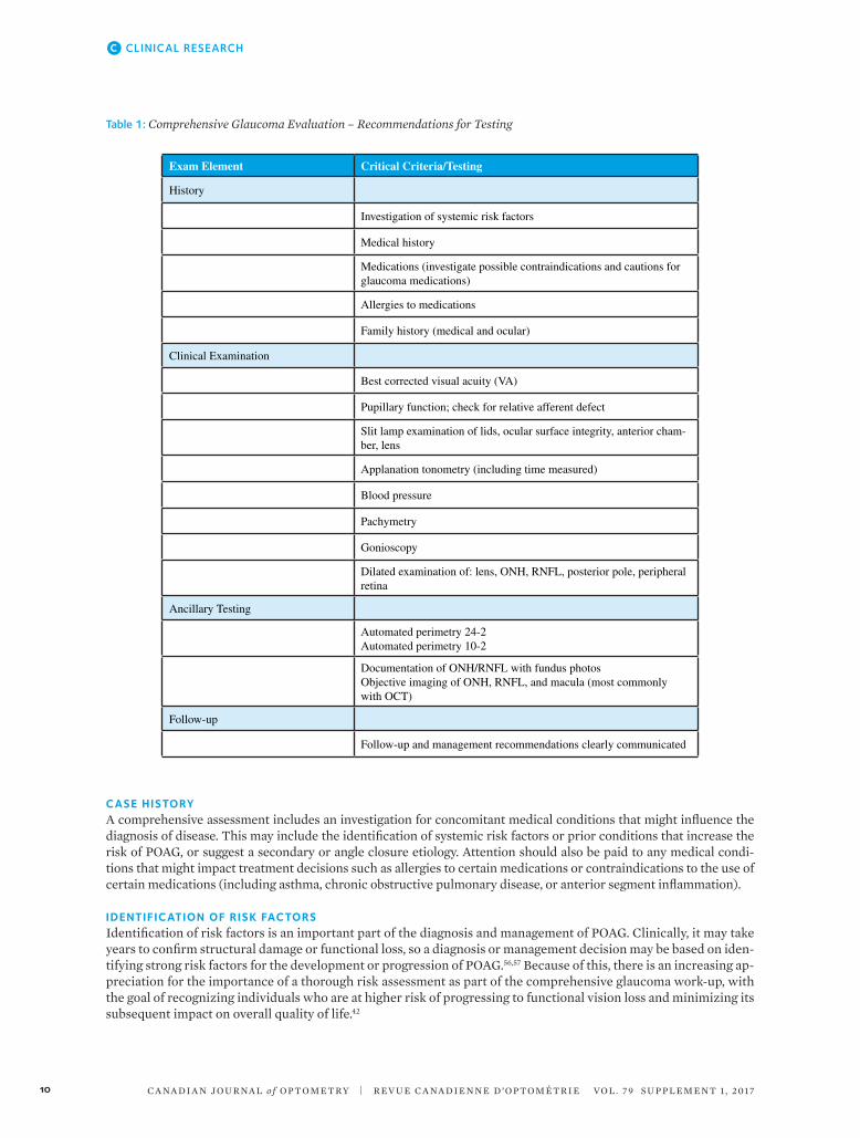
CLINICAL EXAMINATION AND CLINICAL FEATURES OF POAGA comprehensive examination for the diagnosis of POAG may be initiated following identification of risk factors and/or clinical characteristics of glaucomatous optic neuropathy in the initial primary eye care examination. De-tailed case history, specific anterior segment examination, tonometry, pachymetry and gonioscopy, as well as dilated fundus examination should be included with a view to ruling out secondary causes of glaucoma and determining the level of suspicion for a diagnosis of POAG. Structural assessment of the optic nerve, retinal nerve fiber layer, and macular ganglion cell layer, and tests of visual function (most commonly visual field analysis) are invaluable in the diagnosis and ongoing management of glaucoma.
The tests outlined below and summarized in Table 1 should be undertaken to further investigate for the presence of disease and begin to develop a solid baseline.
C A NA D I A N JO U R NA L o f O P T O M E T RY | R EV U E C A NA D I E N N E D ’O P T O M É T R I E VO L . 7 9 S U P P L E M E N T 1 , 2 0 1 7 9
CLINICAL RESEARCHC
Table 1: Comprehensive Glaucoma Evaluation – Recommendations for Testing
Exam Element Critical Criteria/Testing
History
Investigation of systemic risk factors
Medical history
Medications (investigate possible contraindications and cautions for glaucoma medications)
Allergies to medications
Family history (medical and ocular)
Clinical Examination
Best corrected visual acuity (VA)
Pupillary function; check for relative afferent defect
Slit lamp examination of lids, ocular surface integrity, anterior cham-ber, lens
Applanation tonometry (including time measured)
Blood pressure
Pachymetry
Gonioscopy
Dilated examination of: lens, ONH, RNFL, posterior pole, peripheral retina
Ancillary Testing
Automated perimetry 24-2Automated perimetry 10-2
Documentation of ONH/RNFL with fundus photosObjective imaging of ONH, RNFL, and macula (most commonly with OCT)
Follow-up
Follow-up and management recommendations clearly communicated
CASE HISTORYA comprehensive assessment includes an investigation for concomitant medical conditions that might influence the diagnosis of disease. This may include the identification of systemic risk factors or prior conditions that increase the risk of POAG, or suggest a secondary or angle closure etiology. Attention should also be paid to any medical condi-tions that might impact treatment decisions such as allergies to certain medications or contraindications to the use of certain medications (including asthma, chronic obstructive pulmonary disease, or anterior segment inflammation).
IDENTIFICATION OF RISK FACTORSIdentification of risk factors is an important part of the diagnosis and management of POAG. Clinically, it may take years to confirm structural damage or functional loss, so a diagnosis or management decision may be based on iden-tifying strong risk factors for the development or progression of POAG.56,57 Because of this, there is an increasing ap-preciation for the importance of a thorough risk assessment as part of the comprehensive glaucoma work-up, with the goal of recognizing individuals who are at higher risk of progressing to functional vision loss and minimizing its subsequent impact on overall quality of life.42
C A NA D I A N JO U R NA L o f O P T O M E T RY | R EV U E C A NA D I E N N E D ’O P T O M É T R I E VO L . 7 9 S U P P L E M E N T 1 , 2 0 1 710

MANAGING OPEN ANGLE GLAUCOMA
Identification of risk factors in an individual begins with patient history, while others will be detected by clinical exami-nation. A number of risk factors for glaucoma have been identified, but it is important to discriminate which of these is supported by strong evidence. Large prospective longitudinal studies have confirmed the following strong risk factors for the development of POAG: increased age, elevated IOP, thin central corneal thickness, and increased cup-to-disc ratio.8,13,58 Race (African-North American, Hispanic heritage) and family history (first-degree relative with glaucoma) are two other strong risk factors to consider.9,59 Table 2 lists the risk factors that should be considered in the glaucoma assessment.
Table 2: Risk Factors and Other Case History Questions for the Glaucoma Work-up
Risk Factors: Strong Evidence
Elevated IOP
Increased age
Race (African-North American, Hispanic for POAG)
Optic nerve head appearance
Thin central corneal thickness
Family history (first degree relatives)
Risk Factors: Mild* EvidenceModerate** Evidence
Low blood pressure (or over-treatment of hypertension)**
High myopia**
Diabetes mellitus**
Vascular dysregulation (i.e. migraine, Raynaud syndrome)**
Sleep apnea**
Cardiovascular disease*
Thyroid (hypo)*
Hypertension*
Other important case history questions
Significant blood loss?
History of ocular trauma?
Prior use of corticosteroid? (topical ophthalmic, inhaled nasal, systemic)
STRONG RISK FACTORS1. Intraocular Pressure
a. Elevated IOP: IOP is one of the strongest, and remains the only readily modifiable, risk factor for glaucoma onset and progression.12,35,59 There is no clear boundary that separates ‘elevated’ from ‘normal’ IOP, yet a well-known correlation exists between increased IOP and optic nerve damage. The traditional definition of elevated IOP is ≥21 mmHg, based upon two standard deviations from the population mean of 15mmHg.8 The OHTS (Ocular Hypertension Treatment Study) identified high IOP to be a strong risk factor for progression to glaucoma with relative risk of 10% for every 1mmHg increase above baseline IOP.8 Similarly, the EMGT also found IOP to be a strong risk factor for progression in individuals newly diagnosed with glaucoma, with a hazard ratio increasing by 11% for every 1mmHg increase in IOP.13 Inter-eye asymmetry in IOP ≥2mmHg is also a risk for primary open angle glaucoma.37 The literature is undecided on the diagnostic significance of IOP fluctuations identified by in-office measurements.60-62 It is likely more important in the identification of the risk of progression than identification of disease.62 Continuous 24-hour IOP monitoring may yield more information about the significance of IOP fluctuation as a risk factor for glaucoma.63-65
C A NA D I A N JO U R NA L o f O P T O M E T RY | R EV U E C A NA D I E N N E D ’O P T O M É T R I E VO L . 7 9 S U P P L E M E N T 1 , 2 0 1 7 11

CLINICAL RESEARCHC
2. Agea. Increasing Age: A number of population-based studies confirm that age is an important risk for the
development and progression of POAG.12,22,59,66 According to the Baltimore Eye Survey the prevalence of POAG increases 3.5 times in individuals over the age of 70.9 The OHTS showed age greater than 55 to be a strong predictor of POAG, and the EMGT found that the older the individual, the greater the prevalence of glaucoma.8,13 Age has also been found to be a significant risk factor in various ethnic groups with Hispanics showing the highest prevalence of POAG among all races for individuals over 80.59
3. Racea. African Origin: POAG has been found to be 4 to 6 times more frequent among individuals of African origin,
and individuals from the Caribbean and US of African descent, than among Caucasians.9,12,23,67 It has also been suggested that African Americans develop POAG at an earlier age and are more likely to go blind from the disease.67,68
b. Hispanic Origin: POAG appears to be more prevalent in the Hispanic than the Caucasian population, especially in individuals over 60.59
c. Asian Origin: Recent population studies have shown that the prevalence of POAG among Asian and Asian Indians ethnicities is greater than was once thought, with prevalence rates approaching those of Caucasians.1,16,69,70 NTG is more prevalent in Japanese and North Korean individuals than in Caucasians.69,70 While ACG remains an important concern in Asian ethnic groups, POAG and NTG is an increasing concern and must not be overlooked.1,16,69,70
See Figure 2 summarizing the relationship between disease prevalence, age, and ethnicity.
Figure 2: The prevalence of glaucoma increases with advancing age. African-Americans age 40 and older are at the high-est risk of developing the disease compared with people of other races. By age 69, nearly six percent of African-Americans have glaucoma; their risk rises to nearly 12 percent after age 80. Courtesy: National Eye Institute, National Institutes of Health (NEI/NIH)
C A NA D I A N JO U R NA L o f O P T O M E T RY | R EV U E C A NA D I E N N E D ’O P T O M É T R I E VO L . 7 9 S U P P L E M E N T 1 , 2 0 1 712

MANAGING OPEN ANGLE GLAUCOMA
4. Family Historya. The inheritance pattern of POAG remains uncertain, but it is accepted that the disease is a complex multifactorial
polygenic disease that commonly manifests in multiple generations of a family.71 The Rotterdam Eye Study found that the lifetime risk of developing POAG at 80 years of age was 10 times higher in individuals with a family history of glaucoma than in those without.72
5. Central Corneal Thickness (CCT)a. Thin CCT (< 555 um): A thin central corneal thickness was found to be a strong and independent risk factor for
conversion from OHT (ocular hypertension) to POAG in the OHTS.8,73 In fact, in a multivariate analysis of all the significant risk factors for progression from OHT to POAG, CCT was the strongest with a relative risk ratio of 70% for every 40μm decrease in thickness from baseline. The results from this model are adapted and shown below in Table 3. When considering relative risk for glaucoma, rather than adjusting IOP to correct for CCT, it is more valuable to simply classify CCT as thin, average or thick.73,74 It is still unclear as to whether thin CCT is only a predictor for progression to glaucoma from OHT, or if it is also a risk factor for progression once glaucoma has been diagnosed.8,58,75
Table 3: OHTS and Central Corneal Thickness (CCT)
IOP>25.75 36% 13% 6%
IOP> 23.75 12% 10% 7%
IOP < 23.7517% 9% 2%
CCT <555 >555 to< 588 >588
Adapted from: Gordon, M. O., et al. (2002) The Ocular Hypertension Treatment Study: baseline factors that predict the onset of primary open-angle glaucoma, Arch Ophthalmol, 120, 714-20.8
OTHER IMPORTANT RISK FACTORS Low Blood Pressure:
• There is an established link between POAG, specifically NTG, and low blood pressure and poor ocular blood flow.29,61 The EMGT found that low blood pressure was an important risk factor for progression among subjects with glaucoma regardless of baseline IOP.76 A patient might have low blood pressure physiologically, or as a result of over treatment for systemic hypertension. If a patient being evaluated for glaucoma is being treated for high blood pressure it is important to identify the type and dosage of the medication, as well as the time of day it is administered.77
• It has been hypothesized that low ocular perfusion pressure (OPP) leads to alterations in blood flow at the optic nerve and contributes to progressive glaucomatous optic nerve damage.29,76 Diastolic ocular perfusion pressure (DOPP) can be quickly estimated in the clinical setting to identify individuals who likely have low vascular perfusion to the optic nerve. This simple estimation involves taking the difference of the diastolic blood pressure (DBP) and IOP (DOPP = DBP - IOP). The Baltimore Eye Survey found that low DOPP was strongly associated with the prevalence of glaucoma.12,29 It has been suggested that DOPP values of less than 56 can be a useful threshold to identify patients at increased risk of progressive glaucomatous optic neuropathy.78
Myopia: • High myopia: Various studies and meta-analyses have demonstrated that subjects with higher myopic
refractive error have a significantly greater prevalence of glaucoma than groups with low myopia or emmetropia.79,80 This association exists as a risk factor for both development and progression of POAG. The underlying hypothesis is that individuals with greater axial length accompanying high myopia have weaker scleral support for retinal ganglion cells at the lamina cribrosa and this weakness increases the susceptibility of the optic nerve to glaucomatous damage.31
C A NA D I A N JO U R NA L o f O P T O M E T RY | R EV U E C A NA D I E N N E D ’O P T O M É T R I E VO L . 7 9 S U P P L E M E N T 1 , 2 0 1 7 13

CLINICAL RESEARCHC
Diabetes Mellitus: • There are conflicting reports in the literature around the association between glaucoma and diabetes
mellitus. The unexpected findings in the OHTS suggested that subjects with self-reported diabetes actually had lower risk of progression to POAG, which would mean that diabetes was not selected as a predictive factor. However, a meta-analysis published in the American Academy of Ophthalmology Journal in 2015 concluded that diabetes, duration of diabetes and elevated fasting blood glucose levels were all associated with a significantly increased risk of glaucoma. They also found that diabetes and elevated fasting blood glucose were associated with a slightly higher IOP.27
Vascular Dysregulation• Migraine and Raynaud syndrome are two conditions that have been identified as risk factors for the
development and progression of glaucoma. It is hypothesized that these conditions might be related to impaired autoregulation of blood flow to the optic nerve, subjecting the tissue to hypoxia and reperfusion injury.81
Sleep Apnea• Studies have shown an association between the presence of sleep apnea and POAG.82 It is not yet known
what the exact clinical significance of this association is and what, if any, impact treating sleep apnea has on slowing the progression of glaucoma.
Clinical Recommendation for risk factor analysis: • Once a patient has been identified as being at risk for glaucoma, a thorough investigation of all risk factors
should be undertaken to help identify individuals at greater risk of glaucoma and assist in developing a targeted approach to management.
TONOMETRYAssessment of IOP is a critical part of the glaucoma examination. Goldmann applanation tonometry (GAT) remains the gold standard for IOP measurement and should be used for those patients in whom a glaucoma risk profile has been iden-tified.95,96 Hand-held applanation tonometers (e.g. Perkins) have been shown to be comparable to GAT and may be useful to measure IOP in those patients who may be unable to sustain positioning in the slit-lamp biomicroscope.97,98
Non-contact tonometry (NCT), i-Care Tonometer, and Tono-Pen are often reliable alternatives. While these show reasonable agreement with GAT in the normal IOP range, they are less accurate and show disparity with GAT at high IOP levels.99,100
Two tonometers, the Pascal® Dynamic Contour Tonometer (DCT; Swiss Microtechnology® AG, Port, Switzerland) and the Ocular Response Analyzer (ORA; Reichert Corporation; New York, USA), have been developed in an at-tempt to overcome the impact of corneal biomechanics. The DCT is a modified type of applanation tonometer. The measurement principle is based on contour matching, which assumes that if the eye were enclosed by a contoured, tight-fitting shell, the forces generated by IOP would act on the shell wall. Replacing part of the shell wall with a curved pressure sensor would enable measurement of these forces and therefore the IOP.100-102 The ORA is a NCT that measures dynamic aspects of corneal deformation using an air pulse to cause two (inward and outward) corne-al applanations. There are four measurements obtained by the ORA: 1) an estimate of Goldmann IOP, 2) an estimate of IOP after correction for corneal biomechanical properties, 3) corneal hysteresis, and 4) corneal resistance factor. While the measurements obtained on both of these instruments may be an addition to your glaucoma tool kit, they do not replace the IOP measurement obtained using GAT.100,101
Factors that influence tonometry measurements include:103
• Central corneal thickness greater or lesser than average • Corneal hysteresis • Squeezing eyelids, holding breath, obesity or straining to reach slit-lamp • Corneal scarring or corneal irregularity • Elevating eye >15 degrees • Excessive or inadequate amount of fluorescein • Inaccurate calibration • Repeated tonometry • Observer bias
C A NA D I A N JO U R NA L o f O P T O M E T RY | R EV U E C A NA D I E N N E D ’O P T O M É T R I E VO L . 7 9 S U P P L E M E N T 1 , 2 0 1 714

MANAGING OPEN ANGLE GLAUCOMA
VALUE OF 2 4-HOUR AMBUL ATORY BLOOD PRESSURE MONITORING AND SLEEP APNEA TESTING IN POAG MANAGEMENT24-hour Ambulatory Blood Pressure (ABP) MonitoringAs previously noted, it is well known that there is an association between low ocular perfusion pressure and POAG.29,61 In clinical practice, in-office blood pressure measurement may help identify individuals who have low blood pressure, but in isolation, a single measure does not give much insight into the individual’s dy-namic blood pressure profile.83 An ambulatory blood pressure monitor is a portable blood pressure record-ing device that automatically measures blood pressure and generates a blood pressure profile over a defined period, usually 24 hours. The optometrist can coordinate ordering the test with the patient’s primary care physician, and review the results to identify instances of low diastolic blood pressure, paying particular attention to the nocturnal time frame. Most patients have a nocturnal BP “dip” of approximately 10% com-pared to daytime readings. This drop in BP may coincide with an increase in IOP, further exacerbating the decrease in ocular perfusion pressure due to the low blood pressure alone.84 It has been suggested that this situation of low DOPP may be more pronounced in patients with or at risk of glaucoma. However, there is a subset of individuals who are “extreme dippers”, dropping more than 20% at night compared to daytime readings.85 It is thought this might be the case in some patients who are progressing despite what appears to be adequate IOP control.86,87 Graham et al suggested that the magnitude of the nocturnal dip in individuals with glaucoma correlates with visual field progression.86 Similarly, Plange et al found that those with NTG had greater variability of nighttime blood pressure measurements compared with controls who were “non-dippers”, and that “extreme dippers” were more likely to progress than those who had a normal dipping pat-tern.87 It has also been shown that central visual field may be affected more severely than peripheral visual field in NTG with higher 24-hour fluctuation of OPP.88,89
Clinical Recommendations for 24-hour ABP study:• Progression is noted despite what appears to be adequate IOP control • Low blood pressure is suspected in a person at risk of NTG • Suspicion that a person with systemic hypertension may be over-treated • A paracentral field defect encroaching on fixation is noted in NTG
OBSTRUCTIVE SLEEP APNEA (OSA)Obstructive sleep apnea is characterized by a complete or partial obstruction of the upper airway during sleep that causes nocturnal hypoxia, elevated levels of CO2 in the blood, increased vascular resistance, and sympa-thetic activation.90 It is associated with hypertension, metabolic syndrome and cardiovascular disease.90,91 In addition to these systemic associations, there is consistent evidence that individuals with OSA are also at a higher risk of developing POAG.92 The underlying etiological mechanisms for the relationship between glau-coma and OSA remain unclear. One hypothesis is that hypoxia leads to increased intracranial pressure during sleep. The increased intracranial pressure subsequently decreases cerebral perfusion pressure and disturbs blood supply to the optic nerve. Another theory is that the increased sympathetic tone observed in patients with OSA can lead to increased blood pressure, vascular resistance and endothelial dysfunction which may cause insufficient perfusion to the optic nerve and RNFL.92-94 The risk of sleep apnea in the development of glaucoma appears to be greater in younger individuals, women and those of Chinese ethnicity.92 OSA might be more strongly associated with POAG when IOPs are less than 21mmHg.82,92 Despite the association of sleep apnea and risk of glaucoma being confirmed in the literature, the benefit of CPAP treatment for glaucoma re-mains unknown.92 There is some evidence suggesting that CPAP treatment may raise nocturnal IOP, but Liu et al concluded that treatment of OSA does not increase the risk of glaucoma.92
Clinical Recommendations for sleep study investigations:• NTG is suspected or progression in glaucoma is detected despite what appear to be controlled IOPs. • Treatment of confirmed OSA should not be avoided in individuals with glaucoma.
C A NA D I A N JO U R NA L o f O P T O M E T RY | R EV U E C A NA D I E N N E D ’O P T O M É T R I E VO L . 7 9 S U P P L E M E N T 1 , 2 0 1 7 15

CLINICAL RESEARCHC
Clinical Recommendations for IOP measurement: • Applanation tonometry remains the gold standard to measure IOP in individuals with or at risk of
developing glaucoma.
• Since a 24-hour diurnal IOP curve is not practical in most clinical settings, the best compromise is to get 4 to 6 IOP readings at different times of the day over several visits. At least 2 of these readings should be done as early in the morning as feasible in order to attempt to capture IOP as close to the presumed high point as possible.
• A modified diurnal may be practical for some clinics: measurements of IOP are taken every two hours during office hours beginning as early as is feasible in the morning.
2 4-HOUR IOP MONITORINGIt is well known that IOP fluctuates throughout the day, typically being higher in the early morning before decreasing gradually throughout the day to its low point in the early evening.104,105 One of the main limita-tions of the current gold standard GAT is the inability to obtain a measurement throughout this diurnal pe-riod. Glaucomatous eyes show a slightly different pattern of circadian IOP fluctuations: higher fluctuations in 24-hour monitoring and a greater nocturnal IOP rise when compared to those without glaucoma.106,107,108 Ideally, a 24-hour diurnal IOP measurement would be obtained for everyone at risk of glaucoma; however, at this time, this is impractical in most clinical situations. 24-hour continuous monitoring is likely more efficacious than a modified in-office diurnal assessment with GAT. Devices such as the Triggerfish (Sen-simed) that use contact lens sensors to obtain 24-hour continuous measurements have been shown to have good tolerability, safety and reproducibility in those with and without glaucoma.109-111 This device does not measure IOP directly, and its output cannot be calibrated into mmHg. Nevertheless, studies have shown that the measurements with this device correlate strongly with tonometry.110,112,113 The clinical availability of ambulatory devices (perhaps including the i-Care tonometer for home use) will address a large unmet need in managing glaucoma and will provide better understanding of the impact of diurnal IOP fluctuations. In the meantime, it is recommended that multiple IOP measurements are obtained at various times of the day to characterize what the IOP profile might look like pre-treatment.
Contemporary Medical Management Considerations for 24-hour IOP monitoring:The prostaglandin analogues have been shown to reduce IOP over the 24-hour cycle, representing another benefit of this class of medications. Fixed-combination (FC) medications have also consistently demon-strated IOP lowering over the 24-hour period. The CAI medications have shown better IOP control through the overnight hours than brimonidine, both alone and in FC with timolol. Indeed, these are important medical considerations when selecting appropriate therapies for glaucoma management.
PACHYMETRYPachymetry is a measurement of central corneal thickness (CCT). The effects of CCT on GAT under- or over-esti-mating IOP are well known. The GAT assumes a corneal thickness of around 520μm, which was felt to be the aver-age value when the tonometer was developed.114 It is now known, however, that CCT varies dramatically across the population. Several attempts have been made to develop a correction factor to adjust IOP measurements based on CCT.115-117 These nomograms are no longer considered valid or useful in the consideration of an individual patient’s management since the relationship between CCT and IOP is likely too complex to characterize with a simple calcu-lation.102,118 Rather than adjusting or correcting IOP for CCT, both IOP and CCT should be recorded in the record as the absolute value measured. Elevated IOP and thin CCT are considered significant risk factors for the development of POAG.
C A NA D I A N JO U R NA L o f O P T O M E T RY | R EV U E C A NA D I E N N E D ’O P T O M É T R I E VO L . 7 9 S U P P L E M E N T 1 , 2 0 1 716

MANAGING OPEN ANGLE GLAUCOMA
CCT can be measured using ultrasound or optical coherence tomography. Ultrasound pachymeters are easy to use, portable and cost-effective instruments. Their accuracy is dependent on the probe being placed perpendicular to the corneal surface. In the seminal studies that showed the importance of CCT in glaucoma, measurements were taken with ultrasound pachymetry.119 Studies have shown that the measurements obtained through anterior seg-ment OCT are generally in good agreement with those obtained through ultrasound, although OCT might under-estimate CCT.120,121
Clinical Recommendations for measurement of central corneal thickness:• Pachymetry should be measured on each eye with the mean of three measurements recorded.95
• CCT should be reassessed intermittently, as it may change over time and with the use of some topical medications.122
• When possible, CCT should be assessed using ultrasound pachymetry to be consistent with large clinical trials.96
CORNEAL BIOMECHANICSCorneal tissue has both viscous and elastic properties to help absorb and dissipate applied energy. This results in corneal biomechanical variables that not only impact the measurement of IOP but may also be independent risk factors for glaucoma. The reason for this is still unknown but may be linked to the weaken-ing and thinning of the lamina cribrosa that occurs as glaucoma progresses.123 Corneal hysteresis (CH) has the most evidence supporting its role as a strong and independent risk factor for glaucoma. CH reflects the viscous damping in the cornea as a measure of its ability to resist and repulse after absorbing an externally applied force. It is calculated as the difference in non-contact air jet pressure producing two corneal appla-nations, one inward and one outward. Studies have demonstrated lower CH in individuals with glaucoma as compared individuals with ocular hypertension or without disease.124 Low CH has also been linked with risk of progression and greater visual field loss in glaucoma.125,126
There is currently only one instrument that measures CH, the Ocular Response Analyser (ORA). The ORA is a non-contact tonometer that produces two corneal biomechanical values and two IOP measurements. The two corneal biomechanical values are corneal hysteresis and corneal resistance factor (CRF). CRF is calculated from CH through a linear combination of both inward and outward pressure and is considered to be a measurement of corneal resistance independent of IOP. The first IOP measurement is an estimate of Goldmann IOP and the second is an estimate of the IOP corrected for the two biomechanical properties.
Clinical Relevance: Despite the association between corneal hysteresis and glaucoma onset and progression, there is still a paucity of clinical evidence to support adding CH measurement to the standard glaucoma work up. In addition, neither IOP measurement on the ORA will replace GAT as gold standard. This means that should the clinician decide to use the ORA in practice, they should do this in combination with obtaining IOP by GAT. The ORA, however, may add valuable clinical insight into management. For example, when managing a patient with high IOPs and seemingly normal optic nerve and fields, a clinician may feel more confident in deferring treatment if the patient also has thick CCT and high CH. Conversely, in a patient with glaucoma that appears to be progressing despite low IOP measurement, a more aggressive target pressure may be warranted, especially in the presence of thin CCT and low CH.
Clinical Recommendation for Corneal Biomechanics:• At the moment, obtaining corneal hysteresis and other corneal biomechanical measurements is
not standard of care in the glaucoma examination, but this topic should be followed closely as our understanding of their clinical relevance evolves.
C A NA D I A N JO U R NA L o f O P T O M E T RY | R EV U E C A NA D I E N N E D ’O P T O M É T R I E VO L . 7 9 S U P P L E M E N T 1 , 2 0 1 7 17

CLINICAL RESEARCHC
GONIOSCOPYEvaluation of the anterior chamber angle is one of the most important components in the examination of patients with, or suspected of having glaucoma. Unfortunately, gonioscopy remains a procedure commonly omitted from the glaucoma examination by both optometrists and ophthalmologists.127,128 Just as objective im-aging of the optic nerve head complements but does not replace ophthalmoscopy, anterior segment ultrasound biomicroscopy and optical coherence tomography of the anterior segment supplement but do not replace go-nioscopy.129 Gonioscopy remains the only method to fully visualize the anterior chamber angle and trabecular meshwork.
Van Herick’s method, an indirect biomicroscopic assessment of anterior chamber depth, is a common compo-nent of a comprehensive examination.130 A narrow slit beam is directed at the peripheral cornea at an angle of approximately 60°, and the width of the space between the posterior cornea and anterior iris is compared to the peripheral corneal thickness. Due to the increased risk of angle closure, gonioscopy is indicated if the width of that space is one-quarter or less of the corneal thickness when measured at the limbus. This is a more common presentation in women, and in those individuals who are hyperopic, of Asian ethnicity, or developing nuclear cataracts.24
Additionally, gonioscopy is indicated at the baseline examination for anyone with or identified as being at risk for POAG, and ideally annually post-diagnosis. Despite being relatively common, POAG is a diagnosis of exclusion made after ruling out angle closure and the presence of any secondary etiology. The latter includes pigment dis-persion and exfoliation, and conditions that are typically unilateral including angle recession, anterior segment inflammation, neovascularization, and angle dysgenesis such as in irido-corneal-endothelial (ICE) syndrome.25,131-134 Gonioscopy is only contraindicated in the presence of suspected globe perforation, hyphema, orbital fracture, or severe corneal compromise.95 Appendix 1 serves as a review of the gonioscopy procedure.
Clinical Recommendation for gonioscopy:• Although POAG may be the most common form of the disease in North America, it remains a diagnosis of
exclusion requiring confirmation of an open and unobstructed anterior chamber angle through gonioscopy.
Under normal circumstances, the principle of total internal reflection precludes visualization of the angle. This optical limitation can be overcome through the use of lenses or prisms in performing direct or indirect gonioscopy. Direct go-nioscopy utilizing a high-plus Koeppe-type contact lens is rarely used in routine clinical practice, but may be employed in the operating room where patients are supine and sedated for procedures including goniotomy (surgically opening the canal of Schlemm). This technique provides a panoramic view with minimal distortion, allows simultaneous com-parison of the two angles, and unlike indirect visualization, provides an upright non-inverted image.135
Indirect gonisoscopy is the technique most commonly performed by optometrists using the magnification of the biomicroscope and a mirrored lens. These lenses provide a reversed image of the angle opposite to the mirror being used, and with practice can become a convenient and expedient means of angle evaluation. Two lens types are available: a large diameter (12 to 15mm), steeply curved (7.4mm) Goldmann one-, two-, or three-mirror lens requiring a more viscous coupling medium (‘scleral’ lenses); and a smaller diameter (9mm) and flatter (7.85mm) Zeiss, Sussman, or Posner four- or six-mirror lens using the patient’s tear layer as the coupling medium can be employed (‘corneal’ lenses). The smaller contact area of the corneal lenses allows for indenta-tion gonioscopy to differentiate appositional from synechial angle closure and identify plateau iris, a rare ana-tomic configuration in which an anteriorly positioned ciliary body forces the peripheral iris into appositional closure. The corollary is that the use of a smaller lens requires gentle pressure to avoid artificially deepening the angle: corneal striae are a sign of excessive pressure. Given that some corneal compression is unavoidable, tonometry should be performed in advance of gonioscopy, as the latter may temporarily reduce intraocular pressure.136,137
Interpretation of Gonioscopic ResultsA number of grading systems have been proposed to correlate the gonioscopic appearance of the angle with the risk of angle closure: the Shaffer system assigns a numerical grade, estimated angular width, and anatomic description, while the more complex Spaeth system includes a description of angular approach, peripheral iris
C A NA D I A N JO U R NA L o f O P T O M E T RY | R EV U E C A NA D I E N N E D ’O P T O M É T R I E VO L . 7 9 S U P P L E M E N T 1 , 2 0 1 718

MANAGING OPEN ANGLE GLAUCOMA
curvature, point of iris insertion, and the results of indentation.138,139 A modification of the Scheie system noting the most posterior visible angle structure in each quadrant and a qualitative description of iris approach and abnormalities including peripheral anterior synechiae (PAS), angle recession, pigmentation, neovasculariza-tion, etc. may be most applicable to clinical practice.140
Qualitative assessment of pigment in the trabecular meshwork (TM) is critical. Increased trabecular pigmentation is most commonly secondary to pigment dispersion (often in young myopic males) or exfoliation (often in elderly Caucasian females). Noting the location of iris transillumination defects (mid-peripheral in pigment dispersion, adjacent to the pupil margin in exfoliation) or the presence of exfoliative material on the anterior lens capsule, pupil margin, and in the angle will help in the differential diagnosis.141 As noted in Appendix 1, the inferior angle is the normally the widest while the superior is the narrowest: if less than half of the TM across more than six clock hours (180°) is visible, the angle is considered at risk of closure.
Clinical Recommendations for gonioscopy:• Gonioscopy is a critical but often overlooked element in the assessment of all patients at risk for or
diagnosed with any type of glaucoma
• Practice makes perfect. Start practicing routinely: being familiar with normal variation facilitates the identification of abnormal findings, and provides the experience to confidently employ the technique when clinically indicated
ANGLE CLOSURE GL AUCOMA (ACG)A primary angle-closure suspect (PACS) will have ‘normal’ intraocular pressure and healthy optic nerve head (no disease), but 180° of non-synechial angle closure: routine monitoring is indicated. Individuals who progress to primary angle closure (PAC) will have elevated IOP (≥21mmHg) and/or PAS accompanying iri-dotrabecular contact, but no evidence of glaucomatous optic neuropathy: prophylactic laser peripheral iri-dotomy (LPI) is normally recommended. Primary angle-closure glaucoma (PACG) is diagnosed in the pres-ence of glaucomatous optic neuropathy (GON) with at least six clock hours of iridotrabecular contact and elevated IOP. Prompt treatment including LPI augmented by medication and/or surgery (including cataract extraction) is indicated in the presence of GON.142,143
PACG, while more common in East Asia, is under-diagnosed in Western populations, and is responsible for a disproportionate amount of significant vision loss. It is categorized according to gonioscopic assessment of the amount of iridotrabecular contact obstructing the pigmented TM.144
Classic signs and symptoms of an acute angle closure (AAC) attack include conjunctival injection, extreme IOP elevation (often ≥40mmHg), corneal edema, blurred vision, eye pain, and vomiting. AAC is a true ocular emergency that necessitates immediate intervention to prevent significant vision loss within hours. Inden-tation gonioscopy may open an appositionally closed angle and allow aqueous to enter the TM, lowering IOP. Medical therapy, decreasing aqueous production through the use of topical (beta-blocker, carbonic anhydrase inhibitor, and alpha agonist) and oral (acetazolamide) agents, should be initiated immediately. A topical steroid is often required, as AAC is invariably accompanied by significant inflammation. In a phakic eye with PAC only, topical pilocarpine is indicated to break pupillary block: miotic agents are only effective after the IOP drops and pressure-induced ischemia of the iris sphincter resolves. Once the acute attack has been broken and the eye is quiet, bilateral LPI (that may be accompanied by laser peripheral iridoplasty and/or cataract extraction in the involved eye) is the definitive treatment.145-147
C A NA D I A N JO U R NA L o f O P T O M E T RY | R EV U E C A NA D I E N N E D ’O P T O M É T R I E VO L . 7 9 S U P P L E M E N T 1 , 2 0 1 7 19

CLINICAL RESEARCHC
POSTERIOR POLE ASSESSMENTOptic Nerve and RNFL EvaluationThe contemporary definition of glaucoma hinges on structural change of the optic nerve complex.148-150 Structural damage is often the presenting sign of glaucoma, and progression of that damage is highly predictive of future functional loss, typically preceding detection of that loss by months to years.151,152 It warrants emphasizing that up to 40% of an individual’s retinal ganglion cells can be lost before a visual field defect is detectable through standard automated perimetry.51 The OHTS highlighted this fact, as two-thirds of the observation cohort who converted to glaucoma did so based on optic nerve head (ONH) appearance alone.153 For these reasons, careful and systematic stereoscopic evaluation of the ONH and retinal nerve fiber layer (RNFL), complemented by routine photography and ancillary structural and functional assessment when clinically indicated, remains essential in the diagnosis and management of glaucoma.
‘The 5 Rs of Optic Nerve Head Assessment’ provides a helpful framework upon which to construct an effective and efficient clinical examination.41 Table 4 summarizes the salient features of this paradigm:
1. Use the scleral Ring to determine the size of the optic nerve head 2. Identify the width of the neuroretinal Rim 3. Examine the Retinal nerve fiber layer 4. Assess the Region of parapapillary atrophy 5. Look for Retinal and disc hemorrhages.
Table 4: Summary of optic nerve features consistent with glaucoma
Nerve Category Sub Features
1. Scleral Ring(optic nerve size)
Estimate of optic nerve sizeOptic nerve size asymmetry between OD and OS
2. Neuroretinal RimDiffuse loss: break down of ISNT rule, excavation of rim tissue Focal loss: bayonetting of blood vessels, baring of blood vesselsNo pallor
3. RNFLDiffuse loss: loss of bright striations, increased clarity of tertiary blood vesselsFocal loss: area of dark RNFL bounded by bright striations
4. Parapapillary atrophy (PPA) Zone-β adjacent to area of focal neuroretinal Rim thinning, wedge RNFL defectExpanding area of Zone-β noted over time
5. Retinal (disc) hemorrhagesPresence of optic nerve hemorrhage or flame shaped hemorrhage in RNFL
1. Use the scleral Ring to determine the size of the optic nerve headAn accurate assessment of the ONH depends upon an understanding of its size and shape, both of which can vary dramatically between patients.154,155 The normally slightly vertically oval disc is delineated by the thin white para-papillary scleral Ring surrounding the ONH, and its size can be qualitatively categorized (small, average, or large) through comparison with the 5° spot size of a direct ophthalmoscope (an ‘average’ ONH) or with the branches of the vascular tree at the ONH margin.156 An ONH of average size will be 10 to 12 blood vessel widths in diameter, while a small disc will be less than 10 and a large disc more than 12.157 Biomicroscopy using handheld lenses allows both qualitative and quantitative assessment: adjusting a thin slit beam to align with the superior and inferior disc margins provides a measurement in millimeters that can be corrected for the magnification of the lens being utilized (60D: ~1x; 78D: ~1.1x; 90D/SuperField: ~1.4x) or directly compared to reference tables seen in Figure 3. (as provided in the European Glaucoma Society Terminology and Guidelines for Glaucoma).11,158,159
C A NA D I A N JO U R NA L o f O P T O M E T RY | R EV U E C A NA D I E N N E D ’O P T O M É T R I E VO L . 7 9 S U P P L E M E N T 1 , 2 0 1 720

MANAGING OPEN ANGLE GLAUCOMA
Figure 3: Optic disc size assessed at the slit lamp biomicroscope with handheld high power convex lens
Generally, Caucasian and highly hyperopic individuals (> +5D) tend to have smaller ONHs, whereas Asian, Hispan-ic, African American, and highly myopic individuals (> -8D) are the opposite.160 Large ONHs may have significant physiologic cupping, while small ONHs with minimal cupping may be glaucomatous: small discs merit particularly close scrutiny, as glaucoma is frequently overlooked.161-164
Although glaucoma is known as a disease of asymmetry, an inter-ocular cupping difference >0.2 is only suspicious in ONHs of equal size, and asymmetries in ONH size are relatively common.165 As a rule, clinicians tend to overesti-mate disc size and underestimate cup size on clinical examination, leading to an optimistic assessment of neuroreti-nal Rim width as thicker than it actually is.
C A NA D I A N JO U R NA L o f O P T O M E T RY | R EV U E C A NA D I E N N E D ’O P T O M É T R I E VO L . 7 9 S U P P L E M E N T 1 , 2 0 1 7 21

CLINICAL RESEARCHC
2. Identify the width of the neuroretinal RimGlaucoma is defined by the loss of retinal ganglion cell axons that comprise the RNFL and neuroretinal Rim (NRR). Diffuse or localized (particularly inferior-temporal) NRR thinning is 87% specific for glaucoma.166 Ex-cavation or undermining of rim tissue is one of the earliest structural changes, while superior or inferior focal notches are essentially pathognomonic for GON and predictive of rapid visual field loss that may threaten fixation.167-169 Focal loss is often easier to spot but is less common than diffuse loss of the neuroretinal Rim.170,171 Scrutinizing the position of intrapapillary blood vessels, noting bayonetting due to rim excavation or baring resulting from rim thinning, helps in the detection of NRR thinning, both baseline and progressive.172 Figure 4 shows an example of neuroretinal Rim thinning.
Figure 4: A patient with POAG OU and advanced rim loss inferior in both eyes, OS (b) worse than OD (a).
Systematic assessment may be aided by the ‘ISNT rule’: a healthy NRR tends to be thickest in the inferior quadrant, followed by superior, nasal, then temporal, meaning that a vertically elongated cup should raise suspicion of glaucomatous optic neuropathy.45,173 A breakdown of the ISNT rule also helps to identify diffuse loss across multiple sectors. In general, a healthy inferior and superior rim should be 1.5 to 2 times the thickness of the nasal and temporal rims.157,170 In early stages of the disease, the inferior and superior rim are preferentially af-fected. The typical pattern of neuroretinal Rim loss is: inferotemporal – superotemporal – temporal horizontal – inferior nasal – superior nasal.47 As damage occurs, the superior and inferior rim width will become a smaller multiple of the temporal width, making loss detectable even though diffuse glaucomatous rim loss may still maintain the ISNT configuration.46
NRR pallor is not a typical feature of glaucomatous optic neuropathy, but rather is strongly suggestive of non-glau-comatous optic neuropathy due to ischemic (AION), compressive, toxic/metabolic, or traumatic etiology.174 Further, these differential diagnoses will not cause a defect in neuroretinal Rim, which is another feature distinguishing them from glaucoma.47 Table 5 reviews other salient clinical features that are not typical of glaucoma development. Given their sight- and potentially life-threatening consequences, the importance of these differential diagnoses cannot be overstated.175
C A NA D I A N JO U R NA L o f O P T O M E T RY | R EV U E C A NA D I E N N E D ’O P T O M É T R I E VO L . 7 9 S U P P L E M E N T 1 , 2 0 1 722

MANAGING OPEN ANGLE GLAUCOMA
Table 5: Findings on glaucoma examination that warrant investigation into other differential diagnoses:176
Test Results
• Presenting BCVA <20/40 • Age <50 years • + RAPD • Optic nerve pallor • Neurological symptoms (headaches, weakness, numbness, etc.) • Visual field defects respecting vertical midline • Abnormal progression of visual field defects
To properly assess the optic nerve, it is critical to define the NRR by contour, noting the deflection of fine blood vessels, rather than pallor.40 A mismatch between central pallor (suggesting a ‘smaller cup’) and NRR margin as delineated by blood vessel deflection (suggesting a ‘larger cup’) can be an early sign of glaucomatous damage. This is best noted with a stereoscopic view of the nerve, which is best obtained with a dilated fundus examina-tion.44 While objective imaging has become invaluable, it is not able to detect rim pallor (or disc hemorrhages) and can be confounded by anomalous ONHs (those that are tilted, highly myopic, or pitted).177 It is critical that OCT is viewed as a complement to, not a replacement for careful clinical evaluation.178
3. Examine the Retinal nerve fiber layerRNFL loss detected through clinical exam and serial photography (using low magnification and aided by red-free illumination) is one of the earliest signs of, although not pathognomonic for glaucoma.179,180 In fact, RNFL loss can precede detectable VF loss by up to 6 years despite the fact that more than half the RNFL thickness must be lost before a defect becomes visible on ophthalmoscopy.181 The RNFL may be difficult to visualize on clinical examination, even with clear media and a dark fundus. Photography offers an oppor-tunity to maximize the visualization of the RNFL and the identification of subtle defects. A normal healthy RNFL will show prominent bright striations as nerve bundles enter the ONH at the inferior and superior poles, with relatively less brightness adjacent to the temporal and nasal quadrants. Defects are more obvi-ous against a darker background of the retinal pigment epithelium (RPE), and are therefore more difficult to detect in lightly pigmented eyes.
Like glaucomatous NRR defects, RNFL defects can be either diffuse or focal. Diffuse thinning dulls the normally bright RNFL striations, enhances visibility of the parapapillary retinal vessels, and typically manifests as asym-metry between superior and inferior hemispheres and between right and left eyes.182 One should pay particular attention to any asymmetries in brightness or blood vessel clarity between the eyes, as well as between the supe-rior and inferior poles of the optic nerve head. Diffuse glaucomatous loss is superimposed on diffuse age-related loss, making its detection challenging. Figure 5 illustrates the appearance of asymmetric diffuse RNFL loss be-tween the right and left eye.
C A NA D I A N JO U R NA L o f O P T O M E T RY | R EV U E C A NA D I E N N E D ’O P T O M É T R I E VO L . 7 9 S U P P L E M E N T 1 , 2 0 1 7 23

CLINICAL RESEARCHC
Figure 5: A 62 year-old Caucasian man with concurrent optic nerve head drusen and ocular hypertension. The diffuse RNFL loss OD is more prominent than OS.
a) In OD the tertiary vessels are clearly visible in the superior and inferior sectors since no RNFL overlies to blur them. There is no obvious brighter pattern adjacent to relatively darker area temporally and nasally.
b) OS shows some asymmetry between the area inferior and superior to the nerve. There is more diffuse loss inferiorly than superiorly with a few visible striations noted superiorly. Tertiary vessels are clearer inferiorly than superiorly.
Localized wedge defects are usually easier to detect. This type of defect is at least the width of a major retinal vessel (smaller slit defects are normal anatomic variations) and will widen as they extend in an arcuate pattern from the poles of the ONH. Most often, wedge defects will appear inferior- and/or superior-temporal.183,184 These represent sites of active glaucomatous damage that are frequently accompanied by focal NRR notching, PPA, DH, and VF defects, and merit close scrutiny for widening or deepening.185,186 Figure 6 shows an example of an inferior wedge defect that is clearly delineated by adjacent areas of prominent RNFL.
Figure 6: A 67 year-old Persian woman with normal tension glaucoma. A well-defined dark wedge defect inferiorly is bor-dered by relatively brighter RNFL striations on either side. This is contrasted to the healthy RNFL striations and blurring of the tertiary vessels noted superiorly.
C A NA D I A N JO U R NA L o f O P T O M E T RY | R EV U E C A NA D I E N N E D ’O P T O M É T R I E VO L . 7 9 S U P P L E M E N T 1 , 2 0 1 724

MANAGING OPEN ANGLE GLAUCOMA
4. Assess the Region of parapapillary atrophy (PPA)There are typically 5 prominent rings that can be identified clinically on the ONH: from central to peripheral they are the cup, the rim, the scleral Ring, zone beta and zone alpha PPA.187 Zone-beta parapapillary atrophy (zone-β PPA) is increased scleral visibility due to degeneration of the RPE and choriocapillaris immediately adjacent to the ONH. Zone-β PPA is rare in healthy eyes, but is more common and extensive in glaucomatous eyes, particularly those with shallow, sloping cups.188,189 On the contrary, zone-alpha (zone-α) PPA, irregular pigmentary change in the RPE alone, is found in the majority of healthy eyes. When both types of PPA are present, zone-α is always peripheral to zone-β. Zone-β PPA is larger in eyes with more advanced disease, and spatially and temporally correlated with RNFL thinning, NRR defects, and optic disc hemorrhages.190-193 Figure 7 illustrates the differentiation between zone-β and zone-α PPA in an eye with glaucomatous damage. VF deterioration is more rapid in the presence of baseline zone-β PPA, and increasing PPA is associated with progressive VF loss. The progression of PPA may be more diagnostic than its presence.194,195 Assessing PPA may be particularly helpful with small ONHs where intrapapillary (cupping) change is difficult to assess, and less valuable with myopic or tilted ONHs and in older individuals where non-glaucomatous zone-β PPA may already exist.196 PPA has historically been a difficult parameter to objectively quantify, but may be qualitatively tracked through serial fundus photography or en face OCT images.197
Figure 7: An 87 year-old Caucasian man with normal tension glaucoma. The thinner arrow points to an area of zone-α PPA, while the thicker arrow points to an area of zone-β PPA. A subtle disc hemorrhage is also noted superior temporal within the neuroretinal Rim.
5. Look for Retinal and disc hemorrhages (DH)There is no question that there is a strong association between optic disc hemorrhages and glaucoma. Optic disc hemorrhages are a complex phenomenon that cannot be explained by IOP, mechanical disruption, or vascular fac-tors alone.198 DH are typically feathery radial RNFL hemorrhages at or crossing the superior and inferior ONH margins (particularly the latter), but may be blot-shaped intrapapillary bleeds at the level of the lamina cribrosa.199 DH are notoriously difficult to detect via ophthalmoscopy, and meticulous examination of photographs, ideally ste-reoscopic, is helpful. In fact, a review of the OHTS data showed that only 16% of DH were detected on both clinical exam by a glaucoma specialist and stereo photography. In contrast, 84% were overlooked on exam and noted only on stereo photography.200 Figure 8 illustrates the importance of reviewing (stereo) photographs following the eye examination.
C A NA D I A N JO U R NA L o f O P T O M E T RY | R EV U E C A NA D I E N N E D ’O P T O M É T R I E VO L . 7 9 S U P P L E M E N T 1 , 2 0 1 7 25

CLINICAL RESEARCHC
Figure 8: a) A 60 year-old woman with NTG and low ocular perfusion pressure. Recurrent disc hemorrhages were noted at 5:30 in the RNFL. Additional IOP lowering medication was added b) A 67 year-old Caucasian man with POAG. A disc hem-orrhage was noted on the neuroretinal Rim at 6:00. IOP not at target and treatment was adjusted. c) A 76 year-old Caucasian woman with asymmetric POAG. Recurrent DH have been noted, always OS, both at the ONH margin and at the level of the lamina, as seen here.
Figure 8 a Figure 8 b Figure 8 c
DH are quite rare in healthy individuals (0.2 to 0.5% prevalence) but more common in those with early to moderate glau-coma, particularly in the presence of ‘normal’ intraocular pressure.201,202 However, given the relatively low preva-lence of glaucoma, the majority of DH are still found in patients who have not yet been diagnosed with the disease. It has been shown that the median time to development of a visual field defect following an optic disc hemorrhage is 38 months.198,203 Further, it has been suggested that more aggressive treatment after the detection of a hemorrhage might slow down visual field progression compared to not changing treatment.204 Differential diagnoses include venous occlusion, diabetic retinopathy, posterior vitreous detachment, ONH drusen, and AION.205,206
DH may be the single strongest risk factor for the progression of established glaucoma and were found more com-monly in eyes that developed glaucoma in OHTS. However, they are not considered a stand-alone diagnostic crite-rion in the absence of other signs of glaucoma.199,200,207-211 Despite their strong association with disease progression, there has long been uncertainty about whether DH are a result of, or factor for progression. At present, it is gener-ally thought that DH are a phenomenon confirming glaucoma disease activity.
Clinical Recommendations for ONH/RNFL assessment:• Diligent and systematic clinical assessment of the ONH and RNFL is a means of early identification of
disease, and one of the cornerstones of effective glaucoma management. Particular attention should be paid to neuroretinal Rim and RNFL changes at the superior and inferior poles, and to the identification of optic disc hemorrhages.
• ‘We argue that ophthalmoscopy and photography remain the gold standard of imaging due to portability, ease of interpretation, and the presence of a large database of images for comparison.’ (Spaeth GL, Reddy SC; 2014).
ANCILLARY TESTING
SPECTRAL DOMAIN OPTICAL COHERENCE TOMOGRAPHY
RNFL and ONHClinical (subjective) assessment of the optic nerve head (ONH) and Retinal nerve fiber layer (RNFL) is critical but challenging: even among glaucoma specialists, significant inter- and intra-observer variability is the rule rather than the exception.212 Ancillary objective imaging (most commonly optical coherence tomography, OCT) has become an invaluable complement in the diagnosis of glaucoma, detecting structural change up to six years before visual field
C A NA D I A N JO U R NA L o f O P T O M E T RY | R EV U E C A NA D I E N N E D ’O P T O M É T R I E VO L . 7 9 S U P P L E M E N T 1 , 2 0 1 726

MANAGING OPEN ANGLE GLAUCOMA
loss is identified.213,214 Please note the deliberate use of the words ‘ancillary’ and ‘complement’: OCT, like automated visual field analysis, is a tool to inform our clinical judgement and decision-making, not replace it. That being said, it is an extremely useful tool, providing accurate (4 to 5μm axial resolution) and reproducible (≤3% inter-scan vari-ability) quantification of ONH, RNFL, and retinal ganglion cell (RGC) parameters.49,215,216
Clinical Recommendation for ancillary imaging in glaucoma:• OCT augments but definitely does not replace clinical examination. Although objective imaging has
become an invaluable ancillary test, ‘… a thorough clinical examination combined with a healthy dose of common sense is superior to imaging technology …’ (Chong GT, Lee RK; 2012).
Just as an AVF analysis must be reliable, an OCT scan must be of high quality. Adequate signal strength is essential, as weak scans can dramatically underestimate RNFL thickness.217 Motion or blink artifacts, improper alignment, or incorrect segmentation algorithms can result in unreliable data: it is critical that each scan be qualitatively assessed to ensure accuracy and quality.218,219 Some examples of common OCT artifacts are shown in Figure 9.
Figure 9: The following figures demonstrate some common errors in OCT acquisition. a) The scan through the fovea looks good, but the colour image shows an error. b) The scan through this part of the fovea is from moving too close to the eye and flipping the image. c) The scan through this section shows another common OCT error where the image has moved off the screen obstructing the view. These errors are common and can influence the results on the glaucoma analysis. It is important to go back to the scan images to ensure errors have not occurred in acquisition, especially if the analysis looks abnormal. d) Error from eye movement. Caused optic nerve alignment to shift completely in the inferior portion of the nerve. e) The deviation map shows entire superior edge of the nerve as ‘outside normal limits’. The corresponding sector graph indicates either 0 or 1. The cause of this is from truncation during image acquisition. Further investigation into user error should be done whenever the values on the image analysis does not make sense (ie. multiple measurements of 0 um RNFL thickness). f ) A blink has caused a black line in the acquisition circle which resulted in a space on the RNFL segmentation map.
Figure 9 a
Figure 9 b
Figure 9 c
C A NA D I A N JO U R NA L o f O P T O M E T RY | R EV U E C A NA D I E N N E D ’O P T O M É T R I E VO L . 7 9 S U P P L E M E N T 1 , 2 0 1 7 27

CLINICAL RESEARCHC
Figure 9 d
Figure 9 e1 Figure 9 e2
Figure 9 f
C A NA D I A N JO U R NA L o f O P T O M E T RY | R EV U E C A NA D I E N N E D ’O P T O M É T R I E VO L . 7 9 S U P P L E M E N T 1 , 2 0 1 728
MANAGING OPEN ANGLE GLAUCOMA
It is also important to remember that glaucoma is a clinical, not a statistical diagnosis, despite the analysis that accompanies each scan. Reference databases are helpful, but have limitations.220 As an example, the Cirrus RNFL reference database comprises only 284 individuals aged 19 to 84: just 31 were older than 70, 43% were Caucasian, and none had any associated ocular or systemic disease. Applying these data to a 49-year old individual, RNFL thickness can decrease by 30% (from 107 to 75μm) yet still remain in the ‘normal’ range. Reference databases and segmentation algorithms are also instrument-specific, meaning that comparing measures from two different instruments is all but impossible.221
Objective imaging can complement a clinical examination based upon the ‘Five Rs’ paradigm.1. Use the scleral Ring to determine the size of the optic nerve head:
OCT quantifies disc size by delineating Bruch’s membrane opening (BMO), the true anatomic bottle-neck through which all RGC axons must pass.222 If nothing else, this has demonstrated that clinicians consistently over-estimate disc size with ophthalmoscopy, which in turn leads to an over-estimation of neuroretinal Rim (NRR) thickness.223 While OCT may be of particular value with anomalous discs where subjective identification of the scleral Ring is difficult, atypical anatomy also makes comparison to refer-ence databases of questionable value.220
2. Identify the width of the neuroretinal Rim
Confocal scanning laser ophthalmoscopy (CSLO: Heidelberg Retinal Tomography, HRT) and OCT can accurately identify glaucomatous NRR thinning: a rim area <1mm2 or a statistically abnormal vertical cup-to-disc ratio should be considered suspicious.224 Progressive NRR thinning is also predictive of fu-ture VF loss: in the Ocular Hypertension Treatment Study, HRT was able to identify structural change up to 8 years before VF assessment detected functional change.225,226
Some of the currently available OCTs have the ability to detect and quantify neuroretinal Rim defects while others do not. The Heidelberg Spectralis utilizes the detection of Bruch’s membrane opening to quantify a parameter known as Bruch’s membrane opening-minimum rim width (BMO-MRW). This value represents the shortest distance between BMO and the internal limiting membrane that forms the anterior border of the ONH.227 BMO-MRW has proven to be as sensitive in detecting glaucomatous dam-age and its progression as RNFL thickness, and may be particularly valuable in highly myopic patients where clinical disc margins may be difficult to delineate and RNFL bundles may be shifted tempo-rally.228,229 However, objective imaging does not replace careful clinical evaluation: reference databases can be confounded by anomalous ONHs and OCT cannot detect the rim pallor that often accompanies non-glaucomatous optic neuropathy.178,230
3. Examine the Retinal nerve fiber layer
The Retinal nerve fiber layer is where objective imaging with OCT initially made its mark, and still shines. Subtle RNFL thinning often precedes VF loss but can be difficult to appreciate clinically; how-ever, both corneal compensated scanning laser polarimetry (SLP: GDx) and OCT are able to detect this thinning much earlier than clinical examination.231,232 When utilizing spectral domain OCT, the param-eter with the best diagnostic accuracy tends to be average RNFL thickness, followed by inferior and superior quadrant thicknesses.233 A follow-up study comparing RNFL, ONH, and ganglion cell complex (GCC) values confirmed the RNFL assessment software as the best at detecting glaucomatous damage.234 Although the use of multiple parameters could increase false-positive results, structural damage may be present in one parameter and not another, meaning that it is helpful to have information from the ONH, RNFL and macula in glaucoma diagnosis.213,235,236
It is important to always subjectively assess the RNFL deviation map (the OCT equivalent of a red-free photograph) and not simply ‘trust the numbers’: over-diagnosis (the ‘red disease’ of anomalous ONHs and high myopia) or under-diagnosis (incorrectly assuming that ‘green is always good’) are definite risks.48,219,237-239 Figure 10 exemplifies a case of ‘green disease’ in a patient with POAG. Relying entirely on summary parameters or reference database comparisons (in fact, on any single structural or functional test result in isolation) is simply not good enough.240 As a general rule, however, an average RNFL thick-
C A NA D I A N JO U R NA L o f O P T O M E T RY | R EV U E C A NA D I E N N E D ’O P T O M É T R I E VO L . 7 9 S U P P L E M E N T 1 , 2 0 1 7 29

CLINICAL RESEARCHC
ness in the mid-70s or an inter-eye asymmetry of 6 to 9μm is considered suspicious.241,242
It is also critical to recognize that localized RNFL loss may not significantly impact global thickness (as noted in Figure 10), but can lead to substantial focal VF loss.243 That being said, in the absence of a definite structure-function correlation, any localized RNFL thinning must be confirmed, as quadrant and clock hour measurements have relatively poor reproducibility as compared to global (average) param-eters.244 The RNFL profile indicating the characteristic “double hump” pattern is a good place to look to identify localized RNFL thinning (as noted in Figure 10).
Figure 10: This 61 year-old Caucasian woman with POAG has a noticeable RNFL wedge defect in the left eye on fundus photography. Red free photo seen in (a). The corresponding SD-OCT is shown in (b). This example highlights the possibil-ity of an RNFL defect potentially being missed (green disease) if other aspects of the OCT are not carefully scrutinized. The RNFL Thickness Map (1) shows inter-eye asymmetry with the superior RNFL bundle in the right eye (more red area of eleva-tion) being thicker than the left eye (less red area of elevation). The RNFL Deviation Map (2) OS clearly outlines a superior RNFL defect that extends from the rim tissue. The RNFL Thickness Asymmetry Profile (3) shows a superior ‘hump’ in the OD RNFL profile and essentially no ‘hump’ in the OS RNFL profile (circled). The focal RNFL defect is also noted at 1:00 of the OS RNFL Clock Hour analysis (4). The key to effective OCT analysis is to look at all features of the RNFL OU Analysis, not simply the summary parameters, to identify potential areas of asymmetry both within the eye and between the eyes. This patient has been treated for nearly 5 years with PGA: no structural or functional progression has been noted.
Figure 10 a
C A NA D I A N JO U R NA L o f O P T O M E T RY | R EV U E C A NA D I E N N E D ’O P T O M É T R I E VO L . 7 9 S U P P L E M E N T 1 , 2 0 1 730

MANAGING OPEN ANGLE GLAUCOMA
Figure 10 b
C A NA D I A N JO U R NA L o f O P T O M E T RY | R EV U E C A NA D I E N N E D ’O P T O M É T R I E VO L . 7 9 S U P P L E M E N T 1 , 2 0 1 7 31

CLINICAL RESEARCHC
4. Assess the Region of parapapillary atrophy (PPA)
PPA may be qualitatively tracked through serial fundus photography, and investigators are beginning to utilize en face enhanced depth imaging (EDI) OCT for both qualitative and quantitative analyses includ-ing the differentiation of glaucomatous from myopic (zone-gamma) and age-related PPA.197,245-247 The present clinical reality, however, is that PPA is often more confounding than diagnostic.248,249
5. Look for Retinal and disc hemorrhages
The transient nature of DH makes clinical detection difficult: unfortunately, OCT is not helpful in iden-tifying DH, although it is able to quantify resultant NRR/RNFL loss.250 Given that DH are not considered independently diagnostic of glaucomatous optic neuropathy, this is more of an observation about, than a limitation of OCT.
Clinical Recommendation for use of OCT in glaucoma:• In suspect patients identified through clinical exam, targeted OCT assessment can identity RNFL thinning
up to six years before a visual field defect is detected on automated visual field analysis.
GL AUCOMA : A DISEASE OF THE MACUL A?While our clinical exam focuses on the ‘Five R’s’ of the ONH and RNFL, OCT has confirmed earlier suspicions that macular damage is common in early glaucoma.251-253 Although RNFL analysis remains a diagnostic cornerstone, as-sessment of the ganglion cell/inner plexiform layer (GCIPL) or ganglion cell complex (GCIPL + macular RNFL) should be part of every baseline, particularly in the presence of an anomalous or focally notched ONH, or suspected NTG.254 Like the NRR and RNFL, the inferior-temporal macula is most susceptible to glaucomatous damage, and asymmetry between eyes is suspicious.255 Unique to macular analysis, intra-eye asymmetry across the horizontal raphe is also suggestive of glaucoma.256,257 However, glaucomatous macular damage can also be diffuse, which may impact vision-related quality of life more than focal loss.258,259 See Figure 11 for a case highlighting the importance of imaging the macula in addition to the RNFL. Regardless of pattern, macular RGC thinning is strongly associated with central visual field loss. For this reason, a 10-2 VF grid is also recommended as part of a baseline assessment.260 Any concurrent macular disease, including age-related macular degeneration, diabetic macular edema, vitreo-mac-ular traction, or epiretinal membrane, can confound ganglion cell analysis.261
When warranted by clinical suspicion of glaucomatous optic neuropathy, given that no single parameter is foolproof in isolation, clinicians are wise to utilize all the tools at their disposal, obtaining baseline RNFL, ONH, and macular RGC (structural) assessments and complementary 24- and 10-2 AVF (functional) analyses.55
C A NA D I A N JO U R NA L o f O P T O M E T RY | R EV U E C A NA D I E N N E D ’O P T O M É T R I E VO L . 7 9 S U P P L E M E N T 1 , 2 0 1 732

MANAGING OPEN ANGLE GLAUCOMA
Figure 11: A 78 year-old Caucasian male with advanced POAG. Both RNFL OCT (a) and GCA OCT (b) are shown. There is obvious advanced disease OS>OD on both RNFL and GCA plots but the extent of the loss appears greater in the GCA plot, showing more advanced disease on the macular scan than on the RNFL plot. This difference in staging could have important implications on treatment and management decisions.
Figure 11 a
C A NA D I A N JO U R NA L o f O P T O M E T RY | R EV U E C A NA D I E N N E D ’O P T O M É T R I E VO L . 7 9 S U P P L E M E N T 1 , 2 0 1 7 33

CLINICAL RESEARCHC
Figure 11 b
C A NA D I A N JO U R NA L o f O P T O M E T RY | R EV U E C A NA D I E N N E D ’O P T O M É T R I E VO L . 7 9 S U P P L E M E N T 1 , 2 0 1 734

MANAGING OPEN ANGLE GLAUCOMA
Clinical Recommendation for OCT and the macula:• Macular damage and accompanying central visual field loss is common in early glaucoma, but easily
overlooked: it is prudent to obtain good quality RNFL and macular OCT scans for all suspects identified through clinical exam.
PERIMETRYAlthough structural (retinal ganglion cell) damage is what defines glaucoma, functional (visual field) loss is what impacts an individual, and preventing vision loss is ultimately the reason why glaucoma treatment should be initiated. This makes reliable automated visual field (AVF) assessment essential at baseline and regularly during follow-up. Despite its many limitations, white-on-white standard automated perimetry (SAP) remains the gold standard.262 Primarily due to its extended testing time, inter-test variability, and the impact of cataract on its reliability, the initial promise of short-wavelength automated perimetry (SWAP, blue-on-yellow) has not been realized.263 Frequency doubling technology (FDT) perimetry may detect glaucomatous VF loss prior to SAP, and can be positioned in screening at-risk suspects who are followed with SAP post-diagnosis.264
The Swedish Interactive Threshold Algorithm (SITA) strategies available on the Humphrey Field Analyzer (HFA) significantly reduce the time required for threshold SAP with little if any loss of sensitivity.265 Conven-tional wisdom has been to screen (pre-diagnosis) with SITA-Fast and follow (post-diagnosis) with SITA-Stan-dard; although SITA-Standard is a more precise testing algorithm, the precision of SITA-Fast appears to allow effective and efficient detection of change through the glaucoma continuum.266 Given that Guided Progression Analysis cannot currently integrate SITA-Fast and -Standard strategies, it may be pragmatic to ensure that the same strategy is used for as long as possible. Figure 12 reviews the single field analysis for a 24-2 SITA-Standard for a patient with glaucoma.
C A NA D I A N JO U R NA L o f O P T O M E T RY | R EV U E C A NA D I E N N E D ’O P T O M É T R I E VO L . 7 9 S U P P L E M E N T 1 , 2 0 1 7 35

CLINICAL RESEARCHC
Figure 12: This figure is a single field analysis of a patient with a superior paracentral defect, extending to a partial arcu-ate, from moderate POAG. The three indices of test reliability (1) show that this is a reliable visual field. Mean deviation (2) is borderline with a value found in less than 2% of an age-matched population. Pattern standard deviation (3) is outside normal limits with a value found in less than 0.5% of an age-matched population. The Glaucoma Hemifield Test (4) has been flagged as outside normal limits, and the Visual Field Index (5) is 94%. The VFI is center-weighted which is why the value is decreased more than it would be if the same cluster of missed points were localized peripherally instead of centrally.
C A NA D I A N JO U R NA L o f O P T O M E T RY | R EV U E C A NA D I E N N E D ’O P T O M É T R I E VO L . 7 9 S U P P L E M E N T 1 , 2 0 1 736

MANAGING OPEN ANGLE GLAUCOMA
REVIEW OF VISUAL FIELD ANALYSISThe HFA provides three indices of test reliability: false positive errors, false negative errors, and fixation losses.
• False positives (FPs) identify ‘trigger happy’ patients, and are the most important reliability index. FPs can make both baseline and follow-up tests appear too good, suggesting false progression or false stability respectively.267 A FP rate in excess of 15% renders a test unreliable, and automatically excludes it from statistical progression analyses.
• False negatives (FNs) have long been considered an indication of patient inattention, but can also result from the variability that characterizes advanced glaucoma.268 In establishing a baseline, FNs can make a normal field look glaucomatous and thus confound diagnosis: such a result should be discarded and replaced with a more reliable test.269
• Fixation losses (FLs) may indicate that the patient’s eye is wandering during the test, but can also result from inaccurate blind spot detection or intra-test variability in patient positioning. While FLs may not be critical in isolation, an index in excess of 20% in the presence of an unstable gaze tracking record calls test reliability into question.
In clinical practice, several important AVF parameters (‘global indices’) help inform the detection of glau-comatous VF loss.
• Mean deviation (MD) is a weighted average of overall deviation from age-matched normal; however, the same MD may result from either shallow generalized or deep focal loss, patterns that impact a patient very differently.270 For this reason, MD (and visual field index, VFI) is more helpful in staging than in diagnosing glaucomatous damage.
• Pattern standard deviation (PSD) identifies focal loss after correcting for generalized depression (such as that attributable to cataract), and is flagged as ‘outside normal limits’ at a level found in less than 5% of an age-matched population.8 PSD, however, remains normal in the presence of diffuse loss that may accompany both early- and late-stage disease.52
• The Glaucoma Hemifield Test (GHT) compares five mirrored zones in the superior and inferior hemifields, identifying asymmetric damage that characterizes early glaucoma with a high degree of sensitivity and specificity.271 It is flagged as ‘outside normal limits’ when at least one zone pair differs by an amount found in less than 1% of an age-matched population.272
It is essential to establish a solid baseline (two reliable and repeatable AVFs) within a time frame too short to allow for disease progression.53
Given their long track record and robust reference databases, 24- or 30-degree threshold strategies remain invaluable in establishing a diagnosis and monitoring disease progression. Glaucomatous visual field loss results from damage to the retinal ganglion cell (RGC) axons at the level of the lamina cribrosa. The characteristic shape and location of these nerve fiber bundle defects is determined by the unique anatomy of the RNFL.50 RGC axons follow an arcuate path around the macula, with longer axons from peripheral RGC lying deeper in the RNFL and forming the more peripheral NRR.
Initial VF loss commonly manifests as shallow and transient localized paracentral, arcuate, and/or nasal step de-fects (from least to most extensive, with superior nasal steps and paracentral defects being most frequent). These defects arise from damage to RGC axons at the crowded and vulnerable inferior and superior poles of the ONH. However, it is important to note that early glaucomatous loss frequently includes a diffuse 1 to 2dB change in MD not attributable to cataract that is quite easy to overlook.273 Although purely focal or diffuse VF defects are rarely found in isolation, localized loss may be associated with focal NRR defects and lower intraocular pressure, whereas diffuse VF depression may accompany concentric NRR loss and ocular hypertension.274
C A NA D I A N JO U R NA L o f O P T O M E T RY | R EV U E C A NA D I E N N E D ’O P T O M É T R I E VO L . 7 9 S U P P L E M E N T 1 , 2 0 1 7 37

CLINICAL RESEARCHC
For practical purposes, a PSD or GHT that is repeatedly ‘outside normal limits’ can be considered as diagnostic of glaucomatous VF loss. The word ‘repeatedly’ is critical: in the Ocular Hypertension Treatment Study, an astonish-ing 86% of patients with one abnormal VF (suggesting conversion of ocular hypertension to manifest glaucoma) reverted to normal on the next assessment.275
Clinical Recommendations for AVF assessment in glaucoma:• Early glaucomatous defects can include arcuate or partial arcuate loss, paracentral scotoma, nasal step,
and/or diffuse visual field depression: be vigilant for an increased mean deviation in the absence of media opacity.273
• The importance of a reliable and repeatable baseline visual field assessment cannot be overstated: without knowing the starting point, it is impossible to accurately identify progression.
Although clinicians have long relied on 24-2 or 30-2 testing strategies, it is now recognized that macular dam-age may be found as frequently as peripheral defects in early glaucoma, providing that the correct testing strategy is employed.260,276 Only four of the 54 points in the relatively coarse 24-2 grid (6° spacing between points) fall in the central 8 to 10° of the macula, versus all 68 points of the finer 10-2 grid (2° spacing). This poor sampling is exacerbated by RGC displacement at the fovea.277 As a result, a small initial paracentral scotoma may be missed by 24-2 but detected by 10-2 analysis, as shown in Figure 13.253,278 In reviewing the anatomy of the RNFL, the basis for early macular damage becomes clear: the majority of RGC axons from the superior macula enter the temporal ONH as the papillomacular bundle, whereas those from the inferior macula project to the inferior-temporal pole, a region that Hood and colleagues have termed the macular vulnerability zone (MVZ).252,259 Loss of RGC in the MVZ leads to a superior arcuate defect that threatens fixation, one of the crite-ria that defines advanced glaucoma, and has a significant impact on vision-related quality of life.258,279
C A NA D I A N JO U R NA L o f O P T O M E T RY | R EV U E C A NA D I E N N E D ’O P T O M É T R I E VO L . 7 9 S U P P L E M E N T 1 , 2 0 1 738

MANAGING OPEN ANGLE GLAUCOMA
Figure 13: A 62-year old man with asymmetric glaucoma developed a paracentral scotoma that was much more pronounced on the 10-2 visual field testing strategy (b) than would be expected from the 24-2 visual field testing strategy (a) alone.
Figure 13 a
C A NA D I A N JO U R NA L o f O P T O M E T RY | R EV U E C A NA D I E N N E D ’O P T O M É T R I E VO L . 7 9 S U P P L E M E N T 1 , 2 0 1 7 39

CLINICAL RESEARCHC
Figure 13 b
C A NA D I A N JO U R NA L o f O P T O M E T RY | R EV U E C A NA D I E N N E D ’O P T O M É T R I E VO L . 7 9 S U P P L E M E N T 1 , 2 0 1 740

MANAGING OPEN ANGLE GLAUCOMA
Paracentral VF loss appears to be particularly common at relatively low IOPs and in the presence of disc hemorrhages, systemic hypotension, and signs of primary vascular dysregulation including migraine, Raynaud’s phenomenon, and sleep apnea.29,210,280,281 In fact, some investigators have proposed that ‘paracentral POAG’ should be considered a dis-tinct subtype of glaucoma. While it would be ideal to obtain a baseline 10-2 for all patients, targeted assessment can be guided by (inferior) ganglion cell/inner plexiform layer (GCIPL) loss on macular OCT scan, any abnormalities of the central 12 points on 24-2 analysis, or patient-reported symptoms that are not commensurate with abnormalities detected on 24-2 testing.282-284
Clinical Recommendation for selecting AVF test strategy in glaucoma:• 24-2 fields are essential, but don’t forget the very central visual field: obtain a 10-2 AVF early, and repeat
intermittently in follow-up, because ‘… clinicians need to be aware that glaucomatous damage to the macula is common, can occur early in the disease, and can be missed and/or underestimated with standard VF tests that use a 6° grid, such as the 24-2 VF test’ (Hood DC, et al.; 2012).
So, what’s better at diagnosing glaucoma, OCT or AVF? While RNFL assessment in microns is linear, AVF assess-ment in decibels is logarithmic: this allows OCT to detect subtle change in early disease with a robust RNFL, but not in the presence of the extreme RNFL thinning that characterizes advanced glaucoma.152,285,286 Conversely, the log scale of AVF analysis compresses (masks) early loss but expands the range at the opposite extreme. Functional loss is present in early glaucoma, but it is simply not detected by current AVF analyses until up to 40% of RGC are lost and RNFL thickness drops to the mid-70s.51,287,288 The prudent clinician will establish a reliable baseline for both structure and function, leveraging OCT early and AVF later in the glaucoma continuum.54,289
MAKING A DIAGNOSIS
Detecting glaucomatous optic neuropathy and/or a corresponding characteristic visual field defect are the primary endpoints when making a diagnosis of glaucoma.153,290 As mentioned earlier, the diagnosis of POAG is often made presumptively based on consideration of the presence of risk factors including strong family history, elevated IOP, and characteristic optic nerve and/or visual field findings. Only when the subtle signs of progression have been confirmed can glaucoma be definitively diagnosed. These signs may include:291
• A confirmed new defect in a previously normal visual field consistent with glaucomatous damage • A confirmed deepening or expansion of a previously ambiguous visual field defect • Progressive optic disc cupping, notching or rim thinning • Progressive thinning of the circumpapillary RNFL or macular ganglion cell layer consistent with a glaucomatous process
It is commonly said that structural change occurs before functional loss, yet many of the large prospective clini-cal studies on glaucoma demonstrate functional loss before structural change.8,14,15 The most likely reason for this is that structural changes were diagnosed based on observable changes to the optic disc rather than on objective (OCT) imaging. In establishing a diagnosis of glaucoma, it is very important to note if there is a correlation between structural change and functional loss, and to be cognizant for the development of functional loss that corresponds to existing structural defects. For example, if while monitoring a patient as a glaucoma suspect, OCT demonstrates thinning of the inferior-temporal sector of the circumpapillary RNFL before any changes are noted on AVF testing, particular attention should be paid to the superior nasal quadrant or superior paracentral region using both 24-2 and the 10-2 testing strategies.
Clinical Recommendation for diagnosing glaucoma:• In the absence of confirmed disease progression, a diagnosis of glaucoma may be made and treatment
initiated presumptively based upon consideration of risk factors and signs suggesting glaucomatous optic neuropathy.
C A NA D I A N JO U R NA L o f O P T O M E T RY | R EV U E C A NA D I E N N E D ’O P T O M É T R I E VO L . 7 9 S U P P L E M E N T 1 , 2 0 1 7 41

CLINICAL RESEARCHC
STAGING GLAUCOMATOUS DAMAGE
A diagnosis of glaucoma cannot be made without a careful consideration of the classification of the severity of disease, which requires careful assessment and documentation of structural and functional damage. There are many different glaucoma staging resources to refer to. The commonality between them is their consideration of the degree of structural and functional damage, and the ultimate risk of losing functional vision.3,291,292 Con-sideration must be given to the extent of optic nerve and RNFL damage and visual field loss (including mean deviation and proximity of the field defect to fixation) when determining the level of glaucomatous damage present.96,157,292 Table 6 is an adaptation of the staging used in the Hodapp Anderson and Parish classification, the Canadian Ophthalmological Society, and the Glaucoma Handbook written by optometrist, Dr. Anthony Litwak.157,291 Staging of glaucoma is critical because it will help in formulating a management plan and guide management decisions including establishing a target IOP and frequency of follow-up. A standardized staging system also facilitates shared management or transfer of care with a common and more objective understand-ing of severity.
Table 6: Recommendation for staging of degree of glaucomatous damage.96,157,291
Stage Visual Field Changes Optic Nerve And RNFL Damage
Early/Mild
• MD < -5dB AND• < 18 points below 5%• < 10 points below 1% on PSD• No central points < 20 dB
• Thinning of superior and/or inferior rim e.g. C/D < 0.65 in an average sized nerve)• No wedge defects
Moderate
• -5dB < MD < -10dB OR• 18-36 points below 5% OR• 10-20 below 1% on PSD OR• Central points between 10-20 dB in one hemifield
• Early notch in superior OR inferior OR relative thinning in both superior or inferior rim (e.g. C/D 0.7 – 0.85 in an average sized nerve)• Prominent wedge superior or inferior
Advanced
• MD > -10dB OR• > 36 points below 5% OR• > 20 points below 1% on PSD OR• < 20 dB in both hemifields centrally OR • Any point in central 5 degrees < 10dB
• Early notch of superior and inferior or complete notch (eg C/D > 0.9 in an average sized nerve)• Complete Wedge
Clinical Recommendation for staging glaucoma:• Careful assessment of structural damage and functional loss allows staging of disease severity, which
subsequently informs all treatment and follow-up decision-making.
PROGRESSION ANALYSIS IN GLAUCOMA
As a rule, all patients with glaucoma will progress if followed long enough and with sensitive enough follow-up techniques. An important consideration for each individual patient is whether the progression is occurring at a rate that puts visual function and quality of life at risk. In some, progression occurs so slowly that visual function will never be affected, while in others progression can be very rapid, leading to significant vision loss despite medical and/or surgical intervention.15 The majority of patients fall between these two extremes. The goal of management is to provide intervention that is adequate enough to slow progression to a rate at which vision will not become compromised in the patient’s lifetime, while at the same time not causing intolerable side effects from treatment.15,17,61
C A NA D I A N JO U R NA L o f O P T O M E T RY | R EV U E C A NA D I E N N E D ’O P T O M É T R I E VO L . 7 9 S U P P L E M E N T 1 , 2 0 1 742

MANAGING OPEN ANGLE GLAUCOMA
Progression is usually identified clinically as a loss of tissue on structural assessment or a decrease in visual function on psychophysical testing. Both structural objective imaging devices (such as optical coherence to-mography or scanning laser ophthalmoscopy) and visual function testing instruments (usually standard auto-mated perimetry) have the capability to monitor for progression once reliable baseline data has been acquired. There are two types of progression analysis in common use: event-based analysis and trend-based analysis. In order to maximize the ability to detect progression it is important to use the same visual field testing strategy (ie. 24-2 SITA-Standard) and imaging instrument to acquire baseline and follow-up data.
Event-based analysis is the type of analysis used in the landmark glaucoma studies to confirm the presence a pro-gression endpoint. It describes a statistically significant change in structure or function from baseline, such as the deepening or enlargement of an existing defect or the development of a new defect.14,15,153,208,293 Event-based analysis is better for detecting slowly progressive change and localized change. It has the advantage of providing early de-tection of change, and fewer tests are required to detect and confirm change. However, event-based analysis can be more variable, and does not provide a rate of change.267,287,294,295
Trend-based analysis identifies the rate of change over time using linear regression analysis. It is better at differentiating fast progressors from slow progressors, and allows extrapolation to predict clinically signifi-cant change over time.287,295,296 Quantifying the rate of change is imperative to making informed management and follow-up decisions. The main disadvantage of trend-based analysis is that it takes more tests and longer follow-up to generate the rate of change.297 It is also less sensitive to focal changes at specific loci and diffuse loss across the entire visual field.
MONITORING FOR PROGRESSION ON VISUAL FIELD ANALYSISFunctional progression is best monitored using both event-based and trend-based analysis in standard automated perimetry.57 Both the Humphrey and Octopus instruments include software to identify progression against an age-matched normative database, and change from baseline. It is advisable to utilize the software available to generate both event-based and trend-based analysis.295
Event-based analysis requires at least 3 reliable visual fields before the analysis will be generated.57 The in-strument will look for change that exceeds the variability of stable glaucoma in a cluster of adjacent points and flag the defects when the change becomes statistically significant.267,298 Trend-based analysis requires at least 5 reliable visual fields to calculate a predicted rate of change.295 The Humphrey Field Analyzer plots the Visual Field Index (VFI, a center-weighted percentage representation of residual visual field) and generates a linear regression analysis to estimate rate of progression over a 5-year period (see Figure 14 for an example of using both trend- and event-based analysis in monitoring glaucoma progression).299,300 The VFI appears to give a similar rate of change to linear regression of mean deviation but may be less susceptible to the influence of cataracts. The Octopus perimeter has customizable progression analysis and can generate rate of progression using different variables to view mean deviation change per year or localized defect change per year. Rate of change for various speeds of progression using mean deviation and VFI change per year are:57,267,299
• -0.2dB MD or 1% VFI per year for mild rate of change • -0.5dB MD or 2% VFI per year for moderate rate of change • -1.0 to -2.0dB MD or 6 to 8% VFI per year for rapid (potentially catastrophic) rate of change
C A NA D I A N JO U R NA L o f O P T O M E T RY | R EV U E C A NA D I E N N E D ’O P T O M É T R I E VO L . 7 9 S U P P L E M E N T 1 , 2 0 1 7 43

CLINICAL RESEARCHC
Figure 14: The visual field Glaucoma Progression Analysis (GPA) for a patient with POAG who has shown slow progression over an 8-year period. The trend-based analysis (a) shows a rate of progression of -0.7+/-0.2%/year. The bar to the right of the graph is a visual representation of the expected visual field loss after 5 years: the top white section is the amount of current visual field loss, the middle gray hatched section is the expected loss of visual field at the current rate of change over 5 years, and the bottom darker gray hatched section is the expected vision to remain after 5 years. The event-based analysis at the bottom (b) shows a cluster of defects that have changed significantly from baseline over one (hollow triangle), two (semi hollow triangle) and three (solid triangle) exams. Both the event- and trend-based analyses flag this patient as having progressed; however, while the trend-based analysis suggests relatively slow global change, the event-based analysis shows significant localized change threatening fixation. This patient was followed with atypical ONHs for 15+ years with IOPs in the low 20s. She eventually converted to manifest glaucoma. Treatment was initiated with SLT but failed, and she is now successfully managed on PGA alone.
Figure a
Figure b
C A NA D I A N JO U R NA L o f O P T O M E T RY | R EV U E C A NA D I E N N E D ’O P T O M É T R I E VO L . 7 9 S U P P L E M E N T 1 , 2 0 1 744

MANAGING OPEN ANGLE GLAUCOMA
Rate of change calculations become more reliable when a greater number of visual field analyses are obtained. The time it takes to detect progression depends on the speed of progression of the glaucoma and the intervals at which visual fields are being tested. It has been demonstrated that it could take as long as 5 years to detect someone dete-riorating very quickly if fields are only being done annually. Increasing testing frequency to every 4 months short-ens that time frame to less than two years. Table 7 demonstrates the number of visual fields needed to detect mild/moderate/fast progressing visual fields at 1/year, 2/year and 3/year testing intervals in reliable field takers. In the scenario of an unreliable visual field taker, the length of time required to detect progression when fields are done annually increases from 13 to 30 years in slow progression and 6 to 13 years in fast progression.57
Table 7: Length of time required to detect different rates of visual field progression at different annual testing frequencies (in low variability (reliable) field takers)
Length of time (years) to visual field progression at intervals of:
VF/year 2 VF/year 3 VF/year
slow (-0.25 dB/year) 13 6.5 4.3
moderate (-0.5dB/year) 9 4.5 3
fast (-1.0dB/year) 6 3 2
adapted from Chauhan 200857
CLINICAL RECOMMENDATIONS FOR FREQUENCY OF AVF ASSESSMENT:• It is recommended that at least 6 reliable fields are obtained in the first 18 to 24 months to establish a solid
baseline and identify rapidly-progressing glaucoma (-2.0dB MD or 6 to 8% VFI per year).
• In the instance of a very poor (highly variable) visual field taker the number of visual fields should be increased.
MONITORING FOR PROGRESSION USING STRUCTURAL ANALYSISIt is evident that structural changes to the ONH and RNFL occur before detectable functional loss using the tests that we have available today.289 The scanning laser ophthalmoscope (HRT) has over 20 years of use in clinical care and longitudinal studies, and is able to provide reliable measurements of rim area and RNFL change. However, despite its ability to provide high quality progression data, it is not an instrument found in many eye care provider’s offices.
In the last decade, there has been significant improvement in the quality of the objective measurements of ONH, RNFL, and macular RGC parameters. Specifically, the advent of SD-OCT has revolutionized glaucoma imaging. Current versions of SD-OCT demonstrate low variability (≤3%) for ONH, RNFL and macular imaging. It has been suggested that SD-OCT can detect a change as small as 5μm in the average RNFL thickness with event-based analy-sis.49,215 If an adequate number of exams are available, very slow rates of change can potentially be quantified with trend-based analysis.238,301In order to improve detection of progression using SD-OCT it is important to use high quality scans (see earlier review of common imaging artifacts).217
Clinical Recommendation for monitoring for structural progression:• Having high quality scans, looking at multiple parameters (optic nerve, RNFL and macular data) and a
higher frequency of test taking will improve the reliability of detecting progression using the SD-OCT.302
CORREL ATING STRUCTURE AND FUNCTION WITH PROGRESSIONThe agreement between SD-OCT and SAP in detecting progression has to date been poor. SD-OCT seems to do a better job at detecting early disease while SAP appears to be better in advanced disease. The limitation of SD-OCT in advanced disease is likely related to the floor effect reached at an RNFL thickness of approximately 50μm, representing residual glial and vascular tissue.286 At this point the instrument is no longer able to discern change, given that there is little viable RNFL remaining. For this reason, in advanced glaucoma macular retinal ganglion cell analysis may be superior to RNFL analysis.303 At this stage, functional (AVF) assessment also becomes more helpful than RNFL analysis.289,302 The logarithmic scale of AVF analysis masks loss in early disease, but amplifies it in more
C A NA D I A N JO U R NA L o f O P T O M E T RY | R EV U E C A NA D I E N N E D ’O P T O M É T R I E VO L . 7 9 S U P P L E M E N T 1 , 2 0 1 7 45

CLINICAL RESEARCHC
advanced disease.285 In other words, functional loss is present in early glaucoma, but not detected by conventional AVF analysis.51 There appears to be a tipping point at an RNFL thickness of approximately 75μm at which time the detection of functional loss improves.288
Clinical Recommendation for monitoring based upon stage of disease:• SD-OCT is likely better at detecting glaucomatous progression in early disease while SAP is better at
detecting glaucomatous progression in later stages.
It has also been shown that SD-OCT might be superior at detecting progression after a shorter number of visits in individuals unable to provide reliable fields.289,302 The clinician should be aware that even in the presence of reliable testing results, 2 to 3 years is typically required to detect progression, or the effect of treatment on slowing progres-sion.237,302 Further, this timeframe assumes ideal circumstances, when testing is frequent (every 3 to 4 months) and results are reliable, two criteria that may not be replicated in day-to-day practice.302
Clinical Recommendation for frequency of testing:• Progression will be detected sooner with more frequent testing: once an initial rate of change has been
established over the first two years (requiring testing every 3 to 4 months), it is recommended that OCT and SAP are done at least every 6 months.
MANAGEMENT
WHEN TO CONSIDER TREATMENTGlaucoma management encompasses all of the steps culminating in the assessment of risk and diagnosis of glau-coma (ideally at the pre-perimetric stage), followed by setting of target pressures and the initiation of treatment. Periodic reassessments over the long-term are scheduled based on the severity of the disease, rate of progression, risk for adverse effects and patient-specific factors such as concerns for non-adherence.
The conventional approach to management begins with topical medications, followed by selective laser trabeculo-plasty and then surgery, although a strong case has been made for initiating treatment with trabeculoplasty.304 The ultimate decision of where to begin will depend on the individual patient’s needs, values and abilities, as well as other factors such as access to care.
Clearly, management of any disease, particularly a chronic disease, requires careful consideration on many levels. While the evidence available around disease diagnosis and the benefits and risks of treatment is in itself important to keep up with, so too is the evidence around the rates of adherence to therapy, patient-reported outcome concerns, and health-related quality of life measures. While these areas are pivotal to successful disease management, they are beyond the scope of this review.
Other important considerations include the rate of ocular surface disease in the population affected by glaucoma. Not only does adherence to topical treatment likely suffer as a result of the presence of ocular surface disease, the IOP control may also suffer if the ocular surface disease is not appropriately managed.305,306 Preservative-free (at minimum, benzalkonium chloride-free) formulations should be considered early in the management plan to reduce the exposure of the ocular surface and anterior chamber tissues to benzalkonium chloride (BAK). Fixed-combination agents reduce the exposure to preservatives and help address wash-out related concerns, while having a positive impact on adherence.307 At present, there remains no treatment for glaucoma, per se; rather the use of intraocular pressure reduction to facilitate a corresponding reduction in risk of progression.
Ultimately, the decision about treatment resides with the individual. Thorough counselling on the risks and benefits of treatment versus carefully monitoring without treatment must be undertaken with each person. Take care to ensure that a family member or care-giver can be present, especially in those patients for whom a decision like this may be challenging. Detailed documentation of the counselling and informed consent procedure(s) is suggested.
C A NA D I A N JO U R NA L o f O P T O M E T RY | R EV U E C A NA D I E N N E D ’O P T O M É T R I E VO L . 7 9 S U P P L E M E N T 1 , 2 0 1 746

MANAGING OPEN ANGLE GLAUCOMA
Clinical Recommendations for initiating treatment:• Consider all risk factors and treatment options and review options with each individual. • Consider risks versus benefits for all treatment options. • Document all counselling diligently.
IOP LOWERING FOR GL AUCOMAIOP lowering remains the only clinically established method to slow the progression of glaucoma. Recall that not all of those with elevated IOP (ocular hypertension; OHT) develop glaucoma, and not all with glaucoma have elevated IOP. Therefore, treatment for open angle glaucoma is not usually initiated until a threat to visual function has been identified. This may be when confirmed structural damage to the optic nerve or functional loss of visual field is noted, or when an individual with a high-risk profile for the development of glaucoma is identified. Ultimately, the goals of treatment should include being cognizant of a person-centred model to balance the goals of the individual and their life expectancy with their risk profile, always mindful of maintaining functional vision for daily activities and maximizing health-related quality of life (HRQoL).
A variety of drug classes with multiple agents in each class are now available for the medical management of glau-coma. Further, new molecules and novel delivery systems are being actively investigated and these are likely to alter our current treatment algorithms in short order.
The effectiveness and limitations of IOP-lowering treatments have been well established in various randomized clinical trials.61,290 These factors will be considered separately in each class of medication.
TARGET PRESSUREBefore starting treatment, a clear management plan should be developed that includes deciding on a “target” intraocular pressure. Target pressure represents the initial IOP anticipated to stabilize the progression of the disease to a point that it will reduce the risk to visual function. Target IOP is unique to each individual patient, and indeed, unique to each individual eye. Randomized control clinical trials inform our decision-making and highlight the importance of setting a target IOP that best reflects the stage of the disease and the likelihood of minimizing progression.8,14,15 In truth, however, there is no way to be certain that maintaining the target IOP will achieve that goal. Indeed, the target pressure is merely a mathematical construct using a ‘best guess’ scenario, and analysis of long-term, repeated measures of structure and function is the only way to truly de-termine the rate of progression for an individual patient, which in turn determines whether the target IOP was adequate. However, setting a target pressure remains a helpful starting point, establishing a reasonable value from which critical re-evaluation can be assessed over time. Accordingly, it is important to bear in mind that a target pressure is a dynamic value. If the measured IOP is not reaching target but minimal to no signs of progression are detected, then it is not likely necessary to adjust treatment to reach that pre-determined value. Similarly, whether the measured IOP is or is not at target is irrelevant if clinically significant disease progres-sion is determined.308 In the case of progression, careful consideration to adherence to treatment should be attended to before alterations to therapy are made.38 A simplified flow chart highlighting an appropriate way to utilize target IOP can be seen in figure 15.
There are limitations to the use of the target pressure construct. As previously noted, many patients have a peak IOP that is not measured within office hours.38 This consideration is not only important for the determination of the target pressure, but larger fluctuations in IOP have been associated with disease progression and must always be kept in mind when analyzing data.38,149
C A NA D I A N JO U R NA L o f O P T O M E T RY | R EV U E C A NA D I E N N E D ’O P T O M É T R I E VO L . 7 9 S U P P L E M E N T 1 , 2 0 1 7 47

CLINICAL RESEARCHC
Figure 15: Monitoring glaucoma using target pressure.309
Diagnose OHT or POAG
Stage disease, consider age/life
expectancy, determine individual concerns, set target
pressure
Target IOP achieved: continue to monitor
for progression
Target pressure not achieved: re-evaluate risk for progressive
loss of structure and/or function
High risk for progression: lower
target pressure determination and/or
alter therapy
Minimal risk for progression: monitor;
no adjustment to treatment or target
IOP
Initiate treatment
Low risk OHT: monitor IOP, structure/function closely
without treatment
An adequate number of IOP measurements must be accumulated in order to develop a reasonably accurate estimate of the baseline IOP. As mentioned above, assessments of 24-hour IOP would be very useful in understanding an in-dividual’s disease, but are not practical within most clinical settings at this time. Accordingly, at least 3 IOP readings should be collected at different times of day with at least two measures taken as early in the morning as possible. Other recommendations suggest 4 to 6 measures at different times of day over at least 2 to 3 visits.
Once the level of glaucoma is staged as mild, moderate or severe (Table 6), the target IOP can be established based on lowering IOP less for those at lower risk, and more for those at higher risk for progression. Generally, the highest IOP reading is used as a pre-treatment baseline, and the target is set to lower IOP as little as 20% in mild/low risk conditions to as much as 50% for severe/high risk situations (see Table 8). Consideration should be made for other risk factors of concern, including young age (<50 years of age), certain races (African North American descent) and if a sibling has advanced glaucoma. Of course, IOP can only be lowered a finite amount with medications and laser. Further, the pressure lowering required in NTG has not been unequivocally determined, though 20 to 30% reduc-tion is generally used, reflecting the conclusions of the CNTGS.19
C A NA D I A N JO U R NA L o f O P T O M E T RY | R EV U E C A NA D I E N N E D ’O P T O M É T R I E VO L . 7 9 S U P P L E M E N T 1 , 2 0 1 748

MANAGING OPEN ANGLE GLAUCOMA
Table 8: Setting Target IOP Based on Stage of Glaucomatous Damage
Stage disease Suggested initial target pressure Consider lowering an additional 10% if:
Mild damage 20-30% lowering• <50 years of age• African North American descent• Sibling with advanced glaucoma
Moderate damage 30-40% lowering
Severe damage 40-50% lowering
Clinical Recommendations for setting target pressure:• Gather sufficient data to determine the baseline pressure
(4 to 6 readings may be preferred; minimum of 3 with two as early in the morning as possible).
• Set a target IOP informed by the landmark trials based on stage of disease.
• Clearly document this value in the record, and reconsider when appropriate.
CHOOSING A THERAPY The initiation of medical intervention in the treatment of glaucoma should not be taken lightly. Once treatment is started it is normally maintained for life. When deciding on an intervention, the following components should be among the general considerations: awareness of the efficacy, benefits and risks of the treatment; the frequency of adverse effects and development of intolerance to the treatment; the many factors contributing to individual adher-ence to therapy and risks for non-adherence; and the ability to conduct regular follow-up.
For many years, the monocular drug trial was employed in an attempt to assess treatment efficacy, comparing a treated to an untreated eye to differentiate therapeutic IOP change from spontaneous fluctuation.310 The legitimacy of this comparison depends upon a number of assumptions, including that each eye fluctuates symmetrically, that fluctuations are repeatable, and that short-term response to treatment in one eye can accurately predict that of both eyes in the long-term. Recently, however, investigators have questioned the validity of these assumptions. current best practice is to obtain a number of pre- and post-treatment IOP measurements to establish a solid baseline and assess the impact of therapy on both eyes.311,312
While laser trabeculoplasty was initially considered as a treatment alternative to delay incisional surgery, more recently, selective laser trabeculoplasty (SLT) has been shown to be a viable first-line treatment for glaucoma.304,313 Indeed, not only does SLT demonstrate a good safely profile, and eliminate exposure to preservatives and the need for adherence to a treatment regimen, it also shows one-year data on efficacy that is comparable to that of the pros-taglandin analogue group of medications.313 One of the main drawbacks is the short-term efficacy of the procedure; however, there is evidence suggesting that certain individuals, including those with initial high IOP and those who responded well to the first procedure may benefit from repeat SLT.314,315 Newer forms of laser trabeculoplasty are being investigated with a view to reducing adverse effects.316
In the majority of cases, monotherapy with a prostaglandin analogue will be the first-line treatment. The decision to start a prostaglandin analogue is based on their superior efficacy, safety and tolerability profile. Due to their sys-temic adverse effects, beta-blockers, while still considered first-line treatment, are generally not used before prosta-glandin analogues unless the latter are ruled out due to contraindications/cautions or patient preference. Appendix 2 reviews and compares the classes of commonly used glaucoma medications available in Canada.
Prior to prescribing any medication and periodically thereafter, consulting the CPS (Compendium of Pharmaceu-ticals and Specialties) via their web portal is recommended to ensure that contemporary prescribing information is reviewed, including indications, contraindications, cautions, dosing, adverse effects and interactions [see: https://www.pharmacists.ca/products-services/compendium-of-pharmaceuticals-and-specialties/ ].
C A NA D I A N JO U R NA L o f O P T O M E T RY | R EV U E C A NA D I E N N E D ’O P T O M É T R I E VO L . 7 9 S U P P L E M E N T 1 , 2 0 1 7 49

CLINICAL RESEARCHC
Clinical Recommendations for initiating therapy:• Before prescribing, review prescribing information in the CPS (Compendium of Pharmacy Sub-specialties,
Canadian Pharmacists Association) or product monographs.
• Consider investigating potential interactions using a multidrug check (Lexicomp): enter your patient’s medication, including the one you are considering prescribing, and receive critical and helpful information about drug interactions.
FIRST-LINE THERAPY CONSIDERATIONSProstaglandin analoguesThe prostaglandin analogues are potent drugs with the distinct advantage over all other IOP-lowering therapies of unmatched IOP-lowering capabilities (31 to 33% reduction).317,318 The second definite advantage is effectiveness over a 24-hour cycle, thus requiring only once per day administration. While their onset of action is 3 to 4 hours, the peak is at approximately 8 hours, making night time dosing preferred.319 Night time use is generally recommended in an attempt to ensure that the peak effectiveness coincides with the early morning hours when IOP is presumed to be highest; however, any time of day that will facilitate good adherence is preferred to merely maintaining the night time administration at the risk of poor adherence.
The primary mechanism of action is increased outflow facility via remodeling of intercellular spaces in the uveo-scleral pathway, so unlike many of the other medications for glaucoma, maximal overall effect is generally not reached until 4 to 6 weeks of use.320 While this is an advantage for the management of intraocular pressure in chron-ic open angle glaucoma, these drugs are not generally helpful in situations of acutely elevated IOP. Systemic adverse effects are uncommon but may include upper respiratory tract infections/cold/flu, muscle/chest pain or rash.321 On the other hand, local effects are common and include conjunctival hyperemia, change in iris colour (especially hazel, mixed-colour grey or light brown irides), hypertrichosis and prostaglandin-associated periorbitopathy (PAP; periocular skin pigmentation and deepening of the upper eyelid sulcus, ptosis and appearance of enophthalmos).322 While the practitioner may deem these localized adverse effects cosmetic, the patient’s adherence to treatment may be affected if comprehensive counselling about their potential is not relayed adequately. Certainly, monocular treat-ment with prostaglandin analogues is generally not recommended. Although caution is advised in individuals with a risk of recurrent herpes simplex keratitis, cystoid macular edema, and a history of uveitis, a causal relationship with these conditions has not been established. Due to the efficacy of prostaglandins in lowering IOP in patients with uveitis and the small likelihood of developing these rare complications, prostaglandin analogues should remain in the treatment algorithm of uveitic glaucoma patients. Though perhaps not a first option in patients with these risks, the effectiveness of the prostaglandin analogues is such that these agents cannot be ignored in the treatment of uveitic glaucoma.323
While the three main prostaglandin analogues (latanoprost 0.005%, travoprost 0.004% and bimatoprost (a prosta-mide) 0.01% and 0.03%) work very similarly in lowering IOP, bimatoprost may have a better response overall.324,325 Recognizing the need for BAK-free if not completely preservative-free formulations for the ocular surface, travo-prost was formulated with Sofzia, a less toxic preservative. Given that there may be differences in receptor popu-lations between individuals, if one agent in this class does not work as expected, switching within the class may be advisable before determining that the drug class as a whole is ineffective in a particular patient and moving to alternate, and potentially less effective rather than concomitant agents.326,327
Fixed-combination (FC) agents are available with timolol 0.5% for all three prostaglandin analogues. These FC low-er IOP more than their component prostaglandin analogues on their own, and there is a suggestion that the bima-toprost/timolol FC lowers IOP more than the other two.328,328 Also of interest is the relative decrease in the common adverse effect of hyperemia in the FC products compared to monotherapy with the prostaglandin analogue alone.329
A newer drug in this class, tafluprost 0.0015%, is available in many jurisdictions worldwide and will likely also be available as a FC agent with timolol.330 The distinct advantage to this drug is the preservative-free formulation, available in unit dose vials.
C A NA D I A N JO U R NA L o f O P T O M E T RY | R EV U E C A NA D I E N N E D ’O P T O M É T R I E VO L . 7 9 S U P P L E M E N T 1 , 2 0 1 750

MANAGING OPEN ANGLE GLAUCOMA
SECOND-LINE OR ADJUNCTIVE TREATMENT OPTIONS Beta-adrenoceptor blocking agentsThe beta-adrenoceptor blocking agents, or beta-blockers, have been work-horses in glaucoma management for de-cades. While IOP-lowering is significant (~27%, range 19 to 29%) with once-daily or twice-daily dosing, their use as a first-line therapy has been essentially usurped by the prostaglandin analogue medications due to their potency for reducing IOP and their relative lack of systemic adverse effects.331,332 The primary mechanism of action for the beta-blockers is decreased aqueous production. They are broadly categorized as either non-selective where both beta-1 and beta-2 receptors are targeted (timolol 0.25% and 0.5%, levobunolol 0.25% and 0.5%) or cardio-selective where beta-1 receptors are preferentially targeted (betaxolol 0.5% solution and 0.25% suspension). While the non-selective agents may be dosed once per day, betaxolol is generally dosed twice. As the production of aqueous is physiologically reduced at night, it follows that morning instillation is recommended when only once per day dos-ing is prescribed. Unlike the prostaglandin analogues, most of the expected lowering of IOP will occur within 2 weeks with timolol.333 The systemic adverse effects of the beta-blockers are well known and include exacerbation of pulmonary conditions such as asthma and chronic obstructive pulmonary disease, precipitation of heart block, and fatigue and impotence. Local adverse effects include stinging, decreased corneal sensation.
The beta-blocker timolol is considered the gold standard in this class. As such, 0.5% timolol can be found in fixed combination with each of the three main prostaglandin analogues, the alpha-adrenoceptor agonist brimonidine, and both carbonic anhydrase inhibitors (CAIs) dorzolamide and brinzolamide. A preservative-free formulation of timolol is also available.
Despite providing slightly less IOP reduction, betaxolol may preserve visual field as well or better than timolol.334
Alpha-adrenoceptor agonists (∞2)The selective (∞2) alpha-adrenoceptor agonist agents, or alpha-agonists, have also been in use for many years. IOP-lowering is initially significant (20 to 30%) but may drop to lower levels over time (~17% reduction).335 Brimonidine 0.2% administered three times per day is considered to be approximately equivalent to timolol 0.5% used twice per day.336 The mechanism of action for the alpha-agonists is both decreased aqueous production and increased outflow through the uveoscleral pathway.337 While apraclonidine (0.5%, 1.0% unit dose) was first produced as a selective alpha-agonist, it soon fell out of favour for the treatment of open angle glaucoma due to high rates of tachyphylaxis and ocular allergy. However, it is very effective at preventing post-operative IOP spikes. Brimonidine (0.2%, 0.15% and 0.1% in US only) is a much more selective agent than apraclonidine and has a lower rate of tachyphylaxis and allergy. The onset of action of the alpha-agonists is quick at 1 hour and peak at 2 to 3 hours, with trough at 10 to 14 hours. This is the reason that dosing is three times per day if used as a single drug therapy; however, evidence suggests that brimonidine can be successfully reduced to twice per day when used as adjunctive therapy.338 The sys-temic adverse effects of the alpha-agonists are relatively uncommon but include fatigue or drowsiness, dry mouth and headache.339 Caution should be taken with patients with severe cardiovascular disease, orthostatic hypotension and Raynaud syndrome. This agent is also contraindicated in children due to significant central nervous system ef-fects including excessive sleepiness and lethargy: these effects have been reported after even a single drop of 0.2% brimonidine in infants.340 Local adverse effects include hyperemia, allergy (in as many as one in four patients), burn-ing on instillation, dryness, visual disturbance, tearing and eyelid edema.
While a number of the requirements to determine if a drug is neuroprotective have been satisfied by research into the use of brimonidine, and some studies have suggested better visual field preservation with brimonidine than timolol, to date no conclusive evidence supporting neuroprotection has been provided in human glaucoma.341
Brimonidine is available in FC with both timolol 0.5% and brinzolamide 1.0%.
Carbonic anhydrase inhibitors (CAIs)The agents that inhibit carbonic anhydrase include dorzolamide (2.0%) and brinzolamide (1.0%). Due to IOP-low-ering that is significantly inferior to that of the prostaglandin analogues or beta-blockers, the CAIs are generally used as adjunctive treatments. However, like the alpha-agonists, if prescribed on their own they should be admin-istered three times per day, while as add-on therapy they can be administered twice.342 Generally, morning and mid-afternoon are considered reasonable dosing times when added to a prostaglandin analogue dosed at bedtime.
C A NA D I A N JO U R NA L o f O P T O M E T RY | R EV U E C A NA D I E N N E D ’O P T O M É T R I E VO L . 7 9 S U P P L E M E N T 1 , 2 0 1 7 51

CLINICAL RESEARCHC
IOP is reduced a moderate amount (15 to 20%); however, one notable benefit of the CAIs is the ability to lower IOP in the night-time hours. This appears to be unique to the CAIs.343 This makes CAIs a desirable agent for those pa-tients in whom progression is occurring despite what appears to be IOP at target, and in those patients with NTG in whom 24-hour blood pressure monitoring has demonstrated that significant nocturnal dips in blood pressure are occurring, increasing the relative risk for optic nerve non-perfusion during the night.344
The mechanism of action for the CAIs is decreased aqueous production. Systemic adverse effects of the topically applied CAIs are generally taste perversion and headache, while discomfort on instillation is the most frequently reported symptom. It is important to relay this expected finding to ensure patients do not stop the medication due to concern that they are causing harm. Blurred vision, hyperemia, and dryness are also common.345
Of interest is the evolving understanding about the issue of allergy to sulfa drugs and the CAIs. Dorzolamide and brinzolamide are sulfonamides, and the components of the sulfonamide antibiotics that cause allergy are present in these non-antibiotic sulfa drugs.346 Those who have been found to have an allergy to sulfa antibiotics and also to CAIs have been deemed to have a tendency to allergy in general rather than this being caused by cross-reactivity with the sulfonamide.347 For this reason, patients with sulfa antibiotic allergies are likely to be able to safely use CAI glaucoma drops.
Both dorzolamide and brinzolamide are available as FC with 0.5% timolol. These are usually dosed every 12 hours. Brinzolamide is also available in FC with brimonidine 0.2%, which is dosed three times per day.
Oral drugs in this class, including acetazolamide (250mg tablets) and methazolamide (50mg), are rarely used for the treatment of primary open angle glaucoma, but may be considered when acute lowering of a very elevated IOP in an emergency situation (or less commonly, to lower IOP in anticipation of a surgical intervention) is required. Oral ad-ministration is very effective, with IOP reduction of 30% noted by 30 minutes, peaking at 2 hours and lasting for 6 to 8 hours.348 Medical considerations are significantly different for systemic use of the CAIs, which are contraindicated in the presence of liver or kidney disease, serum electrolyte imbalance and other less common systemic conditions. The most common adverse effects include malaise, diarrhea, anorexia, metallic taste and polyuria; however, drowsi-ness or dizziness and depression are also possible. Further CNS, dermatologic, and hematologic adverse effects, as well as metabolic acidosis may occur. Consultation with the patient’s family physician prior to administering these agents is recommended, especially in older patients and those for whom medical history is not known.
Muscarinic agonistsDirect- and indirect-acting muscarinic agonists have been used to treat glaucoma since the prior millennium. Only one agent remains on the market: various concentrations and mechanisms of delivery of pilocarpine (0.25% to 10%; drops and sustained-release inserts). The mechanism of action of pilocarpine is to increase conventional (trabecular meshwork) outflow by increased tension on the scleral spur, physically opening the meshwork. The onset of action of pilocarpine is prompt with peak lowering within 2 hours, but the duration is short, lasting for only 8 hours.349
While systemic adverse effects are uncommon and these are inexpensive drugs, the need for dosing four times per day and the significant local adverse effects have led to these drugs being essentially shelved in the management of open angle glaucoma.350 However, when glaucoma is progressive, maximal medical therapy has been reached and surgical interventions are not possible, these agents can help to lower eye pressure and reduce risk of progression.351
Pilocarpine is particularly important in primary angle closure glaucoma due to plateau iris or pupillary block once any acute IOP elevation has been addressed. It could be used effectively (albeit not comfortably) in pigment dispersion syndrome. The use of pilocarpine should be avoided in uveitic or neovascular glaucoma, and any secondary angle clo-sure whereby the lens-iris diaphragm is displaced anteriorly would be a relative contraindication for a miotic agent.352
Clinical Recommendations for IOP lowering with medications:317,318 IOP reduction for the various classes of medications is approximately:
• Prostaglandin analogues (31-33%): bimatoprost (33%), latanoprost and travoprost (31%) • Beta-blockers: timolol (27%); betaxolol (23%) • Alpha agonist: brimonidine (25%) • CAIs: dorzolamide (21%), brinzolamide (17%); additional nocturnal IOP lowering is an added benefit
C A NA D I A N JO U R NA L o f O P T O M E T RY | R EV U E C A NA D I E N N E D ’O P T O M É T R I E VO L . 7 9 S U P P L E M E N T 1 , 2 0 1 752
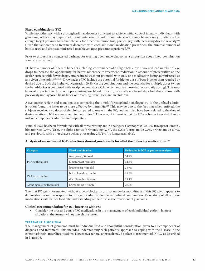
MANAGING OPEN ANGLE GLAUCOMA
Fixed combinations (FC)While monotherapy with a prostaglandin analogue is sufficient to achieve initial control in many individuals with glaucoma, others may require additional intervention. Additional intervention may be necessary to attain a low enough target pressure to reduce the risk for functional vision loss, particularly with increasing disease severity.353 Given that adherence to treatment decreases with each additional medication prescribed, the minimal number of bottles used and drops administered to achieve target pressure is preferred.354
Prior to discussing a suggested pathway for treating open angle glaucoma, a discussion about fixed-combination agents is warranted.
FC have a number of inherent benefits including: convenience of a single bottle over two, reduced number of eye drops to increase the opportunity for better adherence to treatment, reduction in amount of preservative on the ocular surface with fewer drops, and reduced washout potential with only one medication being administered at any given time point.330,355,356 Drawbacks of FC include the potential for higher dose of beta-blocker than required or desired due to both the higher concentration (0.5%) in the combinations and the potential for multiple doses (when the beta-blocker is combined with an alpha-agonist or a CAI, which require more than once-daily dosing). This may be most important in those with pre-existing low blood pressure, especially nocturnal dips, but also in those with previously undiagnosed heart block or breathing difficulties, and in children.
A systematic review and meta-analysis comparing the timolol/prostaglandin analogue FC to the unfixed admin-istration found the latter to be more effective by 1-2mmHg.357 This may be due to the fact that when unfixed, the subjects received two doses of timolol compared to one with the FC, and may also have been related to the time of dosing relative to IOP measurement in the studies.357 However, of interest is that the FC was better tolerated than its unfixed components administered separately.
Timolol 0.5% has been formulated with all three prostaglandin analogues (latanoprost 0.005%, travoprost 0.004%, bimatoprost 0.03% (US)), the alpha agonist (brimonidine 0.2%), the CAIs (dorzolamide 2.0%, brinzolamide 1.0%), and previously with other drugs such as pilocarpine 2%/4% (no longer available).
Analysis of mean diurnal IOP reductions showed good results for all of the following medications: 358
Category Fixed combination Reduction in IOP as per meta-analyses
PGA with timolol
travoprost / timolol 34.9%
bimatoprost / timolol 34.2%
latanoprost / timolol 33.9%
CAI with timololbrinzolamide / timolol 32.7%
dorzolamide / timolol 29.9%
Alpha agonist with timolol brimonidine / timolol 28.1%
The first FC agent formulated without a beta-blocker is brinzolamide/brimonidine and this FC agent appears to demonstrate a similar response to the agents administered as an unfixed combination. More study of all of these medications will further facilitate understanding of their use in the treatment of glaucoma.
Clinical Recommendation for IOP lowering with FC:• Consider the pros and cons of FC medications in the management of each individual patient: in most
situations, the former will outweigh the latter.
TREATMENT ALGORITHMThe management of glaucoma must be individualized and thoughtful consideration given to all components of diagnosis and treatment. This includes understanding each patient’s approach to coping with the disease in the context of their larger life situations. However, a general approach may be taken to treatment of POAG, as described in Figure 16.
C A NA D I A N JO U R NA L o f O P T O M E T RY | R EV U E C A NA D I E N N E D ’O P T O M É T R I E VO L . 7 9 S U P P L E M E N T 1 , 2 0 1 7 53

CLINICAL RESEARCHC
To properly assess the effectiveness of each drug in an individual treatment regimen, single medications are nor-mally added one at a time. Having said that, however, the algorithm does note the addition of FC medications. The clinician should be aware that while some granularity will be lost with respect to the effect of individual medications in the FC, the IOP response is expected to be fast and significant. This is perhaps a more important consideration in severe disease and when encountering very high eye pressures. Not apparent in the algorithm is the fact that SLT, minimally invasive glaucoma surgeries (MIGS) and other surgical interventions may be pursued at any time during the management continuum. Certainly, those patients for whom progression continues to be documented despite maximum tolerated medical therapy (MTMT), should be referred for surgical consultation. MTMT is defined as the largest number of medications that the patient can tolerate and consistently administer.
Currently, the first-line treatment for glaucoma is a prostaglandin analogue (PGA; see STEP 1). Given that these medi-cations allow for the largest reduction in IOP with a single drop per day, are well-tolerated, lower IOP over 24 hours, and have relatively few systemic adverse effects, this is the starting point for the treatment algorithm. If progression is suspected or targets not achieved despite a reasonable follow-up period, it is recommended to switch within the prostaglandin analogue class prior to moving to STEP 2. Clinicians begin to differ after this point, but STEP 2 may be one of three options. Adding a beta-blocker in the morning allows the clinician to tailor this treatment to some extent with choice of agent, concentration and dosing frequency as well as assess for adverse effects. This is also a relatively inexpensive option to consider. The second option is to switch the prostaglandin analogue to a FC agent with that medication and timolol. This provides a number of advantages including fewer bottles and fewer drops (one per day rather than two), thereby reducing the exposure of the ocular surface to preservatives. The third and fourth options include leaving the PGA in place but adding either a single agent CAI or alpha agonist, or proceeding directly to a FC of one of these agents with timolol. Progress through STEPS 3 and 4 are dependent on this choice as per Figure 16. If it is determined that a beta-blocker is not an option at STEP 2, a single agent CAI or alpha agonist may be added. In some circumstances, the fixed combination without beta-blocker (brimonidine / brinzolamide) may be started right away.
Regardless of what treatment is employed, a thoughtful, evidence-based approach must be taken and all compo-nents discussed with the individual and informed consent obtained. The consideration of preexisting and iatrogenic ocular surface disease must also remain an ongoing focus.
Figure 16: General approach to medical management 359
C A NA D I A N JO U R NA L o f O P T O M E T RY | R EV U E C A NA D I E N N E D ’O P T O M É T R I E VO L . 7 9 S U P P L E M E N T 1 , 2 0 1 754

MANAGING OPEN ANGLE GLAUCOMA
Other management considerations
a. Impact of ocular surface disease on managementWhile dry eye disease is frequently recognized in a large segment of our patient population, we are just learning about how common it is in the group with and at risk for glaucoma. Not only are some of the primary cases of ocular surface disease associated with glaucoma, such as chemical or thermal trauma, aniridia and autoimmune conjunc-tivitis, but the treatments for these conditions may exacerbate glaucoma (e.g. topical steroids increasing IOP) and vice versa (e.g. preservatives in glaucoma medications exacerbating ocular surface disease).360 While it is under-stood that dry eye disease may be present in 15% of the population, that percentage increases significantly when looking at the population with glaucoma.361,362 Estimates vary depending on what parameter is evaluated, but in one population studied, 59% of those with glaucoma had symptoms of dry eye in at least one eye, 61% had reduced tear volume on Schirmer test, 78% had reduced tear stability (TBUT), and 65% were noted to have a significant decrease in tear quality.363 In another study, severity of ocular surface disease was directly related to elevated IOP, with 63% of patients with severe glaucoma and 41% with mild glaucoma exhibiting symptoms of OSD.364 These numbers need to be corroborated by others as the relationship between the comorbidities of ocular surface disease and glaucoma is only beginning to be elucidated.
It is well established that BAK-containing eye drops are toxic to the ocular surface, yet this preservative remains the cornerstone of virtually all topical ocular therapeutic agents.365 BAK is known to be toxic to the conjunctiva, cornea, and trabecular meshwork.364,366,367 In fact, chronic conjunctival inflammation secondary to BAK exposure has been implicated in reducing the success of glaucoma surgical procedures.368 Ocular surface disease is related to number of glaucoma medications prescribed and duration of glaucoma treatment, both related to BAK exposure.369 Despite newer non-BAK and preservative-free glaucoma medications being developed, lack of uptake of these agents in the US market has highlighted a need for increased awareness of the significance of treating ocular surface disease in the management of patients with concomitant glaucoma.370 Compounding this challenge, certain medications such as preservative-free tafluprost are not yet available to our patients in Canada.
Ocular surface disease exacerbated glaucoma is a novel term used to describe those patients with severe OSD who also have glaucoma that is refractory to medical treatment.306 These patients respond to medications poorly and are more likely to have compromised success during glaucoma filtering procedures. Inflammation, the root of dry eye disease, is also at the root of glaucoma. The notion of successful glaucoma treatment hinging on successful maintenance of ocular surface health is new, but makes a great deal of sense. While our knowledge of the increasing prevalence and impact of dry eye disease is growing quickly, it follows that we need to be far more vigilant about concurrent disease, elevating the importance of its adequate management, not in spite of but because of the glau-coma.306 A shift away from focusing solely on IOP-lowering to the inevitable detriment of the ocular surface must occur if ‘preserving visual function … without causing untoward side effects from treatment’ is to be the true focus in the management of glaucomatous disease.154
Treatment for the inflammation of ocular surface disease in the presence of glaucoma cannot be ignored. Topical cy-closporine 0.05% has been shown to have a beneficial effect on the ocular surface in the presence of surface-altering glaucoma treatments, improving many measurable elements of dry eye disease including symptom questionnaires, tear stability, tear volume, corneal and conjunctival staining, and corneal morphology.371
Clinical Recommendations related to ocular surface disease and glaucoma:• As soon as a patient is identified as being at risk for glaucoma, ocular surface disease parameters should be
included in the clinical assessment (may include the use of standardized questionnaires, measurement of tear osmolarity, corneal/conjunctival staining, assessment of tear stability).
• Treat the ocular surface disease proactively and aggressively with all available treatments, including agents that safely target chronic inflammation.
• Once glaucoma is diagnosed, consider the status of the ocular surface in every management decision, including the initiation of preservative-free formulations and perhaps earlier consideration for SLT.
b. Role of generic medicationsWhile bioequivalence is the only parameter that a generic drug manufacturer needs to demonstrate to prove that
C A NA D I A N JO U R NA L o f O P T O M E T RY | R EV U E C A NA D I E N N E D ’O P T O M É T R I E VO L . 7 9 S U P P L E M E N T 1 , 2 0 1 7 55

CLINICAL RESEARCHC
their eye drops are ‘equivalent’ to trademarked drugs, evidence is mounting for the significance of the differences found between the two. Not only is bioequivalence not always achieved in generic formulations, but supporting components for a number of medications (drop size, vehicle, pH, preservatives, etc.) may differ from the trade drug.372-374 Evidence is emerging to show that these pharmacokinetic variables may be highly significant when mea-suring response to a medication, both in stabilization of disease and in adverse effects. With the number of drugs soon to be available as generics, more study is required to help to understand the role of generics and enable the clinician to make informed decisions about choices of glaucoma medications.
Clinical Recommendation on generic medications:• If IOP does not respond as anticipated and/or adverse effects are encountered with a generic formulation,
consider changing the treatment to the trade drug prior to eliminating the medication as an option for your patient’s glaucoma management
c. In the pipelineDespite decades of research, no new class of drug molecule has been approved for the treatment of glaucoma in North America for over two decades. Significant advancements have been made in the science around neuroprotection, yet no agents intended to support the optic nerve and Retinal nerve fiber layer have emerged in either systemic or topical form.372-375
There has been no applicable advancements towards a cure for this group of diseases; nevertheless, novel drug for-mulations continue to be actively pursued and tested with the hope that better treatment options emerge.376 Most recently, new classes of agents targeting trabecular outflow are being pursued (see SIDEBAR: On the horizon).
ON THE HORIZON – A GLIMPSE INTO THE FUTURE OF GL AUCOMA TREATMENTThe Rho kinase inhibitors are one such group that act on the contractile tone of smooth muscle to both enhance aqueous humour drainage through the trabecular meshwork and also lower episcleral venous pres-sure.377 Again piquing our interest is the investigation into the possible but yet to be proven neuroprotective effects of these agents in retarding degeneration (and/or promoting regeneration) of axons and improving ocular blood flow.378 Ripasudil is the first Rho kinase inhibitor in the world that has been approved for the treatment of glaucoma (Japan). One agent is currently is making its way through the approval process in the US: Netarsudil is both a Rho kinase inhibitor and a norepinephrine transporter inhibitor, which acts to also decrease aqueous production.379 A combination product incorporating Netarsudil with latanoprost is also under development.
Latanoprostene bunod is a dual mechanism, dual pathway molecule with the remodeling activity of a con-ventional prostaglandin analog but also possessing nitric oxide donating capability to improve both uveo-scleral and conventional outflow facility.380,381
Trabodenoson is an adenosine receptor agonist being investigated for its ability to increase conventional tra-becular outflow. Prostanoid receptors other than the FP receptor used by the current prostaglandin analogs are being targeted, including the EP1, EP2 and EP3 receptors, all by different agents and investigations.376
Even new targeted beta-adrenoceptor blocking agents, including Bamosiran, are being investigated to lo-cally target receptors in the ciliary body, thereby reducing the risk of systemic adverse effects.379
Unique delivery systems targeting the ease of administration of pharmaceuticals and reduction in adverse effects are emerging. Impregnated topical ring inserts and lacrimal plugs are already being developed. Erod-able and non-erodable subconjunctival and intraocular implants have already been developed for other drugs. The new delivery systems for current glaucoma drugs aspire to allow for convenient, measured drug delivery and increased adherence to treatment.376 Nanoparticle technology is an active area of research that aims to overcome the barriers to ocular drug delivery, facilitating high penetration rates, increased com-fort and decreased toxicity, and reduced frequency of administration.382 Ultimately, we all hope for better outcomes for our patients with glaucoma but the theoretical benefits of novel drug delivery systems will certainly require further study.
C A NA D I A N JO U R NA L o f O P T O M E T RY | R EV U E C A NA D I E N N E D ’O P T O M É T R I E VO L . 7 9 S U P P L E M E N T 1 , 2 0 1 756

MANAGING OPEN ANGLE GLAUCOMA
d. Adherence“Drugs don’t work in patients who don’t take them”, once said C. Everett Koop, former U.S. Surgeon General.
“More than half of the progression in treated OAG may be attributable to poor adherence with treatment” (World Glau-coma Congress, Vancouver, 2013).
It is hard to argue with these statements, just as it is hard to argue with the fact that non-adherence to medical therapy is essentially preventable.
That being said, studies on adherence to glaucoma treatment are many, and have consistently concluded that be-tween 5% and 80% of patients are non-adherent to their treatment regimen for glaucoma.307 Unfortunately, a far lower percentage of the clinicians that treat glaucoma feel that their patients are non-adherent, compounding the problem. There is no question that eye drop instillation is a challenge, and for many a burden, and eliminating that element of glaucoma management would invariably support the protection of visual function in our patients.
The four categories of issues related to adherence include: situational or environmental (e.g. cost); patient-related (e.g. lack of understanding of the treatments, outcomes and need for follow-up, and others such as self-reported symptoms of depression); treatment-related (complexity of regimen, adverse effects); and provider-related factors (communication, engagement).383,384
Adherence to the treatment regimen for any disease condition is challenging, and sustaining this adherence in the presence of a chronic disease is an even more formidable obstacle. Glaucoma treatment poses the ultimate chal-lenge because its successful management requires not only sustained adherence to a treatment regimen for a chron-ic disease, but adherence in the face of a disease that is essentially asymptomatic until its very late stages.
Follow-up ConsiderationsConsiderations for appropriate follow-up have been discussed through this guideline. Some key considerations re-garding frequency of follow-up and recommended testing can be categorized under:
• current IOP and relationship to target; • structural assessment and relationship to baseline; • assessment of visual function through timely and appropriate visual field strategies; • repeat gonioscopy to ensure nothing has changed in the original diagnosis or prognosis.
It is helpful to consider how to follow those patients who are high risk for developing glaucoma and those who have been recently diagnosed.
Summary: Recommendations for follow-up
IOP Structure Function Gonioscopy
Applanation tonometry at every visit (consider re-measuring pachymetry once every 1-2 years)
Objective imaging every 6 months
AVF depending upon reliability and risk of progression (assuming baseline 6 VF established in first 18-24 months)
Approximately annually
• Baseline • Set target• Post-treatment (sooner for faster acting therapies, 4-6 weeks for prostaglandin analogs)
At least every 6 months; more often if progression suspected or severe disease• Dilated assessment • Fundus photographs • OCT
• 24-2 SS every 6 months if stable/mild but more frequently (3-4 months if unstable and/or moderate/severe • 10-2 SS once per year, once baseline established
Consider sooner if history changes, anterior segment findings warrant, unusual findings noted
C A NA D I A N JO U R NA L o f O P T O M E T RY | R EV U E C A NA D I E N N E D ’O P T O M É T R I E VO L . 7 9 S U P P L E M E N T 1 , 2 0 1 7 57

CLINICAL RESEARCHC
Conclusion
Glaucoma management begins with identification of individuals at risk of glaucoma in the primary eye exam. A comprehensive glaucoma assessment is recommended after identification of individuals at risk or with signs of the disease to gather baseline information and confirm diagnosis. A treatment plan can then readily be tailored to each individual based on the collection of this information.
As mentioned earlier, the purpose of this evidence-based guideline is to continue to build upon the Canadian Optometrist's competence and confidence in the diagnosis and management of primary open angle glaucoma. The authors hope they have succeeded in this purpose and that readers will find it a useful resource to refer back to at various stages of the glaucoma journey.
APPENDIX 1: GONIOSCOPY
REVIEW OF THE GONIOSCOPY PROCEDURE To perform indirect gonioscopy:
• Anesthetic is instilled in both eyes.
• The patient is positioned at the biomicroscope, and the lens (with or without coupling solution depending on type of lens used) is placed on the cornea. It helps to have the patient look up while the lens is positioned above the lower lid, then look straight ahead while fixating with the fellow eye.
• A scleral lens must be rotated, whereas the slit beam is moved to each of the four mirrors of a corneal lens, to view different aspects of the angle.
• A systematic approach beginning with the superior mirror to assess the normally widest and most pigmented inferior angle (where angle structures tend to be easiest to identify) and proceeding clockwise is recommended.
• Tilting the lens away from, or having the patient look slightly toward the mirror being used facilitates visualization of deeper angle structures in patients with steeper mid-peripheral irides.
• This will not be the case in those with true angle closure. If excessive lens tilt is required, the angle should be considered narrow and potentially occludable.
• Ambient lighting should be low and directing the short and narrow slit beam through the pupil should be avoided, as pupillary constriction can temporarily deepen the angle.385,386
• Mid- to high (10 to 25x) magnification is required to accurately visualize detailed angle anatomy.
• Interface bubbles are common, particularly with corneal lenses: tilting the lens toward the bubble, flattening the lens surface on the cornea, will help eliminate them. An occasional bubble is actually a good sign that the pressure being exerted is adequate but not excessive.
STRUCTURES SEEN ON GONIOSCOPYWith the lens in position, the angle structures can be identified: the following description will proceed from pos-terior (more open) to anterior (less open). The root of the iris defines the posterior extent of the angle while the termination of Descemet membrane (Schwalbe line) marks its anterior border.
• Iris: • Observing the slit beam on the surface of the iris will help identify its contour: myopic eyes often have a deep chamber, concave iris, and an increased risk of pigment dispersion due to friction between the posterior iris and zonules, while hyperopic eyes have a shallower chamber, convex iris, and increased risk of angle closure.
C A NA D I A N JO U R NA L o f O P T O M E T RY | R EV U E C A NA D I E N N E D ’O P T O M É T R I E VO L . 7 9 S U P P L E M E N T 1 , 2 0 1 758

MANAGING OPEN ANGLE GLAUCOMA
• Ciliary body: • After the iris, the most posterior structure seen during gonioscopic assessment of a wide-open angle is the ciliary body (CB).
• The CB appears as a brownish-grey band at the root of the normally less-pigmented iris.
• It is more obvious in deeper angles.
• An extremely wide CB band or intra- or inter-ocular asymmetries in CB visibility, particularly following blunt trauma, may indicate angle recession, or irido- or cyclodialysis.387 Blunt trauma may also result in ‘balls’ of angle pigmentation (breakdown products of red blood cells following hyphema) and increased intraocular pressure representing ghost cell glaucoma.388
• Scleral spur: • The scleral spur (SS) is the insertion site of the ciliary muscle, and is visualized as a white line lying between the CB and the posterior (pigmented) trabecular meshwork (TM).
• It is an important and often quite conspicuous landmark, identifying everything anterior to the SS as TM.
• Benign iris processes (fine pigmented strands running from the iris root to posterior TM) or pathologic peripheral anterior synechiae (PAS, broad-based adhesions between the iris and TM resulting from chronic appositional closure (most often seen superiorly) or inflammation (most often seen inferiorly)) may obscure the SS.389
• As previously noted, indentation gonioscopy can help differentiate appositional from synechial angle closure: • in the former, pressure on a small diameter goniolens will force the lens-iris diaphragm posteriorly and open the angle, while the angle will remain closed in areas of PAS.390
• patients with appositional closure usually benefit from laser peripheral iridotomy (LPI), while those with synechial closure may require incisional surgery.391
• Trabecular meshwork (TM): • Anterior to the SS is found the trabecular meshwork, which is divided into the posterior (functional) TM and the anterior (non-functional) TM.
• The pigmented functional uveal TM, the posterior two-thirds of the TM, overlies canal of Schlemm and as the descriptor “functional” suggests, is the portion of the TM that filters aqueous.
• Anterior to that lies the less-pigmented non-functional corneoscleral TM: its light and even bluish- grey pigmentation of youth normally increases with age. • TM pigment can pathologically increase due to trauma, inflammation, pigment dispersion, and exfoliation.
• In some lightly pigmented eyes, canal of Schlemm may be visible as a slightly darker or red line (the latter in the presence of increased episcleral venous pressure forcing blood into the canal) deeper to the posterior TM.392
• Pharmacologic pupil dilation is typically safe if the posterior pigmented TM is visible in at least two full quadrants (180°) of the angle.
C A NA D I A N JO U R NA L o f O P T O M E T RY | R EV U E C A NA D I E N N E D ’O P T O M É T R I E VO L . 7 9 S U P P L E M E N T 1 , 2 0 1 7 59

CLINICAL RESEARCHC
• Schwalbe line: • The most anterior angle structure is Schwalbe line (SL).
• SL is the peripheral termination of Descemet membrane and the anterior border of the TM.
• It appears as a fine opaque line with variable pigmentation, particularly inferiorly. A pigmented SL is also known as Sampaolesi line, and is suggestive of exfoliation or pigment dispersion.
• In cases where the SL is difficult to visualize, the ‘corneal wedge’ formed by the intersection of off-axis slit beam reflections from the anterior and posterior surfaces of the cornea can aid in its detection.
• If SL is the only visible structure, the angle is considered narrow and at risk of closure.
• A prominent and anteriorly displaced SL (posterior embryotoxon) may be a variation of normal found in as many as one in four individuals, or associated with angle anomalies including Axenfeld-Rieger syndrome.393
Normal iris blood vessels are more visible in eyes with less pigment, tend to be thick, and run both circumferentially at the root of the iris and radially in the iris stroma.394 Normal vessels and tissue do not cross SL, which can help in the differential diagnosis of feathery and meandering neovascularization from normal vessels, and synechiae from iris processes. Angle neovascularization should be suspected whenever there is posterior segment ischemia: two common examples are proliferative diabetic retinopathy and ischemic central retinal vein occlusion.395
Clinical Recommendation for the performance of gonioscopy:• For an excellent resource on the principles, performance, and interpretation of indirect gonioscopy, please
visit Dr. W. Alward’s www.gonioscopy.org, a site that offers a wealth of information in video format.
C A NA D I A N JO U R NA L o f O P T O M E T RY | R EV U E C A NA D I E N N E D ’O P T O M É T R I E VO L . 7 9 S U P P L E M E N T 1 , 2 0 1 760

MANAGING OPEN ANGLE GLAUCOMA
Appendix 2 – Topical Glaucoma Medication Review
Glaucoma
Prostanoids/Prostamides
Drug Year Concentration Brand Name Dosing Bottle Size Preservative
latanoprost (ester)
1996 0.005% XALATAN (and generics)
QD, hs 2.5 mL BAK
travoprost (ester)travoprost (ester)
20010.004%0.004%
(generics only)TRAVATAN Z (and generics)
QD (hs)QD (hs)
2.5 mL2.5 mL
BAK
SOFZIA (boric acid, propylene glycol, sorbitol, zinc chloride)
bimatoprost (amide)bimatoprost (amide)
20022012
0.03%0.01%
LUMIGAN (and generics)LUMIGAN RC
QD (hs)QD (hs)
3 mL3 mL
0.005% BAK0.02% BAK
tafluprost (ester) 2012 0.0015%ZIOPTAN (US) or SAFLUTAN (elsewhere)
QD (hs) 10 x 0.3mL/vial PF
Beta-Adrenergic Blocking Agents (non-selective; ß1 & ß2)
timolol1978 +
0.25 0.5% TIMOPTIC (and generics)
QD (am)
5, 10 mL BAK
0.25% 0.5% Timoptic XE (q.d. dosing)
QD (am)
5mL (GEL); OCUDOSE PLUS
levobunolol HCl 0.25 0.5% BETAGAN (and generics)QD (am)
5, 10, 15 mL BAK
Beta-Adrenoceptorc Blocking Agents (cardioselective; ß1)
betaxolol 1998 0.25% Betoptic S BID Susp 5, 10 mL BAK
betaxolol 0.5% (generic only) BID ? BAK
Adrenomimetics Alpha2-selective Adrenergic Agonists (note: non-selective epinephrine, dipivefrin not used)
dipivefrin 1992 0.1 % Propine (and generics) BID 10, 15 mL BAK
apraclonidine 19940.5% 1.0% Iopidine 0.5% BID
10, 15 mL ampules (PF)
BAK PF
brimonidine 1996 +0.15%0.2%, 0.15%
ALPHAGAN-P (Canada)(generics)
TID (or BID if adding)
5, 10, 15 mLPurite BAK
Carbonic Anhydrase Inhibitors – Topical
dorzolamide 1994 2% TRUSOPTTID (or BID if adding)
5 mL BAK
brinzolamide 2000 1% AZOPTTID (or BID if adding
Susp – 2.5, 5, 10, 15 mL
BAK
C A NA D I A N JO U R NA L o f O P T O M E T RY | R EV U E C A NA D I E N N E D ’O P T O M É T R I E VO L . 7 9 S U P P L E M E N T 1 , 2 0 1 7 61

CLINICAL RESEARCHC
Carbonic Anhydrase Inhibitors – Oral
acetazolamide 1955 250 mg Diamox (and generics) QID Tabs N/A
methazolamide 50mg (generics) TID Tabs N/A
Miotics
pilocarpine
1, 2, 4% (6 no longer available)
MIOCARPINE (and generics)
QID 15 mL BAK
4% PILOPINE HS QD hs Gel 5 g tube BAK
Combination Anti-glaucoma Drugs
latanoprost / timolol
2001 0.005% / 0.5% XALACOM QD (am) 2.5mL BAK
travoprost / timolol
2003 0.004%/0.5% DUOTRAV PQ QD (am) 2.5, 5mLPolyquad, SofZia
brimonidine / timolol
2006 0.2% / 0.5% COMBIGAN BID 5, 10mL BAK
dorzolamide / timolol
1998 2% / 0.5%COSOPT (and generics)
BID5, 10mL BAK
COSOPT PF 0.4mL x 30 vials PF
brinzolamide / timolol
2008 1% / 0.5% AZARGA BID 5mL BAK
brinzolamide / brimonidine
2014 1% / 0.2% SIMBRINZA BID Susp 10mL BAK
C A NA D I A N JO U R NA L o f O P T O M E T RY | R EV U E C A NA D I E N N E D ’O P T O M É T R I E VO L . 7 9 S U P P L E M E N T 1 , 2 0 1 762

MANAGING OPEN ANGLE GLAUCOMA
REFERENCES
1. Quigley HA, Broman AT. The number of people with glaucoma worldwide in 2010 and 2020. Br J Ophthalmol. 2006;90(3):262-267.
2. Resnikoff S, Pascolini D, Etya’Ale D, et al. Global data on vi-sual impairment in the year 2002. Bull World Health Organ. 2004;82(11):844-851.
3. Foster PJ, Buhrmann R, Quigley HA, Johnson GJ. The definition and classification of glaucoma in prevalence surveys. Br J Ophthal-mol. 2002;86(2):238-242.
4. Vijaya L, George R, Paul PG, et al. Prevalence of open-angle glau-coma in a rural south indian population. Invest Ophthalmol Vis Sci. 2005;46(12):4461-4467.
5. Cruess AF, Gordon KD, Bellan L, Mitchell S, Pezzullo ML. The cost of vision loss in canada. 2. results. Canadian Journal of Ophthalmol-ogy/Journal canadien d’ophtalmologie. 2011;46(4):315-318.
6. Iskedjian M, Walker J, Vicente C, et al. Cost of glaucoma in canada: Analyses based on visual field and physician’s assessment. J Glau-coma. 2003;12(6):456-462.
7. Al Ali A, Hallingham S, Buys YM. Workforce supply of eye care pro-viders in canada: Optometrists, ophthalmologists, and subspecialty ophthalmologists. Canadian Journal of Ophthalmology/Journal Canadien d’Ophtalmologie. 2015;50(6):422-428.
8. Gordon MO, Beiser JA, Brandt JD, et al. The ocular hypertension treatment study: Baseline factors that predict the onset of primary open-angle glaucoma. Arch Ophthalmol. 2002;120(6):714-720.
9. Tielsch JM, Katz J, Singh K, et al. A population-based evaluation of glaucoma screening: The baltimore eye survey. Am J Epidemiol. 1991;134(10):1102-1110.
10. Klein BE, Klein R, Sponsel WE, et al. Prevalence of glaucoma: The beaver dam eye study. Ophthalmology. 1992;99(10):1499-1504.
11. European glaucoma society terminology and guidelines for glau-coma, 4th edition - chapter 3: Treatment principles and options-Supported by the EGS foundation: Part 1: Foreword; introduction; glossary; chapter 3 treatment principles and options. Br J Ophthal-mol. 2017;101(6):130-195.
12. Leske MC, Wu S, Hennis A, Honkanen R, Nemesure B, BESs Study Group. Risk factors for incident open-angle glaucoma: The barba-dos eye studies. Ophthalmology. 2008;115(1):85-93.
13. Leske MC, Heijl A, Hussein M, et al. Factors for glaucoma progres-sion and the effect of treatment. Evidence-Based Ophthalmology. 2003;4(3):134-136.
14. Leske MC, Heijl A, Hussein M, Bengtsson B, Hyman L, Komaroff E. Factors for glaucoma progression and the effect of treatment: The early manifest glaucoma trial. Arch Ophthalmol. 2003;121(1):48-56.
15. Agis Investigators. The advanced glaucoma intervention study (AGIS): 7. the relationship between control of intraocular pressure and visual field deterioration. the AGIS investigators. Am J Ophthal-mol. 2000;130:429-440.
16. Tham Y, Li X, Wong TY, Quigley HA, Aung T, Cheng C. Global prevalence of glaucoma and projections of glaucoma burden through 2040: A systematic review and meta-analysis. Ophthalmol-ogy. 2014;121(11):2081-2090.
17. Heijl A, Bengtsson B, Hyman L, Leske MC, Early Manifest Glaucoma Trial Group. Natural history of open-angle glaucoma. Ophthalmology. 2009;116(12):2271-2276.
18. Collaborative Normal-Tension Glaucoma Study Group. Natural his-tory of normal-tension glaucoma. Ophthalmology. 2001;108(2):247-253.
19. Group, Collaborative Normal-Tension Glaucoma Study. Comparison of glaucomatous progression between untreated patients with nor-mal-tension glaucoma and patients with therapeutically reduced intraocular pressures. Am J Ophthalmol. 1998;126(4):487-497.
20. Hollands H, Johnson D, Hollands S, Simel DL, Jinapriya D, Sharma S. Do findings on routine examination identify patients at risk for primary open-angle glaucoma?: The rational clinical examination systematic review. JAMA. 2013;309(19):2035-2042.
21. Meier KL, Greenfield DS, Hilmantel G, et al. Special commentary: Food and drug administration and american glaucoma society co-sponsored workshop: The validity, reliability, and usability of glaucoma imaging devices. Ophthalmology. 2014;121(11):2116-2123.
22. Coleman AL, Miglior S. Risk factors for glaucoma onset and pro-
gression. Surv Ophthalmol. 2008;53(6):S3-S10.23. Sommer A, Tielsch JM, Katz J, et al. Racial differences in the cause-
specific prevalence of blindness in east baltimore. N Engl J Med. 1991;325(20):1412-1417.
24. He M, Foster PJ, Ge J, et al. Gonioscopy in adult chinese: The liwan eye study. Invest Ophthalmol Vis Sci. 2006;47(11):4772-4779.
25. Tumbocon JAJ, Latina MA. Angle recession glaucoma. Int Ophthal-mol Clin. 2002;42(3):69-78.
26. Kersey J, Broadway D. Corticosteroid-induced glaucoma: A review of the literature. Eye. 2006;20(4):407-416.
27. Zhao D, Cho J, Kim MH, Friedman DS, Guallar E. Diabetes, fasting glucose, and the risk of glaucoma: A meta-analysis. Ophthalmology. 2015;122(1):72-78.
28. Shi Y, Liu P, Guan J, Lu Y, Su K. Association between glaucoma and obstructive sleep apnea syndrome: A meta-analysis and systematic review. PloS one. 2015;10(2):e0115625.
29. Memarzadeh F, Ying-Lai M, Chung J, Azen SP, Varma R. Blood pressure, perfusion pressure, and open-angle glaucoma: The los angeles latino eye study. Invest Ophthalmol Vis Sci. 2010;51(6):2872-2877.
30. Buys YM, Harasymowycz P, Gaspo R, et al. Comparison of newly diagnosed ocular hypertension and open-angle glaucoma: Ocular variables, risk factors, and disease severity. J Ophthalmol. 2012;2012:757106.
31. Marcus MW, de Vries MM, Montolio FGJ, Jansonius NM. Myopia as a risk factor for open-angle glaucoma: A systematic review and meta-analysis. Ophthalmology. 2011;118(10):1989-1994. e2.
32. Salmon JF. Predisposing factors for chronic angle-closure glau-coma. Prog Retin Eye Res. 1999;18(1):121-132.
33. Chang EE, Goldberg JL. Glaucoma 2.0: Neuroprotection, neurore-generation, neuroenhancement. Ophthalmology. 2012;119(5):979-986.
34. Chauhan BC, Mikelberg FS, Balaszi AG, LeBlanc RP, Lesk MR, Trope GE. Canadian glaucoma study: 2. risk factors for the progres-sion of open-angle glaucoma. Arch Ophthalmol. 2008;126(8):1030-1036.
35. Mitchell P, Smith W, Attebo K, Healey PR. Prevalence of open-angle glaucoma in australia: The blue mountains eye study. Ophthalmol-ogy. 1996;103(10):1661-1669.
36. Jonas JB, Budde W, Stroux A, Oberacher-Velten IM, Jünemann A. Single intraocular pressure measurements and diurnal intraocular pressure profiles. Am J Ophthalmol. 2005;139(6):1136-1137.
37. Levine RA, Demirel S, Fan J, Keltner JL, Johnson CA, Kass MA. Asymmetries and visual field summaries as predictors of glaucoma in the ocular hypertension treatment study. Invest Ophthalmol Vis Sci. 2006;47(9):3896-3903.
38. Caprioli J, Coleman AL. Intraocular pressure fluctuation: A risk factor for visual field progression at low intraocular pressures in the advanced glaucoma intervention study. Ophthalmology. 2008;115(7):1123-1129. e3.
39. Spaeth GL, Reddy SC. Imaging of the optic disk in caring for patients with glaucoma: Ophthalmoscopy and photography remain the gold standard. Surv Ophthalmol. 2014;59(4):454-458.
40. Reis AS, Toren A, Nicolela MT. Clinical optic disc evaluation in glaucoma. European Ophthalmic Review. 2012;6(2):92-97.
41. Fingeret M, Medeiros FA, Susanna R, Weinreb RN. Five rules to evaluate the optic disc and retinal nerve fiber layer for glaucoma. Optometry-Journal of the American Optometric Association. 2005;76(11):661-668.
42. Caprioli J, Garway-Heath DF, International Glaucoma Think Tank. A critical reevaluation of current glaucoma management: International glaucoma think tank, july 27-29, 2006, taormina, sicily. Ophthalmology. 2007;114(11 Suppl):S1-41.
43. Garway-Heath DF, Ruben ST, Viswanathan A, Hitchings RA. Vertical cup/disc ratio in relation to optic disc size: Its value in the assessment of the glaucoma suspect. Br J Ophthalmol. 1998;82(10):1118-1124.
44. Varma R, Steinmann WC, Scott IU. Expert agreement in evaluating the optic disc for glaucoma. Ophthalmology. 1992;99(2):215-221.
45. Harizman N, Oliveira C, Chiang A, et al. The ISNT rule and dif-
C A NA D I A N JO U R NA L o f O P T O M E T RY | R EV U E C A NA D I E N N E D ’O P T O M É T R I E VO L . 7 9 S U P P L E M E N T 1 , 2 0 1 7 63

CLINICAL RESEARCHC
ferentiation of normal from glaucomatous eyes. Arch Ophthalmol. 2006;124(11):1579-1583.
46. Sihota R, Srinivasan G, Dada T, Gupta V, Ghate D, Sharma A. Is the ISNT rule violated in early primary open-angle glaucoma—a scan-ning laser tomography study. Eye. 2008;22(6):819-824.
47. Jonas JB, Budde WM. Diagnosis and pathogenesis of glaucoma-tous optic neuropathy: Morphological aspects. Prog Retin Eye Res. 2000;19(1):1-40.
48. Chong GT, Lee RK. Glaucoma versus red disease: Imaging and glaucoma diagnosis. Curr Opin Ophthalmol. 2012;23(2):79-88.
49. Wong E, Yoshioka N, Kalloniatis M, Zangerl B. Cirrus HD-OCT short-term repeatability of clinical retinal nerve fiber layer mea-surements. Optom Vis Sci. 2015;92(1):83-88.
50. Hart WM, Becker B. The onset and evolution of glaucomatous visual field defects. Ophthalmology. 1982;89(3):268-279.
51. Kerrigan–Baumrind LA, Quigley HA, Pease ME, Kerrigan DF, Mitchell RS. Number of ganglion cells in glaucoma eyes compared with threshold visual field tests in the same persons. Invest Oph-thalmol Vis Sci. 2000;41(3):741-748.
52. Artes PH, Chauhan BC, Keltner JL, et al. Longitudinal and cross-sectional analyses of visual field progression in participants of the ocular hypertension treatment study. Arch Ophthalmol. 2010;128(12):1528-1532.
53. Nouri-Mahdavi K, Nassiri N, Giangiacomo A, Caprioli J. Detection of visual field progression in glaucoma with standard achromatic perimetry: A review and practical implications. Graefe’s Archive for Clinical and Experimental Ophthalmology. 2011;249(11):1593.
54. Medeiros FA, Lisboa R, Weinreb RN, Girkin CA, Liebmann JM, Zangwill LM. A combined index of structure and function for stag-ing glaucomatous damage. Arch Ophthalmol. 2012;130(9):1107-1116.
55. Rao HL, Yadav RK, Addepalli UK, et al. Comparing spectral-domain optical coherence tomography and standard automated perimetry to diagnose glaucomatous optic neuropathy. J Glaucoma. 2015;24(5):e69-74.
56. Leung CKS, Liu S, Weinreb RN, et al. Evaluation of retinal nerve fiber layer progression in glaucoma: A prospective analysis with neuroretinal rim and visual field progression. Ophthalmology. 2011;118(8):1551-1557.
57. Chauhan BC, Garway-Heath DF, Goni FJ, et al. Practical recommen-dations for measuring rates of visual field change in glaucoma. Br J Ophthalmol. 2008;92(4):569-573.
58. European Glaucoma Prevention Study (EGPS) Group. Predictive factors for open-angle glaucoma among patients with ocular hyper-tension in the european glaucoma prevention study. Ophthalmology. 2007;114(1):3-9.
59. Quigley HA, West SK, Rodriguez J, Munoz B, Klein R, Snyder R. The prevalence of glaucoma in a population-based study of hispanic subjects: Proyecto VER. Arch Ophthalmol. 2001;119(12):1819-1826.
60. Hasegawa K, Ishida K, Sawada A, Kawase K, Yamamoto T. Diurnal variation of intraocular pressure in suspected normal-tension glau-coma. Jpn J Ophthalmol. 2006;50(5):449-454.
61. Leske MC, Heijl A, Hyman L, et al. Predictors of long-term progression in the early manifest glaucoma trial. Ophthalmology. 2007;114(11):1965-1972.
62. Nouri-Mahdavi K, Hoffman D, Coleman AL, et al. Predictive factors for glaucomatous visual field progression in the advanced glaucoma intervention study. Ophthalmology. 2004;111(9):1627-1635.
63. Barkana Y, Anis S, Liebmann J, Tello C, Ritch R. Clinical utility of intraocular pressure monitoring outside of normal office hours in patients with glaucoma. Arch Ophthalmol. 2006;124(6):793-797.
64. Bagga H, Liu JH, Weinreb RN. Intraocular pressure measurements throughout the 24 h. Curr Opin Ophthalmol. 2009;20(2):79-83.
65. Dinn RB, Zimmerman MB, Shuba LM, et al. Concordance of diurnal intraocular pressure between fellow eyes in primary open-angle glaucoma. Ophthalmology. 2007;114(5):915-920.
66. Friedman DS, Jampel HD, Munoz B, West SK. The prevalence of open-angle glaucoma among blacks and whites 73 years and older: The salisbury eye evaluation glaucoma study. Arch Ophthalmol. 2006;124(11):1625-1630.
67. Rotchford AP, Johnson GJ. Glaucoma in zulus: A population-based cross-sectional survey in a rural district in south africa. Arch Oph-thalmol. 2002;120(4):471-478.
68. Wilson R, Richardson TM, Hertzmark E, Grant WM. Race as a risk factor for progressive glaucomatous damage. Ann Ophthalmol. 1985;17(10):653-659.
69. Kim C, Seong GJ, Lee N, Song K, Society KG, Namil Study Group. Prevalence of primary open-angle glaucoma in central south korea: The namil study. Ophthalmology. 2011;118(6):1024-1030.
70. Shiose Y, Kitazawa Y, Tsukahara S, et al. Epidemiology of glau-coma in japan--a nationwide glaucoma survey. Jpn J Ophthalmol. 1991;35(2):133-155.
71. Tielsch JM, Katz J, Sommer A, Quigley HA, Javitt JC. Family his-tory and risk of primary open angle glaucoma: The baltimore eye survey. Arch Ophthalmol. 1994;112(1):69-73.
72. Wolfs RC, Klaver CC, Ramrattan RS, van Duijn CM, Hofman A, de Jong PT. Genetic risk of primary open-angle glaucoma: Population-based familial aggregation study. Arch Ophthalmol. 1998;116(12):1640-1645.
73. Brandt JD, Beiser JA, Kass MA, Gordon MO, Ocular Hyperten-sion Treatment Study (OHTS) Group. Central corneal thickness in the ocular hypertension treatment study (OHTS). Ophthalmology. 2001;108(10):1779-1788.
74. Francis BA, Varma R, Chopra V, et al. Intraocular pressure, central corneal thickness, and prevalence of open-angle glaucoma: The los angeles latino eye study. Am J Ophthalmol. 2008;146(5):741-746.
75. Ocular Hypertension Treatment Study Group, European Glaucoma Prevention Study Group. Validated prediction model for the devel-opment of primary open-angle glaucoma in individuals with ocular hypertension. Ophthalmology. 2007;114(1):10-19. e2.
76. Leske MC. Ocular perfusion pressure and glaucoma: Clinical trial and epidemiologic findings. Curr Opin Ophthalmol. 2009;20(2):73-78.
77. Krasińska B, Karolczak-Kulesza M, Krasiński Z, et al. Effects of the time of antihypertensive drugs administration on the stage of pri-mary open-angle glaucoma in patients with arterial hypertension. Blood Press. 2012;21(4):240-248.
78. Quaid P, Simpson T, Freddo T. Relationship between diastolic per-fusion pressure and progressive optic neuropathy as determined by heidelberg retinal tomography topographic change AnalysisGlauco-matous optic neuropathy predicted by HRT. Invest Ophthalmol Vis Sci. 2013;54(1):789-798.
79. Xu L, Wang Y, Wang S, Wang Y, Jonas JB. High myopia and glaucoma susceptibility: The beijing eye study. Ophthalmology. 2007;114(2):216-220.
80. Mitchell P, Hourihan F, Sandbach J, Wang JJ. The relationship between glaucoma and myopia: The blue mountains eye study. Ophthalmology. 1999;106(10):2010-2015.
81. Wang JJ, Mitchell P, Smith W. Is there an association between mi-graine headache and open-angle glaucoma?: Findings from the blue mountains eye study. Ophthalmology. 1997;104(10):1714-1719.
82. Hashim S, Al Mansouri F, Farouk M, Al Hashemi A, Singh R. Preva-lence of glaucoma in patients with moderate to severe obstructive sleep apnea: Ocular morbidity and outcomes in a 3 year follow-up study. Eye. 2014;28(11):1304-1309.
83. Leung AA, Daskalopoulou SS, Dasgupta K, et al. Hypertension can-ada’s 2017 guidelines for diagnosis, risk assessment, prevention, and treatment of hypertension in adults. Can J Cardiol. 2017;33(5):557-576.
84. Mosaed S, Liu JH, Weinreb RN. Correlation between office and peak nocturnal intraocular pressures in healthy subjects and glau-coma patients. Am J Ophthalmol. 2005;139(2):320-324.
85. Kario K, Shimada K. Risers and extremeńdippers of nocturnal blood pressure in hypertension: Antihypertensive strategy for nocturnal blood pressure. Clin Exp Hypertens. 2004;26(2):177-189.
86. Graham S, Drance S. Nocturnal hypotension: Role in glaucoma progression. Surv Ophthalmol. 1999;43:S10-S16.
87. Plange N, Kaup M, Daneljan L, Predel H, Remky A, Arend O. 24-h blood pressure monitoring in normal tension glaucoma: Night-time blood pressure variability. J Hum Hypertens. 2006;20(2):137-142.
88. Kang JH, Loomis SJ, Rosner BA, Wiggs JL, Pasquale LR. Com-parison of risk factor profiles for primary open-angle glaucoma subtypes defined by pattern of visual field loss: A prospective StudyDeterminants of POAG subtypes by visual field loss. Invest Ophthalmol Vis Sci. 2015;56(4):2439-2448.
89. Jin SW, Seo HR, Rho SS, Rho SH. The effects of nocturnal dip and blood pressure variability on paracentral scotoma in early open-angle glaucoma. . 2016:1-7.
90. Chervin RD, Guilleminault C. Obstructive sleep apnea and related disorders. Neurol Clin. 1996;14(3):583-609.
91. Surani SR. Diabetes, sleep apnea, obesity and cardiovascular
C A NA D I A N JO U R NA L o f O P T O M E T RY | R EV U E C A NA D I E N N E D ’O P T O M É T R I E VO L . 7 9 S U P P L E M E N T 1 , 2 0 1 764

MANAGING OPEN ANGLE GLAUCOMA
disease: Why not address them together? World J Diabetes. 2014;5(3):381-384.
92. Liu S, Lin Y, Liu X. Meta-analysis of association of obstructive sleep apnea with glaucoma. J Glaucoma. 2016;25(1):1-7.
93. Kario K. Obstructive sleep apnea syndrome and hypertension: Ambulatory blood pressure. Hypertension research. 2009;32(6):428-432.
94. Karakucuk S, Goktas S, Aksu M, et al. Ocular blood flow in patients with obstructive sleep apnea syndrome (OSAS). Graefe’s Archive for Clinical and Experimental Ophthalmology. 2008;246(1):129-134.
95. American Academy of Ophthalmology. Preferred Practice patterns: Primary open-angle glaucoma suspect. san francisco, CA: American academy of ophthalmology. . 2015:1-71.
96. Canadian Ophthalmological Society Glaucoma Clinical Practice Guideline Expert Committee. Canadian ophthalmological society evidence-based clinical practice guidelines for the management of glaucoma in the adult eye. Canadian Journal of Ophthalmology/Journal Canadien d’Ophtalmologie. 2009;44:S7-S54.
97. Baskett JS, Goen TM, Terry JE. A comparison of perkins and gold-mann applanation tonometry. J Am Optom Assoc. 1986;57(11):832-834.
98. dos Santos MG, Makk S, Berghold A, Eckhardt M, Haas A. Intraocu-lar pressure difference in goldmann applanation tonometry versus perkins hand-held applanation tonometry in overweight patients. Ophthalmology. 1998;105(12):2260-2263.
99. Tonnu PA, Ho T, Sharma K, White E, Bunce C, Garway-Heath D. A comparison of four methods of tonometry: Method agreement and interobserver variability. Br J Ophthalmol. 2005;89(7):847-850.
100. Kotecha A, White E, Schlottmann PG, Garway-Heath DF. Intraocu-lar pressure measurement precision with the goldmann applana-tion, dynamic contour, and ocular response analyzer tonometers. Ophthalmology. 2010;117(4):730-737.
101. Kanngiesser HE, Kniestedt C, Robert YC. Dynamic contour tonom-etry: Presentation of a new tonometer. J Glaucoma. 2005;14(5):344-350.
102. Chihara E. Assessment of true intraocular pressure: The gap be-tween theory and practical data. Surv Ophthalmol. 2008;53(3):203-218.
103. Whitacre MM, Stein R. Sources of error with use of goldmann-type tonometers. Surv Ophthalmol. 1993;38(1):1-30.
104. Liu JH, Kripke DF, Twa MD, et al. Twenty-four–hour pattern of intraocular pressure in the aging population. Invest Ophthalmol Vis Sci. 1999;40(12):2912-2917.
105. Liu J, Kripke DF, Hoffman RE, et al. Nocturnal elevation of intraocular pressure in young adults. Invest Ophthalmol Vis Sci. 1998;39(13):2707-2712.
106. Drance S. The significance of the diurnal tension variations in nor-mal and glaucomatous eyes. Arch Ophthalmol. 1960;64(4):494-501.
107. Wilensky JT. The role of diurnal pressure measurements in the management of open angle glaucoma. Curr Opin Ophthalmol. 2004;15(2):90-92.
108. Liu JH, Zhang X, Kripke DF, Weinreb RN. Twenty-four-hour intraocular pressure pattern associated with early glaucomatous changes. Invest Ophthalmol Vis Sci. 2003;44(4):1586-1590.
109. Mansouri K, Weinreb RN, Liu JH. Efficacy of a contact lens sensor for monitoring 24-h intraocular pressure related patterns. PLoS One. 2015;10(5):e0125530.
110. Mansouri K, Medeiros FA, Tafreshi A, Weinreb RN. Continuous 24-hour monitoring of intraocular pressure patterns with a contact lens sensor: Safety, tolerability, and reproducibility in patients with glaucoma. Arch Ophthalmol. 2012;130(12):1534-1539.
111. Agnifili L, Mastropasqua R, Frezzotti P, et al. Circadian intraocu-lar pressure patterns in healthy subjects, primary open angle and normal tension glaucoma patients with a contact lens sensor. Acta Ophthalmol. 2015;93(1).
112. Leonardi M, Pitchon EM, Bertsch A, Renaud P, Mermoud A. Wire-less contact lens sensor for intraocular pressure monitoring: Assess-ment on enucleated pig eyes. Acta Ophthalmol. 2009;87(4):433-437.
113. Mansouri K, Shaarawy T. Continuous intraocular pressure monitoring with a wireless ocular telemetry sensor: Initial clinical experience in patients with open angle glaucoma. Br J Ophthalmol. 2011;95(5):627-629.
114. Lleó A, Marcos A, Calatayud M, Alonso L, Ranhal SM, SanchisńGimeno JA. The relationship between central corneal thickness and goldmann applanation tonometry. Clinical and Experimental
Optometry. 2003;86(2):104-108.115. Stodtmeister R. Applanation tonometry and correction according to
corneal thickness. Acta Ophthalmol. 1998;76(3):319-324.116. Shimmyo M, Ross AJ, Moy A, Mostafavi R. Intraocular pressure,
goldmann applanation tension, corneal thickness, and corneal cur-vature in caucasians, asians, hispanics, and african americans. Am J Ophthalmol. 2003;136(4):603-613.
117. Orssengo GJ, Pye DC. Determination of the true intraocular pres-sure and modulus of elasticity of the human cornea in vivo. Bull Math Biol. 1999;61(3):551-572.
118. Shah S, Chatterjee A, Mathai M, et al. Relationship between corneal thickness and measured intraocular pressure in a general ophthal-mology clinic. Ophthalmology. 1999;106(11):2154-2160.
119. Brandt JD, Gordon MO, Gao F, et al. Adjusting intraocular pressure for central corneal thickness does not improve prediction models for primary open-angle glaucoma. Ophthalmology. 2012;119(3):437-442.
120. Zhao PS, Wong TY, Wong W, Saw S, Aung T. Comparison of central corneal thickness measurements by visante anterior segment optical coherence tomography with ultrasound pachymetry. Am J Ophthalmol. 2007;143(6):1047-1049.
121. Kim HY, Budenz DL, Lee PS, Feuer WJ, Barton K. Comparison of central corneal thickness using anterior segment optical coher-ence tomography vs ultrasound pachymetry. Am J Ophthalmol. 2008;145(2):228-232. e1.
122. Brandt JD, Gordon MO, Beiser JA, et al. Changes in central corneal thickness over time: The ocular hypertension treatment study. Ophthalmology. 2008;115(9):1550-1556. e1.
123. Wells AP, Garway-Heath DF, Poostchi A, Wong T, Chan KC, Sachdev N. Corneal hysteresis but not corneal thickness correlates with optic nerve surface compliance in glaucoma patients. Invest Ophthalmol Vis Sci. 2008;49(8):3262-3268.
124. Grise-Dulac A, Saad A, Abitbol O, et al. Assessment of corneal bio-mechanical properties in normal tension glaucoma and comparison with open-angle glaucoma, ocular hypertension, and normal eyes. J Glaucoma. 2012;21(7):486-489.
125. Park JH, Jun RM, Choi KR. Significance of corneal biomechanical properties in patients with progressive normal-tension glaucoma. Br J Ophthalmol. 2015;99(6):746-751.
126. Brown KE, Congdon NG. Corneal structure and biomechanics: Impact on the diagnosis and management of glaucoma. Curr Opin Ophthalmol. 2006;17(4):338-343.
127. Fremont AM, Lee PP, Mangione CM, et al. Patterns of care for open-angle glaucoma in managed care. Arch Ophthalmol. 2003;121(6):777-783.
128. Coleman AL, Yu F, Evans SJ. Use of gonioscopy in medicare benefi-ciaries before glaucoma surgery. J Glaucoma. 2006;15(6):486-493.
129. Park SB, Sung KR, Kang SY, Jo JW, Lee KS, Kook MS. Assessment of narrow angles by gonioscopy, van herick method and ante-rior segment optical coherence tomography. Jpn J Ophthalmol. 2011;55(4):343-350.
130. van Herick W, Shaffer RN, Schwartz A. Estimation of width of angle of anterior chamber: Incidence and significance of the narrow angle. Am J Ophthalmol. 1969;68(4):626-629.
131. Scheie HG, Cameron JD. Pigment dispersion syndrome: A clinical study. Br J Ophthalmol. 1981;65(4):264-269.
132. Siddique SS, Suelves AM, Baheti U, Foster CS. Glaucoma and uve-itis. Surv Ophthalmol. 2013;58(1):1-10.
133. Sivak-Callcott JA, O’Day DM, Gass JDM, Tsai JC. Evidence-based recommendations for the diagnosis and treatment of neovascular glaucoma. Ophthalmology. 2001;108(10):1767-1776.
134. Laganowski HC, Muir MGK, Hitchings RA. Glaucoma and the iri-docorneal endothelial syndrome. Arch Ophthalmol. 1992;110(3):346-350.
135. Ahmad S. Gonioscopy - a primer. US Ophthalmic Review. 2017;10:42-5.
136. Schirmer KE. Gonioscopy and artefacts. Br J Ophthalmol. 1967;51(1):50-53.
137. Elliott DB. Clinical procedures in primary eye care. 2nd ed. Elsevier Canada; 2013:336.
138. SHAFFER RN. Primary glaucomas. gonioscopy, ophthalmoscopy and perimetry. Trans Am Acad Ophthalmol Otolaryngol. 1960;64:112-127.
139. Spaeth GL. The normal development of the human anterior cham-ber angle: A new system of descriptive grading. Trans Ophthalmol
C A NA D I A N JO U R NA L o f O P T O M E T RY | R EV U E C A NA D I E N N E D ’O P T O M É T R I E VO L . 7 9 S U P P L E M E N T 1 , 2 0 1 7 65

CLINICAL RESEARCHC
Soc U K. 1971;91:709-739.140. SCHEIE HG. Width and pigmentation of the angle of the anterior
chamber: A system of grading by gonioscopy. AMA archives of ophthalmology. 1957;58(4):510-512.
141. Ritch R, Schlötzer-Schrehardt U. Exfoliation syndrome. Surv Oph-thalmol. 2001;45(4):265-315.
142. Thomas R, Parikh R, Muliyil J, Kumar RS. Fiveńyear risk of progres-sion of primary angle closure to primary angle closure glaucoma: A populationńbased study. Acta Ophthalmol. 2003;81(5):480-485.
143. Azuara-Blanco A, Burr J, Ramsay C, et al. Effectiveness of early lens extraction for the treatment of primary angle-closure glaucoma (EAGLE): A randomised controlled trial. The Lancet. 2016;388(10052):1389-1397.
144. Emanuel ME, Parrish RK,2nd, Gedde SJ. Evidence-based manage-ment of primary angle closure glaucoma. Curr Opin Ophthalmol. 2014;25(2):89-92.
145. Saw S, Gazzard G, Friedman DS. Interventions for angle-closure glaucoma: An evidence-based update. Ophthalmology. 2003;110(10):1869-1879.
146. Ritch R, Tham CC, Lam DS. Argon laser peripheral iridoplasty (ALPI): An update. Surv Ophthalmol. 2007;52(3):279-288.
147. Harasymowycz PJ, Papamatheakis DG, Ahmed I, et al. Phacoemul-sification and goniosynechialysis in the management of unrespon-sive primary angle closure. J Glaucoma. 2005;14(3):186-189.
148. Weinreb RN, Aung T, Medeiros FA. The pathophysiology and treat-ment of glaucoma: A review. JAMA. 2014;311(18):1901-1911.
149. Rao HL, Addepalli UK, Jonnadula GB, Kumbar T, Senthil S, Garudadri CS. Relationship between intraocular pressure and rate of visual field progression in treated glaucoma. J Glaucoma. 2013;22(9):719-724.
150. Lichter PR. Expectations from clinical trials: Results of the early manifest glaucoma trial. Arch Ophthalmol. 2002;120(10):1371-1372.
151. Medeiros FA, Alencar LM, Zangwill LM, Bowd C, Sample PA, Weinreb RN. Prediction of functional loss in glaucoma from progressive optic disc damage. Arch Ophthalmol. 2009;127(10):1250-1256.
152. Medeiros FA, Zangwill LM, Bowd C, Mansouri K, Weinreb RN. The structure and function relationship in glaucoma: Implications for detection of progression and measurement of rates of ChangeStruc-ture and function relationship in glaucoma. Invest Ophthalmol Vis Sci. 2012;53(11):6939-6946.
153. Kass MA, Heuer DK, Higginbotham EJ, et al. The ocular hyperten-sion treatment study: A randomized trial determines that topical ocular hypotensive medication delays or prevents the onset of pri-mary open-angle glaucoma. Arch Ophthalmol. 2002;120(6):701-713.
154. Weinreb RN, Khaw PT. Primary open-angle glaucoma. The Lancet. 2004;363(9422):1711-1720.
155. Jonas JB, Gusek GC, Naumann G. Optic disc, cup and neuroretinal rim size, configuration and correlations in normal eyes. Invest Ophthalmol Vis Sci. 1988;29(7):1151-1158.
156. Jonas JB, Papastathopoulos K. Ophthalmoscopic measurement of the optic disc. Ophthalmology. 1995;102(7):1102-1106.
157. Litwak AB. Glaucoma management and treatment. In: Anthony Lit-wak, ed. The glaucoma handbook. 1st ed. Philadelphia: Pennsylvania College of Optometry; 2001:219-238.
158. Ansari–Shahrezaei S, Maar N, Biowski R, Stur M. Biomicroscopic measurement of the optic disc with a high-power positive lens. Invest Ophthalmol Vis Sci. 2001;42(1):153-157.
159. Spencer AF, Vernon SA. Optic disc measurement: A com-parison of indirect ophthalmoscopic methods. Br J Ophthalmol. 1995;79(10):910-915.
160. Jonas JB, Gusek GC, Naumann GO. Optic disk morphometry in high myopia. Graefe’s archive for clinical and experimental ophthalmology. 1988;226(6):587-590.
161. Quigley HA, Coleman AL, Dorman-Pease ME. Larger optic nerve heads have more nerve fibers in normal monkey eyes. Arch Ophthal-mol. 1991;109(10):1441-1443.
162. Yücel YH, Gupta N, Kalichman MW, et al. Relationship of optic disc topography to optic nerve fiber number in glaucoma. Arch Ophthal-mol. 1998;116(4):493-497.
163. Jonas JB, Fernandez MC, Naumann GO. Glaucomatous optic nerve atrophy in small discs with low cup-to-disc ratios. Ophthalmology. 1990;97(9):1211-1215.
164. Nixon GJ, Watanabe RK, Sullivan-Mee M, DeWilde A, Young L, Mitchell GL. Influence of optic disc size on identifying glaucoma-
tous optic neuropathy. Optometry Vision Sci. 2017;94(6):654-663.165. Ong LS, Mitchell P, Healey PR, Cumming RG. Asymmetry in optic
disc parameters: The blue mountains eye study. Invest Ophthalmol Vis Sci. 1999;40(5):849-857.
166. Trobe JD, Glaser JS, Cassady J, Herschler J, Anderson DR. Nonglaucomatous excavation of the optic disc. Arch Ophthalmol. 1980;98(6):1046-1050.
167. Lloyd MJ, Mansberger SL, Fortune BA, et al. Features of optic disc progression in patients with ocular hypertension and early glau-coma. J Glaucoma. 2013;22(5):343-348.
168. Healey PR, Mitchell P. Presence of an optic disc notch and glau-coma. J Glaucoma. 2015;24(4):262-266.
169. Reis AS, Artes PH, Belliveau AC, et al. Rates of change in the visual field and optic disc in patients with distinct patterns of glaucoma-tous optic disc damage. Ophthalmology. 2012;119(2):294-303.
170. Jonas JB, Fernández MC, Stürmer J. Pattern of glaucomatous neu-roretinal rim loss. Ophthalmology. 1993;100(1):63-68.
171. Nicolela MT, McCormick TA, Drance SM, Ferrier SN, LeBlanc RP, Chauhan BC. Visual field and optic disc progression in patients with different types of optic disc damage: A longitudinal prospective study. Ophthalmology. 2003;110(11):2178-2184.
172. Nicolela MT, Drance SM. Various glaucomatous optic nerve appear-ances: Clinical correlations. Ophthalmology. 1996;103(4):640-649.
173. Jonas JB, Bergua A, Schmitz–Valckenberg P, Papastathopoulos KI, Budde WM. Ranking of optic disc variables for detection of glaucomatous optic nerve damage. Invest Ophthalmol Vis Sci. 2000;41(7):1764-1773.
174. Greenfield DS. Glaucomatous versus nonglaucomatous optic disc cupping: Clinical differentiation. . 1999;14(2):95-108.
175. Fraser CL, White AJ, Plant GT, Martin KR. Optic nerve cupping and the neuro-ophthalmologist. J Neuroophthalmol. 2013;33(4):377-389.
176. Ahmed IIK, Feldman F, Kucharczyk W, Trope GE. Neuroradiologic screening in normal-pressure glaucoma: Study results and literature review. J Glaucoma. 2002;11(4):279-286.
177. Dutton G. Congenital disorders of the optic nerve: Excavations and hypoplasia. Eye. 2004;18(11):1038-1048.
178. Kotowski J, Wollstein G, Ishikawa H, Schuman JS. Imaging of the optic nerve and retinal nerve fiber layer: An essential part of glau-coma diagnosis and monitoring. Surv Ophthalmol. 2014;59(4):458-467.
179. Sommer A, Miller NR, Pollack I, Maumenee AE, George T. The nerve fiber layer in the diagnosis of glaucoma. Arch Ophthalmol. 1977;95(12):2149-2156.
180. Quigley HA, Addicks EM. Quantitative studies of retinal nerve fiber layer defects. Arch Ophthalmol. 1982;100(5):807-814.
181. Sommer A, Katz J, Quigley HA, et al. Clinically detectable nerve fiber atrophy precedes the onset of glaucomatous field loss. Arch Ophthalmol. 1991;109(1):77-83.
182. Jonas JB, Dichtl A. Evaluation of the retinal nerve fiber layer. Surv Ophthalmol. 1996;40(5):369-378.
183. Sommer A, Quigley HA, Robin AL, Miller NR, Katz J, Arkell S. Evaluation of nerve fiber layer assessment. Arch Ophthalmol. 1984;102(12):1766-1771.
184. Quigley HA, Sommer A. How to use nerve fiber layer examina-tion in the management of glaucoma. Trans Am Ophthalmol Soc. 1987;85:254-272.
185. De Moraes CG, Liebmann JM, Ritch R. Predictive factors within the optic nerve complex for glaucoma progression: Disc hemor-rhage and parapapillary atrophy. Asia Pac J Ophthalmol (Phila). 2012;1(2):105-112.
186. Suh M, Kim D, Kim Y, Kim T, Park K. Patterns of progression of localized retinal nerve fibre layer defect on red-free fundus photo-graphs in normal-tension glaucoma. Eye. 2010;24(5):857-863.
187. Damji K, Friedman S, Moroi S, Rhee D. Chapter 4 - optic nerve, ret-ina and choroid. In: Allingham R, ed. Shields textbook of glaucoma. 6th ed. Philadelphia, PA: Lippincott Williams & Wilkins; 2012.
188. Jonas JB, Nguyen XN, Gusek GC, Naumann GO. Parapapillary cho-rioretinal atrophy in normal and glaucoma eyes. I. morphometric data. Invest Ophthalmol Vis Sci. 1989;30(5):908-918.
189. Jonas JB, Fernández MC, Naumann GO. Glaucomatous parapap-illary atrophy: Occurrence and correlations. Arch Ophthalmol. 1992;110(2):214-222.
190. Park KH, Tomita G, Liou SY, Kitazawa Y. Correlation between peripapillary atrophy and optic nerve damage in normal-tension glaucoma. Ophthalmology. 1996;103(11):1899-1906.
C A NA D I A N JO U R NA L o f O P T O M E T RY | R EV U E C A NA D I E N N E D ’O P T O M É T R I E VO L . 7 9 S U P P L E M E N T 1 , 2 0 1 766

MANAGING OPEN ANGLE GLAUCOMA
191. Cho B, Park KH. Topographic correlation between β-zone parapap-illary atrophy and retinal nerve fiber layer defect. Ophthalmology. 2013;120(3):528-534.
192. Radcliffe NM, Liebmann JM, Rozenbaum I, et al. Anatomic rela-tionships between disc hemorrhage and parapapillary atrophy. Am J Ophthalmol. 2008;146(5):735-740. e2.
193. Tezel G, Kolker AE, Kass MA, Wax MB, Gordon M, Siegmund KD. Parapapillary chorioretinal atrophy in patients with ocular hyper-tension: I. an evaluation as a predictive factor for the development of glaucomatous damage. Arch Ophthalmol. 1997;115(12):1503-1508.
194. Teng CC, De Moraes, Carlos Gustavo V, Prata TS, Tello C, Ritch R,
Liebmann JM. β-Zone parapapillary atrophy and the velocity of glaucoma progression. Ophthalmology. 2010;117(5):909-915.
195. Uchida H, Ugurlu S, Caprioli J. Increasing peripapillary atro-phy is associated with progressive glaucoma. Ophthalmology. 1998;105(8):1541-1545.
196. Jonas J, Naumann G. Parapapillary chorioretinal atrophy in normal and glaucoma eyes. II. correlations. Invest Ophthalmol Vis Sci. 1989;30(5):919-926.
197. Manjunath V, Shah H, Fujimoto JG, Duker JS. Analysis of peripapil-lary atrophy using spectral domain optical coherence tomography. Ophthalmology. 2011;118(3):531-536.
198. Kim KE, Park KH. Optic disc hemorrhage in glaucoma: Patho-physiology and prognostic significance. Curr Opin Ophthalmol. 2017;28(2):105-112.
199. Jonas JB, Xu L. Optic disk hemorrhages in glaucoma. Am J Ophthal-mol. 1994;118(1):1-8.
200. Budenz DL, Anderson DR, Feuer WJ, et al. Detection and prognos-tic significance of optic disc hemorrhages during the ocular hyper-tension treatment study. Ophthalmology. 2006;113(12):2137-2143.
201. Suh MH, Park KH. Pathogenesis and clinical implications of optic disk hemorrhage in glaucoma. Surv Ophthalmol. 2014;59(1):19-29.
202. Drance SM. Disc hemorrhages in the glaucomas. Surv Ophthalmol. 1989;33(5):331-337.
203. Kim HJ, Song YJ, Kim YK, Jeoung JW, Park KH. Development of visual field defect after first-detected optic disc hemorrhage in preperimetric open-angle glaucoma. Jpn J Ophthalmol. 2017:1-7.
204. Medeiros FA, Alencar LM, Sample PA, Zangwill LM, Susanna R, Weinreb RN. The relationship between intraocular pressure reduc-tion and rates of progressive visual field loss in eyes with optic disc hemorrhage. Ophthalmology. 2010;117(11):2061-2066.
205. Siegner SW, Netland PA. Optic disc hemorrhages and progression of glaucoma. Ophthalmology. 1996;103(7):1014-1024.
206. Healey PR, Mitchell P, Smith W, Wang JJ. Optic disc hemorrhages in a population with and without signs of glaucoma. Ophthalmology. 1998;105(2):216-223.
207. Drance S, Anderson DR, Schulzer M, Collaborative Normal-Tension Glaucoma Study Group. Risk factors for progression of visual field abnormalities in normal-tension glaucoma. Am J Ophthalmol. 2001;131(6):699-708.
208. De Moraes CG, Demirel S, Gardiner SK, et al. Rate of visual field progression in eyes with optic disc hemorrhages in the ocular hypertension treatment study. Arch Ophthalmol. 2012;130(12):1541-1546.
209. De Moraes, Carlos Gustavo V, Juthani VJ, Liebmann JM, et al. Risk factors for visual field progression in treated glaucoma. Arch Oph-thalmol. 2011;129(5):562-568.
210. Ishida K, Yamamoto T, Sugiyama K, Kitazawa Y. Disk hemorrhage is a significantly negative prognostic factor in normal-tension glau-coma. Am J Ophthalmol. 2000;129(6):707-714.
211. De Moraes CG, Liebmann JM, Park SC, et al. Optic disc progression and rates of visual field change in treated glaucoma. Acta Ophthal-mol. 2013;91(2):e86-e91.
212. Jampel HD, Friedman D, Quigley H, et al. Agreement among glaucoma specialists in assessing progressive disc changes from photographs in open-angle glaucoma patients. Am J Ophthalmol. 2009;147(1):39-44. e1.
213. Kuang TM, Zhang C, Zangwill LM, Weinreb RN, Medeiros FA. Estimating lead time gained by optical coherence tomography in detecting glaucoma before development of visual field defects. Ophthalmology. 2015;122(10):2002-2009.
214. Lisboa R, Leite MT, Zangwill LM, Tafreshi A, Weinreb RN, Medeiros FA. Diagnosing preperimetric glaucoma with spec-tral domain optical coherence tomography. Ophthalmology.
2012;119(11):2261-2269.215. Mwanza J, Chang RT, Budenz DL, et al. Reproducibility of peri-
papillary retinal nerve fiber layer thickness and optic nerve head parameters measured with cirrus HD-OCT in glaucomatous eyes. Invest Ophthalmol Vis Sci. 2010;51(11):5724-5730.
216. Mwanza J, Oakley JD, Budenz DL, Chang RT, O’Rese JK, Feuer WJ. Macular ganglion cell–inner plexiform layer: Automated detec-tion and thickness reproducibility with spectral domain–optical coherence tomography in glaucoma. Invest Ophthalmol Vis Sci. 2011;52(11):8323-8329.
217. Rao HL, Addepalli UK, Yadav RK, Senthil S, Choudhari NS, Garudadri CS. Effect of scan quality on diagnostic accuracy of spectral-domain optical coherence tomography in glaucoma. Am J Ophthalmol. 2014;157(3):719-727. e1.
218. Hardin JS, Taibbi G, Nelson SC, Chao D, Vizzeri G. Factors affect-ing cirrus-HD OCT optic disc scan quality: A review with case examples. Journal of ophthalmology. 2015;2015.
219. Hood DC, Raza AS. On improving the use of OCT imaging for detecting glaucomatous damage. Br J Ophthalmol. 2014;98 Suppl 2:ii1-9.
220. Realini T, Zangwill LM, Flanagan JG, et al. Normative databases for imaging instrumentation. J Glaucoma. 2015;24(6):480-483.
221. Pierro L, Gagliardi M, Iuliano L, Ambrosi A, Bandello F. Retinal nerve fiber layer thickness reproducibility using seven different OCT InstrumentsRNFL thickness assessment by OCT instrument model. Invest Ophthalmol Vis Sci. 2012;53(9):5912-5920.
222. Chauhan BC, Burgoyne CF. From clinical examination of the optic disc to clinical assessment of the optic nerve head: A paradigm change. Am J Ophthalmol. 2013;156(2):218-227. e2.
223. Reis AS, Sharpe GP, Yang H, Nicolela MT, Burgoyne CF, Chauhan BC. Optic disc margin anatomy in patients with glaucoma and nor-mal controls with spectral domain optical coherence tomography. Ophthalmology. 2012;119(4):738-747.
224. Mwanza J, Oakley JD, Budenz DL, Anderson DR, Cirrus Optical Coherence Tomography Normative Database Study Group. Ability of cirrus HD-OCT optic nerve head parameters to discriminate normal from glaucomatous eyes. Ophthalmology. 2011;118(2):241-248. e1.
225. Medeiros FA, Lisboa R, Zangwill LM, et al. Evaluation of progres-sive neuroretinal rim loss as a surrogate end point for development of visual field loss in glaucoma. Ophthalmology. 2014;121(1):100-109.
226. Zangwill LM, Weinreb RN, Beiser JA, et al. Baseline topographic optic disc measurements are associated with the development of primary open-angle glaucoma: The confocal scanning laser oph-thalmoscopy ancillary study to the ocular hypertension treatment study. Arch Ophthalmol. 2005;123(9):1188-1197.
227. Chauhan BC, O’Leary N, AlMobarak FA, et al. Enhanced detection of open-angle glaucoma with an anatomically accurate optical coherence tomography–derived neuroretinal rim parameter. Oph-thalmology. 2013;120(3):535-543.
228. Pollet-Villard F, Chiquet C, Romanet J, Noel C, Aptel F. Structure-function relationships with spectral-domain optical coherence tomography retinal nerve fiber layer and optic nerve head Mea-surementsStructure-function relationships with SD-OCT. Invest Ophthalmol Vis Sci. 2014;55(5):2953-2962.
229. Malik R, Belliveau AC, Sharpe GP, Shuba LM, Chauhan BC, Nicolela MT. Diagnostic accuracy of optical coherence tomography and scanning laser tomography for identifying glaucoma in myopic eyes. Ophthalmology. 2016;123(6):1181-1189.
230. Rao HL, Kumar AU, Bonala SR, Yogesh K, Lakshmi B. Repeatability of spectral domain optical coherence tomography measurements in high myopia. J Glaucoma. 2016;25(5):e526-30.
231. Blumberg DM, De Moraes CG, Liebmann JM, et al. Technology and the glaucoma SuspectTechnology and the glaucoma suspect. Invest Ophthalmol Vis Sci. 2016;57(9):OCT80-OCT85.
232. Leung CK, Cheung CY, Weinreb RN, et al. Retinal nerve fiber layer imaging with spectral-domain optical coherence tomography: A variability and diagnostic performance study. Ophthalmology. 2009;116(7):1257-1263. e2.
233. Rao HL, Zangwill LM, Weinreb RN, Sample PA, Alencar LM, Medeiros FA. Comparison of different spectral domain optical coherence tomography scanning areas for glaucoma diagnosis. Ophthalmology. 2010;117(9):1692-1699. e1.
234. Lisboa R, Paranhos A, Weinreb RN, Zangwill LM, Leite MT, Medeiros FA. Comparison of different spectral domain OCT scan-
C A NA D I A N JO U R NA L o f O P T O M E T RY | R EV U E C A NA D I E N N E D ’O P T O M É T R I E VO L . 7 9 S U P P L E M E N T 1 , 2 0 1 7 67

CLINICAL RESEARCHC
ning protocols for diagnosing preperimetric GlaucomaSDOCT for preperimetric glaucoma diagnosis. Invest Ophthalmol Vis Sci. 2013;54(5):3417-3425.
235. Dong ZM, Wollstein G, Schuman JS. Clinical utility of optical co-herence tomography in GlaucomaOptical coherence tomography in glaucoma. Invest Ophthalmol Vis Sci. 2016;57(9):OCT556-OCT567.
236. Mwanza JC, Budenz DL. Optical coherence tomography platforms and parameters for glaucoma diagnosis and progression. Curr Opin Ophthalmol. 2016;27(2):102-110.
237. Leung CK, Lam S, Weinreb RN, et al. Retinal nerve fiber layer imaging with spectral-domain optical coherence tomography: Analysis of the retinal nerve fiber layer map for glaucoma detection. Ophthalmology. 2010;117(9):1684-1691.
238. Leung CK, Yu M, Weinreb RN, et al. Retinal nerve fiber layer imag-ing with spectral-domain optical coherence tomography: Interpret-ing the RNFL maps in healthy myopic EyesInterpreting RNFL maps in myopic eyes. Invest Ophthalmol Vis Sci. 2012;53(11):7194-7200.
239. Sayed MS, Margolis M, Lee RK. Green disease in optical coher-ence tomography diagnosis of glaucoma. Curr Opin Ophthalmol. 2017;28(2):139-153.
240. Medeiros FA, Zangwill LM, Bowd C, Vessani RM, Susanna R, Wein-reb RN. Evaluation of retinal nerve fiber layer, optic nerve head, and macular thickness measurements for glaucoma detection using optical coherence tomography. Am J Ophthalmol. 2005;139(1):44-55.
241. Mwanza J, Durbin MK, Budenz DL, Cirrus OCT Normative Data-base Study Group. Interocular symmetry in peripapillary retinal nerve fiber layer thickness measured with the cirrus HD-OCT in healthy eyes. Am J Ophthalmol. 2011;151(3):514-521. e1.
242. Field MG, Alasil T, Baniasadi N, et al. Facilitating glaucoma diagno-sis with intereye retinal nerve fiber layer asymmetry using spectral-domain optical coherence tomography. J Glaucoma. 2016;25(2):167-176.
243. Kotowski J, Wollstein G, Folio LS, Ishikawa H, Schuman JS. Clinical use of OCT in assessing glaucoma progression. Ophthalmic Surgery, Lasers and Imaging Retina. 2011;42(4):S6-S14.
244. Leung CK, Choi N, Weinreb RN, et al. Retinal nerve fiber layer im-aging with spectral-domain optical coherence tomography: Pattern of RNFL defects in glaucoma. Ophthalmology. 2010;117(12):2337-2344.
245. Dai Y, Jonas JB, Huang H, Wang M, Sun X. Microstructure of para-papillary atrophy: Beta zone and gamma ZoneParapapillary atrophy. Invest Ophthalmol Vis Sci. 2013;54(3).
246. Hayashi K, Tomidokoro A, Lee KY, et al. Spectral-domain optical
coherence tomography ofβ-zone peripapillary atrophy: Influence of myopiaand glaucoma. Invest Ophthalmol Vis Sci. 2012;53(3):1499-1505.
247. Miki A, Ikuno Y, Weinreb RN, et al. Measurements of the parapapil-lary atrophy zones in en face optical coherence tomography images. PloS one. 2017;12(4):e0175347.
248. Shoji T, Nagaoka Y, Sato H, Chihara E. Impact of high myopia on the performance of SD-OCT parameters to detect glaucoma. Graefe’s Archive for Clinical and Experimental Ophthalmology. 2012;250(12):1843-1849.
249. Hung KC, Wu PC, Poon YC, et al. Macular diagnostic ability in OCT for assessing glaucoma in high myopia. Optom Vis Sci. 2016;93(2):126-135.
250. Hwang YH, Kim YY, Kim HK, Sohn YH. Changes in retinal nerve fiber layer thickness after optic disc hemorrhage in glaucomatous eyes. J Glaucoma. 2014;23(8):547-552.
251. Aulhorn E, Karmeyer H. Frequency distribution in early glaucoma-tous visual field defects. Doc Ophthalmol Proc Ser. 1977;14:75-83.
252. Hood DC, Raza AS, de Moraes, Carlos Gustavo V, Liebmann JM, Ritch R. Glaucomatous damage of the macula. Prog Retin Eye Res. 2013;32:1-21.
253. Hood DC, Raza AS, de Moraes, Carlos Gustavo V, et al. Initial arcuate defects within the central 10 degrees in glaucoma. Invest Ophthalmol Vis Sci. 2011;52(2):940-946.
254. Mwanza J, Durbin MK, Budenz DL, et al. Glaucoma diagnostic ac-curacy of ganglion cell–inner plexiform layer thickness: Compari-son with nerve fiber layer and optic nerve head. Ophthalmology. 2012;119(6):1151-1158.
255. Jung HH, Sung MS, Heo H, Park SW. Macular inner plexiform and retinal nerve fiber layer thickness in glaucoma. Optom Vis Sci.
2014;91(11):1320-1327.256. Sullivan-Mee M, Ruegg CC, Pensyl D, Halverson K, Qualls C. Diag-
nostic precision of retinal nerve fiber layer and macular thickness asymmetry parameters for identifying early primary open-angle glaucoma. Am J Ophthalmol. 2013;156(3):567-577. e1.
257. Kim YK, Yoo BW, Jeoung JW, Kim HC, Kim HJ, Park KH. Glauco-ma-diagnostic ability of ganglion cell-inner plexiform layer thick-ness difference across temporal raphe in highly myopic EyesGCIPL hemifield test in highly myopic glaucoma. Invest Ophthalmol Vis Sci. 2016;57(14):5856-5863.
258. Prager AJ, Hood DC, Liebmann JM, et al. Association of glaucoma-related, optical coherence tomography–measured macular damage with vision-related quality of life. JAMA ophthalmology. 2017.
259. Hood DC, Slobodnick A, Raza AS, de Moraes CG, Teng CC, Ritch R. Early glaucoma involves both deep local, and shallow widespread, retinal nerve fiber damage of the macular RegionCircumpapillary glaucomatous defects. Invest Ophthalmol Vis Sci. 2014;55(2):632-649.
260. Traynis I, De Moraes CG, Raza AS, Liebmann JM, Ritch R, Hood DC. Prevalence and nature of early glaucomatous defects in the central 10 of the visual field. JAMA ophthalmology. 2014;132(3):291-297.
261. Hwang YH, Kim MK. Segmentation errors in macular ganglion cell analysis as determined by optical coherence tomography. Ophthal-mology. 2016;123(5):950-958.
262. Nouri-Mahdavi K. Selecting visual field tests and assessing visual field deterioration in glaucoma. Canadian Journal of Ophthalmol-ogy/Journal Canadien d’Ophtalmologie. 2014;49(6):497-505.
263. Liu S, Lam S, Weinreb RN, et al. Comparison of standard automated perimetry, frequency-doubling technology perimetry, and short-wavelength automated perimetry for detection of glaucoma. Invest Ophthalmol Vis Sci. 2011;52(10):7325-7331.
264. Medeiros FA, Sample PA, Weinreb RN. Frequency doubling technology perimetry abnormalities as predictors of glaucomatous visual field loss. Am J Ophthalmol. 2004;137(5):863-871.
265. Bengtsson B, Olsson J, Heijl A, Rootzén H. A new generation of algorithms for computerized threshold perimetry, SITA. Acta Oph-thalmol. 1997;75(4):368-375.
266. Saunders LJ, Russell RA, Crabb DP. Measurement precision in a se-ries of visual fields acquired by the standard and fast versions of the swedish interactive thresholding algorithm: Analysis of large-scale data from clinics. JAMA ophthalmology. 2015;133(1):74-80.
267. Artes PH, O’Leary N, Nicolela MT, Chauhan BC, Crabb DP. Visual field progression in glaucoma: What is the specificity of the guided progression analysis? Ophthalmology. 2014;121(10):2023-2027.
268. Bengtsson B, Heijl A. False-negative responses in glaucoma perim-etry: Indicators of patient performance or test reliability? Invest Ophthalmol Vis Sci. 2000;41(8):2201-2204.
269. Rao HL, Yadav RK, Begum VU, et al. Role of visual field reli-ability indices in ruling out glaucoma. JAMA ophthalmology. 2015;133(1):40-44.
270. Saunders LJ ea. Detecting visual field progression in glaucoma – using the right tools for the job. European Ophthalmic Review. 2013;7(1):20–6.
271. Katz J, Sommer A, Gaasterland DE, Anderson DR. Comparison of analytic algorithms for detecting glaucomatous visual field loss. Arch Ophthalmol. 1991;109(12):1684-1689.
272. Åsman P, Heijl A. Glaucoma hemifield test: Automated visual field evaluation. Arch Ophthalmol. 1992;110(6):812-819.
273. Henson DB, Artes PH, Chauhan BC. Diffuse loss of sensitivity in early glaucoma. Invest Ophthalmol Vis Sci. 1999;40(13):3147-3151.
274. Caprioli J, Sears M. Patterns of early visual field loss in open-angle glaucoma. Trans Am Ophthalmol Soc. 1986;84:133-145.
275. Keltner JL, Johnson CA, Quigg JM, Cello KE, Kass MA, Gordon MO. Confirmation of visual field abnormalities in the ocular hyper-tension treatment study. Arch Ophthalmol. 2000;118(9):1187-1194.
276. Schiefer U, Papageorgiou E, Sample PA, et al. Spatial pattern of glaucomatous visual field loss obtained with regionally condensed stimulus arrangements. Invest Ophthalmol Vis Sci. 2010;51(11):5685-5689.
277. Drasdo N, Millican CL, Katholi CR, Curcio CA. The length of henle fibers in the human retina and a model of ganglion receptive field density in the visual field. Vision Res. 2007;47(22):2901-2911.
278. Grillo LM, Wang DL, Ramachandran R, et al. The 24-2 visual field test misses central macular damage confirmed by the 10-2 visual
C A NA D I A N JO U R NA L o f O P T O M E T RY | R EV U E C A NA D I E N N E D ’O P T O M É T R I E VO L . 7 9 S U P P L E M E N T 1 , 2 0 1 768

MANAGING OPEN ANGLE GLAUCOMA
field test and optical coherence tomography. Translational vision science & technology. 2016;5(2):15-15.
279. LeBlanc RP, Forum CGS. A canadian glaucoma strategy. Canadian Journal of Ophthalmology/Journal Canadien d’Ophtalmologie. 2007;42(1):60-65.
280. Ramli N, Nurull B, Hairi NN, Mimiwati Z. Low nocturnal ocular perfusion pressure as a risk factor for normal tension glaucoma. Prev Med. 2013;57:S47-S49.
281. Lin PW, Friedman M, Lin HC, Chang HW, Wilson M, Lin MC. Normal tension glaucoma in patients with obstructive sleep apnea/hypopnea syndrome. J Glaucoma. 2011;20(9):553-558.
282. Kimura Y, Hangai M, Matsumoto A, et al. Macular structure pa-rameters as an automated indicator of paracentral scotoma in early glaucoma. Am J Ophthalmol. 2013;156(5):907-917. e1.
283. Park HL, Hwang B, Shin H, Park CK. Clinical clues to predict the presence of parafoveal scotoma on humphrey 10-2 visual field using a humphrey 24-2 visual field. Am J Ophthalmol. 2016;161:150-159.
284. Blumberg DM, De Moraes CG, Prager AJ, et al. Association between undetected 10-2 visual field damage and vision-related quality of life in patients with glaucoma. JAMA ophthalmology. 2017.
285. Harwerth RS, CarterńDawson L, Smith EL, Crawford ML. Scaling the structure− function relationship for clinical perimetry. Acta Ophthalmol Scand. 2005;83(4):448-455.
286. Mwanza JC, Budenz DL, Warren JL, et al. Retinal nerve fibre layer thickness floor and corresponding functional loss in glaucoma. Br J Ophthalmol. 2015;99(6):732-737.
287. Medeiros FA, Zangwill LM, Anderson DR, et al. Estimating the rate of retinal ganglion cell loss in glaucoma. Am J Ophthalmol. 2012;154(5):814-824. e1.
288. Wollstein G, Kagemann L, Bilonick RA, et al. Retinal nerve fibre layer and visual function loss in glaucoma: The tipping point. Br J Ophthalmol. 2012;96(1):47-52.
289. Abe RY, Diniz-Filho A, Zangwill LM, et al. The relative odds of progressing by structural and functional tests in GlaucomaOdds of progressing by OCT and SAP in glaucoma. Invest Ophthalmol Vis Sci. 2016;57(9):OCT421-OCT428.
290. Cioffi GA, Liebmann JM. Translating the OHTS results into clinical practice. 2002.
291. Brusini P, Johnson CA. Staging functional damage in glaucoma: Review of different classification methods. Surv Ophthalmol. 2007;52(2):156-179.
292. Bayer A, Harasymowycz P, Henderer JD, Steinmann WG, Spaeth GL. Validity of a new disk grading scale for estimating glaucoma-tous damage: Correlation with visual field damage. Am J Ophthal-mol. 2002;133(6):758-763.
293. European Glaucoma Prevention Study (EGPS) Group. Results of the european glaucoma prevention study. Ophthalmology. 2005;112(3):366-375.
294. Casas-Llera P, Rebolleda G, Munoz-Negrete FJ, Arnalich-Montiel F, Perez-Lopez M, Fernandez-Buenaga R. Visual field index rate and event-based glaucoma progression analysis: Comparison in a glaucoma population. Br J Ophthalmol. 2009;93(12):1576-1579.
295. Medeiros FA, Weinreb RN, Moore G, Liebmann JM, Girkin CA, Zangwill LM. Integrating event-and trend-based analyses to improve detection of glaucomatous visual field progression. Oph-thalmology. 2012;119(3):458-467.
296. Bengtsson B, Patella VM, Heijl A. Prediction of glaucomatous visual field loss by extrapolation of linear trends. Arch Ophthalmol. 2009;127(12):1610-1615.
297. Gardiner SK, Crabb DP. Frequency of testing for detecting visual field progression. Br J Ophthalmol. 2002;86(5):560-564.
298. Arnalich-Montiel F, Casas-Llera P, Muñoz-Negrete FJ, Rebolleda G. Performance of glaucoma progression analysis software in a glaucoma population. Graefe’s Archive for Clinical and Experimental Ophthalmology. 2009;247(3):391-397.
299. Artes P, Chauhan B. Properties of the statpac visual field index (VFI). Invest Ophthalmol Vis Sci. 2010;51(13):5494-5494.
300. Bengtsson B, Heijl A. A visual field index for calculation of glau-coma rate of progression. Am J Ophthalmol. 2008;145(2):343-353.
301. Leung CK. Diagnosing glaucoma progression with optical coher-ence tomography. Curr Opin Ophthalmol. 2014;25(2):104-111.
302. Nouri-Mahdavi K, Caprioli J. Measuring rates of structural and functional change in glaucoma. Br J Ophthalmol. 2015;99(7):893-898.
303. Sung KR, Sun JH, Na JH, Lee JY, Lee Y. Progression detection
capability of macular thickness in advanced glaucomatous eyes. Ophthalmology. 2012;119(2):308-313.
304. Zhou Y, Aref AA. A review of selective laser trabeculoplasty: Recent findings and current perspectives. Ophthalmology and therapy. 2017:1-14.
305. Baudouin C. Detrimental effect of preservatives in eyedrops: Implications for the treatment of glaucoma. Acta Ophthalmol. 2008;86(7):716-726.
306. Batra R, Tailor R, Mohamed S. Ocular surface disease exacerbated glaucoma: Optimizing the ocular surface improves intraocular pres-sure control. J Glaucoma. 2014;23(1):56-60.
307. Joseph A, Pasquale LR. Attributes associated with adherence to glaucoma medical therapy and its effects on glaucoma outcomes: An evidence-based review and potential strategies to improve adherence. . 2017;32(1):86-90.
308. Anand N. Target intraocular pressure. In: Shaarwary T, Sherwood M, Hitchings R, Crowston J, eds. Glaucoma. Second ed. Elsevier Saunders; 2014:492-498.
309. Shaarawy T, Sherwood M, Hitchings R, Crowston J. Glaucoma. Vol 1. 2nd ed. Elsevier Saunders; 2015.
310. Bhorade AM. The monocular trial controversy: A critical review. Curr Opin Ophthalmol. 2009;20(2):104-109.
311. King AJ, Uppal S, Rotchford AP, Lakshumanan A, Abedin A, Henry E. Monocular trial of intraocular pressure-lowering medication: A prospective study. Ophthalmology. 2011;118(11):2190-2195.
312. Realini T, Weinreb RN, Wisniewski SR. Diurnal intraocular pressure patterns are not repeatable in the short term in healthy individuals. Ophthalmology. 2010;117(9):1700-1704.
313. Freitas AL, Ushida M, Almeida I, et al. Selective laser trabeculo-plasty as an initial treatment option for open-angle glaucoma. Arq Bras Oftalmol. 2016;79(6):417-421.
314. Avery N, Ang GS, Nicholas S, Wells A. Repeatability of primary selective laser trabeculoplasty in patients with primary open-angle glaucoma. Int Ophthalmol. 2013;33(5):501-506.
315. Hong BK, Winer JC, Martone JF, Wand M, Altman B, Shields B. Repeat selective laser trabeculoplasty. J Glaucoma. 2009;18(3):180-183.
316. Tsang S, Cheng J, Lee JW. Developments in laser trabeculoplasty. Br J Ophthalmol. 2016;100(1):94-97.
317. Ishida N, OdanińKawabata N, Shimazaki A, Hara H. Prostanoids in the therapy of glaucoma. Cardiovascular Therapeutics. 2006;24(1):1-10.
318. van der Valk R, Webers CA, Schouten JS, Zeegers MP, Hendrikse F, Prins MH. Intraocular pressure–lowering effects of all commonly used glaucoma drugs: A meta-analysis of randomized clinical trials. Ophthalmology. 2005;112(7):1177-1185.
319. Alm A. Prostaglandin derivates as ocular hypotensive agents. Prog Retin Eye Res. 1998;17(3):291-312.
320. Watson P, Stjernschantz J, Latanoprost Study Group. A six-month, randomized, double-masked study comparing latanoprost with timolol in open-angle glaucoma and ocular hypertension. Ophthal-mology. 1996;103(1):126-137.
321. Paul S, Wand M, Emerick GT, Richter JM. The role of latanoprost in an inflammatory bowel disease flare. Gastroenterology report. 2014;2(3):232-234.
322. Peplinski LS, Smith KA. Deepening of lid sulcus from topical bima-toprost therapy. Optometry & Vision Science. 2004;81(8):574-577.
323. Horsley MB, Chen TC. The use of prostaglandin analogs in the uveitic patient. . 2011;26(4-5):285-289.
324. Noecker RS, Dirks MS, Choplin NT, et al. A six-month randomized clinical trial comparing the intraocular pressure-lowering efficacy of bimatoprost and latanoprost in patients with ocular hypertension or glaucoma. Am J Ophthalmol. 2003;135(1):55-63.
325. Dubiner H, Cooke D, Dirks M, Stewart WC, VanDenburgh AM, Felix C. Efficacy and safety of bimatoprost in patients with elevated intraocular pressure: A 30-day comparison with latanoprost. Surv Ophthalmol. 2001;45:S353-S360.
326. Shaarawy T, Flammer J. Glaucoma therapy: Current issues and controversies. Taylor & Francis; 2003.
327. Germano RA, Susanna R,Jr, De Moraes CG, Susanna BN, Susanna CN, Chibana MN. Effect of switching from latanoprost to bimato-prost in primary open-angle glaucoma patients who experienced intraocular pressure elevation during treatment. J Glaucoma. 2016;25(4):e359-66.
328. Lou H, Wang H, Zong Y, Cheng J, Wei R. Efficacy and tolerability of
C A NA D I A N JO U R NA L o f O P T O M E T RY | R EV U E C A NA D I E N N E D ’O P T O M É T R I E VO L . 7 9 S U P P L E M E N T 1 , 2 0 1 7 69

CLINICAL RESEARCHC
prostaglandin–timolol fixed combinations: An updated systematic review and meta-analysis. Curr Med Res Opin. 2015;31(6):1139-1147.
329. Aptel F, Cucherat M, Denis P. Efficacy and tolerability of prostaglan-din-timolol fixed combinations: A meta-analysis of randomized clinical trials. Eur J Ophthalmol. 2012;22(1):5-18.
330. Holló G, Topouzis F, Fechtner RD. Fixed-combination intraocular pressure-lowering therapy for glaucoma and ocular hyperten-sion: Advantages in clinical practice. Expert Opin Pharmacother. 2014;15(12):1737-1747.
331. Mills KB. Blind randomised non-crossover long-term trial compar-ing topical timolol 0.25% with timolol 0.5% in the treatment of simple chronic glaucoma. Br J Ophthalmol. 1983;67(4):216-219.
332. Stewart RH, Kimbrough RL, Ward RL. Betaxolol vs timolol: A six-month double-blind comparison. Arch Ophthalmol. 1986;104(1):46-48.
333. Sherwood M, Brandt J. Six-month comparison of bimatoprost once-daily and twice-daily with timolol twice-daily in patients with elevated intraocular pressure. Surv Ophthalmol. 2001;45:S361-S368.
334. Grieshaber MC, Flammer J. Is the medication used to achieve the target intraocular pressure in glaucoma therapy of relevance?–an exemplary analysis on the basis of two beta-blockers. Prog Retin Eye Res. 2010;29(1):79-93.
335. Arthur S, Cantor LB. Update on the role of alpha-agonists in glau-coma management. Exp Eye Res. 2011;93(3):271-283.
336. Konstas AG, Stewart WC, Topouzis F, Tersis I, Holmes KT, Stangos NT. Brimonidine 0.2% given two or three times daily versus timolol maleate 0.5% in primary open-angle glaucoma. Am J Ophthalmol. 2001;131(6):729-733.
337. Rahman MQ, Ramaesh K, Montgomery DM. Brimonidine for glau-coma. Expert opinion on drug safety. 2010;9(3):483-491.
338. Simmons ST, Earl ML, Alphagan/Xalatan Study Group. Three-month comparison of brimonidine and latanoprost as adjunctive therapy in glaucoma and ocular hypertension patients uncontrolled
on β-blockers: Tolerance and peak intraocular pressure lowering1 1The investigators have no proprietary interest in brimonidine, latanoprost, or allergan, inc. Ophthalmology. 2002;109(2):307-314.
339. Cantor LB, Liu C, Batoosingh AL, Hollander DA. Safety and tolerability of brimonidine purite 0.1% and brimonidine purite 0.15%: A meta-analysis of two phase 3 studies. Curr Med Res Opin. 2009;25(7):1615-1620.
340. Bowman R, Cope J, Nischal K. Ocular and systemic side effects of brimonidine 0.2% eye drops (alphagan®) in children. Eye. 2004;18(1):24.
341. Krupin T, Liebmann JM, Greenfield DS, Ritch R, Gardiner S, Low-Pressure Glaucoma Study Group. A randomized trial of brimonidine versus timolol in preserving visual function: Results from the low-pressure glaucoma treatment study. Am J Ophthalmol. 2011;151(4):671-681.
342. Silver LH, Group, The Brinzolamide Primary Therapy Study. Clinical efficacy and safety of brinzolamide (azopt™), a new topical carbonic anhydrase inhibitor for primary open-angle glaucoma and ocular hypertension. Am J Ophthalmol. 1998;126(3):400-408.
343. Quaranta L, Gandolfo F, Turano R, et al. Effects of topical hypoten-sive drugs on circadian IOP, blood pressure, and calculated diastolic ocular perfusion pressure in patients with glaucoma. Invest Oph-thalmol Vis Sci. 2006;47(7):2917-2923.
344. Siesky B, Harris A, Brizendine E, et al. Literature review and meta-analysis of topical carbonic anhydrase inhibitors and ocular blood flow. Surv Ophthalmol. 2009;54(1):33-46.
345. Silver LH, Group, The Brinzolamide Primary Therapy Study. Clinical efficacy and safety of brinzolamide (azopt™), a new topical carbonic anhydrase inhibitor for primary open-angle glaucoma and ocular hypertension. Am J Ophthalmol. 1998;126(3):400-408.
346. Strom BL, Schinnar R, Apter AJ, et al. Absence of cross-reactivity between sulfonamide antibiotics and sulfonamide nonantibiotics. N Engl J Med. 2003;349(17):1628-1635.
347. Brackett CC, Singh H, Block JH. Likelihood and mechanisms of CrossńAllergenicity between sulfonamide antibiotics and other drugs containing a sulfonamide functional group. Pharmaco-therapy: The Journal of Human Pharmacology and Drug Therapy. 2004;24(7):856-870.
348. Becker B. Decrease in intraocular pressure in man by a carbonic an-hydrase inhibitor, diamox*: A preliminary report. Am J Ophthalmol. 1954;37(1):13-15.
349. Boger WP, Steinert RF, Puliafito CA, Pavan-Langston D. Clinical trial comparing timolol ophthalmic solution to pilocarpine in open-angle glaucoma. Am J Ophthalmol. 1978;86(1):8-18.
350. Kini MM, Dahl AA, Roberts CR, Lehwalder LW, Grant WM. Echo-thiophate, pilocarpine, and open-angle glaucoma. Arch Ophthalmol. 1973;89(3):190-192.
351. Toris CB, Zhan G, Zhao J, Camras CB, Yablonski ME. Potential mechanism for the additivity of pilocarpine and latanoprost. Am J Ophthalmol. 2001;131(6):722-728.
352. Mori M, Araie M, Sakurai M, Oshika T. Effects of pilocarpine and tropicamide on blood-aqueous barrier permeability in man. Invest Ophthalmol Vis Sci. 1992;33(2):416-423.
353. Tanna AP, Lin AB. Medical therapy for glaucoma: What to add after a prostaglandin analogs? Curr Opin Ophthalmol. 2015;26(2):116-120.
354. Newman-Casey PA, Blachley T, Lee PP, Heisler M, Farris KB, Stein JD. Patterns of glaucoma medication adherence over four years of follow-up. Ophthalmology. 2015;122(10):2010-2021.
355. Claxton AJ, Cramer J, Pierce C. A systematic review of the associa-tions between dose regimens and medication compliance. Clin Ther. 2001;23(8):1296-1310.
356. Bangalore S, Kamalakkannan G, Parkar S, Messerli FH. Fixed-dose combinations improve medication compliance: A meta-analysis. Am J Med. 2007;120(8):713-719.
357. Quaranta L, Biagioli E, Riva I, et al. Prostaglandin analogs and timolol-fixed versus unfixed combinations or monotherapy for open-angle glaucoma: A systematic review and meta-analysis. Jour-nal of Ocular Pharmacology and Therapeutics. 2013;29(4):382-389.
358. Cheng J, Cheng S, Gao L, Lu G, Wei R. Intraocular pressure-lower-ing effects of commonly used fixed-combination drugs with timolol: A systematic review and meta-analysis. Plos one. 2012;7(9):e45079.
359. Harasymowycz P, Birt C, Gooi P, et al. Medical management of glaucoma in the 21st century from a canadian perspective. Journal of ophthalmology. 2016;2016.
360. Tsai JH, Derby E, Holland EJ, Khatana AK. Incidence and prevalence of glaucoma in severe ocular surface disease. Cornea. 2006;25(5):530-532.
361. Moss SE, Klein R, Klein BE. Prevalence of and risk factors for dry eye syndrome. Arch Ophthalmol. 2000;118(9):1264-1268.
362. Fechtner RD, Godfrey DG, Budenz D, Stewart JA, Stewart WC, Jasek MC. Prevalence of ocular surface complaints in patients with glaucoma using topical intraocular pressure-lowering medications. Cornea. 2010;29(6):618-621.
363. Leung EW, Medeiros FA, Weinreb RN. Prevalence of ocular surface disease in glaucoma patients. J Glaucoma. 2008;17(5):350-355.
364. Baudouin C, Labbé A, Liang H, Pauly A, Brignole-Baudouin F. Pre-servatives in eyedrops: The good, the bad and the ugly. Prog Retin Eye Res. 2010;29(4):312-334.
365. Noecker R, Miller KV. Benzalkonium chloride in glaucoma medica-tions. The ocular surface. 2011;9(3):159-162.
366. Zhu W, Kong X, Xu J, Sun X. Effects of long-term antiglaucoma eye drops on conjunctival structures: An in vivo confocal microscopy study. J Ophthalmol. 2015;2015:165475.
367. Pinheiro R, Panfil C, Schrage N, Dutescu RM. The impact of glaucoma medications on corneal wound healing. J Glaucoma. 2016;25(1):122-127.
368. Boimer C, Birt CM. Preservative exposure and surgical outcomes in glaucoma patients: The PESO study. J Glaucoma. 2013;22(9):730-735.
369. Rossi G, Pasinetti GM, Scudeller L, Raimondi M, Lanteri S, Bianchi PE. Risk factors to develop ocular surface disease in treated glaucoma or ocular hypertension patients. Eur J Ophthalmol. 2013;23(3):296-302.
370. Goldshtein I, Shalev V, Zigman N, Chodick G, Levkovitch-Verbin H. The maccabi glaucoma study: Treatment patterns and persistence with glaucoma therapy in a large israeli health maintenance organi-zation. J Glaucoma. 2016;25(4):e386-91.
371. Saini M, Dhiman R, Dada T, Tandon R, Vanathi M. Topical cyclo-sporine to control ocular surface disease in patients with chronic glaucoma after long-term usage of topical ocular hypotensive medications. Eye (Lond). 2015;29(6):808-814.
372. Queen JH, Feldman RM, Lee DA. Variation in number of doses, bottle volume, and calculated yearly cost of generic and branded latanoprost for glaucoma. Am J Ophthalmol. 2016;163:70-74. e1.
373. Moore DB, Beck J, Kryscio RJ. An objective assessment of the vari-ability in number of drops per bottle of glaucoma medication. BMC
C A NA D I A N JO U R NA L o f O P T O M E T RY | R EV U E C A NA D I E N N E D ’O P T O M É T R I E VO L . 7 9 S U P P L E M E N T 1 , 2 0 1 770

MANAGING OPEN ANGLE GLAUCOMA
ophthalmology. 2017;17(1):78.374. Angmo D, Wadhwani M, Velpandian T, Kotnal A, Sihota R, Dada
T. Evaluation of physical properties and dose equivalency of generic versus branded latanoprost formulations. Int Ophthalmol. 2017;37(2):423-428.
375. Sena DF, Lindsley K. Neuroprotection for treatment of glaucoma in adults. Cochrane Database Syst Rev. 2013;2(2).
376. Lusthaus JA, Goldberg I. Emerging drugs to treat glaucoma: Target-ing prostaglandin F and E receptors. Expert opinion on emerging drugs. 2016;21(1):117-128.
377. Cholkar K, Trinh HM, Pal D, Mitra AK. Discovery of novel inhibi-tors for the treatment of glaucoma. Expert opinion on drug discov-ery. 2015;10(3):293-313.
378. Van de Velde S, De Groef L, Stalmans I, Moons L, Van Hove I. Towards axonal regeneration and neuroprotection in glaucoma: Rho kinase inhibitors as promising therapeutics. Prog Neurobiol. 2015;131:105-119.
379. Schehlein EM, Novack GD, Robin AL. New classes of glaucoma medications. Curr Opin Ophthalmol. 2017;28(2):161-168.
380. Garcia GA, Ngai P, Mosaed S, Lin KY. Critical evaluation of latano-prostene bunod in the treatment of glaucoma. Clinical ophthalmol-ogy (Auckland, NZ). 2016;10:2035.
381. Kaufman PL. Latanoprostene bunod ophthalmic solution 0.024% for IOP lowering in glaucoma and ocular hypertension. Expert Opin Pharmacother. 2017;18(4):433-444.
382. Ikuta Y, Aoyagi S, Tanaka Y, et al. Creation of nano eye-drops and ef-fective drug delivery to the interior of the eye. Sci Rep. 2017;7:44229.
383. Tsai JC. A comprehensive perspective on patient adherence to topi-cal glaucoma therapy. Ophthalmology. 2009;116(11):S30-S36.
384. Tsai JC, McClure CA, Ramos SE, Schlundt DG, Pichert JW. Compli-ance barriers in glaucoma: A systematic classification. J Glaucoma. 2003;12(5):393-398.
385. Barkana Y, Dorairaj SK, Gerber Y, Liebmann JM, Ritch R. Agree-ment between gonioscopy and ultrasound biomicroscopy in detect-ing iridotrabecular apposition. Arch Ophthalmol. 2007;125(10):1331-1335.
386. Leung CK, Cheung CYL, Li H, et al. Dynamic analysis of dark–light changes of the anterior chamber angle with anterior segment OCT. Invest Ophthalmol Vis Sci. 2007;48(9):4116-4122.
387. WOLFF SM, ZIMMERMAN LE. Chronic secondary glaucoma. associated with retrodisplacement of iris root and deepening of the anterior chamber angle secondary to contusion. Am J Ophthalmol. 1962;54:547-563.
388. Campbell DG. Ghost cell glaucoma following trauma. Ophthalmol-ogy. 1981;88(11):1151-1158.
389. Lichter PR. Iris processes in 340 eyes. Am J Ophthalmol. 1969;68(5):872-878.
390. Forbes M. Gonioscopy with corneal indentation. A method for distinguishing between appositional closure and synechial closure. Arch Ophthalmol. 1966;76(4):488-492.
391. Campbell DG, Vela A. Modern goniosynechialysis for the treat-ment of synechial angle-closure glaucoma. Ophthalmology. 1984;91(9):1052-1060.
392. Greenfield DS. Glaucoma associated with elevated episcleral venous pressure. J Glaucoma. 2000;9(2):190-194.
393. Rennie C, Chowdhury S, Khan J, et al. The prevalence and associ-ated features of posterior embryotoxon in the general ophthalmic clinic. Eye. 2005;19(4):396-399.
394. HENKIND P. Angle vessels in normal eyes. a gonioscopic evalua-tion and anatomic correlation. Br J Ophthalmol. 1964;48:551-557.
395. Hayreh SS. Neovascular glaucoma. Prog Retin Eye Res. 2007;26(5):470-485.
C A NA D I A N JO U R NA L o f O P T O M E T RY | R EV U E C A NA D I E N N E D ’O P T O M É T R I E VO L . 7 9 S U P P L E M E N T 1 , 2 0 1 7 71
