gill rakers and teeth of three pleuronectiform species ... · gill rakers and teeth of three...
TRANSCRIPT

Estonian Journal of Earth Sciences, 2017, 66, 1, 21–46 https://doi.org/10.3176/earth.2017.01
21
Gill rakers and teeth of three pleuronectiform species (Teleostei) of the Baltic Sea: a microichthyological approach
Tiiu Märssa, Mark V. H. Wilsonb, Toomas Saata and Heli Špileva
a Estonian Marine Institute, University of Tartu, Mäealuse St. 14, 12618 Tallinn, Estonia; [email protected], [email protected],
[email protected] b Department of Biological Sciences and Laboratory for Vertebrate Paleontology, University of Alberta, Edmonton, Alberta
T6G 2E9, Canada, and Department of Biology, Loyola University Chicago, Chicago, Illinois, USA; [email protected] Received 16 September 2016, accepted 14 November 2016 Abstract. In this microichthyological study the teeth and bony cores of gill rakers of three pleuronectiform species [European plaice Pleuronectes platessa Linnaeus, 1758 and European flounder Platichthys flesus trachurus (Duncer, 1892), both in the Pleuronectidae, and turbot Scophthalmus maximus (Linnaeus, 1758) in the Scophthalmidae] of the Baltic Sea are SEM imaged, described and compared for the first time. The shape and number of teeth in jaws and on pharyngeal tooth plates as well as the shape, size and number of the bony cores of gill rakers in these taxa differ. The European plaice and European flounder carry incisiform teeth anteriorly in their jaws and smoothly rounded, molariform teeth on pharyngeal tooth plates; the teeth of the plaice are more robust. The gill rakers have similar gross morphology, occurring as separate conical thornlets on gill arches. The bony cores of these thornlets (rakers) consist of vertical ribs with connective segments between them. The cores of gill rakers of the plaice and flounder reveal some differences in details. The plaice has cores with one peak, simple vertical ribs, and nodules on their lower thicker parts, while the flounder has cores with a side-branch and fine vertical ribs, which have parallel ribbing and tend to twist around the lower part of cores. The teeth of the jaws and pharyngeal tooth plates and the raker cores of the turbot are completely different from those of the plaice and flounder. In the turbot two main types of complex gill rakers are attached to the gill arches: one type has ‘sail’-shaped, high elements with one to two rows of fine conical teeth set in sockets; the other type has low tubercles with the same type of teeth. The differences among the species can be useful for studies of taxonomy and phylogeny, as well as for understanding their feeding habits. Key words: Pleuronectiformes, teeth, gill rakers, Baltic Sea, microichthyology, SEM study.
INTRODUCTION Methods and approaches developed and used successfully for the comparative, taxonomic and biostratigraphic study of microscopic bony structures (ossicles) of Palaeozoic marine fishes (e.g., Märss et al. 2006, 2007, 2014) have great potential also for similar studies of fishes from younger geological strata, provided that baseline comparative data are available from related taxa of extant fishes. A previous investigation of the flatfish species [European plaice Pleuronectes platessa Linnaeus, 1758 and European flounder Platichthys flesus trachurus (Duncer, 1892), both in the Pleuronectidae, and turbot Scophthalmus maximus (Linnaeus, 1758) in the Scophthalmidae] (Pleuronectiformes) of the Baltic Sea (Märss et al. 2015) revealed a variety of their ossicles such as scales, lateral-line scales, tubercles and swivel-joint platelets. Herein we extend our study of the same pleuronectiform taxa by presenting the results of our examination of the gill rakers, jaw teeth and the teeth of
the pharyngeal tooth plates, aspects previously not studied but potentially useful for the investigation of taxonomy, phylogeny and diet.
The gill rakers of fishes, varying greatly in number, height, form and arrangement, are known as an important source of systematic characters for the identification and classifications of fishes (e.g., Eastman 1977; Hughes 1984). It has also been long recognized that the develop-ment of the branchial apparatus during fish ontogeny leads to increase in the number of gill rakers and decrease in the space between them, while the height of the rakers increases and the shape can become more complex due to the addition of side processes (e.g., Scofield 1934, pp. 26–29). It is also known that the branchial apparatus reflects the environmental conditions of the fish and that gill rakers may undergo morphological changes as a result of pollution (Kashulin 1997). Given the large amount of variation, including ontogenetic changes and responses to environment, there remains the question of to what extent gill rakers and teeth can
© 2017 Authors. This is an Open Access article distributed under the terms and conditions of the Creative Commons Attribution4.0 International Licence (http://creativecommons.org/licenses/by/4.0).

Estonian Journal of Earth Sciences, 2017, 66, 1, 21–46
22
be utilized in taxonomy and in phylogeny, i.e., which characteristics of both gill rakers and teeth are reliable for such purposes.
A very large number of papers and books have been devoted to various aspects of the physiology of fish gills. This study for the first time focuses on morpho-logy as revealed by SEM imaging. We describe the mineralized substance of the cores of the gill rakers, which give them their shape, image and describe the teeth of the jaws and the branchial tooth plates and compare them to the teeth on the rakers. We follow how the rakers change from the 1st to 4th gill arches in three flatfish taxa and how some features change during ontogeny. The taxa are compared and taxon-specific (at the generic level) characters found, which in the future can be exploited in taxonomic and phylo-genetic studies or in palaeozoological identifications. As previous studies of many authors have shown, characters of the teeth and rakers are correlated with and useful for understanding feeding adaptations and dietary preferences.
RESEARCH ON GILL RAKERS The physiology of the gills of actinopterygian fishes has been well studied. Also numerous investigations on gill arches and gill rakers are available, confirming their importance in feeding as well as in taxonomy and phylogeny. The following brief survey focuses on previous research on gill-arch skeletons and their tooth plates, teeth and gill rakers in Pleuronectiformes and other teleosts (classification follows Nelson et al. 2016).
Important research was performed by Scofield (1934, pp. 26–29), who studied the development of the gill rakers during ontogeny. He discovered that the gill rakers of sardine specimens (Clupeiformes: Clupeidae: Sardina) began to form on the gill arch when the larva was about 20 mm long. Later, as the gill arches enlarged, the gill rakers also enlarged, became longer, increased in number and became more complicated in shape due to the addition of side processes.
In several landmark studies, the Netherlands scientist S. J. de Groot (e.g., de Groot 1971), examined the diets, guts and gill rakers of some flatfishes (Pleuronectiformes). He found that the turbot and brill eat polychaetes and molluscs when young but eat mainly fishes when older. They have large mouths with sharp, inwardly curving teeth and strongly toothed gill rakers that prevent the escape of the prey (Yazdani 1969; M. Ladle web page: http://www.mikeladle.com/, 2014). According to de Groot (1971), the flounder, dab and plaice all tended to feed on the same sort of animals (crustaceans, molluscs, polychaetes). The flounder and dab have blunt, conical
teeth adapted to the capture of shrimps and other crustaceans while the plaice has chisel-like teeth in the ‘lower’ (blind-side) jaw (Yazdani 1969) that it uses for biting bits off the siphons of clams and cockles (http://www.mikeladle.com/). Conical, all-purpose throat teeth occur in the flounder and crushing molars for breaking up the shells of molluscs occur in the plaice (de Groot 1971)
De Groot (1971) considered also the size and shape of flatfish gill rakers. Bothidae, the group in which de Groot (1971) placed the turbot Scophthalmus maximus (Linnaeus), has gill rakers with small teeth on each raker. The Pleuronectidae were divided by de Groot (1971) into three ecological and morphological groups: (1) the feeders on fishes having large gill rakers, (2) the feeders on crustaceans and to a lesser extent molluscs and polychaetes having smaller-toothed gill rakers and (3) the feeders on polychaetes, molluscs and crustaceans with even smaller-toothed rakers.
Frame et al. (1978) described and illustrated by means of drawings the skeleton of the American plaice Hippoglossoides platessoides (Fabricius, 1780) (Pleuronectiformes: Pleuronectidae). They also charac-terized the tooth-bearing premaxillaries, dentaries and branchial toothplates, as well as the gill rakers on the epibranchials, ceratobranchials, hypobranchials and pharyngobranchials.
Sakamoto (1984) utilized data on the branchial apparatus in his phylogenetic studies of Pleuronectidae, exploiting seven characters: 5th ceratobranchials, the shape of the 1st epibranchial, teeth on the 3rd epibranchial, gill rakers on the upper limb of the branchial arch, spines on gill rakers, shape of gill rakers and bony plates on the branchial arches.
Abuzinadah (1995) studied gill-raker morphology in 26 Red Sea fish species simultaneously with their gut and stomach contents to correlate the shape and number of the gill rakers with the nature of consumed food. In his work the rakers of each species (representing carnivorous, planktivorous, herbivorous and coral-feeding fishes) were briefly described with the shape of rakers very well exemplified by drawings.
When describing the skeleton of the flounder Tephrinectes sinensis (Lacépède) (Pleuronectiformes: Paralichthyidae), Hoshino & Amaoka (1998) also treated the pharyngeal apparatus and gave illustrations of its branchial toothplates and stout, serrated gill rakers.
Cooper & Chapleau (1998) studied the interrelation-ships of the family Pleuronectidae, from the Northern Hemisphere. Among a high number of morphological and osteological characters, they also used the morphology of rakers and teeth, the number of gill rakers and teeth and the number of rows of the rakers on gill arches as well as the rows of teeth on tooth plates.

T. Märss et al.: Gill rakers and teeth of Baltic pleuronectiforms
23
Vandewalle et al. (2000) presented the role of elements of the branchial basket in teleost feeding, giving special attention to the lower and upper pharyngeal jaws and gill rakers. They reviewed differences in the bone development of more primitive and advanced teleosts, the fusion or differentiation of bones and variations in the structure and organization of the gill rakers.
An informative paper by Tibbetts & Carseldine (2003) described the pharyngeal anatomy of the hemiramphid Arrhamphus sclerolepis krefftii (Steindachner) (Beloni-formes: Hemiramphidae) with four tooth-bearing bones (a large medial and two smaller lateral plates in the roof of the pharynx and a single large plate from the pharyngeal floor). Plate morphology and tooth shapes and orientations were discussed in detail.
Amundsen et al. (2004) studied the gill raker morphology and feeding ecology of two morphs of the European whitefish Coregonus lavaretus (Salmoniformes: Salmonidae: Coregoninae). They found that the morpho-logy of gill rakers was correlated to the raker number in such a way that a sparsely rakered fish had shorter and thicker rakers than a densely rakered fish, which had longer and narrower rakers with less distance between the rakers. The authors concluded that gill raker number and morphology are reliable features for identifying ecologically and genetically different European whitefish morphs. They also found that raker length, distance and breadth – all three measurements – increased with the increasing length of the fish.
Salman et al. (2005) investigated the functional morphology of gill rakers in eight marine teleost species collected from the Red Sea coasts of Yemen. Consider-able variation was described in the structure, number, length and width of gill rakers. When studying the possible role of gill rakers in feeding strategy, the authors found that cylindrical rakers with hook-like ends and hard structure were characteristic of carnivorous fishes. Blade-like rakers and narrowly triangular shapes occurred in omnivorous fishes. Dense, comb-like rakers were the rule for planktivorous filter feeders. The total number of gill rakers varied significantly among the eight teleost species studied, as also did the length and width of the rakers and their density on the arches.
Pichugin & Sidorov (2006) described for the Sakhalin taimen Hucho perryi (Salmoniformes: Salmonidae: Salmoninae) the meristic characters of two types of gill rakers (small rakers and large scrapers) from the lower, middle and upper part of the arch, noting strong differences in their form and size. They found new evidence that the number of rakers decreased as fish length increased when small rakers combined with the neighbouring scrapers.
In a microichthyological study Märss et al. (2010a, 2010b) investigated the ossicles of the Cottidae,
Cyclopteridae and Liparidae (Scorpaeniformes) and SEM imaged the taxonomically important tooth plates and gill raker tubercles of these fishes. They showed that the morphology of the gill rakers differed significantly among the studied taxa.
Conditions such as contamination by heavy metals and river or lake eutrophication may create stress conditions for the vital functions of fishes. Hadi & Alwan (2012) recorded pathological abnormalities manifested by changes in the liver, kidney and gills of fishes. Gill rakers may also be changed under conditions of pollution, as demonstrated by Kashulin (1997).
Meunier (2012) and Meunier et al. (2013a, 2013b) compared the morphological and histological structure of lingual dentigerous plates in extant and extinct teleosts and discussed the adaptation of plates to durophagy. They characterized the hyoid and branchial skeleton, tooth plates and the position of gill rakers of different sizes and shapes on gill arches.
Elsheikh (2013) studied in detail the surface structures of the gill arches and gill rakers of three species, Oreochromis niloticus (Cichliformes: Cichlidae) and Chrysichthys auratus and Clarias gariepinus (Siluriformes: Claroteidae and Clariidae, respectively) from the Nile River in Egypt, finding that these structures are correlated with the food and feeding habits of the fishes. He also characterized and introduced a terminology for the shapes of the teeth (caniniform, villiform, papilliform) and gill rakers (short and tuberous, short with a broad base, long and cylindrical). The descriptions were accompanied by good SEM illustrations.
Yelnikov & Khanaychenko (2013) first investigated the neural and visceral cephalic skeleton of the Black Sea turbot Scophthalmus maximus var. maeotica (Pleuronectiformes: Scophthalmidae), introducing a nomenclature of its bone elements. They also briefly treated the gill apparatus of that fish.
Liston (2013) reviewed published data on phenotypic and ontogenetic variation in gill rakers and raised the question, ‘If parameters of spacing, length, frequency and number are not conserved within the adult life of the animal, how much taxonomic value can they really have?’ (ibid. p. 6). Focusing primarily on an extinct (Jurassic) species of giant suspension-feeding fish (Leedsichthys problematicus) and other fossils that have been referred to that species, he concluded that gill rakers are a particularly unreliable source of taxonomic characters in such fishes.
The above literature survey demonstrates that tooth shapes on jaws and gill arches and gill raker densities and shapes are valuable indicators of taxonomic differ-ences and dietary preferences among taxa, and in some cases of differences among developmental stages or morphs within a single species. Our examination of such

Estonian Journal of Earth Sciences, 2017, 66, 1, 21–46
24
structures in the Pleuronectiformes herein will suggest whether they can be useful in flatfishes not only as dietary indicators but also as taxonomic and phylogenetic characters, unlike the example discussed by Liston (2013).
MATERIAL AND METHODS
Material Pleuronectes platessa Linnaeus, 1758 and Platichthys flesus trachurus (Duncker, 1892) of the family Pleuronectidae, and Scophthalmus maximus (Linnaeus, 1758) of the family Scophthalmidae were studied with respect to their gill rakers and teeth (the jaw teeth and the pharyngeal tooth-plate teeth). Mostly the entire adult specimens were studied, originating geographically from the waters of Paslepa Bay and Muuga Bay, Gulf of Finland, from west and north (Küdema Bay) of Saaremaa Island and from Pärnu Bay, Gulf of Riga, Baltic Sea. The plaice is a rare element in the ichthyofauna of the eastern Baltic Sea and the number of available specimens was low. Therefore, a few specimens from Denmark, southern Sweden, the southern Baltic Sea, and two specimens from the Norwegian Sea were obtained and studied as well and used for comparison. The flounder and turbot are common species in the Baltic Sea and available from research catches as well as from fish markets. The collected specimens (GIT 584-collection number; TL – total length, cm; F – female; M – male/if available/) are as below.
Pleuronectes platessa. Eleven specimens of plaice from the Baltic Sea and Norwegian Sea with total length between 26.5 and 43.0 cm were studied. Specimens of plaice are right-eyed, only. (GIT 584-89, M, TL = 27.0) from Valgerand, Pärnu Bay; (GIT 584-121, F, TL = 29.0; GIT 584-122, M, TL = 26.5) from Paslepa Bay; (GIT 584-98, TL = 36.0; GIT 584-99, TL = 29.0; GIT 584-163, F, TL = 43.0; GIT 584-164, F, TL = 43.0) from Denmark; (GIT 584-94, TL = 34.5; GIT 584-97, TL = 34.0) from southern Sweden; (GIT 584-141, TL = 29.0; GIT 584-144, TL = 37.0) from the Norwegian Sea.
Platichthys flesus trachurus. Twenty specimens from Estonian waters with total length between 16.0 and 36.0 cm were used for this study. Most specimens were right-eyed but two were left-eyed. (GIT 584-93, TL = 16.0; GIT 584-118, TL = 36.0) from Pärnu Bay; (GIT 584-123; GIT 584-137, F, TL = 31.5; GIT 584-138; GIT 584-139; GIT 584-142, F, TL = 31.0; GIT 584-143, F, TL = 32.5; GIT 584-150, M, TL = 32.0; GIT 584-151, TL = 33.0; GIT 584-152, TL = 29.0; GIT 584-153, TL = 27.5; GIT 584-154, TL = 28.0; GIT 584-155, F, TL = 30.0; GIT 584-156, F, TL = 36.0; GIT 584-158, F, TL = 32.5) from west of Saaremaa; (GIT 584-159, M,
TL = 22.0; GIT 584-160, M, TL = 27.4; GIT 584-161, F, TL = 27.0; GIT 584-162, F, TL = 27.2) from Muuga Bay.
Scophthalmus maximus. Ten specimens of turbot from the Baltic Sea with total length between 7.2 and 36.0 cm were studied. Specimens of turbot were left-eyed. (GIT 584-88, F, TL = 31.5) from Pärnu Bay; (GIT 584-116, M, TL = 24.7; GIT 584-117, F, TL = 27.0) from Küdema Bay; (GIT 584-131, TL = 7.2; GIT 584-132, F, TL = 36.0; GIT 584-133, M, 25.7; GIT 584-134, M, TL = 25.0; GIT 584-135, M, TL = 19.8; GIT 584-140, TL = 31.0; GIT 584-157, F, TL = 32.0) from west of Saaremaa.
The specimens are housed in the Institute of Geology at Tallinn University of Technology and carry the collection number GIT 584, followed by the articulated specimen number, and by the unit number of that specimen, which may also have a sub-unit number.
Methods The chemical treatment of fish samples for the recovery of ossicles was described by us earlier (Märss et al. 2010a, 2010b, 2015; Lees et al. 2011). However, the present study required some procedural modifications. The gill arches with rakers, the tooth plates and the jaws with teeth were separately taken from the blind side and eye side of the head to discover possible differences in the ossicles of asymmetrical fishes. The chemical treatment time was about 1 h to 2 h, depending on how much soft tissue the sample contained. The residue with ossicles was washed with tap water in sieves with a mesh size of 0.01 mm. The raker and teeth counts were made before the chemical processing but the numerical data on teeth are still somewhat approximate due to difficulties in counting shed and/or not fully erupted teeth. The teeth on jaws and on tooth plates were counted and SEM imaged to get complete data on teeth of each taxon for comparison. The gill raker count was carried out on the entire length of each arch; the rudimentary rakers at both ends of an arch were counted and summarized with the rest of the rakers on that arch. The count started from the left side of the fish gills and proceeded straight to the right side. The angle between the gill arch and rakers changed from about zero to over 90°, and therefore this angle is not regarded as an important character. The ossicles were imaged using a Zeiss EVO MA15 scanning electron microscope and the images were trimmed at 200% in Adobe Photoshop to give black backgrounds. Methods for thin-sectioning of jaw teeth were as used in palaeoichthyological studies: a specimen in a certain orientation was glued on the glass with Canada Balsam, polished, then heated, turned over, oriented and polished on its other side. Thin and light specimens such as the teeth of turbot were directly drawn into heated Canada

T. Märss et al.: Gill rakers and teeth of Baltic pleuronectiforms
25
Balsam and covered with glass. The microstructure of ossicles was photographed using a Nikon Eclipse 50i transmitted-light microscope and a Nikon Digital Sight DS-Fi1 camera.
TERMINOLOGY Recently our working group adopted the term of Traquair (1865) ‘ossicles’, for all of the very small bony elements of recent fishes despite their embryological origin or function (Märss et al. 2010a, 2010b, 2015). Herein we propose the term microichthyology for a branch of ichthyology consisting in the study of the microscopic ossicles of recent fishes for the purpose of using the data in fish taxonomy, systematics, phylogeny, trophic analyses, migrations, biogeographical recon-structions, etc. Prior to this, the term was used when Christian Pander was regarded as the father of micro-ichthyology because his first notable contribution on conodonts and fish microscopic fossils in 1856 set the standard for later works in both groups (Blieck & Turner 2000, p. 1). Our use of the term ‘microichthyology’ is for recent fish studies such as the present one.
The Dictionary of Ichthyology (Coad & McAllister 2015; http://www.briancoad.com/dictionary/complete dictionary.htm) and FishBase (Froese & Pauly 2015) (www.fishbase.org, version 10/2015) were used for definitions and explanations in the section below and hereinafter. Some terms have been taken directly from other authors as cited. The following terms are used, with explanations given in Fig. 1:
acrodine cap, hypermineralized hard tissue on top of teeth (by Ørvig 1978); basal part, the lower, usually also wider part of a gill raker core; base, the lowest attachment structure, often with a spur-like projection; crown, the top of a tooth or of a gill-raker tubercle; cutting edge (by Cooper & Chapleau 1998), tooth row forming a sharp edge for biting; gill arch, the skeletal support of the gill which bears the gill filaments and the gill rakers; gill raker, bony or cartilaginous projection extending from a gill arch and covered with soft tissue (epithelium); gill raker core, bony mineralized substance of a gill raker; gill raker row, the gill rakers arranged in one or two rows on a gill arch (outer and inner row of Frame et al. 1978; or: lateral and medial, or: anterior and posterior); gill raker branch, ramification at the peak of a raker core; gill raker teeth (by Munshi et al. 1984), the teeth on rakers usually of predatory fishes; gill raker tubercle, tooth-bearing structure, smaller than a plate; pharyngeal tooth plate, tooth-bearing pharyngeal bone on the floor or roof of the pharynx; pharyngo-branchial tooth plate, a bony plate covered with teeth and situated at the top of the gill arch; rib, vertical
straight or twisted bone structure on the cores of rakers; rudimentary gill raker, weakly developed, not yet ossified tiny rakers at one or both ends of an arch; tooth plate (= dentigerous plate), a general term for a flattened bony tooth-bearing structure on the floor or roof of the pharynx; tooth socket, the depression which holds the root of a tooth.
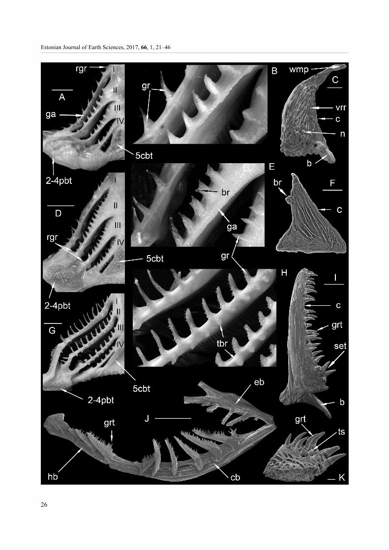
RESULTS The morphology of jaw teeth, teeth of pharyngeal tooth plates and cores of gill rakers of the European plaice Pleuronectes platessa, European flounder Platichthys flesus trachurus and turbot Scophthalmus maximus have basic similarities as well as significant differences. In the branchial region these fishes all have paired symmetrical gill arch bones that support pharyngeal tooth plates, gill rakers and gill filaments (e.g., studied by Frame et al. 1978; Sakamoto 1984; Yelnikov & Khanaychenko 2013). The gill arch skeleton of flatfishes comprises the following components: three pairs of hypobranchials, five pairs of ceratobranchials of which the 5th pair on the floor of the pharynx is tooth-bearing, four pairs of epibranchials and four pairs of pharyngo-branchials of which the 2nd, 3rd and 4th in the roof of the pharynx support a triad of tooth-bearing bony plates (Frame et al. 1978). The hypobranchial and the cerato-branchial form the lower limb of a branchial arch and the epibranchial forms the upper limb (Sakamoto 1984). The gill rakers are located on four gill arches, either in one or two rows, in the studied pleuronectiforms.
Order PLEURONECTIFORMES Family PLEURONECTIDAE Rafinesque, 1815
(cited in van der Laan et al. 2014)
Pleuronectes platessa Linnaeus, 1758 Figures 1A–C; 2–4; 5A
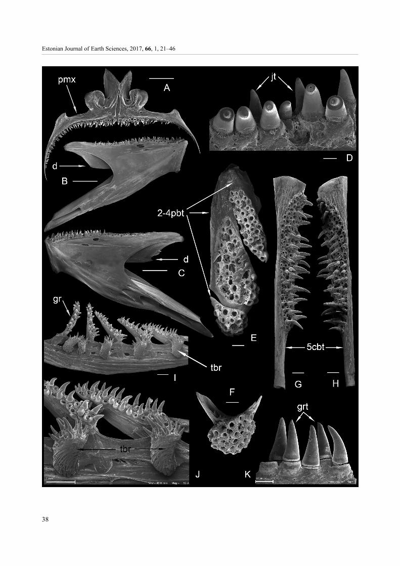
Jaw bones and teeth The mouth is asymmetrical and small. The blind (left) jaw bones, both the premaxilla and dentary, are signi-ficantly longer and bear more teeth (Fig. 2B, E) and also have stronger curvature than the eye-side (right) jaw bones (Fig. 2C, F). The blind-side premaxilla has 19–20 teeth and the dentary has 20–26 teeth, while the eye-side premaxilla has 3–6 teeth and the dentary has 4–7 teeth (Table 1), the higher number of teeth being in GIT 584-163. The teeth in the jaws are incisor-like. They occur in one row forming a cutting edge (term by Cooper & Chapleau 1998) on the blind side; the tooth row on the eye side of the premaxilla holds a somewhat posterior position as compared to the blind-side row. The posterior-
Estonian Journal of Earth Sciences, 2017, 66, 1, 21–46
26

T. Märss et al.: Gill rakers and teeth of Baltic pleuronectiforms
27
most teeth in the rows of the blind and eye sides of the upper and lower jaw bones become slightly roundish and conical with their tips curved towards the mouth cavity. The teeth, especially anteriorly placed ones, have a pillar-like bulge lingually. The teeth are equipped with an acrodine cap (Fig. 2D). Pharyngeal tooth plates and teeth On the floor of the pharynx the 5th ceratobranchials bear a tooth plate on each side. The plates in Fig. 2G have remained together due to a brief chemical treatment but the suture between them is still traceable. The plates on both sides have the shape of an elongate triangle (Fig. 2G in dorsal view), the medial and posterior margins forming an angle of 120–125° (in two Norwegian plaice specimens – 105–110°), the lateral, i.e., the longest margin being 10.5 mm long. The plates carry two rows of teeth. Specimen GIT 584-122 bears 16 (+5 shed) teeth on the left plate and 16 (+6 shed) teeth on the right plate (Table 1). The medial tooth rows form an angle similar to that of the plates. The roundish to irregular molariform teeth on plates are of different sizes, the biggest situated along the medial margin of the plates and the smallest along the posterior margin. Some additional counts on the 5th ceratobranchial tooth plates are in Table 1. In most specimens the number of teeth is slightly larger on the eye side.
On the roof of the pharynx the plaice has three tooth plates in line on each side (Fig. 2H, I). Three plates contact each other via overlapped and overlapping areas. In GIT 584-89 and GIT 584-122 the shape of the anteriormost plate is narrower anteriorly and smoothly widening posteriorly; its inner margin extends farther posteriorly than the outer one. This plate bears one complete row of teeth and one single roundish tooth. The middle plate is wider, extending arch-like laterally and anteriorly, where it has a rather wide margin free of teeth. It has two distinct oblique rows of teeth. The posteriormost plate is the smallest and narrowest, crescent-shaped and has a single row of teeth. The teeth follow the same course as on two anteriorly placed plates, running obliquely from anterior to posterior. The teeth on both the floor and roof tooth plates are rather large.
As on the floor, the teeth on the roof tooth plates also are not exactly of the same size. The biggest teeth are on the middle and the smallest on the posteriormost plate. The teeth have a roundish to irregularly molariform crown shape, with a crest on the surface (Fig. 2A). The crest may be a remnant of a developmental process or may function in weakening harder exoskeletons or shells. Teeth surfaces often show abrasion marks.
In the whole-head sample of the European plaice from southern Sweden treated in an earlier study, we documented small roundish to elongate low teeth with a marginal rim and with a convex crown surface equipped with a triangular sharp carinate ridge (Fig. 2J–L). Such teeth have fine, rather straight, tightly placed dentine tubules and a thin covering layer of transparent hyper-mineralized tissue.
Gill rakers and cores of rakers The gill rakers on the 1st, 2nd and 3rd arches occur in a single outer row, and on the 4th arch in two rows, the outer and the inner ones (Fig. 1A, B). The tallest rakers in the form of slender finger-like thornlets occur on the 1st arch; their size becomes shorter towards the 4th arch, the smallest rakers being on the inner row of that arch. Very short rudimentary rakers are placed at the very ends of all arches. The number of rakers starting on the left from the 1st to the 4th arch and then on the right side from the 4th to the 1st arch is as follows (the slash separates the numbers of rakers in outer and inner rows on the 4th arch): 10 + 8 + 7 + 5/3 on the blind side and 3/6 + 7 + 9 + 9 on the eye side for GIT 584-141, and 8 + 8 + 6 + 5/3 on the blind side and 3/6 + 6 + 8 + 9 on the eye side for GIT 584-144. The number of rakers on both sides is almost equal, being 1–2 rakers more on the eye side. There are 3 : 4 : 5 rakers per 1 cm on the 1st to the 3rd arch, respectively, in GIT 584-141.
In the European plaice the shape of the cores of dissociated gill rakers on the 1st arch, both on the eye side (EI in Fig. 3A–E) and blind side (BI in Fig. 4A–C), is simple. The bony cores of rakers appear as slender, relatively high, conical finger-like thornlets with the acuminate apex, which somewhat curves inwards, the peak often distorted due to its weak ossification; the
___________________________
Fig. 1. Terms and abbreviations of gill arches and gill rakers of three pleuronectiforms. A–C, Pleuronectes platessa Linnaeus;D–F, Platichthys flesus trachurus (Duncker); G–K, Scophthalmus maximus (Linnaeus). A = B, GIT 584-141; C, GIT 584-121-17;D = E, GIT 584-142; F, GIT 584-118-24; G = H, GIT 584-140; I, GIT 584-116-13; J, GIT 584-116-49; K, GIT 584-116-39. Scalebar for A, D, G is 1 cm, for C, F, K 200 µm, for I 1 mm and for J 5 mm; B is close-up of A; E is close-up of D; H is close-up of G.Abbreviations: b, base; br, branch of the gill raker core; c, core; cb, ceratobranchial; eb, epibranchial; ga, gill arch; gr, gill raker;grt, gill raker tooth; hb, hypobranchial; n, nodule on the raker core; rgr, rudimentary gill raker; set, higher set of tightly packedteeth; tbr, tubercle-shape gill raker; ts, tooth socket; vrr, vertical ribs of the gill raker core; wmp, weakly mineralized peak of thecore; 2-4pbt, tooth plates on the 2nd to 4th pharyngobranchials; 5cbt, tooth plates on the 5th ceratobranchials; I–IV, 1st to 4th gillarches with two rows of rakers on the 4th (A, D) arch, and on the 2nd and 3rd arches (G).

Estonian Journal of Earth Sciences, 2017, 66, 1, 21–46
28
Fig. 2. Pleuronectes platessa Linnaeus. A, close-up of a tooth from the left pharyngobranchial toothplate; B, C, lingual aspectof the left and right premaxillae; D, a jaw tooth; E, F, lingual aspect of the left and right dentaries; G, dorsal aspect of the leftand right lower pharyngeal tooth plates; H, I, ventral aspect of sets of left (H) and right (I) pharyngobranchial tooth plates;J–L, small single teeth from the unknown position in the mouth. A–H, GIT 584-89-54; B, GIT 584-163-1-1; C, GIT 584-163-1-2;D, GIT 584-89-49; E, GIT 584-163-2-1; F, GIT 584-163-2-2; G, GIT 584-122-3; H, GIT 584-89-54; I, GIT 584-122-4;J, GIT 584-94-45; K, GIT 584-94-33; L, GIT 584-94-34. Scale bar for A is 100 µm, for B, C, E–I 1 mm, for D 200 µm, forJ–L 200 µm; A is close-up of H. Abbreviations: ant., anterior; d, dentary; jt, jaw tooth; pmx, premaxilla. For other abbreviationssee Fig. 1.

T. Märss et al.: Gill rakers and teeth of Baltic pleuronectiforms
29
Fig. 3. Pleuronectes platessa Linnaeus. Cores of gill rakers from the 1st to 4th arches of the eye side (EI–EIV). A = E, GIT 584-121-3; B, GIT 584-121-4; C, GIT 584-121-2; D, GIT 584-121-1; F, GIT 584-121-8; G, GIT 584-121-10; H = I, GIT 584-121-11;J, GIT 584-122-6; K, GIT 584-121-15; L, GIT 584-121-16; M, GIT 584-121-21; N, GIT 584-121-20; O, GIT 584-121-23. E isclose-up of A; I is close-up of H. Scale bar for A, B is 1 mm, for C, D, F–O 200 µm. For abbreviations see Fig. 1.

Estonian Journal of Earth Sciences, 2017, 66, 1, 21–46
30
Fig. 4. Pleuronectes platessa Linnaeus. Cores of gill rakers from the 1st to 4th arches of the blind side (BI–BIV). A, GIT 584-121-5; B, GIT 584-121-7; C, GIT 584-121-6; D, GIT 584-121-12; E, GIT 584-121-13; F, GIT 584-121-14; G, GIT 584-121-17;H, GIT 584-121-18; I, GIT 584-122-9; J, GIT 584-121-25; K, GIT 584-121-26; L, GIT 584-89-44; M, GIT 584-89-47; N, GIT584-89-52; O, GIT 584-89-46; P, GIT 584-89-51; Q, GIT 584-89-48; R, GIT 584-89-45. Scale bar for A is 1 mm, for B–Q200 μm. For abbreviations see Fig. 1.

T. Märss et al.: Gill rakers and teeth of Baltic pleuronectiforms
31
basal part of the cores is slightly to moderately thickened. The base resting on the arch may be more extended or have a spur-like process (Figs 3C, 4A). Besides these slender and high cores, there occur also short conical ones (Fig. 3D). The cores of rakers differ in size, being 0.9–4.3 mm high (incl. spur a maximum 0.6 mm) on the 1st arch.
The shape of the cores of rakers on both the eye side (EII in Fig. 3F–J) and blind side (BII in Fig. 4D–F) of the 2nd arch is conical but with a stouter lower part and a fine, slender, relatively high upper part. The cores have a reversed L-shape in their upper parts, which is due to the soft tips allowing such a bend. In some cores the width is half the height (Fig. 4E), which varies from 0.7 to 2.5 mm. The basal spurs of cores may be very deep; for example, for a core height of 1.8 mm the spur can be 0.65 mm deep (Fig. 4D).
The shape of the cores of rakers on the eye side (EIII in Fig. 3K, L) and blind side (BIII in Fig. 4G–I) of the 3rd arch is rather similar to those on the 2nd arch. Their measured height is 0.5–1.6 mm plus a spur up to 0.4 mm deep. The surface structure of some cores is covered with a thin organic layer (Fig. 4I).
The cores of rakers on the 4th arch are smaller in the inner row than in the outer one and smaller than in any other rows (except rudimentary rakers). Their shape on the eye side (EIV in Fig. 3M–O) and blind side (BIV in Fig. 4J, K) is much more irregular than given above, so that their conical form is difficult to recognize. The height remains between 0.5 and 1.2 mm. A very few specimens have a horizontal spur (Fig. 4J).
The ossicles in Fig. 4L–R are from a whole-sample of the gill apparatus; they are well cleaned of organic matter in H2O2 and show sharp ribs on the surface. The peaks of cores have a distinct boundary between the ossified and non-ossified parts.
The core structure of gill rakers is of spongy bone with rather strong and long vertical ribs, which start at the base and either extend along the whole height
upwards, or more often, occur as short segments. The ribs are simple (Fig. 3E) and have a convex surface. Elongate grooves, shallow depressions, or deep hollows occur between the ribs. In some cases the ribs are curved at the bases of cores (Figs 3F, H, I; 4N, P). Often the cores have nodules in the lower half, especially on the 2nd arch (Figs 3F–J; 4F, J) but less so in other rakers (Figs 3D, K, O; 4G, J, M, P). Irregular, short, oblique connective segments tie the vertical ribs. The cores from the 4th arch are the most spongy.
A few cores were studied by thin-sectioning. As the core was hollow and cobweb-like, it was polished only from one side. In the reticular outer side no osteocyte lacunae were seen in these thin sections (Fig. 5A).
Platichthys flesus trachurus (Duncker, 1892)
Figures 1D–F; 5B–E; 6–8
The population of Platichthys flesus trachurus (Duncker) in the Baltic Sea has been considered as a valid sub-species of P. flesus (Linnaeus, 1758) by Ojaveer & Drevs (2003) and herein.
Jaw bones and teeth The jaw bones, the premaxilla and dentary, on the blind side (Fig. 6A, E) are longer and carry many more teeth than on the eye side (Fig. 6B, F; Table 2). The teeth are larger and stronger on the blind side than on the eye side; between the blind and eye sides there is a short gap in the rows of teeth. The teeth are mainly aligned in a row while a few teeth are found out of it, giving the dentition a wry-toothed appearance. The shape of the anterior larger teeth is incisor-like (Fig. 6K) but posteriorly they become smaller and their shape conical, with their crowns curved towards the middle of the mouth cavity (Fig. 6A, D–F). The pillar-like bulges seen lingually in anterior teeth are rather thin.
Table 1. Count of teeth on jaws and pharyngeal tooth plates of the European plaice Pleuronectes platessa. The count of tooth sockets or holes is given in parentheses. A dash indicates that no data are available. Abbreviations: ant., anteriormost; cb., ceratobranchial; dent., dentary; mid., middlemost; pb., pharyngobranchial; post., posteriormost; premax., premaxilla; B, blind side; E, eye side; * – specimens from the Norwegian Sea
Bones GIT 584-89 B
GIT 584-89 E
GIT 584-122 B
GIT 584-122 E
GIT 584-141* B
GIT 584-141* E
GIT 584-144* B
GIT 584-144* E
premax. – – 20 3 (19) (3) 19 5 dent. – – 20 6 (22) (4) 22 6 pb. ant. 5 – 6 5 4 5 4 4 pb. mid. 9 – 9 8(13) 10 8(10) 9 8 pb. post. 6 – 4 5 5 5 3 4 cb. 13 14 16(5) 16(6) 17 17 11 15

Estonian Journal of Earth Sciences, 2017, 66, 1, 21–46
32

T. Märss et al.: Gill rakers and teeth of Baltic pleuronectiforms
33
Teeth from the roof of the pharynx (Fig. 5B) and from the dentary (Fig. 5C) have very fine dentine tubules, which are very densely packed in the upper part of the teeth. Traces that are ramified with three branches or nearly straight occur at a certain level in these teeth. They are suggested to be the result of parasite activity.
Small, 0.5 mm long but low teeth are placed lingually next to the socket of each tooth along the tooth rows (herein figured left premaxilla in Fig. 6D; a single tooth is in 6C) representing possibly an early stage of replace-ment teeth. They were buried in the soft tissue but were uncovered by the chemical treatment of samples. Pharyngeal tooth plates and teeth A pair of tooth plates on the 5th ceratobranchials on the floor of the pharynx are in the shape of an elongate triangle, the medial and posterior margins forming an angle of 130–140°, the lateral, longest margin varying in length (7 and 10 mm in GIT 584-93 and GIT 584-123, respectively). The tooth plates consist of lamellar bone (Fig. 6G, H). They carry usually 40–80 teeth but the number may reach 112–114 (cb, Table 2). The finest teeth, in a maximum of three crowded rows, occur along the posterior margin, with their peaks curving anteriorly towards the tongue. These rows may not be at all visible if covered by soft tissue and not oxidized. The strongest teeth occur along the medial margins; they have ground-down roundish caps, often with scratches. Medium-sized teeth are distributed along the longest side and in the middle of plates. The finest teeth may greatly outnumber the larger ones. Acrodine caps top the teeth.
On the roof of the pharynx, three tooth plates are placed in a line on each side (Fig. 6I, the left triad, and Fig. 6J, a single right posteriormost tooth plate). They contact each other by means of overlapping and over-lapped marginal areas. The teeth on each plate are in rows in smaller fish. The larger fish (e.g., GIT 584-118-21 in Fig. 6I) have a more disordered arrangement of small and big teeth together; the teeth occur in an arch-shaped row along the posterior margin of the middle plate.
In addition to the teeth described above, the European flounder has very small single teeth in its mouth, which differ from the jaw teeth in being low and
stubby or high with a peaked or carinate crest (Fig. 6L–O). These separate teeth are composed of dentine and are covered with a stronger, transparent mineralized layer at the tip (Fig. 5D, E).
Gill rakers and cores of rakers Similar to the plaice, the flounder has one row of gill rakers on the 1st, 2nd and 3rd arches and two rows on the 4th arch. The highest rakers are on the 1st arch, their length (height) becoming lower on more lingually situated arches; the smallest rakers are on the inner side of the 4th arch, where also their number is the smallest. The rudimentary rakers situated at the ends of arches are the tiniest. The peaks of rakers point to the middle of the mouth cavity. The complete raker counts for three specimens are as follows (the slash separates the numbers of rakers in outer and inner rows on the 4th arch): 16 + 15 + 12 + 10/6 on the blind side and 6/10 + 12 + 15 + 16 on the eye side for GIT 584-142; 14 + 11 + 11 + 9/4 on the blind side and 5/9 + 11 + 12 + 14 on the eye side for GIT 584-143; 13 + 12 + 12 + 10/7 on the blind side and 8/10 + 11 + 12 + 13 on the eye side for GIT 584-150. There are 5 : 6 : 7 rakers per 1 cm on the 1st, 2nd and 3rd arches, respectively, in flounder GIT 584-142.
Most cores of the dissociated gill rakers on the 1st arch of both the eye side (EI in Fig. 7A–E) and blind side (BI in Fig. 8A–E) are slender cones with a few cores having a side branch (Fig. 7A, B). Weakly mineralized soft peaks bend towards the base to differing degrees. The bases of cores are concave to match the shape of the arch (Figs 7C, 8E). The cores of rakers on both the eye side and the blind side are 1–4 mm high including a short spur.
The cores of rakers of the 2nd arch on both the eye side and the blind side (EII in Fig. 7F–I; BII in Fig. 8F, G) are shorter than those on the first arch, cores having a side branch of considerable size (Figs 1D, E; 7F–I). The base is slightly convex, pillow-like (Fig. 7G, H) or nearly flat (Figs 7I, 8G) and may have a spur (Fig. 8F). The basal part of the core has, as characteristic of the flounder, a structure of twisted (tortuous) ribs, which may be short (Fig. 7G, H) or relatively long, bending around the core (Figs 7I; 8F, G). The height of the core of the rakers on the 2nd arch is 0.9–2.2 mm.
__________________________
Fig. 5. Thin sections of a gill raker and selected teeth. A, Pleuronectes platessa Linnaeus, section along the longer axis of the gillraker from EI (GIT 584-121-27). B–E, Platichthys flesus trachurus (Duncker): B, vertical section of a tooth from the tooth platein the roof of the pharynx (GIT 584-139-1); C, vertical section of a tooth from the dentary (GIT 584-139-2); D, E, small teethfrom the floor of the mouth in vertical (GIT 584-139-3) and horizontal (GIT 584-139-4) section. F–H, Scophthalmus maximus(Linnaeus) gill raker teeth of GIT 584-157-1; F shows the apical portion of a tooth. Scale bar 0.1 mm. GIT 584-121, Paslepa Bay;GIT 584-139, GIT 584-157, west of Saaremaa. Abbreviations: acr, acrodine; b, base; br pc, branch of the pulp cavity; dt, dentinetubules; dt acr, dentine tubules penetrating into acrodine (Ørvig 1978); gw, growth lines; l, limitation of acrodine; pc, pulp cavity;pd, pulp depression; vp, vestige of parasite activities.

Estonian Journal of Earth Sciences, 2017, 66, 1, 21–46
34
Fig. 6. Platichthys flesus trachurus (Duncker). A, B, lingual aspect of the left and right premaxillae; D, lingual aspect of a leftpremaxilla with (C) close-up of a small tooth from the lingual row; E, F, lingual aspect of the left and right dentaries; G, H, dorsalaspect of the left and right lower pharyngeal tooth plates; I, ventral aspect of the set of the left pharyngobranchial tooth plates;J, a posteriormost right pharyngobranchial tooth plate; K, teeth of the left dentary; L–O, small single teeth from the unknownposition in the mouth. A, GIT 584-123-3-1; B, GIT 584-123-3-2; C = D, GIT 584-138-1-1; E, GIT 584-123-2-1; F, GIT 584-123-2-2;G, GIT 584-93-19-1; H, GIT 584-93-19-2; I, GIT 584-118-21; J, GIT 584-118-18; K, GIT 584-93-1-1; L, GIT 584-93-17; M, GIT584-93-13; N, GIT 584-93-18; O, GIT 584-93-16. Scale bar for A, B, D–J is 1 mm, for C, K–O 100 μm. Abbreviations: d, dentary;pmx, premaxilla; jt, jaw teeth. For other abbreviations see Fig. 1.

T. Märss et al.: Gill rakers and teeth of Baltic pleuronectiforms
35
The appearance of the cores on the 3rd arch, due to their small height, is stout (EIII in Fig. 7J, K; BIII in Fig. 8H, I). All the other characteristics, the nearly flat (Figs 7J, 8I) or pillow-like base (Figs 7K, 8H) without or with a spur-like projection (Fig. 7K, less so in Fig. 8I), having the side branch (Fig. 7J), and twisted ribs in the basal part of cores (Figs 7K, 8H), are present. In Fig. 7J and 8I the basal part is covered by organic matter and not well or at all exposed. The height of the cores
remains between 0.6 and 1.9 mm (the latter includes a spur of 0.5 mm).
The 4th arch has rakers in two rows, the inner and outer ones. The inner row lies towards the 5th cerato-branchial, the outer row towards the 1st gill arch. The rakers on the outer row are somewhat larger (Fig. 1D ); their height is 1.2 mm, while the inner row rakers are 0.8 mm high. The cores of the rakers are 0.6–1.1 mm high. Also the cores from the 4th arch of the eye side
Fig. 7. Platichthys flesus trachurus (Duncker). Cores of gill rakers from the 1st to 4th arches of the eye side (EI–EIV). A = B,GIT 584-118-1; C, GIT 584-118-2; D = E, GIT 584-118-3; F = I, GIT 584-118-10; G, GIT 584-118-8; H, GIT 584-118-9;J, GIT 584-118-16; K, GIT 584-118-15; L, GIT 584-118-20. Scale bar for A, C, D, G–L is 200 µm, for B, E, F 100 μm.Abbreviation: twr, twisted ribs of gill raker core. For other abbreviations see Fig. 1.

Estonian Journal of Earth Sciences, 2017, 66, 1, 21–46
36
Fig. 8. Platichthys flesus trachurus (Duncker). Cores of gill rakers from the 1st to 4th arches of the blind side (BI–BIV).A, GIT 584-118-5; B, GIT 584-118-6; C = D, GIT 584-118-4; E, GIT 584-117-7; F, GIT 584-118-11; G, GIT 584-118-12;H, GIT 584-118-14; I, GIT 584-118-17; J, GIT 584-118-23; K, GIT 584-118-24; L, GIT 584-118-25; M, GIT 584-93-10;N, GIT 584-93-14. Scale bar for A is 1 mm, for B–N 200 µm. Abbreviation: twr, twisted ribs of gill raker core. For otherabbreviations see Fig. 1.

T. Märss et al.: Gill rakers and teeth of Baltic pleuronectiforms
37
and blind side may carry a side branch (Figs 7L, 8K), and they have twisted ribs in their basal part (Figs 7L; 8J, L). The rudimentary rakers in all arches are very tiny: 0.3–0.9 mm high measured on the arches of unoxidized samples.
The surfaces of cores are spongy and ‘lacy’. As a rule, vertical ribs cover the whole height of a core, starting at the base and ending at the peak (Figs 7A, C, D; 8A, B, K); connecting segments tie the ribs together. Figure 7M, N shows a side projection and a very distinct pattern of ribs. The vertical ribs in the flounder are straight and occur tightly and often have double, parallel riblets. Nodules rarely occur at the basal part; in most cases they are absent. Twisted ribs also cover the enlarged base. The spur for attachment to the arch is the deepest part of the base.
Family SCOPHTHALMIDAE Chabanaud, 1934
Scophthalmus maximus (Linnaeus, 1758) Figures 1G–K; 5F–H; 9–11
Jaw bones and teeth
On the eye-side premaxilla the tooth row extends a little more posteriorly than on the blind side, while on the eye- and blind-side dentaries the tooth rows are almost of the same length (Fig. 9A–C). The turbot has the premaxilla with a little steeper curvature anteriorly on the blind side than on the eye side, where it is gently curved. The dentary has steeper curvature anteriorly on the eye side than on the blind side. The premaxilla and dentary carry teeth, which are just slightly larger and stronger anteriorly on both jaw bones while posteriorly they become smaller. The teeth are distributed in one long full row and two to four indistinct shorter rows anteriorly. The number of rows depends on the age (total length) of the fish. Fewer rows occur in smaller specimens, e.g., two rows in a 7.2 cm long specimen
GIT 584-131 and up to four indistinct rows in a 36.0 cm long specimen GIT 584-132. The teeth are conical (Fig. 9D), with crown peaks curved strongly towards the middle of the mouth cavity. The teeth are topped by an acrodine cap. The number of teeth in some specimens is given in Table 3. Pharyngeal tooth plates and teeth
Three tooth plates in line on both sides on the roof of the pharynx contact each other, overlapping the adjacent plate. The smallest fish GIT 584-131 has two rows and three tooth rows, respectively, on the anteriormost and middle plates; the posteriormost one bears just a few teeth. Larger specimens have rather disordered rows of small and big teeth together. The SEM images illustrate the triad from the eye side of GIT 584-117 and a single plate from the blind side of GIT 584-88 (Fig. 9E, F). The teeth on the anteriormost elongate plate, on the middle plate of complicated configuration and on the posteriormost roundish plate form disarranged oblique rows, evidenced by the remaining tooth sockets and a few surviving teeth pointing towards the pharynx. Specimen GIT 584-117 has approximately five tooth rows on the anteriormost plate and approximately seven rows on the middle plate. The teeth are less numerous in the oldest/ largest fish, with plenty of empty space and sockets between them. Such contradiction may be a result of more loosely attached teeth which were lost during processing, or maybe they were frequently lost during feeding (see comment on p. 43). The numerical data on tooth number of a few specimens are given in Table 3.
On the floor of the pharynx the tooth plates on the fifth ceratobranchials are elongate rod-like structures, 18.0 and 18.5 mm long and 3.0 and 2.8 mm wide at one end and 1.1 mm wide at the other end in GIT 584-116 (Fig. 9G, H). The plates gradually narrow posteriorly with just a slight curve at the end of the toothed area along the inner margin. The number of teeth varies,
Table 2. Number of teeth on jaws and pharyngeal tooth plates of the European flounder Platichthys flesus trachurus. The counts of tooth sockets or notches are given in parentheses. Abbreviations: pb., pharyngobranchial; cb., ceratobranchial; dent., dentary; premax., premaxilla; B, blind side; R, eye side; ant., anteriormost; mid., middle; post., posteriormost
Dextral Sinistral Bones
GIT 584-138 B
GIT 584-138 E
GIT 584-142 B
GIT 584-142 E
GIT 584-143 B
GIT 584-143 E
GIT 584-150 E
GIT 584-150 B
premax. 22 13 17 9 22 10 10 20 dent. 23 17 17(2) 11(2) 22 17 14 23 pb. ant. 15 14(2) 15 14 9 11 11 12 pb. mid. 16(3) 15(?) 17 17 20 18 14 17 pb. post. 15(2) 21(4?) 17 21 13 12 10 9 cb. 112(8) 114(17) 67 75 65 65? 41(5) 42(5?)
Estonian Journal of Earth Sciences, 2017, 66, 1, 21–46
38

T. Märss et al.: Gill rakers and teeth of Baltic pleuronectiforms
39
depending mainly on the age/length of the fish and may reach a hundred (cb, Table 3). The largest (tallest) teeth occur along the inner margin, with the peaks curved posteriorly towards the pharynx. Gill rakers and cores of gill rakers The rakers on the 1st arch form one row on the outer upper edge of that arch but a single raker may occur on its inner side (Fig. 1G, H, J). The 2nd and the 3rd arch are the most complete because both the inner and outer rows of rakers are present (Figs 1G, H; 9I, J). The 4th arch has one short row on the outer side (Fig. 1G). The tiniest rudimentary rakers are preserved only in chemically untreated fish samples. Besides the single raker on the inner side of the 1st arch, such can also be found on the outer side of the 5th ceratobranchial. The tips of rakers on the outer side of the 1st to the 4th arch slant towards the tongue but the teeth of gill rakers curve towards the pharynx; the rakers (tubercles) on the inner row of the 2nd and the 3rd arch of the turbot stand at a right angle (Figs 1G, H; 9I, J).
The gill rakers of the turbot are complex structures (Fig. 1G–K), which on the 1st arch have a high, triangular ‘sail’-shape. On the 2nd, 3rd and 4th arches the ‘sails’ become gradually lower. The rakers situated on the inner sides of the 2nd and the 3rd arch are much smaller and tubercle-like. Unlike the rakers of the plaice and flounder, which lack teeth, the rakers of the turbot have very small conical teeth standing in tooth sockets; the teeth are of similar size to those on the tooth plates of the 5th ceratobranchials on the floor and pharyngobranchial tooth plates on the roof of the pharynx. The ‘sail’-shaped rakers lie obliquely on all the outer rows, while their peaks point anteriorly towards the tongue (Fig. 1G). The teeth of these rakers on the lower limb point to the roof of the mouth cavity, and the teeth of rakers on the upper limb to the opposite, the floor. The tubercle-shaped rakers on the inner side of the 2nd arch also lie obliquely, while those on the 3rd arch rest crosswise (Fig. 1G, J).
The complete raker count from the left to right of three specimens of different sizes is as follows: 15/2+ 11/9 + 9/7 + 6/0 on the left side and 0/5 + 7/9 + 8/11 + 1/16 on the right side of GIT 584-131 (‘baby’-turbot); 17/2 + 14/12 + 10/8 + 7/0 on the left side and 0/7 + 10/10 + 10/14 + 1/18 on the right side of GIT 584-140;
16/2 + 12/9 + 10/8 + 6/0 on the left side and 0/6 + 6/8 + 10/13 + 2/16 on the right side of GIT 584-132.
An additional low tubercle occurs at the outer side of the 5th ceratobranchials of both larger specimens. As the fish aged, a few rakers were added to an arch but the change in the size of rakers is conspicuous; they may grow more than three-fold. The tiniest rudimentary rakers were found in a 7.2 cm long young turbot.
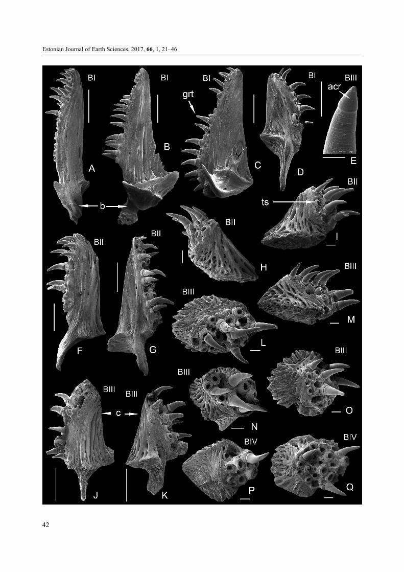
The cores of dissociated gill rakers on the 1st arch on the eye side (EI in Fig. 10A–E) and blind side (BI in Fig. 11A–D) do not differ very strongly. Measurements taken from GIT 584-116 show that on the eye side the bony core of rakers is 1.2–8.5 mm high, including the spur 1.5 mm high, and on the blind side it is 2–6 mm high, including the spur. The cores are smaller at both ends of the gill arches, i.e., on the hypobranchial and epibranchial bones, and the largest on the ceratobranchials. Small sets of teeth which resemble tubercles occur at the lower part of the core above the basal spur (Figs 1I, 10A). The bases of the highest cores are distinct and concave (Fig. 10A, B, D), with a spur directed obliquely or straight downwards. Rakers from the blind side may have a basal projection directed to the side and shaped like a scoop (Fig. 11B, C).
The cores of the gill rakers of the 2nd arch on both the eye side (EII in Fig. 10F–L) and blind side (BII in Fig. 11F–I), originating from the inner and outer sides of that arch, are well differentiated by size. The tall (Figs 10F–H; 11F, G) cores have a maximum measured height of 5.2 mm (for GIT 584-116) and the short, tubercle-like cores (Figs 10I–L; 11H, I) have a maximum measured height of 1.8 mm for the same fish. The bases of high cores have a spur and those of short tubercle-shaped cores are slightly convex or nearly flat. The cores of the rakers from the outer side of the 2nd arch have smooth surfaces (Figs 10F–H; 11F, G), like the cores from the 1st arch. The rakers on the 1st arch also have a set of small teeth at the lower part above the basal spur (Fig. 10F).
Two types of cores occur on the 3rd arch but their height differences are much smaller than in the previously described cores on the 2nd arch. If found isolated, the most distinguishable feature is the triangular shape and smooth surfaces of the cores from the outer row (Figs 10M, O; 11J, K) and the low, tubercle-shaped appearance for the inner row. The first cores of rakers
__________________________
Fig. 9. Scophthalmus maximus (Linnaeus). A, lingual aspect of the left and right premaxillae; B, lingual aspect of the left andC, the right dentary; D, close-up of the left dentary; E, ventral aspect of the set of left pharyngobranchial tooth plates; F, ventralaspect of the posteriormost pharyngobranchial tooth plate; G, H, dorsal aspect of the left and right lower pharyngeal tooth plates;I, J, gill rakers from BII; K, teeth on the gill raker core from BI. A, GIT 584-116-47; B–D, GIT 584-116-48-1; C, GIT 584-116-48-2;E, GIT 584-117-4; F, GIT 584-88-7; G, GIT 584-116-43-1; H, GIT 584-116-43-2; I, J, GIT 584-116-50; K, GIT 584-116-14.Scale bar for A–C is 5 mm, for D, K 200 µm, for E–J 1 mm. Abbreviations: d, dentary; jt, jaw teeth; pmx, premaxilla. For otherabbreviations see Fig. 1.

Estonian Journal of Earth Sciences, 2017, 66, 1, 21–46
40

T. Märss et al.: Gill rakers and teeth of Baltic pleuronectiforms
41
have almost a flat basal surface or a spur-like projection at the base. The height of the cores remains between 0.6 and 3.5 mm (the latter includes a spur of 1.0 mm), while the height may be less than the horizontal measurement (width) of an element. The 4th arch has cores on the outer side, which are rather low but still triangular in shape.
The cores of the rakers on the first arch have a smooth compact surface of somewhat stronger bone tissue (Figs 10A, B, F, G; 11A–C, F, G). The cores of tubercles from the inner side of the 2nd and 3rd arches have a ‘lacy’ spongy structure with relatively long ribs running from the base towards the tip; the ribs are connected by segments (Figs 10I–L; 11H, I). The lacy structure is also characteristic of the cores from the inner row of the 3rd arch (Figs 10P; 11L, M–O) and of the 4th arch (Figs 10Q, R; 11P, Q). The cores have radially arranged ribs in the basal part (Figs 10Q, R; 11P, Q). The cores are 0.6–1.4 mm wide measured horizontally.
The teeth on the rakers of the turbot are true teeth similar to jaw teeth, with well-developed dentine tubules arising from the pulp cavity and covered with an acrodine cap (Fig. 5F–H). The contact surface between the acrodine and dentine is distinct (Figs 5F–H, 9D).
The attachment to the sockets differs in jaw teeth vs those of raker cores (compare Fig. 9K and 9D) and tooth plates. The teeth of rakers and tooth plates appear to have a much sharper line of attachment – there is no zone of ‘bone of attachment’ and probably only a ligamentous connection (Type 2 tooth attachment of Fink 1981).
DISCUSSION As reviewed above, the bony or cartilaginous gill rakers on the gill arches function in feeding, and the number of rakers on arches, their density and shape, have been said to be good indications of a fish’s diet (e.g., de Groot 1971; Abuzinadah 1995; Amundsen et al. 2004; Salman et al. 2005; Elsheikh 2013). Thus, a similar diet and similar rakers are strongly correlated, especially among closely related species. Yazdani (1969) also found correlated differences in bone, muscle and tooth morphology among the jaws of a variety of species of pleuronectiforms, associated also with the diet.
Kobelkowski (2007, pp. 47–48) found that (1) the families Bothidae, Paralichthyidae, Achiridae and Cynoglossidae have longer upper and lower jaws on the
blind side than on the eye side, (2) common to the Bothidae and Paralichthyidae is to have a slight curvature of the mouth on both sides, and to share a similar number and size of teeth on the premaxilla and dentary on both sides and (3) common to the Achiridae and Cynoglossidae is to have a stronger curvature of the mouth on the blind side, more abundant teeth on the premaxilla and dentary of the blind side and few teeth or no teeth on the eye side. Among members of the family Pleuronectidae, the mouth of the European flounder is asymmetrical and small but somewhat larger than that of the European plaice. In always dextral plaice and mainly dextral flounder the blind-side premaxilla and dentary are significantly longer than on the eye side, as in the four families studied by Kobelkowski (2007). They bear more teeth and have a much stronger curvature on the blind-side jaw bones than on the eye-side jaw bones, as in the Achiridae and Cynoglossidae (ibid.). The European flounder bears more teeth in rows than does the European plaice. The teeth occur in one complete row in jaws of both the plaice and flounder, but the flounder has teeth situated also lingually of that row. The jaw teeth of both taxa are incisor-like anteriorly, with a strong pillar-like bulge lingually in the plaice.
In the family Scophthalmidae, the mouth of the turbot is slightly asymmetrical and small, but larger than in the plaice or flounder. It has a premaxilla with a slightly steeper curvature anteriorly on the blind side than on the eye side; the eye-side premaxilla is just gently curved. Tooth rows extend more posteriorly on the eye-side premaxilla than on the blind-side premaxilla, while on the dentary the tooth rows are nearly of the same length on the eye and blind sides. These features are closer to those of the Bothidae and Paralichyidae studied by Kobelkowski (2007). The teeth in the turbot occur in one to two complete, long rows and up to four shorter rows anteriorly on the jaws. The teeth of the turbot on jaws, tooth plates and (only in the turbot in this study) on gill rakers are conical, fine and have a hollow pulp cavity, from which dentine tubules run towards the outer surface. Most of the teeth exhibit well-developed, smooth, shiny caps interpreted as the characteristic acrodine caps of actinopterygians (e.g., Ørvig 1978). The tooth structure, whether on jaws, tooth plates, or gill rakers, is that of true teeth (Sire & Huysseune 2003).
Fink (1981) classified several pleuronectiform species as having Type 2 teeth (tooth not fully ankylosed to the bone, but with a small area of unmineralized collagen
_________________________
Fig. 10. Scophthalmus maximus (Linnaeus). Cores of gill rakers from the 1st to 4th arches of the eye side (EI–EIV). A, GIT 584-116-13; B = C, GIT 584-116-12; D, GIT 584-116-10; E, GIT 584-116-11; F, GIT 584-116-18; G, GIT 584-116-19; H, GIT 584-116-21; I, GIT 584-116-22; J, GIT 584-116-20; K, GIT 584-116-23; L, GIT 584-116-24; M, GIT 584-116-30; N, GIT 584-116-33;O, GIT 584-116-32; P, GIT 584-116-35; Q, GIT 584-117-1; R, GIT 584-117-3. Scale bar for A, B, F, G, M is 1 mm, for C 100 µm,for D, E, H–L, N–R 200 µm. For abbreviations see Fig. 1.
Estonian Journal of Earth Sciences, 2017, 66, 1, 21–46
42

T. Märss et al.: Gill rakers and teeth of Baltic pleuronectiforms
43
at its base), both in their jaws and on their pharyngeal tooth plates. However, on the oral jaws of the plaice (Fig. 2B, C, E, F), flounder (Fig. 6A, B, D–F) and turbot (Fig. 9A–D) the teeth are, perhaps, more firmly attached than on the rakers and tooth plates; radiating ridges around the base of the jaw tooth indicate ligaments or ‘bone of attachment’. However, there is still a visible division between the crown and the base, unlike the true Type 1 tooth attachment of Fink (1981). The more loosely attached raker teeth and probably also the tooth-plate teeth are more likely to be detached from their bases during specimen processing and during life, allowing the prey to pass into the stomach.
Three pharyngobranchials (2nd to 4th) and the 5th ceratobranchials of the gill arch skeleton of flatfishes support tooth-bearing plates: a triad of tooth plates on each side on the roof and a pair of tooth plates on the floor of the pharynx. No plates are fused together. The movable 2nd to 4th plates on the roof of the pharynx are conjoined by means of overlapped areas; the plates of the 5th ceratobranchials on the floor of the pharynx are tied by a ligament. Although the pattern of plates is similar, the shapes, numbers and sizes of teeth on the pharyngeal bones depend on the taxon. The lower tooth plates are triangular in the European plaice and European flounder and strongly elongate and rectangular in the turbot. In the plaice the medial and posterior margins of tooth plates on the 5th ceratobranchials form an angle of 120–125° in the Baltic Sea specimens (105–110° in two plaice from the Norwegian Sea). The number of teeth is low (16 + 5 or + 6) and they are large. In the flounder the angle between the medial and posterior margins is 130–140°; the tooth plates carry usually 40–80 teeth but the number may reach 112–114. The same applies to the
teeth on the upper tooth plates – the plaice has fewer teeth of rather similar shape but larger, in contrast to the flounder, which has a higher number, smaller size and more variable shape of teeth. The orientation of tooth peaks on the lower tooth plates in the flounder is interesting, with high and fine teeth along the posterior margins pointing anteriorly, which should counteract the prey moving to the oesophagus, but perhaps serve to ‘rake’ the prey so as to damage its exoskeleton.
The number of gill raker rows on arches is similar in the two pleuronectids, with the European plaice and European flounder both having one row on the 1st, 2nd and 3rd arches and two rows on the 4th arch. In the turbot the number of raker rows as well as the raker number per arch and their shapes are strikingly different from those of the plaice and flounder. The turbot has one row of rakers on the outer edge of the 1st arch plus a few rakers on its inner edge; the 2nd and the 3rd arch have both inner and outer rows; the 4th arch has one short row of rakers on the outer surface.
The height of the gill rakers is largest on the first gill arch and smallest on the fourth arch; the number of rakers decreases in the same direction, from the 1st to the 4th arch. The number of gill rakers in the European plaice on the left (= blind) and right (= eye) sides is almost equal, with just a few (1–2) more rakers on the eye side. This number in the European flounder is either equal or insignificantly different, as in the plaice, with a few more rakers in the eye side. The rakers are spaced a little more tightly in the flounder than in the plaice.
The oxidation method provided us with previously unavailable material revealing new features of the bony cores of gill rakers, which may contribute new species- or genus-specific characters. The new material showed
Table 3. Approximate numbers of teeth on jaws and pharyngeal tooth plates of the turbot Scophthalmus maximus. Abbreviations: pb., pharyngobranchial; cb., ceratobranchial; dent., dentary; premax., premaxilla; E, eye side; B, blind side; ant., anteriormost; mid., middle; post., posteriormost
Bones GIT
584-131 E GIT
584-131 B GIT
584-132 E GIT
584-132 B GIT
584-140 E GIT
584-140 B
premax. 50 45 100 78 124 112 dent. 46 46 97 93 100 92 pb. ant. 11 11 42 40 62 63 pb. mid. 12 16 47 42 72 58 pb. post. 8 9 18 18 29 24 cb. 15 15 77 89 100 84
__________________________
Fig. 11. Scophthalmus maximus (Linnaeus). Cores of gill rakers from the 1st to 4th arches of the blind side (BI–BIV). A, GIT584-116-16; B, GIT 584-116-14; C, GIT 584-116-15; D, GIT 584-116-17; E = M, GIT 584-116-39; F, GIT 584-116-25; G, GIT584-116-26; H, GIT 584-116-29; I, GIT 584-116-28; J, GIT 584-116-37; K, GIT 584-116-36; L, GIT 584-116-41; M, GIT 584-116-39; N, GIT 584-116-40; O, GIT 584-116-38; P, GIT 584-116-45; Q, GIT 584-116-46. Scale bar for A–D, F, G, J, K is 1 mm,for E 100 µm, for H, I, L–Q 200 µm. Abbreviation: acr, acrodine cap. For other abbreviations see Fig. 1.

Estonian Journal of Earth Sciences, 2017, 66, 1, 21–46
44
no notable morphological differences in cores between the eye side and the blind side of these three asymmetrical fish species except small differences in the height of the rakers. However, the raker cores differed significantly in shape and structure among the taxa. The raker cores of the European flounder have a side branch in their upper parts; such are absent in the plaice. The basal part of the core has a characteristic (of the flounder) structure of horizontally (in relation to the longer vertical axis of the core) twisted (tortuous) ribs of variable length. In the European plaice the cores of rakers often have nodules in the lower half, especially on the 2nd arch but less so in other rakers. The surfaces of vertical ribs on the cores of the plaice are simple, while the rib surface in the flounder has two fine parallel riblets. The tooth-bearing raker cores of the turbot have a smooth, compact surface on the 1st arch, but a lacy, ribbed structure on the 2nd and 3rd arches.
CONCLUSIONS
In this work we extend our microichthyological research to emphasize the morphology of jaw teeth, pharyngeal teeth, gill rakers and their bony cores in flatfishes. Three flatfish taxa from the Baltic Sea, the European plaice Pleuronectes platessa, European flounder Platichthys flesus trachurus (Pleuronectidae) and turbot Scophthalmus maximus (Scophthalmidae) were studied and compared. The investigation of the European plaice, a very rare element of the ichthyofauna, was complemented with specimens from the southern Baltic (Denmark and Southern Sweden) and Norwegian Sea.
We have answered the question, ‘Can gill rakers and teeth of pleuronectiforms be useful in taxonomy and phylogeny?’ We have found that specimens revealed features that separate the plaice from the flounder, and distinctively characterize the turbot, which has ossicles completely different from the others. We discovered that the morphology of the mineralized substance of the gill rakers as well as that of the teeth on jaws and branchial tooth plates have taxon-specific characters (studied here at the generic level). Reliable characteristics are the shapes of raker cores, the pattern of fine structures on raker cores and the size, shape and placement of teeth in jaws and tooth plates. Among the three studied pleuronectiforms only one (turbot) has true teeth on its rakers.
This microichthyological study along with our earlier investigations (Märss et al. 2015) allows the identification of pleuronectiforms from their dermal tubercles, the morphology and arrangement of teeth on jaws and on tooth plates in the pharynx, the gill rakers covered with soft tissue, with or without teeth, and the bony cores of gill rakers. When similar detailed SEM
studies are completed in related taxa, these features can be utilized not only in taxonomy but also in phylogeny, palaeontology and zooarchaeology. We can also confirm the previous conclusions of many authors that ossicle characters can be useful when estimating and under-standing different feeding preferences and relevant differences occurring in jaw teeth, branchial tooth plates and gill rakers. We further conclude that the teeth found on branchial toothplates in all three studied species and in the turbot on the gill rakers are true teeth (Sire & Huysseune 2003) with acrodine caps and Type 2 tooth attachment (Fink 1981).
Acknowledgements. The Estonian Marine Institute (EMI) in Tartu is thanked for finances and the Institute of Geology for study facilities to TM. The study devices (microscopes, computer) were obtained under Estonian Science Foundation grant 7334 (2008–2010): ‘Ultrasculpture on the exoskeleton of recent fishes, and its value in the taxonomy and systematics of fishes’. Funds for publication were contributed by Natural Sciences and Engineering Research Council of Canada (NSERC) Discovery Grant A9180 to MVHW. We acknowledge the help by the Commission on Ichthyological Terminology of the EMI with discussing the Estonian terms that were used for the first time in this paper devoted to the microichthyology. Valdek Mikli, Mart Viljus and Janek Lees are thanked for taking SEM images, Gennadi Baranov for preparing images for publication and Tenno Drevs and Tarmo Luks for providing fish specimens. Dr Alexander Bannikov and two anonymous referees are thanked for objectively commenting on and helping to improve the manuscript. The publication costs of this article were covered by the Estonian Academy of Sciences.
REFERENCES
Abuzinadah, O. A. 1995. Gill raker morphology in some Red
Sea fishes of different feeding preferences. Journal of King Abdulaziz University: Marine Sciences, 6, 93–122.
Amundsen, P.-A., Bøhn, T. & Våga, G. H. 2004. Gill raker morphology and feeding ecology of two sympatric morphs of European whitefish (Coregonus lavaretus). Annales Zoologici Fennici, 41, 291–300.
Blieck, A. & Turner, S. 2000. IGCP 328: Palaeozoic Micro-vertebrates final scientific report – Introduction. In Palaeozoic Vertebrate Biochronology and Global Marine/ Non-Marine Correlation. Final Report of IGCP 328 (1991–1996) (Blieck, A. & Turner, S., eds), Courier Forschungsinstitut Senckenberg, 223, 1–67.
Coad, B. W. & McAllister, D. E. 2015. The Dictionary of Ichthyology; http://www.briancoad.com/dictionary/complete dictionary.htm, revised 3 October 2015 [accessed 10 November 2015].
Cooper, J. A. & Chapleau, F. 1998. Monophyly and intrarelationships of the family Pleuronectidae (Pleuronectiformes), with a revised classification.

T. Märss et al.: Gill rakers and teeth of Baltic pleuronectiforms
45
Fishery Bulletin, National Oceanic and Atmospheric Administration, 96, 686–726.
De Groot, S. J. 1971. On the interrelationships between morphology of the alimentary tract, food and feeding behaviour in flatfishes (Pisces: Pleuronectiformes). Netherlands Journal of Sea Research, 5, 121–196.
Duncker, G. 1892. Der Elbbutt, eine Varietät der Flunder. (Pleuronectes flesus L. var. leiurus). Schriften des Naturwissenschaftlichen Vereins für Schleswig-Holstein, 9, 275–291.
Eastman, J. T. 1977. The pharyngeal bones and teeth of catostomid fishes. American Midland Naturalist, 97, 68–88.
Elsheikh, E. H. 2013. Scanning electron microscopic studies of gill arches and rakers in relation to feeding habits of some fresh water fishes. The Journal of Basic & Applied Zoology, 66, 121–130.
Fabricius, O. 1780. Fauna groenlandica, systematice sistens animalia Groenlandiae occidentalis. Copenhagen and Leipzig. i–xvi + 1–452, 1 pl.
Fink, W. L. 1981. Ontogeny and phylogeny of tooth attach-ment modes in actinopterygian fishes. Journal of Morphology, 169, 167–184.
Frame, D. W., Andrews, T. J. & Cole, C. F. 1978. Osteology of the American Plaice, Hippoglossoides platessoides. Postilla, 173, 1–32.
Froese, R. & Pauly, D. (eds). 2015. FishBase. World Wide Web electronic publication; www.fishbase.org, version (10/2015) [accessed 10 September 2016].
Hadi, A. A. & Alwan, S. F. 2012. Histopathological changes in gills, liver and kidney of fresh water fish, Tilapia zillii, exposed to aluminum. International Journal of Pharmacy and Life Sciences, 3, 2071–2081.
Hoshino, K. & Amaoka, K. 1998. Osteology of flounder, Tephrinectes sinensis (Lacepede) (Teleostei: Pleuronecti-formes), with comments on its relationships. Ichthyological Research, 45, 69–77.
Hughes, G. M. 1984. General anatomy of the fish gills. In Fish Physiology (Hoar, W. S. & Randall, D. J., eds), pp. 1–72. Academic Press, London.
Kashulin, N. (comp.). 1997. The Pasvik River System: Pollution Impacts and Responses of Freshwater Fish. Institute of North Industrial Ecology Problems, INEP, Apatity, in cooperation with Norwegian College of Fishery Science, University of Tromse, Svanhovd Environmental Centre and Norwegian Institute For Water Research, NIVA, Oslo.
Kobelkowski, A. 2007. Morphology of the digestive system of the Mexican flounder, Cyclopsetta chittendeni (Teleostei: Paralichthyidae). Bulletin of Fish Biology, 5, 39–49.
Lees, J., Märss, T., Wilson, M. V. H., Saat, T. & Špilev, H. 2011. The sculpture and morphology of postcranial dermal armor plates and associated bones in gasterostei-forms and syngnathiforms inhabiting Estonian coastal waters. Acta Zoologica, 93, 422–435.
Linnaeus, C. 1758. Systema naturae per regna tria naturae, secundum classes, ordinus, genera, species, cum charac-teribus, differentiis, synonymis, locis. Tomus I. Editio decima, reformata. Impensis Direct. Laurentii Salvii, Holmiae, 532 pp.
Liston, J. 2013. The plasticity of gill raker characteristics in suspension feeders: implications for Pachycormiformes. In Mesozoic Fishes 5. Global Diversity and Evolution (Arratia, G., Schultze, H.-P. & Wilson, M. V. H., eds),
pp. 121–143. Verlag Dr. Friedrich Pfeil, München, Germany.
Märss, T., Wilson, M. V. H. & Thorsteinsson, R. 2006. Silurian and Lower Devonian thelodonts and putative chondrichthyans from the Canadian Arctic Archipelago. Special Papers in Palaeontology, 75, 1–140.
Märss, T., Turner, S. & Karatajūte-Talimaa, V. 2007. “Agnatha” II. Thelodonti. In Handbook of Paleoichthyology, Vol. 1B (Schultze H.-P., ed.), pp. 1–143. Verlag Dr. Friedrich Pfeil, München.
Märss, T., Lees, J., Wilson, M. V. H., Saat, T. & Špilev, H. 2010a. The morphology and sculpture of ossicles in the Cottidae (Teleostei) of the Baltic Sea. Estonian Journal of Earth Sciences, 59, 216–237.
Märss, T., Lees, J., Wilson, M. V. H., Saat, T. & Špilev, H. 2010b. The morphology and sculpture of ossicles in the Cyclopteridae and Liparidae (Teleostei) of the Baltic Sea. Estonian Journal of Earth Sciences, 59, 263–276.
Märss, T., Afanassieva, O. & Blom, H. 2014. Biodiversity of the Silurian osteostracans of the East Baltic. Earth and Environmental Science Transactions of the Royal Society of Edinburgh, 105, 73–148.
Märss, T., Wilson, M. V. H., Lees, J., Saat, T. & Špilev, H. 2015. The morphology and ultrasculpture of small ossicles in the Pleuronectiformes (Teleostei) of the Baltic Sea, a comparative SEM study. Proceedings of the Estonian Academy of Sciences, 64, 495–517.
Meunier, F. J. 2012. Comparative morphological and histological structure of “coalesced teeth” in extant and extinct teleosts: an adaptation to durophagy. In Actes des 13èmes Journees des Sciences de la Mer et 2ème Rencontre Tuniso-française d’Ichthyologie (Mahdia, Tunisie 26–29 novembre 2011), Bulletin de l’ Institut National des Sciences et Technologies de la Mer (I.N.S.T.M.Salammbô), Numéro Spécial, 15, 28–34.
Meunier, F. J., Brito, P. M. & Leal, M.-E. C. 2013a. Morpho-logical and histological data on the structure of the lingual toothplate of Arapaima gigas (Osteoglossidae; Teleostei). Cybium, 37, 263–271.
Meunier, F. J., Dutheil, D. & Brito, P. M. 2013b. Histological study of the median lingual dental plate of the Cretaceous fish †Palaeonotopterus greenwoodi (Teleostei: Osteo-glossomorpha) from the Kem-Kem beds, Morocco. Cybium, 37, 121–125.
Munshi, J. S. D., Ojha, J., Ghosh, T. K., Roy, P. K. & Mishra, A. K. 1984. Scanning electron microscopic observations on the structure of gill-rakers of some freshwater teleostean fishes. The Proceedings of the Indian National Academy of Sciences, B50, 549–554.
Nelson, J. S., Grande, T. C., & Wilson, M. V. H. 2016. Fishes of the World, 5th Edition. John Wiley & Sons, Hoboken, New Jersey, 707 pp.
Ojaveer, E. & Drevs, T. 2003. Flounder, Platichthys flesus trachurus (Duncker). In Fishes of Estonia (Ojaveer, E., Pihu, E. & Saat, T., eds), pp. 362–370. Estonian Academy Publishers, Tallinn.
Ørvig, T. 1978. Microstructure and growth of the dermal skeleton in fossil actinopterygian fishes: Birgeria and Scanilepis. Zoologica Scripta, 7, 33–56.
Pichugin, M. Yu. & Sidorov, L. K. 2006. On the number and form of gill rakers in Sakhalin trout Parahucho perryi. Journal of Ichthyology, 46, 133–135.
Estonian Journal of Earth Sciences, 2017, 66, 1, 21–46
46
Rafinesque, C. S. 1815. Analyse de la nature, ou tableau de l’univers et des corps organises. Palerme, 224 pp.
Sakamoto, K. 1984. Interrelationships of the family Pleuro-nectidae (Pisces: Pleuronectiformes). Memoirs of the Faculty of Fisheries Hokkaido University, 31, 95–215.
Salman, N. A., Al-Mahdawi, G. J. & Heba, H. M. 2005. Gill rakers morphometry and filtering mechanism in some marine teleosts from Red Sea coasts of Yemen. Egyptian Journal of Aquatic Research, 31, 286–296.
Scofield, E. C. 1934. Food of the Larvae and Adult Sardines. Calisphere, California Digital Library, University of California, 45 pp. http://content.cdlib.org/view?docId= kt9c6006rh&chunk.id=d0e312&brand=calisphere&doc. view=entire_text [accessed 10 September 2016].
Sire, J.-Y. & Huysseune, A. 2003. Formation of dermal skeletal and dental tissues in fish: a comparative and evolutionary approach. Biological Reviews, 78, 219–249.
Tibbetts, J. R. & Carseldine, L. 2003. Anatomy of a hemiramphid pharyngeal mill with reference to
Arrhamphus sclerolepis krefftii (Steindachner) (Teleostei: Hemiramphidae). Journal of Morphology, 255, 228–243.
Traquair, R. H. 1865. On the asymmetry of the Pleuronectidae as elucidated by an examination of the skeleton in the turbot, halibut and plaice. Transactions of the Linnean Society, 25, 263–269.
Van der Laan, R., Eschmeyer, W. N. & Fricke, R. 2014. Family-group names of Recent fishes. Zootaxa, 3882(1), 1–230, http://dx.doi.org/10.11646/zootaxa.3882.1.1 [accessed 10 January 2017].
Vandewalle, P., Parmentier, E. & Chardon, M. 2000. The branchial basket in teleost feeding. Cybium, 24, 319–342.
Yazdani, G. M. 1969. Adaptation in the jaws of flatfish (Pleuronectiformes). Journal of Zoology, 159, 181–222.
Yelnikov, D. V. & Khanaychenko, A. N. 2013. Morphological features of cephalic skeleton of the adult Black Sea turbot (kalkan) Scophthalmus maximus var. maeotica (Pleuronectiformes, Scophthalmidae). Vestnik Zoology, 47, 42–51.
Kolme Läänemere lestalise (Pleuronectiformes, Teleostei) lõpusepiid ja hambad: mikroihtüoloogiline käsitlus
Tiiu Märss, Mark V. H. Wilson, Toomas Saat ja Heli Špilev
Antud mikroihtüoloogilises uuringus on kirjeldatud ja võrreldud kolme lestalise liigi [merilesta Pleuronectes platessa Linnaeus, lesta Platichthys flesus trachurus (Duncer), Pleuronectidae ja kammelja Scophthalmus maximus (Linnaeus), Scophthalmidae] hambaid ning lõpusepiide luulisi tuumi. Neid elemente on esmakordselt uuritud skaneeriva elektron-mikroskoobiga. Hammaste kuju ja arv kalade lõualuudes ning neelu hambaplaatidel erineb nii nagu ka lõpusepiide tuuma kuju ja arv nendel taksonitel. Euroopa merilesta ja Euroopa lesta üla- ning alalõualuude esiosas asuvad hambad on lõikehammastekujulised ja neelu hambaplaatidel on need ümarjad ning merilestal ka tugevamad, purihammaste-kujulised. Nende kahe taksoni lõpusepiide luuliste tuumade üldkuju on sarnane ja piid paiknevad lõpusekaartel eraldi-seisvate koonusekujuliste ogadena. Luuliste tuumade peenepitsiline struktuur moodustub vertikaalribidest ja neid omavahel ühendavatest horisontaalsetest liidestest. Detailides merilesta ja lesta lõpusepiide tuumad siiski erinevad. Kui merilestal on kõik tuumad ühe tipuga ja tuumade allosas on ümarjad luuköbrukesed, siis lesta lõpusepiide tuumad on tihti ühe külgharuga ning peened vertikaalsed ribid käänduvad ümber tuuma alumise osa. Kammelja hammastik lõualuudel ja neeluplaatidel ning ka lõpusepiid erinevad täiesti Euroopa merilesta ja Euroopa lesta omadest. Kammeljal on kaks tüüpi kompleksseid lõpusepiisid ja vastavalt nende tuumi, neist ühed on kõrged “purjekujulised” elemendid. Teine tüüp on madalatest tuberkulitest, mis on samuti eelmisega sarnaste hammastega kaetud. Lõpusepii tüüp sõltub sellest, mitmendal kaarel ja millises reas, kas välimises või sisemises, see pii paikneb.