giant pulmonary hamartoma with ... - mj-med-u-tokai.commj-med-u-tokai.com/pdf/430101.pdf · m....
TRANSCRIPT

―1―
Tokai J Exp Clin Med., Vol. 43, No. 1, pp. 1-4, 2018
Giant Pulmonary Hamartoma with Dominant CD34- Positive Smooth Muscle Cell Component
Masashi MIYAOKA*1, Go OGURA*1, Rurika HAMANAKA*2, Tomoki NAKAGAWA*2, Ryota MASUDA*2, Masayuki IWAZAKI*2 and Naoya NAKAMURA*1
*1Department of Pathology and *2Division of General Thoracic Surgery, Department of Surgery, Tokai University School of Medicine
(Received September 26, 2017; Accepted November 15, 2017)
Pulmonary hamartoma (PH) is usually a solid mass of less than 4 cm in size that contains cartilage compo-nents. A 44-year-old Japanese woman received surgical resection of a well-demarcated cystic tumor in the right lung. Resected tissue contained a 13 x 10 x 8 cm-sized solid mass with a prominent unilocular cyst (8 x 6.5 x 5 cm). The tumor was composed of a dominant smooth muscle cell (SMC) component with entrapped glandular respiratory epithelium. There was little cartilaginous or fatty tissue. Immunohistochemically, SMC was positive for smooth muscle actin (SMA) and desmin, as well as CD34. We report a unique case of giant pulmonary hamartoma with a dominant CD34 (+) SMC component.
Key words: pulmonary hamartoma, giant, CD34, smooth muscle cell
INTRODUCTION
Pulmonary hamartoma (PH) is the most common benign tumor found in the lungs [1]. Although PH is usually a solid mass of less than 4 cm in size that contains many cartilaginous components [2], giant PHs, up to 26 cm in diameter, have been reported [1, 3-8]. Tumors grow gradually in size and then become cystic [9], and giant hamartomas with mutilocular cysts have also been reported [6, 7]. PHs sometimes contain smooth muscle cell (SMC) components that are positive for smooth muscle actin (SMA) and desmin, but nega-tive for CD34, immunohistochemically [13-15]. It was reported that CD34-reactive mesenchymal cells are not involved in the histogenesis of chondromatous PH [15].
We herein report a unique case of giant PH with a prominent unilocular cyst. The stroma was composed of CD34 (+) SMC.
CASE REPORT
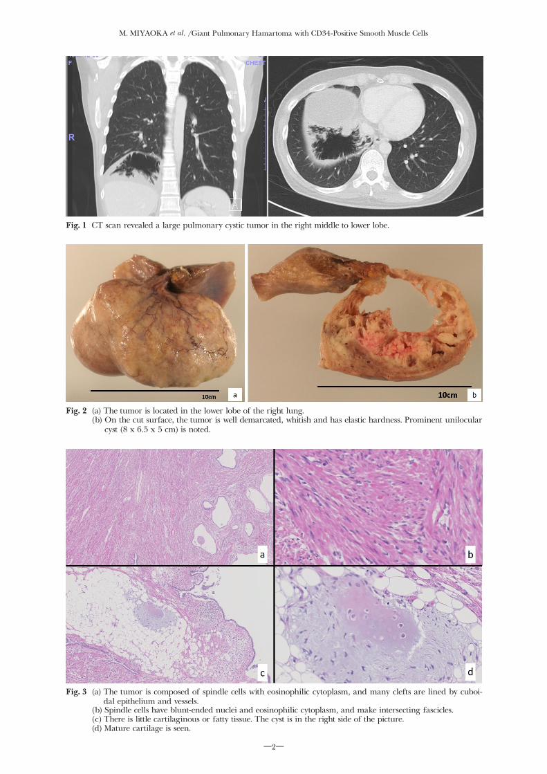
A 44-year-old Japanese woman, with no previous history, was examined for pulmonary masses. The mass observed by chest radiography in the physical checkup was revealed to be a 13 x 12 x 9 cm pul-monary cystic tumor in the middle to lower lobe of the right lung by systemic CT scan (Fig. 1). Uterine leiomyoma was also found. The pulmonary lesion was clinically diagnosed as “congenital cystic adenomatoid malformation (CCAM)” or “bronchopulmonary seques-tration”, and surgical excision was carried out.
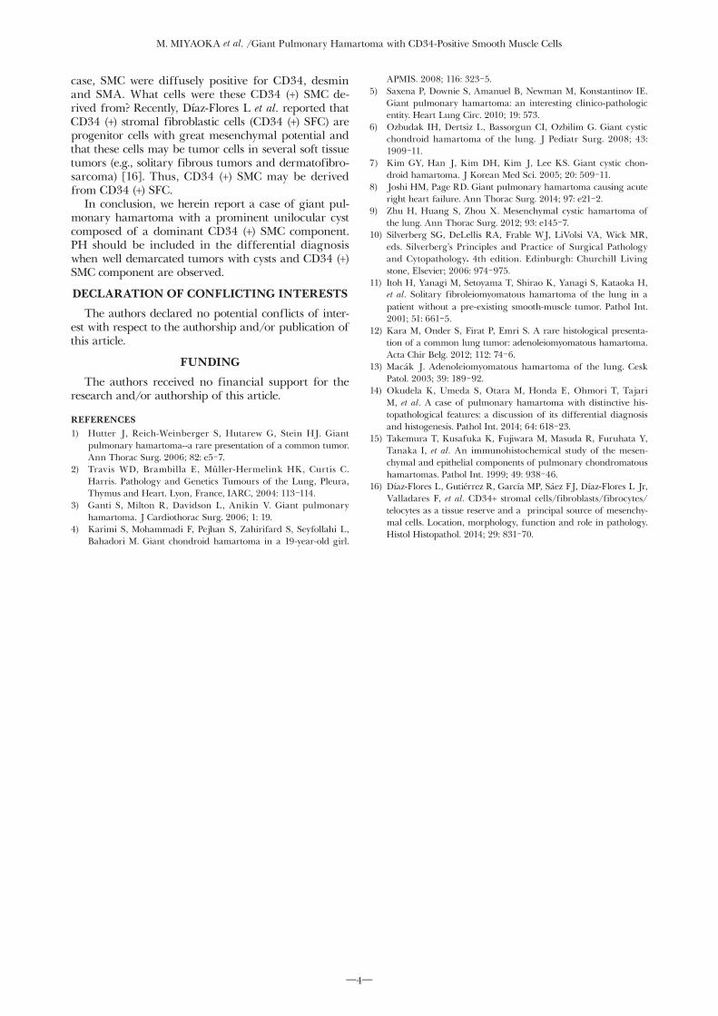
Grossly, a well demarcated and elastic hard tumor (13 x 10 x 8 cm) with a prominent unilocular cyst (8 x 6.5 x 6 cm) was located in the lower lobe of the right lung (Fig. 2). Necrosis and hemorrhage were not ob-served. Microscopically, the solid area was mainly com-posed of proliferated spindle cells with blunt-ended
nuclei and eosinophilic cytoplasm, and a small area of adipose and cartilaginous tissue (Fig. 3). Spindle cells made intersecting fascicles (Figs. 3 and 4). The cellu-larity was not high. There were many clefts lined by cuboidal ciliated epithelium and vessels in the spindle cell component (Fig. 4). Cellular atypia was not appar-ent. The cyst was lined by cuboidal ciliated epithelium. Immunohistochemically, spindle cells were positive for smooth muscle actin (SMA), desmin and CD34, and weakly positive for estrogen receptor, but negative for HMB-45, Melan A and progesterone receptor (Table, Fig. 4). Spindle cells were derived from SMC. TTF-1 reacted with ciliated epithelium, indicating respiratory epithelium (Table, Fig. 4), and D2-40 reacted with the endothelium of vessels, indicating lymphatics (Table, Fig. 4).
DISCUSSION
We report a giant cystic tumor in the lung that con-tained CD34 (+) SMC. Differential diagnoses of pul-monary hamartoma, benign metastasizing leiomyoma and solitary fibrous tumor were suggested; however, the final diagnosis was pulmonary harmatoma due to mesenchymal components of SMC, fat and cartilage.
PH is generally composed of lobules of sparsely cellular, spindled, fibromyxoid or fibrous stroma, often with cartilage and adipose tissue compo-nents [10]. “Adenoleiomyomatous hamartoma” and “Fibroleiomyomatous hamartoma” refer to PH com-posed of dominant SMC components with glandular respiratory epithelium and/or fibrous tissue [11-14]. It was reported that the SMC component of pulmonary hamartoma was positive for SMA and desmin, but negative for CD34 [13-15]. Takemura et al. reported that CD34-reactive mesenchymal cells are not involved in the histogenesis of chondromatous PH [15]. In our
Masashi MIYAOKA, Department of Pathology, Tokai University School of Medicine,143 Shimokasuya, Isehara, Kanagawa 259-1193, JapanTel: + 81-463-93-1121 Fax: +81-463-91-1370 E-mail: [email protected]
M. MIYAOKA et al. /Giant Pulmonary Hamartoma with CD34-Positive Smooth Muscle Cells
―2―
Fig. 1 CT scan revealed a large pulmonary cystic tumor in the right middle to lower lobe.
Fig. 2 (a) The tumor is located in the lower lobe of the right lung. (b) On the cut surface, the tumor is well demarcated, whitish and has elastic hardness. Prominent unilocular
cyst (8 x 6.5 x 5 cm) is noted.
Fig. 3 (a) The tumor is composed of spindle cells with eosinophilic cytoplasm, and many clefts are lined by cuboi-dal epithelium and vessels.
(b) Spindle cells have blunt-ended nuclei and eosinophilic cytoplasm, and make intersecting fascicles. (c) There is little cartilaginous or fatty tissue. The cyst is in the right side of the picture. (d) Mature cartilage is seen.

M. MIYAOKA et al. /Giant Pulmonary Hamartoma with CD34-Positive Smooth Muscle Cells
―3―
Table Immunohistochemistry results for tumor component Spindle cell Cuboidal epithelium VesselSMA + ‒ ‒Desmin + ‒ ‒CD34 + ‒ +ER + (weak) ‒ ‒PgR ‒ ‒ ‒HMB-45 ‒ ‒ ‒Melan A ‒ ‒ ‒TTF-1 ‒ + ‒D2-40 ‒ ‒ + +: positive −: negative
Fig. 4 (a)-(d) Results of immunohistochemistry. Spindle cells are diffusely positive for desmin (b), SMA (c) and CD34 (d). (e) There are many clefts lined by cuboidal epithelium and vessels in the spindle cells. (f) The cuboidal epithelium has cilia. (g) Immunohistochemically, ciliated cuboidal epithelium is positive for TTF-1. (h) Immunohistochemically, endothelial cells of vessels are positive for D2-40.
M. MIYAOKA et al. /Giant Pulmonary Hamartoma with CD34-Positive Smooth Muscle Cells
―4―
case, SMC were diffusely positive for CD34, desmin and SMA. What cells were these CD34 (+) SMC de-rived from? Recently, Díaz-Flores L et al. reported that CD34 (+) stromal fibroblastic cells (CD34 (+) SFC) are progenitor cells with great mesenchymal potential and that these cells may be tumor cells in several soft tissue tumors (e.g., solitary fibrous tumors and dermatofibro-sarcoma) [16]. Thus, CD34 (+) SMC may be derived from CD34 (+) SFC.
In conclusion, we herein report a case of giant pul-monary hamartoma with a prominent unilocular cyst composed of a dominant CD34 (+) SMC component. PH should be included in the differential diagnosis when well demarcated tumors with cysts and CD34 (+) SMC component are observed.
DECLARATION OF CONFLICTING INTERESTS
The authors declared no potential conflicts of inter-est with respect to the authorship and/or publication of this article.
FUNDING
The authors received no financial support for the research and/or authorship of this article.
REFERENCES1) Hutter J, Reich-Weinberger S, Hutarew G, Stein HJ. Giant
pulmonary hamartoma--a rare presentation of a common tumor. Ann Thorac Surg. 2006; 82: e5-7.
2) Travis WD, Brambilla E, Müller-Hermelink HK, Curtis C . Harris. Pathology and Genetics Tumours of the Lung, Pleura, Thymus and Heart. Lyon, France, IARC, 2004: 113-114.
3) Ganti S, Milton R, Davidson L, Anikin V. Giant pulmonary hamartoma. J Cardiothorac Surg. 2006; 1: 19.
4) Karimi S, Mohammadi F, Pejhan S, Zahirifard S, Seyfollahi L, Bahadori M. Giant chondroid hamartoma in a 19-year-old girl.
APMIS. 2008; 116: 323-5.5) Saxena P, Downie S, Amanuel B, Newman M, Konstantinov IE.
Giant pulmonary hamartoma: an interesting clinico-pathologic entity. Heart Lung Circ. 2010; 19: 573.
6) Ozbudak IH, Dertsiz L, Bassorgun CI, Ozbilim G. Giant cystic chondroid hamartoma of the lung. J Pediatr Surg. 2008; 43: 1909-11.
7) Kim GY, Han J, Kim DH, Kim J, Lee KS. Giant cystic chon-droid hamartoma. J Korean Med Sci. 2005; 20: 509-11.
8) Joshi HM, Page RD. Giant pulmonary hamartoma causing acute right heart failure. Ann Thorac Surg. 2014; 97: e21-2.
9) Zhu H, Huang S, Zhou X. Mesenchymal cystic hamartoma of the lung. Ann Thorac Surg. 2012; 93: e145-7.
10) Silverberg SG, DeLellis RA, Frable WJ, LiVolsi VA, Wick MR, eds. Silverberg’s Principles and Practice of Surgical Pathology and Cytopathology. 4th edition. Edinburgh: Churchill Living stone, Elsevier; 2006: 974-975.
11) Itoh H, Yanagi M, Setoyama T, Shirao K, Yanagi S, Kataoka H, et al. Solitary fibroleiomyomatous hamartoma of the lung in a patient without a pre-existing smooth-muscle tumor. Pathol Int. 2001; 51: 661-5.
12) Kara M, Onder S, Firat P, Emri S. A rare histological presenta-tion of a common lung tumor: adenoleiomyomatous hamartoma. Acta Chir Belg. 2012; 112: 74-6.
13) Macák J. Adenoleiomyomatous hamartoma of the lung. Cesk Patol. 2003; 39: 189-92.
14) Okudela K, Umeda S, Otara M, Honda E, Ohmori T, Tajari M, et al. A case of pulmonary hamartoma with distinctive his-topathological features: a discussion of its differential diagnosis and histogenesis. Pathol Int. 2014; 64: 618-23.
15) Takemura T, Kusafuka K, Fujiwara M, Masuda R, Furuhata Y, Tanaka I, et al. An immunohistochemical study of the mesen-chymal and epithelial components of pulmonary chondromatous hamartomas. Pathol Int. 1999; 49: 938-46.
16) Díaz-Flores L, Gutiérrez R, García MP, Sáez FJ, Díaz-Flores L Jr, Valladares F, et al. CD34+ stromal cells/fibroblasts/fibrocytes/telocytes as a tissue reserve and a principal source of mesenchy-mal cells. Location, morphology, function and role in pathology. Histol Histopathol. 2014; 29: 831-70.