ghent university faculty of veterinary ... university faculty of veterinary medicine academic year...
TRANSCRIPT

GHENT UNIVERSITY FACULTY OF VETERINARY MEDICINE
Academic year 2014 – 2015
Reproductive characteristics of female African Elephant and Rhinoceros and the possibilities of gamete cryopreservation.
By
Maaike DE SCHEPPER
Supervisor: Prof. dr. Peter E.J. Bols. Co-Supervisor: Dr. T. Rijsselaere
Literature Review as part of the Master's Dissertation
© 2015 Maaike de Schepper.


Universiteit Gent, its employees and/or students, give no warranty that the information provided in this
thesis is accurate or exhaustive, nor that the content of this thesis will not constitute or result in any
infringement of third-party rights.
Universiteit Gent, its employees and/or students do not accept any liability or responsibility for any use
which may be made of the content or information given in the thesis, nor for any reliance which may
be placed on any advice or information provided in this thesis.

GHENT UNIVERSITY FACULTY OF VETERINARY MEDICINE
Academic year 2014 – 2015
Reproductive characteristics of female African Elephant and Rhinoceros and the possibilities of gamete cryopreservation.
By
Maaike DE SCHEPPER
Supervisor: Prof. dr. Peter E.J. Bols. Co-Supervisor: Dr. T. Rijsselaere
Literature Review as part of the Master's Dissertation
© 2015 Maaike de Schepper

Preface
First of all, I want to thank Prof. dr. Wouter van Hoven from the University of Pretoria, who lectured me
about rhino and elephant poaching and made me think of new possible ways to preserve both
species. Because of him I started to think further than rhino farms and the standard ways of saving
animals.
Second of all, I want to thank my promotor Prof. dr. Peter Bols from the University of Antwerp, who
jumped on the crazy rhino and elephant train with me. As well as I want to thank my co-promotor dr.
Tom Rijsselaere, for reading my thesis and correct my English.
I also really want to thank veterinarian Cyriel Ververs, who helped me with my thesis without any
hesitation and always had answers ready for all my questions. As well as veterinarian Margot Van De
Velde, who corrected my thesis and gave me things to think about during the writing process.
Many thanks to Prof. dr. Ronald de Krijger for reading my (almost) final version and dot my I’s and
cross my t’s!
And last but not least, I want to thank my roommates, friends and boyfriend for their motivational talks
when I couldn’t find light in all my articles and writing. But also my parents, especially my dad, who
gave me some new insights when I was stuck with the writing process.

Table of contents
Preface ....................................................................................................................................................
Table of contents ....................................................................................................................................
Abstract ................................................................................................................................................. 1
Samenvatting ........................................................................................................................................ 2
Introduction ........................................................................................................................................... 4
1. Overview of follicular development, activation and growth ......................................................... 7 1.1. A general overview: the concept of an oestrous cycle .......................................................... 7
1.1.1. Oocyte formation ................................................................................................................... 8 1.1.2. The follicular phase ............................................................................................................... 9 1.1.3. The luteal phase .................................................................................................................. 10
1.2. Elephant .................................................................................................................................... 10 1.3. Rhinoceroses ........................................................................................................................... 13
1.3.1. White rhinoceros ................................................................................................................. 13 1.3.2. Black rhinoceros .................................................................................................................. 16
2. Fertility preservation through cryopreservation ......................................................................... 18 2.0.1. Controlled-rate freezing or slow cooling .............................................................................. 19 2.0.2. Vitrification ........................................................................................................................... 19 2.0.3. The role of cryoprotectant agents (CPA) ............................................................................. 20
2.1. Embryo cryopreservation ....................................................................................................... 20 2.2. Oocyte cryopreservation ........................................................................................................ 21 2.3. Gonadal tissue cryopreservation ........................................................................................... 21
2.3.1. Elephant .............................................................................................................................. 22 2.3.2. Rhinoceros .......................................................................................................................... 22
3. Genome Resource Bank ................................................................................................................ 23 Cloning .......................................................................................................................................... 23
4. Conclusion and future perspectives ............................................................................................ 24
References .......................................................................................................................................... 26

1
Abstract
Poaching is the biggest treat to elephants and rhinoceroses nowadays in Africa. A possible way to
preserve the female genes is cryopreservation, by vitrification or slow-cooling, of female genetic
material such as embryos, oocytes and ovaries. The preferred method depends on the environment
and the physical characteristics of the sexual cycle. In field environment vitrification is recommended,
because there is no need for computer controlled cooling. Vitrification requires liquid nitrogen and a
cryoprotectant for very fast cooling.
In contrast to horses, there are no studies have yet been conducted on embryo cryopreservation in
rhinoceroses and elephants. To establish an embryo cryopreservation protocol for rhinoceroses the
horse protocol can therefore be used as an example.
There are different opinions among scientists regarding oocyte cryopreservation. Some say it is better
to preserve immature oocytes (Pereira and Marques, 2008; Aerts and Bols, 2010a), however, others
say it is preferable to preserve mature oocytes (Lim et al., 1992; Ledda et al., 2000; Pereira and
Marques, 2008). Another method is to harvest and cryopreserve the ovaries post-mortem from
deceased or killed animals. A complete ovary can be cryopreserved as well as small ovarian pieces.
Viable oocytes can be recovered from ovarian tissue after transplantation of cryopreserved ovaries.
To maximize success of assisted reproductive techniques (ARTs) a thorough understanding of the
normal reproductive cycle is crucial. As for the elephant (Loxodonta africana) it displays an oestrous
cycle of 13-18 weeks, with a double luteinising hormone (LH) surge. A non-ovulatory LH surge, 10-20
days after the progesterone decrease and an ovulatory LH surge, 19-22 days after the non-ovulatory
surge (Hildebrandt et al., 2010). Also the oestrous cycle of the black rhinoceros (Diceros bicornis) is
fairly well defined. It takes 25 days with a single LH surge (Hindle et al., 1992; Schwarzenberger et al.,
1993). However, scientist share different views about the cycle length of the white rhinoceros
(Ceratotherium simum). Generally, the assumption is made that there are two different cycle types;
type I, a regular cycle, taking 31-35 days and a type II, an irregular cycle, taking 65-70 days (Radcliffe
et al., 1997; Patton et al., 1999; Brown et al., 2001; Morrow et al., 2008). On the other hand,
Schwarzenberger et al. (1998) stated that a regular cycle takes 70 days while an irregular one takes
30-70 days. There happens to be only one LH surge to induce ovulation.
Keywords: Cryopreservation, Elephant (Loxondota africana), Fertility preservation, Oestrous cycle, Rhinoceros (Ceratotherium simum and Diceros bicornis).

2
Samenvatting
De laatste decennia is het stropen van olifanten (Loxondota africana) en neushoorns (de witte
neushoorn: Ceratotherium simum en de zwarte neushoorn: Diceros bicornis), om hun slagtanden en
hoorn in Afrika exponentieel toegenomen. In de afgelopen honderd jaar is de olifantenpopulatie met
95% gedaald. Het aantal neushoorns dat slachtoffer is geworden van stropers was in 2014 in Zuid-
Afrika alleen al 1,225 dieren. In 2015 zijn tussen 1 januari en 1 april, volgens onofficiële cijfers, 240
neushoorns gestroopt. Als het doden van deze dieren in dit tempo doorgaat zullen beide diersoorten
in de nabije toekomst in het wild uitsterven. Om de stropers te ontmoedigen kunnen de neushoorns
onthoornd worden. De hoorn, bestaande uit keratine, groeit terug aan. Ook kan er overwogen om
ivoren slagtanden van olifanten te verwijderen of in te korten. Verder valt een translocatie van de
dieren naar veiligere locaties te overwegen. Dit heeft als bijkomend voordeel dat er nieuwe genen in
een bestaande groep worden geïntroduceerd, wat een vernauwing van de genetische basis kan
voorkomen.
Daarnaast moet men ook het preserveren van vrouwelijk genetisch materiaal zoals embryo’s, oocyten
en ovaria als belangrijke strategie overwegen. Dit kan door middel van cryopreservatie, via slow-
cooling of vitrificatie. Vitrificatie is aan te raden als de preservatie in het veld moet gebeuren omdat
hiervoor geen computer gestuurde koeling bij nodig is. Hierbij wordt het te preserveren monster
doordrongen met een cryoprotectant en vervolgens in vloeibare stikstof overgebracht. Door de super
snelle koeling wordt een glasachtige staat gecreëerd binnen de cellen, de koelingssnelheid kan
oplopen van 100° en 10.000° Celsius per minuut, afhankelijk van de procedure, het volume en de
oplossing die gebruikt wordt (Saragusty and Arav, 2011). Embryo cryopreservatie is tot nog toe niet
uitgevoerd bij olifanten of neushoorns. Bij het paard, het dier dat aanzien wordt als het best
gerelateerde gedomesticeerd dier ten opzichte van de neushoorn (O’Brien and Roth, 2000; Stoops et
al., 2011), is dit wel mogelijk. Het paardenprotocol zou mogelijk, in licht aangepaste vorm, voor het
cryopreserveren van olifanten- en neushoorn embryos gebruikt kunnen worden. Over het
cryopreserveren van oocyten bestaan verschillende opvattingen binnen de wetenschap. Zo zijn er
wetenschappers die beweren dat het beter is om mature oocyten te cryopreserveren (Lim et al., 1992;
Ledda et al., 2000; Pereira and Marques, 2008; Saragusty and Arav, 2011), terwijl andere meer
voordelen zien in de cryopreservatie van immature eicellen (Pereira and Marques, 2008; Aerts and
Bols, 2010a). Een andere mogelijkheid is het cryopreserveren van stukjes ovarieel weefsel. Deze
techniek heeft het voordeel dat zowel gebruik gemaakt kan worden van weefsel van seksueel mature
als immature dieren. Ook is het eenvoudiger om post-mortem de ovaria uit de dieren te verwijderen
dan embryo’s te kweken en/of oocyten te isoleren. De ovariële weefsel stukjes kunnen ter plekke, in
het veld, worden gecryopreserveerd door middel van vitrificatie of individueel steriel verpakt worden in
fosfaat-gebufferde zoutoplossing (PBS). Dit kan op kamertemperatuur vervoerd worden naar een
beter uitgerust onderzoeks laboratorium (Stoop et al., 2011). Na het ontdooien kunnen immature
oocyten op een cultuurmedium gerijpt worden en later eventueel ingebracht worden bij een ander
seksueel actief vrouwelijk dier.

3
Om met deze technieken van ‘geassisteerde voortplanting’ voldoende succes te kunnen behalen is
het absoluut noodzakelijk een goed inzicht te krijgen in het verloop van de normale seksuele cyclus bij
beide species. Voor de olifant (Loxodonta africana) is dit redelijk het geval. Hier is sprake van een 13-
18 weken durende oestrale cyclus, waarvan de luteale fase 6-12 weken en de folliculaire fase 4-6
weken duurt. Gedurende de folliculaire fase zijn er twee verschillende luteïniserend hormoon (LH)
pieken, de non-ovulatoire, 10-20 dagen na de daling van progesteron concentratie, en de ovulatoire
LH piek, die 19-22 dagen na de non-ovulatoire volgt (Hildebrandt et al., 2010).
De oestrale cyclus van de zwarte neushoorn (Diceros bicornis) is ook goed gekend. Deze duurt
gemiddeld 25 dagen, waarvan de luteale fase 18 ± 1.1 dagen duurt en de folliculaire fase 5 ± 1 dagen
duurt (Garnier et al., 2002), met één enkele LH piek om de ovulatie te induceren (Hindle et al., 1992;
Schwarzenberger et al., 1993).
Over de oestrus cyclus van de witte neushoorn (Ceratotherium simum) bestaat enige controversie
tussen de verschillende wetenschappers. Er wordt algemeen aangenomen dat er twee verschillende
types cycli zijn, namelijk het type I dewelke regelmatig is en rond de 31-35 dagen duurt. Een type II
cyclus is onregelmatig en duurt 65-70 dagen (Radcliffe et al., 1997; Patton et al., 1999; Brown et al.,
2001; Morrow et al., 2008). Daarentegen beweren Schwarzenberger et al. (1998) dat de regelmatige
70 dagen duurt en dat de onregelmatige cyclus 30-70 dagen duurt. De ovulatie wordt geïnduceerd
door één enkele LH piek.
4
Introduction
During a two month stay (November-December 2013), I took up an internship at the University of
Pretoria, South Africa. During this trip, I spoke with several people, such as Prof. van Hoven and Ralf
Kalwa, the former head of the Kruger National Park rangers, about the increase in the frequency of
poaching of elephants and rhinoceroses. Those people made me aware about how big the poaching
problem actually is. We discussed a lot about different options to preserve those precious species,
varying from translocation, dehorning, legalizing the ivory and rhino horn trade and starting breeding
farms. This experience made me realise that different solutions must be explored, such as genetic
preservation.
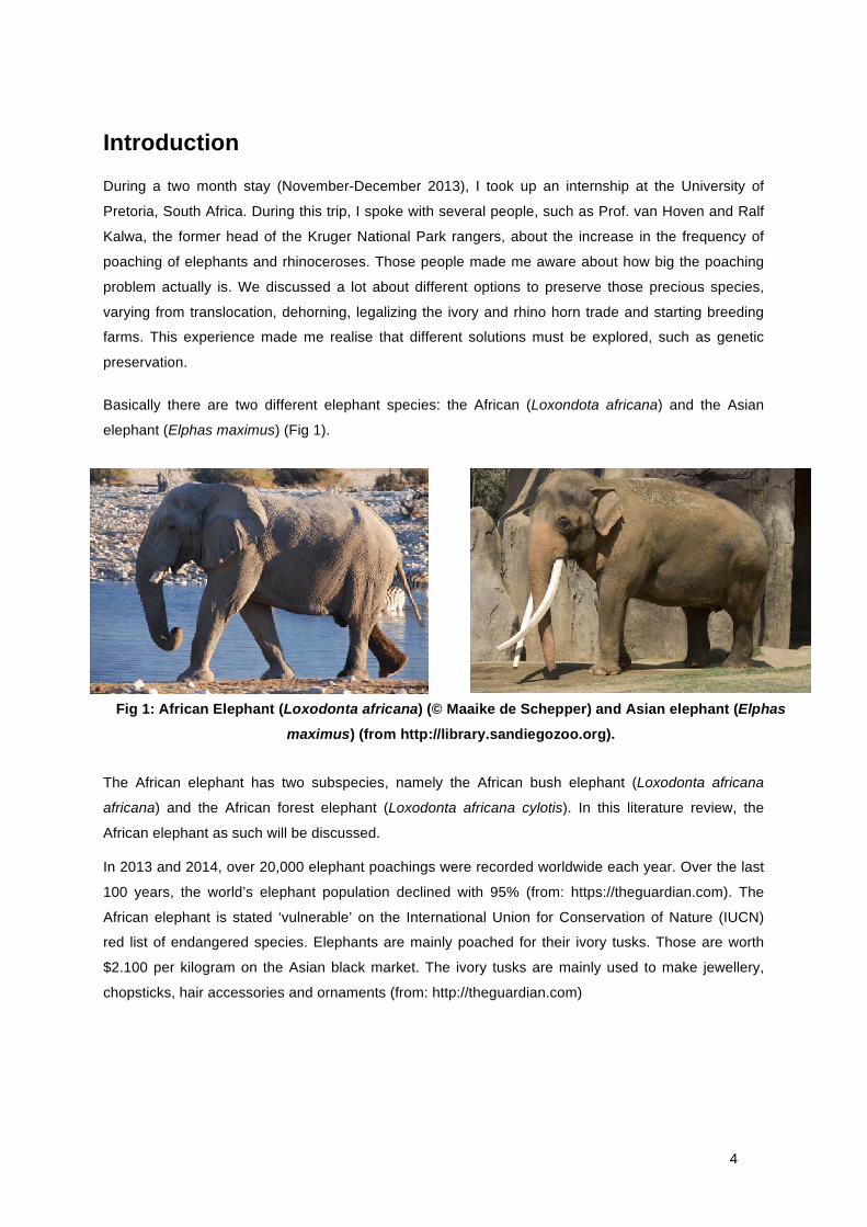
Basically there are two different elephant species: the African (Loxondota africana) and the Asian
elephant (Elphas maximus) (Fig 1).
The African elephant has two subspecies, namely the African bush elephant (Loxodonta africana
africana) and the African forest elephant (Loxodonta africana cylotis). In this literature review, the
African elephant as such will be discussed.
In 2013 and 2014, over 20,000 elephant poachings were recorded worldwide each year. Over the last
100 years, the world’s elephant population declined with 95% (from: https://theguardian.com). The
African elephant is stated ‘vulnerable’ on the International Union for Conservation of Nature (IUCN)
red list of endangered species. Elephants are mainly poached for their ivory tusks. Those are worth
$2.100 per kilogram on the Asian black market. The ivory tusks are mainly used to make jewellery,
chopsticks, hair accessories and ornaments (from: http://theguardian.com)
Fig 1: African Elephant (Loxodonta africana) (© Maaike de Schepper) and Asian elephant (Elphas
maximus) (from http://library.sandiegozoo.org).

5
There are five different rhinoceros species: the White (Ceratotherium simum), the Black (Diceros
bicornis), the Greater one-horned (Rhinoceros unicornis), the Sumatran (Dicerorhinus sumatrensis)
and the Javan (Rhinoceros sondaicus) (Fig 2).
Fig 2: From left to right: Sumatran rhinoceros (Dicerorhinus sumatrensis), Indian rhinoceros
(Rhinoceros unicornis), White rhinoceros (Creratotherium simum), Javan rhinoceros
(Rhinoceros sondaicus) and Black rhinoceros (Diceros bicornis) (from:
http://rhinoresourcecenter.com).
Both the white and the black rhinoceros have several subspecies. The white rhinoceros can be divided
in the Northern (Ceratotherium simum cotoni) and the Southern white rhinoceros (Ceratotherium
simum simum). The black rhinoceros can be divided in the Eastern (Diceros bicornis michaeli), the
South central (Diceros bicornis minor) and the South western black rhinoceros (Diceros bicornis
bicornis). In this literature review the black and the white rhinoceros will be discussed in general.
The numbers of poached rhinoceros, in South Africa alone, increase yearly (Fig 3). In 2014, 1,225
recorded rhinoceroses where poached, which means one every eight hours! The real numbers might
even be higher, and is underestimated because not all findings are recorded and not all killed animals
are found. In 2015, the unofficial statistics stated that there are 240 poached rhinoceroses between
January 1st and April 1st, in South Africa. The most recent official numbers are: 49 killed rhinoceroses
until the January 22 2015 (Department of Environmental Affairs, South Africa).
The white rhinoceros is stated as to be ‘almost threatened’ and the black rhinoceros is stated ‘critically
endangered’ on the IUCN red list however, the South western black rhinoceros is declared extinct
since April 2015 by the IUCN. The rhinoceroses are poached for their horns. That contains only
keratin, the same substance where human hair or nails are made of. However for one kilogram of
rhinoceros horn, around $65.000 is paid for on the black market in Asia. The rhinoceros horn is mainly
sold in China and Vietnam. Where it is used as a drug to cure hangovers, cancer and to stimulate the
libido. However there is no scientific basis for any medical benefits. In addition the horn is also seen
as a status symbol for wealthy people. Since 2013 the poaching rate is higher then the birth rate,
which will lead in ultimately to extinction in the wild in the near future.
The battle against poachers has taken immense proportions. They are using advanced equipment,
like helicopters, night vision binoculars and several types of machine guns and the poachers don’t
hesitate to take a human life to reach their goal.

6
Fig 3: Number of rhinoceros poached in South Africa (from Save the rhino, South African
Department of Environmental Affairs, 2015)
The shocking numbers mentioned above illustrate the overall failure of preventive measures, like
translocation, dehorning and security of the animals. This means that preservation of both species
needs to be taken to another level. Fertility preservation from animals that die unexpectedly or animals
that are still alive but not sexually active might be a very valuable strategy. This way genetic material
can be used to re-establish a sustainable population.
In this literature review I will focus on three different topics. Firstly I will review the basic characteristics
of the oestrous cycle of the elephant and the rhinoceros. Secondly, fertility preservation and how it can
contribute to preserve the elephant and the rhinoceros in the future will be discussed. Finally, several
future perspectives will be briefly commented upon.
7
1. Overview of follicular development, activation and growth
1.1. A general overview: the concept of an oestrous cycle
In adult, female mammals, mature oocytes are released periodically from the ovaries until the female
becomes pregnant (Sjaastad et al., 2010). The time between two released oocytes is called the
oestrous cycle i.e. the time interval between the start of one oestrous cycle until the next; in non-
pregnant mammals, it is defined as the ‘length’ of an oestrous cycle. During the oestrus cycle the
female is receptive to mating while her behaviour is changing, which can be used as an oestrus
indicator in several mammalian species. Most of the mammals will ovulate during or shortly after
oestrous, maximizing the change of fertilization.
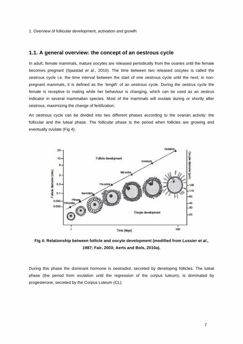
An oestrous cycle can be divided into two different phases according to the ovarian activity: the
follicular and the luteal phase. The follicular phase is the period when follicles are growing and
eventually ovulate (Fig 4).
Fig 4: Relationship between follicle and oocyte development (modified from Lussier et al.,
1987; Fair, 2003; Aerts and Bols, 2010a).
During this phase the dominant hormone is oestradiol, secreted by developing follicles. The luteal
phase (the period from ovulation until the regression of the corpus luteum), is dominated by
progesterone, secreted by the Corpus Luteum (CL).

8
1.1.1. Oocyte formation
Cells divide and reproduce in two possible ways, mitosis and meiosis (Fig 5). During mitosis the
complete DNA set is doubled and transferred into two daughter cells. Consequently, the number of
cells rises to a large number of identical cells in the body.
Fig 5: Development of oocytes (from Sjaastad, 2010).
The oocytes are formed during meiosis. From the moment the germ cells start the first stage of
meiosis, at the end of embryonic life, they are called primary oocytes. The chromosomes are
duplicated. In contrast to mitosis, in meiosis the division of cytoplasm, cytokinesis, does not take
place.
The ovaries contain a large reserve of non-growing primordial follicles (Aerts and Bols, 2010a).
Follicles can be divided in four different morphological stages: the primordial, primary, secondary and
the tertiary follicle. A primordial follicle is surrounded by a single layer of flattened granulosa cells,
meanwhile containing an immature oocyte. The development or activation from a primordial follicle to
a growing follicle is a gradual process, continuous during the whole reproductive life. It is assumed
that the primordial follicles first need to go in the first meiotic arrest before they can be activated (Yang
and Fortune, 2008). Primordial follicle activation into a primary follicle is facilitated by the proliferation
and differentiation of the granulosa cells. The rising number of granulosa cells influences the
transformation form flattened into cuboidal granulosa cells. Additionally the volume expansion of the
oocyte influences this transformation. The granulosa cells contain Follicle Stimulating Hormone (FSH)
receptors and take part in the follicle growth. The greater number of the activated primary follicles
develops into an antral follicle, detectable by the cavity or antrum. Along the way, most follicles
regress and start apoptosis, including the growing follicles in a follicular growth wave (Aerts and Bols,
2010a). During the transition from the primary to the secondary stage the follicle expands and the
zona pellucida is formed between the oocyte and the growing layers of granulosa cells. Tertiary
follicles can be divided in small or large antral follicles, consisting of an antrum filled with follicular fluid.
Mature antral follicles are called Graafian follicles. The mural granulosa cells within the mature antral

9
follicles display LH receptors. Their number increases with follicle maturation, which means the follicle
is sensitive for the pre-ovulatory LH-peak.
1.1.2. The follicular phase
The follicular phase covers of only 20% of the duration of the oestrous cycle and can be divided into
two stages: the pro-oestrous and the oestrous. The pro-oestrous starts when the luteolysis of the old
corpus luteum starts. Pro-oestrous is known for the transition from progesterone to oestrogen
dominance. This transition is covered by the rise of Follicle Stimulating Hormone (FSH) and
Luteinizing Hormone (LH). The anterior pituitary gland produces both FSH and LH, which is induced
by Gonadotropin Releasing Hormone (GnRH), secreted by the hypothalamus (Fig 6). The granulosa
cells in the growing follicle have specific hormone receptors in the cell membrane. FSH binds to these
receptors and promotes follicle growth and proliferation. Due to LH secretion, the theca cells of the
follicle produce testosterone, which is transfered to the granulosa cells. Due to FSH, testosterone is
converted into oestrogen in the granulosa cells. Oestrogen stimulates follicular development and
diffuses into the blood stream (Fig 6). During the pro-oestrous, follicles for ovulation are selected.
During the follicular phase, oestradiol is the dominant hormone, exteriorising the oestrous behaviour
and the willingness of the female tot copulate.
The selected, expanding follicle also produces the hormone inhibin. Inhibin has a negative feedback
effect on the secretion of FSH in the anterior pituitary gland. Oestrogen has a negative feedback on
the hypothalamus and both hormones cause a decrease in FSH secretion. Due to this decrease in
FSH new follicles do not mature and atresia is triggered. The low levels of FSH are however, still
sufficient to convert testosterone into oestrogen.
Fig 6: Hormone secretion during pro-oestrous (from Senger, Pathways to pregnancy and
parturition, 2005)

10
When LH secretion increases, so does oestrogen, mainly due to the fact that there are more hormone-
producing granulosa cells present in the developing follicle. At the end of the follicular phase the
oestrogen concentration rapidly increases. When the oestrogen concentration reaches a threshold
value, oestrogen initiates a positive feedback effect on the GnRH surge centre in the brain, causing a
preovulatory LH surge (Sjaastad et al., 2010). Because of the inhibitory effect of inhibin the positive
feedback effect is smaller on the FSH increase (Sjaastad et al., 2010).
The preovulatory LH surge induces an increased blood flow to the ovary resulting in an accumulation
of fluid from the surrounding tissue in the mature follicle. Due to this accumulation, the hydrostatic
pressure in the follicle increases. The enzyme collagenase is released and weakens the follicle wall.
These changes finally result in a sudden burst of the follicle wall and a release of the follicular fluid and
the oocyte into the peritoneal cavity: ovulation. The oocyte is surrounded by the granulosa cells (the
corona radiata) at the moment of ovulation. It is captured by the fimbriae at the end of the tuba uterina.
Once the released oocyte is present in the oviduct, it can be fertilized when mating has occurred. The
remaining cells in the ruptured follicle undergo luteinisation and start to form the corpus luteum.
1.1.3. The luteal phase
The luteal phase covers approximately 80% of the oestrous cycle and can be divided into two different
stages: the metoestrous and the dioestrous. During metoestrous the ovulated follicle is converted into
a CL, a temporary intra-ovarian endocrine gland, which starts to secrete progesterone. During di-
oestrous which is the longest period of the cycle, the CL is fully functional and secretes significant
quantities of progesterone. The dioestrous ends when the CL starts to luteolyse due to the increased
amount of circulating prostaglandin F2α that is produced by the non-pregnant uterus. Due to the
regression of the CL, the concentration of progesterone decreases and its negative feedback on
GnRH disappears. The secretion of FSH and LH by the anterior pituitary gland rise again and follicular
development will start all over in the next oestrous cycle in continuous breeders.
1.2. Elephant
The sexual cycle as described above, describes the general pattern of the oestrous cycle of most
mammals. However, the oestrous cycle of the elephant differs in several aspects. Elephants are
continuous breeders. Female elephants reach sexual maturity approximately at 10-12 years of age.
The oestrous cycle of the elephant cow lasts for 13-18 weeks, including a luteal phase with a duration
of approximately 6-12 weeks and a follicular phase of 4-6 weeks (Plotka et al., 1988). Consequently,
the cow only has three to four oestrous cycles per year. During each of these reproductive cycles
multiple corpora lutea are present in the ovaries.
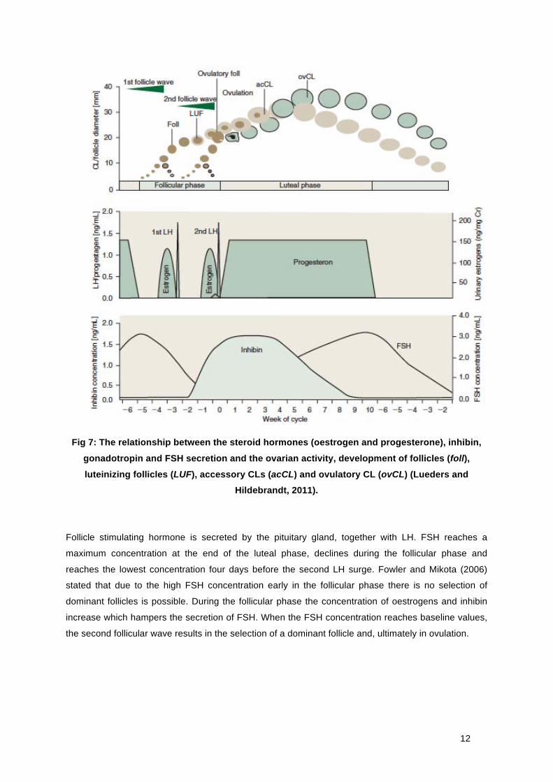
During the follicular phase (the non-luteal phase), two different LH surges occur (Fig 7). The first surge
is a non-ovulatory LH surge appearing 10-20 days after the progesterone decrease. During this
follicular wave follicles grow reaching a maximum diameter of 13.7 ± 0.7 mm while none of them
ovulate (Hildebrandt et al., 2010).
11
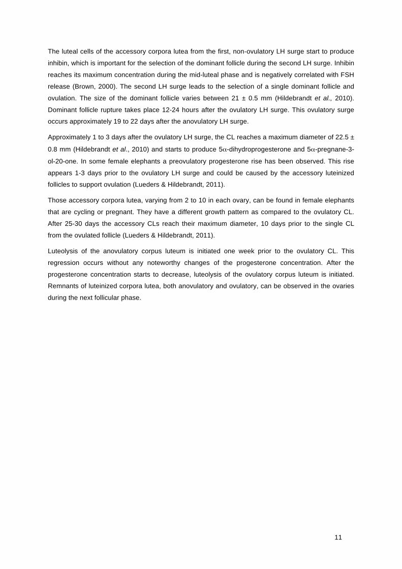
The luteal cells of the accessory corpora lutea from the first, non-ovulatory LH surge start to produce
inhibin, which is important for the selection of the dominant follicle during the second LH surge. Inhibin
reaches its maximum concentration during the mid-luteal phase and is negatively correlated with FSH
release (Brown, 2000). The second LH surge leads to the selection of a single dominant follicle and
ovulation. The size of the dominant follicle varies between 21 ± 0.5 mm (Hildebrandt et al., 2010).
Dominant follicle rupture takes place 12-24 hours after the ovulatory LH surge. This ovulatory surge
occurs approximately 19 to 22 days after the anovulatory LH surge.
Approximately 1 to 3 days after the ovulatory LH surge, the CL reaches a maximum diameter of 22.5 ±
0.8 mm (Hildebrandt et al., 2010) and starts to produce 5α-dihydroprogesterone and 5α-pregnane-3-
ol-20-one. In some female elephants a preovulatory progesterone rise has been observed. This rise
appears 1-3 days prior to the ovulatory LH surge and could be caused by the accessory luteinized
follicles to support ovulation (Lueders & Hildebrandt, 2011).
Those accessory corpora lutea, varying from 2 to 10 in each ovary, can be found in female elephants
that are cycling or pregnant. They have a different growth pattern as compared to the ovulatory CL.
After 25-30 days the accessory CLs reach their maximum diameter, 10 days prior to the single CL
from the ovulated follicle (Lueders & Hildebrandt, 2011).
Luteolysis of the anovulatory corpus luteum is initiated one week prior to the ovulatory CL. This
regression occurs without any noteworthy changes of the progesterone concentration. After the
progesterone concentration starts to decrease, luteolysis of the ovulatory corpus luteum is initiated.
Remnants of luteinized corpora lutea, both anovulatory and ovulatory, can be observed in the ovaries
during the next follicular phase.
12
Fig 7: The relationship between the steroid hormones (oestrogen and progesterone), inhibin,
gonadotropin and FSH secretion and the ovarian activity, development of follicles (foll),
luteinizing follicles (LUF), accessory CLs (acCL) and ovulatory CL (ovCL) (Lueders and Hildebrandt, 2011).
Follicle stimulating hormone is secreted by the pituitary gland, together with LH. FSH reaches a
maximum concentration at the end of the luteal phase, declines during the follicular phase and
reaches the lowest concentration four days before the second LH surge. Fowler and Mikota (2006)
stated that due to the high FSH concentration early in the follicular phase there is no selection of
dominant follicles is possible. During the follicular phase the concentration of oestrogens and inhibin
increase which hampers the secretion of FSH. When the FSH concentration reaches baseline values,
the second follicular wave results in the selection of a dominant follicle and, ultimately in ovulation.

13
1.3. Rhinoceroses
In contrast to the elephant’s cycle, the oestrous cycle of the rhinoceros corresponds more to the
general cycle of most mammalian species as stated above. Female rhinoceroses are defined as
continuous breeders (Garnier et al., 2002). Ovulation in rhinoceroses is induced by a single, pre-
ovulatory LH surge occurring at the end of the follicular phase (Hermes et al., 2007). However, the
white and black rhinoceros species do differ in certain aspects of their cycle.
1.3.1. White rhinoceros
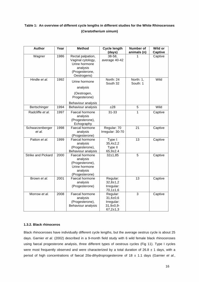
Several different opinions exist about oestrous cycle length of the white rhinoceros, as summarized in
Table 1. Two different oestrus cycle patterns can be distinguished for animals in captivity: cycle type I
lasting 31-35 and type II lasting 65-70 days (Fig 8). There is no difference in the inter-luteal phase
length between the two different cycles. The noted difference is associated with the variation in luteal
phase length (Patton et al., 1999).
Fig 8: Concentrations of faecal progesterone in the white rhinoceros illustrating type I and type II cycles. Darkened triangle indicates copulation, open triangle indicates mounting (Patton et
al. 1999).
There are different causes for the extended luteal phase during the type II cycle. Radcliffe et al. (1997)
reported on a female rhinoceros with uterine inflammation that expressed a type II cycle. Another
female rhinoceros in the same study experienced two times an embryonic loss, both one month post-
conception and showed the type II cycle as well. Foetal resorption, as diagnosed by ultrasonic
examination, also resulted in an extended luteal phase. In contrast, Schwarzenberger et al. (1998)
stated that the ‘normal’ oestrous cycle takes up to 10 weeks in the white rhinoceros (Fig 9), instead of
four weeks. This discrepancy can be caused by the length of the observation period, as
Schwarzenberger et al. (1998) monitored different female animals for more than four years while

14
Hindle et al. (1992) and Radcliffe et al. (1997) observed the animals for only one to two months which
means that these studies were only based on one or two cycles per female.
Fig 9: Concentrations of faecal 20-oxo-Progesterone in a white rhinoceros (Pistol), classified
as category I (Schwarzenberger et al. 1998).
MacDonald et al. (2008) stated that wild female rhinoceroses usually show the 35 day cycle. This
differs from the cycle in females in captivity that display a low reproductive efficiency. In wild
rhinoceroses pregnancy and lactation are dominating the endocrine status (Hermes et al., 2004).
Pregnancies take 16 months and the calf is weaned after a lactation of 12 months. This means that a
regular oestrous cycle in wild females is an uncommon occurrence, with around 30 full cycles in the
entire reproductive life span, comparing to 310 oestrous cycles in captive non-reproducing females
(Hermes et al., 2004; Hermes et al., 2007) (Fig 10).
Due to recurring influences of the steroid hormones, asymmetric aging processes in the reproductive
organs occur, as well as a reduction of the follicular stock on the ovaries. Also, steroid hormone
dependent tumours can develop due to the recurring concentrations of oestrogens and progesterone.
This could explain why captive females show a 65-70 day cycle.

15
Fig 10: The reproductive aging process in reproducing and non-reproducing female rhinoceros
(Hermes et al, 2004).
The preovulatory follicle reaches a diameter of 30 ± 2 mm within 48 hours before oestrus (Radcliffe et
al., 1997). During the 48 hours before ovulation the shape of the mature follicle changes from
spherical to pear shaped. The progesterone metabolite 20α-dihydroprogesterone concentration is low
during the follicular phase and increases after ovulation with a peak concentration at 8 to 12 days
post-ovulation (called the mid-luteal phase). Oestradiol-17β, an oestrogen metabolite, increases
during the basal levels of 20α-dihydroprogesterone; the highest concentration of oestradiol is reached
on the day before or the day of the 20α-dihydroprogesterone rise (Hindle et al., 1992).
16
Table 1: An overview of different cycle lengths in different studies for the White Rhinoceroses
(Ceratotherium simum)
Author Year Method Cycle length (days)
Number of animals (n)
Wild or Captive
Wagner 1986 Rectal palpation, Vaginal cytology, Urine hormone
analysis (Progesterone,
Oestrogens)
38-58, average 40-42
1 Captive
Hindle et al. 1992 Urine hormone
analysis
(Oestrogen, Progesterone)
Behaviour analysis
North: 24 South 32
North: 1, South: 1
Wild
Bertschinger 1994 Behaviour analysis ±28 5 Wild Radcliffe et al. 1997 Faecal hormone
analysis (Progesterone),
Echography
31-33 1 Captive
Schwarzenberger et al.
1998 Faecal hormone analysis
(Progesterone)
Regular: 70 Irregular: 30-70
21 Captive
Patton et al. 1999 Faecal hormone analysis
(Progesterone), Behaviour analysis
Type I: 35,4±2,2 Type II
65,9±2.4
13 Captive
Strike and Pickard 2000 Faecal hormone analysis
(Progesterone), Urine hormone
analysis (Progesterone)
32±1,85 5 Captive
Brown et al. 2001 Faecal hormone analysis
(Progesterone)
Regular: 32,8±1,2 Irregular: 70,1±1,6
13 Captive
Morrow et al. 2008 Faecal hormone analysis
(Progesterone), Behaviour analysis
Regular: 31,6±0,6 Irregular: 31,9±0,9-67,2±1,3
3 Captive
1.3.2. Black rhinoceros
Black rhinoceroses have individually different cycle lengths, but the average oestrus cycle is about 25
days. Garnier et al. (2002) described in a 9-month field study with 6 wild female black rhinoceroses
using faecal progesterone analysis, three different types of oestrous cycles (Fig 11). Type I cycles
were most frequently observed and were characterized by a total duration of 26.8 ± 1 days, with a
period of high concentrations of faecal 20α-dihydroprogesterone of 18 ± 1.1 days (Garnier et al.,
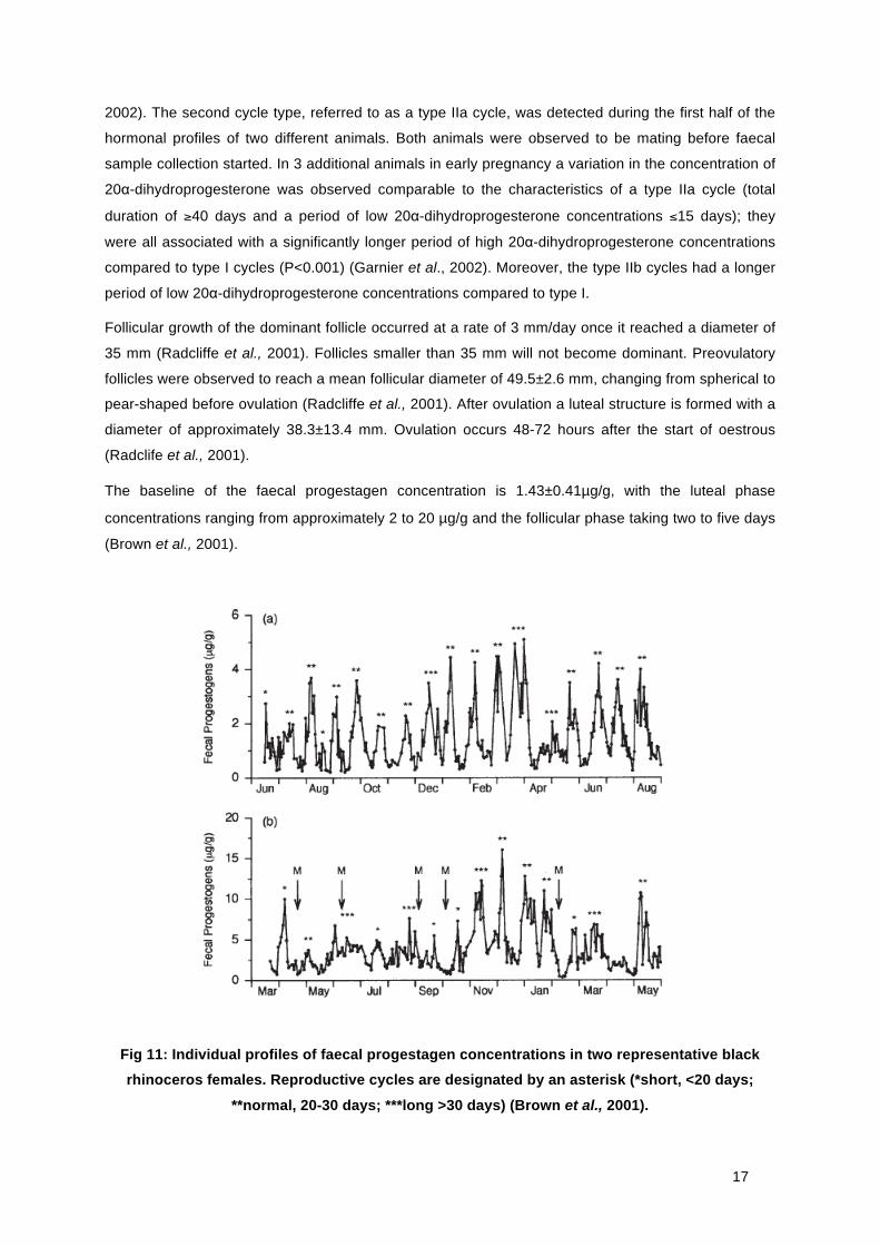
17
2002). The second cycle type, referred to as a type IIa cycle, was detected during the first half of the
hormonal profiles of two different animals. Both animals were observed to be mating before faecal
sample collection started. In 3 additional animals in early pregnancy a variation in the concentration of
20α-dihydroprogesterone was observed comparable to the characteristics of a type IIa cycle (total
duration of ≥40 days and a period of low 20α-dihydroprogesterone concentrations ≤15 days); they
were all associated with a significantly longer period of high 20α-dihydroprogesterone concentrations
compared to type I cycles (P<0.001) (Garnier et al., 2002). Moreover, the type IIb cycles had a longer
period of low 20α-dihydroprogesterone concentrations compared to type I.
Follicular growth of the dominant follicle occurred at a rate of 3 mm/day once it reached a diameter of
35 mm (Radcliffe et al., 2001). Follicles smaller than 35 mm will not become dominant. Preovulatory
follicles were observed to reach a mean follicular diameter of 49.5±2.6 mm, changing from spherical to
pear-shaped before ovulation (Radcliffe et al., 2001). After ovulation a luteal structure is formed with a
diameter of approximately 38.3±13.4 mm. Ovulation occurs 48-72 hours after the start of oestrous
(Radclife et al., 2001).
The baseline of the faecal progestagen concentration is 1.43±0.41µg/g, with the luteal phase
concentrations ranging from approximately 2 to 20 µg/g and the follicular phase taking two to five days
(Brown et al., 2001).
Fig 11: Individual profiles of faecal progestagen concentrations in two representative black rhinoceros females. Reproductive cycles are designated by an asterisk (*short, <20 days;
**normal, 20-30 days; ***long >30 days) (Brown et al., 2001).

18
Table 2: An overview of different cycle lengths as determined by different studies in the Black
Rhinoceros (Diceros bicornis)
Author Year Method Cycle length (days)
Number of animals (n)
Wild or Captive
Hitchins and Anderson
1983 Behaviour analysis 26-46 average of 35
5 Wild
Hindle et al. 1992 Urine hormone analysis (Oestrogen,
Progesterone), Behaviour analysis.
25 2 Wild
Schwarzenberger et al.
1993 Faecal hormone analysis
(Progesterone)
24-26,5 3 Captive
Berkeley et al. 1997 Faecal hormone analysis
(Progesterone), Ultrasound,
Behaviour analysis
26 14 Captive
Brown et al. 2001 Faecal hormone analysis
(Progesterone)
26,8±0,5 (18% cycles <20, 21% cycles
>32)
16 Captive
Lance et al. 2001 Faecal hormone analysis
(Progesterone)
26-27 1 Captive
Radcliffe et al. 2001 Faecal hormone analysis
(Progesterone), Ultrasound
25 2 Captive
Garnier et al. 2002 Faecal hormone analysis
(Progesterone)
Type I: 26,8±1 Type II: >40
Type III: 53,0±6,6
6 Wild
2. Fertility preservation through cryopreservation
Due to the excessive amount of poached animals during the last decades, the genetic diversity in the
elephant and rhinoceros population is decreasing rapidly. A narrow genetic basis results in an
increase in homozygosity, accompanied by deleterious effects, caused by recessive genes (Demirci et
al., 2003), and resulting in lower reproductive efficiency and survival rates.
Several ways to preserve female genes and reproductive tissue have been described such as embryo,
oocyte or gonadal tissue cryopreservation. The method of choice depends on the particular situation
of the animals: living in a zoo or in the wild, cycling or anoestrous, young or old. ‘Fertility Preservation’
(FP) as such can be described as methods for an individual animal, male or female, to reproduce
notwithstanding iatrogenic or pathological loss of fertility.
Several guidelines need to be followed to successfully preserve mammalian reproductive tissue.
Firstly, the tissue needs to be cooled and stored at a temperature of –196 degrees Celsius so that all
intracellular chemical reactions are inhibited (Demicri et al., 2003). Cell survival during and following
19
freezing is determined by the risk of intracellular ice crystal formation and/or extreme dehydration, as
well as mechanical and/or physical damage (Saragusty et al., 2011). In general there are two different
freezing methods: controlled-rate freezing and vitrification (Table 3).
2.0.1. Controlled-rate freezing or slow cooling
Controlled-rate freezing uses a computer device that controls gradual temperature changes during the
freezing and thawing process. The cooling rate differs among cells of different sizes and water
permeability. The tissue that needs to be frozen is stored in small volume straws and cooled to -5° to -
7° Celsius and kept at this temperature for several minutes to equilibrate. Subsequently, it is cooled at
a rate of -0.3°/-0.5° Celsius per minute to around -30° and -65° Celsius (Jewgenow et al., 2010;
Saragusty and Arav, 2011). When the correct temperature is reached, the straws are transferred to
and stored in liquid nitrogen. This method ensures freezing takes place outside the cells due to the
extracellular water crystallizing and the osmotic pressure changes, drawing intracellular water
extracellular, resulting in gradual cell dehydration and leaving the intracellular matrix vitrified.
Directional freezing is a type of controlled-rate freezing which ensures the temperature decreases
rapidly from 5° to -50° Celsius, avoiding intracellular crystal growth, and moving the cold front through
the specimen in stead of working its way from the outside to the inner part.
2.0.2. Vitrification
Vitrification is a cryopreservation method that can be used in the field because there is no need for
computer controlled cooling. Vitrification requires liquid nitrogen and a cryoprotectant for very fast
cooling, creating a glass-state within the cells. The cooling rate can be around 100° to 10.000°Celsius
per minute, depending on the container, the volume, the thermal conductivity and the solution
(Saragusty and Arav, 2011). The tissue samples are loaded with high concentrations of cryoprotective
agents, which can be toxic and need to be removed very quickly after warming the samples
(Jewgenow et al., 2010). Due to the rapid cooling much less ice-crystals are formed.
Table 1: Oocyte and embryo cryopreservation methods (Pereira and Marques, 2008) Table 3: Oocyte and embryo cryopreservation methods (Pereira and Marques, 2008)
20
2.0.3. The role of cryoprotectant agents (CPA)
The aim of CPAs is to reduce the formation of ice-crystals due to an increase of the solution of the
H2O phase in the mammalian cells. There are two categories of cryoprotectants: permeating and
nonpermeating. Permeating CPAs are small molecules that readily penetrate the membranes of the
cells, form hydrogen bonds with intracellular water molecules and lower the freezing temperature of
the resulting mixture, minimizing ice crystallization (Pereira and Marques, 2008). The most commonly
used permeating cryoprotectant is propylene glycol (1,2-propanediol), which is usually at a
concentration of 1.5 M. At this point the toxicity is low but it interferes only poorly with the ice crystal
formation. Several other permeating cryoprotectants are commonly used as well, like ethylene glycol,
glycerol and dimethyl sulfoxide (DMSO). Nonpermeating CPAs are used for the osmotic effect; they
draw the intracellular water out of the cell, resulting in cell dehydration. Sucrose, galactose and
threalose are used as nonpermeating cryoprotectants. Both cryoprotectant classes are used in
combination to increase the net concentration of the permeating cryoprotectant inside the cell and also
to prevent ice crystal formation (Pereira and Marques, 2008).
2.1. Embryo cryopreservation
The main advantage of embryo cryopreservation is the preservation of the genome of both parents in
one embryo. However, embryo cryopreservation is no cure for inbreeding if the stock of the gametes is
limited.
In contrast to horses, there are no studies conducted yet on embryo cryopreservation in rhinoceroses
and elephants. The horse is considered to be the closest domestic relative of the rhinoceros (O’Brien
and Roth, 2000; Stoops et al., 2011). To establish an embryo cryopreservation protocol for
rhinoceroses the horse protocol can therefore be used as an example. Working with horse embryos
showed that smaller, six day old embryos are better permeable for the CPAs than the larger seven
day old embryos. Between day six and seven a cellular capsule is formed, which may impair
movement of the cryoprotectant into the embryo (O’Brien and Roth, 2000). Meira et al. (1993)
concluded that glycerol was superior to 1,2 propanediol as CPA for equine embryo cryopreservation.
Squires et al. (1999) stated that the step-down equilibration method using vitrification is the most
promising method for preservation of large equine embryos.
To improve the survival of cryopreserved embryos, integrated FP strategies need to be put in place.
The most sensitive embryonic structures are the cellular membrane, the cytoskeleton, intracellular
lipids, intracellular water and manipulations to in vitro culture conditions (Saragusty and Arav, 2011).
21
2.2. Oocyte cryopreservation
Oocytes are the largest mammalian cells. Therefore they have a small surface-to-volume ratio and a
correspondingly higher sensitivity to chilling and intracellular ice formation (Songsasen and Comizzoli,
2009). Moreover, due to the delicate cytoskeleton of the oocyte the volumetric resistance is reduced.
Both the zona pellucida, a thick and protective wall around the oocyte, and the plasma membrane,
have a low permeability coefficient and prevent the movement of cryoprotectant and water into the
oocyte (Songsasen & Comizzoli, 2009). Additionally, oocytes are highly delicate to osmotic damage
and the meiotic spindle, formed during metaphase II, is temperature-sensitive (Pereira and Marques,
2008; Saragusty et al., 2011; Comizzoli et al., 2012). In addition, problems arise post-cryopreservation
in fertilization and embryonic development, due to hardening of the zona pellucida, preventing sperm
penetration as well as changes in the organization of the organelles. The cytoskeleton is often
damaged. An alternative approach is cryopreservation of immature (germinal vesicles) oocytes
because at this stage the oocytes have lower microtubular chilling-sensitivity due to a smaller size,
their lower metabolic rate, no zona pellucida and no meiotic spindle (Pereira and Marques, 2008;
Aerts and Bols, 2010a). However, several studies reported immature oocytes to be more sensitive to
cryopreservation than matured ones (Lim et al., 1992; Ledda et al., 2000; Pereira and Marques, 2008),
probably due to a lower cell membrane stability, their particular cytoskeletal formation, the damage
and/or interruption of cumulus cell projections, that control the intercellular communication between
cumulus cells and the maturing oocyte (Ledda et al., 2000).
Cold tolerance studies of oocytes in elephants and rhinoceroses are however, limited due to the low
availability of oocytes for research purposes.
2.3. Gonadal tissue cryopreservation
A possible solution to preserve genetic material from sexually mature or immature female elephants
and rhinoceroses is to harvest and cryopreserve the ovaries post-mortem. Cryopreserved gonadal
tissue can be used for post-thawing oocyte retrieval (Jewgenow et al., 2010). A complete ovary can
be cryopreserved as well as small ovarian pieces. Viable oocytes can be recovered from ovarian
tissue after transplantation of cryopreserved ovaries. Another technique to cryopreserve ovaries is
‘directional freezing’. This technique is based on thermodynamics whereby an ovarian tissue piece or
a whole ovary moves through a preprogrammed temperature gradient at a speed that determines the
cooling rate (Maffei et al., 2013). Due to the moving cold front the ovary or ovarian sample is cooled
precisely and in a uniform way. The cryopreserved tissue can be stored in a tissue bank. These banks
can be used as a source for germ cells that can be cultured in vitro or grafted into a host. After
maturation the cells can be used for IVF (in-vitro fertilisation). However, an alternative way to develop
and recover oocytes from ovarian tissue could be xenografting ovarian tissue to an immune
compromised animal, unable to reject xenografts (Gunasena et al., 1998).

22
There are several advantages in cryopreserving ovarian tissue instead of oocytes or embryos. First of
all, the ovaries contain a large amount of oocytes enclosed in primordial follicles. Secondly, ovarian
tissue can be collected from animals of different ages, dead or alive (Santos et al., 2010). Thirdly, due
to the rather inactive metabolism, the low amounts of lipids, the lack of a zona pellucida, cortical
granules and a meiotic spindle, primordial follicles might be more resistant to cryopreservation.
2.3.1. Elephant
Gunsasena et al. (1998) conducted a study on xenografted, cryopreserved ovarian tissue of three
female elephants from the Kruger National Park, South Africa. The goal of this study was to
investigate if an immune compromised nude mouse model would be suited as a host to develop antral
follicles originating from cryopreserved elephant ovarian tissue. The three adult female elephants were
culled and their ovaries were examined and cryopreserved. On the ovaries of two elephants several
corpora lutea were visible and the weight of the ovaries was between 28-188 grams (Gunsasena et
al., 1998). The ovaries were frozen in liquid nitrogen vapour and cryovials. After thawing their cortical
section it was minced (1 mm x 1-1.5 mm) to fit in the bursa ovarica of the athymic nude mouse that
was used as a host (Gunsasena et al., 1998). The ovarian tissue of the elephant was placed in the
pocket of the bursa ovarica in the mouse and covered by the membrane. About 10-11 weeks after
transplantation, well developed antral follicles and several small follicles were observed in different
grafts. Several oocytes were retrieved despite the fact that they had a poor morphological
appearance.
2.3.2. Rhinoceros
Stoops et al. (2011) conducted a 10-year study on post-mortem ovaries in captive African black
rhinoceroses. In this study the ovaries were shipped at the recommended transport temperature
(22°C) for the horse, (Carnevale et al., 2004) and in vitro maturation was conducted for 32 to 36 hours
(Stoops et al., 2011). Ovaries obtained from five adult African black rhinoceros yielded 74 oocytes,
14.8 ± 7.2 per female (Stoops et al., 2011). The oocyte quality was classified inyo three different
grades. Grade 1: oocytes that were medium to darkly pigmented and completely surrounded by
expanded cumulus cells; grade 2, dark to lightly pigmented oocytes surrounded by several layers of
compact cumulus cells; and grade 3, dark to lightly pigmented oocytes with either no cumulus or
corona radiata cells or only a single layer of corona radiata cells (Stoops et al., 2011). By grading the
oocytes the chance on successful maturation and embryo formation after fertilisation can be predicted.
Because of to the high number of retrieved oocytes, the ovaries it can be considered as a good source
for female gametes. Transported at the recommended temperature will result in oocytes with a distinct
chromatin and in this study at least one oocyte per female achieved nuclear maturation (Stoops et al.,
2011). Many of the oocytes reached the MI phase and several displayed further development in
culture. The researchers concluded that rhinoceros oocytes can remain viable 24 to 48 hours post-
mortem (Stoops et al., 2011).

23
3. Genome Resource Bank
A genome resource bank (GRB) refers to the collection, processing, storage and use of gametes,
embryos and other biological material with the intention to use them in a future breeding programme
(Comizzoli et al., 2000). A GRB can be used to manage the global gene pool of elephants and
rhinoceroses, due to exchange of genetic diversity. In a Genome Resource Bank spermatocytes,
oocytes, embryos or tissue grafts can be stored and a database with important biological information
can be created. GRB can be used in combination with assisted reproductive techniques for in situ (in
nature) and ex situ (in managed captive programs) conservation.
Cloning
A complimentary alternative to a genome resource bank is cloning or so-called somatic cell nuclear
transfer (SCNT) of living elephants and rhinoceroses. During this process the nucleus (DNA) is moved
from a donor cell to an enucleated recipient cell (Andrabi and Maxwell, 2007). If cloning results in a
viable embryo, the genome of the embryo is a copy of the genome of the donor, apart from the
mitochondrial DNA. This DNA is copied from the animal that was used as a recipient. By transferring a
somatic cell nucleus into the enucleated egg of a genetic stock, a closely related species or another
subspecies, the recovery of the complete genome of the donor can be obtained without the genetic
dilution that would occur in producing biparental hybrids (Corley-Smith and Brandhorst, 1999).
This means that the genome of non-cycling, but healthy, elephants and rhinoceroses, or animals in
anoestrous can be used to create offspring that in turn can reproduce itself. In this way, no vital DNA
is lost and inbreeding in the population or the creation of a small founder population is prevented.
A downside of cloning is that it leads to a stagnation or decline of genetic diversity, especially if non-
healthy animals will be cloned. In addition, the production of viable offspring using cloning uses a lot of
germ cells, that are not available in elephants and rhinoceroses.

24
4. Conclusion and future perspectives
Nowadays the interest in preserving genetic material of endangered species is rising, mostly due to
the rapid decline in numbers of wild living elephants and rhinoceroses because of the intensive
poaching. With each poached animal a considerable amount of important genetics is irreversibly lost.
Cryopreservation, through vitrification of ovarian tissue of recently killed animals and the development
of a genome resource bank could be a solution to prevent lost of genetic diversity.
However, regardless of the source of the germ cells, oocytes, embryos or ovarian tissue, a thorough
knowledge of the fundamental reproductive cycle of both animals is necessary to be able to produce
viable offspring (Comizzoli and Wildt, 2014). The oestrous cycle length and the time of ovulation are
two important aspects. The reproductive cycle is well known for the elephant, and characterized by an
oestrous cycle length of 13-18 weeks, with an anovulatory LH surge 19-22 days before the ovulatory
LH surge (Plotka et al., 1988; Hildebrandt et al., 2010). As for the black rhinoceros, the oestrous cycle
covers 25 days with a single ovulatory LH surge (Hindle et al., 1992; Schwarzenberger et al., 1993).
However, there are different opinions among scientists regarding the white rhinoceros reproductive
cycle length, and differences between captive and wild animals.
To increase the overall success rate with cryopreserving elephant and rhinoceros genetic material,
more studies on this topic should be conducted. Retrieving oocytes from in-vivo donor rhinoceros and
elephant is a sensitive issue, due to the high value of the animals and the lack of knowledge on this
topic. It is conducted in one study; Hermes et al. (2007) performed a trans-rectal ultrasound-guided
follicle aspiration in an infertile black rhinoceros. Multiple follicles were punctured, but it did not result
in an embryo yet. This can be promising in a future perspective. Another strategy could focus on the
optimal use of ovaries from deceased animals in zoos and sanctuaries. A cryopreserving protocol
should be made specifically for the two different animal species. In this protocol the most suited
cryoprotectant, the optimal method of freezing, the cooling rate and the size of the ovarian tissue
samples should be determined. In addition a universal scenario should be agreed upon by which
scientists all over the world can recover gonads from freshly slaughtered or poached possible donors
under the most optimal conditions. After the veterinarian conducted all these steps the ovaries could
be transported to a central laboratory where further research can be performed.
Another solution could be composing a GRB including different biological products, like blood, tissue
and DNA. These biomaterials can be used in phylogenetic, systematic and disease studies related to
conservation (Wildt, 2000). They also can give insight in gene flow, selection and mating.
A difficulty in this solution is that in Europe all rhinoceroses and elephants are owned by the EAZA
(European Association of Zoos and Aquaria) or privately owned. Also, it is prohibited to transport
biological materials over long distances, due to the possible risk of spreading infectious diseases
throughout Europe. In Africa, the animals that are living in wild parks or around lodges are mostly
privately owned. The government owns all the animals that are living in the national parks. It could be
difficult to come close to recently poached animals in the national parks, this because the deceased or
25
poached animal and it surroundings is treated as an official crime scene. All traces that can lead to the
poachers are secured.
To solve both problems a central umbrella organisation should be founded. This organisation needs to
keep track of all poached animals, and therefore work closely whith the police, the government, the
wild parks and national parks in Africa. On the other hand, in Europe the organisation must keep track
of all deceased animals in zoos and work closely with the house veterinarians and the laboratories. If
all this can be established, there is an opportunity to save those animals.

26
References
• Aerts, J.M.J., Bols, P.E.J. (2010a). Ovarian follicular dynamics: a review with emphasis on the
bovine species. Part I: folliculogenesis and pre-antral follicle development. Reproduction of
Domestic Animals, 45, 171-179.
• Andrabi, S.M.H., Maxwell, W.M.C. (2007). A review on reproductive biotechnologies for
conservation of endangered mammalian species. Animal Reproductive Science 99, 223-243.
• Berkeley, E.V., Kirkpatrick, J.F., Schaffer, N.E., Bryant, W.M., Threlfall, W.R. Serum and fecal
steroid analysis of ovulation, pregnancy and Parturition in the Black Rhinoceros (Diceros
bicornis). (1997). Zoo Biology 16, 121-132.
• Bertschinger, H.J. (1994). Reproduction in black and white rhinos: a review. Proceedings of a
symposium on “Rhinos as game ranch animals”, Onderstepoort Pretoria.
• Brown, J.L. (2000). Reproductive Endocrine Monitoring of Elephants: an Essential Tool for
Assisting Captive Management. Zoo Biology 19, 347-367.
• Brown, J.L., Bellem, A.C., Fouraker, M., Wildt, D.E., Roth, T.L. (2001). Comparative analysis
of gonadal and adrenal activity in the black and white rhinoceros in North America by
noninvasive endocrine monitoring. Zoo Biology 20, 463-486.
• Carnevale, E.M., Coutinho da Silva, M.A., Preis, K.A., Stokes, J.E., Squires, E.L. (2004).
Establishment of pregnancies from oocytes collected from the ovaries of euthanized mares.
Proceedings of the American Association of Equine Practioners 50, 531-533.
• Comizzoli, P., Mermillod, P., Mauget, R. (2000). Reproductive biotechnologies for endangered
mammalian species. Reproduction Nutrition Development 40, 493-504.
• Comizzoli, P., Songsasen, N., Hagedorn, M., Wildt, D.E. (2012). Comparative cryobiological
traits and requirements for gametes and gonadal tissues collected from wildlife species.
Theriogenology 78, 1666-1681.
• Comizzoli, P., Wildt, D.E. (2014). Mammalian fertility preservation through cryobiology: value
of classical comparative studies and the need for new preservation options. Reproduction,
Fertility and Development 26, 91-98.
• Corley-Smith, G., Brandhorst, B. (1999). Preservation of endangered species and populations:
a role for genome banking, somatic cell cloning, and androgenesis? Molecular Reproduction
and Development 53, 363-367.
• Demirci, B., Lornage, J., Salle, B., Poirel, M.T., Guerin, J.F., Franck, M. (2003). The
cryopreservation of ovarian tissue: uses and indications in veterinary medicine.
Theriogenology 60, 999-1010.
• Department of Environmental Affairs, South Africa. Internet referene:
http://www.defenceweb.co.za, consulted at 16/04/2015.
• Fair, T. (2003). Follicular oocyte growth and acquisition of developmental competence. Animal
Reproduction Science, 78, 203-216.

27
• Fowler, M.E., Mikota, S.K. (2006). Biology, Medicine and Surgery of Elephants. Blackwell
Publishing, Oxford, United Kingdom p. 377-388.
• Garnier, J.N., Holt, W.V., Watson, P.F. (2002). Non-invasive assessment of oestrous cycles
and evaluation of reproductive seasonality in the female wild black rhinoceros (Diceros
bicornis minor). Reproduction 123, 877-889.
• Gunasena, K.T., Lakey, J.R.T., Villines, P.M., Bush, M., Raath, C., Critser, E.S., McGann,
L.E., Critser, J.K. (1998) Antral follicles develop in xenografted cryopreserved African elephant
(Loxodonta africana) ovarian tissue. Animal Reproduction Science 53, 265-275.
• Goot van der, A.C., Dalerum, F., Ganswindt, A., Martin, G.B., Millar, R.P., Paris, M.C.J.
(2013). Fecal progestagen profiles in wild southern white rhinoceroses (Ceratotherium simum
simum). African Zoology 48, 143-151.
• Hermes, R. and Hildebrandt, T.B. (2001). Chapter 71: Rhinoceros Theriogenology. In: Fowler,
M. and Miller, E. (eds). Zoo and Wild Animal Medicine, current therapy. 7th Edition, Saunders,
St. Louis, USA, 546-561.
• Hermes, R., Hildebrandt, T.B., Göritz, F. (2004). Reproducutive problems directly attributable
to long-term captivity-asymmetric reproductive aging. Animal Reproduction Science 82-83, 49-
60.
• Hermes, R., Göritz, F., Streich, W.J., Hildebrandt, T.B. (2007). Assisted Reproduction in
Female Rhinoceros and Elephants – Current Status and Future Perspective. Reproductive in
Domestic Animals 42, 33-44.
• Hildebrandt, T., Lueders, I., Hermes, R., Goeritz, J., Saragusty, J. (2010). Reproductive Cycle
of the Elephant. Animal Reproductive Science 124, 176-183.
• Hindle, J.E., Möstl, E., Hodges, J.K. (1992). Measurment of urinary oestrogens and 20α-
dihydroprogesterone during ovarian cycles of black (Diceros bicornis) and white
(Ceratotherium simum) rhinoceroses. Journals of Reproductive & Fertility 94, 237-249.
• Hitchins, P.M., Andersond, J.L. (1983). Reproduction, population characteristics and
management of the black rhinoceros Diceros bicornis minor in the Hluhluwe/Corridor/Umfolozi
game reserve complex. South African Journal of Wildlife Research, 13, 78-85.
• Jewgenow, K., Wiedeman, C., Bertelsen, M.F., Ringleb, J. (2010). Cryopreservation of
mammalian ovaries and oocytes. International Zoo Yearbook 45, 124-132.
• Lance, V.A., Patton, M.L., Hagey, L.R. (2001). Identification of a series of C21O2 pregnanes
from fecal extracts of a pregnant black rhinoceros (Diceros bicornis minor). Steroids 66, 875-
881.
• Ledda, S., Leoni, G., Bogliolo, L., Naitana, S. (2000). Oocyte cryopreservation and Ovarian
tissue banking. Theriogenology 55, 1359-1371.
• Lim, J.M., Fukui, Y., Ono, H. (1992). Developmental competence of bovine oocytes at various
maturation stages followed by in vitro maturation and fertilization. Theriogenology 37, 351-
362.
28
• Lueders, I., Hildebrandt, T.B. (2011). Chapter 66: Female elephant reproduction update. In:
Fowler, M. and Miller, E. (eds). Zoo and Wild Animal Medicine, current therapy. 7th Edition,
Saunders, St. Louis, USA.
• Lussier, J.G., Matton, P., Dufour, J.J. (1987). Growth rates of follicles int ovary of the cow.
Journal of Reproduction and Fertilisation, 81, 301-307.
• Maffei, S., Hanenberg, M., Pennarossa, G., Silva, J.R., Brevini, T.A., Arav, A., Gandolfi, F.
(2013). Direct comparative analysis of conventional and directional freezing for the
cryopreservation of whole ovaries. Fertility and Sterility 100, 1122-1131.
• Meira, C., Alvarenga, M.A., Pappa, O.F., Abba, E., Landon, E., Alvarenga, F.C. (1993).
Cryopreservation of equine embryos using glycerol and 1,2 propanediol as cryoprotectnts.
Equine Veterinary Journal 15, 64-66.
• Morrow, C., Kudeweh, S., Goold, M., Standley, S. (2008). Reproductive cycles, pregnancy
and reversal of long term acyclicity in captive southern white rhinoceros at Hamilton Zoo.
Reproduction Fertility and Development 21 (1), 180.
• O’Brien, J.K., Roth, T.L. (2000). Post-coital sperm recovery and cryopreservation in the
Sumatran rhonceros (Dicerorhinus sumatrensis) and application to gamete rescue in the
African black rhinoceros (Diceros bicornis). Journal of Reproduction and Fertility 118, 263-
271.
• Patton, M.L., Swaisgood, R.R., Czekala, N.M., White, A.M., Fetter, G.A., Montagne, J.P.,
Rieches, R.G., Lance, V.A. (1999). Reproductive Cycle Length and Pregnancy in the
Southern White Rhinoceros (Ceratotherium simum simum) as Determined by Fecal Pregnane
Analysis and Observations of Mating Behavior. Zoo Biology 18, 111-127.
• Pereira, R.M., Marques, C.C. (2008). Animal oocyte and embryo cryopreservation. Cell Tissue
Banking 9, 267-277.
• Plotka, E.D., Seal, U.S., Zarembka, E.D., Simmons, L.G., Teare, A., Phillips, L.G., Hinshaw,
K.C., Wood, D.G. (1988). Ovarian Function in the Elephant: Luteinizing Hormone and
Progestrone Cyclus in African and Asian Elephants. Biology of Reproduction 38, 309-314.
• Radcliffe, R.W., Czekla, N.M., Osofsky, S.A. (1997). Combined Serial Ultrasonography and
Faecal Progestin Analysis for Reproductive Evaluation of the Female White Rhinoceroses
(Ceratotherium simum simum): Preliminary Results. Zoo Biology 16, 445-456.
• Radcliffe, R.W., Eyres, A.I., Patton, M.L., Czekala, N.M., Emslie, R.H. (2001).
Ultrasonographic Characterization of Ovarian Events and Fetal Gestational Parameters in
Two Southern Black Rhinoceroses (Diceros bicornis minor) and Correlation to Fecal
Progesterone. Theriogenology 55, 1033-1049.
• Rhino Resource Center. (2014). Internet reference: http://rhinoresourcecenter.com, consulted
at 14/04/2015.
• Rodriguez-Wallberg, K.A., Oktay, K. (2012). Recent advances in oocyte and ovarian tissue
cryopreservation and transplantation. Best practice & research clinical obstetrics and
gynaecology 26, 391-405.

29
• San Diego Zoo Library. (2008). Internet reference: http://library.sandiegozoo.org, consulted at
14/04/2015.
• Santos, R.R., Amorim, C., Cecconi, S., Fassbender, M., Imhof, M., Lornage, J., Paris, M.,
Schoenfeldt, V., Martinez-Madrid, B. (2010). Cryopreservation of ovarian tissue: An emerging
technology for female germline preservation of endangerd species and breeds. Animal
Reproduction Science 122, 151-163.
• Saragusty, J., Arav, A. (2011). Current progress in oocyte and embryo cryopreservation by
slow freezing and vitrification. Reproduction 141, 1-19.
• Saragusty, J., Hermes, R., Göritz, F., Hildebrandt, T.B. (2011). Mammalian reproduction out of
cryopreserved cells and tissues: current state of the art and future options. International Zoo
Yearbook 45, 133-153.
• Save the Rhino. (2015). Internet reference: http://www.savetherhino.org, consulted at
27/03/2015.
• Schwarzenberger, F., Francke, R., Göltenboth, R. (1993). Concentrations of faecal
immunreactive progestagen metabolites during the oestrous cycle and pregnancy in the black
rhinoceros (Diceros bicornis minor). Journal of Reproduction and Fertility 98, 285-291.
• Schwarzenberger, F., Walzer, C., Tomasova, K., Vahala, J., Meister, J., Goodrowe, K.L.,
Zima, J., Strauss, G., Lynch, M. (1998). Faecal progesterone metabolite analysis for non-
invasive monitoring of reproductive function in the white rhinoceros (Ceratotherium simum).
Animal Reproduction Science 53, 173-190.
• Senger, P.L. (2005). Pathways tot pregnancy and parturition. Second revised edition. Current
Conceptions Inc., Pullman, United States of America.
• Sjaastad, Ø.V., Sand, O., Hove, K. (2010). Physiology of Domestic Animals. 2nd edition.
Scandinavian Veterinary Press, Oslo, Norway. p. 684-734.
• Songsasen, N., Comizzoli, P. (2009). A historic overview of embryos and oocyte preservation
in the world of mammalian in vitro fertilization and biotechnology. In: Borini, A., Coticchio, G.
(eds). Preservation of human oocytes. London: Informa Healthcare.
• Squires, E.L., McCue, P.M., Vanderwall D. (1999). The current status of equine embryo
transfer. Theriogenology 5, 91-104.
• Stoops, M.A., O’Brien, J.K., Roth, T.L. (2011). Gamete rescue in the African black rhinoceros
(Diceros bicornis). Theriogenology. 76, 1258-1265.
• Strike, T., Pickared, A. (2000). Non-invasive hormone analysis for reproductive monitoring in
femal southern white rhinoceros (Ceratotherium simum simum). Proceedings of the 2nd
Annual Symposium on Zoo Research, Devon.
• Wagner, R.A. (1986). The reproductive cycle of one southern white rhinoceros. Proceedings
of the American Association of Zoo Veterinarians, p. 14-16.
• Wildt, D.E. (2000). Genome resource banking for wildlife research, management, and
conservation. Cryobiology of Embryos, Germ Cells and Ovaries 41(4), 228-234.

30
• Yang, M.Y., Fortune, J.E. (2008). The capacity of primordial follicles in fetal bovine ovaries
initiate growth in vitro develops during mid-gestation and is associated with meiotic arrest of
oocytes. Biology of Reproduction, 78, 1153-1161.