ggr biennial critical review: analytical developments
TRANSCRIPT

Acc
epte
d A
rtic
le
This article has been accepted for publication and undergone full peer review but has not been through the copyediting, typesetting, pagination and proofreading process, which may lead to differences between this version and the Version of Record. Please cite this article as doi: 10.1111/j.1751-908X.2014.00347.x This article is protected by copyright. All rights reserved.
Received Date: 30/10/2014 Accepted Date: 30/10/2014
Article Type: Original Article
GGR Biennial Critical Review: Analytical Developments Since 2012
Michael Wiedenbeck (1)*, L. Paul Bédard (2) Roxana Bugoi (3), Mary Horan (4), Kathryn Linge (5), Silke Merchel (6), Luiz F.G. Morales (1), Dany Savard (2), A. Kate Souders (7) and Paul Sylvester (8) (1) Helmholtz Zentrum Potsdam, Deutsches GeoForschungsZentrum, Telegrafenberg, 14473
Potsdam, Germany (2) Sciences de la Terre, Université du Québec à Chicoutimi, Chicoutimi, QC, Canada (3) Roxana Bugoi, Horia Hulubei National Institute for Nuclear Physics and Engineering,
P.O. Box MG-6, Bucharest-Măgurele, RO-077125, Romania (4) Department of Terrestrial Magnetism, Carnegie Institution of Washington, 5241 Broad
Branch Road NW, Washington DC, 20015, USA (5) Curtin Water Quality Research Centre, Curtin University, GPO Box U1987 Perth,
Western Australia, 6845, Australia (6) Helmholtz-Zentrum Dresden-Rossendorf, Helmholtz Institute for Resource Technology,
Bautzner Landstrasse 400, 01328 Dresden, Germany (7) Center for Meteorite Studies, School of Earth and Space Exploration, Arizona State
University, Tempe, Arizona, 85287-1404, USA (8) Department of Geosciences, Texas Tech University, 125 Science Building, Lubbock,
Texas, 79409-1053, USA * Corresponding author. e-mail: [email protected]
Advances in the chemical, crystallographic and isotopic characterisation of geological
and environmental materials can often be ascribed to technological improvements in
analytical hardware or to innovative approaches to data acquisition and/or its
interpretation. This biennial review addresses key laboratory methods that form much
of the foundation for analytical geochemistry; again this contribution is presented as a
compendium of laboratory techniques. We highlight advances that have appeared since
January 2012 and that are of particular significance for the chemical and isotopic
characterisation of geomaterials. Prominent scientists from the selected analytical fields
present publications they judge to be particular noteworthy, providing background
information about the method and assessing where further opportunities might be
anticipated. In addition to well established technologies such as thermal ionisation mass
spectrometry and plasma emission spectroscopy, this publication also presents new or
rapidly growing methods such as electron backscattered diffraction analysis and atom
probe tomography – a very sensitive method providing atomic scale information.
Acc
epte
d A
rtic
le
This article is protected by copyright. All rights reserved.
Keywords: ICP-MS, laser ablation, mass spectrometry, ICP-AES, microwave plasma source,
TIMS, isotopic analysis, geochronology, trace element analysis, calibration, XRF,
environmental sampling, AMS, Ion Beam Analysis, radionuclides, SIMS, ion probe,
microanalysis, FIB, EBSD, atom probe tomography.
Received 23 Oct 14 – Accepted 30 Oct 14
This contribution marks the continuation of Geostandards and Geoanalytical Research’s
biennial review series, which describes advances in both methods and instrumentation for the
characterisation of geomaterials. Such reviews in Geostandards first appeared in 2006, where
five individual contributions devoted to specific analytical methods were presented. The most
recent contribution (Wiedenbeck et al. 2012) was presented as a single compilation, and this
format has been continued here. One change from the review that appeared two years
previously is that the segment devoted to reference materials in geoanalytical and
environmental research has been removed, making this current contribution focussed entirely
on laboratory instrumentation and innovative strategies that advance data quality. Specific
chapters contained in this review, and their corresponding page numbers, are:
• Plasma source mass spectrometry and laser ablation sampling page xxx
• Plasma source emission spectroscopy page xxx
• X-ray fluorescence methods page xxx
• TIMS ion detection page xxx
• Accelerator-based methods page xxx
• Secondary ion mass spectrometry page xxx
• Focused ion beam method page xxx
• Electron backscatter diffraction page xxx
• Atom probe tomography page xxx
Advances in plasma source mass spectrometry and laser ablation sampling (Contribution by: P.J. Sylvester and A.K. Souders)
Overview of ICP-MS and laser ablation ICP-MS
Inductively coupled plasma source-mass spectrometry (ICP-MS) is used principally for rapid,
precise and accurate trace element determination in solid, liquid and gas samples. It is also
Acc
epte
d A
rtic
le
This article is protected by copyright. All rights reserved.
used increasingly for both isotopic and speciation determinations. The technique became
commercially available in the mid-1980s, and since then has grown rapidly as a technique
capable of element and isotopic measurements over a wide dynamic range (major to sub-trace
contents) at a range of spatial resolutions (bulk rock to micrometre-size inclusions). Samples
are introduced to the ICP-MS as solids dissolved in acidic solutions, sometimes purified to
separate particular elements of analytical interest, or as fine aerosols produced by laser
ablation. For solutions, a nebuliser converts the liquid into a fine spray of droplets, the largest
of which are removed by a spray chamber, before the remaining aerosol is injected into the
ICP where the atoms of the elements in the sample are converted into ions.
In general, a mass spectrometer has four main parts: the ion source, mass filter, detector, and
vacuum system. ICP mass spectrometers come as single-collector (detector) instruments,
which detect ions sequentially by mass, and multi-collector (detector) instruments capable of
simultaneous detection of ions. Quadrupole and magnetic sector ICP-MS instruments are the
two most common mass analysers used in the geosciences. A quadrupole mass analyser
consists of four parallel rods with an AC or DC voltage applied to each set of rods so as to
create an electrostatic field between the rods (Linge and Jarvis 2009). For given combination
of AC and DC voltages, only ions of a certain mass-to-charge ratio will pass through the
quadrupole; all other ions are filtered from their original path. A magnetic sector separates
ions in a magnetic field as a function of their mass-to-charge ratio between poles of a magnet
(Rehkämper et al. 2001).
Three different detector types are found in ICP-MS instruments. Discrete-dynode and
continuous dynode detectors operate in both pulse-counting and analogue modes that have
the advantage of both an instantaneous response time and a large dynamic range capable of
detecting concentrations from sub-pg/g to high- μg/g. Faraday cups are the third type of
detector in common use in ICP mass spectrometer, they have the ability to deal with
excessively high count rates (109 to 1012 cps), are effectively 100% efficient, and have stable
response characteristics. Unfortunately response of the Faraday cups is not also
instantaneous, meaning Faraday cup readouts lag behind that of electron multipliers,
increasing analytical uncertainty when the two different detector types are used for the same
measurement.
Laser ablation – ICP-MS (LA-ICP-MS) is an in situ analytical technique in which laser
pulses are used to remove small volumes of a sample, and the resulting aerosol is introduced
into the ICP-MS for analysis. In the geosciences this technique is used for determining the
elemental and isotopic composition of micrometre-sized areas of minerals, volcanic glasses,
metal alloys, fused rocks, fluid inclusions, experimental charges and variety of environmental
Acc
epte
d A
rtic
le
This article is protected by copyright. All rights reserved.
proxies such as bones, teeth and calcareous shells (Sylvester 2008). Samples are typically
prepared as a thin polished slice or slab, and are often imaged using light optical and/or
scanning electron microscopy in advance so as to identify domains the best domains for laser
sampling.
A laser ablation system is connected to an ICP-MS via an airtight sample cell, where the
samples are ablated, and plastic tubing, which carries the ablated aerosols to the ICP in a
helium + argon gas stream. As with solution nebulisation, the aerosols are ionised in the ICP
torch, and then separated based on their mass to charge ratios by a mass spectrometer and
measured with a detector. A quadrupole mass filter is the most common type of mass
spectrometer used for LA-ICP-MS, but magnetic sector field mass analysers incorporating
single- or multiple-collectors (MC) are increasingly employed because of their superior
sensitivity and precision for isotope ratio measurements. The detector in single-collector
instruments is typically a secondary electron multiplier, whereas multiple Faraday cups (± ion
counters) are used in MC devices.
Deep ultraviolet (UV) nanosecond pulsed lasers are most commonly used for LA-ICP-MS,
either a neodymium-doped yttrium aluminium garnet (Nd:YAG) solid-state laser using the
frequency quintupled 213 nm output, or an argon-fluoride (ArF) excimer gas laser using its
fundamental 193 nm wavelength. UV light is absorbed more completely than infrared light
by many geomaterials, particularly those that are transparent or poor in iron. There has been
recent interest in using femtosecond pulsed lasers based on evidence that they produce
aerosols with a large proportion of small particle sizes and negligible sample melting at the
ablation site, improving analytical performance (Shaheen et al. 2012).
The moderate capital cost, robust design, multi-element capability, short analysis times,
minimal sample preparation requirements, and good precision and accuracy of LA-ICP-MS
compared with competing techniques for in situ micrometre-scale, chemical analysis has led
to its widespread use in university, government and industry research laboratories worldwide.
Five notable areas of developments in plasma source mass spectrometry and laser sampling
for geochemistry and geochronology since 2012 are presented in this contribution: (1)
Sample digestion for solution ICP-MS analysis; (2) Performance of the new generation MC-
ICP-MS instruments; (3) Improved spatial resolution of laser analysis; (4) Inter-laboratory
comparison of in situ U-Pb zircon geochronology; and (5) New data processing software for
LA-ICP-MS. Finally, we note that a recent thematic issue in Geostandards and Geoanalytical
Research (Sylvester 2014 and associated contributions) gives a recent perspective on a
variety of key aspects of laser-based sampling methods as applied to geomaterials.

Acc
epte
d A
rtic
le
This article is protected by copyright. All rights reserved.
Sample digestion for solution ICP-MS analysis
ICP-MS is used routinely for multi-element and isotopic analysis of geological materials.
LA-ICP-MS has rapidly become a popular choice for the in situ determination of elemental
and isotopic compositions of individual minerals, while solution ICP-MS remains the
preferred method of sample introduction for characterising the chemical compositions of
whole rocks and high-precision isotopic analyses of geological materials. Sample digestion is
a critical step when preparing geological samples for solution ICP-MS analysis; it is most
often the limiting factor in obtaining rapid and accurate results. Techniques that have been
developed include fusion and sintering procedures, open and closed vessel acid digestion,
microwave digestion and high-pressure acid digestion.
Acid digestions of geological samples are not without challenges. Complete digestion of rock
samples with a high proportion of refractory phases such as zircon, rutile or garnet can be
difficult and typically require acid attack in closed-vessel PTFE bombs at high temperature
for multiple days. Hydrofluoric acid (HF), an effective acid for breaking Si–O bonds, is
commonly combined with other acids (e.g., HNO3, HClO4, HCl) to break down geological
samples, with the choice of acid mixtures strongly dependent on sample matrix. The
formation of insoluble fluoride complexes can present a major challenge to achieving full
dissolution. Insoluble phases (e.g., AlF3, MgF2, CaAlF5) typically incorporate large
proportions of trace elements and lead to low sample recoveries and inaccurate solution
analyses.
The importance of complete sample digestion in order to obtain accurate analytical results
has been demonstrated by several recent investigations. Zhang et al. (2012) investigated the
capabilities of HF and HF–HNO3 in high-pressure digestion systems, or PTFE-lined stainless
steel bombs. They focused on the complete digestion of felsic rocks, which have a reputation
of being difficult to digest completely due to the presence of refractory phases such as zircon.
Several parameters, such as HF–HNO3 ratio, digestion time, digestion temperature, sample
size, and presence/absence of insoluble fluorides were systematically evaluated through
solution, multi-element ICP-MS analysis of the felsic USGS reference material GSP-2. For
comparison, a series of reference materials covering the compositional spectrum of igneous
rocks (basic-intermediate-acidic) plus two common sedimentary rock reference materials
(shales) were also digested and evaluated using the same analytical strategy.
Variations on three basic digestion procedures (A: 1 ml HF + 1 ml HNO3; B: 1 ml HF; C: 2
ml HF) were used for the decomposition of the rock reference materials. The final solution of
each experimental digestion product were analysed for elemental concentrations of 37

Acc
epte
d A
rtic
le
This article is protected by copyright. All rights reserved.
isotopes using an Agilent 7700X ICP-MS. Using Zr recovery as an approximation for
refractory phase digestion, Zhang et al. (2012) conclude that using HF alone is the most
efficient and cost effective method for attacking felsic rock samples. Results demonstrate that
for complete digestion of a rock sample with < 100 mg mass, it would take 8–12 hours at
190°C in a closed high-pressure vessel for complete Zr recovery. It was also found that
sequential addition of HNO3 (0.2–6 ml) to the HF acid digestion solution lowered the
digestion capability of the HF, as noted by the decreased recovery of Zr with increasing
amounts of HNO3 to the acid mixture.
The presence of insoluble fluoride phases were observed in the final solution for all
experimental digestions using a > 100 mg test portion, regardless of digestion solution used.
Low recoveries of Nb and Ta suggest that a second HClO4 acid attack for samples > 100 mg
suppresses, but does not eliminate, fluoride formation. Zhang et al. (2012) conclude that HF
alone can be used successfully in high-pressure digestion of a range of igneous rock
compositions. They note that it is important that sample aliquots are < 100 mg of ultra-fine
fractions (< 30 µm), which raises the question of how representative a small sample can be,
knowing that most reference materials have not been tested for homogeneity at the milligram
scale (Cotta et al. 2007).
The formation of insoluble fluorides during HF–HNO3 digestion is controlled by the
proportions of Al:Mg:Ca in the sample, specifically the [(Mg + Ca)/Al]M (Takei et al. 2001).
By adding Mg to the sample prior to digestion so [(Mg + Ca)/Al]M ≈ 1, Takei et al. (2001)
were able to suppress the formation of AlF3, the most resistant fluoride formed during high
temperature digestion of felsic silicate rocks. Recognising that the formation of insoluble
fluoride phases is a major challenge to obtaining full sample recovery using traditional HF–
HNO3 digestion protocol, Cotta and Enzweiler (2012) incorporated the Mg-addition
technique of Takei et al. (2001) into a new HF–HNO3 digestion procedure using a High
Pressure Acid System (HPA-S), which had been previously used successful for the digestion
of biological samples (e.g., Amarasiriwardena et al. 1994) and platinum-group element
bearing minerals in geological materials (e.g., Meisel et al. 2001).
During development of the HPA-S method, geological reference materials of both granitic
and basaltic composition were used to evaluate the effect of digestion temperature, HClO4
addition and Mg-addition prior to sample dissolution; the concentrations of 41 trace elements
were determine in the final digestion solution. Element concentrations were measured using a
Thermo Scientific X series ICP-QMS. The recovery results were then compared with
recovery data from digestions of the same materials using traditional HF–HNO3 digestion
techniques (sintering, open vessel acid decomposition, closed vessel acid decomposition).

Acc
epte
d A
rtic
le
This article is protected by copyright. All rights reserved.
The results of Cotta and Enzweiler (2012) show complete recovery (± 10%) of most
measured elements. Fluoride precipitation was inhibited using the HPA-S by finding the
appropriate digestion temperature, adding Mg to samples prior to digestion and evaporation
with HClO4. The HPA-S was demonstrated to be well suited for the determination of trace
elements in geological samples with both mafic and felsic compositions as well as felsic
samples with a high proportion of acid-resistant phases.
An alternative to traditional HF-HNO3 sample digestion procedures is the open-vessel acid
digestion method using ammonium fluoride (NH4F) presented by Hu et al. (2013). NH4F is a
solid reagent with a boiling point of 260°C that can be purified using a PFA sub-boiling
purification system prior to use for sample digestion. The high boiling point of NH4F enables
use of a high temperature, open-vessel (Savillex screw-top Teflon vials) acid digestion
method, eliminating the use of high-pressure PTFE digestion bombs and HF. Because NH4F
has not been routinely used to dissolve geological materials, Hu et al. (2013) presented a
systematic study of NH4F for the decomposition of the felsic geological reference material
GSP-2. Using an open-vessel digestion method they document the effects of digestion
temperature, digestion time, addition of nitric acid, amount of NH4F used, presence of
insoluble fluorides and NH4F purification.
Geological reference material USGS GSP-2 (granodiorite) was chosen to evaluate the
decomposition capabilities of NH4F because of its high Zr content (580 μg g-1). To further
test the accuracy and precision of the NH4F dissolution method, eleven additional geological
reference materials covering a range of igneous rocks, shale and soils were also analysed.
Multi-element analyses of experimental digestion products were conducted on acidic
solutions, using an Agilent 7700x ICP-MS. Analytical precision of measured element
concentrations was generally better than 10% (1 RSD) for all of the reference materials
analysed. The accuracy of measured elemental concentrations was better than 10% for most
elements.
The optimised procedure involves adding NH4F and sample powder (< 74 µm) into a screw
top PFA vial at a ratio of 6:1 (NH4F:sample). Complete digestion of refractory minerals using
the NH4F method is controlled by the combination of time and temperature, with temperature
being a critical factor. The proportion of refractory mineral zircon digested controls the
recovery of Zr. A digestion time of 1.5 hr and temperature of 250 °C was found to be
optimal, which is approximately twelve times faster than traditional HF–HNO3 digestion
using a high-pressure PTFE digestion bomb at 190 °C. Hu et al. (2013) discovered that
addition of HNO3 has a negative effect on the digestion capacity of NH4F: Zr recovery
decreased with the addition of just 0.2 ml HNO3. Perhaps the greatest advantage of the NH4F

Acc
epte
d A
rtic
le
This article is protected by copyright. All rights reserved.
digestion protocol is that no insoluble fluorides were found during the digestion of even 200
mg samples. Limiting fluoride precipitation is key to obtaining accurate trace element data
for silicate rock samples. Results indicate this is possible using NH4F without sacrificing
time, money, or sample homogeneity.
Performance of new generation MC-ICP-MS instruments
The general trend in ICP-MS technology is toward more sensitive instruments to maximise
measurement accuracy and precision while minimising the sample volume being analysed in
case of solution analyses; in the case of in situ analyses improved spatial resolution is a key
factor. The two manufacturers of multi-collector instruments have both produced second-
generation instruments (Thermo Scientific – Neptune Plus; Nu Instruments – Nu Plasma II
ES) or have made upgrade packages available for existing multi-collector instruments, with
new technology aimed at improved sensitivity. Examples of new hardware to increase overall
instrument sensitivity include improvements in ion transmission by redesign of the ICP
interface and ion extraction region (e.g., cone geometry and source lens), improved vacuum
within the interface region, the development of compact ion counting detectors installed
within the Faraday detector array and the installation of low noise amplifiers with 1012 Ω
resistors for improved signal/noise ratios on Faraday detectors at low intensity signals.
Zhang et al. (2014) demonstrated the capabilities of the Thermo Scientific Neptune Plus MC-
ICP-MS by measuring Pb isotopes in melt inclusions in olivine by laser ablation. Their ion
counting method takes advantage of the new compact discrete dynode (CDD) detectors that
can be installed within the focal plane of the mass analyser of the MC-ICP-MS. The optional
CDD multipliers replace the Channeltron continuous dynode ion counters that were installed
as an option on the original Neptune instrument. The CDD multipliers are expected to give
improved stability and linearity over a wider dynamic range (up to 106 cps).
The Neptune Plus MC-ICP-MS used for the in situ Pb isotope measurements has 8 ion
counters installed within the detector block: Five CDD multipliers attached to Faraday cup
L4 to collect isotopes 202Hg, 204Pb, 206Pb, 207Pb, and 208Pb, 2 CDDs attached to Faraday cup
H3 to collect 232Th and 235U, and 1 CDD attached to H4 to measure 238U. Using this multi-ion
counting geometry and a Resonetics RESOlution M-50 193 nm ArF excimer laser, Zhang et
al. (2014) were able to achieve precisions on 20XPb/204Pb ratios better than 1.3% (2RSD) and
precisions for 20XPb/206Pb ratios better than 0.23% (2RSD) with a 208Pb signal intensity >
200,000 cps. These results are similar to those achieved by Souders and Sylvester (2008) for
in situ Pb isotope ratio measurements using Channeltron ion counters on the original Neptune
instrument. The data reported by Zhang et al. (2014) is significant because it was collected at
Acc
epte
d A
rtic
le
This article is protected by copyright. All rights reserved.
much higher spatial resolution (24–45 µm laser spot for Zhang et al. (2014) vs. 49–99 µm
laser spot for Souders and Sylvester (2008)), representing a significant decrease in the
volume of sample consumed while achieving similar levels of accuracy and precision. Zhang
et al. (2014) did not address the linearity, short- or long-term stability of the CDD electron
multipliers, which still remain to be demonstrated.
Two new features to enhance instrument sensitivity, now standard on the Neptune Plus, are a
new ‘Jet’ sample cone and a large dry interface pump (100 m3/h pumping speed) that
improves pumping capacity at the interface. Several investigations, including Zhang et al.
(2014), have reported improved sensitivity using the new Jet sample cone in combination
with either the H or X skimmer cone when compared with the conventional sample cone and
H skimmer cone set-up. For in situ Pb isotope measurements in silicate glass reference
materials, the measured 208Pb signal intensity more than doubled after the Jet sample + X
skimmer cone combination was installed. The repeatability of in situ Pb isotope ratios
improved by at least a factor of two. Corresponding sensitivity increases were observed for
Hf, Yb and Lu during in situ MC-ICP-MS Hf isotope measurements (Hu et al. 2012) and for
Nd during solution MC-ICP-MS analysis (Newman 2012).
Gases such as N2 or H2 have been shown to increase instrument sensitivity when added to one
of the gas flows into the ICP (e.g., Iizuka and Hirata 2005, Guillong and Heinrich 2007). N2
addition has been documented to improve precision and accuracy of LA-MC-ICP-MS Hf
isotope determination in zircon (Iizuka and Hirata 2005). The relative merits of three
different cone combinations (1: standard sample + H skimmer; 2: standard sample + X
skimmer; 3: Jet sample + X skimmer) plus the addition of N2 to the central gas flow rate of
the ICP on the accuracy and precision of in situ Hf isotope ratio analysis of zircon was
explored by Hu et al. (2012) using a Neptune Plus and a GeoLas 193 nm ArF excimer laser
ablation system (44 µm laser spot). Analytical precision is strongly dependent on signal
intensity, so flow rates for both the admixed N2 and Ar were optimised for maximum 180Hf
sensitivity for each of the 3 cone combinations. Signal sensitivities for 173Yb, 175Lu and 180Hf
prior to the addition of N2 improved 1.4 times using the standard sample + X skimmer, and
2.5 times using the Jet sample + X skimmer, when compared with the standard sample + H
skimmer cone set-up. In the presence of N2, the relative sensitivity was found to be element
specific with Hf observed to have greater signal enhancement in the presence of N2 than
either Yb or Lu. 176Hf/177Hf ratios in zircon reference materials 91500 and Mud Tank were
measured using each cone configuration and corresponding optimum Ar makeup gas flow
rate for N2 flow rates of 0 ml min-1, 2 ml min-1 and 4 ml min-1. Results for the 176Hf/177Hf
ratios for the standard sample + H skimmer cone set up and the standard sample + X skimmer
cone set up, both with and without N2 addition, were within acceptable limits of accuracy and

Acc
epte
d A
rtic
le
This article is protected by copyright. All rights reserved.
precision. The 176Hf/177Hf ratios for the Jet sample + X skimmer in the presence of N2 were
also acceptable. However, Hu et al. (2012) found large deviations (410 ppm for 91500; 470
ppm for Mud Tank) in Hf isotope ratios relative the reference values when using the Jet
sample + X skimmer without nitrogen addition. Because the deviation was not a linear
function of mass, it could not be corrected using standard mass fraction laws. Fortunately, the
non-linear mass-dependent fractionation of Hf isotope ratios can be suppressed with the
addition of N2 to the central channel (Ar make-up) gas. It was found that the non-linear mass-
dependent fractionation exhibited by the Jet sample + X skimmer was related to the Ar make-
up flow rate and not the result of improper corrections for isobaric interferences of 176Yb and 176Lu on 176Hf.
The origins of this non-linear mass dependent fractionation are not fully understood.
Increased analytical sensitivity for Nd isotope measurements and non-linear mass dependant
fractionation on Nd isotope ratios are not instrument specific, having been observed using
enhanced sensitivity, modified geometry cones on the Neptune Plus, Nu Plasma HR and Nu
Plasma 1700 MC-ICP-MS instruments (Newman et al. 2009, Newman 2012). Such non-
linearity was attributed to increased oxide formation when high sensitivity cones are used. A
small amount of N2 added to the carrier gas during LA-MC-ICP-MS analysis can reduce
oxide formation (Nesbitt et al. 1997, Hu et al. 2008) and it is possible that the N2 addition to
the central channel gas reported by Hu et al. (2012) reduced both oxide formation and non-
linear mass dependent fractionation of the Hf isotopes; this could perhaps bring similar
improvements for Nd, but further work is needed.
Modifications for increasing ion transmission, and thereby enhancing sensitivity, have also
been made for the latest generation Nu Plasma HR MC-ICP-MS. Changes have been made to
both skimmer and sample cone geometries as well as a redesign of the interface chamber
(‘ES’ interface) so as to improve the intermediate vacuum. There have also been
modifications to the source lens, leading to improved ion transmission to the mass
spectrometer. The vendor claims that with the new ES interface elemental sensitivity has
improved at least 1.8 times over the entire mass range (Nu Instruments AN27). Cottle et al.
(2013) reported measurements on the same Nu Plasma HR MC-ICP-MS instrument before
(HR) and after (HR-ES) the enhanced sensitivity modifications were made. They found a 2.3
times increase in sensitivity for solution Hf isotope measurements and a 1.6–1.8 times
improvement for laser analyses (Lu-Hf and U-Th-Pb in zircon) with no loss in accuracy. The
improved signal/noise ratios resulted in ~ two-fold improvement in analytical precision when
the same laser ablation conditions were used for HR and HR-ES analyses.

Acc
epte
d A
rtic
le
This article is protected by copyright. All rights reserved.
Improved spatial resolution of laser analyses
One of the most sought-after goals of LA-ICP-MS researchers is analysis with better spatial
resolution (e.g., Johnston et al. 2009). Typical ablations are either spots that are 30 to 60 μm
wide and 20 to 50 μm deep or line scans that are 10 to 30 μm wide, and 2 to 10 μm deep
(Košler 2008). Ablations at this scale may mix components of a sample that have different
origins, compositions or ages, particularly as the laser drills to depths beneath the sample
surface that are not accessed by surface imaging methods (backscattered electron,
cathodoluminescence, energy dispersive X-ray spectrometer) provided by scanning electron
microscopy (SEM).
LA-ICP-MS system properties that are desirable for sampling on smaller scales include:
• high ICP-MS sensitivity for measuring small ablation volumes accurately and precisely;
• integrated imaging capabilities to avoid laser beam positioning on surface domains with
heterogeneous compositions or ages;
• rapid washout ablation cells for separation of signals derived from successive laser pulses
in a sample;
• proper data reduction protocols for “spectral skew” (non-simultaneous detection of
isotope ratio pairs of interest including unknown element/known internal standard
element ratios needed for concentration measurements) when using instruments
recording analytes sequentially with a single detector or simultaneously using two or
more detectors with different response times.
Significant improvements can still be made in each of these aspects of laser ablation systems.
Single shot ablations: It has been long recognised that perhaps the most desirable
(albeit not the easiest) way to carry out spatially resolved laser analysis is by single shot
ablation of a discrete, homogenous domain (e.g., Leach and Hieftje 2000, Liu et al. 2001,
Cottle et al. 2009). A single ablation shot removes only the first micrometre or so of sample
material so that SEM imaging can reliably document the homogeneity of the targeted ablation
volume, even if a “pre-ablation” shot or two are used to remove sample surface
contamination. Also, single shot ablation virtually eliminates laser-induced volatile/refractory
element fractionation, which progresses as pit depth increases, degrading data quality for
some applications, most notably U-Pb geochronology (Košler and Sylvester 2003).
Nonetheless, the reliability of single shot analysis has remained suspect for many analysts
because of potential effects from factors such as variable shot-to-shot laser irradiance, low
total analyte counts (compared with conventional continuous, multi-pulse analyses), improper
corrections for spectral skew, and different particle size distributions between samples and
reference materials.

Acc
epte
d A
rtic
le
This article is protected by copyright. All rights reserved.
The recent study of Cottle et al. (2012) gave a useful demonstration of the capabilities
of single shot laser ablation using current state-of-the-art instrumentation, highlighting an
application that may emerge as a niche area for the method: reconnaissance screening of U-
Pb ages of detrital zircon and monazite for provenance studies. Their experiments were
carried out with a Nu Plasma HR MC-ICP-MS and Photon Machines 193nm ArF excimer
laser system fitted with a two-volume Helex cell. Zircon and monazite RMs and unknown
samples were measured for U-Pb ages using both conventional continuous and single shot
laser ablation. For single-shot analyses, the laser was fired (fluence of 3.1 J cm-2) once every
10 s and then moved to the next sample location. Ablation spots were 31 μm wide in zircon
and 8 μm wide in monazite, with depths of 95–120 nm.
Analyte signals were measured with a mixed Faraday (238U)/ion counter (206Pb and 207Pb) detector array. Peak signal and decay (Tau) curves are offset in time between the two
collectors types, because Faraday response is ~ 0.2 s slower than that for the ion counters.
Thus, total signals for each isotope must be integrated from before-shot baseline to after-shot
baseline, thereby avoiding any systematic offset of the resulting isotopic ratios (Cottle et al.
2009). To avoid manual and subjective selection of peaks and backgrounds and so as to
streamline data reduction, the authors wrote a new MatLab code (SLaPChron) that
automatically identifies the baseline and the peaks, calculates total counts, performs
corrections for mass bias and instrumental drift, normalises the unknowns to a primary
reference material and calculates ages. (The code is not, unfortunately, published with the
paper as an on-line electronic file supplement.)
When applied to detrital mineral populations, Cottle et al. (2012) found that the time
required to determine U-Pb zircon and Th-Pb monazite ages (120 unknowns and some two
dozen RMs) was reduced from ~ 2 hr using a protocol consisting of 15–40 s of continuous
ablation for each spot to ~ 20 min using single shot ablations. There was only a modest
increase in individual data point uncertainty with single shot ablations (from ~ 2% to ~ 4%)
and no loss of sub-population identification (for detrital components representing > 5% of the
total at the 95% confidence interval) compared with the continuous ablations.
The authors noted that the ability of the single shot method to screen large numbers of
zircons rapidly would make it a powerful tool for identifying age populations of zircon of
specific interest within large, diverse zircon populations such as magmatic grains in ash beds
carrying large numbers of inherited grains, and Hadean grains in ancient quartzites dominated
overwhelmingly by Archaean grains.

Acc
epte
d A
rtic
le
This article is protected by copyright. All rights reserved.
Pulse-by-pulse data processing: Another strategy for improving spatial resolution of
laser analyses involves processing data from a continuous ablation on a pulse-by-pulse basis.
Two useful applications of this approach would be depth profiling as the laser progressively
drills a pit into the target (Mason and Mank 2001) and 2- or 3-dimensional (2,3D)
chemical/isotopic mapping as the laser makes linear tracks back and forth across a predefined
area of the sample surface (Woodhead et al. 2007). The spatial resolution of these
applications is, however, currently limited by the extent of mixing of pulse responses at the
ablation site and in the ablation cell and transfer tubing. For instance, Rittner and Müller
(2012) presented a mapping example using a scan speed of 67 μm s-1 with a 74-μm spot and a
mass spectrometer sweep time of 0.496 s, which resulted in a 33% overlap of ablation sites.
Van Malderen et al. (2015) noted that washout times for commercial ablation cells are
typically 1–2 s, whereas laser repetition rates above 1–2 Hz are commonly used for laser
sampling, leading inevitably to overlap of pulse responses generated by sequential laser
pulses. They reported the design of a low dispersion ablation cell and transfer tubing that
achieves 99% washout of ablated aerosols in only ~ 6 ms, providing a potential lateral
resolution of ~ 0.3 μm during laser scanning. Wang et al. (2013) described a “tube cell” for
laser ablation with wash-out times of 30 ms. For depth profiling, Johnstone et al. (2013)
examined methods for carefully determining laser-drilling rates, documenting the extent of
non-linearity of drill rate with depth.
Tollstrup et al. (2012) described a different application for pulse-by-pulse data
processing: measurement of U/Pb isotopic ratios in discordant domains of single zircon
grains for determining upper and lower intercept ages on U-Pb Concordia diagrams. Here, the
interest is not in mapping the distribution of the discordant domains in either 2- or 3-
dimensions within the grains, but instead simply defining their U/Pb isotopic ratios to define
a Discordia array as accurately as possible. The authors note that such discordant grains are
commonly discarded in detrital zircon provenance and high-grade metamorphism studies,
which might bias results.
Their analyses were carried out using a ThermoScientific Element XR ICP-MS and
Photon Machines Analyte 193H laser system equipped with a dual volume HelEx ablation
cell. A total of 350 scans of data were acquired during a 76 s acquisition period (25–30 s of
gas background and ~ 46–51 s of sample ablation), corresponding to ~ 0.21 s for each data
point, approximately equivalent to a laser pulse (for a 5 Hz repetition rate). Downhole
fractionation factors for 206Pb/238U and 207Pb/235U were calculated for multiple zircon
reference materials and the average ratios were used to correct down-hole U/Pb fractionation
of unknown zircon analyses back to the time of the start of ablation. The software program
Isoplot (Ludwig 2009) was then used to calculate upper and lower intercept ages for a

Acc
epte
d A
rtic
le
This article is protected by copyright. All rights reserved.
Discordia line fit to each data point obtained from a single laser ablation spot and,
alternatively, for each consecutive set of five binned measurements. The method was applied
to discordant zircons from metasedimentary xenoliths from a Miocene diatreme in the Sierra
Nevada batholith. Upper and lower intercept ages calculated using all of the individual mass
scan data (upper: 2780 ± 40 Ma; lower: 106 ± 38 Ma) were found to be identical within error
to ages calculated using integrated data (the average of every five mass scans) (upper: 2810 ±
64 Ma; lower: 125 ± 43 Ma).
Although uncertainties on the reported ages are large for the discordant zircons, and
their accuracy is difficult to assess without independent measurements by another technique
such as isotope dilution-thermal ionisation mass spectrometry (ID-TIMS), the method could
prove useful for some U-Pb zircon geochronology studies.
Tollstrup et al. (2012) split the ablated aerosol stream from their analyses so that a
ThermoScientific Neptune Plus MC-ICP-MS measured Hf isotopes concurrently with the U-
Pb analyses. “Split-stream” analysis is becoming an increasingly popular way of linking
analyses of U-Pb age, Hf isotope and/or trace element composition of zircon domains directly
(e.g., Kylander-Clark et al. 2013), although the trade-offs in reduced accuracy and precision
compared with conventional single-stream analysis (e.g., Košler 2012) are not well
established. As in many previous studies (e.g., Gerdes and Zeh 2009), the Hf isotopic
compositions of the discordant domains in the zircon grains were uniform, indicating that
variable Pb-loss did not disturb the Hf isotopic ratios.
Inter-laboratory comparison of in situ U-Pb zircon geochronology
This year’s review of LA-ICP-MS attests to the substantial interest of the geoscience
community in applying the method to U-Pb zircon geochronology. It is therefore somewhat
surprising that the quality of U-Pb zircon age data produced by LA-ICP-MS and its
limitations have only rarely been examined in a systematic and rigorous fashion. An example
is the study of Košler et al. (2013), which compared U-Pb age data from eight LA-ICP-MS
and two SIMS laboratories on 1 inch round epoxy mounts of selected mixtures of grains from
six zircon RMs of known ID-TIMS age, simulating the equivalent of a detrital zircon
population.
The study addressed several important issues dealing with the accuracy and precision of U-Pb
zircon geochronology by LA-ICP-MS and SIMS. First, in general terms, the accuracy of the
in situ methods is ~ 2%, as determined by comparison with ID-TIMS ages for the same RMs.
Certainly the 2% estimate is a function of the number of grains analysed as well as the Pb

Acc
epte
d A
rtic
le
This article is protected by copyright. All rights reserved.
concentrations in the grains (which is in turn a function of U content and age of the RM). The
results of Košler et al. (2013) suggest that, at least for the LA-ICP-MS analyses, accuracy is
poorer than 2% for grains with comparatively low Pb concentrations and where
comparatively few grains of a particular age are analysed. Future work should attempt to
quantify accuracy of U-Pb zircon analyses by LA-ICP-MS as a function of both Pb
concentration and number of analyses.
The laboratories performed well at identifying all of the correct age populations of zircon in
the sample. Even for two age populations that made up only 5% and 3% of the zircon, all
participating laboratories identified both populations after 40 and 70 analyses, respectively.
This should give the community some confidence that the 75-100 analyses that are
commonly reported for a detrital zircon sample is likely to have identified at least one grain
from all populations present at the 3% level and above. Some concerns with the accuracy of
age peak identification were identified however. Two laboratories reported two false age
peaks, albeit from discordant analyses that may well have been discarded from the
interpretation of an actual detrital zircon sample. Also, two age populations in the sample
mount with a difference in age of only 5% were not well separated by half of the laboratories.
The implication is that apparent single age populations with a broader range of ages than
other populations in actual detrital zircon samples should be checked carefully to ensure that
they do not represent mixtures of two or more discrete age populations.
A test of the ability of the participating laboratories to determine the relative proportions of
the zircon age populations in the sample mount produced mixed results. Probability Density
Plots (PDPs), which are the most common way that the spectrum of detrital zircon ages for a
sample are reported today (e.g., in Isoplot; Ludwig 2009), were qualitatively similar for the
results from all ten laboratories. Peak intensities in the PDPs for the two dominant age
populations in the sample, each representing 24% of the zircon present, were biased toward
the population with a greater proportion of larger (> 100 μm) grains, indicating some degree
of non-representative sampling by the analysts. The bias in the peak intensities for the two
dominant age populations is also a result of the way that they are calculated in a PDP, being
based both on the relative abundance and precision of age determinations. Vermeesch (2012)
discussed this problem and suggested that Kernel Density Estimation (KDE), which does not
explicitly take into account the analytical uncertainties, is a more appropriate way of plotting
detrital zircon age results with respect to estimating relative abundances. His paper provided
software to make KDE plots, and it would be instructive for future studies to compare detrital
age populations using both the PDP and KDE approaches.

Acc
epte
d A
rtic
le
This article is protected by copyright. All rights reserved.
Finally, Košler et al. (2013) noted that there was no obvious improvement in the accuracy
and precision of LA-ICP-MS U-Pb zircon data collected using multi-collector or single-
collector magnetic sector ICP-MS versus quadrupole ICP-MS and an 193 nm excimer laser
versus a 213 nm Nd-YAG laser, as might have been expected based on the theoretical
capabilities of the instrumentation. The authors attributed this result to the strong control that
treatment of U/Pb fractionation has on data quality, and suggested that improved (and more
uniform) data handling and uncertainty propagation routines are needed (e.g., Horstwood
2008, Ulianov et al. 2012).
New data processing software
Data handling for U-Th-Pb analyses: LA-ICP-MS U-Pb age analyses for accessory
minerals produce large data sets of time-resolved analyte count rates. In broad terms, there
are two tasks involved in processing such data. In the first, intervals of analyte count rates
during ablation are selected and then (1) corrected for the gas background and the isobaric
interference of 204Hg on 204Pb, (2) corrected for laser-induced fractionation of U/Pb and the
presence of common Pb (if the measured 204Pb is significant) and (3) converted to 206Pb/238U, 207Pb/235U, 208Pb/232Th and 207Pb/206Pb ratios, which are calibrated against the known values
of these ratios in one or more reference materials analysed along with the unknown minerals;
appropriate analytical uncertainties must be propagated to the final corrected ratios. The
second task involves calculating 206Pb/238U, 207Pb/235U, 208Pb/232Th and 207Pb/206Pb ages and
their uncertainties from the respective ratios that can then be displayed on Wetherill, Tera-
Wasserburg and/or 3D U-Th-Pb Concordia diagrams, or as weighted averages, histograms
and PDPs.
More than a dozen data reduction programs are in use for the first task, including some
summarised in Sylvester (2008) and more recent workshops (Horstwood et al. 2010, Bowring
et al. 2013). The lack of consensus as to which methodologies in these programs are most
appropriate has almost certainly led to biases between laboratories for U-Th-Pb age data
reduction (Košler et al. 2013). In stark contrast, the Excel-based Isoplot program of Ludwig
(2009) is used almost exclusively for the second task. Here, the problem is that Isoplot is no
longer being updated to retain full functionality with successive versions of Excel.
In this context, Petrus and Kamber (2012) developed a data reduction scheme (DRS)
for Iolite (Paton et al. 2011), which is one of the more popular software packages used for the
first task above. The new DRS, VizualAge calculates 207Pb/206Pb ages and common Pb
corrections for each time-slice of raw data. More importantly it displays the data on the same
type of plots made by Isoplot but in “live” fashion for the Concordia diagram so that the user

Acc
epte
d A
rtic
le
This article is protected by copyright. All rights reserved.
can visualise the results as the selected interval of the analyte signal is adjusted. This allows
discrete zones of discordance, inheritance and rim overgrowths within the ablated zone of the
grain to be identified more easily. The new DRS is a significant advance because it combines
the two data reduction tasks described above within a single software package.
More recently, Chew et al. (2014) presented a modified version of the VizualAge
(VizualAge_UcomPbine) that can correct for variable amounts of common Pb in any U–Pb
accessory mineral RM as long as the material is concordant in the U/Pb (and Th/Pb) systems
following common Pb correction. Common Pb correction can be undertaken using the 204Pb, 207Pb or 208Pb methods on raw data files from all commonly used multi-collector and single-
collector ICP-MS instruments. The DRS is open-source, freeware but the new version of
Iolite (v3) is a commercial product.
Imaging Software: As noted earlier in this review, 2 and 3D chemical/isotopic mapping
of minerals is an application of much interest in the geosciences (Woodhead et al. 2007).
Chemical mapping by LA-ICP-MS was first developed for the biological and medical
sciences (Becker et al. 2010). In that field, software is available to construct images from raw
data obtained by scanning a sample line-by-line and further analyse those images by standard
image analysis (Osterholt et al. 2011).
Two papers recently described imaging software for geoscience applications. Paul et al.
(2012) described a module for Iolite referred to as CellSpace that creates images by
synchronising the position on the sample with the measured mass spectrometer data, which is
plotted as a circular spot representing the size of the area ablated. Individual 2D maps for
each element or isotope ratio of interest can be created. Laser ablation data can be plotted
over optical or scanning electron microscope images of the sample.
Rittner and Müller (2012) presented an open-source, freeware software package
(LAICPMS) for the R language for statistical computing, which can also make 2D element
maps of LA-ICP-MS data. A difference with CellSpace is that the LAICPMS software uses an
empirical cumulative density function for optimised colour coding of the maps rather than a
linear or logarithmic scale, maximising the visibility of element-specific detail.
Advances in plasma source emission spectroscopy (Contribution by: K. Linge)
Acc
epte
d A
rtic
le
This article is protected by copyright. All rights reserved.
Plasma source emission spectroscopy in geochemistry
Since the mid-1960s, inductively coupled plasma atomic emission spectroscopy (ICP-AES)
has made major contributions to geochemistry by providing major, minor and trace element
analysis of rocks and minerals. Since commercialisation in the mid-1970s, ICP-AES has been
the instrument of choice for many laboratories, especially for its ability to determine between
twenty to sixty elements within 2–3 min. Compared with flame AAS, ICP-AES provides
lower detection limits, multi-element capability, a wider linear dynamic range and it is able to
analyse samples with higher total dissolved solids than ICP-MS with better precision. As an
established analytical technique, advances in ICP-AES instrumentation are now rare. While
other plasma sources and types have been developed and tested (Jankowski and Reszke 2013,
Montaser et al. 1992), commercial hardware is focused solely on instruments using argon
based ICP. Recently, however, a commercial microwave plasma (MP) AES system has been
introduced onto the market, which appears to provide an attractive alternative to flame AAS
and ICP-AES. The MP-AES system utilises a relatively new design of plasma torch, utilising
nitrogen gas, previously reported by Hammer (2008), and several geochemical applications
of the instrument have now been reported.
Recent applications for commercial MP-AES
Developments in instrumentation for MP sources have been reviewed by Jankowski and
Reszke (2013). Historically, plasmas have used energy from microwave electric field, leading
to very thin, rod-like plasmas. Such plasmas are not well suited for the introduction of a wet
aerosol, which undergoes rapid heating and expansion as it approaches the plasma,
preventing entrainment of the sample stream into the plasma. The development of annular
plasmas has been particularly important for optimising plasma-aerosol interactions, where the
central channel must be large enough to allow sample injection, but small enough to ensure
good thermal coupling. While MP-AES systems capable of accepting a wet aerosol have
existed since the early 1990’s, they are more susceptible to matrix interferences than argon
ICP systems, in general leading to inferior detection limits. In contrast to previously reported
systems, the newly commercialised MP-AES system couples energy from the microwave
magnetic field rather than the microwave electric field, using a hollow rectangular metal
section called a waveguide (Hammer 2008). The magnetic field is reflected at the end of the
waveguide at a closed ‘short-circuit’ end, which is designed to create standing waves within
the guide, and a magnetic field maximum at half a wavelength from the short-circuited end.
The quartz torch is positioned within the magnetic field maximum and obstructions placed
within the waveguide alter the field patterns, acting as capacitors or inductors. The
harmonically oscillating magnetic field (2450 MHz) generated by the conductor induces an
electric field in which the plasma electron are accelerated (Jankowski and Reszke 2013). The

Acc
epte
d A
rtic
le
This article is protected by copyright. All rights reserved.
resulting plasma exists as a hollow tube that can be held away from the torch wall by a layer
of fast flowing sheath gas (Figure 1). Using argon as the plasma gas resulted in a plasma
thickness of only a few mm, and poor coupling to the central channel. However, plasma
thickness increased when using nitrogen as the plasma gas, due to its reduced electron density
and plasma conductivity. Increased power was required to sustain the nitrogen plasma (1 kW)
compared with the argon plasma (200 W). The coupling between the sample and plasma was
also improved by distorting the shape of the plasma by combining an axial magnetic field
with a transverse electric field, which creates a plasma with an elliptical cross section. This
was achieved using a resonant iris near the torch.
<<Figure 1 approximately here>>
The gas flows and torch used in the new MP-AES system are very similar to those used in
ICP-AES, although the injector tip in the torch needs to be positioned closer to the plasma
(Hammer 2008). Whereas ICP typically requires cooling through the copper induction coil,
no additional cooling is required for the MP waveform cavity other than natural air
convection, resulting in a maximum temperature ~ 40 °C hotter than ambient temperature as
the MP torch is efficiently cooled by the sheath gas flow. Theoretically the MP may be
viewed either axially or radially through an opening in the cavity end wall that is designed to
attenuate microwave energy. However, in practise, the commercial MP-AES system utilises
axial viewing as this offers better sensitivity, with the viewing position able to be modified
using an optical mirror (Donati et al. 2013).
The most sensitive emission lines for MP-AES are typically atom lines rather than ion lines
(Hammer 2008), probably related to the lower temperature of the MP (~ 5000 K, Donati et al.
2013). While the background for ICP-AES includes both continuum and structured
background signals, mostly in the visible region near 400 nm, the MP-AES background is
dominated by highly structured emission lines of NO and N2 in the UV region, which can
influence limits of detection (LOD) for some wavelengths. Detection limits of the prototype
MP-AES system for fourteen elements ranged between 0.09 and 140 ng ml-1. In comparison,
an ICP-AES system using the same sample introduction system and polychromator achieved
comparable LODs for Ba, Cr, Cu, Mo, Ni, Pb, and Sr (Hammer 2008). The ICP-AES system
had better detection limits (by a factor of 5 or more) for As, Cd, Co, Mn, Se and Zn, while
MP-AES had a better detection limit for Al by a factor of 5.7.
The new MP-AES instrument has been demonstrated using several different matrices,
including rocks, ores and other geochemical materials (Balaram et al. 2014; Balaram et al.
2013), as well as ethanol and gasoline (Donati et al. 2013). Analyses of precious metals in

Acc
epte
d A
rtic
le
This article is protected by copyright. All rights reserved.
several rock and ore reference materials utilised an aqua regia digestion without further
separation or pre-concentration for Ag and Au analysis, whereas a NiS fire-assay followed by
tellurium co-precipitation was used for Pd and Pt (Balaram et al. 2013). To minimise matrix
effects and interferences, calibration was undertaken using solutions of identically prepared
international precious metal reference materials, which is a common method of overcoming
matrix effects in ICP-AES. Quantitative results were similar for Ag and Au when calibrated
with either matrix-matched reference materials or when using aqueous calibrants; this was
not the case for Pd and Pt, although no hypothesis was given by the authors for these
differences. All precious metal results compared closely to both the reference values and the
analytical results from established techniques (e.g., flame AAS and ICP-MS) when
calibrations were based on matrix matched calibrants. Balaram et al. (2014) also reported
analyses of major, minor, and some trace elements in eight international rock, sediment and
soil reference materials using MP-AES after either digestion utilising a multi-acid mixture in
a closed vessel or lithium metaborate fusion in a glassy carbon crucible. In that study
wavelengths were selected based on sensitivity, and were predominantly atomic lines. A
study of interference effects from concomitant elements present in the samples found
significant signal enhancements in the presence of alkali and alkaline earth elements. Such
signal enhancements were attributed to a shift in the equilibrium between atom and ions,
caused by reduced plasma temperature and increased electron density. Other major elements,
such as Ti, Fe, Al and Mn, had similar, but smaller enhancement effects on signals from
atomic lines. LODs ranged from 0.1 to 33 μg l-1, which is superior to flame AAS and
approaches the LOD values typical of ICP-AES. Repeatabilities of ≤ 3% RSD were obtained
for major and minor elements (Al, Ca, Fe, K, Mg, Mn, Na, P, Ti) and ≤ 6% RSD for trace
elements (Ba, Be, Cr, Cu, Ga, Hf, Li, Mo, Nb, Ni, Pb, Rb, Sc, Sr, Ta, Zr, Zn). There was very
good agreement between concentrations determined by MP-AES and certified values for the
reference materials.
Analyses of Cr, Ni, Pb and V in ethanol and gasoline by MP-AES utilised an external gas
control module that introduce air into the plasma (Donati et al. 2013). This promoted the
formation of CO and CO2, thereby suppressing C interferences and soot formation in the
torch. Ethanol samples were diluted 10-fold in 1% HNO3 solution, while gasoline samples
were prepared as micro-emulsions with aqueous HNO3 (20%), Triton X-100 and n-propanol.
LODs for Cr, Ni, Pb and V (0.3-60 μg/L) were lower than previously reported for ICP-AES,
which may indicate that the lower sensitivity of MP-AES can be overcome in part by an
optimisation of the sample introduction system or integration time. The linear dynamic range
of such MP-AES analyses was between 1.3 and 3.5 orders of magnitude, close to that
expected from flame AAS, but much poorer than ICP-AES (typically considered to have a

Acc
epte
d A
rtic
le
This article is protected by copyright. All rights reserved.
linear dynamic range of 6 orders of magnitude). Accuracies for spiked ethanol and gasoline
samples were generally within 10%, even without matrix matched calibration materials.
Future outlook for plasma source emission spectroscopy
While MP-AES holds promise as an alternative analytical method to AAS and ICP-AES, it is
still very much in its infancy, with only 9 publications in the Web of Science™ database
reporting the use of the new MP-AES system up until the time of writing (September 2014).
The new system is primarily being marketed as an AAS replacement; it is not yet clear how
successfully it will be able to compete as an alternative for ICP-AES in geochemical
applications. Hopefully, as the instrument is adopted by research organisations in particular,
future publications will provide more detailed comparisons of MP-AES with AAS, ICP-AES
and even ICP-MS. Despite this uncertainty, the MP-AES instrument has a number of obvious
advantages for commercial use. Use of a nitrogen MP represents a considerable cost savings
compared with an argon ICP, particularly as a nitrogen generator with air compressor can be
used to supply the nitrogen plasma, effectively eliminating on-going gas costs. Compared to
AAS, the nitrogen MP does not require the use of flammable gases such as acetylene,
providing a safer laboratory environment. MP-AES is likely to find particular application in
remote sites and mobile laboratories, where regular supply of analytical grade gases can be
both difficult and expensive. Given the similarity of sample introduction systems between the
MP-AES and conventional ICP-AES, it is anticipated sample introduction and sample pre-
concentration strategies already validated for ICP-AES will be equally robust for MP-AES,
which would make MP-AES an even more attractive alternative technique.
Advances in X-ray fluorescence methods (Contribution by L.P. Bédard and D. Savard)
Overview of X-ray based methods
X-ray fluorescence (XRF) is a mature technique that has seen its most significant
developments a few decades ago. Nonetheless, it remains an important tool for quantifying
major elements and some trace elements in geomaterials. Now to be considered a fully
matured method, XRF-based methods can be found in a variety of instruments such as micro-
XRF (Figure 2A), portable XRF (PXRF), handheld XRF (HHXRF, Figure 2B), core scanners
and as an attachment for electron beam instruments such as electron probe micro-analysis
(EPMA) and scanning electron microscope (SEM), where characteristic X-rays are
stimulated by electron bombardment (Table 1). Atoms excited by an X-ray beam will expel

Acc
epte
d A
rtic
le
This article is protected by copyright. All rights reserved.
electrons. Outer shell electrons of the unstable atom will “fall” towards lower orbitals,
resulting in the emission of X-ray photons that are characteristic of the element with a signal
intensity that is a function of the abundance of the element in the excited volume. Two types
of detectors are currently used: wavelength dispersive spectrometers (WDS) and energy
dispersive spectrometers (EDS), where the EDS detection system in particular has seen major
technical advances in the last decade with the introduction of the silicon drift detector (SDD).
Sample preparation for laboratory XRF instruments involves the preparation of either glass
disks or pressed pellets while microbeam instruments (EPMA, SEM, micro-XRF) generally
employ polished samples or thin sections. HHXRF can be used to analyse prepared glass
disks, pressed pellets or even unprepared samples in the field. In micro-XRF analysis the X-
ray beam is focussed through a polycapillary lens that produces a small beam (~ 50 µm),
thereby providing a mapping tool for working at the mm to sub-mm scale. A recent review of
the analytical field of XRF is presented in West et al. (2014). Synchrotron XRF is not
included in this review.
Recent publications based on X-ray methods
A study by Pleßow (2013) provides an explanation for the difficulties in measuring F, Cl and
B by XRF. Repeated determinations of these analytes in rocks and soils yielded poor
reproducibility: results were systematically drifting, often increasing but also decreasing in
some cases. X-ray ionising radiation is known to induce diffusion of fluorine, boron and
chloride in bombarded samples; Pleßow (2013) puts forward that the diffusion of radiolysis
products toward the sample surface was the cause for poor reproducibly for these elements.
Methods capable of mapping phase assemblages in polished or thin sections using SEM
combined with EDS XRF (Mineral Liberation Analysis (MLA from MINTEK) and
Quantitative Evaluation of Minerals by SCANning electron microscopy (QEMSCAN®, a
trade mark of FEI company) are popular techniques that are used quite intensively in mining
geochemistry. Lund et al. (2013) described an analytical protocol that can be applied to iron
ores for producing a modal mineralogy from XRF results. They based the element to mineral
conversion on a set of linear expressions and least-square equations using EPMA mineral
determinations, going on to validate the modal results against QEMSCAN® analyses. The
authors claim that their modal estimates using XRF results gave equivalent results to those
obtained using QEMSCAN® in 95% of cases.
Portable and hand held XRF: Interest in field portable XRF has exploded in the
fields of environment and mining exploration geoanalysis; in particularly HHXRF has
contributed data towards hundred of papers over the past two years. The main themes of these
publications have been instrument performance, calibration schemes and case studies. Here

Acc
epte
d A
rtic
le
This article is protected by copyright. All rights reserved.
we note that the journal “Geochemistry: Exploration, Environment, Analysis” recently
published a special issue devoted to HHXRF (August 2014, vol. 14). In terms of technical
innovation, PXRF instrumentation is approaching a high level of maturity. The capacity to
measure directly in the field opens new opportunities for alternative sampling strategies. The
most interesting development is a paradigm change where an instrument producing lower
quality results (PXRF or HHXRF) than large laboratory XRF instrumentation can be
successfully used thanks to an improved sampling strategy (Ramsey and Boon 2012). If one
considers sampling uncertainty as part of the total uncertainty budget, then the lower
sampling uncertainty might compensate, in some cases, for higher instrumental uncertainty.
In some cases, the HHXRF (in situ measurement) can be more “fit for purpose” than
laboratory analyses (ex situ measurements). Ross et al. (2013, 2014a, 2014b) presented a
series of publications on an integrated core logging system that determines volumetric
magnetic susceptibility, density, chemical composition (using HHXRF) and mineral
identification (using visible/near infrared spectrometry) on drill core specimens. They found
that the analytical uncertainty provided by HHXRF is adequate, though corrections may be
necessary as the measurement are done directly on core surface without sample pulverisation
or any further preparation. Their more recent publications address HHXRF, where they
evaluate the performance of one HHXRF instrument and explain their corrections (Ross et al.
2014a) while in the companion publication (Ross et al. 2014b) they provide a case study from
the Matagami mining camp (Canada). An important issue they raise is that of mineralogical
heterogeneity, which especially impacts samples that are coarser grain as compare to the
HHXRF beam size. Not surprisingly, these authors confirm that when grain size approaches
or exceeds the sampling size of the technique the overall “scatter” in the data increases,
leading these authors to apply a three-point moving average to reduce the effects
mineralogical variations. Ross et al. (2013, 2014a, 2014b) compared in situ results (HHXRF)
with ex situ data and conclude that HHXRF can provide quite useful information.
Among the numerous case studies published using HHXRF applied to geomaterials, Le
Vaillant et al. (2014) presented a well-documented case devoted to the exploration of nickel
sulphide deposits. They developed a strict protocol to analyse chalcophile and lithophile
elements, concluding that if a strict protocol is established and followed then HHXRF can
deliver fit for purpose results. A look at other recent literature suggests that the determination
of detection limits for HHXRF is not well understood by many users; Le Vaillant et al.
(2014) are among the few authors who have proposed a realistic procedure for estimating
overall analytical uncertainty.
Micro-XRF: Many acronyms for micro X-ray fluorescence instrumentation can be
found in the scientific literature: micro-XRF, M-XRF, MXRF, µ-XRF, µXRF, etc., and these
Acc
epte
d A
rtic
le
This article is protected by copyright. All rights reserved.
become even more numerous when the detector and/or source of X-rays is specified as in
micro-EDXRF, µEDX, micro-WDXRF, micro-SXRF, SRµXRF, etc. Finally, as three
dimensions analyses can be achieved, the acronyms become even more varied: 3D-micro-
XRF, 2D-micro-XRF, etc. Considering that the abbreviation “micro-XRF” appears to be the
most commonly used in the literature, it will be used as a generic acronym in this review.
Physical configurations of micro-XRF have multiplied significantly, mainly due to
combining it with other techniques such as XANES (X-ray Absorption Near Edge Structure).
One of the more interesting recent developments, confocal micro-XRF was used for 3D layer
imaging by incorporating a second X-ray optical system in front of the energy dispersive X-
ray (EDX) detector. Having the foci of the incoming and outgoing optical systems coincide
results in a small, well defined volume for the analysis. Such developments have meant that
micro-XRF has been a very active field, with hundreds of publications devoted to the topic in
the past two years; the year 2013 brought ~ 600 publications on the topic. Numerous reviews
covering micro-XRF applications and experimental setups have been published recently,
including West et al. (2014), Cnudde and Boone (2013), Holt et al. (2013), Smolek et al.
(2014), Kanngießer et al. (2012), Huber et al. (2014), Smolek et al. (2012), Nakazawa and
Tsuji (2013), Miot et al. (2014), Obst and Schmid (2014) and Dehlinger et al. (2013). Some
of these publications are not devoted to geoscience topics, but they contain useful information
and descriptions of interesting developments that might prove useful to geoscientists in the
future.
The scope of this review of micro-XRF has necessarily been limited to those key
developments that are most relevant to its application in the field of analytical geochemistry.
Readers seeking more thorough or more general reviews of the micro-XRF technique should
refer to the aforementioned publications. Jean-Soro et al. (2013) used micro-XRF to map
elements in soils in an experiment where they doped soils to simulated car catalyst pollution
in an urban environment. Their goal was to locate platinum-group elements in order to
understand better their behaviour in the environment. Using micro-XRF maps, kaolinite was
found to be the substance that adsorbs the most Pt followed by hematite and humic acid.
Ichikawa and Nakamura (2014) used WDXRF with a < 1 mm diameter microbeam to
determine major and trace element contents in siliceous samples as part of an archaeological
investigation. They used lithium borate to prepare fused microdisks from 1.1 mg of sample
material. The main advantage reported by these authors was that WDXRF yielded
reproducible data on such small samples, which is often a limiting factor in quantitative
archaeology. Ichikawa and Nakamura (2014) were able to verify that their disk thickness was
sufficient to satisfy the infinite thickness concept. Results on their small disks were compared
with commonly used larger disk sizes and they reported reproducibilities better than 95%
and, using the established large disk method as a benchmark, accuracies better than 1% for
Acc
epte
d A
rtic
le
This article is protected by copyright. All rights reserved.
SiO2, Al2O3, TiO2, MnO, K2O, better than 3.5% for P2O5 and CaO, and better than 5% for
Fe2O3 and Na2O. Their sample technique could probably be easily modified for use with the
more common polycapillary focussed micro-XRF. MacIntosh et al. (2014) presented a
technique for analysing Pu in microfluidic samples, which they went on to test on two
instruments: a high resolution monochromatic wavelength dispersive X-ray fluorescence and
a commercial micro-XRF (Eagle III from EDAX Inc., Figure 2A). Because Pu is radioactive,
Sr was used as a proxy for evaluating instrument sensitivity, yielding a detection limit of ~ 5
µg ml-1 in the liquid sample. The accurate determination of solute contents in natural liquids
is often of interest in geological and environmental studies. Devès et al. (2012) investigated
the geochemistry of speleothems by combining micro-XRF and autoradiography imaging.
They recorded the distribution of U, Ca, Sr and Si in speleothems, observing an inverse
correlation between Ca and U and between Ca and Si, with a significant correlation detected
between U and Si; these authors interpreted this correlation to be the result of a volcanic
event that released uranium-rich ejecta that was easily soluble, thereby provided both Si and
U sources for the speleothems. Considering the importance of speleothems as climate
archives, micro-XRF studies may develop into a powerful tool both for understanding
geochemical processes and for improved correlation of radiometric ages.
Characterising material heterogeneity is a major aspect in reference material development.
However, many geochemical RM development projects do not attempt a detailed
quantification of material heterogeneity at fine-scale sampling sizes appropriate for
microanalytical work. Micro-XRF was used by Bédard and Néron (2014) to quantify
heterogeneities in powdered reference materials. They used chemical maps of reference
materials pressed into pellets to characterise the spatial distribution of heterogeneities in the
RM, from which they could define a minimum test portion mass for representative results.
Their approach could be applied to microbeam RMs such as glasses, where spatial
heterogeneity is an important issue.
Advances in TIMS ion detection (Contribution by: M. Horan)
In thermal ionisation mass spectrometry (TIMS) the element of interest is ionised from a
heated filament, usually a refractory metal. Prior to mass spectrometric analysis, the element
is purified from its matrix using wet chemical techniques. Modern TIMS instrumentation can
analyse either positively or negatively charged ions and molecules; the ionised species is
accelerated by a high voltage, focused into a collimated beam by ion optic components in the
ion source region and deflected through a perpendicular magnetic field where the ion beam is
separated by mass into its constituent isotopes. For higher ion beam intensities the individual

Acc
epte
d A
rtic
le
This article is protected by copyright. All rights reserved.
isotopes are quantified by Faraday cups detectors; for lower intensities secondary electron
multipliers (SEM) or Daly photomultiplier are generally used. Modern commercial TIMS,
including the Thermo Fisher Triton, IsotopX Phoenix and Nu Instruments Nu TIMS, are
equipped with an array of Faraday cups so that multiple isotopes can be simultaneously
collected. Each Faraday cup detector employs a current amplifier equipped with a high-ohm
feedback resistor, traditionally a 1011 Ω resistor. The high precision capability of a Faraday
cup detector using a 1011 Ω resistor is, however, compromised by thermal noise at signal
intensities below circa 10-12 amps, yet such ion currents are still too large for detection using
an SEM. This section describes recent progress using higher resistance current amplifiers,
expanding the ability of TIMS to provide precise quantification of lower intensity ion beams
which may be imposed by very low isotopic abundances or small sample sizes.
Noise in the Faraday cup amplifiers (Figure 3) is the dominant factor limiting instrumental
measurement uncertainty at low signal intensities in TIMS. The high-ohm feedback resistor
in the amplifier is the main source of that noise, called Johnson Nyquist noise. The intensity
of the noise scales with the square root of both the ambient temperature and the value of the
resistor. For a given signal intensity, increasing the resistance by a factor of 10, therefore,
should improve the signal to noise ratio by a factor of more than 3. Reproducibility of
baselines, with integration times of several minutes, collected using 1012 Ω resistors is
typically 3–5 times better than using 1011 Ω resistors (Koornneef, et al. 2013). The main
disadvantage of using 1012 Ω resistors is their significantly slower response time, requiring
3–5 times longer for decay of a signal, relative to 1011 Ω resistors (Koornneef et al. 2013),
along with greater temperature dependence of their resistance. For TIMS analyses performed
in static mode (i.e., without peak hopping) on multiple Faraday cups, the slower response
time can be regarded as only a minor issue.
<<Figure 3 approximately here>>
Regarding the comparison of 1012 versus 1011 Ω resistors, Koornneef et al. (2013)
investigated measurement repeatability as a function of beam intensity for analyses of Sr and
Nd reference samples. They found that if the intensity of the less abundant isotope was
smaller than 100 mV, as measured on a 1011 Ω resistor, then the internal precision of
measured isotope ratios was improved by using 1012 Ω resistors. Internal precision of a 4 mV
signal, for example, was improved by a factor of more than 2 by using a 1012 Ω resistor. The
repeatability for multiple analyses at a beam intensity of 3 mV using 1011 Ω resistors was
improved by a factor of 3, to ± 0.4‰ (2s) when the amplifiers were swapped with 1012 Ω
resistors. This work suggests that employing a full array of detectors having 1012 Ω resistors

Acc
epte
d A
rtic
le
This article is protected by copyright. All rights reserved.
will allow resolution of Nd and Sr isotope ratios to the fourth decimal place in quantities of
these elements as little as 100 pg.
Liu et al. (2014) compared results for static analyses of small quantities of Os using 1012 Ω
resistors with those obtained by conventional single collector analyses using a SEM in pulse
counting mode. For many terrestrial studies, repeatability and internal precision of the 187Os/188Os ratio are acceptable at the ± 1–2‰ level. In the study by Lui et al. (2014),
baselines were acquired for 22 min on the Faraday cup detectors between one and a few times
per day. Analyses of reference materials and samples were then conducted using a ten-
minute, static multi-collector method for Os data acquisition. This analytical protocol yielded
a repeatability better than ± 0.6‰. Intensities equivalent to 1 to 2 mV on 1011 Ω resistors
were achieved for sample sizes as low as 0.1 ng Os. For Faraday cups equipped with 1012 Ω
resistors, a minimum sample size of 0.025 ng of total Os appeared necessary to achieve
uncertainties that were comparable to or better than SEM measurements.
Naumenko et al. (2013) re-evaluated the 40K/39K ratio used for K-Ar and K-Ca dating by
employing two amplifier configurations having different feedback resistors. The current
IUPAC recommendation for the isotopic abundance of 40K has an uncertainty of ± 0.35%,
which is the largest contributor to the overall uncertainty in K-Ar dates. The isotope
composition of K is difficult to measure because mass dependent fractionation during
analysis is large and because 40K is a very minor isotope of K (0.01%), requiring precise
quantification of ion beams over a large dynamic range. The most accurate results obtained in
this study used amplifiers with a 1010 Ω resistor for 39K, 1012 Ω for 40K, and 1011 Ω for 41K,
which optimised the performance of the amplifier assigned to each ion beam species.
Separate measurement protocols controlled for mass dependent fractionation of the reference
material during analysis, using either total evaporation or an in-run mass bias correction.
These authors proposed a new 40K/39K ratio for SRM 985 that agrees with the previously
accepted IUPAC value, but reduced its uncertainty to ± 0.05%.
These early studies suggest that the use of Faraday cup current amplifiers with 1012 Ω
feedback resistors may help bridge the gap between traditional Faraday cup and SEM
detection in TIMS. Such high resistance amplifiers offer two key advantages. First, for some
low abundance TIMS work, an array of 1012 Ω detectors may lessen the need for SEM
collectors, which can require more tuning, longer analysis times and more frequent
replacement. Second, a combination of Faraday cup detectors having different value feedback
resistors may be useful for isotope characterisation of elements that have large ranges in their
isotope abundances, such as some lower-mass elements or transition metals, and for some
especially low-abundance isotopes, such as those produced by the p-process in

Acc
epte
d A
rtic
le
This article is protected by copyright. All rights reserved.
nucleosynthesis. In such cases, the resistors could be tailored to a particular application. It
remains to be seen, however, whether the additional dynamic range offered by high resistance
current amplifiers also allows for sufficient flexibility in laboratories that must cater to a
range of analytical needs.
Advances in accelerator-based methods (Contribution by: R. Bugoi and S. Merchel)
Overview of accelerator-based methods
A brief introduction in accelerator-based methods has been given in the last GGR Biennial
Review (Wiedenbeck et al. 2012). The review papers and books mentioned therein have been
supplemented by reviews by Granger et al. (2013a) and Kutschera (2013a, b) for accelerator
mass spectrometry (AMS) and by Zucchiatti and Redondo-Cubero (2014) for ion beam
analysis (IBA).
AMS is mainly used for the detection of long-lived radionuclides; the most prominent
example being radiocarbon (14C). Radionuclides need to be enriched and chemically isolated
from the sample matrix before they can be converted into a chemically stable form and
placed in a Cs-sputter ion source, from which negative ions (molecules or elements) are
extracted. The ions are injected into a tandem accelerator, where they gain MeV-energies
and, by passing through “stripper” matter (gas or thin foil), transmute into multiple-positively
charged ions, which are further accelerated towards the exit. All molecules are destroyed by
this stripping process, thereby allowing isotope ratios to be measured from a much-simplified
spectrum. Stable nuclides are detected using Faraday cups whereas the much, much less
abundant radionuclides are usually detected using ionisation chambers. AMS provides the
lowest detection limit of all mass spectrometry methods, reaching atomic abundances as low
as 10-16.
If AMS is used for the detection of stable elements, it is named Trace Element AMS
(TEAMS, e.g., Ott et al. 2012) or if the Cs-beam is focussed so as to provide spatial
resolution, Accelerator-SIMS or Super-SIMS, which provides detection limits some orders of
magnitude better than is the case for traditional dynamic SIMS.
IBA provides information on the composition and spatial distribution of chemical elements in
a sample using swift ions produced by a particle accelerator with energies ranging from
hundreds of keV up to several MeV. The particles and/or electromagnetic radiation generated

Acc
epte
d A
rtic
le
This article is protected by copyright. All rights reserved.
by the nuclear and atomic interactions resulting from the ion beam striking the sample are
then used to deduce information about the “unknown” material. The most widely employed
IBA methods are Rutherford Backscattering Spectrometry (RBS), Elastic Recoil Detection
(ERD), Particle Induced X-ray Emission (PIXE), Particle Induced Gamma-Emission (PIGE),
Nuclear Reaction Analysis (NRA), Scanning Transmission Ion Microscopy (STIM) and Ion
Beam Induced Luminescence (IBIL)/Ionoluminescence (IL). IBA techniques offer a number
of important advantages with respect to geochemical applications:
1 Compositional characterisation with good mass resolution (isotope separation is
possible with some IBA techniques) and sensitivity (from 0.1 at.% down to the µg/g
level)
2 Low atomic number element detection and quantification
3 Extended 2D mapping capability with high lateral resolution down to ~ 30 nm
4 Depth profiling resolution that can go below 1 nm
5 The capability of covering the entire periodic table through judicious use of
complimentary IBA techniques
6 Generally short acquisition times (~ 10–20 min)
7 Non-destructive in nature.
Recent trends and challenges facing by IBA techniques have been reviewed by Zucchiatti and
Redondo-Cubero (2014). Key advances of IBA with regards to geochemical applications are
related to the implementation of new detectors for particle and electromagnetic radiation that
had been developed initially for nuclear and particle physics research, permitting better
characterisation of the geological materials in terms of detection limits, mapping capabilities,
chemical speciation, shorter acquisition etc. For example, segmented X-ray detectors allow
an enormous gain in efficiency per solid angle while providing concurrent, high spatial
resolution mapping of multiple elements (Pichon et al. 2014, Ryan et al. 2013, Siddons et al.
2014). New types of calorimeter-based EDX detectors characterised by high-energy
resolution (e.g., several eV energy resolution for Mn Kα line) come at the expense of a
reduced detector surface and limited counting rates. Such energy resolution, comparable to
the binding energy of electrons, is opening the way for many new applications in PIXE for
solving chemical speciation issues. Parallel developments have also taken place with respect
to charged particle detection: optimised light particle multi-channel detectors are providing
higher counting rates, leading to improved signal-to-background ratios resulting in better
detection limits. The last couple of years have also witnessed advances in terms of the control
and acquisition systems of nuclear microprobe setups (Laird et al. 2013, Pichon et al. 2014).
Acc
epte
d A
rtic
le
This article is protected by copyright. All rights reserved.
As the key element of both AMS and IBA is an accelerator with a terminal voltage between
200 kV and 25 MV, both methods are often performed in the same laboratory. The number of
available facilities and, thus easier access, continues to increase thanks to the transformation
of old accelerators formerly used for fundamental research and, especially for AMS, the
installation of dedicated machines. To date about 100 AMS facilities operate worldwide or
are under installation. Although a few medium-sized machines have recently been developed
which are capable of measuring radionuclides (e.g., 36Cl and its troublesome isobars 36S), the
trend is clearly towards compact machines with accelerators having terminal voltages up to
1 MV only. These facilities can reach excellent background levels (e.g., on the order of low
10-16 for 10Be/9Be) and have real multi-isotope measurement capabilities (Klein et al. 2014).
Thus, generally easier access and cheaper measurements can be expected in the near future.
Advances in AMS ion source design
With respect to AMS, key advances have been seen in ion source design. Also significant,
using highly efficient He-stripping, Christl et al. (2014) have reported Am and the Cm
analyses using the low-energy TANDY AMS facility; they demonstrated that Am- and Cm-
isotopes could be quasi-simultaneously measured using a 243Am tracer.
Sometimes the ion source itself fortuitously accounts for the main isobar suppression (e.g., 14N eliminated form 14C investigations). Beyond the need for high and stable beam currents,
ion source designs must take into consideration the minimisation of isobaric interferences,
cross-contamination and memory effects. For the first application of 39Ar, which can be used
for dating of ocean water samples, Collon et al. (2012) used a gas-filled magnet for
suppressing the main isobar (39K) suppression. However, another important isobar reduction
of a factor of 130 could be achieved by using a quartz liner in the plasma chamber of the
electron cyclotron resonance (ECR) ion source. By combining these two strategies, detection
limits of 4 x 10-17 could be reached. Further modifications, such as replacing specific
components in the ion source with ultrapure materials, unfortunately did not contribute to
even lower potassium contamination levels. Generally, these kinds of investigations are not
only relevant for 39Ar determination, but could be relevant for the more widespread
determination of 41Ca where 41K is the troublesome isobar. Though, in contrast to ECR
sources, for Cs-sputter ion sources the contamination of the Cs itself may well be the central
issue.
In the case of volatile elements such as chlorine and iodine the key issue is cross-
contamination and long-term memory (e.g., Finkel et al. 2013, Wilcken et al. 2013). Pavetich
et al. (2014) modified their commercial ion source to reduce these effects, subsequently

Acc
epte
d A
rtic
le
This article is protected by copyright. All rights reserved.
demonstrating the advantages of their new design by measuring samples of highly different
stable chlorine ratios (35Cl/37Cl).
It would be major advance if carbon-containing environmental samples, such as speleothems
or corals, could be directly introduced into an AMS source for direct 14C measurements.
Wacker et al. (2013) and Münsterer et al. (2014) have reported on a pulsed laser ablation
system coupled to the gas ion source of the MICADAS system (200 kV) for direct and
continuous CO2 introduction by He flushing. The first results on two stalagmites are very
promising, though the authors are claiming that there is scope for further technical
improvements. In addition to saving time and costs as compared with conventional treatment,
this new technique allows radiocarbon analyses at significantly improved spatial resolutions.
Improvements in IBA detection and acquisition systems
Pichon et al. (2014) have reported on the complete refurbishment and upgrades to the
detector set-up of the AGLAE system in Paris, which have led to improved spatial resolution
for this system. The new acquisition system allows better quality X-ray spectra with reduced
beam currents. They have a multi-particle detection system with one Silicon Drift Detector
(SDD) for PIXE analyses of the low Z elements, four filtered SDD’s for higher Z (minor and
trace) elements, a high purity Ge detector for PIGE, an annular detector for RBS and an IL
spectrometer. The system has position stability controls and permits automated, unsupervised
operation for sequential applications. At best, large maps (200 mm × 200 mm) of (6 µm ×
3 µm) pixels can be achieved. A multi-parameter system saves each event from the X-ray, γ-
ray and particle detectors, time-tagged with the X and Y positions of the beam. The resulting
large data sets meant that new software solutions had to be developed, including powerful
event-by-event reconstruction software. The new AGLAE setup can be used for the
characterisation of geological samples, mapping trace elements at concentration levels tens of
µg g-1 (Zucchiatti et al. 2014) and allowing mineral phase identification through the analysis
of micro-PIXE quantitative elemental maps (Calligaro et al. 2014).
Ryan et al. (2013) and Siddons et al. (2014) have reported on upgrades to their large solid-
angle (1.2 sr) detector array Maia. The new data acquisition and detection system will allow
capturing fine details in large area images (> 100 M pixels) of complex mineral samples
obtained at counting rates up to 10 M s-1. The dynamic analyses implemented in the
GeoPIXE software permit the real-time display of deconvoluted elemental distribution maps.
By combining the deep penetration of MeV ions, the wide range of take-off angles accessible
to Maia and the X-rays self-absorption in the sample, it will be possible to identify particles
(e.g., PGE grains) located well below mineral surfaces, providing an alternative for

Acc
epte
d A
rtic
le
This article is protected by copyright. All rights reserved.
destructive depth profiling methods. In parallel to these detector developments, Laird et al.
(2013) reported the development of a new Field Programmable Gate Array-based Data
Acquisition System (DAS), integrated with both target positioning and beam transport system
at CSIRO Nuclear Micro Probe. This new system supports the increasingly complex
experimental configurations associated with IBA, featuring a high level of synchronisation
needed for quantitative accuracy and cross-correlation. The performances of the new DAS
have been demonstrated by micro-PIXE mapping of an apatite sample.
A detailed description of the High-Resolution High-Energy (HRHE) PIXE setup at CTN,
Lisbon is given by Chaves et al. (2014a, b). This detection system covers a wide energy
range (from below 1 keV up to 120 keV) based on a CdTe detector with an efficiency plateau
up to roughly 70 keV and an X-ray Microcalorimeter Spectrometer (XMS) with energy
resolution better than 15 eV at 1.486 keV (Al-Kα) and less than 40 eV at 10.550 keV (Pb-Lα).
The CdTe detector can be used to measure the K X-ray lines of REE (instead of the usual L
lines), allowing accurate quantification of REE with detection limits in the range of tens of
µg g-1. This detector arrangement extends the useful energy window for the PIXE technique
to the medium and high Z elements. The XMS detector -- a transition edge sensor that
measures the temperature rise in a 250 µm3 Au cube when hit by a single X-ray photon -- can
detect X-ray lines with energies as low as 800 eV. The newly installed X-Y positioning
system allows the mapping of geological samples of macroscopic dimensions up to several
cm2. The capabilities of the system for the analysis of geological materials were demonstrated
on a sample of UO2 with well-resolved M and N lines for uranium (XMS detector), K lines
for uranium (CdTe detector) and by mapping medium and high Z elements in a
metapyroxenite target including, REEs at hundreds of µg g-1 concentration levels. This
HRHE PIXE set-up raised the need for a revision of the software used to fit the PIXE spectra.
The new DT2 package (Reis et al. 2014), with a user-friendly interface, includes both fitting
and data handling codes.
The LIBAF detection system in Lund was recently upgraded with a segmented annular
Double Sided Silicon Strip Detector (DSSSD) that can provide angular information and a
large solid angle of detection. The performances of this position sensitive detector for pp-
scattering were tested by Borysiuk et al. (2013) who measured the H content in several
geological samples (a 13 µm thick olivine sample with 24 µg g-1 H and a 10 µm thick garnet
with 380 µg g-1 H).
The analyses of B with high spatial resolution is interesting from a geochemical perspective
as B is an important tracer for chemical cycling, having a high solubility in both aqueous
fluids and silicate melts. LIBAF has made use of the 11B(p,2α)α nuclear resonant reaction in

Acc
epte
d A
rtic
le
This article is protected by copyright. All rights reserved.
conjunction with the recently upgraded DSSSD that allows higher counting rates. This new
set-up both increases the statistics imaging and improves the detection limits for a given
acquisition time. Kristiansson et al. (2014) estimated detection limits for 11B in geological
samples to be ~ 5 µg g-1, as well as documenting high-resolution maps of 11B in sillimanite.
D/H ratios are of interest in geology as they can record information about the behaviour and
mass transport of H during Earth’s evolution. Simultaneous measurements of D/H ratio by
coincidence measurements using pp-scattering and pd-scattering or dd-scattering and dp-
scattering were presented by Ros et al. (2013). They employed a DSSSD detector providing
both position and energy information. The judicious placement of the detector allowed the
simultaneous detection of H and D coincidences. Their experiments showed that deuterons
are the more suitable projectiles for the measurement of samples with low D/H ratios (no
need to change the projectile while avoiding uncertainties imposed by charge normalisation).
Ros et al. (2014) reported measurements on both a deuterated polyethylene reference material
with D/H ratio close to natural abundance and on a thin sample of muscovite with 4800 µg g-1
H, showing that D/H ratios as low as 2.0 x 10⁻⁴ (corresponding to a D content of 1 µg g-1) can
be measured. Here we note that this coincidence method has the same advantages as pp-
scattering (i.e., high cross-section, low background and low sample damage) (Reichart and
Dollinger 2009), making it suitable for nuclear microprobe measurements of D/H ratio in thin
geological samples.
PIGE and NRA are ion beam methods that are extensively used for light element
concentration and isotope ratio determinations. Photon tagged nuclear reaction analysis
(pNRA) fuses PIGE and NRA into a single technique, resulting in improved detection limits
for certain light elements (Kristiansson and Martinsson 1997). This method relies on time
resolved γ-rays detection and the parallel charged particle detection resulting from the same
nuclear reaction, correlating the arrival time of the particles in the detectors with the energy
of the charged particle. Tagging the charged particle with the coincident γ-ray is a way of
suppressing background, allowing the separation of interesting reactions from background
interferences inherent in complex targets. Borysiuk et al. (2014) describe a new pNRA setup
that benefits from advances in detector technology that have occurred over the past 20 years.
Specifically, a LaBr3 scintillation detector with large γ-ray capture efficiency and a DSSSD
with large solid angle coverage were used for the coincident detection of the γ-ray and
charged particle resulting from the same nuclear reaction, respectively, leading to the
significant reduction of the background and to the improvement of the detection limits. Thin
samples of known areal concentrations were used to deduce the detection limits of the pNRA
setup, ranging from 0.36 (µg cm-2)/µC for Na in a NaCl matrix to 20 (µg cm-2)/µC for Al in a
high-purity Al target. The pNRA technique could also be applied for the analysis of isotopic

Acc
epte
d A
rtic
le
This article is protected by copyright. All rights reserved.
ratios of light elements such as B and O in geological samples. In the near future this same
group of researchers envision that δ11B measurements will be possible with pNRA by
combining 10B(p,αγ)7Be nuclear reaction in conjunction with NRA on 11B. Such studies
would be of interest for geochemistry, since the measurements of hydrous B-bearing
minerals, melts and aqueous fluids show that isotopic fractionation of B is strongly dependent
on the coordination state, as 10B has a marked preference for the tetrahedral BO4
configuration and 11B for the trigonal BO3 groups.
As part of an investigation into meteorite samples, Wunderlich et al. (2013) describe a
solution for correcting the lateral dead-time based on a noise-triggered pulse generator.
Silicate and oxide crystals located in the close proximity of massive Fe-Ni grains are a
challenge for IBA mapping due to the strongly varying dead-time of the detector electronics,
leading to inaccurate quantitative PIXE and RBS maps on highly heterogeneous samples.
Wunderlich et al. (2013) compared the number of generated and processed pulses, thereby
generating a dead-time map that was then used to correct the collected spectrum on a pixel-
per-pixel basis. In addition, recording the dead-time map allowed the use of high beam
currents providing higher counting rates and leading to a reduction in the overall
measurement time. The proper functioning of this approach was validated by measurements
on a synthetic test specimen as well as on a chondrule from the Açfer 094 meteorite. With
such a setup, accurate quantitative maps of different chemical elements in a wide range of
inhomogeneous targets could be recorded using IBA methods.
IBA detector dead-time correction
As part of an investigation into meteorite samples, Wunderlich et al. (2013) describe a setup
for detector dead-time correction based on a noise-triggered pulse generator. Silicate and
oxide crystals located in close proximity to massive Fe-Ni grains are a challenge for IBA
methods due to the strongly varying dead-time of the detector electronics on highly
heterogeneous samples; this leads to incorrect PIXE and RBS maps on a local scale.
Wunderlich et al. (2013) compared the number of generated and processed pulses, thereby
generating a dead-time map that could be used to correct the collected spectrum on a pixel-
per-pixel basis. In addition, recording the dead-time map allowed the use of high beam
currents (higher counting rates), leading to a reduction in the overall measurement time. With
such a setup, accurate spatial distributions of different chemical elements were recorded in a
wide variety of specimens with inhomogeneous dead-time distributions (e.g., meteorite
samples). The proper functioning of the technique was validated by measurements on a well
characterised test material and on a chondrule from the Açfer 094 meteorite.

Acc
epte
d A
rtic
le
This article is protected by copyright. All rights reserved.
Fundamental research devoted to cosmogenic radionuclides
Half-life determinations: Efforts continuing towards refining the half-life estimates
of a variety of radionuclides that are yet to be satisfactorily constrained. An initiative reported
by Kinoshita et al. (2012) has measured a significantly shorter half-life for 146Sm as
compared with the earlier accepted value (i.e., (67 ± 7) Ma vs. (103 ± 5) Ma, respectively).
They have used three different nuclear beams – 50-MeV electron bremsstrahlung, 21-MeV
protons and fast neutrons – to produce 146Sm from 147Sm-enriched targets. The authors
subsequently performed α-activity measurements for establishing 146Sm/147Sm along with
sophisticated AMS measurements using a gas-filled magnet to overcome the interference
from 146Nd. The authors conclude that the new shorter half-life of 146Sm, which usually
serves as an astrophysical and geochemical chronometer by measuring isotopic anomalies of
its α-decay daughter 142Nd, implies a higher initial 146Sm abundance in the early solar system.
New and revisited minerals: As cosmogenic radionuclide studies continue to be
dominated by only a few mineral types (e.g., quartz for 10Be and 26Al, calcite and feldspars
for 36Cl), every study focussing on other nuclide-mineral combinations is a clear highlight.
Thus, the work of Zerathe et al. (2013) reinvestigating the sometimes erratic 10Be results in
chert merit mentioning. By using 36Cl data from coexisting limestone, they could identify the
well-known problems of atmospheric 10Be contaminations in their chert samples. The authors
demonstrated grain size dependence for contamination and also suggested a chemical
cleaning method based on KOH treatment to reduce the contamination. This proposed
cleaning protocol, however, was not fully validated as the data set consisted of two samples
only; however even the limited amount of data suggests that further efforts to investigate such
cleaning procedures would be work well invested. It is clear that earlier described
atmospheric 10Be contamination for all other mineral phases (e.g., calcite, chert, flint, but not
quartz) might be a key issue leading to systematic overestimations of cosmogenic 10Be
concentrations and ages. That 10Be contamination originating from the atmosphere must be
carefully removed prior to AMS analyses could also be demonstrated by the yet preliminary
but remarkable work of Granger et al. (2013b) on magnetite.
Nuclear production models: For geomorphology, tectonics, glaciology and climate
applications an accurate knowledge of the so-called production rate (the production of a
given cosmogenic nuclide per year and gram of target material at the specific exposure site)
is an absolute prerequisite. Thus, there is on-going research to determine local production
rates at independently dated calibration sites even for rather remote areas such as Greenland
(Briner et al. 2012) and the Arctic (Young et al. 2013). These publications report 10Be
production rates of (3.98 ± 0.24) atoms g-1 a-1 and (3.96 ± 0.15) atoms g-1 a-1 (normalised to

Acc
epte
d A
rtic
le
This article is protected by copyright. All rights reserved.
sea level high latitude), respectively.
To use these production rates at other nearby sites for undated events and unknown erosion
processes requires scaling models that are still under constant improvement. Lifton et al.
(2014) developed a flexible framework based on analytical approximations for atmospheric
cosmic-ray particle spectra constraining in situ produced cosmogenic nuclides. The new
model includes updated geomagnetic and solar modulation, thus taking into account temporal
variability. Using their model they investigated discrepancies between earlier results,
concluding that purely neutron monitor-based scaling models generally agree best with data
at high geomagnetic latitudes, whereas underestimate the flux dependency on the altitude,
especially at > 2000 m. On the contrary, Lifton’s model showed best constancy with the
oldest model (Lal 1991) at low geomagnetic latitudes. Although scaling models continue to
improve, the best advice still remains that on should use a production rate from a calibration
site as “equal” as possible to the unknown site, the hope being that this will avoid any
systematic offsets induced by temporal variations in primary flux and magnetic field
characteristics or any changes in secondary flux due to altitude and latitude influences.
Parameters influencing the actual production of cosmogenic radionuclides near the terminus
of their pathway have been addressed by two recent papers. Firstly, Dunai et al. (2014)
measured thermal neutron fluxes in experimental granite piles to study the effect of
hydrogen-rich covers. They simulated hydrogen-rich covers by using polyethylene and water
as proxies for snow and plant matter. These authors showed a significant increase in the
thermal neutron flux in an underlying rock surface. They concluded that the rock itself is the
main source of the neutrons, whereas the hydrogen-rich cover behaves as a neutron reflector.
Their findings are important for future applications based on such nuclides as 3He, 36Cl and 41Ca, which are sensitive to thermal neutrons fluxes. Thus, hydrogen-rich covers, especially
snow, will greatly enhance production rates if target elements like 6Li, 35Cl and 40Ca are
abundant. It will be essential to consider such elevated production rates if accurate dating is
to be achieved. From an alternative perspective, this new realisation might open new
possibilities for the reconstruction of the size and time span of such hydrogen-rich covers for
climate reconstruction, by using paired radionuclide schemes in which one system is sensitive
to thermal neutron and the second (mainly high-energy particle spallation produced)
cosmogenic nuclide is unaffected by thermal neutrons.
Delunel et al. (2014) addressed the problem of snow cover using a different approach. They
present long-term neutron-detector monitoring data from a natural setting from July 2001 to
May 2008 at seven stations located throughout the Ecrins-Pelvoux massif (French Western
Alps) and its surroundings with elevations extending from 200 to 2500 m. Their results

Acc
epte
d A
rtic
le
This article is protected by copyright. All rights reserved.
suggest that the fast neutrons are more strongly attenuated in snow than predicted by a
conventional mass-shielding approach and the attenuation length estimated from the
atmosphere. For future applications the authors propose an empirical model allowing the
calculation of snow-shielding correction factors as a function of snow water-equivalent
values for studies using 21Ne-analyses in Mg-rich and 36Cl-analyses in K-rich minerals from
mafic and ultramafic rocks. This study, along side that of Dunai et al. (2014), is of utmost
importance for climate and geomorphology applications, where hydrogen-rich covers are
omnipresent.
Applications of cosmogenic radionuclides
Fukushima, Chernobyl and nuclear bomb tests: The disastrous accident at the
Fukushima Daiichi nuclear plant in March 2011 led to numerous papers investigating the
release of P and 129I. Steinhauser (2014) very recently summarised most of the Fukushima-
studies – including those using AMS for Pu and 129I. Remarkably, Miyake et al. (2012)
showed that the long-lived iodine isotope 129I (half-life = 15.7 Ma) can also be an excellent
tool for reconstructing the radiation dosimetry of shorter lived 131I (half-life = 8 days) by
analysing several surface soil samples collected around Fukushima for which the 131I level
had yet to have been measured. In addition to Pu, Shinonaga et al. (2014) identified non-
natural uranium from Fukushima in aerosol samples at a distance of 120 km just a few days
after the accident.
That this unwanted release will bother us for a very long time is obvious. However, the
importance of on-going investigations of long-lived radionuclides is highlighted by the paper
of Roux et al. (2014). They found 36Cl/Cl ratios in groundwater, trench soil and leaf leachates
one to five orders of magnitude higher than the theoretical natural 36Cl/Cl ratio 28 years after
the explosion of the Chernobyl nuclear facility. The authors conclude that 36Cl, due to its
conservative property, is an extremely good tool for determining the maximal extent of the
contamination plume from the trench in groundwater.
An anthropogenic radionuclide release more than 60 years ago was investigated by Lachner
et al. (2014). They measured 10Be, 14C and 41Ca concentrations in coral sand from the Bikini
atoll to reconstruct the neutron flux caused by the 1954 Castle Bravo nuclear bomb
explosion. 41Ca from coral CaCO3 was shown to be to be the best neutron flux monitor. These
authors found 10Be to be of limited use due to possible atmospheric contamination, whereas
high 14C concentrations indicated additional production ways besides neutron capture on 13C,
meaning that both lighter nuclides are less-suitable for retrospective dosimetry.

Acc
epte
d A
rtic
le
This article is protected by copyright. All rights reserved.
Marine environment and oceanography: Anthropogenic 236U has recently been
identified as another powerful transient and extremely conservative oceanic tracer (e.g.,
Sakagushi et al. 2012, Winkler et al. 2012). Subsequent studies, made possible by the fact
that 236U can be measured at rather small (0.5 MV) AMS facilities, have confirmed this.
Among these, are the first data from the North Sea (Christl et al. 2013) and the first transect
of 236U in the North Atlantic Ocean composed of 70 seawater samples collected from nine
depth profiles (Casacuberta et al. 2014).
Geomorphology and young climate events: As mentioned in the last review
(Wiedenbeck et al. 2012), Akçar et al. (2012) successfully analysed low 10Be concentrations
in quartz-rich boulders originating from a historic rock fall in AD 1717. However, in their
latest publication (Akçar et al. 2014) about this project they had to reject their original site for
calibrating the 10B system. They concluded that 10Be production rates at the rock fall site
could only be interpreted as an upper bound for production rates in the Massif region over the
past 300 years, as it is very probably influenced by inheritance. This interpretation agrees
with studies by Merchel et al. (2013, 2014) that also found biased results from medieval rock
falls as calibration sites 36Cl production, which is even more sensitive to inheritance. Of
course, problems of inherited nuclides influence not only calibration site studies, but also
generally all application works: the shorter the exposure, the higher is the risk of inheritance-
induced systematic age bias. However, Schimmelpfennig et al. (2014) have analysed 10Be in
fourteen boulders from different moraines from the Steingletscher (Central Alps,
Switzerland), deducing ages of 140–580 years without any hints of being influenced by
inheritance. Thus, the authors argue, 10Be-dating of young moraines can be a very valuable
method to directly link palaeo-glacier-chronologies to historical records and present-day
glacier evolution.
Glaciology and climate studies: Numerous applications of cosmogenic radionuclides
to glaciology and climate research, some of them investigating contemporary climate change,
have been published in the last years in highest-ranking journals. The general idea is that the
reconstruction of ancient ice-sheets responses to climate perturbations might help scientists
better understand current and future cryospheric responses to climate changes. Although the
experimental data is of outstanding quality, the interpretations often remain debatable.
Johnson et al. (2014) analysed 10Be in glacially transported rocks from the Pine Island
Glacier, a major outlet of the West Antarctic Ice Sheet. They interpreted their data as
recording a rapid thinning at an average rate of more than 1 m per year for decades in the
early Holocene (~ 8 ka ago). Young et al. (2012) - with the same AMS team on board - found
that during the 150 yr long cold reversal occurring 8.2 ka ago (proven by earlier ice core

Acc
epte
d A
rtic
le
This article is protected by copyright. All rights reserved.
data), the colder temperatures were associated with the advance of mountain glaciers at
Baffin Bay, in arctic Canada. They also 10Be-dated erratics from moraines. The authors
concluded that during the 8.2-ka event mountain glaciers had expanded even further than at
the time of the Younger Dryas cold interval (11.7–12.9 ka ago). Finally, Jomelli et al. (2014)
present a new 10Be moraine chronology from Colombia, showing that glaciers in the northern
tropical Andes expanded to a larger extent during the Antarctic cold reversal (12.9–14.5 ka
ago) than during the Younger Dryas. They even assert that during the Younger Dryas the
northern tropical Andes glaciers on the contrary “retreated owing to abrupt regional warming
in response to reduced precipitation and land-surface feedbacks triggered by a weakened
Atlantic meridional overturning circulation”, i.e., as a result of progressive warming,
probably influenced by an increase in atmospheric CO2.
Bierman et al. (2014) measured atmospherically produced 10Be, C and N from soil preserved
in basal ice recovered from the Summit locality in Greenland. Their very exceptional data
interpretation was used to conclude that pre-glacial landscapes are preserved for long periods
under continental ice sheets due to ice sheet stability through the whole Pleistocene (the past
2.7 Ma).
Jansen et al. (2014) addresses the on-going debate over the origins of inner gorges covered by
glaciers through 10Be exposure dating of seven gorges positioned along ~ 500 km of the
former Fennoscandian ice sheet margin. They interpret their data, measured as duplicates at
two AMS facilities, as proof for the cutting of the gorges by channelised subglacial meltwater
while being covered by ice, rejecting fluvial erosion by subaerial rivers.
A rather indirect, but interesting climate application is the work by Rybczynski et al. (2013).
These authors determined 10Be and 26Al on five samples of coarse sand from two fossil sites
for burial dating. They also reported the first evidence of a High Arctic giant camel living in a
forested Arctic (identified using collagen fingerprinting), which originated in North America
and dispersed to Eurasia via the Bering land bridge between Alaska and Siberia.
Larsen et al. (2014) investigated 10Be and Zr concentrations from soils and sediments in the
western Southern Alps of New Zealand, from which they deduce catchment-wide denudation
rates as high as 2.5 mm a-1. Such high rates support the theory that mountains from
tectonically uplifting landscapes play a key role in global-scale chemical weathering, thereby
influencing the global carbon cycle and climate.
Anthropology: Of course, the classical 14C dating of remnants of human beings has
produced thousands of remarkable results since the invention of AMS. However, the short-

Acc
epte
d A
rtic
le
This article is protected by copyright. All rights reserved.
half-life of 14C (~ 5.7 ka) means this decay system is unable to address any material older
than roughly 60 ka. Thus, indirect dating methods using longer-lived radionuclides are of
great interest to anthropologists. One of the most active research groups in this domain is at
ASTER, the French national AMS facility, which has recently reported two major
breakthroughs. Firstly, Lebatard et al. (2014) could show the presence of Homo erectus
(Kocabaş hominid) at the Anatolian Peninsula more than 1 Myr ago. They performed a
multidisciplinary study including sedimentological, palaeontological and
palaeoanthropological observations accompanied by palaeomagnetic measurements and
determinations of cosmogenic nuclide concentration for 26Al-10Be-burial ages. The work led
to an absolute chronological framework for the upper fossiliferous Travertine unit where the
skull and fauna had been found. The age of the fossil-bearing section is, thus, very likely in
the 1.1 to 1.3 Ma range, which is in perfect agreement with the late Villafranchian age
inferred for the mammalian fauna. Secondly, Vallverdú et al. (2014) investigated the arrival
of the Acheulian in Europe by analysing samples from the Barranc de la Boella site
(Catalonia, Spain), a lithic assemblage containing large cutting tools. Burial ages and
denudation rates were calculated based on 10Be and 26Al concentrations in samples of sand
and fine gravel. The authors showed that the Acheulian technical tradition first appeared in
Europe ~ 0.9 Myr ago matching hominine colonisation of the subcontinent.
Based on the same cosmogenic nuclide burial dating technique, Gibbon et al. (2014)
presented new radiometric age estimates of 2.19, 1.80 and 0.96 Ma for different members of
the Swartkrans Formation in South Africa and compared them with published data provided
by other dating techniques. Their new dates provide the complete age range for the extinct
hominin, Paranthropus robustus. Also important, this study indicates ages for both the first
appearance of the genus Homo itself and for manufacturing and use of bone digging tools in
southern Africa. The authors proved “some of the earliest evidence of stone tool use and large
animal butchery in South Africa; and one of the earliest archaeological indications of the
domestication of fire in the world” (Gibbon et al. 2014).
Future perspectives for accelerator-based methods
Not only is 129I the focus of much research due to the Fukushima incident, this radionuclide
seems to be increasingly targeted, especially for environmental and geological applications
(e.g., review of Fan et al. 2013). As already mentioned in the introduction, even very small
AMS facilities now have the capability of producing high-precision 129I data. The improved
situation regarding machine access has been accompanied by several studies devoted to 129I
advances in sample preparation using preconcentration or coprecipitation steps (Bamba et al.
2014, Mostafa et al. 2014, Zhang et al. 2013), and including protocols for chemical

Acc
epte
d A
rtic
le
This article is protected by copyright. All rights reserved.
separation procedures that also allow for valuable speciation analysis (e.g., Schwehr et al.
2014). That chemical sample preparation is a key issues in iodine analysis was shown years
ago (Roberts et al. 1997, Roberts and Caffee 2000) by performing interlaboratory
comparisons that resulted in orders of magnitude variation in results. Thus, these “advanced
recipes” still have to prove that they produce accurate data for several kinds of matrices and
can be easy and robustly copied by other laboratories.
However, as mentioned above, a second key issue highlighted in several investigations of
chlorine (Finkel et al. 2013, Pavetich et al. 2014, Wilcken et al. 2013) is the risks from cross-
contamination and long-term memory in the ion source. Similar concerns apply to other
volatile elements such as iodine. Therefore, advanced measurement strategies including
“wasting” precious accelerator time on cleaning the ion source between samples and the use
of additional quality control materials that are fit-for-purpose having variable 129I/127I ratios
(Schwehr et al. 2014) need to be developed further. Such fundamental work needs to be
undertaken so that robust analytical protocols will assure optimal 129I data from unknown
samples. Ultimately, it should be standard practice that all normalisation standards shall be
traceable to primary standards and, furthermore, quality assurance should be fostered by
regular participation in round-robin exercises, which the “AMS community” should initiate
and encourage. Of course, the latter is also true for all other radionuclides, not only for 129I,
and for AMS and IBA analyses of stable elements.
Advances in secondary ion mass spectrometry (Contribution by: M. Wiedenbeck)
Overview of the SIMS method
Secondary ion mass spectrometry (SIMS, also known as the ion probe or ion microprobe
method) is a well established techniques in the geosciences stretching back into the early
1970’s, with some of the earliest applications devoted to the then recently returned Lunar
materials (e.g., Andersen and Hinthorne 1972). Basically, SIMS instruments employ a
primary ion beam with energies up to a few tens of keV, which is focused on the surface of a
polished sample under vacuum. A small fraction of the material eroded from the sample is
ionised during the so-called sputtering process and these ions are accelerated directly into a
mass spectrometer for analysis. This arrangement makes possible a number of analytical
strategies including: (1) trace element quantifications with limits of quantification typically in
the tens up to perhaps 100 ng g-1, (2) isotope ratio determinations with major elements
repeatabilities reaching to below ± 0.2‰ (1s) using the most modern multi-collection

Acc
epte
d A
rtic
le
This article is protected by copyright. All rights reserved.
instruments, and (3) depth profiling analyses where the depth resolution of a few tens of
nanometres is often defined by the surface roughness of the sample. Certain instrument
designs can directly image element distributions in ion microscope mode, capable of
providing distribution maps with spatial resolutions of a few µm at concentrations down to
µg g-1 levels. Compared with the main competing technology of laser ablation sampling,
SIMS has 1 to 2 orders of magnitude better sensitivity, meaning that total sampling masses
for SIMS analyses are typically a few ng or less. Significant drawbacks of SIMS compared
with LA-ICP-MS determinations are the ion probe’s need for very carefully prepared
samples, the need to matrix-match the calibration material to the sample of interest and, in
general, a higher initial investment cost.
Over the decades a spectrum of SIMS instruments has become commercially available, with
each design having its specific advantages in terms of for example spatial resolution, isotopic
ratio uncertainty or technical complexity. An early review of the SIMS method as applied to
geochemical studies is provided by Hinton (1995). A good overview of both modern SIMS
instrumentation and SIMS applications to geochemical research is provided by Fayek (2009).
A review specific to the Cameca NanoSIMS instrument is provided by Hoppe et al. (2013),
with a particular focus on applications in cosmochemistry and biological geochemistry.
World-wide there are currently some four dozen SIMS facilities active in the field of
geochemistry.
Milestone SIMS applications in geochemistry since 2012
High precision isotope studies: Large geometry SIMS instruments, representing top-
end hardware for this application, continue to focus much of their work on high precision
isotope determinations. State-of-the-art instrumentation can today produce data sets with
repeatabilities routinely better than ± 0.2‰ (1s) for major elements from a variety of
materials. Common applications include δ18O determinations on silicates and δ34S
determinations on sulfides, both of which can be conducted on ~ 200 pg total sampling
masses equivalent to a spatial resolution of ≤ 10 µm. A recent such report by Whitehouse
(2013) devoted to sulphur isotope ratio determinations on sulphides is illustrative of both
current technological capabilities as well as analytical issues related to investigating minor
isotopes such as 36S. For this work Whitehouse (2013) used a Cameca 1280 instrument
operating in static multi-collector mode both without and with an NMR magnetic field
controller. A constraint on this study was the goal of marinating a beam diameter of 10 µm –
equivalent to a 2.5 nA 133Cs+ primary beam current, as had been reported in earlier sulphur
isotope ratio studies. The multi-collector configuration included three Faraday cups and a
narrow format Hamamatsu electron multiplier positioned at the L’2, L1, C and H2 positions
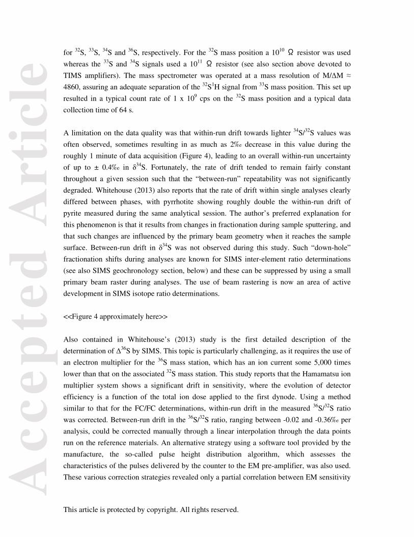
Acc
epte
d A
rtic
le
This article is protected by copyright. All rights reserved.
for 32S, 33S, 34S and 36S, respectively. For the 32S mass position a 1010 Ω resistor was used
whereas the 33S and 34S signals used a 1011 Ω resistor (see also section above devoted to
TIMS amplifiers). The mass spectrometer was operated at a mass resolution of M/ΔM ≈
4860, assuring an adequate separation of the 32S1H signal from 33S mass position. This set up
resulted in a typical count rate of 1 x 10⁹ cps on the 32S mass position and a typical data
collection time of 64 s.
A limitation on the data quality was that within-run drift towards lighter 34S/32S values was
often observed, sometimes resulting in as much as 2‰ decrease in this value during the
roughly 1 minute of data acquisition (Figure 4), leading to an overall within-run uncertainty
of up to ± 0.4‰ in δ34S. Fortunately, the rate of drift tended to remain fairly constant
throughout a given session such that the “between-run” repeatability was not significantly
degraded. Whitehouse (2013) also reports that the rate of drift within single analyses clearly
differed between phases, with pyrrhotite showing roughly double the within-run drift of
pyrite measured during the same analytical session. The author’s preferred explanation for
this phenomenon is that it results from changes in fractionation during sample sputtering, and
that such changes are influenced by the primary beam geometry when it reaches the sample
surface. Between-run drift in δ34S was not observed during this study. Such “down-hole”
fractionation shifts during analyses are known for SIMS inter-element ratio determinations
(see also SIMS geochronology section, below) and these can be suppressed by using a small
primary beam raster during analyses. The use of beam rastering is now an area of active
development in SIMS isotope ratio determinations.
<<Figure 4 approximately here>>
Also contained in Whitehouse’s (2013) study is the first detailed description of the
determination of Δ36S by SIMS. This topic is particularly challenging, as it requires the use of
an electron multiplier for the 36S mass station, which has an ion current some 5,000 times
lower than that on the associated 32S mass station. This study reports that the Hamamatsu ion
multiplier system shows a significant drift in sensitivity, where the evolution of detector
efficiency is a function of the total ion dose applied to the first dynode. Using a method
similar to that for the FC/FC determinations, within-run drift in the measured 36S/32S ratio
was corrected. Between-run drift in the 36S/32S ratio, ranging between -0.02 and -0.36‰ per
analysis, could be corrected manually through a linear interpolation through the data points
run on the reference materials. An alternative strategy using a software tool provided by the
manufacture, the so-called pulse height distribution algorithm, which assesses the
characteristics of the pulses delivered by the counter to the EM pre-amplifier, was also used.
These various correction strategies revealed only a partial correlation between EM sensitivity

Acc
epte
d A
rtic
le
This article is protected by copyright. All rights reserved.
and integrated dose; the repeatability of the 36S/32S ratio fell in the range of ± 0.2 to ± 0.7‰
(1s) for the 18 analytical session in which 36S was determined. Nonetheless, this work
demonstrates that with Cameca 1280 instruments Δ36S can be acquired with a data quality
sufficient to permit geological process to be discerned (Figure 4). This author foresees further
improvements in data quality for EM/FC determinations from either exchanging the small
format Hamamatsu detectors for more robust detector designs or from further developments
in software correction algorithms.
The overall between-run repeatability on 1254 measurements on the Ruttan pyrite (Crowe
and Vaughan 1996, Ruttan pyrite δ34SCDT = +1.2 ± 0.1‰) reported by Whitehouse (2013) of
± 0.35‰ and ± 0.22‰ (2s) for δ34S and Δ33S, respectively, clearly shows that modern SIMS
instrumentation is capable of producing isotope ratio determinations on major elements with
individual uncertainties of better than ± 0.2‰ (1s) on sampling sizes on the order of a few
hundred picograms and as measured by repeatability of numerous determinations on
supposedly homogeneous materials. This observation leads to three further conclusions,
namely (1) SIMS instrument technology is no longer the limiting factor in defining total
analytical uncertainty, but rather the quality of the RM characterisation is now becoming the
dominant contributor, (2) the quality of isotope data from well equipped and experienced
SIMS facility is such that direct comparison between labs at stated uncertainty levels can only
be meaningfully made if the same calibrant is used or if the chain of traceability is kept very
short, and (3) refined methods for homogeneity testing are needed so that this component can
be eliminated from the total uncertainty budget assigned to micro-analytical RMs. Finally, in
the case of isotopic systems requiring corrections for major element compositions (see SIMS
matrix effects section below) there is little to be gained by further improvements in
instrument data quality unless both the matrix effect correction models and the RMs upon
which they are based are improve.
Uranium-lead geochronology: As has been the case for several decades, U-Th-Pb
dating of geological materials, and in particular of zircon, continues to be a major focus of
many SIMS investigations. Efforts to improve both data quality as well as the software for
converting raw SIMS data into geologically meaningful results are on-going. One possible
trend that might be emerging are efforts by some SIMS facilities to recover data at sampling
scales significantly smaller than the 20 to 30 µm diameter pits, which have long been the
norm for SIMS zircon analyses. One strategy towards finer sampling dimensions has
employed NanoSIMS instrumentation; such development work has been reported by Yang et
al. (2012), who investigated both the zircon and baddeleyite systems. In the case of zircon,
these authors used a ~ 500 pA Gaussian 16O- primary probe which they could focus to a < 2
µm diameter beam at the sample surface with a total time of ~ 30 min per analysis. The

Acc
epte
d A
rtic
le
This article is protected by copyright. All rights reserved.
primary beam was rastered over a 3 µm x 3 µm area during the analyses in order to suppress
variations in U-Pb fractionation during the course of single analyses, yielding an overall
spatial resolution for their analyses of around 5 µm. Compared with “conventional” SIMS
zircon analyses using large geometry instruments, such as the Cameca 1280 or SHRIMP
instruments, Yang et al. (2012) found their NanoSIMS 50L approach benefitted from a
higher primary beam density. Disadvantages of using the NanoSIMS were a loss in
sensitivity relative to 1280-class instruments due to the absence of oxygen flooding in the
sample chamber and the inability to do multi-collection on the Pb-spectrum because
NanoSIMS instruments do not provide sufficient dispersion between single Dalton mass
differences at such heavy masses. Yang et al. (2012) also report operating at a mass
resolution of 5000 at mass 206Pb, but a mass scan showed extensive overlap between this peak
and the 178Hf28Si+ peak located M/ΔM = 3830 below the 206Pb mass position. This
discrepancy may be due to the different means of defining “mass resolution” vs. “mass
resolving power” as used by the NanoSIMS community. Another issue perhaps worth
considering is why a 16O- primary beam was preferred over a 16O2- as would be used by large
geometry instruments due to the higher sensitivity it offers for Pb – is it a matter that 16O-
provides a small beam diameter for a given primary beam current? One final observation is
that no attempt was made to include 232Th/208Pb decay system in these analyses, which could
have been done without lengthening the total analysis time. These observations aside, this
paper contributes valuable understanding of the capabilities of NanoSIMS instrumentation
with regards to U-Pb dating, an approach that holds much promise when only small sample
domains are available to the analyst.
A second approach towards fine scale U-Pb analyses in zircon employing a Cameca 1280
instrument has been described by Liu et al. (2011), and this methodology has now been used
for the dating of very grained zircons recovered from five eucrite meteorites (Zhou et al.
2013). These latter workers employed a 100 to 200 pA, Gaussian primary beam that could be
focused to a < 5 µm diameter at the sample surface. Oxygen flooding was used in order to
improve sensitivity for the Pb isotopes by roughly a factor of ~ 2 above that obtained under
high vacuum conditions in the secondary ion source. Zhou et al. (2013) actually report on
two different strategies for obtaining dates from the small zircons inherent to their samples.
For determining 207Pb/206Pb ages Zhou et al. (2013) used a static, multi-collection set-up
which required ~ 15 min of data acquisition, subdivided into eighty cycles. Mass resolution
for the multi-collection system was ~ 8000 at 50% peak height. Four electron multipliers
were used for the 204Pb+, 206Pb+, 207Pb+ and 90Zr216O2
+ mass stations and daily calibrations of
instrumental fractionation and any relative drift on the four detectors employed Phalaborwa
baddeleyite, which the authors knew to behave similarly to zircon in term of isotopic matrix
effects. Corrections for common Pb were based on the observed 204Pb+ signal. Despite the

Acc
epte
d A
rtic
le
This article is protected by copyright. All rights reserved.
very small sampling volume, using this strategy it was possible for these authors to obtain 207Pb/206Pb age uncertainties of around ± 20 Ma (1s) on 4.5 Ga old zircons.
Zhou et al. (2013) also presented a second method for small scale geochronology based on U-
Pb dating operating in mono-collection mode. Fifteen cycles of the peak stepping sequence 180Hf16O+ (used for correcting for magnetic field drift), 92Zr2
16O+, 204Pb+, 206Pb+, 207Pb+, 208Pb+, 238U+, and 238U16O2
+ were conducted for each analysis, leading to a total analysis time
of ~ 15 min. Mass resolution in multi-collector mode was set at 7000 at 50% of peak height.
Correction for common Pb made use of the observed 204Pb/206Pb for each individual analysis
and the U/Pb calibration was based on the M257 zircon reference material (Nasdala et al.
2008). Using their mono-collection protocol, Zhou et al. (2013) reported typical analytical
uncertainties for 207Pb/206Pb ages of ± 25 Ma and for 206Pb/238U ages of ± 100 Ma (1s) on
their ~ 4.5 Ga old zircon grains. Some of the samples yielded analyses that are significantly
reverse discordant.
These examples document the possibility and the limitations at greatly reduced sampling
volumes for SIMS-based zircon geochronology. Clearly the reduction of the total mass
consumed down to the low picogram level is associated with poorer data statistics;
nonetheless, these studies demonstrate that useful data can be recovered even when one is
faced with grain sized of less than 10 µm. It seems probable that further advances will be
made in the realm of fine-scale dating in the coming years. Improvements in our
understanding of the sputtering process as well as refinements to calibration protocols may
likely prove key for major advances in this field.
Matrix effects: One of the greatest challenges facing SIMS as a tool in the
geosciences is the common necessity to correct for the presence of “matrix effects”, whereby
the relative sputtering yields of distinct elements or of individual isotopes is significantly
influenced by the chemical composition of the material being analysed. (Matrix effects
resulting from the cryptographic orientation in certain mineral/trace element systems have
been reported, but such phenomena are not discussed here.) For those mineral phases with
little or no variation in their major element contents (e.g., quartz, zircon) there is generally
little need to worry about the issue of matrix effects as long as the sample materials is the
same phase as the RM used for calibrating the SIMS instrument. When dealing with samples
with extended compositional ranges, there are basically two means for correcting for the
influence of major element composition on the sputtering of geological materials: (1) by
using a “closely matched” RM for each material being analysed, or (2) using a spectrum of
RM which cover the most of the compositional space of the given phase and then to develop
a mathematical correction for interpolating between RM compositions to define a correction

Acc
epte
d A
rtic
le
This article is protected by copyright. All rights reserved.
factor for each individual location being analysed from the sample material. The first of these
strategies suffers from two problems. Firstly, one must define the concept of “closely
matched” such that any differences between the calibration and sample material are small
enough that variations in sputtering behaviour are negligible. Secondly, one is compelled to
produce a very large number of well characterised RMs in order to cover all possible
compositions within a given mineral/isotope system. Thus, the second strategy, of using a
mathematical model to interpolate a correction coefficient, is generally the more realistic
approach – albeit this comes with the need to propagate any analytical uncertainties
introduced by the selected correction scheme.
The determination of δ18O in natural glasses, and in particular in melt inclusions, can provide
key information for understanding magmatic systems. As SIMS is uniquely capable of
determining the 18O/16O ratio at the microanalytical scale, it is essential that the effect of
chemical composition on the overall instrumental mass fractionation (IMF) should be
understood. The contribution by Hartley et al. (2012) provides valuable guidance in this
regard. These authors investigated a suite of glass RMs that had either been characterised
earlier for their δ18O compositions or that had been measured as part of this study. Their
twelve glass RMs spanned a compositional range covering from basaltic thorough rhyolitic,
all of which had well determined major element compositions. This study found variability in
the IMF of up to 5.3‰ where, in general, the more basic materials measure a more positive
IMF than was the case for more silica-rich compositions. These authors evaluated a variety of
corrections schemes, including both univariate and multivariate approaches based on the
chemical compositions as well as assessing correlations between other parameters such as
sputtering rate and material density. Hartley et al. (2012) found strong correlations in a
number of the corrections schemes that they investigated. A multivariate approach using five
elements (Fe, Mg, Ca, Na and K) was found to give the best correlation between composition
and IMF (R2 ≈ 0.981) for intermediate compositions, however this approach was found to
yield poor perditions of IMF when investigating materials at the ends of the compositional
range. These authors ultimately recommended the use of the univariate parameter of the SiO2
content of the sample in order to correct for composition induced variations in IMF, for
which the obtained R2 ≈ 0.962. Using the SiO2 content alone, they found that 75% of the RM
results were within ± 0.2‰ of the values predicted by their model and that 95% were within
± 0.4‰. This is a fairly encouraging result, as it implies that such a matrix correction is not a
major source of additional uncertainty. For many SIMS determined δ18O value the published
uncertainties are on the order of ± 0.2‰ (1s); adding in quadrature an additional uncertainty
of ~ 0.2‰ would result in a total analytical uncertainty of circa ± 0.3‰, which still might be
of sufficient quality to address important geochemical topics. Clearly, advancing the overall
data quality in such cases where matrix effects have an impact will require improving both

Acc
epte
d A
rtic
le
This article is protected by copyright. All rights reserved.
the robustness of the correction models as well as the assigned uncertainties of the myriad of
materials upon which such models are based. New measurement methods, for example major
improvement in ion yields or using heavy molecular ions, which are less sensitive to matrix
chemistry, might also help improve the situation. Finally, here we make a few additional
observations on further work that might already be undertaken to advance such IMF
correction schemes. Firstly, the role of using a small beam raster during the analyses needs to
be clarified. In Hartley et al. (2012) it was not clear whether Köhler illumination was used on
the primary beam or whether a small raster was employed during the analyses. Such a
strategy might well change the slope of the relationship between SiO2 content and IMF.
Secondly, it would seem plausible to determine the 16O-/28Si- or the 16O-/28Si2- ratio so as to
obviate the need for determining the absolute SiO2 content of the sample material in advance,
though this would prevent the use of the NMR magnetic field probe on Cameca large
geometry instruments.
A second noteworthy investigation into matrix corrections for SIMS isotope analyses is
reported by Ickert and Stern (2013); they investigated the nature of oxygen isotope
fractionation affecting δ18O determinations on garnet. These authors provide a detailed report
on the nature and stability of the IMF value they observed during δ18O garnet analyses using
their Cameca 1280 instrument; particularly valuable is their detailed portrait of all uncertainty
propagation aspects of their correction model. Ickert and Stern (2013) employed 17 garnet
materials in their study. In addition to assessing the impact that variations in major element
composition have on the overall IMF, they also investigated the role of varying specific
parameter of the secondary ion optics such as system magnification and the width of the
energy band-pass. The key result of their study is that Ca-Mg-Fe rich garnets show a
systematic variation in their IMF, which correlates strongly with the Ca/(Mg+Fe2++Ca)
atomic ratios. Using a quadratic best-fit relationship the correction was found to be reliable to
± 0.2‰ at the 95% confidence interval from across the compositional spectrum investigated.
This level of reliability is similar to that possible for glass analyses as discussed above,
suggesting that δ18O determinations on garnet can be achieved using a fairly straightforward
matrix correction. Likewise, as is the case for geological glasses, the need for a matrix
correction in garnet will introduce an additional uncertainty source similar to that derived
from analytical repeatability. A limitation of the approach described by Ickert and Stern
(2013) is that the quadratic relationship between IMF and major element composition could
accommodate neither Cr-rich nor Mn-rich garnets; these end members remain a subject for
future studies.

Acc
epte
d A
rtic
le
This article is protected by copyright. All rights reserved.
The focused ion beam method in the geosciences
(Contribution by: Luiz F.G. Morales)
The focused ion beam (FIB) technique was initially developed in the 1980s by the semi-
conductor industry and has been widely employed since to patch and/or modify semi-
conductors and even µm-scale cooling systems. It was only in the 2000s, however, that such
techniques were first used in the Earth sciences, initially limited to the preparation of in situ
transmission electron microscopy foils (e.g., Wirth 2009). More recently, with the advent of
so-called “Dual Beam®” machines, geoscientists realised that a focused ion beam integrated
with the imaging capabilities of the electron source has vastly broader utility within the
geosciences, and in particular for 3D imaging and nanotomography. A Dual Beam® machine
essentially consists of a vertical electron column, as in a standard scanning electron
microscope (SEM), together with a tilted ion beam column. In addition, a number of different
accessories and detectors are normally present in these machines, such as micromanipulators
and gas injection needles, as well as energy dispersive X-ray spectrometers (EDS) and
electron backscattered diffraction (EBSD) detectors (Figure 5A).
FIB optics resembles that of a SEM, but where the later uses a focused beam of electron to
generate images, the former uses a finely focused beam of ions. Most FIB instruments in
geoscience departments use a liquid-metal ion source (LMIS) normally based on gallium,
although gold, iridium and lighter elements can also be used for similar purposes. The LMIS
reservoir is heated to near evaporation causing the liquid metal to flow to the tip of a very
sharp, heat resistant tungsten needle. The electric field at the tip of this needle (~ 2 nm radius)
induces field emission ionisation of the metal atoms. At low beam currents, the interaction of
the incident ions on the target material produces secondary electron emission that can be used
for imaging, similarly to the process in a SEM. However, if the beam current is high enough,
the incident ions (due to their mass) are capable of sputtering atoms from the target material
at the atomic scale. Three-dimensional imaging with FIB/SEM is achieved by repetitively
sputtering material from the target with the ion beam and then imaging the region of interest
with the electron beam between each step of sputtering; the amount of material removed by
sputtering before the next image acquisition can be controlled by the user. For a
representative volume at the micrometre scale a variable thickness between 50 to 100 nm of
material is typically removed during a given step, where the optimal spacing of the SEM

Acc
epte
d A
rtic
le
This article is protected by copyright. All rights reserved.
imaging planes depends on the grain sizes and the level of detail required for the given
investigation. A detailed description of sample preparation and key setup parameters for
successful analyses of most of geological materials is given by Wirth and Morales (2012).
<<Figure 5 approximately here; colour>>
FIB 3D microstructural characterisation of geomaterials
Pore space geometry: Although FIB investigations of geological materials have been
reported for some time, sample preparation for TEM studies have been its dominant
application. However, in the last two years fundamental 3D microstructural investigations
using the nanotomography method, coupling FIB and SEM, have been carried out both on
fine-grained lithologies (e.g., shales) and on partially-melted rocks. In the last few years there
has been an increasing focus on shales, with research foci on radioactive waste disposal, the
production of hydrocarbon gas and CO2 sequestration. In all such cases the common goal is a
complete characterisation of the geometry of the intergranular pore space in clay-rock
formations. Due to inherent mechanical anisotropies and very small grain sizes, shales are
highly complex rocks to prepare; it is exceedingly difficult to polish thin-sections to the level
where nanoporosity can be well-defined. Keller et al. (2013) reported a fundamental study on
the application of FIB technique (couple with other methods not discussed here) in shales,
including a detailed study of a sample of Opalinus Clay. Due to its favourable properties for
the deep disposal of radioactive wastes, the Opalinus Clay of Switzerland is a well-studied
shale example: it has an extremely low permeability, is practically watertight and has self-
sealing properties. In their contribution, Keller et al. (2013) have used a multi-scale approach
in order to achieve a highly detailed three-dimensional characterisation of the porosity and
phase distribution at variable scales. They tested several tomographic methods, spanning
from the nanometre to millimetre-scale resolutions. One important aspect of this study is the
impact of increasing resolution in tomography methods and the inherent decrease in the total
sample volume. Because shales are very heterogeneous in terms of phase distribution and
because the sample size analysed by the FIB is rather small (maximum dimensions of a few
tens of µm), the authors performed standard SEM analyses in broad ion beam milled surfaces
prior the FIB nanotomography. This allowed the 2D characterisation of the microstructures
enabling a better targeting of sites for the 3D methods. The authors chose two sites with
contrasting degrees of compaction, which they document at the nanotomographic scale. Their

Acc
epte
d A
rtic
le
This article is protected by copyright. All rights reserved.
work indicated that porosity ranges between 2 and 3%, which is significantly lower than the
porosity determined via lower resolution methods (~ 10%). Even with the sample in a
position not favourable for backscatter imaging (as the sample is tilted 52˚ from the standard
parallel position with relation to the backscattered detector), the authors were capable of
distinguishing different phases such as carbonates, quartz, large clay crystals and different
organic materials within the clay matrix. The segmentation of the individual images acquired
during the nanotomography via FIB also allowed the authors to model the pore path
orientation distribution and pore geometry (Figure 5B). The pore path density distribution
was shown to have a preferred orientation parallel to the bedding, whereas geometrical
convolutedness of pore paths was significantly higher in the direction normal to bedding.
Using the FIB method Keller et al. (2013) demonstrated that nanopore might not represent
the main transport pathways in this Opalinus Clay, despite the fact that the nanopores
between the clay laths represent a large fraction of the general porosity in this lithology.
Furthermore, Keller et al. (2013) demonstrated that the coarser porosity observed in the FIB
nanotomography is not interconnected and that porosity is concentrated along contacts
between clay minerals and the other phases. Together these features indicate strong pore
connectivity anisotropy, with higher values parallel to the bedding. Equally significant, it was
shown that a significant fraction of the porosity is not interconnected, which would certainly
influence gas extraction from such rocks. One additional aspect of this paper is that the
authors clearly show that an up-scaling of FIB observations is not straightforward due to the
very small observational scale; it may be necessary to use other tomography techniques to
assure that the observations from the nanoscale are correctly extrapolated to the µm and mm
scales.
Melt distribution: A second contribution making use of the FIB nanotomography
methods was presented in by Garapic et al. (2013), who made extensive use of this technique
for the 3D characterisation of melt distribution in partially molten mantle rocks. The
dynamics of such rocks is directly related to the amount of melt retained in the solid matrix,
affecting both physical (e.g., buoyancy, seismic velocities) and chemical (e.g., trace elements
and isotopic) properties of the material. Garapic et al. (2013) performed experiments on
olivine aggregates containing 2 and 4% of basaltic melt using a piston cylinder apparatus.
The samples were then polished using only colloidal silica, thereby avoiding physical damage
of the surfaces, and then analysed via FIB. Such melt-containing samples are very tricky to
prepare for two reasons. The first is that because the quenched melt (a glass) is softer than the

Acc
epte
d A
rtic
le
This article is protected by copyright. All rights reserved.
solid matrix it is preferentially removed during polishing, introducing topographic effects
onto the sample surface. Hence, the electron illumination during oblique imaging is not
homogeneous for both the melt and the solid matrix part, particularly near crystal-glass
interfaces. This may cause serious problems during image segmentation (the statistical
evaluation whereby a digital image is partitioned into multiple sets of pixels), hindering an
accurate determination of 3D melt volumes. Secondly, some experiment-produced melts
types can be very reactive such that they are prone to damage and charging during focused
ion beam sputtering, potentially resulting in artefacts related to image drift. A careful tuning
of both the ion and electron sources (by means of changing accelerating voltages and beam
currents) is therefore essential for successful tomography studies in melt-rich rocks. In order
to convert the typical grey-scale images from the SEM to binary images Garapic et al. (2013)
performed a hand digitisation of the melt-solid interfaces prior to processing. FIB
nanotomography clearly showed that the thin films separating neighbouring grains could be
traced in 3D. These films were found to persist over distances of more than 10 µm, thereby
demonstrating that such features are not simply triple-junction melt pockets, which are on the
order of 1µm in diameter. Rather, such melt accumulations were seen to form thin films
separating olivine grains, where such films can be classified as wetted two-grain boundaries.
Such wetted two-grain boundaries exist in aggregates with dihedral angles > 0°, and are
apparently a dynamic feature resulting from grain growth driven by surface energy reduction.
One minor drawback encountered in Garapic et al.’s (2013) work was the fact that, due to the
relative coarse olivine grains, only a few grains could be visualised at once; for this reason
the “volume” of wetted grains imaged was rather limited.
These papers by Keller et al. (2013) and Garapic et al. (2013) show the strong potential of the
FIB nanotomography for the microstructural quantification of fine-grained rocks having
complex mineralogies for characterising melt distributions in partially molten rocks. These
examples also show clearly that this technique can be handicapped in terms of dimensional
volumes that can be analysed, mostly because the sputtering process at low beam currents
coupled with high-resolution imaging is very time consuming. To give an idea, FIB
nanotomography of a 25 x 25 x 15 µm box, with 100 nm thick slices being removed at each
step prior imaging, takes about 10 hr. Nonetheless, the level of detail that can be attained is
impressive, and characterisation of nanoscale porosity and mineralogy in three dimensions is
now possible in almost any sort of geomaterial.

Acc
epte
d A
rtic
le
This article is protected by copyright. All rights reserved.
Outlook for focused ion beam techniques in the geosciences
Thanks to “Dual Beam®” equipment, geoscientists now have the ability to combine both
focused ion and the electron beam sources, providing access to both 3D compositional and
crystallographic information within micrometre-sized volumes. 3D chemical analyses using a
SEM/FIB system are normally obtained by quantifying the excited X-ray photons with EDX.
The acquisition of a 3D chemical composition map follows the same procedure as with the
nanotomography, with an additional step that a full EDX map from the frontal surface of the
target is acquired between each sputtering step with the FIB. This approach can be
particularly useful in those cases where the phases present in a rock specimen do not generate
enough contrast in the secondary or back-scattered electron image. As an example, quartz,
calcite and feldspars may appear with similar grey levels in SE/BSE imaging, and it is
virtually impossible to separate the phases by colour contrast. In such cases the chemical
compositional mapping via EDX signal can be used to uniquely identify phases. The 3D
resolution will depend on the milling resolution of the focused ion beam and the EDX
analyses will depend on the accelerating voltage of the electron beam and on the composition
of the target material to be analysed. Because the interaction between the electron beam and
sputtered surface occur at an angle, the depth of interaction and emission of X-rays in these
conditions is lower, commonly on the order of few 100’s nanometres. One drawback of this
method is that it requires very stable SEM imaging, free of electrical charging and imaging
drift. Because most geological materials are non-conductive, this can be an issue that may
hinder successful analyses. Furthermore, because a complete BSE or SE image has to be
acquired and one full EDX map has to be generated from the frontal surface of the studied
volume after each sputtering step, this procedure is very time consuming, limiting its
application to carefully selected small regions in fine-grained rocks. Future developments
addressing the challenges of sample charging and image stability while increasing the rate at
which chemical maps can be acquired would represent significant advances in the field of
FIB-based imaging. A better integration of FIB-based imaging methods with finer-scale
methods (e.g., SIMS, atom probe) would also mark a significant advance.

Acc
epte
d A
rtic
le
This article is protected by copyright. All rights reserved.
Electron backscatter diffraction in the geosciences
(Contribution by: Luiz F.G. Morales)
Electron backscatter diffraction (EBSD) analysis is a microstructural-crystallographic
technique normally conducted in a scanning electron microscope; EBSD can be used to
determine the crystal orientation of virtually any crystalline material at the µm-scale. This is
often the method of choice for crystal orientation determinations, grain boundary mapping,
intra-crystalline defect characterisation, phase identification and micro-strain mapping
studies. As is the case for many of the analytical techniques used in geosciences, EBSD
started as a tool for material sciences, and it has now become the most widely used method
for determining the crystallographic orientations of minerals and rocks. In the EBSD
technique an ultra-polished crystalline specimen is placed inside the SEM chamber at a 70°
angle from the horizontal, facing towards the diffraction detector. The detector, which consist
of a phosphor screen combined with a low luminosity CCD camera that together image the
diffraction patterns, is position in the specimen chamber at ~ 90° to the SEM pole piece. In
this configuration, some of the electrons that enter the sample will be backscattered and may
continue on to escape from the sample. The probe diameter of the incident electron beam is
typically one to tens of nm, which is roughly one order of magnitude larger than the Å-scale
intra-crystalline spacing typical in minerals. Because of their wave-like properties, the
escaping electrons undergo Bragg diffraction by all the intra-crystalline planes at the sample
target point, and these diffracted electrons form a characteristic pattern displaying different
Kikuchi bands, each of which corresponds to the projection of a diffracting lattice plane.
Through comparison of the observed Kikuchi pattern with all possible patterns for a given
(known) phase it is possible to index the patterns by Miller indices to determine the
orientation of the target domain in 3D space with good precision. All the theoretical details
are beyond the scope of this review, for which the reader is referred to publications by Engler
and Randle (2009) and Prior et al. (1999). The EBSD technique as it is known today was first
described in a series of papers in the beginning of 1990s (Wright and Adams 1992, Adams et
al. 1993, Kunze et al. 1993). These papers describe the first system with a computer-
controlled SEM, a low-light CCD camera, a video card for on-line diffraction pattern
acquisition and dedicate software for automatic indexation. Over the intervening years many
other advances have occurred, including higher speeds for data acquisition, refinement of the
Kikuchi bands analyses, the combination of EBSD and in situ chemical analyses via
EDS/WDS for phase indexation and the development of the dynamical theory for EBSD
Acc
epte
d A
rtic
le
This article is protected by copyright. All rights reserved.
diffraction patterns. Here we report on two recent publications devoted to (1) cryo-EBSD
study of ice samples deformed experimentally and naturally and (2) ultra-high resolution
EBSD in FIB-prepared foils.
Recent milestone EBSD applications in geochemistry
Cryo-EBSD and crystallographic orientations in ice: Natural ice is a major
component of the Earth’s polar regions and it plays a fundamental role in our planet’s climate
system, affecting sea level and consequently the ocean currents that control temperatures,
seasons and rainfall distribution on our planet. The effect of polar ice discharge into the
oceans is nevertheless poorly understood, mainly because of an incomplete understanding of
solid-state flow in ice sheets. In essence, ice flow involves either sliding over rocks at the
base of the ice or internal flow, usually involving deformation and recrystallisation within the
ice body. Such mechanisms operate essentially at the macroscopic scale, but a complete
understanding of the physical processes involved in such deformation requires that the
microstructures and mechanical properties of naturally and experimentally deformed ice be
investigated. Furthermore, a better understanding of the rheological behaviour of ice may
yield insights about the dynamics of giant gas planets and icy moons. Up until a few years
ago the study of ice was very challenging, as it requires a cryogenic system coupled with the
analytical or deformation tool, further requiring controls on temperature, pressure, water
fugacity, etc. The first attempts to measure crystallographic orientations via EBSD on ice
were made in the early 2000s (Iliescu et al. 2004), but it was not before the work of Weikusat
et al. (2010) that the method became established. Only recently has the routine mapping of
crystallographic orientations in ice via cryo-EBSD become available, lead by the group at the
University of Otago, New Zealand (see also Prior et al. 2012).
Prior et al. (submitted) have describe the procedures they developed and successfully applied
to the analyses of ~ 100 samples of synthetic and natural ice, addressing both a range of grain
sizes and levels of impurities. EBSD mapping needs flat sample surfaces free of mechanical
damage, as the electron backscatter diffraction signal arises ≤ 20 nm from the sample surface.
Sample preparation for rocks normally evolves a mechanical and a chemical-mechanical
polishing step, where some materials may require etching, electro- or ion-polishing carried
out under carefully controlled conditions. It was therefore necessary that such methods
should be adapted for ice sample preparation, where a very important factor is that the sample

Acc
epte
d A
rtic
le
This article is protected by copyright. All rights reserved.
surface must be free of frost particles. During preparation of ice samples it is essential to
track the thermal history of the sample, from synthesis and storage to cutting and polishing,
as microstructural modifications are prone to occur even if only small temperature variations
occur. After synthesis sample storage is a very important consideration; fine-grained samples
(10–20 µm grain sizes) require storage in liquid nitrogen, whereas standard ice (grain sizes
around 500 µm) can be stored in low temperature freezers (-80 °C). For sample cutting the
authors used both a scroll saw designed to cut wood and a circular saw with a diamond-
sintered blade, as would be used in rock sample preparation. For obvious reasons, cutting
(and polishing) has to be done in cold rooms, with temperatures varying from -10 to -15 °C.
In addition, a series of precautions/procedures have to be considered so as to avoid any
modifications to the microstructure of the samples (pre-chilled transfer cool boxes, pre-
cooled saw guide, etc.). Optimal cutting routines depend on the sample properties, where
medium grained samples without mechanical anisotropies might be cut with a circular saw
whereas more delicate, fine grained samples need to be cut with a scroll saw. Care during
sample mounting is crucial if microstructures are to be preserved, hence Prior et al.
(submitted) developed cooper sample holders to which the samples could be attached and
stored, without the need to remount the samples in the case of reanalysis. Sample mounting is
involved the melting and refreezing of a thin layer at the bottom of the sample to make the
bond between the material being investigated and the holder. To minimise thermal impact,
the very cold ice is pushed against the holder at 5–10 °C until a melt layer < 1 mm is created.
Then, as soon as the bond is made, the sample holder was pressed against another sample
holder at liquid nitrogen temperature. To obtain flat surfaces in their samples, particularly in
coarse grained samples, the authors used a cryo-microtome that could be cooled to -30 °C.
Flat surface samples are also obtained by grinding the samples on a metal plate cooled with
liquid nitrogen, assuring that the sample surface remains below 100 °C during the first 20 s of
grinding, which is enough time to generate a flat surface. For the sample transfer from the
storage boxes to the SEM, the authors used a nitrogen glove box (Figure 6A) that could be
fixed directly to the vacuum chamber door, allowing sample exchanging under a controlled
atmosphere. This arrangement allowed the use of the main SEM vacuum chamber door,
rather than through the commonly used airlock, thereby overcoming the problem of some
cryo-transfer systems that limit sample tilt and movement, precluding EBSD measurements.
The cryo-stage is made of Cu with a liquid nitrogen reservoir with an internal thermocouple
and a heating element. The setup procedure for the ice EBSD took between 1 and 2 hr,
depending on the experience of the users. Prior et al. (submitted) report that the last step

Acc
epte
d A
rtic
le
This article is protected by copyright. All rights reserved.
before the EBSD mapping or imaging involves surface sublimation to remove frost and
damage caused by cutting and grinding. Sublimation, employing the cryo-stage’s internal
heater operating under high-vacuum, was found to be effective for coarse-grained samples
but not successful in fine-grained ice. These authors go on to describe two strategies for
inducing sublimation.
<<Figure 6 approximately here; colour>>
All analytical work was carried with a Zeiss Sigma VP FEG-SEM coupled with a NordlysF
camera using AZTEC software from Oxford Instruments. Image acquisition and EBSD
mapping were conducted in the variable pressure mode, usually with N as process gas. For
the image acquisition Prior et al. (submitted) used different accelerating voltages and beam
currents, whereas all EBSD measurements were acquired with a 30 kV accelerating voltage
and a 90 nA beam current, albeit using variable working distances. An example from an ice
orientation map is shown in Figure 6B, where the colour variations record changes in
crystallographic orientation. These authors demonstrated that cryo-EBSD is a powerful
technique for understanding ice microstructural behaviour and ice transformation
mechanisms. In addition, this analytical approach makes possible the study of
recrystallisation processes and the orientation between parent and recrystallised grains.
Although this is also possible in normal EBSD analyses, unlike most natural minerals some
crystallographic types of ice are free of twinning, allowing the isolated study of
recrystallisation free of the twinning effects. Ice EBSD is also well suited for better
constraining the microstructural evolution of experimentally and naturally deformed samples.
Such work further constrains the kinetics and mechanical behaviour related to grain size
reduction during ice deformation. One minor issue for this technique is the fact that the grain
sizes in some natural samples (e.g., glacial ice) reach up to the cm-scale, making truly
representative EBSD mapping difficult. Nevertheless, the authors successfully measured the
crystallographic orientations of samples larger than 7 cm in length, with very high resolution.
This is the first time that EBSD mapping has been carried out at this scale. One can expect
much more research to be reported on this topic in the coming few years.
Ultra-high resolution EBSD in FIB-prepared foils: Since the advent of EBSD
techniques a couple decades ago, scientists have struggled to achieve near-nanometre
resolution in crystallographic orientation determinations using scanning electron

Acc
epte
d A
rtic
le
This article is protected by copyright. All rights reserved.
microscopes. Although constant improvements have been made on both field emission gun
technologies (the best electron source so far) and EBSD detectors since the 1980s, the basic
principles of electron-matter interaction in SEMs (volume interaction) and the high tilt angle
associated with this technique have prevented resolutions below some tens of nanometres.
For many years, material researchers, and more recently Earth scientists, have been interested
in ultra-fine grained materials, as their physical properties (e.g., strength and plasticity) have
very different behaviours to those observed at coarser grained scales. As an example,
nanocrystalline particles are thought to have a major effect on the strength of upper crustal
faults, and their presence or absence may influence the seismic vs. aseismic nature in these
places. In addition, nanocrystals are commonly present in biominerals, they occur as
inclusions in many different minerals and they can influence the early stages of
crystallisation. Although standard EBSD has been commonly applied to the characterisation
of fine-grained materials, its spatial resolution has not been sufficient for the routine
characterisation of nanostructured materials, meaning most such studies have had to be based
on transmission electron microscopy. In a recent paper Trimby (2012) proposed a solution for
this problem, employing what the author has termed “transmission Kikuchi diffraction”
(TKD); this seems to offer a much better spatial resolution than standard EBSD analyses. The
key innovation in the TKD method is its use of transparent samples prepared for TEM
analyses in conjunction with the sample being mounted at 90° from the traditional EBSD
position (Figure 6C). Under these conditions, good quality diffraction patterns could be
obtained from domains below 10 nm, allowing the detailed mapping of crystallographic
orientations at such a fine scale. The author used two TEM-prepared foils of aluminium alloy
and nanocrystalline nickel as illustrative test materials, from which he went on to measure
successfully samples of diamond with nano-inclusions of iron oxides and sulfides (Figure
6D). These foils were mounted using a micro-clamp SEM sub-stage attached to a standard
70° tilted sample holder. The SEM stage is then tilted 20° towards the EBSD detector, so the
TEM foils lie in a horizontal position (Figure 6C). The author used a very small working
distance (5 mm), whereas the EBSD camera was located in the standard operating position.
Trimby (2012) used accelerating voltages between 20–28 kV and beam currents between 1–
10 nA on the University of Sydney’s Zeiss Ultra Plus FEG SEM equipped with a Nordlys-S
EBSD detector and Channel 5 suite of programs, both from Oxford Instruments. As the
sample lies in the horizontal, no tilt correction or dynamic focus correction was necessary.
The EBSD patterns produced with the TKD-SEM technique was very sharp, and it was
shown that the thickness of the sample plays an important role in determine the quality of the

Acc
epte
d A
rtic
le
This article is protected by copyright. All rights reserved.
patterns. If the sample is too thick, two or more superimposed patterns may occur and the
indexation becomes impossible, whereas if the sample is too thin the diffraction contrast is
low and the patterns are again difficult to index. According to the author, good diffraction
patterns can be obtained in samples ranging from 75 to 400 nm thick when using a 22 kV
accelerating voltage, however the optimal thickness values were found to depend on the
accelerating voltage used during analysis. High resolution mapping of both samples (with an
average grain size of 200 nm) was achieved and, as can be seen in Figure 6D, the same
resolution could also be obtained from minerals. The results from Trimby (2012) indicate a
true spatial resolution of 5-10 nm for the nanocrystalline nickel sample, and around 10 nm for
the Al alloy; it would appear that similar resolutions are possible for minerals in general. The
work performed by the author suggests that this technique can be used to map grains well
below 100 nm – this technique will certainly attract the attention of Earth scientists in the
near future.
Outlook for EBSD in the Geosciences
With respect to EBSD investigations of ice, some issues remain regarding porous samples
(e.g., snow) that are very delicate, and it appears further work will be needed on refining the
sample preparation before these will be successfully measured. In addition, “dirty ice”
(mixture of ice and rock particles) has also proven challenging to prepare because the
hardness contrast between the clasts and ice. This means that standard preparation strategies
may lead to topographic effects on the sample surface, inhibiting precise crystal orientation
determinations. EBSD on different ice polymorphs is also something warranting further
exploration, but this will require modifications being made on existing cryo-systems (e.g.,
lower temperatures). Other types of ices, such as CO2 and CH4, which are major components
on giant gas planets, have yet to be explored; here there seems to be great potential for future
studies.
The transmission Kikuchi diffraction in SEM is likely to attract a lot of interest in the near
future due to its high spatial resolution and ease of data acquisition; importantly, such
measurements can be made using a standard SEM with field emission gun and EBSD
detector. The study performed by Trimby (2012) has clearly shown that the TKD can resolve
structures that had previously proven difficult or impossible to resolve using standard EBSD
methodologies. The effect of high intra-crystalline defect densities and the study of samples

Acc
epte
d A
rtic
le
This article is protected by copyright. All rights reserved.
whose degree of crystallinity is so low that individual grains cannot be resolved by traditional
EBSD are potential candidates to be studied with the TKD-SEM technique. In addition, this
method may prove ideal for the study of FIB milled tips prior to atom probe analyses (e.g.,
Yao et al. 2011, see also atom probe chapter in this paper). As the sample during TKD
investigation lies in a horizontal position, EDS mapping coupled with EBSD techniques can
be used to separate phases with similar crystal structures at high spatial resolution.
Atom probe tomography in the geosciences (Contribution by: M. Wiedenbeck)
Overview of the atom probe method
Although it has only recently been taken up in the geosciences, atom probe tomography
(APT) has actually existed as an analytical tool since roughly 1970. This technology offers
the unique and amazing capability of mapping the chemical and structural composition of a
material at the atomic scale, generating massive data sets describing the position and isobaric
identity of individual atoms within a 3-dimensional volume. Basically, an atom probe
consists of a field emission ion source (the sample) coupled to a time-of-flight mass
spectrometer, where ion detection uses a position sensitive detector capable of recording the
arrival times of individual ions. The ion optics of such a system are designed to provide a
magnification on the order of 106, allowing the X-Y position recorded by the ion detection
system to be transposed to the original location of a particular atom at a sub-nanometre scale
(Figure 7). As the system depends on a field emission ion source, the material under
investigation must be prepared as a sharp tip with a diameter on the scale of up to a few 100’s
of nanometre in diameter and with a length typically around 1000 nm, meaning sample
preparation generally requires ion milling using focused ion beam technology. Another
challenge of APT technology had earlier been the need to pulse the voltage applied to the
sample in order to generate a start time-stamp for the time-of-flight system. This need to
apply rapid voltage shifts to the sample tip largely precluded the analysis of non-conducting
materials, as they tend to break under due to mechanical stress. However, with the advent of
pulsed laser evaporation, the attractiveness of APT technology for investigating semi-
conductors and non-conductors has recently improved. APT systems have been commercially
available for perhaps a decade, meaning the atomic-scale characterisation of non-conducting
geomaterials is now realistic. A detailed description of APT technology is provided by Gault
et al. (2012) and a brief overview of the historical background is provided by Wiedenbeck
(2011).

Acc
epte
d A
rtic
le
This article is protected by copyright. All rights reserved.
<<Figure 7 approximately here; colour>>
APT applications in mineralogy and geochemistry
As laser assisted APT has existed for only a relatively short time, until now this technology
has found few applications for Earth science topics; this is the first time that APT technology
has been reported in this biennial review series. A fundamental investigation of the potential
of the atom probe as applied to mineralogy was published by Bachhav et al. (2011), who
report an APT study of wüstite (Fe1-xO), which is of interest in both the material sciences as
well as the Earth sciences. These authors assessed the significance of such parameters as laser
energy, sample temperature and the voltage applied to the sample during analysis. Their
paper documents that by varying the laser conditions used during analysis one alters
significantly the quantitative results – in this particular study the goal having been the
determination of the absolute oxygen content of the sample. Another valuable contribution by
Bachhav et al. (2011) is their description of the sample preparation requirements of their
instrument. Although somewhat on the brief side, this description gives good insight into the
complexity of the atom probe method. Another interesting observation presented in this work
is that a large spectrum of both multiply charged and molecular ions could be detected,
despite the relatively simple chemistry of the wüstite material. This work lays the foundations
for future fundamental studies needed in order to convert APT, which currently can be seen
as an extremely high spatial resolution visualisation tool, into a truly quantitative analytical
technique. Obvious points for further investigation are the role of the system’s mass resolving
power and the detection limits for minor components, which are largely defined by detector
noise. One shortcoming acknowledged by Bachhav et al. (2011) was the inability of the
system to reliably detect the minor isotope 18O, which would otherwise have been useful for
distinguishing between 16O+ and 16O22+ on the m/e = 16 mass station; distinguishing between
these two species was of fundamental importance in view of the goal of quantifying the
absolute oxygen content of the material. Though not discussed by these authors, information
about the presence or absence of a signal at the m/e = 17 mass station would have been
highly relevant, revealing the abundance of the 16O18O2+ species. Future investigation into
detector background, system mass resolution, ion through-put rates and factors affecting the
detection of minor and trace components will be valuable contributions towards
understanding the strengths and limitation of the atom probe method.
Until now only a single paper using APT for a purely Earth science application has appeared
in the scientific literature. Valley et al. (2014) used laser assisted APT to image
nanostructures within the core of a zircon that had previous been dated by SIMS to be nearly
4.4 Ga. This publication is not only noteworthy for its pioneering use of APT technology, but

Acc
epte
d A
rtic
le
This article is protected by copyright. All rights reserved.
is also valuable for an appendix describing the analytical method in detail. These authors
found a plethora of trace element enriched clusters (Figure 8) spread throughout the volume
that they analysed. These enriched zones were defined by the authors as those volumes that
their imaging software determined to have 3 at% or more of the trace element yttrium, which
is one to two orders more enriched than that detected in the surrounding matrix. Many other
trace elements were found to behave similarly. Of particular interest was that the 207Pb/206Pb
ratio – measured using the 206Pb2+ and 207Pb2+ mass stations, as these were largely free of
isobaric interferences – differed between the two classes of material. The 207Pb/206Pb ratio
was found to be 1.25 ± 0.05 and 0.30 ± 0.05 (2s) for the cores of yttrium-rich clusters and the
non-enriched matrix, respectively. Valley et al. (2014) interpret their results from the yttrium-
rich volumes as recording radiogenic Pb produced between 4.4 and 3.4 Ga, whereas that in
the case of the matrix material recording uranium decay over the past 3.4 Ga. The age of 3.4
Ga had been previously determined by SIMS on an overgrowth on this particular zircon and
has been interpreted as the time of a high temperature event of regional significance. The
cluster-like nature of the trace element enriched volumes is postulated to be the result of
localised lattice damage resulting from α-recoil cascades, which were annealed during the
subsequent metamorphism.
<<Figure 8 approximately here; colour>>
The work by Valley et al. (2014) may well prove to be a watershed publication, for the first
time undertaking isotopic analyses at the near-atomic scale. This suggests great potential of
the APT method as applied to geoscience research. Equally clear from this contribution is the
huge amount of work needed prior to this method becoming a “routine” method. Many
questions remain unaddressed, including the issues of detector backgrounds, magnitude and
potential means for suppressing polyatomic interferences and the relative abundances of
various charge states of the elements of interest. Also of potentially great significance is the
topic of using the time-of-flight nature of APT in organic geochemistry – is any useful
molecular information retained through the field emission process? The low ion production
rate of ~ 0.008 ions per cycle reported in this study, albeit with the laser operating at 200
kHz, suggests that progress in reducing total analysis time to well below a full day might be
possible. Increasing the ion yield above the reported level of ~ 37% would also be of obvious
benefit. Finally, for the specific case of the 207Pb/206Pb = 1.25 ± 0.05 (2s) determined for the
yttrium-rich clusters reported by Valley et al. (2014), the value for radiogenic 207Pb/206Pb that
evolved from 4.4 to 3.4 Ga would be closer to 207Pb/206Pb = 1.218. Though the experimental
result is within its assigned 2s uncertainty of this value, this might suggest that other
systematic processes such as unresolved isobars or detector background may warrant
additional attention.

Acc
epte
d A
rtic
le
This article is protected by copyright. All rights reserved.
Outlook for atom probe applications in mineralogy
Clearly the APT method has the potential to become a key method for the characterisation of
geomaterials at the nanometre scale. As this technology has only recently become suitable for
the analysis of electrically non-conducting specimens, many parameters remain to be
investigated in order to optimise this approach for geochemical applications. The
investigation of mineral grain boundaries, short-range diffusion and the characterisation of
nano-inclusions – all of which require nm-scale lateral resolution – can be expected to benefit
from atom probe technology. The ability to visualise structures in 3-dimensions at nanometre
scales will also likely provide new perspectives on diffusion and reprecipitation mechanisms.
One drawback to be highlighted about atom probe technology, however, is the need for
extensive supporting infrastructure. Such instruments are not prohibitively expensive in
themselves, but one needs to keep in mind that at a very minimum a scanning electron
microscope plus focused ion beam capability is essential for their operation. Hence, it can be
expected that APT will in the short to medium term be largely limited to either multi-user
facilities or to exceptionally well-equipped laboratories with a focus on nano-scale processes.
Nonetheless, as this class of instrumentation becomes more widely available, one can
reasonably expect many new breakthrough discoveries from APT within the geosciences.
Acknowledgements
R. Bugoi and S. Merchel would like to thank discussions with C. Andermann, D.L. Bourlès,
R. Braucher, M. Christl, F. Munnik and P. Steier.
References
Adams B.L., Wright S.I. and Kunze K. (1993)
Orientation imaging: The emergence of a new microscopy. Metallurgical Transactions,
24A, 819–831.
Akçar N., Deline P., Ivy-Ochs S., Alfimov V., Hajdas I., Kubik P.W., Christl M. and
Schlüchter C. (2012)
The AD 1717 rock avalanche deposits in the upper Ferret Valley (Italy): A dating approach
with cosmogenic 10Be. Journal of Quaternary Science, 27, 383–392.

Acc
epte
d A
rtic
le
This article is protected by copyright. All rights reserved.
Akçar N., Ivy-Ochs S., Deline P., Alfimov V., Kubik P.W., Christl M. and Schlüchter C.
(2014)
Minor inheritance inhibits the calibration of the 10Be production rate from the AD 1717 Val
Ferret rock avalanche, European Alps. Journal of Quaternary Science, 29, 318–328.
Amarasiriwardena D., Krushevska A., Argentine M. and Barnes R.M. (1994)
Vapor-phase acid digestion of micro samples of biological-material in a high-temperature,
high pressure asher for inductively coupled plasma-atomic emission spectrometry. The
Analyst, 119, 1017–1021.
Andersen C.A. and Hinthorne J.R. (1972)
U, Th, Pb and REE abundances and 207Pb/206Pb ages of individual minerals in returned Lunar
material by ion microprobe mass analysis. Earth and Planetary Science Letters, 14, 195–
200.
Bachhav M., Danoix R., Danoix F., Hannoyer B., Ogale S. and Vurpillot F. (2011)
Investigation of wüstite (Fe1-xO) by femtosecond laser assisted atom probe tomography.
Ultramicroscopy, 111, 584–588.
Balaram V., Vummiti D., Roy P., Taylor C., Kamala C.T., Satyanarayanan M., Kar P.,
Subramanyam K.S.V., Raju A.K. and Abburi K. (2014)
Analysis of geochemical samples by microwave plasma-AES. Atomic Spectroscopy, 35,
65–78.
Balaram V., Vummiti D., Roy P., Taylor C., Kar P., Raju A.K. and Abburi K. (2013)
Determination of precious metals in rocks and ores by microwave plasma-atomic emission
spectrometry for geochemical prospecting studies. Current Science, 104, 1207–1215.
Bamba S., Yamaguchi K.E. and Amano H. (2014)
Determination of 129I/127I in environmental water before and after the 2011 Fukushima
Daiichi nuclear power plant accident with a solid extraction disk. Journal of
Radioanalytical and Nuclear Chemistry, 301, 75–80.
Becker J.S., Zoriy M., Matusch A., Wu B., Salber D. and Palm C. (2010)
Bioimaging of metals by laser ablation inductively coupled plasma-mass spectrometry (LA-
ICP-MS). Mass Spectrometry Reviews, 29, 156–175.
Bédard L.P. and Néron A. (2014)

Acc
epte
d A
rtic
le
This article is protected by copyright. All rights reserved.
Spatial geochemistry: A characterisation of heterogeneity in reference materials.
Geostandards and Geoanalytical Research, 38, 177–188.
Bierman P.R., Corbett L.B., Graly J.A., Neumann T.A., Lini A., Crosby B.T. and Rood
D.H. (2014)
Preservation of a preglacial landscape under the center of the Greenland ice sheet. Science,
344, 402–405.
Borysiuk M., Ros L., Kristiansson P., Skogby H., Abdel N., Elfman M., Golubev P.,
Nilsson E.J.C. and Pallon J. (2013)
Hydrogen analysis and profiling with a position sensitive detector. Nuclear Instruments and
Methods in Physics Research B, 306, 49–53.
Borysiuk M., Kristiansson P., Ros L., Abdel N., Elfman M., Nilsson E.J.C. and Pallon J.
(2014)
Evaluation of a setup for pNRA at LIBAF for applications in geosciences. Nuclear
Instruments and Methods in Physics Research B, 332, 202–206.
Bowring J., Horstwood M. and Gehrels G. (2013)
Resolving bias in laser ablation geochronology: Workshop on data handling in LA-ICPMS
U-Th-Pb geochronology, Charleston, South Carolina, 6–8 March 2013. EOS, Transactions
American Geophysical Union, 94 (24), 217.
Briner J.P., Young N.E., Goehring B.M. and Schaefer J.M. (2012)
Constraining Holocene 10Be production rates in Greenland. Journal of Quaternary Science,
27, 2–6.
Calligaro T., Coquinot Y., Pichon L, Pierrat-Bonnefois G., de Campos P., Re A. and
Angelici D. (2014)
Characterization of the lapis lazuli from the Egyptian treasure of Tôd and its alteration using
external μ-PIXE and μ-IBIL. Nuclear Instruments and Methods in Physics Research B,
318, 139–144.
Casacuberta N., Christl M., Lachner J., Rutgers van der Loeff M., Masqué P. and Synal
H.-A. (2014)
A first transect of 236U in the North Atlantic Ocean. Geochimica et Cosmochimica Acta,
133, 34–46.

Acc
epte
d A
rtic
le
This article is protected by copyright. All rights reserved.
Chaves P.C., Taborda A., de Oliveira D.P.S. and Reis M.A. (2014a)
CdTe detector based PIXE mapping of geological samples. Nuclear Instruments and
Methods in Physics Research B, 318, 37–41.
Chaves P.C., Taborda A., Marques J.P. and Reis M.A. (2014b)
N to K Uranium PIXE spectra obtained at the high resolution high energy PIXE setup.
Nuclear Instruments and Methods in Physics Research B, 318, 60–64.
Chew D.M., Petrus J.A. and Kamber B.S. (2014)
U–Pb LA–ICPMS dating using accessory mineral standards with variable common Pb.
Chemical Geology, 363, 185–199.
Christl M., Lachner J., Vockenhuber C., Goroncy I., Herrmann J. and Synal H.-A.
(2013)
First data of uranium-236 in the North Sea. Nuclear Instruments and Methods in Physics
Research B, 294, 530–536.
Christl M., Dai X., Lachner J., Kramer-Tremblay S. and Synal H.-A. (2014)
Low energy AMS of americium and curium. Nuclear Instruments and Methods in Physics
Research B 331, 225–232.
Cnudde V. and Boone M.N. (2013)
High-resolution X-ray computed tomography in geosciences: A review of the current
technology and applications. Earth-Science Reviews, 123, 1–17.
Collon P., Bowers M., Calaprice F., Galbiati C., Henderson D., Hohman T., Jiang C.L.,
Kutschera W., Lee H.Y., Loer B., Pardo R.C., Paul M., Rehm E., Robertson D., Schmitt
C., Scott R. and Vondrasek R. (2012)
Reducing potassium contamination for AMS detection of 39Ar with an electron-cyclotron-
resonance ion source. Nuclear Instruments and Methods in Physics Research B, 283, 77-
83.
Cotta A.J.B. and Enzweiler J. (2012)
Classical and new procedures of whole rock dissolution for trace element determination by
ICP-MS. Geostandards and Geoanalytical Research, 36, 27–50.
Cotta A.J.B., Enzweiler J., Wilson S.A., Perez C.A., Nardy A.J.R. and Larizzatti J.H.
(2007)

Acc
epte
d A
rtic
le
This article is protected by copyright. All rights reserved.
Homogeneity of the geochemical reference material BRP-1 (Parana Basin Basalt) and
assessment of minimum mass. Geostandards and Geoanalytical Research, 31, 379–393.
Cottle J.M., Horstwood M.S.A. and Parrish R.R. (2009)
A new approach to single shot laser ablation analysis and its application to in situ Pb/U
geochronology. Journal of Analytical Atomic Spectrometry, 24, 1355–1363.
Cottle J.M., Kylander-Clark A.R. and Vrijmoed J.C. (2012)
U–Th/Pb geochronology of detrital zircon and monazite by single shot laser ablation
inductively coupled plasma-mass spectrometry (SS-LA-ICP-MS). Chemical Geology, 332,
136–147.
Cottle J.M., Burrows A.J., Kylander-Clark A., Freedman P.A. and Cohen R.S. (2013)
Enhanced sensitivity in laser ablation multi-collector inductively coupled plasma-mass
spectrometry. Journal of Analytical Atomic Spectrometry, 28, 1700–1706.
Crowe D.E. and Vaughan R.G. (1996)
Characterization and use of isotopically homogeneous standards for in situ laser microprobe
analysis of 34S/32S ratios. American Mineralogist, 81, 187–193.
Dehlinger M., Fauquet C., Jandard F., Bjeoumikhov A., Bjeoumikhova S., Gubzhokov
R., Erko A., Zizak I., Pailharey D., Ferrero S. and Tonneau D. (2013)
Toward sub‐micro‐XRF working at nanometer range using capillary optics. X‐Ray
Spectrometry, 42, 456–461.
Delunel R., Bourlès D.L., van der Beek P.A., Schlunegger F., Leya I., Masarik J. and
Paquet E. (2014)
Snow shielding factors for cosmogenic nuclide dating inferred from long-term neutron
detector monitoring. Quaternary Geochronology, 24, 16–26.
Devès G., Perroux A.S., Bacquart T., Plaisir C., Rose J., Jaillet S., Ghaleb B., Ortega R.
and Maire R. (2012)
Chemical element imaging for speleothem geochemistry: Application to a uranium-bearing
corallite with aragonite diagenesis to opal (Eastern Siberia, Russia). Chemical Geology, 294,
190–202.
Donati G.L., Amais R.S., Schiavo D. and Nobrega J.A. (2013)

Acc
epte
d A
rtic
le
This article is protected by copyright. All rights reserved.
Determination of Cr, Ni, Pb and V in gasoline and ethanol fuel by microwave plasma optical
emission spectrometry. Journal of Analytical Atomic Spectrometry, 28, 755–759.
Dunai T.J., Binnie S.A., Hein A.S. and Paling S.M. (2014)
The effects of a hydrogen-rich ground cover on cosmogenic thermal neutrons: Implications
for exposure dating. Quaternary Geochronology, 22, 183–191.
Engler O. and Randle V. (2009)
Introduction to texture analysis macrotexture, microtexture, and orientation mapping. CRC
Press, Taylor and Francis Group (Boca Raton, USA).
Fan Y., Hou X. and Zhou W. (2013)
Progress on 129I analysis and its application in environmental and geological researches.
Desalination, 321, 32–46.
Fayek M. (2009)
Secondary ion mass spectrometry in the Earth sciences. Mineralogical Association of
Canada, Short Course Series, 41, 148pp.
Finkel R., Arnold M., Aumaître G., Benedetti L., Bourlès D., Keddadouche K. and
Merchel S. (2013)
Improved 36Cl performance at the ASTER HVE 5 MV accelerator mass spectrometer national
facility. Nuclear Instruments and Methods in Physics Research B, 294, 121–125.
Garapic G., Faul U.H. and Brisson E. (2013)
High-resolution imaging of the melt distribution in partially molten upper mantle rocks:
Evidence for wetted two-grain boundaries. Geochemistry Geophysics Geosystems, 14,
doi:10.1029/2012GC004547.
Gault B., Moody M.P., Cairney J.M. and Ringer S.P. (2012)
Atom probe microscopy. Springer Series in Material Sciences. Springer Verlag, 396pp.
Gerdes A. and Zeh A. (2009)
Zircon formation versus zircon alteration – New insights from combined U-Pb and Lu-Hf in-
situ LA-ICP-MS analyses, and consequences for the interpretation of Archaean zircon from
the Central Zone of the Limpopo Belt. Chemical Geology, 261, 230–243.

Acc
epte
d A
rtic
le
This article is protected by copyright. All rights reserved.
Gibbon R.J., Pickering T.R., Sutton M.B., Heaton J.L., Kuman K., Clarke R.J., Brain
C.K. and Granger D.E. (2014)
Cosmogenic nuclide burial dating of hominin-bearing Pleistocene cave deposits at
Swartkrans, South Africa. Quaternary Geochronology, 24, 10-15.
Granger D.E., Lifton N.A. and Willenbring J.K (2013a)
A cosmic trip: 25 years of cosmogenic nuclides in geology. Bulletin of the Geological
Society of America, 125, 1379–1402.
Granger D.E., Rogers H.E., Riebe C.S. and Lifton N.A (2013b)
Production rate of cosmogenic 10Be in magnetite. American Geophysical Union, Fall
Meeting 2013, abstract #EP53A-0736.
Guillong M. and Heinrich C.A. (2007)
Sensitivity enhancement in laser ablation ICP-MS using small amounts of hydrogen in the
carrier gas. Journal of Analytical Atomic Spectrometry, 22, 1488–1494.
Hammer M.R. (2008)
A magnetically excited microwave plasma source for atomic emission spectroscopy with
performance approaching that of the inductively coupled plasma. Spectrochimica Acta Part
B, 63, 456–464.
Hartley M.E., Thordarson T., Taylor C., Fitton J.G. and EIMF (2012)
Evaluation of the effects of composition on the instrumental mass fractionation during SIMS
oxygen isotope analyses of glasses. Chemical Geology, 334, 312–323.
Hinton R. (1995)
Ion microprobe analysis in geology. In: Potts P.J., Bowles J.F.W., Reed S.J.B and Cave
M.R. (eds), Microprobe techniques in the Earth sciences. Chapman and Hall, 235–289.
Holt M., Harder R., Winarski R. and Rose V. (2013)
Nanoscale hard X-ray microscopy methods for materials studies. Annual Review of
Materials Research, 43, 183–211.
Hoppe P., Cohen S. and Meibom A (2013)
NanoSIMS: Technical aspects and applications in cosmochemistry and biological
geochemistry. Geostandards and Geoanalytical Research, 37, 111–154.

Acc
epte
d A
rtic
le
This article is protected by copyright. All rights reserved.
Horstwood M.S.A. (2008)
Data reduction strategies, uncertainty assessment and resolution of LA-(MC-)ICP-MS isotope
data. In: Sylvester P. (ed.), Laser ablation ICP-MS in the Earth sciences: Current practices
and outstanding issues. Mineralogical Association of Canada, Short Course Series, 40,
283–300.
Horstwood M., Gehrels G. and Bowring J. (2010)
Improving consistency in laser ablation geochronology: Workshop on data handling in LA-
ICP-MS U-Th-Pb geochronology, San Francisco, California, 12–13 December 2009. EOS,
Transactions American Geophysical Union, 91 (28), 247.
Hu Z.C., Gao S., Liu Y.S., Hu S.H., Chen H.H. and Yuan H.L. (2008)
Signal enhancement in laser ablation ICP-MS by addition of nitrogen in the central channel
gas. Journal of Analytical Atomic Spectrometry, 23, 1093–1101.
Hu Z., Liu Y., Gao S., Liu W., Zhang W., Tong X., Lin L., Zong K., Li M., Chen H.,
Zhou L. and Yang L. (2012)
Improved in situ Hf isotope ratio analysis of zircon using newly designed X skimmer cone
and jet sample cone in combination with the addition of nitrogen by laser ablation multiple
collector ICP-MS. Journal of Analytical Atomic Spectrometry, 27, 1391–1399.
Hu Z., Zhang W., Liu Y., Chen H., Gaschnig R.M., Zong K., Li M., Gao S. and Hu S.
(2013)
Rapid bulk rock decomposition by ammonium fluoride (NH4F) in open vessels at an elevated
digestion temperature. Chemical Geology, 355, 144–152.
Huber C., Smolek S. and Streli C. (2014)
Simulation of layer measurement with confocal micro‐XRF. X‐Ray Spectrometry, 43, 175–
179.
Ichikawa S. and Nakamura T. (2014)
X-ray fluorescence analysis with micro glass beads using milligram-scale siliceous samples
for archaeology and geochemistry. Spectrochimica Acta Part B, 96, 40–50.
Ickert R.B. and Stern R.A. (2013)
Matrix corrections and error analysis in high-precision SIMS 18O/16O Measurements of Ca–
Mg–Fe Garnet. Geostandards and Geoanalytical Research, 37, 429–448.

Acc
epte
d A
rtic
le
This article is protected by copyright. All rights reserved.
Iizuka T. and Hirata T. (2005)
Improvements in precision and accuracy in in situ Hf isotope microanalysis of zircon using
the laser ablation-MC-ICPMS technique. Chemical Geology, 220, 121–137.
Iliescu D., Baker I. and Chang H. (2004)
Determining the orientations of ice crystals using electron backscatter patterns. Microscopy
Research Technology, 63, 183–187.
Jankowski K. and Reszke E. (2013)
Recent developments in instrumentation of microwave plasma sources for optical emission
and mass spectrometry: Tutorial review. Journal of Analytical Atomic Spectrometry, 28,
1196–1212.
Jansen J.D., Codilean A.T., Stroeven A.P., Fabel D., Hättestrand C., Kleman J., Harbor
J.M., Heyman J., Kubik P.W. and Xu S. (2014)
Inner gorges cut by subglacial meltwater during Fennoscandian ice sheet decay. Nature
Communications, 5, 3815.
Jean-Soro L., Oleron-Hamdous A., Béchet B. and Legret M. (2013)
Evaluation of platinum distribution between different soil components. Journal of Soils and
Sediments, 13, 569–574.
Johnson J.S., Bentley M.J., Smith J.A., Finkel R.C., Rood D.H., Gohl K., Balco G.,
Larter R.D. and Schaefer J.M. (2014)
Rapid thinning of Pine Island glacier in the early Holocene. Science, 343, 999–1001.
Johnston S., Gehrels G., Valencia V. and Ruiz J. (2009)
Small-volume U-Pb zircon geochronology by laser ablation-multicollector-ICP-MS.
Chemical Geology, 259, 218–229.
Johnstone S., Hourigan J. and Gallagher C. (2013)
LA-ICP-MS depth profile analysis of apatite: Protocol and implications for (U–Th)/He
thermochronometry. Geochimica et Cosmochimica Acta, 109, 143–161.
Jomelli V., Favier V., Vuille M., Braucher R., Martin L., Blard P.-H., Colose C.,
Brunstein D., He F., Khodri M., Bourlès D.L., Leanni L., Rinterknecht V., Grancher D.,
Francou B., Ceballos J.L., Fonseca H., Liu Z. and Otto-Bliesner B.L. (2014)

Acc
epte
d A
rtic
le
This article is protected by copyright. All rights reserved.
A major advance of tropical Andean glaciers during the Antarctic cold reversal. Nature, 513,
225–228.
Kanngießer B., Malzer W., Mantouvalou I., Sokaras D. and Karydas A.G. (2012)
A deep view in cultural heritage – Confocal micro X-ray spectroscopy for depth resolved
elemental analysis. Applied Physics A, 106, 325–338.
Keller L.M., Schütz P., Erni R., Rossell M.D., Lucas F., Gasser P. and Holzer L. (2013)
Characterization of multi-scale microstructural features in Opalinus Clay.
Microporous and Mesoporous Materials, 170, 83–94.
Kinoshita N., Paul M., Kashiv Y., Collon P., Deibel C.M., DiGiovine B., Greene J.P.,
Henderson D.J., Jiang C.L., Marley S.T., Nakanishi T., Pardo R.C., Rehm K.E.,
Robertson D., Scott R., Schmitt C., Tang X.D., Vondrasek R. and Yokoyama A. (2012)
A shorter 146Sm half-life measured and implications for 146Sm-142Nd chronology in the Solar
System. Science, 335, 1614–1617.
Klein M., Heinemeier J., Gottdang A., Mous D.J.W. and Olsen J. (2014)
Extension of the HVE 1 MV multi-element AMS system for low background. Nuclear
Instruments and Methods in Physics Research B, 331 204–208.
Koornneef J.M., Bouman C., Schwieters J.B. and Davies G.R. (2013)
Use of 1012 ohm current amplifiers in Sr and Nd isotope analyses by TIMS for application to
sub-nanogram samples. Journal of Analytical and Atomic Spectrometry, 28, 749–754.
Košler J. and Sylvester P.J. (2003)
Present trends and the future of zircon in geochronology: Laser ablation ICP-MS. In:
Hanchar J.M. and Hoskin P.W.O. (eds), Zircon. Reviews in Mineralogy and
Geochemistry, 53, 243–275.
Košler J. (2008)
Laser ablation sampling strategies for concentration and isotope ratio analyses by ICP-MS.
In: Sylvester P. (ed.), Laser ablation ICP-MS in the Earth sciences: Current practices
and outstanding issues. Mineralogical Association of Canada, Short Course Series, 40,
79-92.
Košler J. (2012)
Acc
epte
d A
rtic
le
This article is protected by copyright. All rights reserved.
U-Pb geochronology and Hf-isotope geochemistry of detrital zircon in sedimentary systems.
In: Sylvester P. (ed.), Quantitative mineralogy and microanalysis of sediments and
sedimentary rocks. Mineralogical Association of Canada, Short Course Series, 42, 185–
202.
Košler J., Sláma J., Belousova E., Corfu F., Gehrels G.E., Gerdes A., Horstwood
M.S.A., Sircombe K.N., Sylvester P.J., Tiepolo M., Whitehouse M.J. and Woodhead,
J.D. (2013)
U-Pb detrital zircon analysis – Results of an inter-laboratory comparison. Geostandards and
Geoanalytical Research, 37, 243–259.
Kristiansson P., Borysiuk M., Hålenius U., Mosenfelder J.L., Ros L., Skogby H., Abdel
N., Elfman M., Nilsson E.J.C. and Pallon J. (2014)
The implementation of a DSSSD in the upgraded boron analysis at LIBAF for applications in
geochemistry. Nuclear Instruments and Methods in Physics Research B, 332, 207–211.
Kristiansson P. and Martinsson B.G. (1997)
Photon-tagged nuclear reaction analysis for trace element determination. Nuclear
Instruments and Methods in Physics Research B, 132, 159–176.
Kunze, K., Wright S.I., Adams B.L. and Dingley D.J. (1993)
Advances in automatic EBSP single orientation measurements. Textures and
Microstuctures, 13, 41–45.
Kutschera W. (2013a)
Applications of accelerator mass spectrometry. International Journal of Mass
Spectrometry 349–350, 203–218.
Kutschera W. (2013b)
Accelerator mass spectrometry – from DNA to astrophysics. EPJ Web of Conferences 63,
03001.
Kylander-Clark A.R., Hacker B.R. and Cottle J.M. (2013)
Laser-ablation split-stream ICP petrochronology. Chemical Geology, 345, 99–112.
Lachner J., Christl M., Alfimov V., Hajdas I., Kubik P.W., Schulze-König T., Wacker
L. and Synal H.-A. (2014)

Acc
epte
d A
rtic
le
This article is protected by copyright. All rights reserved.
41Ca, 14C and 10Be concentrations in coral sand from the Bikini atoll. Journal of
Environmental Radioactivity, 129, 68–72.
Laird J.S., Szymanski R., Ryan C.G. and Gonzalez-Alvarez I. (2013)
A Labview based FPGA data acquisition with integrated stage and beam transport control.
Nuclear Instruments and Methods in Physics Research B, 306, 71–75.
Lal D. (1991)
Cosmic ray labelling of erosion surfaces: In situ nuclide production rates and erosion models.
Earth and Planetary Science Letters, 104, 424–439.
Larsen I.J., Almond P.C., Eger A., Stone J.O., Montgomery D.R. and Malcolm B. (2014)
Rapid soil production and weathering in the Southern Alps, New Zealand. Science, 343, 637–
640.
Leach A.M. and Hieftje G.M. (2000)
Methods for shot-to-shot normalization in laser ablation with an inductively coupled plasma
time-of-flight mass spectrometer. Journal of Analytical Atomic Spectrometry, 15, 1121–
1124.
Lebatard A.-E., Alçiçek M.C., Rochette P., Khatib S., Vialet A., Boulbes N., Bourlès
D.L., Demory F., Guipert G., Mayda S., Titov V.V., Vidal L. and de Lumley H. (2014)
Dating the Homo erectus bearing travertine from Kocabaş (Denizli, Turkey) at least 1.1 Ma.
Earth and Planetary Science Letters, 390, 8–18.
Le Vaillant M., Barnes S.J., Fisher L., Fiorentini M.L. and Caruso S. (2014)
Use and calibration of portable X-Ray fluorescence analysers: Application to
lithogeochemical exploration for komatiite-hosted nickel sulphide deposits. Geochemistry:
Exploration, Environment, Analysis, 14, 199–209.
Lifton N., Sato T. and Dunai, T. (2014)
Scaling in situ cosmogenic nuclide production rates using analytical approximations to
atmospheric cosmic-ray fluxes. Earth and Planetary Science Letters, 386, 149-160.
Linge K.L. and Jarvis K.E. (2009)
Quadrupole ICP-MS: Introduction to instrumentation, measurement techniques and analytical
capabilities. Geostandards and Geoanalytical Research, 33, 445–467.
Acc
epte
d A
rtic
le
This article is protected by copyright. All rights reserved.
Liu H., Mao X. and Russo R.E. (2001)
Representative sampling using single-pulse laser ablation with inductively coupled plasma
mass spectrometry. Journal of Analytical Atomic Spectrometry, 16, 1115–1120.
Liu J. and Pearson D.G. (2014)
Rapid, precise and accurate Os isotope ratio measurements of nanogram to sub-nanogram
amounts using multiple Faraday collectors and amplifiers equipped with 1012 Ω resistors by
N-TIMS. Chemical Geology, 363, 301–311.
Liu Y., Li X.-H., Li Q.-L., Tang G.-Q. And Yin Q.Z. (2011)
Precise U-Pb zircon dating at a scale of < 5 micron by the CAMECA 1280 SIMS using a
Gaussian illumination probe. Journal of Analytical and Atomic Spectrometry, 26, 845–
851.
Ludwig K.R. (2009)
Isoplot 4.1. A geochronological toolkit for Microsoft Excel. Berkeley Geochronology
Center Special Publication (Berkeley), 4, 76pp.
Lund C., Lamberg P. and Lindberg T. (2013)
Practical way to quantify minerals from chemical assays at Malmberget iron ore operations–
An important tool for the geometallurgical program. Minerals Engineering, 49, 7–16.
Mason P.R.D. and Mank A.J.G. (2001)
Depth-resolved analysis in multi-layered glass and metal materials using laser ablation
inductively coupled plasma mass spectrometry (LA-ICP-MS). Journal of Analytical Atomic
Spectrometry, 16, 1381–1388.
McIntosh K.G., Neal J.A., Nath P. and Havrilla G.J. (2014)
Microfluidic sample preparation for elemental analysis in liquid samples using micro X-ray
fluorescence spectrometry. X-Ray Spectrometry. doi: 10.1002/xrs.2559.
Meisel T., Moser J., Fellner N., Wegscheider W. and Schoenberg R. (2001)
Simplified method for the determination of Ru, Pd, Re, Os, Ir and Pt in chromitites and other
geological materials by isotope dilution ICP-MS and acid digestion. The Analyst, 126, 322–
328.
Merchel S., Braucher R., Alfimov V., Bichler M., Bourlès D.L. and Reitner J.M. (2013)

Acc
epte
d A
rtic
le
This article is protected by copyright. All rights reserved.
The potential of historic rock avalanches and man-made structures as chlorine-36 production
rate calibration sites. Quaternary Geochronology 18, 54-62.
Merchel S., Mrak I., Braucher R., Benedetti L., Repe B., Bourlès D.L. and Reitner J.M.
(2014)
Surface exposure dating of the Veliki vrh rock avalanche in Slovenia associated with the
1348 earthquake. Quaternary Geochronology 22, 33–42.
Miot J., Benzerara K. and Kappler A. (2014)
Investigating microbe-mineral interactions: Recent advances in X-ray and electron
microscopy and redox-sensitive methods. Annual Review of Earth and Planetary Sciences,
42, 271–289.
Miyake Y., Matsuzaki H., Fujiwara T., Saito T., Yamagata T., Honda M. and
Muramatsu Y. (2012)
Isotopic ratio of radioactive iodine (129I/131I) released from Fukushima Daiichi NPP accident.
Geochemical Journal, 46, 327–333.
Montaser A., Ohls K.D. and Golightly D.W. (1992)
Inductively coupled plasmas in gases other than argon. In: Montaser A. and Golightly
D.W. (eds), Inductively coupled plasmas in analytical atomic spectrometry. VCH
Publishers (New York, USA), 877–948.
Mostafa M., El-gharbawy A.F., El-Absy M.A., Soliman S.E. and Aly F.A. (2014)
Preconcentration of radioiodine as AgI and purification from radiotellurium waste. Journal
of Radioanalytical and Nuclear Chemistry, 300, 1089–1097.
Münsterer C., Wacker L., Hattendorf B., Christl M., Koch J., Dietiker R., Synal H.-A.
and Günther D. (2014)
Rapid revelation of radiocarbon records with laser ablation accelerator mass spectrometry.
CHIMIA International Journal for Chemistry, 68, 215–216.
Nakazawa T. and Tsuji K. (2013)
Depth‐selective elemental imaging of microSD card by confocal micro XRF analysis. X‐Ray
Spectrometry, 42, 123–127.

Acc
epte
d A
rtic
le
This article is protected by copyright. All rights reserved.
Nasdala L., Hofmeister W., Norberg N., Martinson J.M., Corfu F., Dörr W., Kamo S.L.,
Kennedy A.K., Kronz A., Reiners P.W., Frei D., Košler J., Wan Y, Götze J., Häger T.,
Kröner A. and Valley J.W. (2008)
Zircon M257 – A homogeneous natural reference material for the ion microprobe U-Pb
analysis of zircon. Geostandards and Geoanalytical Research, 32, 247–265.
Naumenko M.O., Mezger K., Nägler T.F. and Villa I.M. (2013)
High precision determination of the terrestrial 40K abundance. Geochimica et
Cosmochimica Acta, 122, 353–362.
Nesbitt R.W., Hirata T., Bulter I.B. and Milton J.A. (1997)
UV laser ablation ICP-MS: some applications in the Earth Sciences. Geostandards
Newsletter: The Journal of Geostandards and Geoanalysis, 20, 231–243.
Newman K. (2012)
Effects of the sample interface in MC-ICP-MS: Relative elemental sensitivities and non-
linear mass dependent fractionation of Nd isotopes. Journal of Analytical Atomic
Spectrometry, 28, 63–70.
Newman K., Freedman P.A., Williams J., Belshawb N.S. and Halliday A.N. (2009)
High sensitivity skimmers and non-linear mass dependent fractionation in ICP-MS. Journal
of Analytical Atomic Spectrometry, 24, 742–751.
Obst M. and Schmid G. (2014)
3D chemical mapping: Application of scanning transmission (soft) X-ray microscopy
(STXM) in combination with angle-scan tomography in bio-, geo-, and environmental
sciences. In: Electron Microscopy. (Humana Press), 757–781.
Osterholt T., Salber D., Matusch A., Becker J. and Palm C. (2011)
IMAGENA: Image generation and analysis – An interactive software tool handling LA-ICP-
MS data. International Journal of Mass Spectrometry, 307, 232–239.
Ott U., Besmehn A., Farouqi K., Hallmann O., Hoppe P., Kratz K.-L., Melber K. and
Wallner A. (2012)
New attempts to understand nanodiamond stardust. Publications of the Astronomical
Society of Australia, 29, 90–97.
Paton C., Hellstrom J., Paul B., Woodhead J. and Hergt J. (2011)

Acc
epte
d A
rtic
le
This article is protected by copyright. All rights reserved.
Iolite: Freeware for the visualisation and processing of mass spectrometric data. Journal of
Analytical Atomic Spectrometry, 26, 2508–2518.
Paul B., Paton C., Norris A., Woodhead J., Hellstrom J., Hergt J. and Greig A. (2012)
CellSpace: A module for creating spatially registered laser ablation images within the Iolite
freeware environment. Journal of Analytical Atomic Spectrometry, 27, 700–706.
Pavetich S., Akhmadaliev S., Arnold M., Aumaître G., Bourlès D., Buchriegler J.,
Golser R., Keddadouche K., Martschini M., Merchel S., Rugel G. and Steier P. (2014)
Interlaboratory study of the ion source memory effect in 36Cl accelerator mass spectrometry.
Nuclear Instruments and Methods in Physics Research B, 329, 22–29.
Petrus J.A. and Kamber B.S. (2012)
VizualAge: A novel approach to laser ablation ICP–MS U–Pb geochronology data reduction.
Geostandards and Geoanalytical Research, 36, 247–270.
Pichon L., Moignard B., Lemasson Q., Pacheco C. and Walter P. (2014)
Development of a multi-detector and a systematic imaging system on the AGLAE external
beam. Nuclear Instruments and Methods in Physics Research B, 318, 27–31.
Pleßow A. (2013)
X-ray-induced alteration of specimens as crucial obstacle in XRF spectrometry of fluorine in
rocks and soils. X-Ray Spectrometry, 42, 19–32.
Prior D.J., Boyle A.P., Brenker F., Cheadle M.C., Austin D., Lopez G., Peruzzo L., Potts
G.J., Reddy S., Spiess R., Timms N.E., Trimby P.W., Wheeler J. and Zetterström L.
(1999)
The application of electron backscatter diffraction and orientation contrast imaging in the
SEM to textural problems in rocks. American Mineralogist, 84, 1741–1759
Prior D.J., Diebold S., Obbard R., Daghlian C., Goldsby D.L., Durham W.B. and Baker
I. (2012)
Insight into the phase transformations between Ice Ih and Ice II from electron backscatter
diffraction data. Scripta Materialia, 66, 69–72.
Prior D.J., Lilly K., Seidmann M., Vaughan M., Becroft L., Easingwood R., Diedold S.,
Obbard R., Daghlian C., Backer I., Caswell T., Golding N., Goldsby D., Durham W.B.,
Piazolo S. and Wilson C.J.L. (2014)

Acc
epte
d A
rtic
le
This article is protected by copyright. All rights reserved.
Making EBSD on water ice routine. Journal of Microscopy, (submitted).
Ramsey M.H. and Boon K.A. (2012)
Can in situ geochemical measurements be more fit-for-purpose than those made ex situ.
Applied Geochemistry, 27, 969–976.
Rehkämper M., Schönbächler M. and Stirling C.H. (2001)
Multiple collector ICP-MS: Introduction to instrumentation, measurement techniques and
analytical capabilities. Geostandards Newsletter: The Journal of Geostandards and
Geoanalysis, 25, 23–40.
Reichart P. and Dollinger G. (2009)
Hydrogen analysis by proton-proton scattering. In: Wang Y. and Nastasi M. (eds),
Handbook of modern ion beam materials analysis (2nd edition), Materials Research
Society (Warrendale, Pennyslvania, USA), 187–206.
Rybczynski N., Gosse J.C., Harington C.R., Wogelius R.A., Hidy A.J. and Buckley M.
(2013)
Mid-Pliocene warm-period deposits in the high Arctic yield insight into camel evolution.
Nature Communications, 4, article 1550.
Reis M.A., Chaves P.C., Taborda A., Marques J.P. and Barradas N.P. (2014)
Fixed and free line ratio DT2 PIXE fitting and simulation package. Nuclear Instruments
and Methods in Physics Research B, 318, 65–69.
Rittner M. and Müller W. (2012)
2D mapping of LA-ICP-MS trace element distributions using R. Computers and
Geosciences, 42, 152–161.
Roberts M.L. and Caffee M.W. (2000) 129I interlaboratory comparison: Phase II results. Nuclear Instruments and Methods in
Physics Research B, 172, 388–394.
Roberts M.L., Caffee M.W. and Proctor I.D. (1997) 129I interlaboratory comparison. Nuclear Instruments and Methods in Physics Research B,
123, 367-370.

Acc
epte
d A
rtic
le
This article is protected by copyright. All rights reserved.
Ros L., Borysiuk M., Kristiansson P., Abdel N., Elfman M., Golubev P., Nilsson E.J.C.
and Pallon J. (2013)
Measurement of hydrogen isotopes, D/H-ratio in thin samples. Nuclear Instruments and
Methods in Physics Research B, 306, 54-58.
Ros L., Borysiuk M., Kristiansson P., Abdel N., Elfman M., Nilsson E.J.C. and Pallon J.
(2014)
Calibration of an elastic recoil setup for D/H-ratios close to natural abundance. Nuclear
Instruments and Methods in Physics Research B, 332, 187–190.
Ross P.-S., Bourke A. and Fresia B. (2013)
A multi-sensor logger for rock cores: Methodology and preliminary results from the
Matagami mining camp, Canada. Ore Geology Reviews, 53, 93–111.
Ross P.-S., Bourke A. and Fresia B. (2014a)
Improving lithological discrimination in exploration drill-cores using portable X-ray
fluorescence measurements: (1) testing three Olympus Innov-X analysers on unprepared
cores. Geochemistry: Exploration, Environment, Analysis, 14, 171–185.
Ross P.-S., Bourke A. and Fresia B. (2014b)
Improving lithological discrimination in exploration drill-cores using portable X-ray
fluorescence measurements: (2) applications to the Zn-Cu Matagami mining camp, Canada.
Geochemistry: Exploration, Environment, Analysis, 14, 187–196.
Roux C., Le Gal La Salle C., Simonucci C., Van Meir N., Fifield L.K., ASTER Team,
Diez O., Bassot S., Simler R., Bugai D., Kashparov V. and Lancelot J. (2014)
High 36Cl/Cl ratios in Chernobyl groundwater. Journal of Environmental Radioactivity,
138, 19–32.
Ryan C.G., Siddons D.P., Kirkham R., Li Z.Y., de Jonge M.D., Paterson D., Cleverley
J.S., Kuczewski A., Dunn P.A., Jensen M., De Geronimo G., Howard D.L., Godel B., Dyl
K.A., Fisher L.A., Hough R.H., Barnes S.J., Bland P.A., Moorhead G., James S.A.,
Spiers K.M., Falkenberg G., Boesenberg U. and Wellenreuther G. (2013)
The Maia detector array and X-ray fluorescence imaging system: locating rare precious metal
phases in complex samples. In: Lai B. (ed.), X-ray nanoimaging: Instruments and
Methods. SPIE Proceedings 8851, 88510Q.

Acc
epte
d A
rtic
le
This article is protected by copyright. All rights reserved.
Sakaguchi A., Kadokura A., Steier P., Takahashi Y., Shizuma K., Hoshi M., Nakakuki
T. and Yamamoto M. (2012)
Uranium-236 as a new oceanic tracer: A first depth profile in the Japan Sea and comparison
with caesium-137. Earth and Planetary Science Letters, 333–334, 165–170.
Schimmelpfennig I., Schaefer J.M., Akçar N., Koffman T., Ivy-Ochs S., Schwartz R.,
Finkel R. C., Zimmerman S. and Schlüchter C. (2014)
A chronology of Holocene and Little Ice Age glacier culminations of the Steingletscher,
Central Alps, Switzerland, based on high-sensitivity beryllium-10 moraine dating. Earth and
Planetary Science Letters, 393, 220–230.
Schwehr K., Otosaka S., Merchel S., Kaplan D.I., Zhang S., Xu C., Li H.-P., Ho Y.-F.,
Yeager C.M., ASTER-Team and Santschi P.H. (2014)
Speciation of iodine isotopes inside and outside of a contaminant plume at the Savannah
River Site. Science of the Total Environment, 497–498, 671–678.
Shaheen M.E., Gagnon J.E. and Fryer B.J. (2012)
Femtosecond (fs) lasers coupled with modern ICP-MS instruments provide new and
improved potential for in situ elemental and isotopic analyses in the geosciences. Chemical
Geology, 330–331, 260-273.
Shinonaga T., Steier P., Lagos M. and Ohkura T. (2014)
Airborne plutonium and non-natural uranium from the Fukushima DNPP Found at 120 km
distance a few days after reactor hydrogen explosions. Environmental Science and
Technology, 48, 3808–3814.
Siddons D.P., Kirkham R., Ryan C.G., De Geronimo G., Dragone A., Kuczewski A. J.,
Li Z.Y., Carini G.A., Pinelli D., Beuttenmuller R., Elliott D., Pfeffer M., Tyson T.A.,
Moorhead G.F. and Dunn P.A. (2014)
Maia X-ray microprobe detector array system. Journal of Physics: Conference Series, 499,
012001.
Smolek S., Nakazawa T., Tabe A., Nakano K., Tsuji K., Streli C. and Wobrauschek P.
(2014)
Comparison of two confocal micro‐XRF spectrometers with different design aspects. X‐Ray
Spectrometry, 43, 93–101.
Smolek S., Pemmer B., Fölser M., Streli C. and Wobrauschek P. (2012)
Acc
epte
d A
rtic
le
This article is protected by copyright. All rights reserved.
Confocal micro-X-ray fluorescence spectrometer for light element analysis. Review of
Scientific Instruments, 83, 083703.
Souders A.K. and Sylvester P.J. (2008)
Improved in situ measurements of lead isotopes in silicate glasses by LA-MC-ICPMS using
multiple ion counters. Journal of Analytical Atomic Spectrometry, 23, 535–543.
Steinhauser G. (2014)
Fukushima’s forgotten radionuclides: A review of the understudied radioactive emissions.
Environmental Science and Technology, 48, 4649–4663.
Sylvester P.J. (ed.) (2008)
Laser ablation ICP-MS in the Earth Sciences: Current practices and outstanding issues.
Mineralogical Association of Canada Short Course, 40, 305–348.
Sylvester P. (2014)
Guest editorial: Laser-based analyses for geochemistry and geochronology. Geostandards
and Geoanalytical Research, 38, 251–252.
Takei H., Yokoyama T., Makishima A. and Nakamura E. (2001)
Formation and suppression of AlF3 during Hf digestion of rock samples in Teflon bombs for
precise trace element analyses by ICP-MS and ID-TIMS. Proceedings of the Japan
Academy, Series B, 77, 13–17.
Tollstrup D.L., Xie L.W., Wimpenny J.B., Chin E., Lee C.T. and Yin, Q.-Z. (2012)
A trio of laser ablation in concert with two ICP-MSs: Simultaneous, pulse-by-pulse
determination of U-Pb discordant ages and a single spot Hf isotope ratio analysis in complex
zircons from petrographic thin sections. Geochemistry, Geophysics, Geosystems, 13,
Q03017, doi:10.1029/2011GC004027.
Trimby P.W. (2012)
Orientation mapping of nanostructured materials using transmission Kikuchi diffraction in
the scanning electron microscope. Ultramicroscopy, 120, 16–24.
Trimby P.W. and Cairney J.M. (2014)
Transmission Kikuchi diffraction in the scanning electron microscope: orientation mapping
on the nanoscale. Advanced Materials and Processes, 172, 13–15.

Acc
epte
d A
rtic
le
This article is protected by copyright. All rights reserved.
Ulianov A., Müntener O., Schaltegger U. and Bussy F. (2012)
The data treatment dependent variability of U–Pb zircon ages obtained using mono-collector,
sector field, laser ablation ICPMS. Journal of Analytical Atomic Spectrometry, 27, 663-
676.
Valley J.W., Cavosie A.J., Ushikubo1 T., Reinhard D.A., Lawrence D.F., Larson D.J.,
Clifton P.H., Kelly T.F., Wilde S.A., Moser D.E. and Spicuzza M.J. (2014)
Hadean age for a post-magma-ocean zircon confirmed by atom-probe tomography. Nature
Geoscience, 7, 219–223.
Vallverdú J., Saladié P., Ross A., Huguet R., Cáceres I., Mosquera M., Garcia-
Tabernero A., Estalrrich A., Lozano-Fernández I., Pineda-Alcalá A., Carrancho A.,
Villalaín J.J., Bourlès D., Braucher R., Lebatard A., Vilalta J., Esteban-Nadal M., Lluc
Bennàsar M., Bastir M., López-Polín L., Ollé A., Maria Vergés J., Ros-Montoya S.,
Martínez-Navarro B., García A., Martinell J., Expósito I., Burjachs F., Agustí J. and
Carbonel E. (2014)
Age and date for early arrival of the Acheulian in Europe (Barranc de la Boella, la Canonja,
Spain), PLOS One, 9, e103634.
Van Malderen S.J.M., van Elteren J.T. and Vanhaecke F. (2015)
Development of a fast laser ablation-inductively coupled plasma-mass spectrometry cell for
sub-μm scanning of layered materials. Journal of Analytical Atomic Spectrometry, DOI:
10.1039/C4JA00137K.
Vermeesch P. (2012)
On the visualisation of detrital age distributions. Chemical Geology, 312–313, 190–194.
Wacker L., Münsterer C., Hattendorf B., Christl M., Günther D. and Synal H.-A.
(2013)
Direct coupling of a laser ablation cell to an AMS. Nuclear Instruments and Methods in
Physics Research B, 294, 287–290.
Wang H.A., Grolimund D., Giesen C., Borca C.N., Shaw-Stewart J.R., Bodenmiller B.
and Günther D. (2013)
Fast chemical imaging at high spatial resolution by laser ablation inductively coupled plasma-
mass spectrometry. Analytical Chemistry, 85, 10107–10116.
Weikusat I., De Winter D.A.M., Pennock G.M., Hayless M., Schneijdenberg C.T.W.M.

Acc
epte
d A
rtic
le
This article is protected by copyright. All rights reserved.
and Drury M.R. (2010)
Cryogenic EBSD on ice: Preserving a stable surface in a low pressure SEM. Journal of
Microscopy, doi: 10.1111/j.1365-2818.2010.03471.x
West M., Ellis A.T., Potts P.J., Streli C., Vanhoof C. and Wobrauschek P. (2014)
2014 atomic spectrometry update – A review of advances in X-ray fluorescence spectrometry
Journal of Analytical Atomic Spectrometry, 29, 1516–1563.
Whitehouse M. (2013)
Multiple sulfur isotope determination by SIMS: Evaluation of reference sulfides for Δ33S with
observations and a case study on the determination of Δ36S. Geostandards and
Geoanalytical Research, 37, 19–33.
Wiedenbeck M. (2011)
Seeing single atoms. Elements, 7, 79–80.
Wiedenbeck M., Bugoi R., Duke J.J.M, Dunai T., Enzweiler J., Horan M., Jochum K.P.,
Linge K., Košler J., Merchel S., Morales L.F.G., Nasdala L., Stalder R., Sylvester P.,
Weis U. and Zoubir A. (2012)
GGR biennial critical review: Analytical developments since 2010. Geostandards and
Geoanalytical Research, 36, 337–398.
Wilcken K.M., Freeman S.P.H.T., Schnabel C., Binnie S.A., Xua S. and Phillips R.J.
(2013) 36Cl accelerator mass spectrometry with a bespoke instrument. Nuclear Instruments and
Methods in Physics Research B, 294, 107–114.
Winkler S.R., Steier P. and Carilli J. (2012)
Bomb fall-out 236U as a global oceanic tracer using an annually resolved coral core. Earth
and Planetary Science Letters, 359-360, 124–130.
Wirth R. (2009)
Focused Ion Beam combined with SEM and TEM: Advanced analytical tools for studies of
chemical composition, microstructure and crystal structure in geomaterials at nanoscale.
Chemical Geology, 261, 217–229.
Wirth R. and Morales L. (2012)
Combined FIB-SEM-TEM techniques: Advanced tools to resolve microstructures

Acc
epte
d A
rtic
le
This article is protected by copyright. All rights reserved.
andminerals phases in sedimentary rocks. Mineralogical Association of Canada Short
Course, 42 (St. John’s NL), 17–34.
Woodhead J., Hellstrom, J., Hergt J., Greig A. and Maas, R. (2007)
Isotopic and elemental imaging of geological materials by laser ablation inductively coupled
plasma-mass spectrometry. Geostandards and Geoanalytical Research, 31, 331–343.
Wright S.I. and Adams B.L. (1992)
Automatic analysis of electron backscatter diffraction patterns. Metallurgical Transactions,
23A, 759–767.
Wunderlich R., Klingner N., Vogt J. and Spemann D. (2013)
Quantitative elemental microscopy on lateral highly inhomogeneous meteorite samples using
ion beam analysis. Nuclear Instruments and Methods in Physics Research B, 306, 85–89.
Yang W., Lin A.-T., Zhang J.-C., Hao. J.-L., Shen W.-J. and Hu S. (2012)
Precise micrometre-sized Pb-Pb and U-Pb dating with NanoSIMS. Journal of Analytical
and Atomic Spectrometry, 27, 479–487.
Young N.E., Briner J.P., Rood D.H. and Finkel R.C. (2012)
Glacier extent during the Younger Dryas and 8.2-ka Event on Baffin Island, Arctic Canada,
Science, 337, 1330–1333.
Young N.E., Schaefer J.M., Briner J.P. and Goehring B.M. (2013)
A 10Be production-rate calibration for the Arctic. Journal of Quaternary Science, 28, 515–
526.
Zerathe S., Braucher R., Lebourg T., Bourlès D., Manetti M. and Léanni L. (2013)
Dating chert (diagenetic silica) using in-situ produced 10Be: Possible complications revealed
through a comparison with 36Cl applied to coexisting limestone. Quaternary
Geochronology, 17, 81–93.
Zhang L., Hou X., Zhou W., Chen N., Liu Q., Luo M., Fan Y. and Fu Y. (2013)
Performance of Accelerator Mass Spectrometry for 129I using AgI–AgCl carrier-free
coprecipitation. Nuclear Instruments and Methods in Physics Research B, 294, 276–280.
Zhang L., Ren Z.-Y., Nichols A.R.L., Zhang Y-H., Zhang Y., Qian S-P. and Liu J.-Q.
(2014)

Acc
epte
d A
rtic
le
This article is protected by copyright. All rights reserved.
Lead isotope analysis of melt inclusions by LA-MC-ICP-MS. Journal of Analytical Atomic
Spectrometry, 29, 1393–1405.
Zhang W., Hu Z., Liu Y., Chen L., Chen H., Li M., Zhao L., Hu S. and Gao S. (2012)
Reassessment of Hf/HNO3 decomposition capability in the high-pressure digestion of felsic
rocks for multi-element determination by ICP-MS. Geostandards and Geoanalytical
Research, 36, 271–289.
Zhou Q., Yin Q.-Z., Young E.D., Li X.-H. Wu F.-Yu, Li Q.-L, Liu Y. and Tang G.-Q.
(2013)
SIMS Pb–Pb and U–Pb age determination of eucrite zircons at < 5 µm scale and the first 50
Ma of the thermal history of Vesta. Geochimica et Cosmochimica Acta, 110, 152–175.
Zucchiatti A. and Redondo-Cubero A. (2014)
Ion beam analysis: New trends and challenges. Nuclear Instruments and Methods in
Physics Research B, 331, 48–54.
Zucchiatti A., Alonso U., Lemasson Q., Missana T., Moignard B., Pacheco C., Pichon L.
and Camarena de la Mora S. (2014)
Detection of actinides and rare earths in natural matrices with the AGLAE new, high
sensitivity detection set-up. Nuclear Instruments and Methods in Physics Research B,
332, 245–250.
Figure Captions
Figure 1. Schematic diagram of the MP-AES waveguide, with axial magnetic excitation and
radial electric field.
Figure 2. Examples of XRF-based devices that have come onto the market in recent years:
(A) microXRF (EDAX Eagle III) with computers. (B) Handheld XRF (Niton XL3t
GOLDD+) with reference materials.
Figure 3. Schematic representation of a Faraday cup detection system. The Faraday cup is
connected to electrical ground by an amplifier that is equipped with a high value resistor
(commonly 1011 or 1012 Ω). The amplified signal in converted to volts by a voltage-to-
frequency converter. Figure adapted from Koornneef et al. 2013.
Acc
epte
d A
rtic
le
This article is protected by copyright. All rights reserved.
Figure 4. Top panel: example of FC/FC runs from a single session in which little or no trend
in 34S/32S was observed over the course of minute-long pyrite analyses. Middle panel:
example for a different analytical session in which a clear trend is seen in the measured 34S/32S, note the differences in the slopes between the pyrite (Py) and pyrrhotite (Po)
analyses. For both top and middle panels the Y-axis is‰ change from the first cycle in the
individual run. Bottom panel: example of δ34S vs. Δ36S for a variety of Archaean pyrite
samples investigated by Whitehouse (2013), showing that the dispersion in the data for both
systems well exceeds the analytical uncertainty. Crosses indicate 2s uncertainty estimates.
MDF = mass dependent fractionation line. All panels courtesy of M. Whitehouse.
Figure 5. (A) Internal view of the vacuum chamber of the Dual Beam machine®, installed at
the GFZ-Potsdam, which is typical for such instrumentation. (B) 3D reconstruction showing
pore space, quartz, calcite and organic matter of a sample of Opalinus Clay analysed via FIB
nanotomography. Image courtesy L. Keller (Zürich University of Applied Sciences,
Switzerland), also published in Keller et al. (2013).
Figure 6. (A) General view of the cryo-SEM-EBSD system Zeiss Sigma VP FEG, including
the nitrogen glove box, operating at the Otago Centre of Electron Microscopy (University of
Otago, New Zealand). (B) EBSD orientation map on experimentally deformed standard ice,
axial load was vertical and parallel to [0001] in the inverse pole figure colour code. Images A
and B courtesy of Prof. D. Prior at University of Otago, New Zealand. (C) Schematic
illustration a SEM chamber illustrating the setup for the Transmission Kikuchi Diffraction
method using a FIB-prepared TEM foil. Image courtesy of P. Trimby, extracted from Trimby
and Cairney (2014). (D) Transmission Kikuchi Diffraction orientation map of a diamond
crystal (blue) with an inclusion of pyrrhotite (pink) and a rim of magnetite nanocrystals.
Crystallographic orientation map using a step size of 10 nm. Sample courtesy of D. Jacob
(Macquarie University), TKD map courtesy P. Trimby, D. Jacob and S. Piazolo at the
Australian Centre for Microscopy and Microanalysis at the University of Sydney.
Figure 7. Top: Overview of the LEAP 5000 HRTM, latest in the line of commercially
available APT instruments from Cameca. Middle: Close up view of a sample array on the left
side and the Local ElectrodeTM for extracting evaporated ions and accelerating them into the
APT time of flight spectrometer. Bottom: High resolution SEM image of a sample tip prior to
APT analysis – in this particular case NIST SRM 2135c Ni/Cr thin film depth profile
standard. All three images courtesy of Cameca.
Figure 8. Example of APT determined element distributions in a 4.4 Ga zircon. Left section
shows the distributions of lead and yttrium as recorded from the entire ~ 1 µm long sample.

Acc
epte
d A
rtic
le
This article is protected by copyright. All rights reserved.
The middle section shows a 100 nm x 100 nm detail from the same data suite. The right side
of the figure is a detail from a single Y-rich cluster (image courtesy J. Valley, see also Valley
et al. 2014).
Table 1.
Summary of main XRF instruments for geoanalysis
Instrument Acronym Portable Detector Sample type
Laboratory X-ray fluorescence
XRF No WDS or EDS Glass disks, pressed pellets or liquids
Micro X-ray fluorescence
Micro-XRF No EDS Glass disks, pressed pellets, polished thin-sections and unprepared samples (Figure 2A)
Handheld X-ray fluorescence
HHXRF Yes EDS Glass disks, pressed pellets and unprepared samples (Figure 2B)
Portable X-ray fluorescence
PXRF Yes EDS Glass disks, pressed pellets and unprepared samples
X-ray fluorescence core scanner
No EDS Split core of rock or unconsolidated sediments
Scanning electron microscope attachment
SEM No EDS or WDS Polished thin sections, grain mounts,
Electron probe micro-analysis
EPMA No WDS or EDS Polished thin sections or grain mounts

Acc
epte
d A
rtic
le
This article is protected by copyright. All rights reserved.

Acc
epte
d A
rtic
le
This article is protected by copyright. All rights reserved.

Acc
epte
d A
rtic
le
This article is protected by copyright. All rights reserved.

Acc
epte
d A
rtic
le
This article is protected by copyright. All rights reserved.

Acc
epte
d A
rtic
le
This article is protected by copyright. All rights reserved.

Acc
epte
d A
rtic
le
This article is protected by copyright. All rights reserved.