getting organized – how bacterial cells move proteins and dna anna buch 25.01.2010 martin...
TRANSCRIPT

Getting organized –how bacterial cells move
proteins and DNA
Anna Buch25.01.2010
Martin Thanbichler and Lucy Shapiro
Nature Reviews, 2008
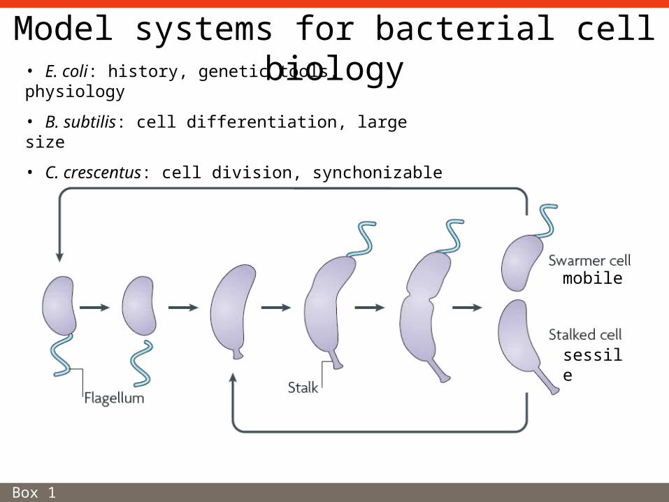
Model systems for bacterial cell biology
Box 1
• E. coli: history, genetic tools, physiology
• B. subtilis: cell differentiation, large size
• C. crescentus: cell division, synchonizable
mobile
sessile

• Diffusion and capture
Assembly of stationary protein complexes
SpoIVB
SpoIIQ
Figure 1
Mother cell
Phagocytosis-likeuptake
Septal membrane

• Targeted membrane insertion
Assembly of stationary protein complexes
SpoIVB
SpoIIIAH
SpoIIQ
Figure 1

• Targeted membrane insertion
Assembly of stationary protein complexes
Steinhauer et al., Mol Microbiol. 1999 32:367-77.; Pollard & Cooper, Science 2009 326:1208-12
IcsA: outer membrane protein, N-term is exposed to host cytoplasm
IcsP: Protease that cleaves off IcsA
Shigella flexneri:facultative intracellular pathogen

Dynamic protein scaffolds and cell shape:Bacterial actin-like cytoskeleton
Figure 2
Bundles of two or more protofilaments.
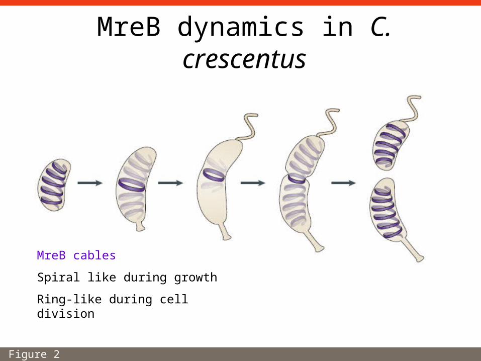
MreB dynamics in C. crescentus
Figure 2
MreB cables
Spiral like during growth
Ring-like during cell division

Architecture of MreB cables
Figure 2; Carballido-Lopez & Errington, Dev Cell. 2003 4:19-28.
B. subtilis, FRAP of GFP-Mbl

Regulation of cell-wall biosynthesis
Carballido-Lopez et al., Dev Cell. 2006 11:399-409
MreB homologues: MreBH and MblLytE: peptidoglycan hydrolase
Peptidoglycan (PG)synthetic machinery
PG-hydrolase subunit
CW binding subdomain
B. subtilis

Role of MreC in bacterial morphogenesis
Divakaruni et al., PNAS 2005 102:18602-7
C. Crescentus
PBC (penicillin-binding protein):
involved in peptidoglykan synthesis
MreC
DAPI

Crescentin
Ausmees et al., Cell. 2003 115:705-13.
C. crescentus:
creS::Tn5 -> no crescentin
creS::Tn5 + creS ->crescentin on plasmid
In-vitro assay
His-CreS filaments,
EM negative stain

Plasmid segregation
• Actin superfamiliy member (type II partitioning system)
• Walker ATPase (type I partitioning system)
• Tubulin homologue

Plasmid segregation by actin-like proteins
Figure 3
Plasmid R1 of E. coli
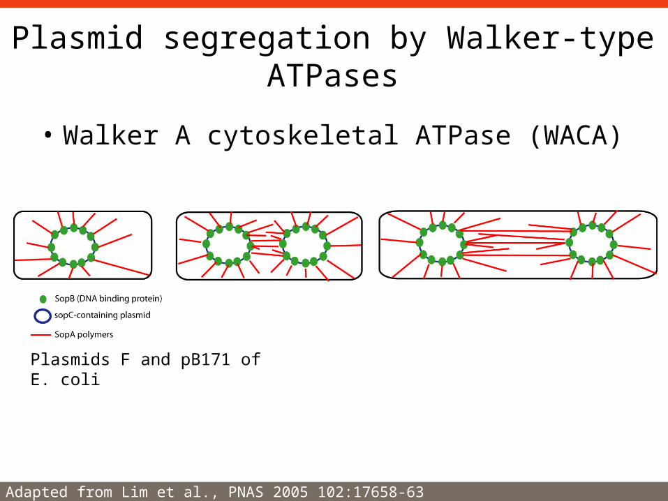
Plasmid segregation by Walker-type ATPases
• Walker A cytoskeletal ATPase (WACA)
Adapted from Lim et al., PNAS 2005 102:17658-63
Plasmids F and pB171 of E. coli
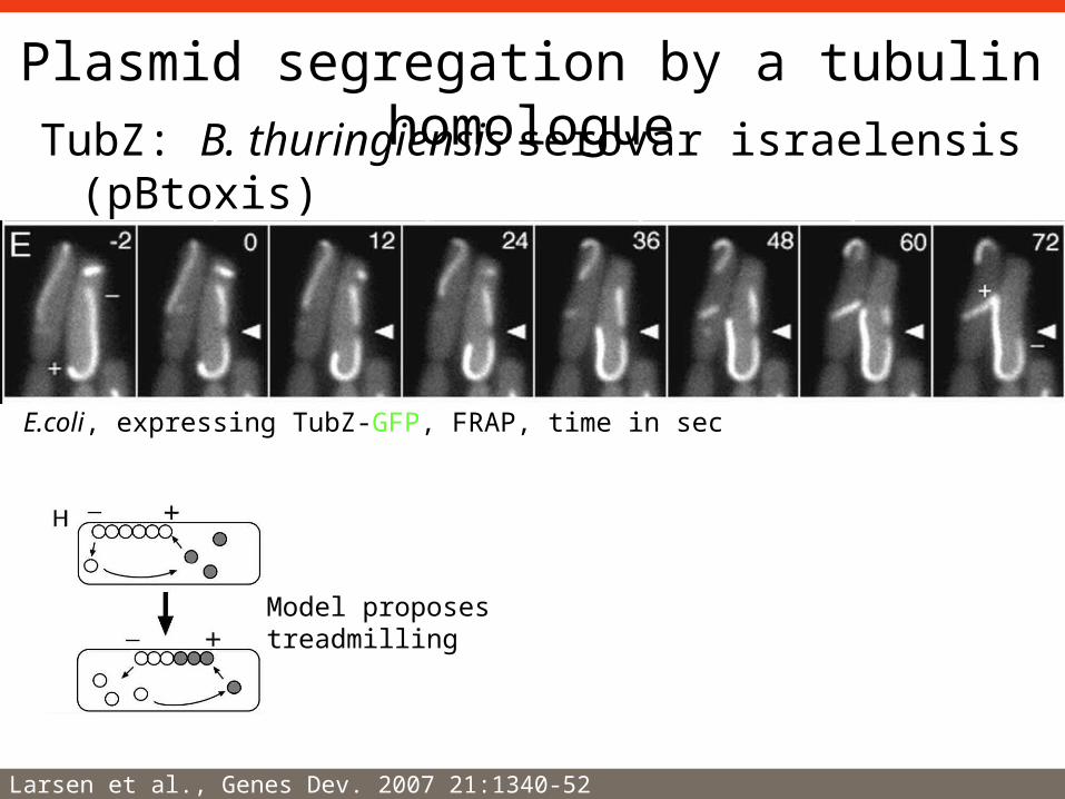
TubZ: B. thuringiensis serovar israelensis (pBtoxis)
Plasmid segregation by a tubulin homologue
Larsen et al., Genes Dev. 2007 21:1340-52
E.coli, expressing TubZ-GFP, FRAP, time in sec
Model proposes treadmilling
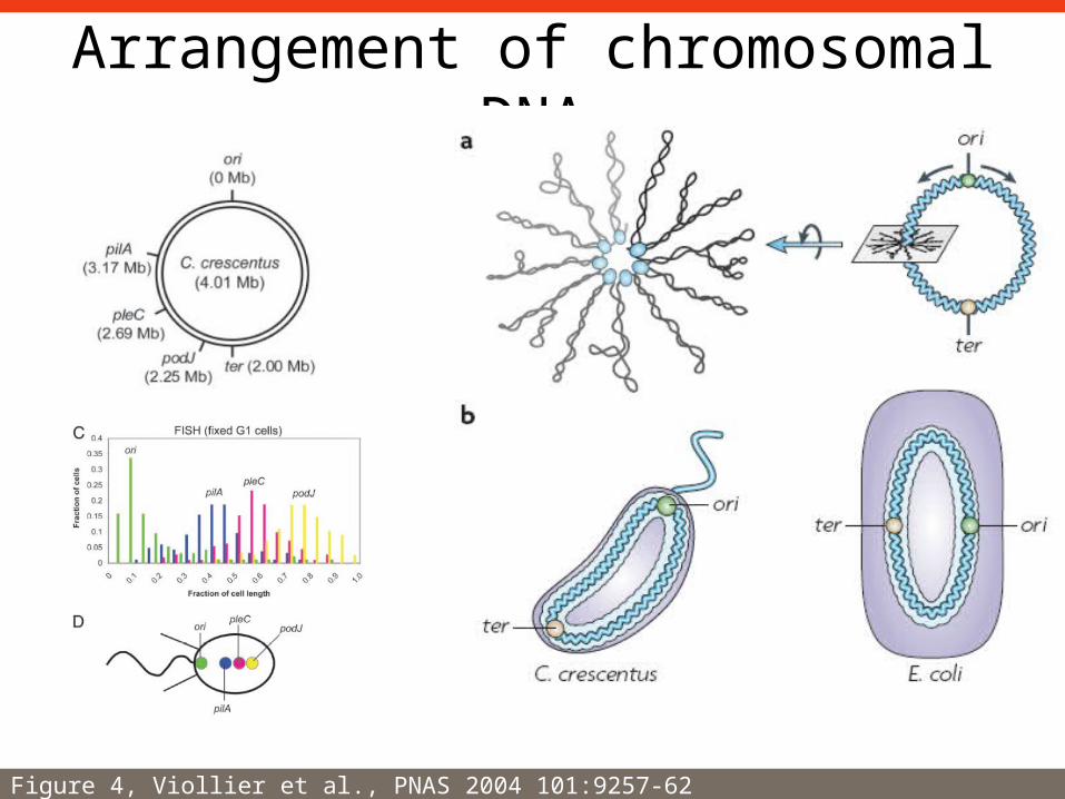
Arrangement of chromosomal DNA
Figure 4, Viollier et al., PNAS 2004 101:9257-62

Divisome: Bacterial cell-division apparatus
Allard & Cytrynbaum PNAS 2009 106:145-50; Erickson, PNAS 2009 106:9238-43
Z-ring: FitsZ filaments
Rod-shaped bacterium (e.g. E. coli)

Division-site placement: The Min system
Figure 5
minCDE operon:
MinD: WACA family
MinCD-complex: inhibit FtsZ-ring formation
MinE: represses MinCD activity
“Fail-safe mechanism”: nucleoid occlusion
B. subtilis: Noc
E. coli: SlmA

Division-site placement: The MipZ system
MipZ: ATPase, inhibits FtsZ-polymerization
ParB: chromosome partitioning protein
parS: cluster of sites, 15 kb away from ori

Conclusions
• Tubulin filaments:– cell-division apparatus, plasmid segregation
• Actin cables:– DNA partitioning, cell-shape determination,
protein localization
• WACA ATPases:– DNA segregation, cell-division plane

Outlook
• Positioning of proteins at cell poles– TipN– Peptidoglycans– Cardiolipin -> ProP
• Biochemical assembly mechanisms– Actin homologues– Tubulin homologues– WACA ATPases

Thank you for your attention!