geol 440 sedimentology and stratigraphyclasses.geology.illinois.edu/17sprgclass/geo440/geol 440 lab...
TRANSCRIPT
1 | P a g e
Name ____________________
GEOL 440
SEDIMENTOLOGY AND STRATIGRAPHY: processes, environments and deposits
Lab #1 – The Petrological Microscope: an Introduction or Refresher
Purpose:
The petrographic microscope is an invaluable tool to an Earth scientist. Many
features of rocks and minerals are much more easily identified with a microscope rather
than the naked eye. The purpose of this lab class is to give you a basic understanding of
how to use a petrographic microscope (advanced use is covered in Geology 436:
Petrology and Petrography) so that you can use it to answer questions about the origin of
sedimentary rocks that we’ll look at in the coming weeks. This week you should aim to:
1) get used as to how to operate and look after a petrological microscope and familiarize
yourself with its operation, and, 2) recognize several principal minerals: quartz, feldspars,
mica and heavy minerals.
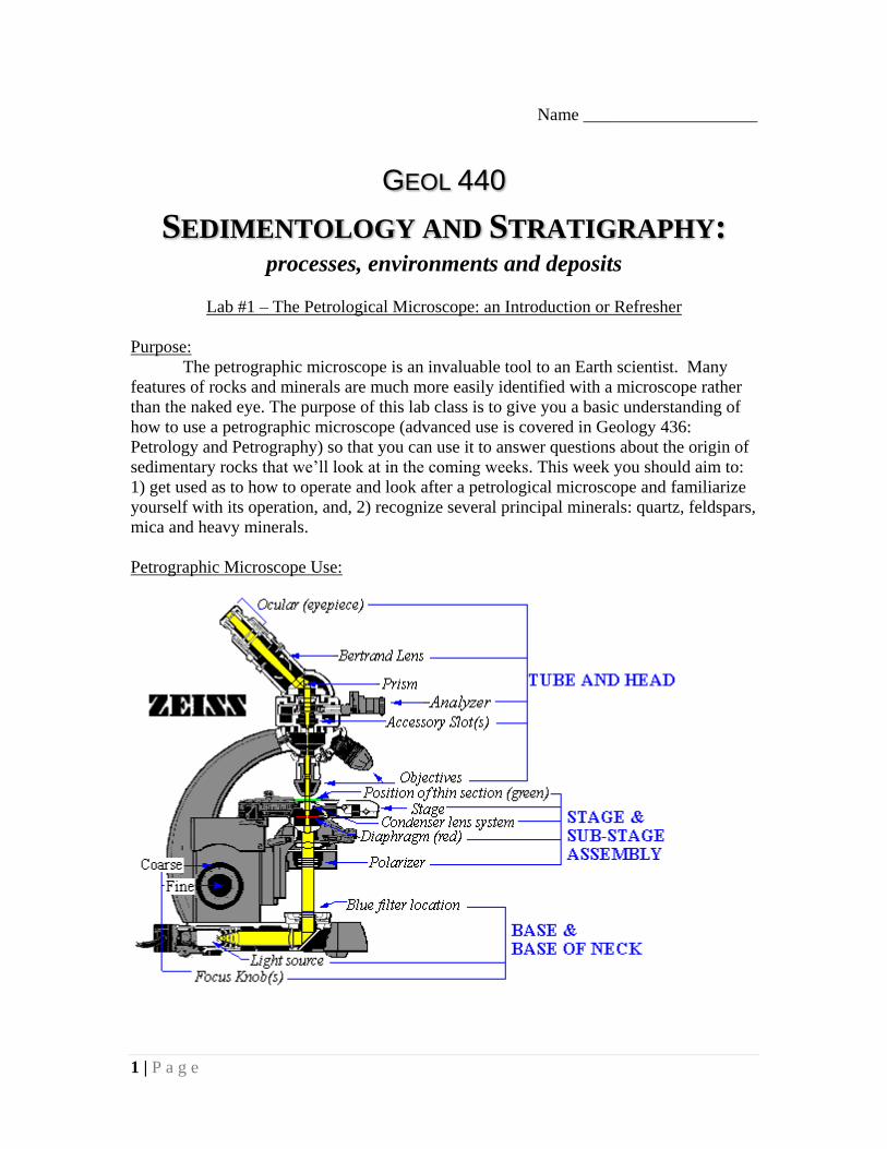
Petrographic Microscope Use:

2 | P a g e
There is a lot of superfluous information on the above diagram. For the purposes
of this class you will need to know how to: turn on the light source, change objectives,
use the coarse and fine focus, rotate the stage, and insert the analyzer.
Turning on the light source – The switch to turn on the light is located on the
base. The switch is adjustable (can be dimmed and brightened), you can reduce the strain
on your eyes if you set the light to a little over half-power.
Changing objectives – this will change the amount of magnification. These
microscopes all have a 2.5x, 10x, and 40x objective. When changing objectives, never
touch the objective – always turn the circular mount that the objectives are set in.
Coarse and fine focus – these are straight forward if you have used any
microscope before. Be careful when using the course focus, you can push the objective
through the slide, cracking or breaking it.
Rotating the stage – the thin section sits upon a stage that can be rotated 360
degrees. Rotating the stage can reveal additional information about the minerals.
Inserting the analyzer – this action can also be called “crossing the polars”. A
petrographic microscope has two plates that polarize light (force it to vibrate in a single
plane rather than in all directions). The first polarizer is located below the stage and
polarizes all the light that will transmit through the thin section. The second polarizer
(analyzer) is located above the objectives and is aligned perpendicular to the polarization
from the first polarizer.
Light that passes through the first polarizer and second polarizer (analyzer)
without being refracted by mineral grains will be totally extinguished (black). This is
what porosity will look like, as well as some uncommon sedimentary minerals called
isotropic minerals (e.g. Garnet). If the light passes through a mineral grain after the first
polarizer, some of the light is refracted (bent) such that when it passes through the second
polarizer (analyzer) not all of it is extinguished, and the mineral grain takes on a color
(birefringence). When the stage is rotated, the light that passed through the mineral will
be totally extinguished by the analyzer four times in a 360-degree rotation.

3 | P a g e
Some other terms that may help in your mineral identification:
Relief – certain minerals may appear to stand out on the thin section. Since the
thin section surface is totally flat, this standing out is the result of different optical
properties. Minerals that stand out on the thin section are described as having high relief.
Birefringence – this is basically the color of the mineral grains; low birefringence
refers to white, gray, or pale yellow while high birefringence refers to pinks, blues,
purples, and greens.
Pleochroism – this is mineral property where the color of a mineral will change
when the stage is rotated under plane-polarized light (without the analyzer inserted).
Reading: see Boggs Chapter 5
Today's Session: this weeks practical is meant to give you time to learn the different
parts of a microscope, how to use the microscope, and begin to get you to recognize the
major minerals we’ll encounter in the next few weeks. If you have used a petrological
microscope before, this will be purely a revision session.
You will be given two thin sections. For these, you should use the books in the lab and
the descriptions below, to be able to recognize 1) Quartz, 2) Feldspars, 3) Micas, 4)
Calcite and 5) Accessory Minerals. The TA will guide you through all of this and show
examples on screen. Your MUST make notes and drawings on each major mineral and its
characteristics under the microscope in both plane polarized light (ppl) and crossed
polars.
There are books available in the Grainger library that shows examples of siliciclastic
rocks under the microscope to help you in your identifications. These books are on the
reserve list handed out on the first day of class. You may need to spend time outside of
class to finish this lab. Please be very careful with the microscopes and thin sections. If
you do break a slide, its OKAY -accidents happen - but please tell the TA as soon as
possible.

4 | P a g e
A quick summary of the identification of grain mineralogy in thin section;
For today, you need to concentrate on identifying the major and accessory minerals: you
should aim to take notes and make sketches of these. A standard procedure for drawing
these in to make a sketch of what you see in the field of view, with one half showing the
characteristics under plane polarized light (ppl) and the other half under crossed polars.
Major grains
Quartz – colorless in plane light; gray, white or pale yellow in crossed polars.
No cleavage.
Feldspar – colorless in plane light; grey, white or pale yellow in crossed polars.
Good 90 deg cleavage and twinned in cross polars (stripes of black and white).
Rock fragments – aggregates of minerals (typically fine grained igneous
(basalt), sedimentary (chert) or metamorphic (shist, slate) rocks). May be
difficult to see grain boundary in crossed polars, can look similar to clay matrix.
Accessory minerals (grains)
Biotite and muscovite – micaceous minerals (perfect one direction of cleavage),
look like “books” under the microscope. May change color in plane light
(pleochroic), biotite is brownish in plane light, muscovite is colorless.
Carbonates – colorless in plane light, pearly (pinkish and purpleish hues) in
crossed polars. Bright colored “twin” planes are common in crossed polars
(especially on big crystals). May be present as fossils (shell bits – aggregates of
small calcite crystals) or cement (typically large crystals).
Green minerals – could be chlorite or glauconite, either way this is not going to
effect our classification greatly (but still include in observations).
Matrix
Fine grained background material – could be fine grained quartz sand in a
conglomerate or clay in a sandstone.
Clay – very small needle-like crystals, brightly colored in crossed polars
(yellow), brownish or green in plane light. Needles will be randomly oriented
when present as a matrix (unless closely packed in which case the needles will
line up parallel to the grain boundary), or be oriented perpendicular to the grain
boundary when present as a cement (uncommon, reflects the growth of the
crystals off of the grain surface).
Cement
Siliciclastic – same petrographic characteristics as quartz. Common as
overgrowths on quartz grains, will have same orientation (blink same time as
grain). Less common is clay cement, characteristics above.
Calcite – same petrographic characteristics as in carbonates, diverse crystal
shapes (blocky, prismatic, dogtooth, etc.). Blue, pink under crossed polars.
Iron oxide – reddish brown, opaque in thin section. Usually a thin coating on
grain surfaces, may give entire sample a reddish color. May be present as large
globs in thin section (common in fine grained samples).
Porosity – transparent in plane light, always black (never blinks) in crossed
polars. May see bubbles, or other weird shapes in pore spaces (caused by
refraction of light around grain boundaries).

5 | P a g e
CLUES TO RECOGNIZING MINERALS IN SMEAR SLIDES (originally from gc.ucsd.edu/Proc/minsPROC.html)
Calcite is highly birefringent so that even tiny fragments appear blue, red, and/or yellow under crossed polars; often they are outlined in black. Most calcite flakes are probably tiny fragments of foraminifer tests: look for associated identifiable foram tests and fragments to confirm this. If fragments are too small to identify as foraminifers (or nannoplankton), lump them as"undifferentiated calcite" in percentage estimates. Pavement calcite appears under crossed polars as a flat bluish grains with irregular black lines (the interlocking pavement blocks). Fragments were probably originally foraminiferss, but now altered to mineral calcite.
Aragonite needles, look like minute stubby rice grains to more elongate shreds. Like calcite, they are highly birefringentand even small fragments show color under crossed polars. (Aragonite is most often seen in the shells of pteropods, see Biogenic components.)
Aragonite needles (Image downloaded from Doug Schnurenberger's web site at http://lrc.geo.umn.edu/smears/.)
Dolomite is rare, but sometimes occurs as tiny equidimensional rhombs or flakes with high relief and very high birefringence, higher even than that of calcite.

6 | P a g e
Dolomite Rhombs
Gypsum crystals in smear slides look like gypsum in hand sample: they have the typical elongated rhombic form (or some twinned variation thereof). Gypsum is colorless in plane polarized light and is distinguished from quartz by its crystal habit. Gypsum fragments can appear as scuzzy yellow under crossed polars. Halite crystals, if they survive the smear-slide preparation, are isotropic and appear as clear squares (cubic) under plane-polarized light and are black under crossed polars.
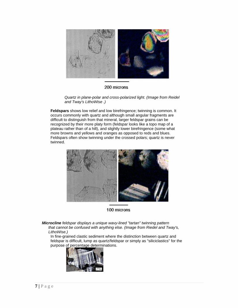
Quartz is very common, especially in sediments derived from terrigenous sources. It has low birefringence and low relief. Grains are usually rounded or subrounded with relatively high sphericity. Small fragments are grey, but typical silt- and sand-sized grains show concentric rings of blue, red, yellow. A sand-sized quartz grain looks like a colorful topographic map of a low hill. Very small fragments of quartz are often difficult to distinguish from feldspar.
7 | P a g e
Quartz in plane-polar and cross-polarized light. (Image from Reidel and Tway's LithoWise .)
Feldspars shows low relief and low birefringence; twinning is common. It occurs commonly with quartz and although small angular fragments are difficult to distinguish from that mineral, larger feldspar grains can be recognized by their more platy form (feldspar looks like a topo map of a plateau rather than of a hill), and slightly lower birefringence (some what more browns and yellows and oranges as opposed to reds and blues. Feldspars often show twinning under the crossed polars; quartz is never twinned.
Microcline feldspar displays a unique wavy-lined "tartan" twinning pattern that cannot be confused with anything else. (Image from Riedel and Tway's, LithoWise.)
In fine-grained clastic sediment where the distinction between quartz and feldspar is difficult, lump as quartz/feldspar or simply as "siliciclastics" for the purpose of percentage determinations.

8 | P a g e
Ferromagnesian Minerals (Amphiboles, Pyroxenes, Olivines) occur as discrete grains in pelagic and terrigenous deposits. Grains are often larger than the "background clay" in pelagic sediments and generally show high relief and high birefringence. Some are gray or greenish gray in plane-polarized light; some are pleochroic (change color lightly when stage is rotated in plane-polarized light). A distinctive "hacksaw" habit is also common. Although we do not often recognize the specific minerals for reconnaissance smear-slide determinations, some clues are
Olivine - green in plane-polarized light and showing high order colors under crossed polars; concoidal fracture.
Amphiboles - (esp.hornblende) - high relief, moderately elongate, prismatic habit, green, greenish gray in plane-polarized light, pleochroic, moderate birefringent colors, but may be masked by mineral color. Green hornblende is relatively common in near-shore sediments
Green hornblende in plane polarized and cross-polarized light (Image from D. Schnurrenberger's web site at http://lrc.geo.umn.edu/smears/.)
Pyroxenes - Most common ferromagnesian mineral in marine sediments. High relief, moderately high birefringence, prismatic. Can be distinquished (sometimes) from amphiboles as the pyroxenes are not usually colored or pleochroic in plane-polarized light. Also individual crystals tend to be more stubby than amphibole crystals.
Heavy Minerals (non-opaque) comprise a variety of minerals of over 2.89 specific gravity. Many (not all) are recognized by very high relief, high birefringence, rounded subhedral grains. They occur mostly in near-shore sediments in association with quartz and feldspar. (High birefringent, high relief mineral grains in deep-sea sediments are more likely to be ferromagnesian minerals, though both heavy and ferromagnesian minerals occur in near-shore sediments.) Some heavy minerals which are recognizable in smear slides are:
Apatite - colorless in plane-polarized light. Moderately high relief, weak birefringence (small fragments are gray, larger fragments are blue

9 | P a g e
and red, yellow.) Apatite is distinguished quartz by its much higher relief. It also occurs frequently as elongated prismatic crystals.
Zircon - rounded grains with very high relief (perifery looks like a Marks-A-Lot line), very high birefringence, colorless in plane-polarized light, inclusions.
Rutile - very high relief, very high birefringence, yellow or brown in plane- polarized light, diagonal striations.
Garnet - very high relief, rounded octahedreal crystal and isotropic. It is the only relatively common heavy mineral which is isotroic (black under crossed polars) May be pink or reddish brown under plane-polarized light and show distinctive v-shaped etched surface.
Tourmaline - elongate prismatic form, moderate relief, moderate birefringence, often colored in plane-polarized light and highly pleochroic, parallel extinction. Distinguished from hornblende by parallel extinction and greater pleochroism.
Fe Oxides - hematite, limonite is reddish brown in plane-polarized light, and may be in the form of small transparrent grains or the red color dissiminated throughout the clay fraction. Highly birefringent, but often this is masked by strong brown mineral color. Iron oxides appear earthy and reddish brown in reflected light. Magnetite and ilmenite are opaque. If gross sediment color is red, it probably contains iron oxides; it doesn’t take much iron oxide in the sediment to give it a red color.
Fe/Mn Micronodules - opaque, spherical to subspherical nodules. Pyrite - opaque, commonly seen in microfossil tests, especially diatom tests, as spherical (framboidal) clumps. Also seen as opaque cubes and octahedra or just opaque spheres and grains scattered throughout the sediment. Appears metallic brassy yellow under reflected light. Sometimes pyrite looks reddish (oxidized) in plane-polarized light.
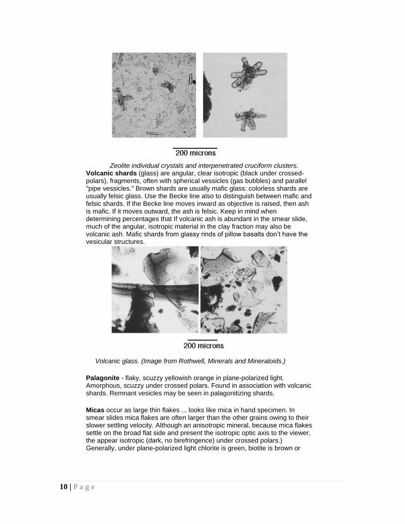
Zeolites - Phillipsite is the most common zeolite in younger marine sediments. It occurs as small transparent (plane-polarized light) prismatic grains with distinctive v-shaped terminations or as distinctive cruciform twins or clusters. Look for zeolites in red and brown clays (sediments deposited below the CCD) and below beds rich in volcanic ash: zeolites are thought to be an alteration produce of ash.
Because Clinoptilolite is more common in older sediments, from below 700m, we rarely see it in the SIO cores. But if a zeolite seen in the form of small, ragged shreds instead of the distinctive phillipsite crystals, suspect clinoptilolite. Usually, at the level of smear-slide descriptions, simply lumping the minerals as zeolites is sufficient.
10 | P a g e
Zeolite individual crystals and interpenetrated cruciform clusters.
Volcanic shards (glass) are angular, clear isotropic (black under crossed-polars), fragments, often with spherical vessicles (gas bubbles) and parallel "pipe vessicles." Brown shards are usually mafic glass: colorless shards are usually felsic glass. Use the Becke line also to distinguish between mafic and felsic shards. If the Becke line moves inward as objective is raised, then ash is mafic. If it moves outward, the ash is felsic. Keep in mind when determining percentages that If volcanic ash is abundant in the smear slide, much of the angular, isotropic material in the clay fraction may also be volcanic ash. Mafic shards from glassy rinds of pillow basalts don’t have the vesicular structures.
Volcanic glass. (Image from Rothwell, Minerals and Mineraloids.)
Palagonite - flaky, scuzzy yellowish orange in plane-polarized light. Amorphous, scuzzy under crossed polars. Found in association with volcanic shards. Remnant vesicles may be seen in palagonitizing shards.
Micas occur as large thin flakes ... looks like mica in hand specimen. In smear slides mica flakes are often larger than the other grains owing to their slower settling velocity. Although an anisotropic mineral, because mica flakes settle on the broad flat side and present the isotropic optic axis to the viewer, the appear isotropic (dark, no birefringence) under crossed polars.) Generally, under plane-polarized light chlorite is green, biotite is brown or

11 | P a g e
brownish green, and muscovite is colorless.
Mica Flake (Image from D. Schnurrenberger's web site.)
Glauconite - grains are a scuzzy olive green in plane-polarized light and isotropic (cryptocrystalline) under crossed polars. Glauconite seen off the Mendocino Fracture zone, however occurs in large (.1 - .5mm) gray-green pellets, often with reddish brown (limonite) sutures. Some pellets appear to be casts of fossil tests or fecal pellets(?).They appear as large opaque masses in the petrographic scope. Look for them under lower in the reflected-light ‘scope. Clay (Component) - Clay, as a component, comprises a suite of specific minerals, but in practice all the very fine-grained background material which can’t be otherwise identified is usually lumped under "clay."
Clay (Image from D. Schnurrenberger's web site at http://lrc.geo.umn.edu/smears/.)
Clay (Texture) - grains less than .004mm. Sometimes even these small grains can be identified as diatoms, volcanic shards or FeOxides so be sure to lump these components back into the clay size when making sand/silt/clay determinations. Clay (component) and clay (texture) percentages may vary for the same slide.
Non-Disaggregated Material - best to recognize as a separate category, as the clumped material can comprise a variety of components.
(Last revised 3/2002 by [email protected])

12 | P a g e